DOI:10.32604/cmc.2020.012585
| Computers, Materials & Continua DOI:10.32604/cmc.2020.012585 | |
| Article |
Intelligent Decision Support System for COVID-19 Empowered with Deep Learning
1School of Computer Science, NCBA&E, Lahore, 54000, Pakistan
2School of Computer Science, Minhaj University Lahore, Lahore, 54000, Pakistan
3Department of Computer Science, Lahore Garrison University, Lahore, 54792, Pakistan
4Department of Computer Science & Information Technology, Superior University, Lahore, 54000, Pakistan
5Computer Science Department, Umm Al-Qura University, Makkah City, 715, Saudi Arabia
*Corresponding Author: Muhammad Adnan Khan. Email: madnankhan@lgu.edu.pk
Received: 05 July 2020; Accepted: 25 July 2020
Abstract: The prompt spread of Coronavirus (COVID-19) subsequently adorns a big threat to the people around the globe. The evolving and the perpetually diagnosis of coronavirus has become a critical challenge for the healthcare sector. Drastically increase of COVID-19 has rendered the necessity to detect the people who are more likely to get infected. Lately, the testing kits for COVID-19 are not available to deal it with required proficiency, along with-it countries have been widely hit by the COVID-19 disruption. To keep in view the need of hour asks for an automatic diagnosis system for early detection of COVID-19. It would be a feather in the cap if the early diagnosis of COVID-19 could reveal that how it has been affecting the masses immensely. According to the apparent clinical research, it has unleashed that most of the COVID-19 cases are more likely to fall for a lung infection. The abrupt changes do require a solution so the technology is out there to pace up, Chest X-ray and Computer tomography (CT) scan images could significantly identify the preliminaries of COVID-19 like lungs infection. CT scan and X-ray images could flourish the cause of detecting at an early stage and it has proved to be helpful to radiologists and the medical practitioners. The unbearable circumstances compel us to flatten the curve of the sufferers so a need to develop is obvious, a quick and highly responsive automatic system based on Artificial Intelligence (AI) is always there to aid against the masses to be prone to COVID-19. The proposed Intelligent decision support system for COVID-19 empowered with deep learning (ID2S-COVID19-DL) study suggests Deep learning (DL) based Convolutional neural network (CNN) approaches for effective and accurate detection to the maximum extent it could be, detection of coronavirus is assisted by using X-ray and CT-scan images. The primary experimental results here have depicted the maximum accuracy for training and is around 98.11 percent and for validation it comes out to be approximately 95.5 percent while statistical parameters like sensitivity and specificity for training is 98.03 percent and 98.20 percent respectively, and for validation 94.38 percent and 97.06 percent respectively. The suggested Deep Learning-based CNN model unleashed here opts for a comparable performance with medical experts and it is helpful to enhance the working productivity of radiologists. It could take the curve down with the downright contribution of radiologists, rapid detection of COVID-19, and to overcome this current pandemic with the proven efficacy.
Keywords: COVID-19; deep learning; convolutional neural network; CT-scan; X-ray; decision support system; ID2S-COVID19-DL
The traumatic pandemic Coronavirus 2019 (COVID-2019) all started from Wuhan city of China back then in December 2019 and it spread as a wildfire and it affected enormously around the globe. According to the World Health Organization (WHO) announcement, the infection can be a respirational disease with the symptoms of cough, fever, and lung infection [1]. The World economy is at the verge of destruction, devastating health is hitting us so hard and everything has just jammed with the prevalence of COVID-19. Momentarily, it is critical to detect the roots of this virus to prevent the prevailing devastating ongoing conditions and the future spread could be diminished, eventually saving a thousand of lives. When seemingly things got out of hand and the health sectors were furiously dumped, WHO declared this gravest epidemic as a health emergency on January 30, 2020 [2]. The truce to this pandemic would genuinely take the graph of the victims to settle down.
The COVID-19 isn’t limited to humans only but it could outspread amongst mammals and reptiles as well -like bats, cats and snakes, COVID-19 rolled out from the live animal market in Wuhan, pacing up the transmission to animal-to-human scenario. And then it switched towards a person-to-person transmission, prompt the infected cases [3]. When the confirmed cases reached 118000 and outrageous deaths toll more than 4000, WHO announced the COVID-19 outbreak as a pandemic on March 11, 2020. The United States of America (USA) despite being a leading a health-sector overtook China and Italy swiftly in the number of mortalities [4].
COVID-19 is another type of virus that belongs to the family of Cornoviridea. These types of viruses infect people with adequate cold Middle east respiratory syndrome (MERS) or Severe acute respiratory syndrome (SARS) [5]. SARS was the first time diagnosed in Southern China in 2003 and breakout in many countries of the world. On the other hand, 858 deaths have been brought to the table due to MARS virus that was the first time reported in Saudi Arabia. It is observed that this virus has originated in bats after analysis [5]. The most common symptoms to COVID-19 reported so far are fever, cough and severe headache [6]. Different detection methods for COVID-19 are used like Nucleic acid test (NAT) and Computed tomography (CT) scans. NAT helps in the detection of fundamental nucleic acid sequence and species of organism, predominately a virus or bacteria that cause disease in blood, tissue, or urine. The role of the detection kit is most significant in detecting COVID-19. On the other hand, a CT scan is important in the detection of severity and degree of lung inflammation associated with this disease [7]. According to the National health commission of China, the radiographic method was used for the clinical diagnostic process of this disease [8], which declares the consequence of CT scan images for the diagnosis of COVID-19.
It has observed that many queues of COVID-19 patients are present in hospitals and clinics for medical tests. It is because there is a threat to spread the virus rapidly in patient-to-patient and medical systems may get collapsed [8]. Furthermore, radiologists are fewer, but COVID-19 patients are in a condemning increase number. The patients are deprived of the treatment [9]. Therefore, hospitals are prioritizing those patients that are more prone to symptoms of fever and shortness of breath [10].
It has become a great challenge for the researchers to diagnose and develop an efficient responsive and more intelligent diagnosis system. The radiologists are using a manual lung infection quantification method to detect the intensity of confirmed COVID-19 cases AI-based pneumonia is being used. These types of algorithms are found better to evaluate the results of CT scan images in a limited time as compared to existing available techniques to find out the different stages of confirmed cases early level to critical level Radiologists use chest CT scan images. The evaluation of these images becomes difficult and time taking by doing the manual calculation of septic regions on the CT scans and X rays, whereas the escalating of lung infection needs various CT scan images [11]. The existing findings show that the spread of this virus is still irrepressible, so there is a need for an accurate and efficient technique for diagnosis of COVID-19. This study suggested a deep learning model for the automatic detection of coronavirus. It depends upon CT-Scan and X-ray images that collected various sources. The present study not only reviewed the existing solutions to COVID-19, and offered an efficient COVID-19 detection technique using deep learning.
Due to the vast spread of COVID-19, WHO has announced a public health emergency in January 2020. The symptoms of this disease were like pneumonia and it had become a great challenge for the medical practitioners to diagnosis the treatment of this disease. Early detection of coronavirus was essential to control its future spread. Image tests considered being the rapid diagnose of COVID-19 to overcome the spread of coronavirus. CT and Chest X-rays have a significant role in the detection of COVID-19 [12]. CT scan method was the best method for diagnosing novel coronavirus pneumonia. Research conducted by the authors in [1] proposed a detection method based on deep learning for ascertaining COVID-19 pneumonia. This technique was more rapid with high-resolution CT scan images in diagnosing COVID19; it was more helpful for the radiologists in their work to control the epidemic, the interesting thing with their system was that it was able to gather 46,096 images from 106 patients, involving 51 cases of laboratory-confirmed COVID-19 for the development, training, and evaluation of the proposed model. Their proposed model based on deep learning techniques has shown the equivalent efficiency while compared with radiologists. In this way, the diagnostic system can overcome the burden of frontline radiologists. An important claim was pointed out of CT scan images in diagnoses of COVID-19, which is a rapid technique as compared to other conventional methods. Moreover, it is more accurate in diagnosing the infection at any level of pneumonia. The results of 140 COVID-19 patients were considered positive. It was more efficient in diagnosing positive cases at an early stage [5]. The researchers introduced a model for the diagnoses COVID-19 which was more accurate and less time consuming, likely to radiologist’s technique. While comparing the efficiency of the proposed model with the expert radiologist, the model takes 65% lesser time in diagnosing COVID-19 cases. There is a chance of betterment in their model during further research. It will become a self-check system that can be accessed individually, assisting medical experts and specialists. Kassani et al. [13] suggested a novel deep learning method consisting of Keras and Tensor Flow for diagnoses COVID-19. In their study, the researchers have taken 50 images and divided them into two parts, 50% images were COVID-19 positive and 50% were negative. On the bases of these X-ray images, the proposed model was trained and validated. The precisions of the proposed model have shown 90%–92% precision.
Hall et al. [14] described that chest X-ray images played an important role in diagnoses COVID-19. The researchers used 455 images, 135 chest X-ray images for COVID-19, and the rest of the images for pneumonia. A deep convolutional neural network was trained on 102 COVID-19 images and 102 pneumonia images in 10-fold cross-validation. The results showed a precision of 89.2% for COVID-19 cases. The proposed study was based on small data size. Because of a lack of information about COVID-19, the study had some weaknesses in it. The study proposed that chest X-ray can be more helpful in diagnosing COVID-19 with the help of more data about good resolution images.
Zheng et al. [15] proposed a novel model for the rapid diagnosing of COVID-19 based on deep learning techniques. For the evaluation of performance, the researchers obtained 100 chest X-ray images of confirmed patients with COVID-19. To fulfill the requirement of more data the researchers obtained 1431 more chest X-ray images and 1008 patients’ images of pneumonia. The basic findings of the suggested model showed 96% precision with COVID-19 cases and 70.65% with non-COVID-19 cases. Ozturk et al. [16] explored a deep learning-based model for the detection and classification of COVID-19 with the help of X-ray images. This model had not any manual feature extraction and was fully automated. The proposed model could handle with binary and multi-class tasks. The precision of the proposed model for binary was 98.08% and the multi-class task was 87.02%. The benefits of the model were that it was accessible by remote places where there was a shortage of radiologists. The drawback of the study was that a few numbers of X-ray images were used more images were required for better results.
Abbas et al. [17] introduced a classification method named Decompose, transfer, and compose (DeTraC) based on deep convolutional neural networks. DeTraC used for the classification of chest X-ray for diagnosis COVID-19. DeTraC handled irregularities in the image and it used a decomposition mechanism to identify images class boundaries. The findings expressed the capability of the proposed model in the diagnosis of COVID-19 in an inclusive way. DeTraC achieved a precision of 95.12% in the diagnosis of COVID-19 from the comprehensive image dataset obtained all over the world.
Apostolopoulos et al. [18] developed an automatic COVID-19 diagnosis system with the help of X-ray images based on transfer learning with deep learning techniques. The assessment of the performance of the convolutional neural network that particularly worked on medical image classification was the objective of the study. For this purpose, the transfer learning method was used to diagnose different abnormalities in small images datasets that produced excellent precisions. Two types of datasets had been used in this study, the first dataset consisted of 224, 700, and 504 images for confirmed COVID-19, common pneumonia and normal conditions, and the second dataset contained on 224, 714 and 504 images for confirmed COVID-19, common pneumonia and normal conditions respectively. The findings concluded that deep learning worked excellently on X-ray images for the detection of COVID-19 and the precision of the results was 96.78%.
Sethy et al. [19] described that there was a need to detect COVID-19 patients on earlier stages for the better prevention of this disease. In this study, the researchers introduced a methodology consisting of a deep learning technique for the diagnosis of COVID-19. The support vector machine using a deep feature identified COVID-19 X-ray images from the dataset. The proposed method was helpful for medical practitioners for the detection of COVID-19 disease. The proposed model obtained a precision of 95.38% that was better from the existing classification models. The review of related studies shows that some irregularities still need to be addressable in the database of the image. These irregularities are affecting the efficiency of deep learning models. The proposed study focuses on detecting the causes of these irregularities and find out the solution in a comprehensive way.
3 Proposed ID2S-COVID19-DL System Model
The advent of artificial intelligence brought about a great change in the field of information technology. The rapid development of deep learning has revolutionized the world of image processing. In the field of medical image processing, deep learning technology is being used in the last few years. It is beneficial to analyze and identify the object in the domain of medical data. The outbreak of COVID-19 surprised the world because of its continuous spread and become a pandemic.
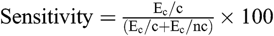
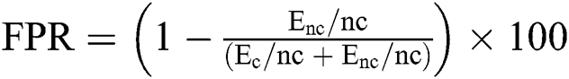
The medical practitioners are still unable to find out its complete treatment. Its rapid spread can be reduced through early detection. Several artificial intelligence techniques have been used to predict the early detection of COVID-19. Deep learning is an effective source to support radiologists in early detection COVID-19. The proposed ID2S-COVID19-DL model provides a forecast for early detection of this pandemic based on the deep learning technique. It consists of three layers named data acquisition layer, pre-processing layer, and application layer. The data acquisition layer collects data from different sources like cameras, X-rays, and CT-scans machines through the Internet of medical things (IoMT). Noise and blurriness occur during the collection of this grapple amount of data. All this gathered data becomes raw data. Pre-processing layer deals with raw data in two steps. In the first step, it mitigates noise using moving average technique, mean, and mode techniques. Step 2, resizes the Red green blue (RGB) image that persists the quality of required information. Next is an application layer that is composed of two sub-layers the performance layer and the prediction layer. The performance layer evaluates root mean square, mean absolute error and, mean absolute percentage error. If the learning criteria do not meet then the model needs to retrain, otherwise, data store in the cloud are shown in Fig. 1.
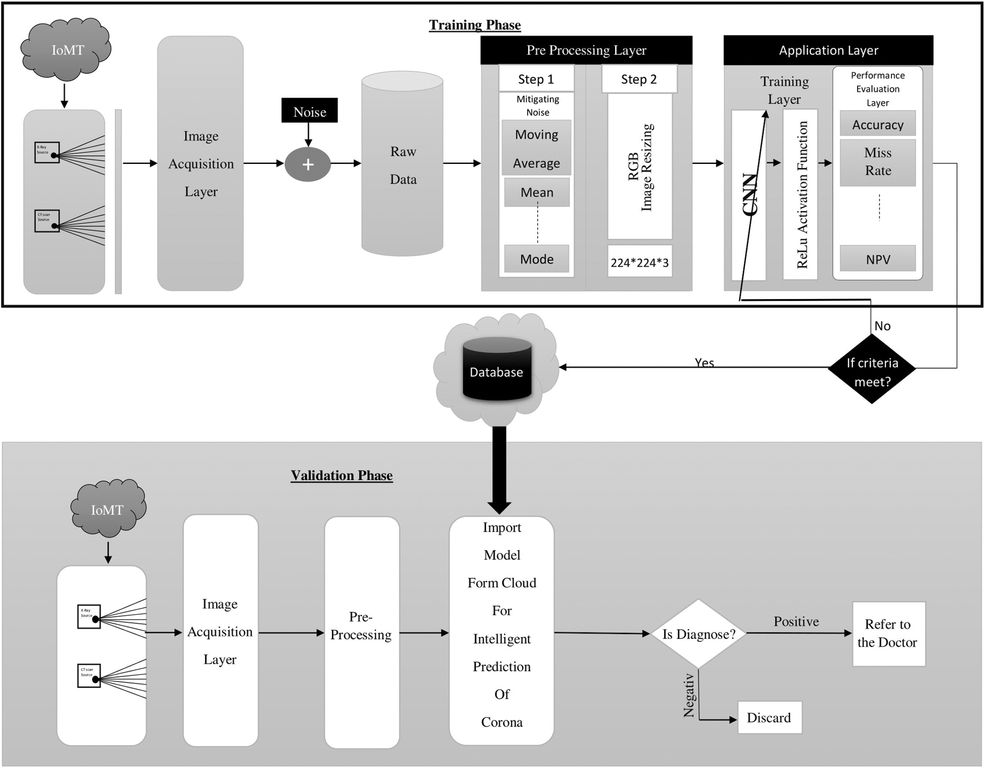
Figure 1: Proposed ID2S-COVID19-DL system model
In the validation phase, there are two layers named data acquisition layer, pre-processing layer. The data acquisition layer collects data from different sources like cameras, X-rays, and CT-scans machines through the IoMT. The pre-processing layer is used to mitigate the noise and blurriness occur, after the pre-processing layer, the proposed ID2S-COVID19-DL System Model import data from the cloud for intelligent prediction of COVID-19. If the intelligent decision support system detects the COVID-19 positive then referred to the doctor and if COVID-19 negative then no need any COVID-19 treatment.
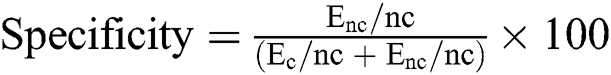
3.1 Convolutional Neural Network
Deep learning (DL) is a popular technique used in several domains of life to predict diseases, transportation, agriculture, and aeronautics, etc. DL is helpful in various areas not only because of its fast learning. Convolution neural network (CNN) comprises of two main components named the conventional layer and pooling layer. Artificial neural network deals only with one dimension and CNN covers two and three dimensions.
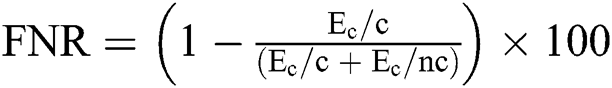
The proposed study has used CNN for the early detection of COVID-19. This network is a compound of an input layer, hidden layers, and an output layer. The input layer can read images taken from the pre-processed dataset. In the pre-processing phase, CT scan and X-ray images are resized. Pre-processing is an essential process to obtain proper datasets with no letter on images. Image resize is required because there is a variation in images and their sources are different. Hence, the size of input images is reformed into 224 × 224 × 3 where 224 × 224 shows the width and height of the input images, and 3 shows the number of channels. Convolution layer has great worth because mostly computations tasks are executed in this layer. The basic objective of the convolution layer is to retrieve features applying filters, preserves spatial relationships amongst pixels.
In Fig. 2 the pooling layer is used to reduce the dimensions of the image. Pooling also reduces the computation time by downsampling. Max pooling and average pooling are the two types of pooling layer. Max pooling is used in the proposed model. The main target of the pooling layer is to retain the specific image features that are captured during the conventional operation. Weights remain constant in the pooling layer because the pooling layer never indulges in the backpropagation process. In the Fully Connected layer, all inputs are connected to entire activation functions (ReLu) from the previous layer. A fully connected layer compiles the data that is extracted from previous layers to formulate the final output. At the last convolution layer, it is converted into a single flatten length of one-dimension array and becomes a fully connected layer then the softmax layer is responsible to transform logits into probabilities. In the ultimate layer of the CNN model, the accuracy values from the previous layer are labeled.
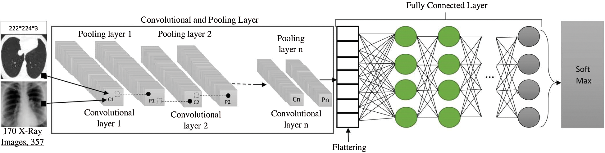
Figure 2: CNN model for intelligent decision support system (ID2S)
In the mathematical model, the target is to back prorogate through taking derivative of Eq. (1) w.r.t. to weights 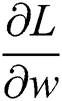 and bias
 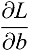 .
.
where c = No. of classes depends upon applications
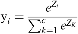
We have softmax transformation as in Eq. (2)
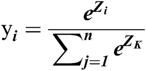
where  represents logits, many logits will transform into probabilities using softmax transformation.
 obtained by interconnected weights with the
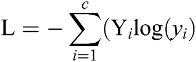
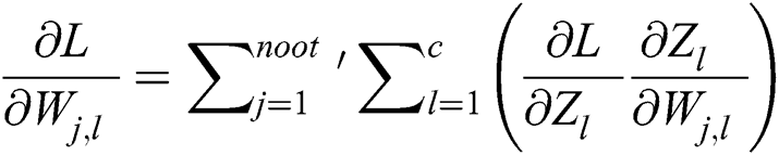
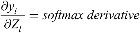
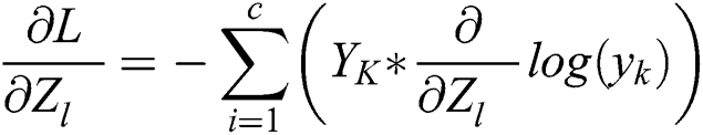
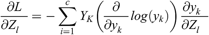
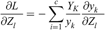
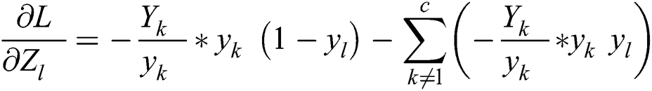
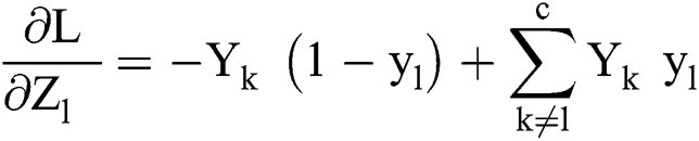
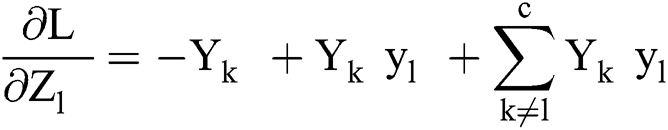
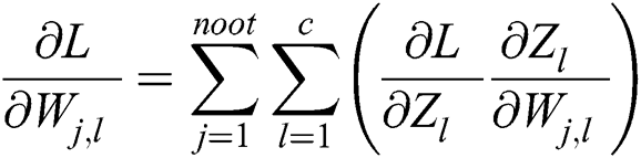
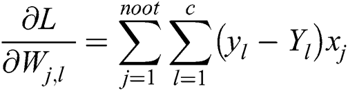
Here we find Loss w.r.t. weights that are based on two summations in Eq. (3). One summation from j = 1 to n out and other l = 1 to c. Then we take the product of two derivatives (Dominator of 1st and 2nd Nominator are same, we apply multiplication association rule, resulted in both are canceled).
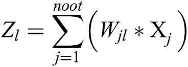
In Eq. (1), Loss having  as its parameter that is indirectly related to
  in terms of the following expression
 is given as
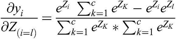
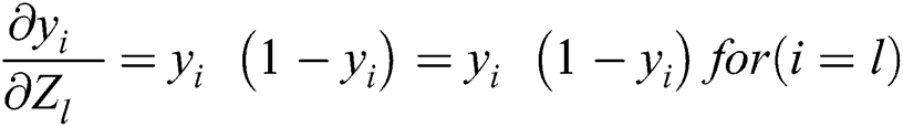
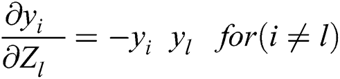
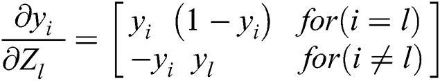
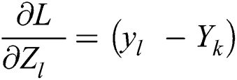
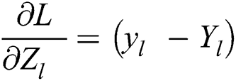
Two cases are important, where case 1, I = l, and case 2 i  when i = lth unit. Where l is the single neuron point of focus in softmax output neurons and 1 neuron has the heights values and rest of them close to zero.
case1: ( )
)
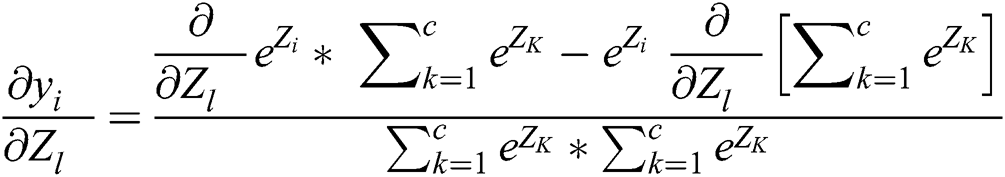
Taking the derivative of Eq. (2) through quotient rules
Taking common 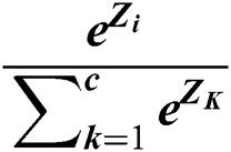 from Eq. (4), we get following
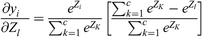
By dividing, we get
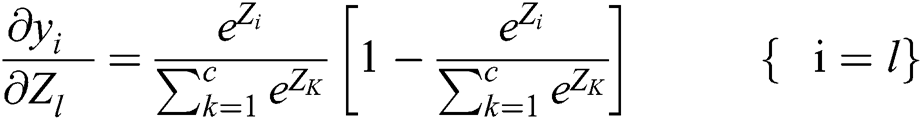
As we know 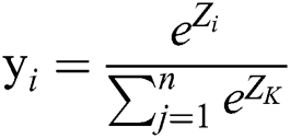 so, the above equation can be further written as.
When i! = lth unit which has low probability where l is the single neuron point of focus in softmax output neurons.
case2 ( ):
):
Taking the derivative of Eq. (3) through quotient rules w.r.t.  .
.
It can be written as,
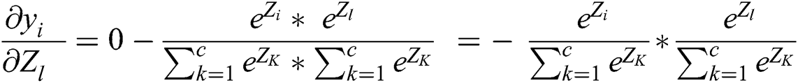
As we know that 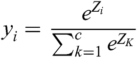 and
and  so we can drive this equation as.
We can summarize Eqs. (5) and (6)
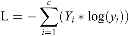
As we know that Cross-Entropy Loss has not any component of  so, we take the partial derivative of
  w.r.t. this expression log (yK)
Taking derivative, the equation becomes
 has already calculated for the softmax gradient. Here we have two cases
 as in Eq. (7). Now we have to divide Eq. (8) in two parts
where,
 for
for 
 for
for 
We can simplify this,
We can further simplify this as,
where  represents 1,
Now put the value of  in Eq. (3)
where  =
=  as input weights.
Eq. (9) is the derivative of Loss w.r.t weights for the fully connected layer.
Matlab Tool 2019a is utilized for simulation and results that are based on an intelligent decision support system for COVID-19 empowered with deep learning based on a convolutional neural network.
4.1 Proposed ID2S-COVID19-DL Results
Intelligent COVID-19 Prediction using a Deep learning model designed for the diagnosis of a novel disease COVID-19. Matlab 2019a tool is being used for the results and prediction purposes. A deep learning-based CCN model has been instigated 527 images of the dataset. The proposed ID2S-COVID19-DL model further divided into two phases named training and validation. In the training phase, 70% (370) images were used and 30% (157) for validation was used. Different statistical evaluation parameters applied compared with existing approaches like precision, miss rate, sensitivity, specificity, Negative prediction value (NPV), Probability prediction value (PPV), False positive ratio (FPR), False negative ratio (FNR), Likelihood ratio positive (LRP), and Likelihood ratio negative (LRN).
The proposed ID2S-COVID19-DL model predicts the COVID-19 disease in the form of positive and negative. Positive represents the COVID and negative shows that non-COVID.
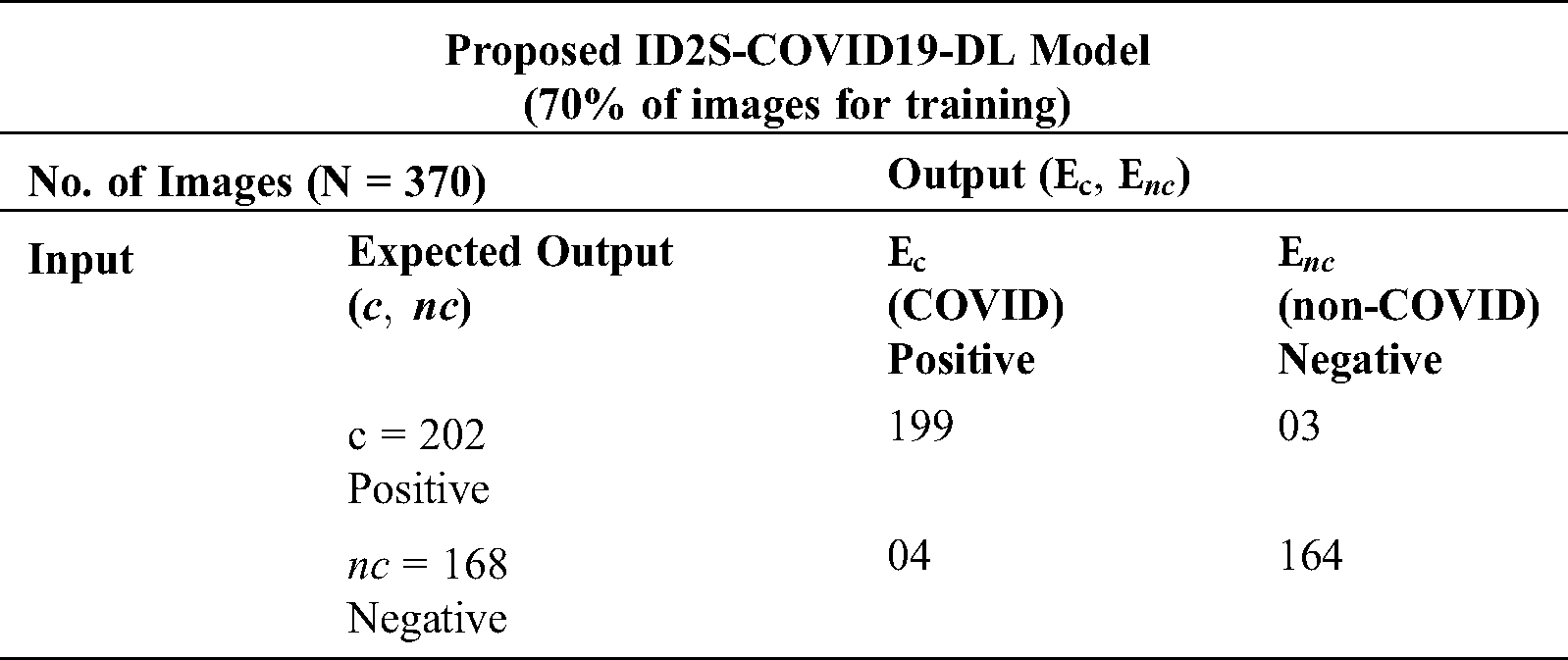
Tab. 1 represents the prediction of the proposed ID2S-COVID19-DL model for training. Total no. of images 370 is obtained to train the proposed ID2S-COVID19-DL model. A total of 370 images are divided into two groups named COVID and Non-COVID. In the COVID group, 202 images used for prediction, the proposed ID2S-COVID19-DL model predicted 199 for COVID Positive and 03 for non-COVID. In the non-COVID group, 168 images used for prediction, the proposed ID2S-COVID19-DL model predicted 04 for COVID Positive and 164 for non-COVID.
Table 1: Decision matrix for proposed ID2S-COVID19-DL (training)
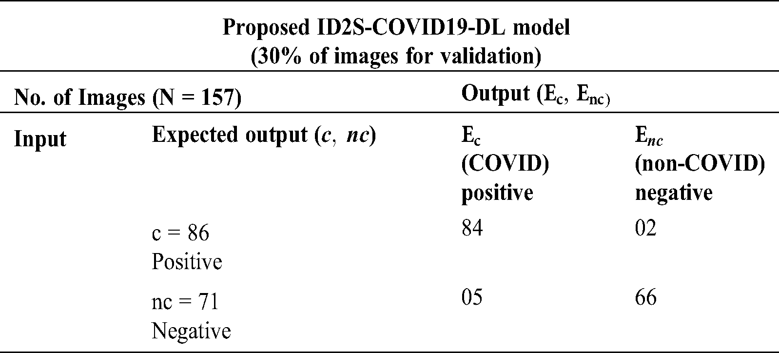
Tab. 2 represents the prediction of the proposed ID2S-COVID19-DL model for validation. Total no. of images 157 is obtained to train the proposed ID2S-COVID19-DL model. A total of 157 images are divided into two groups named COVID and Non-COVID. In the COVID group, 86 images used for prediction, the proposed ID2S-COVID19-DL model predicted 84 images for COVID Positive and 02 for non-COVID. In the non-COVID group, 71 images used for prediction, the proposed ID2S-COVID19-DL model predicted 05 for COVID Positive and 66 for non-COVID.
Table 2: Decision matrix for proposed ID2S-COVID19-DL (validation)
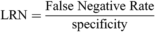
Tab. 3 represents the statistical measures of various parameters like accuracy, miss rate, sensitivity, specificity, PPV, NPV, FPR, NPR, LRP, and LRN for proposed ID2S-COVID19-DL system model performance. The proposed ID2S-COVID19-DL system model provides Accuracy of 98.11% for training and 95.50% for validation purposes.
Table 3: Performance Evaluation of statistical parameters for proposed ID2S-COVID19-DL model in Validation & Training
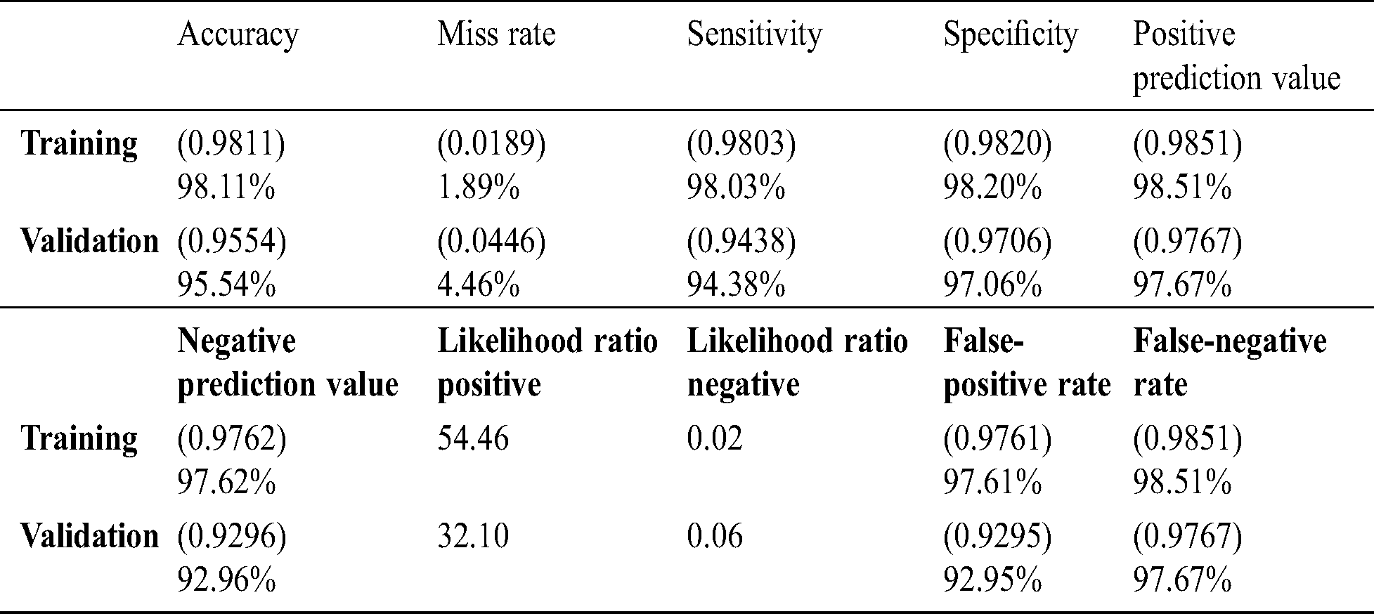
Along with other statistical parameters miss rate, sensitivity, specificity, PPV, NPV, FPR, NPR, LRP and LRN for validation are 4.46%, 94.38%, 97.06%, 97.67%, 92.96%, 32.10, 0.06, 92.95%, 97.67% respectively. During the training miss rate, sensitivity, specificity, PPV, NPV, FPR, NPR, LRP and LRN for 1.89%, 98.03%, 98.20%, 98.51%, 97.62%, 54.46%, 0.02%, 97.61% and 98.51% respectively.
Tab. 4 and Fig. 3 shows the performance of the proposed accuracy chart model using deep learning-based CNN, it gives the 98.11 percent accuracy for the training phase and 95.54 percent accuracy for the validation phase.
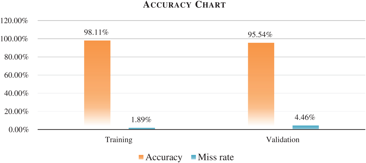
Figure 3: Proposed ID2S-COVID19-DL model accuracy chart
Table 4: In Contrast with literature for the proposed ID2S-COVID19-DL system model
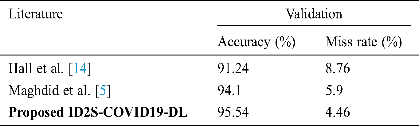
The lives of people are at high-level risk because of the rapid spread of COVID-19 around the world. CT scan and X-ray images are an effective tool to detect COVID-19. The CT scan and X-ray images are utilized for simulation collected from various sources. The CT scan and X-ray images based on deep learning are used to detect coronavirus because these images are fast, accurate, comparatively low-cost diagnosis system. The proposed intelligent decision support system COVID-19 empowered with a deep learning-based convolutional neural network system model achieves the accuracy 95.54 percent with a sensitivity of 94.38 percent, and specificity of 97.06 percent on the X-ray and CT scan datasets. The accuracy is quite satisfied while compared with other existing approaches. The proposed ID2S-COVID19-DL model can greatly beneficial for the medical staff and control and stop the COVID-19 epidemic.
Acknowledgement: Thanks to our families & colleagues who supported us morally.
Funding Statement: This Work is supported by Data and Artificial Intelligence Scientific Chair at Umm AlQura University.
Conflicts of Interest: The authors declare that they have no conflicts of interest to report regarding the present study.
1. J. Chen, L. Wu, J. Zhang, L. Zhang, D. Gong et al. (2020). , “Deep learning-based model for detecting 2019 novel coronavirus pneumonia on high-resolution computed tomography: A prospective study,” Medical, Educational, and Development of Resources through International Exchange, vol. 2020, pp. 1–27. [Google Scholar]
2. M. L. Holshue, C. DeBolt, S. Lindquist, K. H. Lofy, J. Wiesman et al. (2020). , “First case of 2019 novel coronavirus in the United States,” New England Journal of Medicine, vol. 382, no. 10, pp. 929–936. [Google Scholar]
3. Novel Coronavirus-China. World Health Organization. (2020). [Online]. Available: https://www.who.int/csr/don/12-january-2020-novel-coronavirus-china/en/. [Google Scholar]
4. CNN Health. (2020). [Online]. Available: https://edition.cnn.com/2020/03/11/health/coronavirus-pandemic-world-health-organization/index.html. [Google Scholar]
5. H. S. Maghdid, K. Z. Ghafoor, A. S. Sadiq, K. Curran, K. Rabie et al. (2020). , “A novel ai-enabled framework to diagnose coronavirus COVID-19 using smartphone embedded sensors: Design study,” vol. 2000, pp. 1–7, , The preprint server for Health Sciences, Cornell University, ArXiv preprint arXiv: 2003.07434. [Google Scholar]
6. S. Tian, N. Hu, J. Lou, K. Chen, X. Kang et al. (2020). , “Characteristics of COVID-19 infection in Beijing,” Journal of Infection, vol. 80, pp. 401–406. [Google Scholar]
7. A. Zumla, J. A. Al-Tawfiq, V. I. Enne, M. Kidd, C. Drosten et al. (2014). , “Rapid point of care diagnostic tests for viral and bacterial respiratory tract infections—Needs, advances, and future prospects,” Lancet Infectious Diseases, vol. 14, no. 11, pp. 1123–1135. [Google Scholar]
8. National Health Commission of China. March, 2020. [Online]. Available: http://www.chinadaily.com.cn/m/chinahealth/index.html. [Google Scholar]
9. Z. Ye, Y. Zhang, Y. Wang, Z. Huang and B. Song. (2020). “Chest CT manifestations of new coronavirus disease 2019 (COVID-19A pictorial review,” European Radiology, vol. 30, no. 8, pp. 1–9. [Google Scholar]
10. A. Filatov, P. Sharma, F. Hindi and P. S. Espinosa. (2020). “Neurological complications of coronavirus disease (COVID-19Encephalopathy,” Cureus, vol. 12, no. 3, pp. 1–7. [Google Scholar]
11. F. Shan, Y. Gao, J. Wang, W. Shi and N. Shi. (2020). “Lung infection quantification of COVID-19 in CT images with deep learning,” vol. 2003, pp. 1–23, , The preprint server for Health Sciences, Cornell University, Arxiv preprint arXiv: 2003.04655. [Google Scholar]
12. World Health Organization. (2020). “Coronavirus disease 2019 (COVID-19Situation report, 72,” . https://www.who.int/ [Google Scholar]
13. S. H. Kassani, P. H. Kassasni, M. J. Wesolowski, K. A. Schneider, R. Deters et al. (2020). , “Automatic detection of coronavirus disease (COVID-19) in X-ray and CT images: A machine learning-based approach,” vol. 2004, pp. 1–15, , The preprint server for Health Sciences, Cornell University, Arxiv preprint arXiv: 2004.10641. [Google Scholar]
14. R. E. Hall, C. I. Jones and P. J. Klenow. (2020). “Trading off consumption and COVID-19 deaths,” National Bureau of Economic Research, vol. 42, no. 1, pp. 1–12. [Google Scholar]
15. C. Zheng, X. Deng, Q. Fu, Q. Zhou, J. Feng et al. (2020). , “Deep learning-based detection for COVID-19 from chest CT using weak label,” MedRxiv, vol. 2020, pp. 1–13. [Google Scholar]
16. T. Ozturk, M. Talo, E. A. Yildirim, U. B. Baloglu, O. Yildirim et al. (2020). , “Automated detection of COVID-19 cases using deep neural networks with X-ray images,” Computers in Biology and Medicine, vol. 121, 103792. [Google Scholar]
17. A. Abbas, M. M. Abdelsamea and M. M. Gaber. (2020). “Classification of COVID-19 in chest X-ray images using DeTraC deep convolutional neural network,” pp. 1–14, , The preprint server for Health Sciences, Cornell University, Arxiv preprint arXiv: 2003.13815. [Google Scholar]
18. I. D. Apostolopoulos and T. A. Mpesiana. (2020). “COVID-19: Automatic detection from X-ray images utilizing transfer learning with convolutional neural networks,” Physical and Engineering Sciences in Medicine, vol. 43, pp. 635–640. [Google Scholar]
19. P. K. Sethy and S. K. Behera. (2020). “Detection of coronavirus (COVID-19) disease based on deep features,” pp. 1–9. [Google Scholar]
 | This work is licensed under a Creative Commons Attribution 4.0 International License, which permits unrestricted use, distribution, and reproduction in any medium, provided the original work is properly cited. |