DOI:10.32604/cmc.2022.029445
| Computers, Materials & Continua DOI:10.32604/cmc.2022.029445 | |
| Article |
A Mathematical Model for COVID-19 Image Enhancement based on Mittag-Leffler-Chebyshev Shift
1Department of Mathematics and Statistics, College of Science, Imam Mohammad Ibn Saud Islamic University, Riyadh, 11432, Saudi Arabia
2Department of Computer System & Technology, Faculty of Computer Science, and Information Technology, Universiti Malaya, 50603, Kuala Lumpur, Malaysia
*Corresponding Author: Hamid A. Jalab. Email: hamidjalab@um.edu.my
Received: 03 March 2022; Accepted: 08 April 2022
Abstract: The lungs CT scan is used to visualize the spread of the disease across the lungs to obtain better knowledge of the state of the COVID-19 infection. Accurately diagnosing of COVID-19 disease is a complex challenge that medical system face during the pandemic time. To address this problem, this paper proposes a COVID-19 image enhancement based on Mittag-Leffler-Chebyshev polynomial as pre-processing step for COVID-19 detection and segmentation. The proposed approach comprises the Mittag-Leffler sum convoluted with Chebyshev polynomial. The idea for using the proposed image enhancement model is that it improves images with low gray-level changes by estimating the probability of each pixel. The proposed image enhancement technique is tested on a variety of lungs computed tomography (CT) scan dataset of varying quality to demonstrate that it is robust and can resist significant quality fluctuations. The blind/referenceless image spatial quality evaluator (BRISQUE), and the natural image quality evaluator (NIQE) measures for CT scans were 38.78, and 7.43 respectively. According to the findings, the proposed image enhancement model produces the best image quality ratings. Overall, this model considerably enhances the details of the given datasets, and it may be able to assist medical professionals in the diagnosing process.
Keywords: CT scans; COVID-19; Mittag-Leffler; Chebyshev polynomial fractional calculus
The infectious disease COVID-19 is one of the world's most devastating pandemics, wreaking havoc on numerous health systems around the world and affecting the health and economics of billions of people. The Computed Tomography (CT) is considered as one of common procedures which is used to diagnose COVID-19 [1]. The CT has shown to be useful in detecting the virus that is causing the COVID-19 pandemic. Automated image enhancement of COVID-19 CT scans is regarded an essential first step in the routine diagnosis of COVID-19 infections, but it is difficult due to CT scan intensity inhomogeneity, artifacts, and closeness in the gray level of distinct soft tissue [2].
The CT technology is one of the most important medical imaging technologies that has been constantly improved over the last five decades and has played a significant role in diagnostic, treatment planning, interventional, screening, and has recently been linked to the COVID-19. Whereas, as image quality has improved and acquisition times have decreased, the clinical uses of CT have grown considerably.
The development of a computer-assisted technique to improve CT scans of the lung remains challenging when a very low contrast between the lesion, the surrounding normal tissue, the ribs, and large pulmonary blood vessels is displayed. Despite the fact that a visual assessment is regarded as an adequate standard, it is more prone to diagnostic error that comes from difficulty to distinguish between lesions even for an experienced radiologist. In the discipline of image processing, the use of fractional calculus operators has risen in popularity. Image enhancement is a type of image processing that tries to improve an image's details. Due to the unpredictable change in the quality of the acquired images, enhancing medical imagery is a difficult undertaking. Based on this, we propose in this study an automated algorithm for enhancement of COVID-19” CT scans using the Mittag-Leffler sum convoluted with Chebyshev polynomial. The following are the study's primary contributions:
1- We introduce a novel low-light image enhancement method that improves the contrast of COVID-19 CT scans.
2- For realistic low-light COVID-19 CT scans enhancement, we present a mathematical model based on the Mittag-Leffler sum convoluted with Chebyshev polynomial.
3- The proposed COVID-19 CT scans enhancement can be applied as an effective pre-processing step for any image processing method.
Most medical images contain noise due to different contrast, light reflections, and patient movement during the capturing process. Such noise causes computational complications and reduces the COVID-19 diagnostic accuracy.
Image enhancement is pre-processing used for improving the image quality for a particular application. Most image enhancement techniques use spatial operations on picture pixels to generate more relevant images than the input image.
Multiple spatial domain algorithms-based picture enhancing methods have been documented in the literature. The most widely used idea in picture enhancing technologies is fractional calculus.
The Riesz fractional model in [3] was proposed to enhance the regions of text in license plate images. In the same approach. For medical images, a kidney region image enhancement was proposed using new local fractional entropy [4]. This method achieved good results for kidney MRI scans with low contrast. Another medical image enhancement based on fractional calculus was proposed by [5]. The method aimed to dynamically enhance the image contents by using new fractional integral entropy. Ibrahim et al., 2022 [6] proposed a new class of fractional partial differential equations as new image enhancement for low contrast medical image of two datasets. However, this method required low resolution images for enhancing the image. Jalab et al., 2021 [7] proposed a new fractional Rényi entropy enhancement model as a pre-processing stage for kidney magnetic resonance imaging (MRI) segmentation. Since the method was developed as a pre-processing for kidney segmentation, it may not work out for COVID-19 detection in X-Ray or in CT scans.
More recently the fractional calculus operators were effectively utilized as a new approach in image enhancement [8–11]. Regardless of the outcomes of the fractional operators as an image enhancement, there is still a possibility for an improvement. Here, we propose a new image enhancement model based on new mathematical model based on Mittag-Leffler-Chebyshev Shift of fractional operator.
The challenges involved in CT scans enhancement include:
1. In CT scans, the infected spots have different textures, sizes, and positions.
2. The low contrast border in the appearance area of the CT scan makes it difficult to distinguish from healthy regions during the enhancing procedure.
3. The level of noise in the infected area is high, which has a significant impact on the COVID-19 diagnostic accuracy.
The image enhancement consists of a collection of algorithms that are applied to enhance the contrast details of an image. Due to the large number of captured medical images for COVID-19 diagnosis, the captured image quality may differ depending on imaging system or due to the process of converting image formats, which will lead to reduced quality of these images [12]. Therefore, it is essential to reduce the effects of these causes and enhance the image texture detail prior to any image process. The detail of the proposed mathematical model of Mittag-Leffler-Chebyshev Shift (MLCS) for image enhancement is described in the following subsection. The MLCS model is defined by special function based on two parameters α and β.
The Mittag-Leffler function is a mathematical function which is the major function in fractional calculus.
where
Definition 1. For the power series in
To continue the definition of Mittag-Leffler-Chebyshev Shift (MLCS), the following power series is considered:
Note that
where
as follows:
Satisfying
where
The proposed image enhancement model is defined in (9), which is based on the MLCS with the fractional parameter α. The idea for using the proposed image enhancement model MLCS for pixel type image improvement is that it enhances images with minor gray-level changes based on the likelihood of each pixel probability.
The algorithm steps are described as follows:
1. Consider the input image (I).
2. Set the tune image enhancement value α as the fractal power.
3. Find the pixel probability value (
4. Calculate the enhanced image using Eq. (3).
The logic behind using of Mittag-Leffler sum convoluted with Chebyshev polynomial as an image enhancement model is to boost low contrast intensities through the estimation of the enhanced each pixel values based on the details of image pixels’ frequency. The novelty of this study is the mathematical model of Mittag-Leffler-Chebyshev Shift (MLCS) for image enhancement which may be used as an effective pre-processing step for any image processing approach.
The fractional power is the parameter α for fine detail enhancement in the proposed enhancement model. In the proposed enhancement model, the fractional power (α) value is the parameter for fine detail enhancement. The value of α, as shown in Fig. 1, is calculated empirically by calculating the average NIQE score for all images in the dataset. It is noted that when the value of α is equal to 2.1, the best score for NIQE is obtained (lower is better).
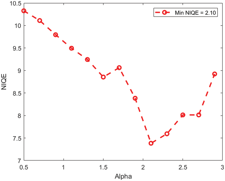
Figure 1: The average NIQE measure for different values of α
The proposed image enhancement code was written in MATLAB 2021b, and the comparison analysis with other approaches was done in both Matlab and Python.
In this study, the standard dataset of COVID-19 CT scan by the ‘Italian society of medical and interventional radiology's radiologists’ has been used in this study [14].
The qualitative results of the proposed MLCS image enhancement are shown in Fig. 2. The figure includes the input images, the enhanced images, as well as the histogram plot. It is clear from Fig. 3 that the input image pixel probability looks dense, while the enhanced image pixel probability looks distributed. This indicates that the image's contrast has been improved by the proposed MLCS method.
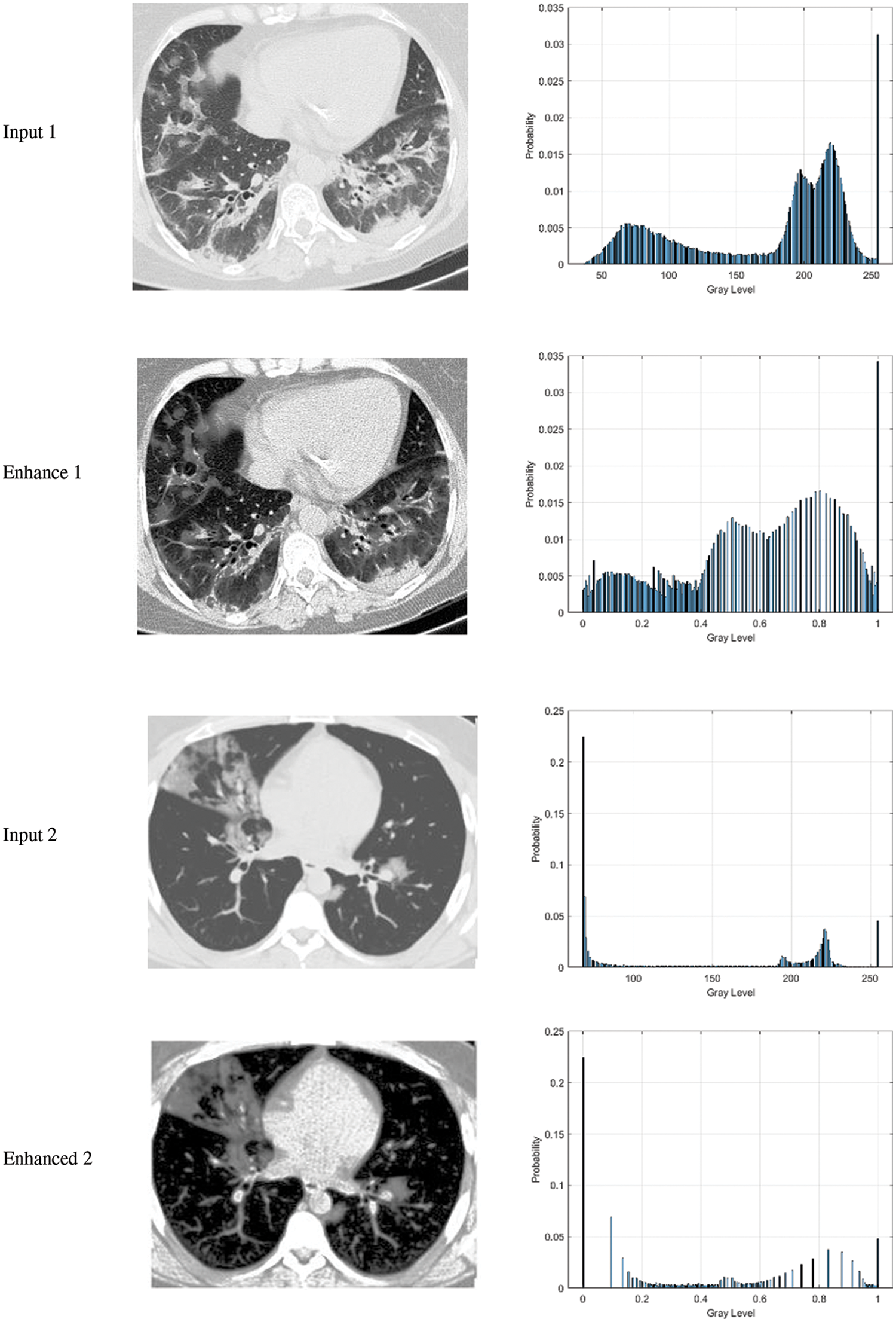
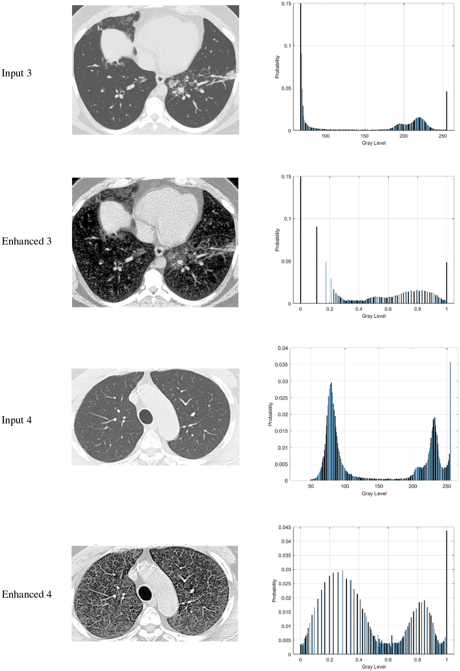
Figure 2: The output of the enhancement proposed algorithm along with histogram analysis for CT scans images
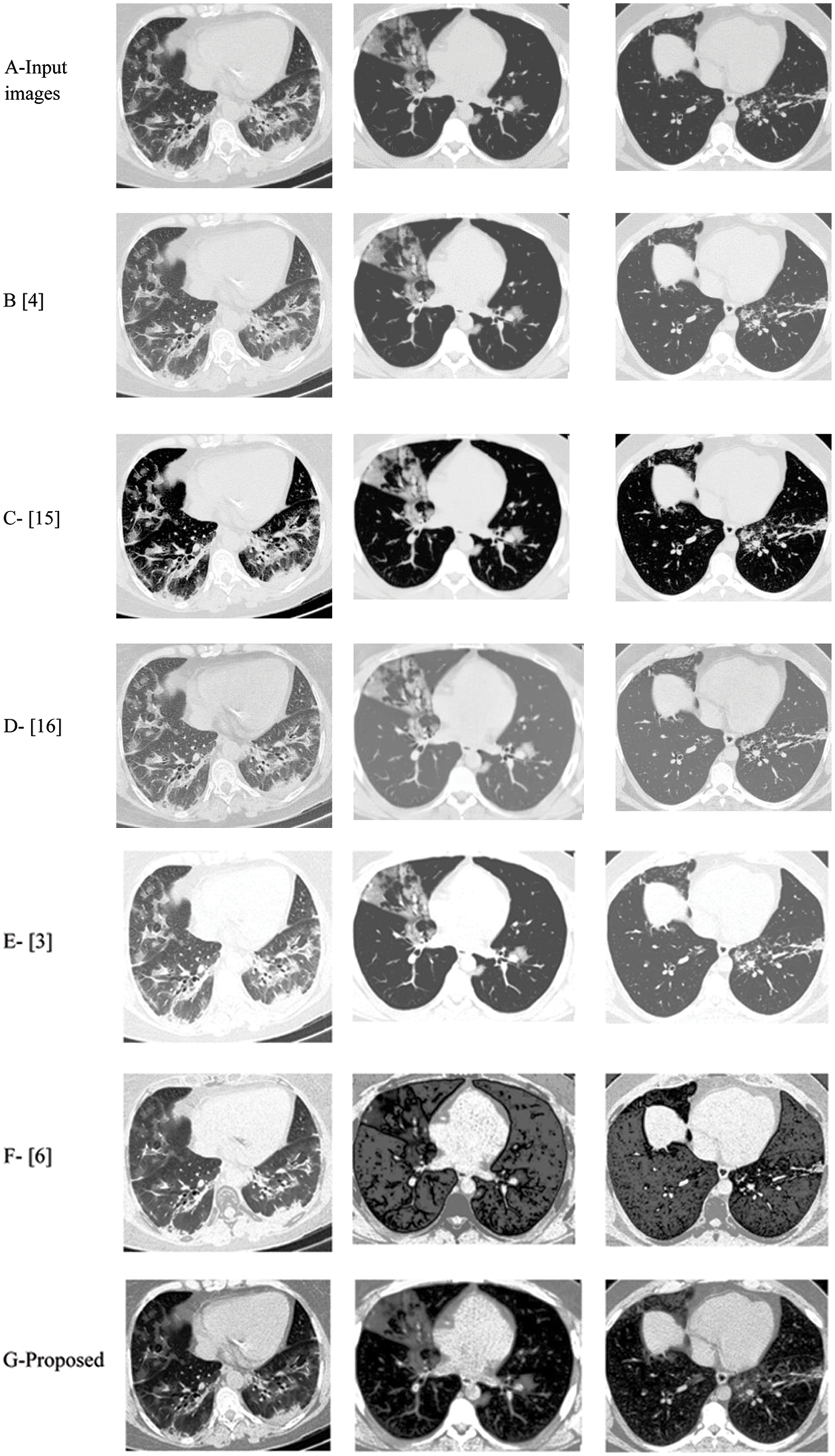
Figure 3: The results of the proposed and existing enhancement models. (A) Input image, (B) Al-Shamasneh et al. [4], (C) Al-Ameen et al. [15], (D) Fu et al. [16], (E) Raghunandan et al. [3]. (F) Ibrahim et al. [6], (G) proposed method
In Fig. 3, the proposed and existing techniques’ qualitative outcomes for CT image input are shown in which all the images exhibit various trends with dark and bright parts.
For the comparative analysis, we used the following existing approaches to show that the proposed enhancement model is effective as a medical picture enhancing tool. Al-Shamasneh et al. (2018) [4] developed a fractional entropy-based approach for enhancing kidney images. Al-Ameen et al. 2016 [15] proposed an MRI brain image enhancing approach. Fu et al. 2016 [16] developed a fusion-based enhancing approach for low-light photos. Raghunandan et al. 2017 [3] developed an image enhancement approach for license plates based on the Riesz fractional operator. Finally, a medical picture improvement model based on the proportional-Caputo hybrid operator's class of fractional partial differential equations (FPDEs) [6].
When existing methods’ enhancement results are compared to the proposed approach's enhancement outcomes, the proposed method outperforms the existing methods in terms of image quality. It may be seen in the instance of the existing approaches that the Al-Ameen et al. [15] Fig. 3C, and Ibrahim et al. [6] Fig. 3F techniques yield over-enhanced images, whereas the suggested method enhances the dark portions of input photos while leaving the brilliant areas alone.
Overall, the proposed model's brightness produces the structures of medical images, which often depict edges, well-defined and clear. This is due to the model's capacity to efficiently capture high frequency features. For images that are poorly lighted, the proposed approach produces reasonable visual results. This is the Mittag-Leffler sum with the fractional parameter α contribution to this study. In Tab. 1, the quantitative outcomes of the proposed enhancement method and existing enhancement models, are summarizes.

The Brisque and Niqe scores were employed for the quantitative comparisons, as shown in Tab. 1. The blind image quality assessment (IQA) of Natural Picture Quality Evaluator (NIQE) is a completely blind image quality analyzer. For practically all CT scans, the proposed approach obtains the best Brisque and Niqe. As shown in Tab. 1, the proposed image enhancement method outperforms the existing methods in terms of Brisque and Niqe scores.
Regardless of dataset or content, the proposed method obtains the best of Brisque and Niqe scores (lower is preferable). In conclusion, the proposed method produces the best results for CT scans of various visual difficulties when compared to the other methods since its results are consistent across different datasets. However, because they were built with a specific application in mind, some existing procedures may give greater results when employed under specific conditions.
The performance of the proposed approach diminishes significantly as the complexity of the input images increases, which is the fundamental limitation of this work.
In this study, we have proposed a new COVID-19 image enhancement based on Mittag-Leffler-Chebyshev polynomial as pre-processing step for COVID-19 detection and segmentation. The proposed approach is based on using the Mittag-Leffler sum convoluted with Chebyshev polynomial. Through the proposed image enhancement model, the images with low gray-level have been improved by estimating the probability of each image pixel. The proposed methodology outperforms existing methods in the wide application of image enhancement, according to testing findings on a variety of lungs CT scans. Future research may customize the current model for specific applications to maximize the benefit of the upgrade.
Funding Statement: This research was supported by the Deanship of Scientific Research, Imam Mohammad Ibn Saud Islamic University (IMSIU), Saudi Arabia, Grant No. (21-13-18-056).
Conflicts of Interest: The authors declare that they have no conflicts of interest to report regarding the present study.
1. A. M. Hasan, M. M. Al-Jawad, H. A. Jalab, H. Shaiba, R. W. Ibrahim et al., “Classification of COVID-19 coronavirus, pneumonia and healthy lungs in CT scans using Q-deformed entropy and deep learning features,” Entropy, vol. 22, no. 5, pp. 517, 2020. [Google Scholar]
2. X. R. Zhang, J. Zhou, W. Sun and S. K. Jha, “A lightweight CNN based on transfer learning for COVID-19 diagnosis,” Computers, Materials & Continua, vol. 72, no. 1, pp. 1123–1137, 2022. [Google Scholar]
3. K. Raghunandan, P. Shivakumara, H. A. Jalab, W. I. Ibrahim, G. H. Kumar et al., “Riesz fractional basedmodel for enhancing license plate detection and recognition,” IEEE Transactions on Circuits and Systems for Video Technology, vol. 28, no. 9, pp. 2276–2288, 2017. [Google Scholar]
4. A. R. Al-Shamasneh, H. A. Jalab, P. Palaiahnakote, U. H. Obaidellah and W. I. Ibrahim, “A new local fractional entropy-based model for kidney MRI image enhancement,” Entropy, vol. 20, no. 5, pp. 344, 2018. [Google Scholar]
5. H. A. Jalab, R. W. Ibrahim, A. M. Hasan, F. K. Karim, A. Al-Shamasneh et al., “A new medical image enhancement algorithm based on fractional calculus,” Computers Materials & Continua, vol. 68, no. 2, pp. 1467–1483, 2021. [Google Scholar]
6. R. W. Ibrahim, H. A. Jalab, F. K. Karim, E. Alabdulkreem and M. N. Ayub, “A medical image enhancement based on generalized class of fractional partial differential equations,” Quantitative Imaging in Medicine and Surgery, vol. 12, no. 1, pp. 172–183, 2022. [Google Scholar]
7. H. A. Jalab, A. R. Al-Shamasneh, H. Shaiba, R. W. Ibrahim and D. Baleanu, “Fractional renyi entropy image enhancement for deep segmentation of kidney MRI,” Computers Materials & Continua, vol. 67, no. 2, pp. 2061–2075, 2021. [Google Scholar]
8. P. N. Chowdhury, P. Shivakumara, H. A. Jalab, R. W. Ibrahim, U. Pal et al., “A new fractal series expansion based enhancement model for license plate recognition,” Signal Processing: Image Communication, vol. 89, no. 3, pp. 1–24, 2020. [Google Scholar]
9. R. W. Ibrahim, H. Yahya, A. Mohammed, N. M. Al-Saidi and D. Baleanu, “Mathematical design enhancing medical images formulated by a fractal flame operator,” Intelligent Automation and Soft Computing, vol. 32, no. 2, pp. 937–995, 2022. [Google Scholar]
10. R. W. Ibrahim, “A new image denoising model utilizing the conformable fractional calculus for multiplicative noise,” SN Applied Sciences, vol. 2, no. 1, pp. 1–11, 2020. [Google Scholar]
11. H. A. Jalab, M. A. Alqarni, R. W. Ibrahim and A. A. Almazroi, “A novel pixel's fractional mean-based image enhancement algorithm for better image splicing detection,” Journal of King Saud University-Science, vol. 34, no. 2, pp. 1–10, 2022. [Google Scholar]
12. I. Aldawish and R. W. Ibrahim, “A new mathematical model of multi-faced COVID-19 formulated by fractional derivative chains,” Advances in Continuous and Discrete Models, vol. 2022, no. 1, pp. 1–10, 2022. [Google Scholar]
13. H. J. Haubold, A. M. Mathai and R. K. Saxena, “Mittag-leffler functions and their applications,” Journal of Applied Mathematics, vol. 2011, no. 1, pp. 1–51, 2011. [Google Scholar]
14. COVID-19 images dataset. Italian Society of Medical and Interventional Radiology. 2020 [Online]. Available: https://www.sirm.org/category/senza-categoria/covid-19/ [Accessed on January 2022]. [Google Scholar]
15. Z. Al-Ameen and G. Sulong, “Ameliorating the dynamic range of magnetic resonance images using a tuned single-scale retinex algorithm,” International Journal of Signal Processing, Image Processing and Pattern Recognition, vol. 9, no. 7, pp. 285–292, 2016. [Google Scholar]
16. X. Fu, D. Zeng, Y. Huang, Y. Liao, X. Ding et al., “A Fusion-based enhancing method for weakly illuminated images,” Signal Processing, vol. 129, no. 1, pp. 82–96, 2016. [Google Scholar]
 | This work is licensed under a Creative Commons Attribution 4.0 International License, which permits unrestricted use, distribution, and reproduction in any medium, provided the original work is properly cited. |