DOI:10.32604/csse.2021.014247
| Computer Systems Science & Engineering DOI:10.32604/csse.2021.014247 | |
| Article |
Cervical Diseases Prediction Using LHVR Techniques
Muthayammal Engineering College, Kakaveri, Rasipuram, 637408, India
*Corresponding Author: Praveena Rajasekaran. Email: praveena.r.ece@mec.edu.in
Received: 08 September 2020; Accepted: 02 December 2020
Abstract: The stabilizing mechanisms of cervical spine spondylosis are involved in the degenerating segmentation vertebra, which often causes pain. Health of the individual is affected, both physically and mentally. Due to depression, nervousness, and psychological damages occur thereby losing their human activity functions. The nucleus pulposus of spinal disc herniation is prolapsed through a deficiency of annulus fibrosus. A jelly-like core part of the disc contains proteins that cause the tissues to become swollen when it touches the nucleus pulposus. The proposed Gradient Linear Classification (GLC) algorithm is used for the efficient automatic classification of disc degeneration herniation of Inter vertebral/ vertebra in a cervical disc. Distance between the disc degeneration is classified through gradient operator and is estimated using the rotation of angles between the correlations. Specialists of the orthopedic spine are searching for high-precision algorithms, which are achieved using proposed Linear Hybrid Vertebra Regression (LHVR) diagnostic techniques to identify the degree of cervical disc degeneration using an accurate location. Our experimental results have been used to determine a high range of classification in predicting the spinal cord which saves handling time and accomplishes high accuracy in detection.
Keywords: Cervical diseases; GLC; LHVR; disc prediction; classify disc degeneration
Cervical Spondylosis (CS) is caused due to disorder in the usual location of the cervical discs and has been surveyed to be affected two-third of population throughout the world. The cervical spine shielding supports the head with a high-range of movement and spinal cord. The entire spinal cord has a supportive cartilage fibrous called disc cartilage. The gel-like center of the disc cartilage called nucleus pulposus of the degenerative process causes disc hernia to rupture through the hardcore outer wall of the layer annulus fibrosus. The soft gel of the nucleus is affected by many reasons such as irregular growth of boil/ bones which affects the cervical spondylolysis. The irregular growth will cause pain in the neck and spinal column of the disc.
Reason for Cervical Spondylosis
• A major risk of cervical spondylosis is age and conditions are called to be a common factor for older people; the osteoporosis is measured with the bone condition.
• Due to the impact of sports or collision of cars causes the sudden pain during the movement of the neck.
• Shoulder leads to neck pain while clenching a neck may cause stress for a long time.
• Sleeping or working in a desk at the awkward angle position with a lengthy period this poor ergonomic posture can cause neck strain.
• Cervical herniation may occur if there is any disease affected in the spinal cord.
The reason for cervical herniation can affect some of the parts of the spinal cord bones, nerves, tendons muscles and ligaments that hinder to allow the neck to be long-term flexible. From evisceration the nucleus pulposus of the inner layer is protected by the outer hardcore of peripheral annulus fibrosus. Also, onset of degeneration cartilages, increasing age, desiccations, and disc herniation ensue occurs.
In medical images the segmentation of novel approaches is explained using articulated shape of the spine, introduced by Samuel et al. The non-linear low dimensional manifolds are established from the training set of mesh models by exploring the pattern of global variations [1–3]. Their approaches depend upon the graphical model of Markov-chainlike used for the lumbar spine of the vertebra and ordered discs.
The classification and automatic detection of the vertebra system generate an effective and efficient detection of a vertebra; hence statistical learning techniques are based on the proposed algorithm of AdaBoost by Huang et al. [4]. Benjelloun et al. [5] detect the vertebra shapes through the ASM model known as a semi-automated model. Srinivas et al. [6] explained detecting the spine of discs using semi-automatic models.
Seifert et al. [7] the spine reconstructed from the MR images are presented from the combination of the segmentation algorithm, anatomical knowledge and recognizing objects in cervical discs. Yiebin et al. [8] used automatic segmentation of vertebra through large volume of CT data as proposed in the survey. The Gaussian images of emphasized valley construct the method of 3D that is separated by adjacent vertebra.
Glocker et al. [9] proposed a technique to identify localized vertebrae in the field of arbitrary using CT scans. The hidden Markov techniques and regression forest method employ partially cropped scans using moderately visible spines. Schmidt et al. [10] introduced a probabilistic inference with the possible measure of locations in the spinal discs from 3D MR images. Their approaches used part based techniques that describes the appearance of discs by tree classifiers [11]. Under a framework of graphical models, they enforced the relationship between the cervical discs. Liu et al. [12] here the sine cosine algorithm is used for the prediction of cervical hyper extension injuries. The proposed model of intelligent prediction of cervical injury is used along with the SVM techniques. Hence it is called as COSCA SVM model. Yang et al. [13] Machine Learning is used in real data driven for the detection and identification of cervical cancer and the random forest algorithm is used to find the diagnostic accuracy.
Aditya et al. [14] using IOHT, the framework for detection and classification of cervical cancer in pap smear images are identified, here transfer learning along with CNN techniques like naïve Bayes, k Nearest Neighbor, SVM &random forest have been used. Talha et al. [15] the cervical cancer is important for increasing of the mortality rates worldwide. Here the combination of data mixing techniques and decision bee algorithm is used to predict the cervical cancers. Here the smote & auroc curve techniques are used for cervical cancer screening due to their high prediction range and other classifiers are used significantly. Abdullah et al. [16] this 714 features and 58 samples were used to enhance the prediction of cervical cancers using random forest and SVM model. Sultana et al. [17] here the HPV vaccinations were used to reduce the presence of cervical cancer related diseases. Vidya et al. [18], supervised machine learning techniques like ID3, C4.5 and Naive Bayer are used for prediction of cervical disease. Shetty et al. [19] here various ML approaches have been made for detecting the asymptomatic nature of cervical disorders. This gives a comparison of various techniques used for the analysis & prediction. Singh et al. [20] here they have used algorithms & decision tree classifiers for the cervical injury stage prediction. The stages were predicted and analyzed based on the outcomes.
3.1 Gradient Linear Classification (GLC)
The model of geometrical representation of vertebra is generally done prior fitting by using spinal cord segmentation technique. The spatial models of the interrelationship between vertebras to edges are analyzed in prior. The gray level features of images are defined as the appearance of spatial information and the shape of the cervical disc. The GLC have often executed as geometrical model and spatial model proceeded by high expressive power to catch the entire range of feasible images that appeared in the cervical disc. The below Fig. 1 demonstrates the comparison of the healthy and unhealthy cervical discs.

Figure 1: Cervical comparison of healthy and disc degeneration
Pattern matching is based on the Gradient Linear Classification (GLC) similarity index term is taken into account gradients of vertical and horizontal images. The formula of GLC index is:

where ∂/∂x and ∂/∂y are the gradient linear operators beside a direction of x and y respectively, ∂V the vertebra template, LC is the Linear correlation operator and can be expressed by the below formula:


where,
x & y: Estimated angles of rotation.
∂V: The vertebra index.
n : Is the number of pixels.
σ : The standard deviations of a vertebra.
The maximum absolute of GLC corresponds to the most excellent location of templates in current image, this way each vertebra is easily been located. The trajectories of the vertebra are obtained in a sequence location along with rigid of time and considering the sagittal plane over a planar motion which is completely described.
3.2 Linear Hybrid Vertebra Regression (LHVR)
The parts of the nucleus pulposus contain proteins that will cause the tissues to become tender and swollen. High pain will causes the proteins to leak out to the nerves along the outer layer of cervical discs. Forces are resisting back to stay flexible in order to assist shock-like absorbers to the bones in spinal columns and discs.

Figure 2: Degenerated cervical disc
The LHVR algorithm categorizes several diseases using their appropriate symptoms. Cervical presented the spinal nerve, bone spurs, spinal cord, annulus, nucleus, herniated disc and anterior opposed with posterior. Let (x, y) are the disc angles which are processed in the degenerated disc to detect the diseases. The layers are generated with the input, weight and output layer to detect the diseases as shown in Fig. 2. Each layer is executed step by step as three phases which are used to detect the diseases along with the position of disc degeneration.
Step 1:
The weights update between the hidden & output layers.
Let Erri be the i-th component of the error vector y–W(x)
Define

The weight updated prototype becomes 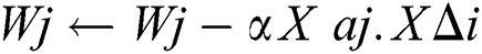 this is parallel to the weight updated of perceptrons.
Step 2:
Linear Regression method of the weighted value
The hidden node of ‘j’ is responsible for some divided error ∆i\x96 in each output node. Thus, division error ∆i\x96 values between the hidden node and the output node are divided according to the power of the joining:

Step 3:
Update the weights (W) between the input and hidden layers. Again this is related to the weight-updated in the Perceptrons

Degenerative Disc Disease Symptoms
The diagnosis method is based on the examination level of physical and medical history which includes the description, symptoms and circumstances of pain started. MRI scans show the damages of discs but this alone cannot confirm the disc degenerated diseases using LHVR which can be analyzed to predict the diseases. Symptoms are commonly concentrated along the low back or neck depending on the degenerated discs.
Table 1: Disease prediction of cervical disc

The above Tab. 1 is the comparison of cervical disc diseases and symptoms. This detection will be useful for the prediction of cervical herniation and can analyze the diseases easily.
Accurate Segmentation of Cervical disc
The proposed algorithm is executed in milliseconds with accurate segmentation of the cervical discs compared with the existing system. The time of execution is compared with segmentation is established in Fig. 3. The below shown comparison chart shows the proposed algorithm using Gradient Linear Classification (GLC) which will take less time for classifying the spinal cord image compared with K-Means and MLR. The GLC illuminates accurate disc degeneration location.

Figure 3: Cervical disc segmentation time
Prediction of Cervical Diseases
The performance level of proposed techniques in predicting cervical diseases is through the classification in accuracy level.

Figure 4: Prediction of cervical diseases
The accuracy level of proposed and existing methods has been illustrated in Fig. 4. From the above comparison chart, the existing algorithms of MRF and BABC showed low prediction accuracy compared with the LHVR that has high precision level for prediction of cervical spondylolysis. Using the above results, it shows high level of prediction in cervical diseases. Due to disc degeneration, several diseases affected are detected using the LHVR algorithm to predict cervical diseases.
The study concluded accurate segmentation and prediction of cervical disc vertebra and inter vertebra. The research is proposed for peoples who are affected by neck or back pain so that they can categorize the diseases by which they were affected. The GLC algorithm is sensitive for high quality images and the accuracy is evaluated for classification of disc degeneration. The LHVR techniques are useful for recognition of disease based symptoms that are detected as a whole part of the human body. These can improve the accuracy in navigation and precision robotics that can detect the best ways to reduce the herniation before acquiring high illness. The disease-based symptoms are classified so that the patients can easily analyze the disease by which they were affected. Therefore the research is useful for medical based cervical analyzed specialists.
Funding Statement: The author(s) received no specific funding for this study.
Conflicts of Interest: The authors declare that they have no conflicts of interest to report regarding the present study.
1. R. B. Cloward. (1958). “Cervical diskography; technique, indications and use in diagnosis of ruptured cervical disks,” American Journal of Roentgenology, Radium Therapy, and Nuclear Medicine, vol. 79, no. 4, pp. 563–574.
2. R. Fejer, K. O. Kyvik and J. Hartvigsen. (2006). “The prevalence of neck pain in the world population: A systematic critical review of the literature,” European Spine Journal, vol. 15, no. 6, pp. 834–848.
3. M. V. Risbud and I. M. Shapiro. (2014). “Role of cytokines in intervertebral disc degeneration: Pain and disc content,” Nature Reviews Rheumatology, vol. 10, no. 1, pp. 44–56.
4. S. Huang, Y. Chu, S. Lai and C. L. Novak. (2009). “Learning-based vertebra detection and iterative normalized-cut segmentation for spinal MRI,” IEEE Transactions on Medical Imaging, vol. 28, no. 10, pp. 1595–1605.
5. M. Benjelloun, S. Mahmoudi and F. Lecron. (2011). “A framework of vertebra segmentation using the active shape model-based approach,” International Journal of Biomedical Imaging, vol. 2011, no. 2, pp. 1–14.
6. R. Srinivas and K. V. Ramana. (2015). “A fully automated new–fangled VESTAL to label cervical vertebrae and intervertebral discs,” in 3rd Int. Conf. on Recent Trends in Computing, vol. 57, pp. 483–492.
7. S. Seifert, I. Wachter, G. Schmelzle and R. Dillmann. (2009). “A knowledge-based approach to soft tissue reconstruction of the cervical spine,” IEEE Transactions on Medical Imaging, vol. 28, no. 4, pp. 494–507.
8. K. Yiebin and K. Dongsung. (2009). “A fully automatic vertebra segmentation method using 3D deformable fences,” Computerized Medical Imaging and Graphics, vol. 33, no. 5, pp. 343–352.
9. B. Glocker, J. Feulner, A. Criminisi, D. R. Haynor and E. Konukoglu. (2012). “Automatic localization and identification of vertebrae in arbitrary field of view CT scans,” in Int. Conf. on Medical Image Computing and Computer-Assisted Intervention, vol. 3, pp. 590–598.
10. S. Schmidt, V. Pekar, S. Dries, C. Schnörr, J. Kappes et al. (2007). , “Spine detection and labeling using a parts-based graphical model,” in Biennial Int. Conf. on Information Processing in Medical Imaging, vol. 4584, pp. 122–133.
11. J. Preetha and S. Selvarajan. (2016). “Computer aided diagnostic system for automatic cervical disc herniation classification,” Journal of Medical Imaging and Health Informatics, vol. 6, no. 7, pp. 1589–1593.
12. G. Liu, W. Jia, M. Wang, A. A. Heidari, H. Chen et al. (2020). , “Predicting cervical hyperextension injury: A covariance guided sine cosine support vector machine,” IEEE Access, vol. 8, pp. 46895–46908.
13. W. Yang, X. Gou, T. Xu, X. Yi and M. Jiang. (2019). “Cervical cancer risk prediction model and analysis of risk factors based on machine learning,” in Int. Conf. on Bioinformatics and Biomedical Technology, pp. 50–54.
14. K. Aditya, G. Deepak, H. C. A. Victor, K. S. Arun and H. J. Rutvij. (2020). “Internet of health things-driven deep learning system for detection and classification of cervical cells using transfer learning,” Journal of Supercomputing, vol. 76, no. 5, pp. 8590–8608.
15. M. A. Talha, M. A. K. Muhammad, A. I. Muhammad, A. Wahab and M. Mubbashar. (2019). “Cervical cancer prediction through different screening methods using data mining,” International Journal of Advanced Computer Science and Applications, vol. 10, no. 2, pp. 388–396.
16. A. A. Abdullah, N. K. Abusabri, W. Khairunizam, I. Zunaidi, Z. M. Razlan et al. (2019). , “Development of predictive models for cervical cancer based on gene expression profiling data,” in IOP Conf. Series: Materials Science and Engineering, vol. 557.
17. F. Sultana, K. Winch, M. Saville and J. M. L. Brotherton. (2019). “Is the positive predictive value of high-grade cytology in predicting high-grade cervical disease falling due to HPV vaccination?,” International Journal of Cancer, vol. 144, no. 12, pp. 2964–2971.
18. R. Vidya and G. M. Nasira. (2016). “Predicting of cervical cancer using machine learning techniques-an analysis,” Global Journal of Pure and Applied Mathematics, vol. 12, no. 3, pp. 934–939.
19. A. Shetty and V. Shah. (2018). “Survey of cervical prediction using machine learning: A comparative approach,” in 2018 9th Int. Conf. on Computing, Communication and Networking Technologies, pp. 1–6.
20. J. Singh and S. Sharma. (2019). “Prediction of cervical cancer using machine learning techniques,” International Journal of Applied Engineering Research, vol. 14, no. 11, pp. 2570–2577.
 | This work is licensed under a Creative Commons Attribution 4.0 International License, which permits unrestricted use, distribution, and reproduction in any medium, provided the original work is properly cited. |