DOI:10.32604/iasc.2021.017467
| Intelligent Automation & Soft Computing DOI:10.32604/iasc.2021.017467 | |
| Article |
AcuRegions: A Novel Cutaneous Region Model Based on Acupoints and Its Application
1Chengdu University of Information Technology, Chengdu, 610103, China
2Beijing University of Chinese Medicine, Beijing, 100029, China
3Institute for Medical Informatics, Statistics and Epidemiology, University Leipzig, Leipzig, 04103 Germany
4Guang’an men Hospital, China Academy of Chinese Medical Sciences, Beijing, 100053,
5China Institute of Information on Traditional Chinese Medicine, China Academy of Chinese Medical Sciences, Beijing, 100700, China
*Corresponding Author: Yan Zhu. Email: zhuyan166@126.com
Received: 27 January 2021; Accepted: 01 March 2021
Abstract: The meridian theory, as an essential part of Traditional Chinese Medicine (TCM) fundamentals, provides an explanation of the spatial and functional relationship between the superficial part and the internal organs based on empiric observations. Cutaneous regions which are the body superficies on which the functions of the meridians are reflected, and the sites where the qi of the collateral’s spreads, play an important role in TCM clinical diagnosis and treatment of skin diseases. The survey of the literature on anatomical site, pathology in patients with skin disease, particularly in TCM perspective, clearly indicates that a better cutaneous region model and tool for analysis is needed to make an improved division of the skin into regions with finer granularity. In this paper, we proposed a new cutaneous region model, AcuRegions, which is based on meridian theory. Firstly, we constructed 3D anatomical models for human body including surface, meridian and acupoint model. Then, skin of human body was divided into 670 regions based on acupoints using Voronoi diagram generation algorithm. Thirdly, a software program for visualization and analysis was developed based on AcuRegions model. Lastly, taking discoid eczema as an example, we carried out a demonstration application, whose visualization and statistic result showed consistent with TCM principles and could provide some guidance and enlightenment for further treatment for skin diseases.
Keywords: Cutaneous region; acupoint; meridian; dermatosis; visualization
According to survey data, 35–69% of patients with dermatologic conditions have used complementary and alternative medicine (CAM) in their lifetime, and the prevalence of CAM use amongst patients is growing. Traditional Chinese Medicine (TCM) is the mainstream throughout East Asia in last thousands of years. There have been numerous studies which reported that TCM therapies appear to have some benefit as well as lower risks than that of many common medical treatments [1].
TCM theory regards the human body as a constellation of dynamic forces called Qi, which flow through the body in meridian system, which form the basis of needle placement in acupuncture [2].The meridian theory, as an essential part of TCM fundamentals, provides an explanation of the spatial and functional relationship between the superficial part and the internal organs based on empiric observations. The main part of the meridian system consists twelve regular meridians, inside which the motion of the qi and the blood is circulative and continuous. Marked by the regular meridians, the whole-body surface is divided into 12 corresponding cutaneous regions. These cutaneous regions are the body superficies on which the functions of the 12 regular meridians are reflected, and the sites where the qi of the collateral’s spreads [3]. Cutaneous regions, collaterals, meridians, and internal organs are interrelated, which interact and influence each other, forming a complete system of "cutaneous region-collateral-meridian-organ." One of important characteristics of clinical diagnosis and treatment of skin diseases is the combination of syndrome, meridian and skin lesion differentiations is one of important characteristics of TCM clinical diagnosis and treatment of skin diseases.
In recent years, there has been an increasing interest in the intelligent classification [4,5], analysis, on skin lesion. Particularly, more and more attention has been drawn to research on distribution characteristics of lesion, syndrome differentiation of skin disease based on cutaneous regions theory. Wei Guoqi [6] and Cai Yige [7] have respectively verified that the lesion’s distribution of vitiligo and psoriasis vulgaris appears obvious regular pattern on twelve cutaneous regions. These studies mainly used conventional 2D pictures or actual model whose history could be traced back to the Illustrated Manual of Points for Acupuncture and Moxibustion on a Bronze Statue with Acupoints [8], an official standardized model of the meridian developed in the Song Dynasty. In dermatologic area of modern medicine, researchers have found the body site location is correlated with prognosis and survival of many skin diseases such as Malignant Melanoma (MM) [9,10], inherited epidermolysis bullosa (EB) [11] over the years. Gillgren et al [12,13] proposed a body division model consisting of 24 separate areas and developed a data program to map and analyze the effect of anatomical locations on prognosis of MM.
With comprehensive application of 3D visualization technology, many studies attempted to build 3D meridian system [14,15] for users to browse. Lee [16] visualized the biomedical information to acupoints on the 3D surface anatomical models. In order to map and analysis the lesion distribution associated with meridian theory, there is requirement to build a 3D cutaneous regions model based on meridian system. However, the granularity of traditional cutaneous regions model is not fine enough to represent the clinical lesion’s distribution since the existing division of only 24 cutaneous regions. It is necessary to build the 3D cutaneous regions based on Acupoints, since there are 670 acupoints belonging to 12 regular meridians and Ren and Du meridians. What’s more, there is a great challenge to divide the body surface model into 3D cutaneous regions based on acupoints using computerized method.
Voronoi diagram, also known as Dirichlet tessellation, is the division of a plane with n points into n regions such that each region contains exactly one generating point and every point in the given region is closer to its generating point than to any other. This diagram has many excellent properties in spatial analysis, which make it a particularly useful spatial data model having diverse applications in a variety of disciplines, including urban planning, engineering geology [17], biological morphology, information retrieval, and so on [18].
In this paper, we suggest a new cutaneous region model, named AcuRegions. We implemented an algorithm for generating Voronoi diagram, in which 670 acupoints were taken as generating points to divide the body surface into 670 regions. Based on the AcuRegions model, a newly developed software program and computerized method b enables us to bridge phenotypic features and epidemiological characteristics of skin disease to a more detailed meridian and acupoint site mapping. We also report a case study on the use of this new model to visualize and analysis the lesion site of discoid eczema.
As illustrated in Fig. 1, the workflow of this study mainly consisted of 3 steps:
1. 3D model construction
2. AcuRegions model generation
3. Clinical data visualization and analysis
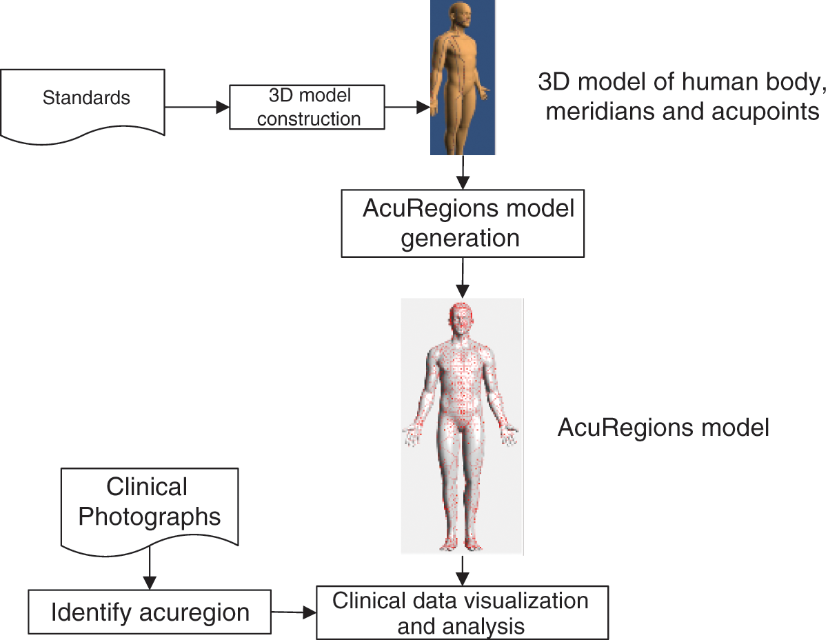
Figure 1: Workflow of study
We collected and followed the World Health Organization (WHO) standard acupuncture point locations in the Western Pacific Region [19] to build 3D model, including the body surface, meridians and acupoints. The newest version of this standard was released by WHO, Regional Office for the Western Pacific in in 2010. It specified the locations of 670 acupoints based on standard measurements of human body (see as in Tab. 1).
Table 1: Statistics of acupoints by meridian & vessel

Using Autodesk 3ds Design 2016 modeling software, as shown in Fig. 2, we constructed 3D model including:
1. Human body. An anatomical surface model, as the base for meridian system, was made in standard measurement of body parts. The model of human was put in position of standing and stretching out hands forward, which is beneficial to display information of the meridian system.
2. Acupoints: 670 acupoints were built in point, the location of which was identified by domain export according to the WHO standard. The code of each acupoint also recorded in the 3D model.
3. Meridians and vessels: 12 regular meridian and Conception vessel and Governor Vessel were built in polyline connecting the belonging acupoints.
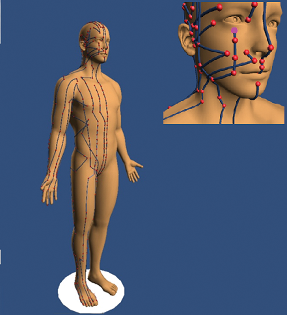
Figure 2: 3D models including human body, meridians and acupoints
2.3 AcuRegions Model Generation
Based on the three-dimensional Voronoi diagram construction method described in the literature [20], the Matlab software of MathWorks is used to implement the Voronoi division algorithm for the AcuRegions model generation. Taking 670 acupoints belonging from 12 meridians and 2 vessels, the human body surface model is divided into 670 regions. Each region, called “acuregion,” is marked by its corresponding central acupoint.
Voronoi diagram is a division of the space plane. Its characteristic is that any position in the polygon divided, name Thiessen polygon, is the closest to the generating point of the polygon, and the distance from the generating point in the adjacent polygon. Each polygon divided contains only one generating point. Because of the equipartition characteristics of the Thiessen polygon in the spatial division, it can be used to solve problems such as the nearest point, the smallest closed circle, and many spatial analysis problems, such as adjacency, proximity, and reachability analysis.
The Delaunay triangulation algorithm mainly refers to the generation of the Delaunay triangle when the Voronoi diagram is generated, and then finds the center of the outer circle of each triangle of the triangle network, and finally connects the center of the circumscribed circle of the adjacent triangles to form a generator with each triangle vertex as the generator Polygonal net. The formed polygonal network is the regional boundary of the acupoint points. In this paper, the Delaunay triangle is generated on the top of the three-dimensional human body model, and countless triangles are used as the basic components of the human body model, as shown in part A of Fig. 3.
This article used the fast-marching algorithm to apply the three-dimensional body model acupoint region division, and utilized the Voronoi division to find the region boundary. First, we use the fast-marching method to start with each acupuncture point and calculate the distance from each vertex to the nearest starting point (acupuncture point). According to the obtained distance, find the maximum distance belonging to each acupuncture point, and then take the maximum distance of each acupuncture point as the constraint, and then use the acupuncture point as the starting point to perform a fast travel algorithm on the vertices of the human model. According to the obtained distance, the three acupuncture points closest to each vertex point in the model are calculated. Then assign each model vertex to the starting point (acupuncture point) closest to it. Then use Voronoi division to further generate the boundary of the acupuncture point region based on the vertices of the obtained acupoint boundary of the human body model. Therefore, we get the area of each acupoint, that is, the skin partition of each acupoint as shown in part B of Fig. 3.
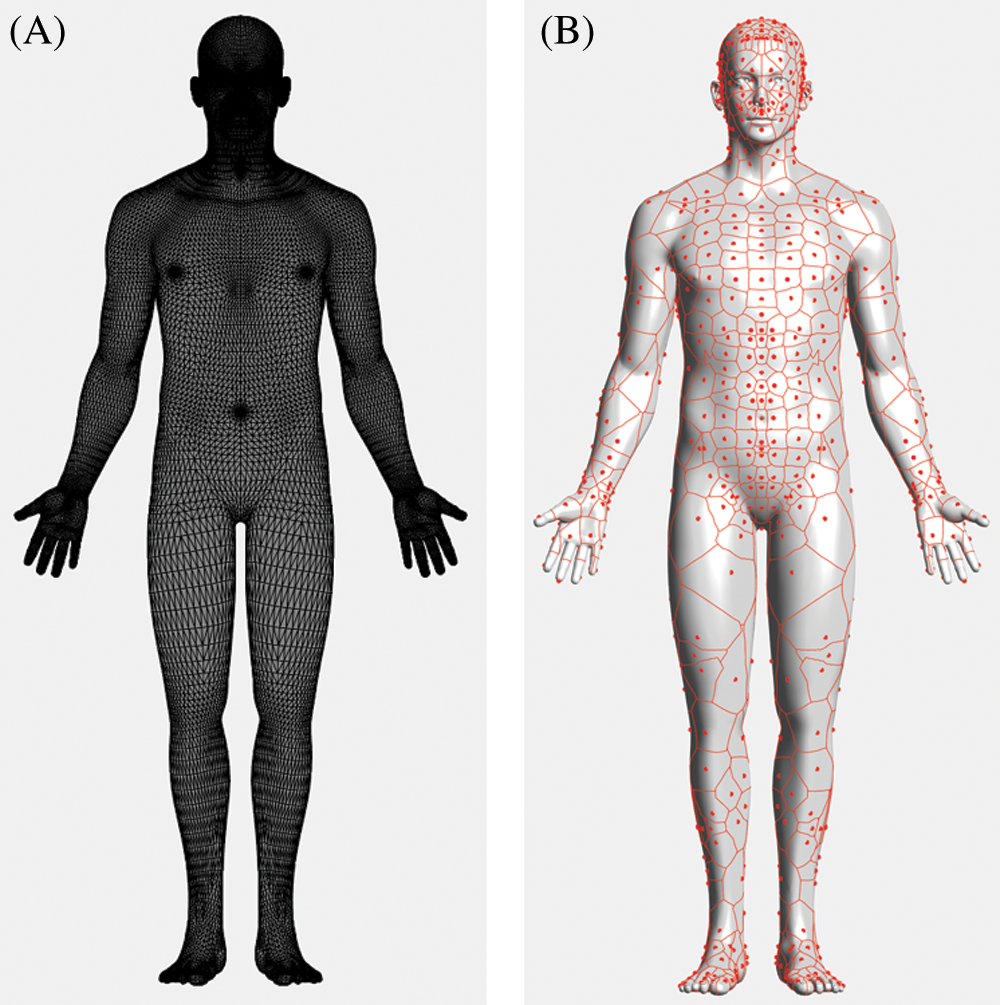
Figure 3: (A) 3D surface model for human in Delaunay triangulation. (B) AcuRegions model generated by Voronoi diagram, points in red are acupoints as generating point.
Use the Matlab development environment to integrate the meridian acupoint knowledge base and the three-dimensional human body model to develop a visualization system. Our acupoint skin partition visualization program contains the following functions:
1. According to the human body of different precision selected by humans, load analysis as a segmented model.
2. Perform Voronoi division on the loaded human body model based on different numbers of meridian acupoint data.
3. Integrate and analyze the meridian acupoint data and the three-dimensional human body model data, so that the meridian data is accurately mapped to the three-dimensional human body model, and the acupoint code/name corresponding to each acupoint is displayed, and finally the visualization of the three-dimensional human body meridian acupoint model is realized.
4. According to the provided data of the most common skin area of lesion, perform statistical analysis on the data of the diseased skin area. According to the severity of the skin area obtained by the statistical analysis, color the diseased skin area in layers to visualize the situation of the diseased area the display is rendered.
5. Statistic and charting functions, such as calculating the frequency by meridian.
3 Application on Visualization and Analysis on Lesion Distribution of Discoid Eczema
Discoid eczema, also called nummular eczema, nummular dermatitis, and discoid dermatitis, is a chronic disease, characterized by distinctive ‘coin-shaped’ eczematous plaques, frequent recurrences, exudative trend, fierce pruritus, etc. The exact cause of discoid eczema is still unknown. What is certain is people who have a family history of allergies and asthma is more susceptible to the discoid eczema and stress has been shown to make it worse. It most usually develops on the lower legs, trunk or forearms, but can also appear on hands and fingers [21].
TCM therapeutic methods, including external and internal herbs, acupuncture and so on, has been long used to treat eczema in China. Many studies [22–24] have been reported TCM can significantly improve the treatment effect, shorten the course of disease, and accelerate the rehabilitation of patients with eczema.
3.1 Data Source and Inclusion Criteria
Initially, raw clinical photographs are downloaded from dermetnz.org using keyword of “Discoid eczema” (https://www.dermnetnz.org/topics/discoid-eczema-images). Criteria for selecting the subject photographs were as follows:
a) The photograph is in color and its quality is high enough to be interpreted by exports.
b) The boundary of lesion is clear.
As a result, 85 photographs meet the inclusion criteria and were selected as clinical data source (see as in Fig. 4).

Figure 4: Samples of selected photographs from DermNet
3.2 Manual Identification for Acuregions
For each photograph, the acuregions where patches located were independently identified by 2 domain experts. The photographs in which acuregions cannot determined clearly and accurately by both experts were excluded. The rest few conflicts of identifying results were all resolved by double checking and discussion.
3.3 Statistic on Acuregion where Discoid Eczema Usually Developing
As a result, 102 acuregions from 59 distinct ones from 32 photographs were identified successfully. As illustrated in Fig. 5, the acuregions where discoid eczema most frequently appeared (over 3 of the 32 cases) were BL57 (5), TE3(5), ST35(4), LI11(4), BL56(4), BL58(4), SP8(4), LI4(4).
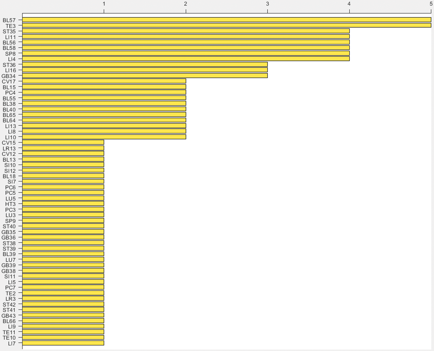
Figure 5: Count of acuregions where discoid eczema most frequently develops
Fig. 6 shows that Bladder Meridian (BL) is most likely meridian where discoid eczema develops, covered more than 28% or 29 of 102 lesions. It followed by Large Intestine Meridian (LI) and Stomach Meridian (ST), 20% and 12% respectively.
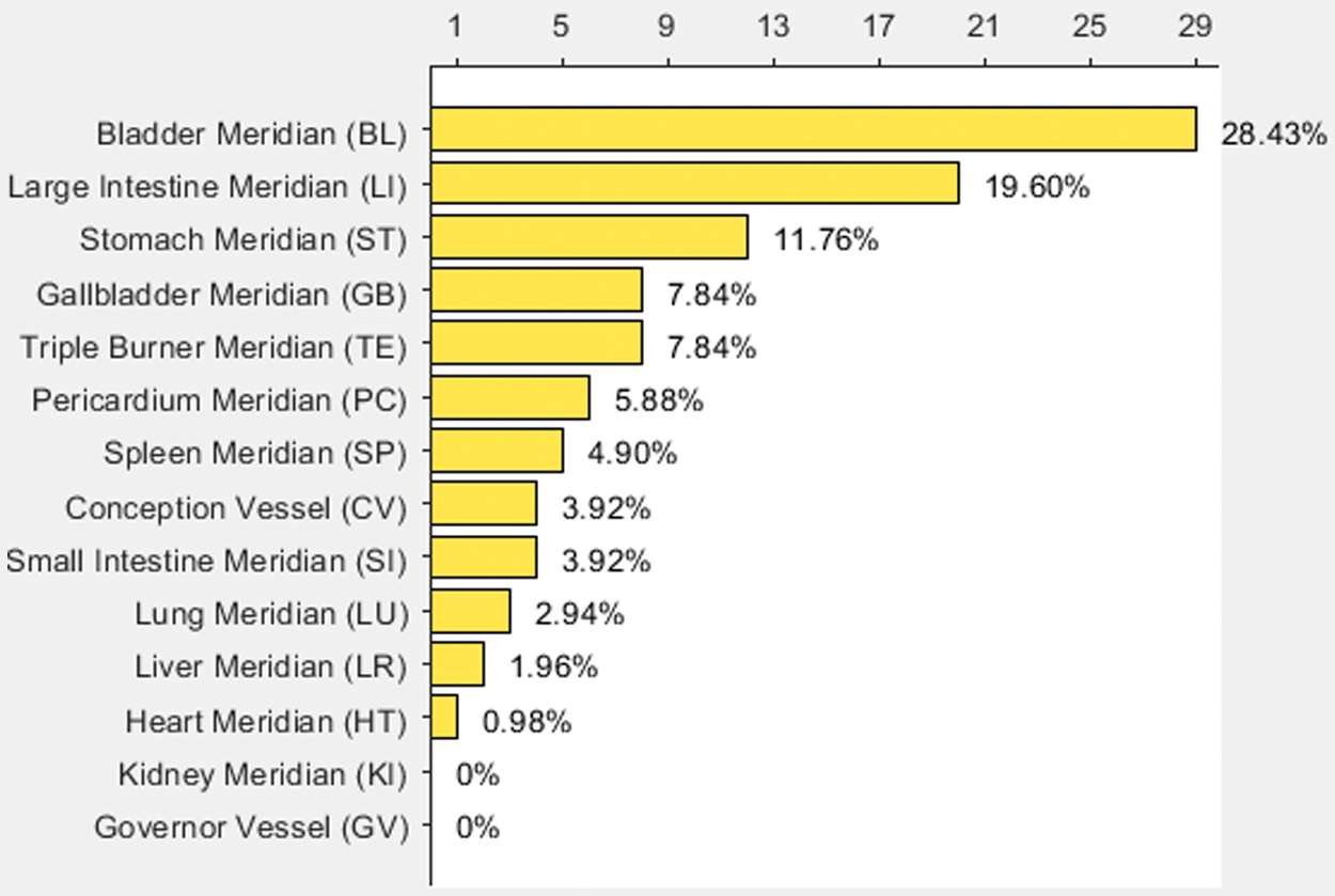
Figure 6: Probability of lesion developed by meridian
Data visualization refers to data that is presented in a visual form, to help people understand the meaning of the data [25]. Based on the AcuRegions model, we generated a 3D visual map to present the site distribution of discoid eczema’s lesion. The color of each acuregion was decided by magnitude of the lesion develops on it. Fig. 7 showed the Top 3 most likely acuregions were BL57 , TE3, ST35 located on the posterior aspect of the leg, dorsum of the hand, anterior aspect of the knee respectively.

Figure 7: 3D visualization for frequency of AcuRegions discoid eczema usually developing
From TCM perspective, discoid eczema belongs to the category of “damp sores (湿疮)” recorded in ancient literature. "dampness pathogen (湿邪)" is thought to be the major cause of eczema, which can block the meridians and accumulate in the skin. According to meridian theory of TCM, Bladder Meridian (BL) governing the external of the whole body, is closed related to skin disease [26]. Besides, Stomach Meridian (ST), Gallbladder Meridian (GB) and Triple Burner Meridian (TE) are the main meridians involved in the body's water and moisture metabolism. Hou’s study [27] also has shown that persistent eczema lesions are often located at these three meridians. The visualization and statistic result from discoid eczema clinical data is consistent with these principles of meridian theory.
In terms of treatment, Guo summarized clinical reports and found the therapeutic effects of acupuncture in treating eczema are significant. Acupoints of Du meridian and Yangming meridian are usually preferred to clear heat [24]. AcuRegions model enable us to introduce TCM theory and principles to anatomical site, providing valuable guidance information for further treatment.
In summary, the existing researches have showed that the information of anatomical site is valuable for diagnosis, prognosis, and treatment with skin disease, particularly in TCM perspective. The meridian theory provides an explanation of the spatial and functional relationship between the superficial part and the internal organs based on empiric observations. Existing cutaneous partition based on regular meridians, the division of the whole-body surface into 12 corresponding cutaneous regions, is too coarse-grained to meet the requirement for clinical data representation and mining in under the background of modernization of TCM.
In this paper, we proposed a new cutaneous region model based on meridian theory, name AcuRegions, which divided skin of human body into 670 regions based on acupoints using Voronoi diagram. Employed 3D visualization and statistical methods, we can utility AcuRegions to enhance the visualization and analysis for the clinical data of skin diseases. A preliminary demonstration application on discoid eczema proved the efficiency and correctness of AcuRegions model further. The model and developed tools based on it are also applicable for to more kinds of diseases integrated with other biomedical information, such as therapeutic and/or efficacy-based information, as well as time series data [28].
Acknowledgement: We thank Mr. Lifeng Zhang for providing efficient help in polishing 3D models.
Author Contributions: Conceptualization, JW and YZ; methodology, JH, LL and ZW; software, JH and LL; validation, WY and JW; resources JW; data curation, JW, LL and YZ; writing—original draft preparation, YZ; writing—review and editing, JH; visualization, LL and YZ; supervision, JH; project administration, JW and YZ; funding acquisition, JW and YZ. All authors have read and agreed to the published version of the manuscript.
Funding Statement: This study was supported by National Natural Science Foundation of China (61701546, YZ received), Fundamental Research Funds for the Central public welfare research institutes (ZZ13-YQ-021, JW received), and Fundamental Research Funds for the Central public welfare research institutes (ZZ13-YQ-126, YZ received).
Conflicts of Interest: The authors declare that they have no conflicts of interest to report regarding the present study.
1. S. Bhuchar, R. Katta and J. Wolf, “Complementary and alternative medicine in dermatology: An overview of selected modalities for the practicing dermatologist,” American Journal of Clinical Dermatology, vol. 13, no. 5, pp. 311–317, 2012. [Google Scholar]
2. G. Sun, D. E. Douglas and Q. Zhang, Fundamentals of Chinese Medicine. Beijing, China: People's Medical Publishing House, 2014. [Google Scholar]
3. F. I. Chen, A. D. Antochi and A. G. Barbilian, “Acupuncture and the retrospect of its modern research,” Romanian Journal of Morphology and Embryology, vol. 60, no. 2, pp. 411–418, 2019. [Google Scholar]
4. F. Liu, J. Yan, W. Wang, J. Liu and A. Yang, “Scalable skin lesion multi-classification recognition system,” Computers Materials and Continua, vol. 61, no. 3, pp. 801–816, 2019. [Google Scholar]
5. J. Liu, W. Wang, J. Chen, G. Sun and A. Yang, “Classification and research of skin lesions based on machine learning,” Computers Materials and Continua, vol. 61, no. 3, pp. 1187–1200, 2019. [Google Scholar]
6. G. Wei and A. Xu, “Relationship between progressive vitiligo and meridians,” Chinese Archives of Traditional Chinese Medicine, vol. 29, no. 10, pp. 2286–2287, 2011. [Google Scholar]
7. Y. Cai, L. Jin, W. Lou and W. Li, “The preliminary study on the regulation of lesions' distribution of recalcitrant psoriasis vulgaris on twelve cutaneous regions,” Journal of Practical Dermatology, vol. 7, no. 1, pp. 46–49, 2014. [Google Scholar]
8. W. H. Liu, “Thinking about standard and standardization of acupuncture and moxibustion,” Chinese Acupuncture & Moxibustion, vol. 29, no. 1, pp. 40–43, 2009. [Google Scholar]
9. B. K. Ragnarsson-Olding, “Spatial density of primary malignant melanoma in sun-shielded body sites: A potential guide to melanoma genesis,” Acta Oncologica, vol. 50, no. 3, pp. 323–328, 2011. [Google Scholar]
10. C. Garbe, B. Petra, J. Bertz, G. Burg, B. Hoedt et al., “Primary cutaneous melanoma: Prognostic classification of anatomic location,” Cancer, vol. 75, no. 10, pp. 2492–2498, 1995. [Google Scholar]
11. D. T. Devries, L. B. Johnson, M. Weiner and J. D. Fine, “Relative extent of skin involvement in inherited epidermolysis bullosa (EBComposite regional anatomic diagrams based on the findings of the National EB registry, 1986 to 2002,” Journal of the American Academy of Dermatology, vol. 50, no. 4, pp. 572–581, 2004. [Google Scholar]
12. P. Gillgren, G. Brattstrom, J. Frisell, J. O. Persson, U. Ringborg et al., “Effect of primary site on prognosis in patients with cutaneous malignant melanoma. a study using a new model to analyse anatomical locations,” Melanoma Research, vol. 15, no. 2, pp. 125–132, 2005. [Google Scholar]
13. P. Gillgren, G. Brattström, E. D. Mårtensson, J. Frisell, J. Palmgren et al., “A new computerized methodology to analyse tumour site in relation to phenotypic traits and epidemiological characteristics of cutaneous malignant melanoma,” British Journal of Dermatology, vol. 146, no. 6, pp. 1023–1030, 2002. [Google Scholar]
14. Y. Zhu, B. Li, M. Cui, Y. Fu and L. Zhu, “Acu3d: A cross-platform three-dimensional visualization system for the meridians and acupoints of human body,” in 2015 7th Int. Conf. on Information Technology in Medicine and Education (ITMEHuangshan, China, pp. 27–31, 2015. [Google Scholar]
15. C. Liu, Z. Chen, G. Jiang and H. Wang, “Interactive exhibition of human meridian based on cult3d,” in 2005 IEEE Engineering in Medicine and Biology 27th Annual Conf., Shanghai, China, pp. 6262–6264, 2006. [Google Scholar]
16. N. Zong, S. Lee and H. G. Kim, “Visualization of the meridian system based on biomedical information about acupuncture treatment,” Evidence-based Complementary and Alternative Medicine, vol. 2013, pp. 697–715, 2013. [Google Scholar]
17. J. Gao and X. Lin, “Mathematical interpolation and correction of three-dimensional modelling of high-speed railway,” Intelligent Automation & Soft Computing, vol. 26, no. 5, pp. 1023–1034, 2020. [Google Scholar]
18. J. Chen, X. Zhao and Z. Li, “An Algorithm for the Generation of Voronoi Diagrams on the Sphere Based on QTM,” Photogrammetric Engineering & Remote Sensing, vol. 69, no. 1, pp. 79–89, 2003. [Google Scholar]
19. WHO Regional Office for the Western Pacific, “WHO standard acupuncture point locations in the western pacific region,” Manila, World Health Organization, 2008. [Google Scholar]
20. Y. Liu, Z. Chen and K. Tang, “Construction of iso-contours, bisectors, and voronoi diagrams on triangulated surfaces,” IEEE Transactions on Pattern Analysis and Machine Intelligence, vol. 33, no. 8, pp. 1502–1517, 2011. [Google Scholar]
21. A. K. C. Leung and W. L. M. Robson, “Nummular eczema, ” in Encyclopedia of Molecular Mechanisms of Disease, F. Lang, Berlin, Heidelberg: Springer, pp.1502–15032009. [Google Scholar]
22. Z. Peng, “Clinical observation of traditional Chinese medicine wet compress in the treatment of eczema in 60 cases,” Chinese Community Doctors, vol. 33, no. 13, pp. 80–82, 2017. [Google Scholar]
23. H. L. Wan, H. Z. Chen and X. Q. Shi, “Study on effect of traditional Chinese medicine Jianpi Chushi decoction and ointment on chronic eczema,” Asian Pacific Journal of Tropical Medicine, vol. 9, no. 9, pp. 920–923, 2016. [Google Scholar]
24. T. P. Guo, L. Zhao, R. Lin, M. Dong, J. Yang et al., “A clinical review of acupuncture in treating eczema in the past decade in China,” European Journal of Integrative Medicine, vol. 2, no. 4, pp. 217, 2010. [Google Scholar]
25. G. Sun, F. Li and W. Jiang, “Brief talk about big data graph analysis and visualization,” Journal on Big Data, vol. 1, no. 1, pp. 25–38, 2019. [Google Scholar]
26. X. Zhang, Q. Wang, H. Zhao and W. Yuan, “The experimental study of ‘the bladder meridian in charge of the body surface’ which based on the theory of the skin immune,” Lishizhen Medicine and Materia Medica Reseach, vol. 27, no. 1, pp. 232–234, 2016. [Google Scholar]
27. T. Hou and G. Zhou, “Investigation of the meridian distribution pattern of 156 cases of long-lasting intractable skin lesions due to chronic eczema,” Xinjiang Journal of Traditional Chinese Medicine, vol. 34, no. 6, pp. 44–46, 2016. [Google Scholar]
28. W. Jiang, J. Wu, G. Sun, Y. Ouyang, J. Li et al., “A survey of time series data visualization methods,” Journal of Quantum Computing, vol. 2, no. 2, pp. 105–117, 2020. [Google Scholar]
 | This work is licensed under a Creative Commons Attribution 4.0 International License, which permits unrestricted use, distribution, and reproduction in any medium, provided the original work is properly cited. |