| Molecular & Cellular Biomechanics |
DOI: 10.32604/mcb.2021.016056
ARTICLE
Comparison of Detection and Classification of Hard Exudates Using Artificial Neural System vs. SVM Radial Basis Function in Diabetic Retinopathy
1Sona College of Technology, Salem, 636201, India
2Muthayammal Engineering College, Rasipuram, 637408, India
*Corresponding Author: V. Sudha. Email: sudha.ece@sonatech.ac.in
Received: 02 February 2021; Accepted: 19 March 2021
Abstract: Diabetic Retinopathy (DR) is a disease that occurs in the eye which results in blindness as it passes to proliferative stage. Diabetes can significantly result in symptoms like blurring of vision, kidney failure, nervous damage. Hence it has become necessary to identify retinal damage that occurs in diabetic eye due to raised glucose level in its initial stage itself. Hence automated detection of anamoly has become very essential. The appearance of crimson and yellow lesions is considered as the earliest symptoms of DR which are called as hemorrhages and exudates. If DR is analysed at initial stage, blindness does not occur. The damage in retina can hinder the light that passes through nerves of the eye leading to visual loss. The motivation behind this research is to reduce the number of false positives by accurate detection which is possible using proposed fuzzy system based on ANN. Though several classifiers are available to detect the exudates this paper makes analysis of support vector machine using radial basis kernel function with proposed ANN technique. Also, adaptive neuro fuzzy inference system segmentation is performed after feature extraction technique, which makes classifer to outperform. The evaluation results showed that proposed artificial neural network based on fuzzy approach attained significant results compared to other classifiers. Moreover, the proposed algorithm has significant accuracy of 94% and minimum error rate has been observed.
Keywords: Support vector machine; radial basis function; neural networks; exudates; segmentation
Diabetes is a common disease that affects quite large population across the world. Raised glucose levels always lead to presence of anamolies in retinal layer of eye. Initial stage of retinopathy is characterized by hard, soft exudates, microaneurysms, haemorrhages. When there occurs growth of tiny blood vessels, it is microaneurysms. Also, leakage of blood from retinal blood vessels is haemorrhage. There are different stages in retinopathy. In stage of nonproliferative retinopathy [1], retinal vessels are damaged hence fatty and protein-based particles are leaked called as exudates. If accumulation of exudates occurs in centre of the retina, it can result in loss of vision in the nonproliferative stage of DR. Exudates are characterised with vascular damage patches resulting in leakage which is viewed as random yellow patches of various sizes and shapes. Exudates are related to retinal edema and visual loss, which is considered as most important retinal lesion detection. The Diabetic Macular Edema is based on feature vector classification produced for images [2]. This exudates are segmented using feature based technique. This comprises of morphological method that uses algorithm based on SVM [3].
Here, two classifier techniques are compared with Bayesian and Artificial neural network and performance was analysed [4]. This methodology used neural network for classification of diseases to get better result than Bayesian classifier. To identify the feaures present in image several preprocessing techniques are used. Using fuzzy, this segmentation process is done [5]. To identify the retinal bright lesions a minimum-distance discriminant classifier was used. The drusen dependent on 16 matrix type was used and trained using neural networks [6]. It achieved 91% accuracy for patch resolution performance using small number of upto 15 retinal images. An automated machine learning-based system is capable of detecting both the types of exudates and distinguish from drusen that is present in color imagesusing ensemble based approach [7].
A diabetic patient has developed approaches to improve the retinal experts performance level. Using additional data sets, machine learning technique can be improved for identifying clinically significant lesions that are bright, thereby improving diagnosis at an early stage to avoid visual loss that occur in diabetes patients. Fig. 1 illustrated normal and abnormal image.
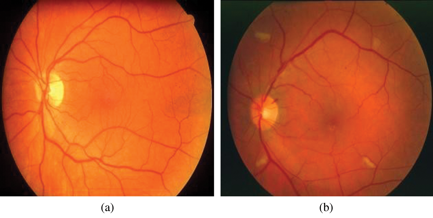
Figure 1: Retinal images a) Normal b) Image showing abnormality
Wang et al. [8] to obtain graded features and classify using random forest that is trained using convolutional neural networks. To extract the features it used 6 convnets layers along with a subsequent sub-sampling layer. DRIVE and STARE databases were used to acquire accuracy of 98.1% and 97.2%, respectively. Complexity is increased by additional preprocessing levels through technique of deblurring prior to detection, segmentation of microvascular condition, improving light intensity using mathematical model and morphological reconstruction [9]. To detect dark-colored lesions named as microaneurysms and haemorrhages thresholding technique along with, adaptive preprocessing was used [10,11] after that the vessels were removed set of input Structures. Using multilayer perceptron analysis vessels are identified [10] along with multiscale morphological closing operation [11]. The major drawback being that most of the false positives detected during segmentation of vessels are identified as lesions. Due to this, lesions are lost and are not retrieved in subsequent processing of blood vessels. Sudha et al. [12] has studied the performance of convolutional neural networks using pretrained VGG-19 for detection of exudates in diabetic retinopathy. Joshi et al. [13] provided a detailed survey on the available techniques used to identify the exudates in diabetic retinopathy. The image dataset used in our research is shown in Tab. 1.
A. Image Preprocessing
The data set required for analysis is obtained from publically available database such as MESSIDOR with largest resolution of 2204 × 1536 and STARE of 600 × 700 resolution database. Preprocessing is a technique to enhance picture information for processing the image. Picture preprocessing techniques can be characterized into four classes: pixel, geometric variation. The original RGB fundus image to HSI image space used colour space transition. The noise band containing noise is removed using median filter over the full image. To enhance the contrast, Contrast limited Adaptive Histogram equalization technique is performed. This technique is different from Gaussian function based methods, as CLAHE avoids saturation in similar areas of the image. As optic disc and exudate uses same characteristics of intensity, RGB is converted to HSI image prior to processing. This conversion does involve RGB to grayscale conversion. Binary operation performed in the HSI image can reduce the effect of noise and artifacts in image. Optic disc can be removed by using large circular components present in the fundus image. Exudates are classified as soft and hard exudates finally using SVM classifier.
2 Classification Using SVM Radial Basis Function
Classification algorithms are used to get images required of various sizes. Classifier of supervisor type needs user input. Unsupervised classifier needs large set of information to estimate the structure. SVM is predicted as a supervised learning technique that is used for regression and classification. A support vector machine improves the detection accuracy by using a regression and classification technique utilising machine learning technique. The technique is simple, with no optimum value with good error controllability. SVM requires kernel function to reduce its drawbacks. Kernel function of SVM is Radial basis function is the kernel function that used cross validation technique. The Fig. 2 showed the flow diagram of proposed research methodology.
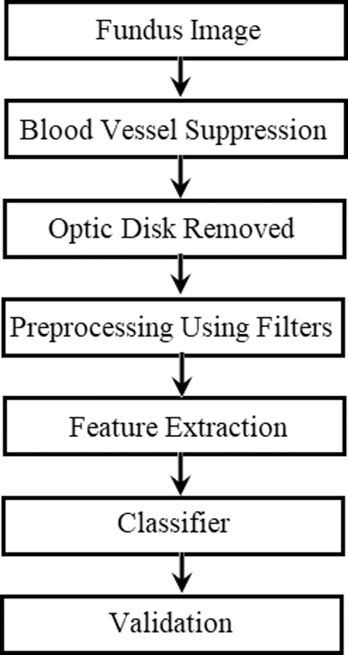
Figure 2: Flow diagram of proposed method
Following the collected dataset and preprocessing certain features respective of shape, intensity, gray tone, and parameters of fourier transform are extracted.
4 Classification Using Artificial Neural Networks
The performance of artificial neural classifier was analysed based on IF-THEN condition. Two factors such as basis and resultant factors are used. The basis factor is based on Fuzzy rule based on IF condition which is given as input and another Fuzzy condition is based on THEN condition is thereby given as resultant factor. The foremost significance of the classifier is that it sufficiently increases capacity of dataset that is used for training and by changing the basis functions and Resultant factors the classifier can operate. Here MESSIDOR database is used for analysis that is publicly available. The adaptive type of fuzzy classifier network has total layers of five in number to perform many computational functions using input signals. The layer three can calculate the Fuzzy strength based on certain conditions that is based on the updations of second layer.
The layer four computes the subsequent factors based on THEN condition of the Fuzzy logic [14]. The layer five computes the combined results of previous nodes as single value, which finds the feature input vector belonging to a class. The diagnostic results of classifier predicted included normal or abnormal image with exudates in Diabetic retinopathy. The basis factor is represented by the first layer and the output layer gives the result which is represented by subsequent factors corresponding to THEN condition. The classifier accuracy is dependent on proper selection of highly unique feature vector and via thorough training process. The neural network can also be analysed using genetic algorithms [15] in medical applications.
Fig. 3 shows ANFIS system that operates on 32 AND fuzzy rules which uses membership functions to get output, that has 5 inputs.
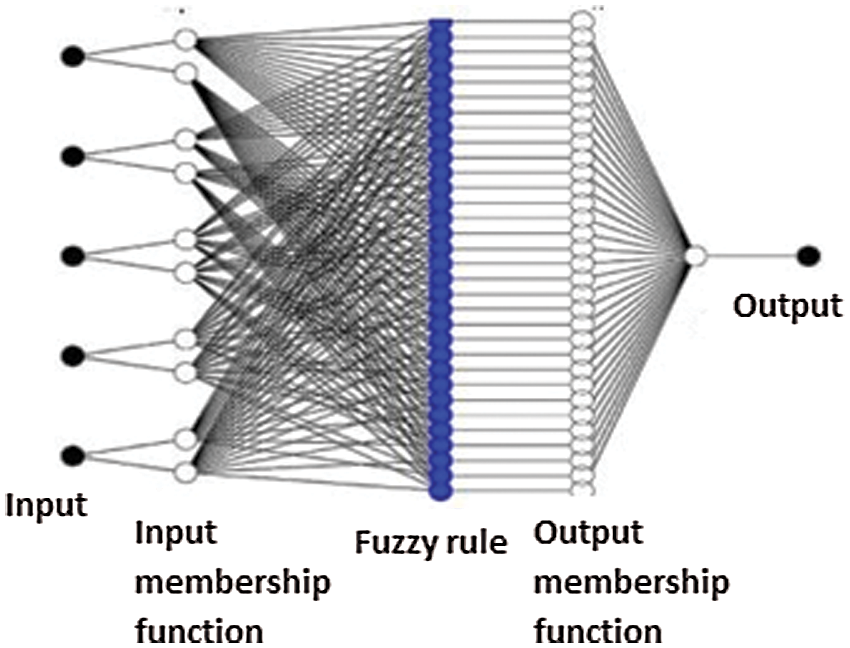
Figure 3: Structure of ANFIS system
Fig. 4 showed green channel extraction from RGB fundus. Here green channel is extracted as it possess better contrast level and good visibility of fundus area.
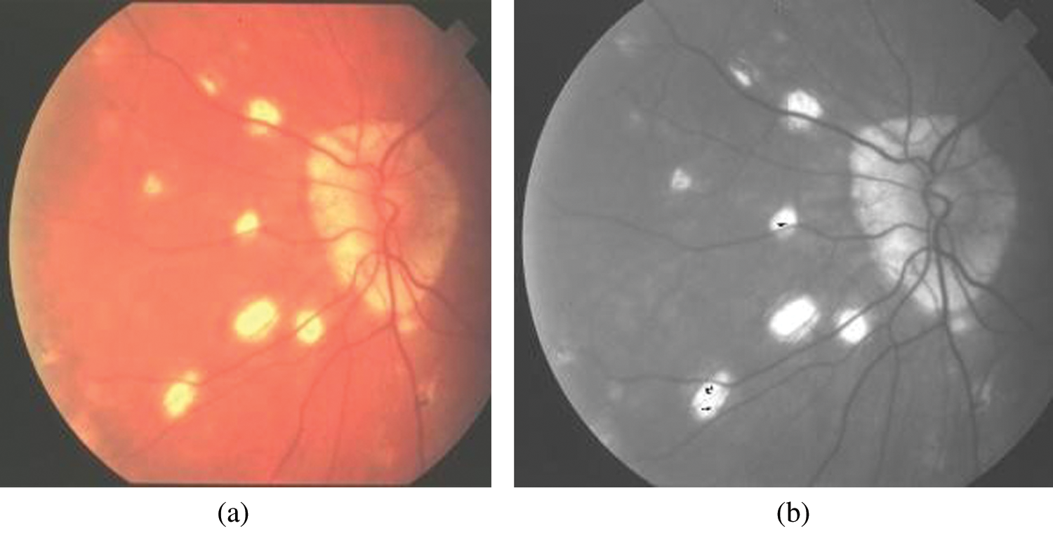
Figure 4: a) RGB fundus image b) Green channel
Fig. 5 showed blood vasculature and non-blood vessels. Using morphological filter blood vessels are extracted so as to identify the exudates more efficiently. Using contourlet type of transform the morphological segmentation is performed. Following the classification, validation is performed to identify the results of diagnosis into two classes where first includes Healthy eyes and second includes exudates lesion.
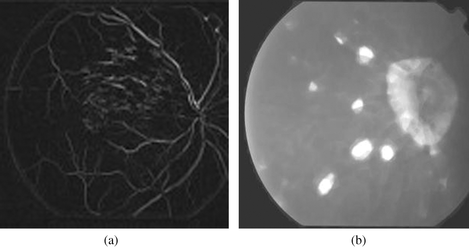
Figure 5: a) Blood vasculature b) Non blood vessels
Tab. 2 shows the comparison of classifier algorithms for various performance parameters with Naive bayes [16] classifier method. The results prove that artificial neural network performance is better compared with other classifiers.
The Tab. 3 depicts the confusion matrix for predicting abnormal ones. Also, the Tab. 4 showed comparison results of techniques which are compared for accuracy, sensitivity and specificity. The results showed that proposed system outperformed existing techniques.
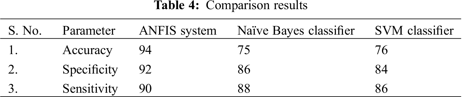
The confusion matrix depicted above showed the various performance metrics like specificity, sensitivity and accuracy. We find that artificial inference system outperforms Radial basis function based on SVM kernel and Naïve Bayes method in terms of classification. The proposed classifier based on fuzzy inference system showed better results in terms of accuracy for correct prediction of patients with anamoly. Also we find specificity, sensitivity corresponding to patients without disease and with disease were estimated as 92% The results prove that fuzzy inference system based on neural networks can perform much better compared with conventionally available classifier techniques like SVM with radial basis function and naïve bayes technique. The analysis proved that less error rate was involved in proposed than conventional technique.
Funding Statement: The authors received no specific funding for this study.
Conflicts of Interest: The authors declare that they have no conflicts of interest to report regarding the present study.
1. Lee, S., Lee, E. T., Wang, Y., Klein, R. (2005). Computer classification of non-proliferative diabetic retinopathy. Journal of Archeology and Ophthalmology, 123(6), 759–764. [Google Scholar]
2. Giancardo, L., Meriaudeau, F., Karnowski, T. P., Li, Y. Q., Garg, S. et al. (2012). Exudate based diabetic macular edema detection in fundus images using publicly available datasets. Medical Image Analysis, 16(1), 216–226. [Google Scholar]
3. Kumar, A., Kumar Gaur, A., Srivastava, M. (2012). A segment based technique for detecting exudate from retinal fundus image. Procedia Science Technology, 6, 1–9. [Google Scholar]
4. Nayak, J., Subbanna Bhat, P., Rajendra Acharya, U., Lim, C. M., Kagathi, M. (2008). Automated identification of diabetic retinopathy stages using digital fundus images. Journal of Medical Systems, 32, 107–115. [Google Scholar]
5. Osare, A., Shadgar, B., Markham, R. (2009). A computational intelligence based approach for detection of exudates in diabetic retinopathy images. IEEE Transactions on Information Technology in Biomedicine, 13(4), 535–545. [Google Scholar]
6. Niemeijer, M., Russell, S. R., Abramoff, M. D. (2007). Automated detection and differentiation of drusen, exudates, and cotton-wool spots in color fundus photographs for diabetic retinopathy diagnosis. Investigative Opthalmology & Visual Science, 48(5), 2260–2267. [Google Scholar]
7. Yang, Y., Wu, H., Fan, W., Zhang, W. (2017). Lesion detection and grading of diabetic retinopathy via two-stage deep convolutional neural networks. Proceedings of the International Conference on Medical Image and Computer Assisted Intervention, vol. 21, pp. 533–540. USA. [Google Scholar]
8. Wang, S., Yin, Y., Cao, G., Wei, B., Yang, G. (2017). Hierarchical retinal blood vessel segmentation based feature and ensemble learning. Journal of Neurocomputing, 226(22), 270–272. [Google Scholar]
9. Sinthanayothin, C. (2002). Automated detection of diabetic retinopathy on digital fundus images. Journal of the Diabetic British Association, 19(2), 105. [Google Scholar]
10. Ravishankar, S., Mittal, A. (2009). Automated feature extraction for early detection of diabetic retinopathy in fundus images. IEEE Conference on Computer Vision and Pattern Recognition, pp. 210–217. [Google Scholar]
11. Wang, H., Hsu, H., Lee, M. (2000). An effective approach to detect lesions in retinal images. Proceedings IEEE Conference of Computer Vision Pattern Recognition, Hilton Head, SC, USA. [Google Scholar]
12. Sudha, V., Ganeshbabu, T. R. (2020). A convolutional neural network classifier VGG-19 architecture for lesion detection and grading in diabetic retinopathy based on deep learning. Computers, Materials & Continua, 66(1), 828–842. [Google Scholar]
13. Joshi, S., Karule, P. T. (2018). A review on exudates detection methods for diabetic retinopathy. Journal of Biomedicine & Pharmacotherapy, 97, 1451–1454. [Google Scholar]
14. Sridevi, S., Nirmala, S. (2016). ANFIS based decision support system for prenatal detection of Truncus Arteriosus congenital heart defect. Journal of Applied Soft Computing, 46, 577–587. [Google Scholar]
15. Verma, B., Zhang, P. (2007). A novel neural-genetic algorithm to find the most significant combination of features in digital mammograms. Applications on Soft Computing, 7, 612–625. [Google Scholar]
16. Sudha, V., Vikram, N., Suryakrishnaan, K., Sangeetha, D. P., Raja, R. (2021). Review on classification methodologies for accurate detection of lesions in diabetic retinopathy. Turkish Journal of Physiotherapy and Rehabilitation, 32(2), 2634–2639. [Google Scholar]
 | This work is licensed under a Creative Commons Attribution 4.0 International License, which permits unrestricted use, distribution, and reproduction in any medium, provided the original work is properly cited. |