| Molecular & Cellular Biomechanics |
DOI: 10.32604/mcb.2022.017044
ARTICLE
Comparative Study on Biomechanics of Two Legs in the Action of Single-Leg Landing in Men’s Badminton
Department of Physical Education, Sichuan Top IT Vocational Institute, Chengdu, 611743, China
*Corresponding Author: Gang He. Email: he24886@163.com
Received: 06 May 2021; Accepted: 03 August 2021
Abstract: This study aims to analyze the biomechanical difference between the two legs of male badminton players when they land on one leg, thereby providing some guidance for preventing sports injury. Ten male badminton players were selected as the subjects. They did the single-leg landing movement successfully three times. The kinematic data were obtained by the Vicon infrared high-speed motion capture system. The kinetic data were obtained by the KISTLER three-dimensional forcing measuring platform. The data were processed and analyzed. The center of gravity of the right leg on the X and Y axes were 0.25 ± 0.05 and 0.21 ± 0.04 m, respectively, which were lower than that of the left leg (p < 0.05). At the moment of landing by a single leg, the hip angle of the left and right legs was 164.78 ± 6.12° and 156.29 ± 6.89°, respectively (p < 0.05), the hip joint speed of the left and right legs was 2.21 ± 0.32 and 1.98 ± 0.31 m/s, respectively (p < 0.05), the knee joint speed of the left and right legs was 2.51 ± 0.21 and 2.21 ± 0.21 m/s, respectively (p < 0.05). Although there was no significant difference in the range of joint motion, the motion range of the right leg was larger than that of the left leg, and the buffering time of the knee joint of the right leg was also significantly less than that of the left leg. The comparison of the kinetic data demonstrated that the ground reaction force (GRF), peak vertical ground reaction force (PVGRF), and lower limb stiffness of the right leg were significantly smaller than those of the left leg, and the time to peak force was greater than that of the left leg (p < 0.05). The injury risk of the left leg is greater than that of the right leg when the athlete land on a single leg. In the process of training, the athlete should strengthen the stability training of two legs, especially the left leg, in order to reduce sports injury.
Keywords: Badminton; single-leg landing; biomechanics; dominant side; knee joint
In the field of sports, the study of biomechanics is a very important part, not only as a means of analysis for sports training [1], but also as a guide for sports injuries and injury prevention [2,3], and biomechanics has been widely used in the study of various sports [4]. Landing after jumping is a common action in daily life. Whether it is landing with one foot or with two feet, the great impact load produced instantaneously will impact the bones and muscles of the lower limbs. In sports, landing after the long jump is also very common and plays an important role, which has been widely used in sports such as basketball, volleyball, and football [5]. In the perspective of safety, when landing with a single foot, the load is completely borne by unilateral limb, which is easy to cause limb injury. However, in most ball games, single-foot landing is more common, which is more conducive to subsequent actions such as stop and direction change. Therefore, the landing stage of various sports has been widely concerned by researchers. Leppänen et al. [6] compared the kinematic differences in vertical drop jump (VDJ) between basketball players and floorball players and found that the knee valgus angle and knee flexion angle of floorball players were larger and that the valgus angle of female players was significantly larger than that of male players. A study of Stephenson et al. [7] analyzed the relationship between the available time to react (ATR) and knee joint biomechanics. They studied 45 jumping experiments of 34 leisure athletes and found that knee torque decreased in the direction of medial jumping with the decrease of ATR but increased in the direction of lateral jumping. Taking 18 healthy women as subjects, Cannon et al. [8] analyzed the effects of the trunk and hip mechanics on the knee joint. They found that the transverse plane rotation stiffness of the hip joint reduced the abduction moment of the knee joint during landing, and the rotation stiffness of the lumbar joint reduced the abduction angle and moment of the knee joint, which verified the importance of proximal joint rotation stiffness in preventing knee abduction during landing. Koshino et al. [9] studied 22 healthy college students and analyzed the kinematics of the hind foot relative to the lower leg, knee, and hip joint during walking and single-leg landing. They found that the valgus/varus and external/internal rotation of the rear foot were closely related to the adduction/abduction of the hip joint during walking but had a weak correlation with the knee joint. During single-leg landing, the correlation became stronger. The coupling angle showed that hip movement relative to rear foot movement was greater than knee movement relative to rear foot movement (p < 0.001). Badminton, as a non-contact sport, has been widely loved by people [10], but it has strong antagonism and high intensity [11]. With “fast” as the core, badminton is easy to cause sports injury [12]. Badminton has high requirements for the explosive power of athletes’ lower limbs.
At present, there has been a lot of research on badminton, such as the recognition of movements [13], the analysis of tactics [14], the effects of fatigue [15], and the development of wearable teaching systems [16]. Single-leg landing is also an important action in badminton, but there are few studies comparing the characteristics of the left and right legs when landing on one leg. Therefore, taking male badminton players as the subjects, this paper compared the biomechanical characteristics of players’ legs when they landed on one leg to understand the differences between the two legs in aspects of buffering ability, stability, etc. This work aims to provide some guidance for making training plans and preventing sports injuries and offer some theoretical support for research on injury prevention and stability control in the field of sports.
Twenty male badminton players were selected as the research subjects. They all took the right hand as the holding hand and the right leg as the dominant leg. They were healthy and in good mental condition. They underwent no physical injury in the past year. They did not take strenuous exercise 48 h before the experiment. All of them understood the content and process of the experiment and signed the informed consent. The general information of the players is shown in Table 1.
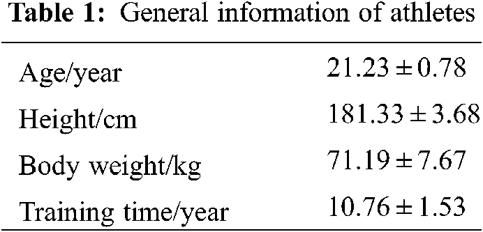
The kinematics related data were obtained using the British Vicon infrared high-speed motion capture system (Model No. T40) (Fig. 1). The system included ten high-resolution infrared cameras. The diameter of the marker was 14 mm, and the sampling frequency was 200 Hz.
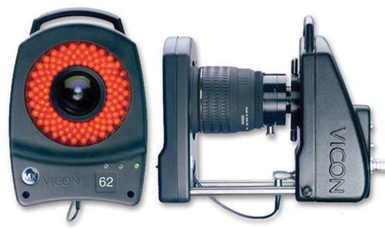
Figure 1: Vicon infrared high-speed motion capture system
The kinetic data were acquired by Switzerland KISTLER three-dimensional force measuring platform (Model No. 9287B) (Fig. 2). It was equipped with an external signal amplifier. The sampling frequency was 1000 Hz. It collected data synchronously with the Vicon system.
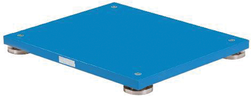
Figure 2: KISTLER three-dimensional force measuring platform
The action studied in this study was a single-leg landing with the ball overhead. The overhead ball is a kind of high ball stroked by the opponent. The player should block the ball in the backfield or midfield with one arm lifted high over the head and return the ball to the opponent’s field (Fig. 3). In the course of the movement, the body leans back, the right foot swings forward, the left foot is supported behind to maintain balance, the hitting point is over the head, and the medial side of the left foot lands on the ground before transitioning to the entire foot. In the process from the lower limbs touching the ground to the knee joint bending to the maximum angle, the lower limbs are often in the limit state, which is easy to be injured. Therefore, comparing the biomechanical characteristics of the two legs can understand the causes of injuries better.
Figure 3: Single-leg landing after returning the overhead ball
The athletes wore the same clothes, warmed up for ten minutes at the speed of 8 km/h, and stretch the body to fully exercise the body’s joints. The research action was single-leg landing after returning the overhead ball. The athletes were asked to completely familiar with the action and practice until they could fluently complete the action.
After the warm-up, the experimenters pasted the markers. Sixteen markers were pasted on left and right anterior superior spines, posterior superior spines, knee joints, thighs, calves, ankle joints, toes, and heels, respectively. In order to ensure the experimental effect, the markers were pasted by the same experimenter. The paste positions of the markers are shown in Fig. 4.
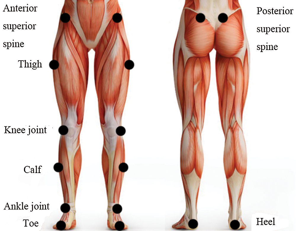
Figure 4: The paste positions of the markers
In all the tests, the ball was served by the same badminton player. The research subject stood in the midfield and backfield near the center line. The high ball, which was sent by the opponent, was blocked by lifting the arm over the head and stroked to the opponent’s field. Three groups of successful data of the test action were collected from each subject, i.e., the left foot fell on the force measuring platform completely. The testing flow is shown in Fig. 5.
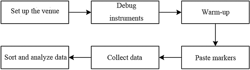
Figure 5: The testing flow
As shown in Fig. 5, researchers first organized and arranged the experimental site and debug the instruments to ensure that the experiment could be carried out smoothly, the athletes warmed up and familiarized themselves with the experimental movements, the lab personnel pasted the markers for data collection, and the collected data were organized and analyzed to obtain the experimental results.
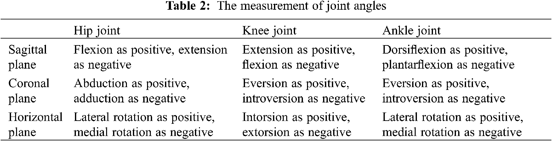
The collected data were processed and calculated in Visual 3D motion analysis software. The measurement of joint angles of the lower limbs are shown in Table 2.
The direction of the joint angular velocity was the same as the joint angle, and the unit was m/s. The coordinate system used the coronal axis as the X axis, the vertical axis as the Y axis, and the sagittal axis as the Z axis, and the three axes were perpendicular to each other.
The data were sorted out and summarized by Excel 2007, and statistically analyzed and processed by SPSS 22.0. The data results were expressed as
The comparison of three-axis gravity center positions at the moment of landing with a single foot is shown in Table 3.
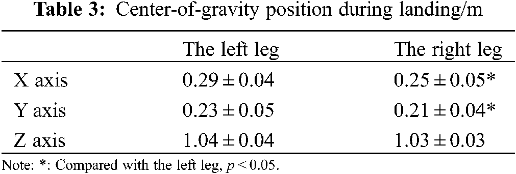
It was seen from Table 3 that the center of gravity of the X and Y axes of the two legs showed no significant differences. In the direction of the X axis, the center-of-gravity position of the right leg was 0.25 ± 0.05 m, which was significantly lower than that of the left leg (0.29 ± 0.04 m); in the direction of the Y axis, the center-of-gravity position of the right leg was 0.21 ± 0.04 m, which was significantly lower than that of the left leg (0.23 ± 0.05 m); but in the direction of the Z axis, the kinematic data of the two legs showed no significant difference (p > 0.05).
The comparison of the angles and velocities of the lower limb joints at the moment of landing is shown in Table 4.
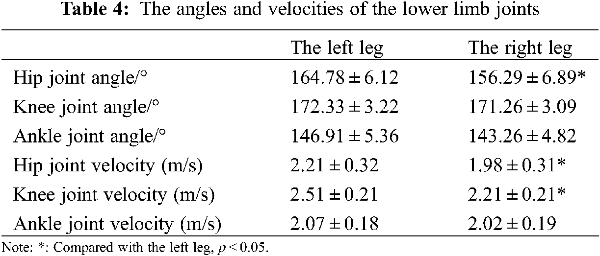
It was seen from Table 4 that the two legs showed significant differences in hip joint angle. The hip joint angle of the right leg was 156.29 ± 6.89°, significantly smaller than the left leg (164.78 ± 6.12°) (p < 0.05). The knee and ankle joint angles of the right leg were also smaller than those of the left leg, but the differences were not significant. The hip joint velocity of the left and right legs was 2.21 ± 0.32 and 1.98 ± 0.31 m/s respectively (p < 0.05); the knee joint velocity of the left and right legs was 2.51 ± 0.21 and 2.21 ± 0.21 m/s respectively (p < 0.05); there was no significant difference in the ankle joint velocity.
Range of joint motion refers to the maximum motion range of a joint during a single-leg landing. The comparison results of the two legs are shown in Table 5.
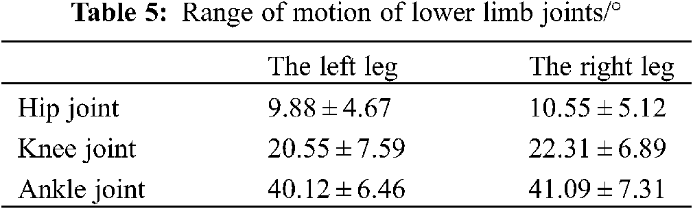
It was seen from Table 5 that there was no significant difference in the range of joint motion between the two legs (p > 0.05), but specifically, the range of joint motion of the right leg was slightly larger than that of the left leg. In the process of single-leg landing, the motion range of the ankle joint was the largest, 40.12 ± 6.46° for the left leg and 41.09 ± 7.31° for the right leg, followed by the knee joint and the hip joint.
The landing buffer time of different joints is shown in Fig. 6.

Figure 6: Comparison of the landing buffer time
Note: *: Compared with the left leg, p < 0.05.
It was seen from Fig. 6 that the landing buffer time of different joints of the left legs was larger than that of the right leg. The buffer time of the hip joint of the left leg was 127.68 ± 22.33 ms, and that of the right leg was 120.33 ± 18.59 ms (p > 0.05). The buffer time of the knee joint of the left leg was 141.28 ± 42.67 ms, and that of the right leg was 126.84 ± 26.33 ms (p < 0.05). The buffer time of the ankle joint of the right leg was 130.67 ± 34.54 ms, which was shorter than that of the left leg (138.94 ± 44.86 ms) (p > 0.05).
The comparison of the kinetic data between the two legs at the moment of landing with a single leg is shown in Table 6.
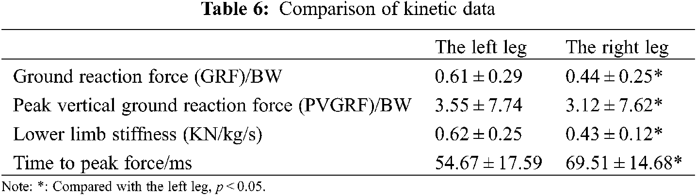
It was seen from Table 6 that the kinetic data of the two legs showed significant differences. The GRF of the right leg was 0.44 ± 0.25 BW, significantly smaller than that of the left leg (0.61 ± 0.29 BW) (p < 0.05). The PVGRF of the right leg was 3.12 ± 7.62 BW, significantly smaller than that of the left leg (3.55 ± 7.74 BW) (p < 0.05). The lower limb stiffness of the left leg was 0.43 ± 0.12 KN/kg/s, significantly smaller than that of the left leg (0.62 ± 0.25 KN/kg/s). The time to peak force of the right leg was 69.51 ± 14.68 ms, which was significantly larger than that of the left leg (54.67 ± 17.59 ms) (p < 0.05).
During human movement, the possibility of sports injury is very high. Especially in the process of competition, the probability of injury is very high. Anterior cruciate ligament (ACL) injury is a very common injury [17], which widely exists in sports such as football [18] and rugby [19]. The actions that are easy to cause ACL injury include landing support, side cutting, take-off, etc. [20,21]. Unreasonable landing methods will increase the risk of ACL, and ACL injury will bring great pressure to the body and mind of athletes [22]. Therefore, it is very important to understand the biomechanical characteristics of landing and prevent ACL injury.
This paper took ten male badminton players as the subjects and compared the biomechanical differences between two legs when they landed on the ground with a single leg. The comparison of the kinematic data demonstrated that the right leg had a lower center-of-gravity position, smaller joint angle and speed, and larger range of joint motion, and the motion range of the ankle joint was the largest, followed by the knee joint and the hip joint. Considering the stability of the human body, the lower the position of the center of gravity is, the greater the stability is, and the more stable the landing is. Therefore, the stability of the right leg is higher during landing, while the unstable left leg is easy to be injured. The range of joint motion of the left leg was small; thus, the left leg would present a relatively rigid posture when landing, which was easy to cause injury. The comparison of various joints suggested that the ankle joint had the largest range of motion and was the first to land, i.e., it borne a great impact force and had a larger injury risk. In general, the joints of the right leg had better flexion and smaller velocity, i.e., the right leg had stronger stability, and the joints of the left leg presented the opposite characteristics, i.e., the left leg had poor stability. The comparison of the kinetic data demonstrated that the right leg had smaller GRF, PVGRF, and lower limb stiffness and longer time to peak force, which was conducive to reducing the vertical impact force of the limb. A larger ground reaction will increase the possibility of cartilage and soft tissue injury, which may cause an ACL tear. A lower limb stiffness refers to the ability of the limb to resist deformation. By comparison, the stiffness of the left leg was significantly greater than that of the right leg (p < 0.05). A larger lower limb stiffness will increase the risk of sports injury. Larger GRB, PVGRF, and lower limb stiffness of the left leg may be caused by the insufficient flexion of the limb.
In general, in the single-leg landing action, the right leg has better stability and smaller injury risk. Therefore, in the training process, athletes should strengthen the ability to control the lower limbs, especially the left leg, to improve the stability of the lower limbs and avoid sports injury.
This paper compared the biomechanics of the two legs of male badminton players when they landed on the ground with a single leg. Through the processing and analysis of the experimental data, the author found that the right leg had a lower center-of-gravity position, smaller joint angles and velocities, a larger range of joint motion, shorter buffer time, smaller GRF, and longer time to peak force, which made the right leg have stronger stability and smaller injury risk. Therefore, in the training process, the players should strengthen the stability training of the left leg to reduce sports injury.
Funding Statement: The author received no specific funding for this study.
Conflicts of Interest: The author declares that they have no conflicts of interest to report regarding the present study.
1. Glazier, P. S., Mehdizadeh, S. (2019). In search of sports biomechanics’ holy grail: Can athlete-specific optimum sports techniques be identified? Journal of Biomechanics, 94, 1–4. DOI 10.1016/j.jbiomech.2019.07.044. [Google Scholar] [CrossRef]
2. Radakovi, R., Filipovic, N. (2020). Sport biomechanics: Experimental and computer simulation of knee joint during jumping and walking-ScienceDirect. Computational Modeling in Bioengineering and Bioinformatics, 387–418. DOI 10.1016/B978-0-12-819583-3.00012-6. [Google Scholar] [CrossRef]
3. Wang, W., Li, Y. (2021). Study on treatment and rehabilitation training of ligament injury of javelin throwers based on sports biomechanics. Measurement, 171(4), 108757. DOI 10.1016/j.measurement.2020.108757. [Google Scholar] [CrossRef]
4. Bandeiras, C. (2019). Technology in sports biomechanics. IEEE Potentials, 38(3), 8–10. DOI 10.1109/MP.45. [Google Scholar] [CrossRef]
5. Fransz, D. P., Huurnink, A., de Boode, V., Kingma, I., van Dieën, J. H. (2016). Time series of ground reaction forces following a single leg drop jump landing in elite youth soccer players consist of four distinct phases. Gait & Posture, 50, 137–144. DOI 10.1016/j.gaitpost.2016.09.002. [Google Scholar] [CrossRef]
6. Leppänen, M., Pasanen, K., Kulmala, J. P., Kujala, U., Krosshaug, T. et al. (2015). Knee control and jump-landing technique in young basketball and floorball players. International Journal of Sports Medicine, 37(4), 334–338. DOI 10.1055/s-00000028. [Google Scholar] [CrossRef]
7. Stephenson, M. L., Hinshaw, T. J., Wadley, H. A., Zhu, Q., Wilson, M. et al. (2017). Effects of timing of signal indicating jump directions on knee biomechanics in jump-landing-jump tasks. Sports Biomechanics, 17(1), 1. DOI 10.1080/14763141.2017.1346141. [Google Scholar] [CrossRef]
8. Cannon, J., Cambridge, E., Mcgill, S. M. (2021). Increased core stability is associated with reduced knee valgus during single-leg landing tasks: Investigating lumbar spine and hip joint rotational stiffness. Journal of Biomechanics, 116, 110240. DOI 10.1016/j.jbiomech.2021.110240. [Google Scholar] [CrossRef]
9. Koshino, Y., Yamanaka, M., Ezawa, Y., Okunuki, T., Ishida, T. et al. (2017). Coupling motion between rearfoot and hip and knee joints during walking and single-leg landing. Journal of Electromyography & Kinesiology, 37, 75. DOI 10.1016/j.jelekin.2017.09.004. [Google Scholar] [CrossRef]
10. Arora, M., Shetty, S. H., Khedekar, R. G., Kale, S. (2015). Over half of badminton players suffer from shoulder pain: Is impingement to blame? Journal of Arthroscopy & Joint Surgery, 2(1), 33–36. DOI 10.1016/j.jajs.2014.12.006. [Google Scholar] [CrossRef]
11. Phomsoupha, M., Laffaye, G. (2015). The science of badminton: Game characteristics, anthropometry, physiology, visual fitness and biomechanics. Sports Medicine, 45(4), 473–495. DOI 10.1007/s40279-014-0287-2. [Google Scholar] [CrossRef]
12. Park, J., Lee, Y. H., Kong, I. D., Park, T., Chang, J. S., et al. (2017). Ultrasonographic changes of upper extremity tendons in recreational badminton players: The effect of hand dominance and comparison with clinical findings. British Journal of Sports Medicine, 51(4). DOI 10.1136/bjsports-2016-097372.219. [Google Scholar] [CrossRef]
13. Rahmad, N. A., As’Ari, M. A. (2020). The new convolutional neural network (CNN) local feature extractor for automated badminton action recognition on vision based data. Journal of Physics Conference Series, 1529, 022021. DOI 10.1088/1742-6596/1529/2/022021. [Google Scholar] [CrossRef]
14. Lei, W., Yu, K. (2020). Analysis and research on badminton spot tactics based on computer association rules mining. Journal of Physics Conference Series, 1648, 032065. DOI 10.1088/1742-6596/1648/3/032065. [Google Scholar] [CrossRef]
15. Kosack, M. H., Staiano, W., Folino, R., Hansen, M. B., Lønbro, S. (2020). The acute effect of mental fatigue on badminton performance in elite players. International Journal of Sports Physiology and Performance, 15(5), 632–638. DOI 10.1123/ijspp.2019-0361. [Google Scholar] [CrossRef]
16. Lin, K. C., Wei, C. W., Lai, C. L., Cheng, I. L., Chen, N. S. (2021). Development of a badminton teaching system with wearable technology for improving students’ badminton doubles skills. Educational Technology Research and Development, 69, 945–969. DOI 10.1007/s11423-020-09935-6. [Google Scholar] [CrossRef]
17. Agel, J., Rockwood, T., Klossner, D. (2016). Collegiate ACL injury rates across 15 sports: National collegiate athletic association injury surveillance system data update (2004–2005 through 2012–2013). Clinical Journal of Sport Medicine Official Journal of the Canadian Academy of Sport Medicine, 26(6), 518. DOI 10.1097/JSM.0000000000000290. [Google Scholar] [CrossRef]
18. Zebis, M. K., Andersen, L. L., Brandt, M., Myklebust, G., Bencke, J. et al. (2016). Effects of evidence-based prevention training on neuromuscular and biomechanical risk factors for ACL injury in adolescent female athletes: A randomised controlled trial. British Journal of Sports Medicine, 50(9), 552–557. DOI 10.1136/bjsports-2015-094776. [Google Scholar] [CrossRef]
19. Montgomery, C., Blackburn, J., Withers, D., Tierney, G., Moran, C. et al. (2016). Mechanisms of ACL injury in professional rugby union: A systematic video analysis of 36 cases. British Journal of Sports Medicine, 50(9), 552–557. DOI 10.1136/bjsports-2016-096425. [Google Scholar] [CrossRef]
20. Eltoukhy, M., Kuenze, C., Oh, J., Butler, L., Signorile, J. F. (2019). Concurrent validity of depth-sensing cameras for noncontact ACL injury screening during side-cut maneuvers in adolescent athletes: A preliminary study. Journal of Applied Biomechanics, 35(1), 2–10. DOI 10.1123/jab.2O18-0105. [Google Scholar] [CrossRef]
21. Bakker, R., Tomescu, S., Brenneman, E., Hangalur, G., Laing, A. C. et al. (2016). Effect of sagittal plane mechanics on ACL strain during jump landing. Journal of Orthopaedic Research Official Publication of the Orthopaedic Research Society, 34(9), 1636–1644. DOI 10.1002/jor.23164. [Google Scholar] [CrossRef]
22. Dicesare, C. A., Bates, N. A., Foss, K. B., Thomas, S. M., Wordeman, S. C. et al. (2015). Reliability of 3-Dimensional measures of single-leg cross drop landing across 3 different institutions: Implications for multicenter biomechanical and epidemiological research on ACL injury prevention. Orthopaedic Journal of Sports Medicine, 3(12). DOI 10.1177/2325967115617905. [Google Scholar] [CrossRef]
 | This work is licensed under a Creative Commons Attribution 4.0 International License, which permits unrestricted use, distribution, and reproduction in any medium, provided the original work is properly cited. |