
 Submit a Paper
 Propose a Special lssue
 Open Access
ARTICLE
Evaluation of Biventricular Volume and Systolic Function in Children with Ventricular Septal Defect and Moderate to Severe Pulmonary Hypertension Using Real-Time Three-Dimensional Echocardiography
1
Children’s Heart Center, Wuhan Children’s Hospital (Wuhan Maternal and Child Healthcare Hospital), Tongji Medical College,
Huazhong University of Science & Technology, Wuhan, China
2
Department of Ultrasound, Wuhan Children’s Hospital (Wuhan Maternal and Child Healthcare Hospital), Tongji Medical College,
Huazhong University of Science & Technology, Wuhan, China
* Corresponding Author: Jun Gao. Email:
# These authors contributed equally
Congenital Heart Disease 2022, 17(6), 697-707. https://doi.org/10.32604/chd.2022.022648
Received 18 March 2022; Accepted 06 June 2022; Issue published 11 October 2022
 View Full Text
 Download PDF
Abstract
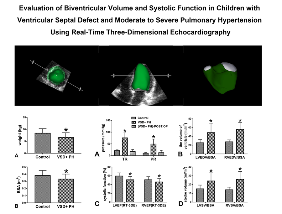
Background: Real-time three-dimensional echocardiography (RT-3DE) could obtain ventricular volume and ejection fraction rapidly and non-invasively without relying on ventricular morphology. This study aims to use RT-3DE to evaluate the changes in biventricular volume and systolic function in children with ventricular septal defect (VSD) and moderate to severe pulmonary hypertension (PH) before surgery. Methods: In this study 18 children with VSD and moderate to severe PH (VSD + PH Group) and 18 healthy children of the same age (Control Group) were recruited. Biventricular volume and systolic function were evaluated by RT-3DE. The measurements included: left and right ventricular volume indexed to body surface area (BSA), stroke volume (SV) indexed to BSA, and ejection fraction (EF). Results: The results showed left and right ventricular volume indexed to BSA and SV indexed to BSA were significantly increased in VSD + PH Group (VSD + PH Group vs. Control Group), LVEDV/BSA (ml/m2 ): 48.67 ± 21.46 vs. 25.59 ± 6.96, RVEDV/BSA (ml/m2 ): 55.98 ± 15.35 vs. 27.69 ± 4.37, LVSV/BSA (ml/m2 ): 24.08 ± 9.30 vs. 15.14 ± 4.29, RVSV/BSA (ml/m2 ): 26.02 ± 8.87 vs. 14.11 ± 2.89, (P < 0.05). While for EF in VSD + PH Group decreased (VSD + PH Group vs. Control Group), LVEF: 50.93 ± 7.50% vs. 59.38 ± 7.24%, RVEF: 45.84 ± 7.71% vs. 51.05 ± 6.90% (P < 0.05). Conclusion: In children with VSD and moderate to severe PH, increased biventricular volume and decreased systolic function were observed with RT-3DE, but biventricular systolic function remained within acceptable limits. The children in this study recovered well after surgery without serious perioperative complications, suggesting that biventricular systolic function may help facilitate the surgical decision-making process in children with VSD and moderate-tosevere PH.Graphic Abstract
Keywords
Cite This Article
 Copyright © 2022 The Author(s). Published by Tech Science Press.
This work is licensed under a Creative Commons Attribution 4.0 International License , which permits unrestricted use, distribution, and reproduction in any medium, provided the original work is properly cited.
 Downloads
 Citation Tools