
 Login
Login Register
 Submit a Paper
 Propose a Special lssue
 Open Access
CASE REPORT
Acute Hypoattenuating Leaflet Thickening after Percutaneous Pulmonary Valve Implantation: A Case Report
1 Cardiology Division, Department of Medicine, Li Ka Shing Faculty of Medicine, The University of Hong Kong, Hong Kong, China
2 Cardiology Division, Department of Medicine, Queen Mary Hospital, Hong Kong, China
3 Cardiology Centre, Department of Paediatrics and Adolescent Medicine, Hong Kong Children’s Hospital, Hong Kong, China
4 Department of Radiology, Queen Mary Hospital, Hong Kong, China
* Corresponding Author: Ka-Chun Un. Email:
Structural and Congenital Heart Disease 2026, 21(4), 5 https://doi.org/10.32604/schd.2026.078486
Received 31 December 2025; Accepted 05 June 2026; Issue published 30 September 2026
 View Full Text
 Download PDF
Abstract
Background: Hypoattenuating leaflet thickening (HALT) is a reported complication following percutaneous pulmonary valve implantation (PPVI) Case description: A 32-year-old man with a history of repaired Tetralogy of Fallot (TOF) underwent PPVI for symptomatic severe pulmonary regurgitation (PR). The postoperative course was complicated by HALT diagnosed by computed tomography (CT) within 24 h of the procedure. Anticoagulation therapy was administered, and reassessment CT at 4 months showed resolution of HALT. Conclusion: HALT following PPVI is managed with anticoagulation yet consensus for optimal management strategy is lacking.Keywords
PPVI is an effective and lower-risk alternative to surgery for treating dysfunctional right ventricular outflow tract (RVOT). As with transcatheter aortic valve implantation (TAVI), leaflet thrombosis following PPVI is a recognized postoperative complication that may compromise valve durability. While hypoattenuating leaflet thickening (HALT) represents subclinical leaflet thrombosis (SLT), hypoattenuation affecting motion (HAM) refers to clinical leaflet dysfunction due to thrombosis. The prevalence of HALT and HAM following PPVI has been reported as 17.4% and 6.5%, respectively, in a retrospective cohort study of 109 patients [1].
A 32-year-old man with repaired tetralogy of Fallot (TOF) was referred to our unit for consideration of pulmonic valve replacement (PVR). He presented with worsening exercise tolerance over the course of one year. Serial transthoracic echocardiography (TTE) showed a progressively dilated right ventricle (RV) with impaired systolic function and severe PR (Fig. 1A—Severe PR). Cardiac magnetic resonance imaging revealed an RV end-diastolic volume index of 166 mL/m2 and a PR fraction of 64%. CT showed a pyramidal-shaped RVOT (Type I) and ectasia of the main pulmonary artery (MPA). Balloon interrogation of the RVOT demonstrated a dilated MPA with a minimal waist of 34 mm. A multidisciplinary team, including cardiothoracic surgeons, pediatric cardiologists, and adult congenital heart specialists, recommended PPVI. The anti-thrombotic regimen consisted of loading doses of aspirin 320 mg and clopidogrel 300 mg once before the procedure, followed by lifelong aspirin 80 mg daily and clopidogrel 75 mg daily for 6 months after the procedure. Subsequently, a Harmony Transcatheter Pulmonary Valve (TPV) 25 (manufactured by Medtronic) was implanted. Intraoperatively, intravenous unfractionated heparin was administered to maintain an activated clotting time (ACT) of 250–350 s. The final angiogram showed good valve position, patency, and competence, with no distortion of the valve frame (Fig. 1B—Intraoperative final angiogram). Immediately post-implantation, invasive hemodynamic assessment demonstrated no pressure gradient across the prosthesis.
On day one, a new-onset grade 2/6 ejection systolic murmur over the pulmonic area was audible. TTE showed stable prosthesis position and conformation, but the maximum and mean Doppler gradients were elevated at 35 mmHg and 17 mmHg, respectively (Fig. 1C—Elevated pressure gradient across TPV on TTE). As the patient was asymptomatic, subclinical leaflet thrombosis (SLT) was suspected. The patient’s body weight and height were 73 kg and 176 cm, respectively, with a body surface area of 1.89 m2. The indexed effective orifice area was 1.58 cm2/m2, arguing against patient–prosthesis mismatch [2]. Urgent computed tomography (CT) revealed HALT without distortion of the valve frame (Fig. 1D,E—Leaflet thickening on CT with 3-D reconstruction in systolic phase and diastolic phase). Genetic testing detected a CYP2C19*1/*1 diplotype with no CYP2C19*2 or CYP2C19*3 alleles, indicating that clopidogrel resistance was unlikely. Systemic anticoagulation with intravenous unfractionated heparin was administered to maintain an activated partial thromboplastin time (APTT) of 50–75 s for one day, followed by oral apixaban 5 mg twice daily. Reassessment CT at 4 months showed resolution of HALT (Fig. 1F,G—Resolution of leaflet thickening on reassessment CT at 4 months with 3-D reconstruction in systolic phase and diastolic phase). Reassessment TTE demonstrated improved maximum and mean gradients of 22 mmHg and 12 mmHg, respectively. The resolution of HALT on CT following anticoagulation strongly suggested thrombotic aetiology. Apixaban was continued at 5 mg twice daily until one year postoperatively, followed by lifelong aspirin 80 mg daily. Gradients across the TPV on TTE at one year remained stable.
Ethics approval is waived because it is a retrospective case analysis complying with the standard of care without the addition of a new intervention. The author(s) have obtained consent for reproduction of images in accordance with the international rules outlined in the Declaration of Helsinki.
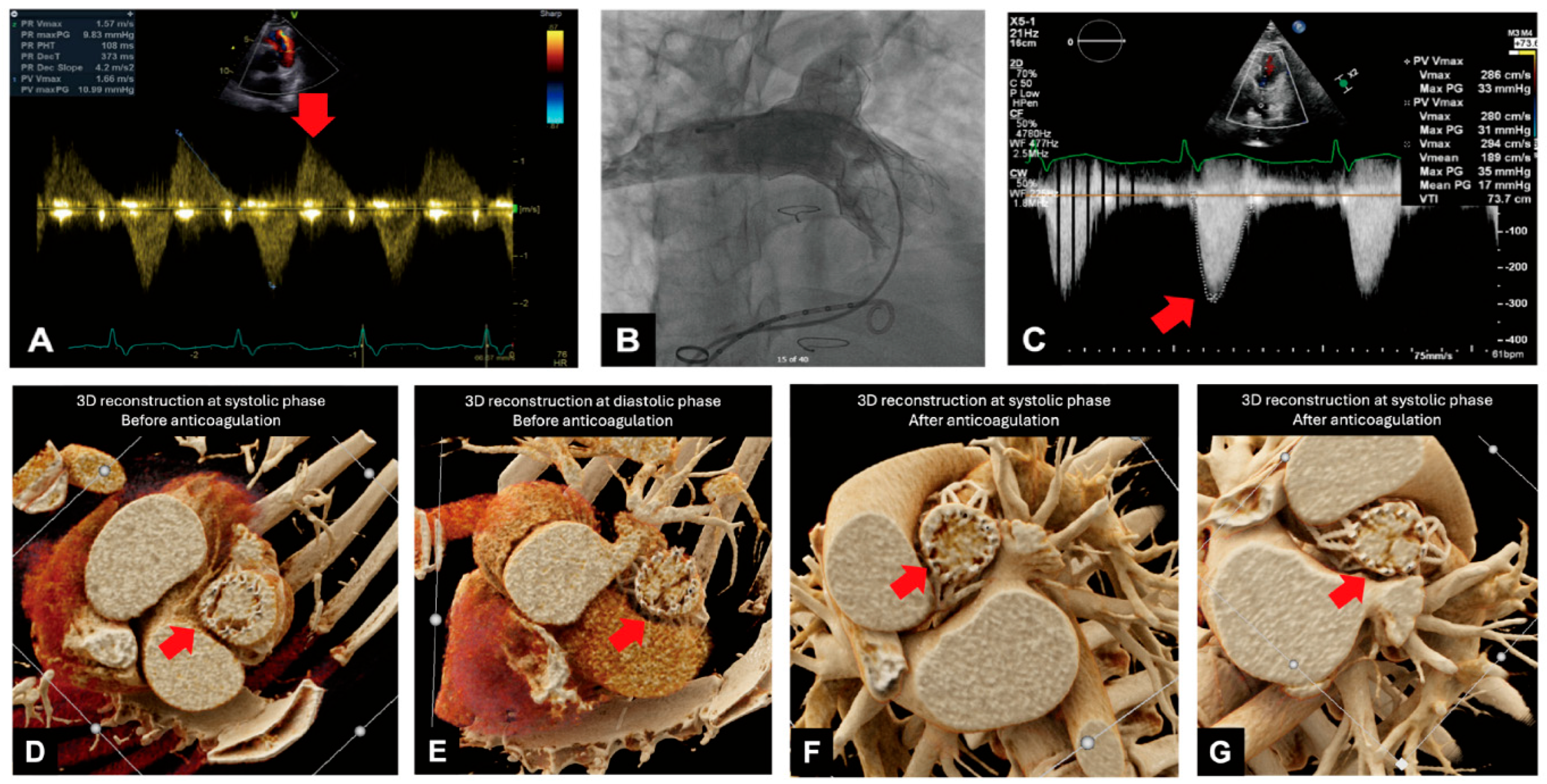
Figure 1: (A) Severe PR (red arrow). (B) Intraoperative final angiogram. (C) Elevated pressure gradient across TPV on TTE (red arrow). (D) Leaflet thickening on CT with 3-D reconstruction in systolic phase (red arrow). (E) Leaflet thickening on CT with 3-D reconstruction in diastolic phase (red arrow). (F) Resolution of leaflet thickening on reassessment CT at 4 months with 3-D reconstruction in systolic phase (red arrow). (G) Resolution of leaflet thickening on reassessment CT at 4 months with 3-D reconstruction in diastolic phase (red arrow).
The Harmony TPV is a porcine pericardial tissue valve sewn onto a self-expanding nitinol frame covered in a polyester knit fabric. Studies have shown that the Harmony TPV yields favorable clinical outcomes at 3–5 years [3]. Despite well-demonstrated device safety, the incidence of Harmony TPV thrombosis was reported to be 3.4% in a previous registry [4]. HALT is not specific to the Harmony TPV; cases involving other transcatheter pulmonary valve platforms have also been reported. In our case, an unexpected and very early rise in valve gradient occurred postoperatively. Possible reasons for early Harmony TPV dysfunction include device under-expansion, valve thrombosis related to an inadequate antithrombotic or anticoagulant regimen, or frame distortion [5,6]. To help prevent these complications, the use of intraoperative intravascular ultrasound (IVUS) has been previously described [7]. IVUS allows precise verification of valve stent expansion, interactions between the valve stent and vessel wall along the landing zone, and areas of inadequate apposition associated with paravalvular leaks. Application of IVUS may guide optimization strategies such as post-dilatation to minimize the risk of leaflet thrombosis.
When valve leaflet thrombosis or HALT is suspected, CT is the mainstay of diagnosis. The utility of CT is supported by a study of 99 patients undergoing TAVI with SAPIEN XT or SAPIEN 3, which used thrombus attenuation to predict HALT resolution [8].
Studies have shown that HALT following TAVI is independently associated with inferior valve durability and long-term mortality [9]. Vitamin K antagonists or non-vitamin K oral anticoagulants remain the cornerstone treatment for leaflet thrombosis after TAVI [10]. This treatment strategy is extrapolated to PPVI. Anticoagulation has been shown to be effective in treating patients with HALT after Venus p-valve implantation [11]. Similarly, a report described treating patients with HALT after PPVI using the Harmony TPV 25 with oral anticoagulation [5]. Nevertheless, evidence regarding the progression of valve dysfunction due to leaflet thrombosis following PPVI and its relationship to optimal antithrombotic therapy remains lacking.
This case represents HALT diagnosed within 24 h following PPVI that was successfully managed with anticoagulation. HALT and HAM after PPVI are emerging clinical entities that require timely recognition, management, and prevention. More real-world data are needed to formulate optimal preoperative regimens and treatment strategies.
Acknowledgement:
Funding Statement: The authors received no specific funding for this study.
Author Contributions: The authors confirm contribution to the paper as follows: Conceptualisation, Yee-Tat Lee, Hay-Son Robin Chen, Ka-Chun Un; writing—original draft preparation, Yee-Tat Lee, Ka-Chun Un; writing—review and editing, Yee-Tat Lee, Ka-Chun Un, Ka-Lam Calvin Leung, Chun-Ka Wong, Ho-On Alston Chiu; Processing of images, Chi-Wai Stephen Cheung, Yuen-Hei Mak, Ka-Lam Calvin Leung. All authors reviewed and approved the final version of the manuscript.
Availability of Data and Materials: The data supporting the findings of this study are available from the corresponding author upon reasonable request.
Ethics Approval: Ethics approval is waived because it is a retrospective case analysis complying with the standard of care without the addition of a new intervention. The authors have obtained consent for reproduction of images in accordance with the international rules outlined in the Declaration of Helsinki.
Conflicts of Interest: The authors declare no conflicts of interest.
References
1. Sefton C , Firth A , Curran K , Rajeswaran J , Das T , Bakhtadze B , et al. Outcomes of hypoattenuating leaflet thickening post-transcatheter pulmonary valve replacement. JACC Adv. 2025; 4( 3): 101599. doi:10.1016/j.jacadv.2025.101599. [Google Scholar] [CrossRef]
2. Takajo D , Forbes TJ , Kobayashi D . Impact of patient prosthesis mismatch on the outcome of transcatheter pulmonic valve implantation. Am J Cardiol. 2021; 151: 93– 9. doi:10.1016/j.amjcard.2021.04.022. [Google Scholar] [CrossRef]
3. Morray BH , Gillespie MJ , Cheatham JP , Salavitabar A , Peng L , Jones TK , et al. Midterm outcomes in a pooled cohort of harmony transcatheter pulmonary valve recipients. Circ Cardiovasc Interv. 2025; 18( 9): e015196. doi:10.1161/CIRCINTERVENTIONS.125.015196. [Google Scholar] [CrossRef]
4. Goldstein BH , McElhinney DB , Gillespie MJ , Aboulhosn JA , Levi DS , Morray BH , et al. Early outcomes from a multicenter transcatheter self-expanding pulmonary valve replacement registry. J Am Coll Cardiol. 2024; 83( 14): 1310– 21. doi:10.1016/j.jacc.2024.02.010. [Google Scholar] [CrossRef]
5. Akazawa Y , Higaki T , Kashiwagi K , Chisaka T , Takata H , Uchita S , et al. Subclinical leaflet thrombosis after transcatheter pulmonary valve implantation: Two clinical concerns. Circ Cardiovasc Imaging. 2024; 17( 6): e016459. doi:10.1161/CIRCIMAGING.123.016459. [Google Scholar] [CrossRef]
6. Steinberg ZL , Cabalka AK , Balzer DT , Asnes JD , Morray BH , Gillespie MJ , et al. Right ventricular outflow tract obstruction associated with neointimal tissue accumulation and distortion of the Harmony TPV25 stent frame: Potential mechanisms and treatment. Catheter Cardiovasc Interv. 2023; 102( 6): 1078– 87. doi:10.1002/ccd.30901. [Google Scholar] [CrossRef]
7. Sadowski KA , Mintz GS , Woźniak O , Warchoł-Celińska E , Biernacka EK , Lipczyńska M , et al. Intravascular ultrasound for guiding Venus-P-valve deployment. JACC Cardiovasc Interv. 2025; 18( 6): 804– 7. doi:10.1016/j.jcin.2024.10.053. [Google Scholar] [CrossRef]
8. Tirado-Conte G , Nombela-Franco L . Can a CT scan characterise and predict HALT evolution? EuroIntervention. 2025; 21( 9): e445– 6. doi:10.4244/EIJ-E-25-00012. [Google Scholar] [CrossRef]
9. Garcia S , Fukui M , Dworak MW , Okeson BK , Garberich R , Hashimoto G , et al. Clinical impact of hypoattenuating leaflet thickening after transcatheter aortic valve replacement. Circ Cardiovasc Interv. 2022; 15( 3): e011480. doi:10.1161/CIRCINTERVENTIONS.121.011480. [Google Scholar] [CrossRef]
10. Adrichem R , Rodes Cabau J , Mehran R , Park DW , Ten Berg JM , de Backer O , et al. Treatment of transcatheter aortic valve thrombosis: JACC review topic of the week. J Am Coll Cardiol. 2024; 84( 9): 848– 61. doi:10.1016/j.jacc.2024.05.064. [Google Scholar] [CrossRef]
11. Spagnolo P , Bevilacqua F , Perillo N , d’Aiello AF , Guglielmi G , Pasqualin G , et al. Hypoattenuating leaflet thickening after transcatheter pulmonary valve implantation: An urgent call to action. J Am Coll Cardiol Case Rep. 2025; 30( 17): 103889. doi:10.1016/j.jaccas.2025.103889. [Google Scholar] [CrossRef]
Cite This Article
 Copyright © 2026 The Author(s). Published by Tech Science Press.
This work is licensed under a Creative Commons Attribution 4.0 International License , which permits unrestricted use, distribution, and reproduction in any medium, provided the original work is properly cited.
 Downloads
 Citation Tools
