
 Login
Login Register
 Submit a Paper
 Propose a Special lssue
 Open Access
ARTICLE
Chronic Fatigue in Adolescents: The Role of Congenital Heart Disease vs. Psychosocial Factors
1 Federal Research Center for Innovator and Emerging Biomedical and Pharmaceutical Technologies, Baltiyskaya str., 8, Moscow, Russia
2 A.N. Bakulev National Medical Research Center for Cardiovascular Surgery of the Russian Ministry of Health, Rublyovskoye shosse, 135, Moscow, Russia
* Corresponding Author: Olga Shevaldova. Email:
(This article belongs to the Special Issue: Mental Health & Behaviour in CHD)
Structural and Congenital Heart Disease 2026, 21(4), 7 https://doi.org/10.32604/schd.2026.079461
Received 21 January 2026; Accepted 18 September 2026; Issue published 30 September 2026
 View Full Text
 Download PDF
Abstract
Background: Fatigue is a prevalent clinical complaint in adolescence, but its determinants—particularly in congenital heart disease (CHD)—remain poorly understood. Objective: This study aimed to compare the severity and multidimensional profile of fatigue symptoms between adolescents with corrected CHD and school-based peers without reported chronic disease, and to evaluate the independent contributions of cardiac severity indices, echocardiographic parameters, and physical work capacity to fatigue severity. Methods: In a cross-sectional comparative study, 229 adolescents (11–17 years), including 154 with corrected CHD (simple, moderate, complex defects) and 75 controls, completed the validated Russian “Degree of Chronic Fatigue” questionnaire. All CHD patients underwent echocardiography (left ventricular ejection fraction (LVEF), valvular gradients, and regurgitation grades) and bicycle ergometry (W/kg). Between-group differences were analyzed using nonparametric tests, false discovery rate (FDR) correction, multivariate analysis of covariance (MANCOVA), and linear regression with heteroscedasticity-consistent (HC3) robust standard errors. Null results were verified by post-hoc power analysis, Bayesian model comparison, equivalence testing. Results: The CHD group reported significantly lower fatigue on four of five domains compared with controls recruited from an academically selective school. Within the CHD cohort, neither CHD complexity, the New York Heart Association (NYHA) class, heart failure stage, nor any echocardiographic parameter correlated with fatigue. Physical work capacity showed only a weak association that did not survive multiple-testing correction. Female sex was the most consistent predictor of higher fatigue across both groups. Conclusions: Cardiac disease severity did not predict fatigue in this cohort. Fatigue appears multifactorial, with sex, functional capacity, and psychosocial context potentially playing greater roles than cardiac anatomy alone. The unexpectedly high fatigue in controls from a highly selective school limits generalizability. Replication in representative samples is needed.Keywords
Fatigue—persistent exhaustion that impairs daily function and is not adequately relieved by rest—is a prevalent clinical complaint in both adult [1,2,3,4] and pediatric populations [5,6].
Myalgic encephalomyelitis/chronic fatigue syndrome (ME/CFS; ICD-11: 8E49) is a formally defined clinical entity with specific diagnostic criteria [7]. The present study, however, focuses on clinically significant fatigue as a dimensional symptom construct assessed over a three-month reporting window, independently of a formal ME/CFS diagnosis [8,9]. This approach is consistent with epidemiological work that treats fatigue as a quantifiable symptom dimension rather than exclusively as a diagnostic category.
Fatigue prevalence is similar across age groups, with comparable rates documented in adults (0.65%) and children (0.55%) [10]. Importantly, fatigue exhibits a transdiagnostic pattern: it arises and persists across diverse underlying conditions, often exceeding disease-specific mechanisms in clinical significance [11]. This diagnostic overlap substantially complicates clinical assessment in pediatric populations.
Fatigue and persistent malaise contribute to school absenteeism, declining academic performance, and impaired social functioning—thereby normative adolescent developmental challenges [12,13]. The multifactorial nature of fatigue in adolescence is particularly evident in the complex interplay among physical activity, sleep quality, daytime somnolence, subjective energy levels, and emotional regulation [14,15]. Fatigue in children and adolescents has been linked to urban environmental stressors, information overload, excessive academic demands, and test anxiety [16,17]. Academic pressure—one of the leading stressors of adolescence—is well-documented as an antecedent of sleep disturbance, anxiety, and depressive symptoms, all of which substantially exacerbate fatigue [18]. A systematic review of 52 studies found a positive association between academic pressure and mental health problems in adolescents in 48 of them [19].
Congenital heart disease (CHD) may be associated with reduced exercise tolerance, impaired cognitive processing speed, and prolonged recovery after physical exertion [20,21]. Fatigue has been documented as a clinically significant concern in children with CHD, including in the post-surgical period [22]. A recent integrative analysis of 442 pediatric CHD patients (aged 2–18 years, more than one-year post-surgery) found fatigue to be present in 32.8% of the sample [23]. Notably, cardiology-focused factors (exercise capacity and comorbidity) explained 13.2% of fatigue variance, whereas transdiagnostic factors—encompassing physical, social, emotional, and cognitive functioning together with sleep quality—explained 61.4% of the variance [23]. This substantial disparity underscores the multifactorial nature of fatigue in pediatric CHD, with transdiagnostic factors demonstrating a more prominent role than cardiac-specific variables.
In adults over 40 years of age with complex CHD, higher frequencies of severe physical fatigue have been observed relative to those with moderate complexity [1,24]; however, between-group differences in overall fatigue levels between CHD patients and healthy peers have not been consistently documented [1]. Data examining the specific contribution of cardiovascular status to fatigue severity in adolescents with CHD remain limited. Taken together, these observations support two plausible and not mutually exclusive accounts of fatigue in adolescents with CHD: a disease-severity account, in which greater cardiac disease burden is expected to be accompanied by greater fatigue, and a broader transdiagnostic account, in which fatigue reflects the combined influence of disease-specific, functional, behavioral, and psychosocial factors [4,23].
To address this gap, the present study aimed to compare the severity and multidimensional profile of fatigue in adolescents with CHD across anatomical complexity categories [25] and with school-based controls, and to examine the extent to which fatigue was associated with measured indices of cardiac disease burden and functional capacity. We approached these analyses from two complementary theoretical perspectives. First, based on the conventional disease-severity model, we hypothesized that fatigue severity and its specific domains would be greater in adolescents with CHD than in controls and would increase with anatomical CHD complexity and functional impairment. Second, in view of emerging evidence that fatigue in pediatric chronic disease is only partly explained by disease-specific factors and may be more strongly associated with broader transdiagnostic determinants [4,23], the study was also designed to evaluate whether fatigue showed only limited correspondence with anatomical complexity, functional status, and measured cardiac parameters. Thus, the study provided an empirical test of the severity-specific hypothesis while interpreting any dissociation between cardiac disease burden and fatigue within a broader biopsychosocial-transdiagnostic framework.
A cross-sectional comparative study was conducted among adolescents aged 11–17 years. Initially, 231 participants were enrolled. Two participants—one from each study group—were excluded because they selected the first response option for every questionnaire item, a response pattern deemed incompatible with attentive completion given the presence of reverse-scored items. The final analytic sample therefore comprised 229 adolescents: 154 patients with a verified CHD diagnosis treated or followed at the A.N. Bakulev National Medical Research Center for Cardiovascular Surgery and 75 school-based controls without a reported chronic disease.
Control participants were recruited from a single academically selective school consistently ranked among the top 100 schools in Russia for the preceding 10 years. The groups were not individually or frequency matched on demographic variables. Clinical and demographic characteristics are presented in Table 1 and Table 2; age, sex, and body mass index (BMI) were included as covariates in all adjusted analyses.
Exclusion criteria were an acute infectious illness during the preceding four weeks, decompensated heart failure, cognitive impairment that could interfere with questionnaire completion, and insufficient Russian-language proficiency.
Exclusion criteria were acute infectious illness during the preceding four weeks, decompensated heart failure, cognitive impairment that could interfere with questionnaire completion, and insufficient Russian-language proficiency.
The study was approved by the Ethics Committee of the Federal Research Center for Innovator and Emerging Biomedical and Pharmaceutical Technologies (P.K. Anokhin Research Institute of Normal Physiology), Moscow, Russian Federation (Protocol No. 18/1, 15 February 2023), and was conducted in accordance with the Declaration of Helsinki. Written informed consent for study participation and publication of results was obtained from legal guardians of all participants; adolescents provided written assent where required by the approved protocol.
Anatomical complexity was classified according to the 2018 American Heart Association (AHA)/American College of Cardiology (ACC) adult CHD anatomic complexity categories [25] into three categories: simple defects (CHD I)-isolated structural anomalies without significant hemodynamic disturbance, not requiring lifelong specialist follow-up after correction; moderate-complexity defects (CHD II)-hemodynamically significant lesions requiring surgical correction and regular follow-up; and complex defects (CHD III)-multi-structural lesions requiring lifelong specialist surveillance because of high complication risk (Table 1).
Table 1: Distribution of congenital heart disease (CHD) group’s distribution by sex, type of the defect type, and its complexity classification.
| Type of Defect | Number of Patients | |
|---|---|---|
| Girls | Boys | |
| I: Simple CHD (CHD I) | ||
| Patent Ductus Arteriosus | 1 | 0 |
| Ventricular Septal Defect (VSD) | 12 | 7 |
| Atrial Septal Defect (ASD) | 7 | 4 |
| VSD + ASD | 0 | 1 |
| Total | 20 | 12 |
| II: Moderate-Complexity CHD (CHD II) | ||
| Ebstein Anomaly | 5 | 1 |
| Congenital Aortic Stenosis | 3 | 8 |
| Congenital Subaortic Stenosis | 2 | 3 |
| Congenital Pulmonary Valve Stenosis | 1 | 0 |
| Congenital Aortic Regurgitation | 3 | 0 |
| Congenital Pulmonary Regurgitation | 0 | 1 |
| Coarctation of the Aorta | 15 | 25 |
| Tetralogy of Fallot | 7 | 16 |
| Partial Anomalous Pulmonary Venous Connection | 2 | 1 |
| Total | 38 | 55 |
| III: Great-Complexity (Complex) CHD (CHD III) | ||
| Coronary Artery Anomalies | 0 | 2 |
| Congenital Pulmonary Atresia | 0 | 1 |
| Congenital Double Outlet Right Ventricle | 1 | 0 |
| Congenital Truncus Arteriosus | 1 | 0 |
| Other Congenital Aortic Anomalies | 0 | 3 |
| Transposition of the Great Arteries (TGA) | 5 | 5 |
| TGA with Prosthetic Heart Valve | 0 | 1 |
| Total Anomalous Pulmonary Venous Connection | 0 | 1 |
| Post-Fontan Operation | 4 | 5 |
| Total | 11 | 18 |
2.3 Clinical and Instrumental Assessment
Functional status was assessed using the New York Heart Association (NYHA) functional classification (classes I–IV), extracted from clinical medical records as part of routine cardiovascular assessment. Although originally developed and validated in adult populations with heart failure, the NYHA classification has been adapted for use in contemporary pediatric and adolescent cardiology to describe exercise tolerance and physical activity limitations. Given the age range of the present cohort (11–17 years) and the established use of NYHA descriptors in pediatric cardiology practice in Russia to document functional capacity in adolescents with CHD, this classification was employed as a clinically relevant indicator of functional status. NYHA classification is based on clinical documentation of symptomatic capacity during physical activity and reflects the clinician’s assessment of exercise-induced symptoms rather than formal cardiopulmonary testing. The ACC/AHA heart failure staging system (stages A–D) was similarly extracted from clinical records as a complementary measure of cardiovascular disease status.
Clinical data-including the number of previous cardiac surgeries, age at first operation, and time since the most recent procedure-were extracted from medical records. Functional status according to NYHA classification (classes I–IV) and ACC/AHA heart failure stage (A–D) were similarly extracted from clinical documentation.
Clinical data-including the number of previous cardiac surgeries, age at first operation, and time since the most recent procedure-were extracted from medical records. Functional status according to NYHA classification (classes I–IV) and ACC/AHA heart failure staging (stages A–D) were similarly extracted from clinical documentation.
Participants with CHD underwent transthoracic echocardiography as part of clinical assessment. Analyzed echocardiographic variables included left ventricular ejection fraction (LVEF), estimated right ventricular systolic pressure, peak pressure gradients across the aortic and pulmonary valves, and recorded grades of tricuspid and pulmonary regurgitation on a 0–3 scale. Peripheral oxygen saturation (SpO2) was measured by pulse oximetry.
Physical work capacity (PWC) was assessed by bicycle ergometry when clinically indicated and clinically feasible. The primary exercise variable was peak workload normalized to body weight (W/kg). Complete cardiopulmonary exercise testing data, including oxygen consumption at peak exercise (VO2peak), were available for only 36 of 154 participants with CHD (23.4%). Because restricting the analysis to this incomplete subgroup could introduce selection bias, VO2peak was not included in primary analyses; W/kg served as the available proxy measure of functional capacity.
The control group did not undergo echocardiography or exercise testing. Information on planned cardiac interventions was not collected systematically and therefore could not be analyzed. Clinical and functional characteristics of the CHD cohort according to anatomical complexity are presented in Table 2.
Table 2: Clinical and functional characteristics of the CHD group.
| Parameter | CHD total (n = 154) Median [Q1; Q3] | CHD I (n = 32) Median [Q1; Q3] | CHD II (n = 93) Median [Q1; Q3] | CHD III (n = 29) Median [Q1; Q3] | p-Value |
|---|---|---|---|---|---|
| Demographics | |||||
| Age (years) | 15.00 [14.00; 16.00] | 15.00 [14.00; 16.00] | 15.00 [13.00; 16.00] | 14.00 [12.00; 15.00] | 0.029 |
| BMI (kg/m2) | 19.80 [17.90; 23.05] | 19.45 [18.16; 21.66] | 20.07 [18.00; 23.33] | 18.71 [17.25; 23.29] | 0.556 |
| Boys, n (%) | 85 (55.2%) | 12 (37.5%) | 55 (59.1%) | 18 (62.1%) | 0.075 |
| Functional Status | |||||
| NYHA class, n (%) | <0.001 | ||||
| I | 12 (7.8%) | 8 (25.0%) | 3 (3.2%) | 1 (3.4%) | |
| II | 138 (89.6%) | 24 (75.0%) | 89 (95.7%) | 25 (86.2%) | |
| III | 4 (2.6%) | 0 (0.0%) | 1 (1.1%) | 3 (10.3%) | |
| Heart failure stage, n (%) | <0.001 | ||||
| A | 11 (7.1%) | 6 (18.8%) | 4 (4.3%) | 1 (3.4%) | |
| B | 36 (23.4%) | 13 (40.6%) | 20 (21.5%) | 3 (10.3%) | |
| C | 107 (69.5%) | 13 (40.6%) | 69 (74.2%) | 25 (86.2%) | |
| Oxygen saturation, SpO2 (%) | 99.00 [98.00; 99.00] | 99.00 [98.00; 99.00] | 99.00 [98.00; 99.00] | 98.00 [96.00; 99.00] | 0.005 |
| Surgical History | |||||
| No. of previous cardiac surgeries | 1.00 [1.00; 2.00] | 1.00 [1.00; 1.00] | 1.00 [1.00; 2.00] | 3.00 [1.00; 4.00] | <0.001 |
| Age at first surgery (years) | 0.85 [0.30; 6.62] | 2.60 [0.70; 12.95] | 1.00 [0.40; 5.90] | 0.10 [0.07; 0.62] | <0.001 |
| Time since most recent surgery (years) | 8.85 [2.00; 13.80] | 11.80 [1.40; 14.75] | 7.30 [2.00; 13.80] | 9.95 [5.38; 12.62] | 0.820 |
| Echocardiography | |||||
| LVEF (%) | 65.20 [62.10; 68.92] | 64.40 [62.10; 66.45] | 66.40 [63.30; 69.40] | 61.00 [54.50; 65.80] | <0.001 |
| Estimated right ventricular systolic pressure (mmHg) | 32.00 [29.25; 33.00] | 32.00 [30.00; 33.00] | 32.00 [29.00; 33.00] | 32.00 [29.00; 35.00] | 0.916 |
| Peak aortic valve gradient (mmHg) | 7.00 [0.00; 11.00] | 5.00 [0.00; 7.00] | 9.00 [0.00; 15.00] | 7.00 [0.00; 8.00] | <0.001 |
| Peak pulmonary valve gradient (mmHg) | 7.00 [4.00; 16.00] | 6.00 [4.50; 8.00] | 7.00 [5.00; 17.00] | 5.00 [0.00; 35.00] | 0.366 |
| Tricuspid regurgitation grade (0–3) | 1.00 [1.00; 1.50] | 1.50 [1.00; 1.50] | 1.00 [1.00; 1.50] | 1.50 [1.00; 2.00] | 0.490 |
| Pulmonary regurgitation grade (0–3) | 1.00 [0.00; 1.50] | 1.00 [0.00; 1.00] | 1.00 [1.00; 1.50] | 1.00 [0.00; 1.50] | 0.230 |
| Functional Parameters | |||||
| Physical work capacity (W/kg) | 1.70 [1.40; 1.90] | 1.60 [1.38; 1.62] | 1.80 [1.50; 1.90] | 1.60 [1.25; 1.70] | 0.004 |
The three CHD complexity groups differed significantly in several demographic, functional, and clinical characteristics. Age differences between the groups were statistically significant (p = 0.029), although the median values were similar and the distributions substantially overlapped; age was therefore included as a covariate in subsequent adjusted models. The distributions of clinically recorded NYHA functional class and heart-failure stage also differed across the anatomical-complexity groups (both permutation p < 0.001). Significant omnibus differences were observed in the number of previous cardiac surgeries, age at first surgery, LVEF, and peak aortic valve gradient (all p < 0.001), as well as in oxygen saturation (p = 0.005) and PWC (p = 0.004). No significant differences were found in sex distribution, BMI, time since the most recent surgery, estimated right ventricular systolic pressure, peak pulmonary valve gradient, or tricuspid and pulmonary regurgitation grades.
Fatigue severity was assessed using the Russian-language ‘Degree of Chronic Fatigue’ questionnaire (DCF), a standardized self-report instrument developed by A.B. Leonova and colleagues [26]. The DCF is designed to identify and characterize fatigue-related symptoms across somatic, cognitive, emotional, motivational, and behavioral domains. The scale structure, conceptual correspondence with the Multidimensional Fatigue Inventory (MFI-20) domains, and internal consistency in the present sample are summarized in Table 3.
The DCF assessment protocol requires participants to evaluate each statement with reference to their physical and psychological state during the preceding three months. This three-month reference period, inherent to the instrument’s standardized administration, operationalizes the chronicity criterion typically employed in epidemiological research to characterize persistent fatigue (duration ≥ 3 months). The resulting fatigue scores reflect the severity and multidimensional profile of fatigue symptoms reported over this reference interval. However, the DCF does not assess continuity or uninterrupted presence of individual symptoms throughout the entire three-month period, nor does it enable formal clinical diagnosis of myalgic encephalomyelitis/chronic fatigue syndrome (ME/CFS) according to established diagnostic criteria. Therefore, the present study evaluated fatigue severity-operationalized through DCF assessment anchored to a three-month evaluation window-as a dimensional symptomatic construct, rather than establishing formal clinical diagnosis of ME/CFS or other discrete fatigue-related disorder.
Table 3: Conceptual correspondence between the Degree of Chronic Fatigue (DCF) subscales and the multidimensional fatigue Inventory (MFI-20) domains with internal consistency in the present sample.
| Scale No. | «Degree of Chronic Fatigue» (DCF): Full Scale Name | DCF’s Internal Consistency (Cronbach’s α, McDonald’s ω) | Conceptual Correspondence to MFI-20 Domain | Text Labels* |
|---|---|---|---|---|
| 1 | Manifestations of physiological discomfort (including sleep-wake cycle disturbances) | α = 0.823 | Physical fatigue | Physical discomfort |
| ω = 0.831 | ||||
| 2 | Reduction in general well-being and cognitive discomfort | α = 0.855 | General fatigue | General and cognitive discomfort |
| ω = 0.862 | Mental fatigue | |||
| 3 | Emotional disturbances | α = 0.811 | Reduced motivation | Emotional fatigue |
| ω = 0.815 | ||||
| 4 | Reduced motivation and social activity | α = 0.695 | Reduced motivation | Reduced motivation and activity |
| 5 | ω = 0.717 | Reduced activity | ||
| 6 | Chronic Fatigue Index | α = 0.928 | Total score | Chronic Fatigue Index (CFI), total score |
| ω = 0.930 |
The DCF comprises 36 items scored on a 3-point response scale (“definite agreement”, “ambiguous agreement”, and “disagreement”) and yields four subscale scores plus a composite index: (1) Physical discomfort-somatic and autonomic manifestations, including sleep-wake cycle disturbances; (2) General and cognitive discomfort-subjective deterioration in well-being and cognitive difficulties; (3) Emotional fatigue-disturbances in affective and motivational domains; (4) Reduced motivation and activity-decreased motivation and social engagement; (5) CFI-a composite summary score.
Because the four symptom subscales differ in item count, subscale raw scores were converted to percentages of their maximum possible scores for cross-subscale comparability. CFI interpretation categories were as follows: absence of symptoms (<17%), initial stage (18–26%), pronounced fatigue (27–36%), high level (37–47%), severe fatigue manifestations (≥48%). These categories represent instrument-specific scoring classifications and do not correspond to formal clinical diagnoses.
To assess internal consistency in the present sample, Cronbach’s alpha (α) and McDonald’s omega (ω) were calculated for all subscales (Table 3). Item-level analysis revealed that within the “Reduced motivation and activity” subscale, item 6 (“I enjoy working in a team”) showed a low item-remainder correlation (r = 0.245, acceptable threshold > 0.3); however, McDonald’s omega for this subscale was 0.717, meeting psychometric standards, and the item did not substantially affect the composite CFI (r = 0.327). No formal cross-cultural validation of the DCF against international instruments (MFI-20, PedsQL Multidimensional Fatigue Scale) has been conducted; test-retest reliability was not calculated owing to the cross-sectional design.
Because most variables deviated from normality (Shapiro–Wilk test; p < 0.0001 for all fatigue scales in the CHD group, skewness 1.10–1.47), data are presented as medians with interquartile range-Median [Q1; Q3].
Primary between-group comparisons employed the Mann–Whitney U test or Welch’s t-test (when both groups approximated normal distributions). Comparisons among the three CHD subgroups used the Kruskal–Wallis test with Dunn’s post-hoc test and Holm correction. Categorical variables were tested using chi-squared tests. The Benjamini–Hochberg false discovery rate (FDR) correction was applied across all test families: a family of 5 outcomes for the primary CHD vs. control comparison, and 15 tests for pairwise comparisons across four groups.
Effect sizes were quantified using Hedges’ g for pairwise comparisons (small: |g| = 0.20–0.49; moderate: 0.50–0.79; large: ≥0.80), epsilon-squared (ε2) for multi-group comparisons, and Cliff’s delta as a nonparametric effect-size measure. Bootstrap confidence intervals for median differences were obtained with 1000 iterations.
Multivariate analysis of covariance (MANCOVA) was conducted for the four DCF domain scores, excluding the composite CFI to avoid redundancy, with age, sex, and BMI as covariates. Adjusted regression models were fitted using ordinary least squares (OLS) with heteroscedasticity-consistent (HC3) robust standard errors. The base model included age, sex, and BMI. Predictor blocks were then tested sequentially: clinical severity (CHD complexity, NYHA class, heart failure stage, oxygen saturation), surgical history, echocardiographic parameters, and PWC (W/kg). Multicollinearity was monitored via variance inflation factor (VIF; exclusion threshold > 5). Standardized β coefficients were computed for cross-predictor comparison; Cohen’s benchmarks: small |β| = 0.10–0.29, moderate = 0.30–0.49, large ≥ 0.50.
Given the central role of null results in this investigation (the absence of association between cardiac parameters and fatigue), three additional verification approaches were employed to strengthen confidence in the primary null findings and ensure that observed non-significant results were not attributable to statistical limitations:
- (1)Post-hoc power analysis for the key comparison ‘CHD complexity → fatigue’ to confirm that the sample had adequate power to detect medium-sized effects if they existed.
- (2)Bayesian model comparison using Jeffreys–Zellner–Siow (JZS) priors with calculation of Bayes factor (BF01) to quantify evidence in favor of the null hypothesis relative to models including the parameter of interest.
- (3)Equivalence testing (two one-sided tests, TOST, with boundaries of ±5 points) for pairwise comparisons among CHD subgroups to formally establish clinical equivalence where appropriate.
These verification analyses provide robust evidence that the null findings reflect true absence of association rather than insufficient statistical power or model specification issues.
Robustness and sensitivity checks to evaluate the stability and generalizability of primary findings comprised: (1) exclusion of influential observations (Cook’s distance, threshold 4/n); (2) \least absolute shrinkage and selection operator (LASSO) and Elastic Net (EN) regularization (l1 = 0.9) with cross-validation across 18 candidate predictors to assess predictor selection stability; (3) multiple imputation of missing data (20 imputations, iterative imputer, Rubin’s rules) to evaluate sensitivity to missing data mechanisms.
Mediation and moderation were assessed via Spearman correlation and OLS HC3 with product-of-predictors interaction terms where theoretically indicated. All statistical analyses were performed using SPSS Statistics, version 31.0 (IBM Corp, Armonk, NY, USA) A two-sided p-value of <0.05 was considered statistically significant.
The final analytic sample comprised 229 adolescents: 154 with CHD and 75 healthy controls. Demographic characteristics of the two groups are presented in Table 4.
Table 4: Demographic, clinical and functional characteristics of participants.
| Parameter | Control (n = 75) | CHD Total (n = 154) | p-Value |
|---|---|---|---|
| Age (years) | 15.00 [14.00; 16.00] | 15.00 [13.00; 17.00] | 0.093 |
| Boys, n (%) | 30 (40.0%) | 85 (55.2%) | 0.031 |
| BMI (kg/m2) | 21.37 [19.77; 23.16] | 19.80 [17.90; 23.05] | 0.006 |
| Oxygen saturation (%) | 99.00 [98.00; 100.00] | 99.00 [98.00; 99.00] | 0.003 |
The two groups did not differ significantly in age (p = 0.093). The CHD group included a higher proportion of boys than the control group (55.2% vs. 40.0%, p = 0.031) and had a lower median BMI (19.80 vs. 21.37 kg/m2, p = 0.006). Peripheral oxygen saturation was slightly lower in the CHD group, although the median was 99% in both groups and distributions substantially overlapped (p = 0.003). Age, sex, and BMI were included as covariates in all adjusted between-group analyses.
Demographic, clinical, and functional characteristics of the CHD cohort according to anatomical complexity are presented in Table 2. The three anatomical-complexity groups differed significantly in several demographic, functional, and clinical characteristics.
3.2 Fatigue Severity in the CHD and Control Groups
Participants with CHD reported lower fatigue scores than the control group across all five DCF outcomes (Table 5). After FDR correction, statistically significant between-group differences remained for the CFI, general and cognitive discomfort, emotional fatigue, and physical discomfort, whereas reduced motivation and activity was not significantly different. Effect sizes were small to moderate, with the largest difference observed for general and cognitive discomfort (Hedges’ g = −0.56), followed by the CFI (g = −0.48), emotional fatigue (g = −0.43), and physical discomfort (g = −0.34).
Table 5: Chronic fatigue scores in the CHD and control groups.
| Scale | CHD (n = 154) Median [Q1; Q3] | Control (n = 75) Median [Q1; Q3] | U | p | FDR Adjusted p | Hedges’ g | Cliff’s δ |
|---|---|---|---|---|---|---|---|
| CFI | 15.00 [9.00; 24.00] | 20.00 [10.00; 39.00] | 4536.0 | 0.008 | 0.025 | −0.48 | −0.21 |
| General and cognitive discomfort | 15.00 [5.00; 30.00] | 25.00 [5.00; 65.00] | 4578.0 | 0.011 | 0.025 | −0.56 | −0.21 |
| Emotional fatigue | 16.67 [8.33; 33.33] | 33.33 [8.33; 66.67] | 4663.5 | 0.017 | 0.025 | −0.43 | −0.19 |
| Physical discomfort | 16.67 [6.67; 33.33] | 23.33 [13.33; 43.33] | 4682.0 | 0.020 | 0.025 | −0.34 | −0.19 |
| Reduced motivation and activity | 40.00 [20.00; 60.00] | 50.00 [30.00; 80.00] | 4948.0 | 0.077 | 0.077 | −0.26 | −0.14 |
MANCOVA including the four DCF domain scores, while excluding the composite CFI to avoid redundancy, showed an overall difference between the CHD and control groups when the four fatigue domains were considered jointly, after adjustment for age, sex, and BMI (Wilks’ λ = 0.923, F(4, 219) = 4.60, p = 0.001). Sex showed the strongest multivariate association with the four fatigue-domain scores considered jointly (Wilks’ λ = 0.863, F(4, 219) = 8.67, p < 0.001), followed by BMI (Wilks’ λ = 0.952, F(4, 219) = 2.77, p = 0.028). Age was not significantly associated with the four domain scores considered jointly (Wilks’ λ = 0.963, F(4, 219) = 2.08, p = 0.085).
3.3 Adjusted Association between Group Membership and Fatigue Scores
After adjustment for age, sex, and BMI, membership in the CHD group remained significantly associated with lower scores on three of the five fatigue outcomes: general and cognitive discomfort, the CFI, and emotional fatigue (Table 6; Fig. 1). The largest adjusted difference was observed for general and cognitive discomfort, whereas standardized group effects were small across all outcomes. Adjusted differences in physical discomfort and reduced motivation and activity did not remain statistically significant after FDR correction.
Table 6: Adjusted effect of CHD group membership on chronic fatigue scores (OLS HC3).
| Scale | β | 95% CI | p | FDR Adjusted p | Standardized β | Adjusted R2 |
|---|---|---|---|---|---|---|
| General and cognitive discomfort | −12.43 | [−20.10; −4.76] | 0.001 | 0.007 | −0.231 | 0.122 |
| CFI | −6.12 | [−10.53;−1.72] | 0.006 | 0.016 | −0.198 | 0.104 |
| Emotional fatigue | −10.30 | [−18.45; −2.15] | 0.013 | 0.022 | −0.174 | 0.105 |
| Physical discomfort | −5.35 | [−10.98; 0.28] | 0.063 | 0.063 | −0.130 | 0.083 |
| Reduced motivation and activity | −7.97 | [−16.09; 0.16] | 0.055 | 0.063 | −0.136 | 0.019 |
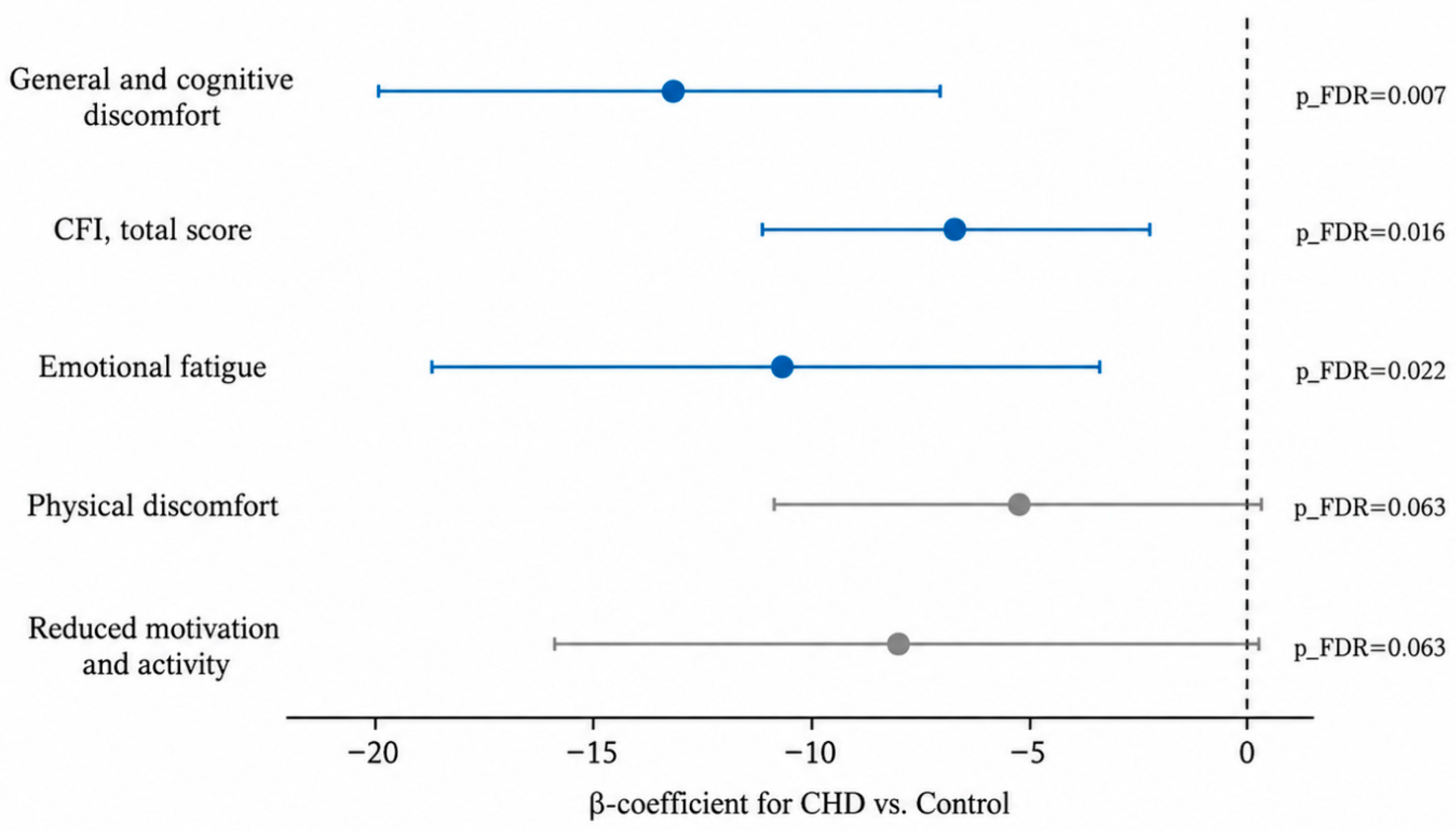
Figure 1: Adjusted differences in fatigue scores between the CHD and control groups. Points represent unstandardized OLS regression coefficients, and horizontal lines indicate 95% confidence intervals. Models were adjusted for age, sex, and BMI and fitted using HC3 heteroscedasticity-robust standard errors. Negative coefficients indicate lower fatigue scores in the CHD group. Blue denotes associations that remained significant after Benjamini–Hochberg FDR correction; grey denotes non-significant associations.
Sex differences in fatigue severity were significant across four of the five outcomes: boys reported significantly lower adjusted scores on the CFI, physical discomfort, general and cognitive discomfort, and emotional fatigue compared to girls (all p < 0.001) (Fig. 2). No significant sex difference was observed for reduced motivation and activity (β = −0.95, 95% CI −8.20 to 6.31, p = 0.798).
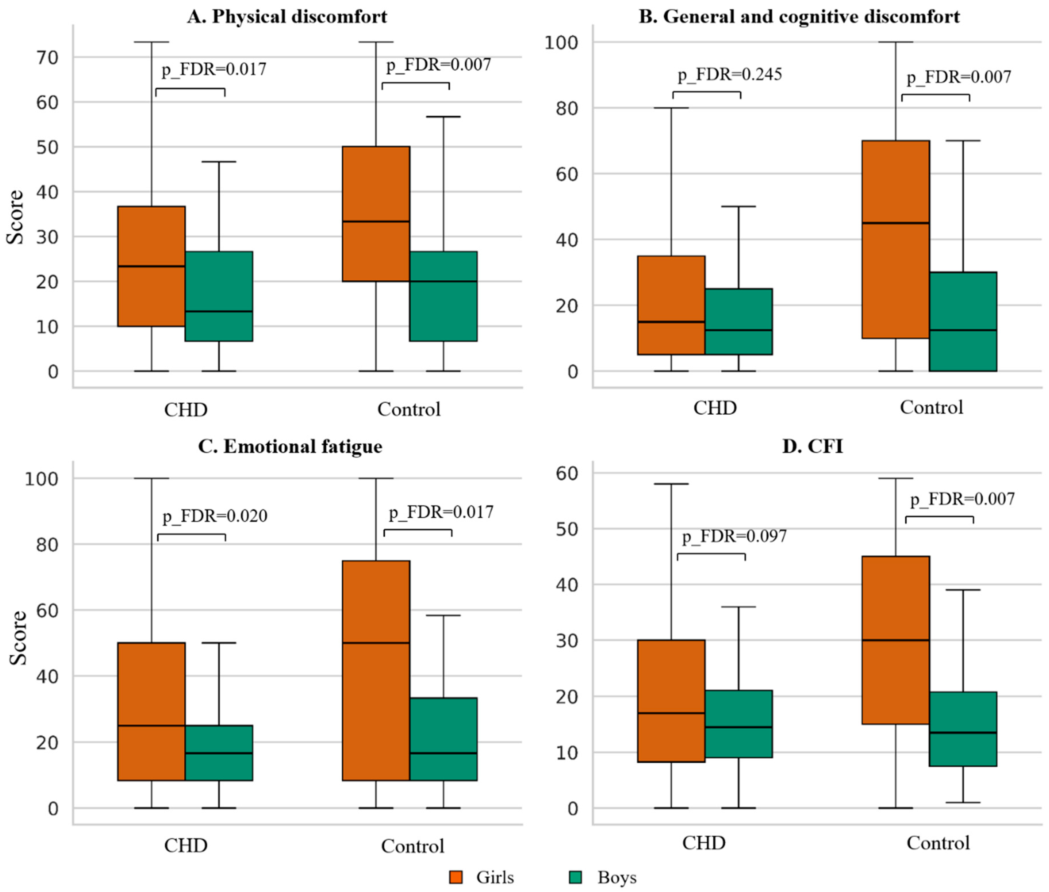
Figure 2: Sex differences in chronic fatigue scores in the CHD and Control groups. Panels (A–D): Physical discomfort, General and cognitive discomfort, Emotional fatigue, Total CFI. Orange—girls, green—boys. Boxplot: median and IQR; brackets—p_FDR (Mann-Whitney test).
3.4 Fatigue Severity by CHD Anatomical Complexity and Bayesian Verification of Null Results
Comparison across four groups (Table 7, Fig. 3) revealed statistically significant fatigue reductions relative to control in the CHD I and CHD II subgroups on the CFI and the general and cognitive discomfort scale. The CHD III group did not differ significantly from controls on any scale. Comparison of the three CHD subgroups against one another (Kruskal–Wallis test) found no significant differences on any scale (H = 0.10–2.56, all p > 0.28, all p_FDR > 0.70; Dunn–Holm post-hoc: all p ≥ 0.41).
Table 7: Chronic fatigue scores by group and adjusted effects of CHD subgroups vs. Control (OLS HC3).
| Scale | Control (n = 75) | CHD I (n = 32) | CHD II (n = 93) | CHD III (n = 29) |
|---|---|---|---|---|
| Median [Q1; Q3] | ||||
| CFI | 20.00 [10.00; 39.00] | 15.00 [7.00; 24.00] | 14.00 [9.00; 23.00] | 18.00 [12.00; 25.00] |
| General and cognitive discomfort | 25.00 [5.00; 65.00] | 10.00 [0.00; 31.25] | 10.00 [5.00; 30.00] | 15.00 [10.00; 35.00] |
| Emotional fatigue | 33.33 [8.33; 66.67] | 16.67 [0.00; 41.67] | 16.67 [8.33; 33.33] | 25.00 [8.33; 50.00] |
| Physical discomfort | 23.33 [13.33; 43.33] | 15.00 [10.00; 33.33] | 20.00 [6.67; 33.33] | 16.67 [6.67; 33.33] |
| Reduced motivation and activity | 50.00 [30.00; 80.00] | 40.00 [27.50; 60.00] | 40.00 [20.00; 60.00] | 50.00 [40.00; 70.00] |
| β vs. Control [95% CI], p | ||||
| CFI (total score) | - | −7.32 [−13.64; −0.99], p = 0.023* | −5.96 [−10.53; −1.40], p = 0.010* | −3.83 [−9.99; 2.33], p = 0.223 |
| General and cognitive discomfort | - | −16.48 [−26.86; −6.10], p = 0.002** | −12.83 [−20.94; −4.71], p = 0.002** | −8.19 [−18.62; 2.24], p = 0.124 |
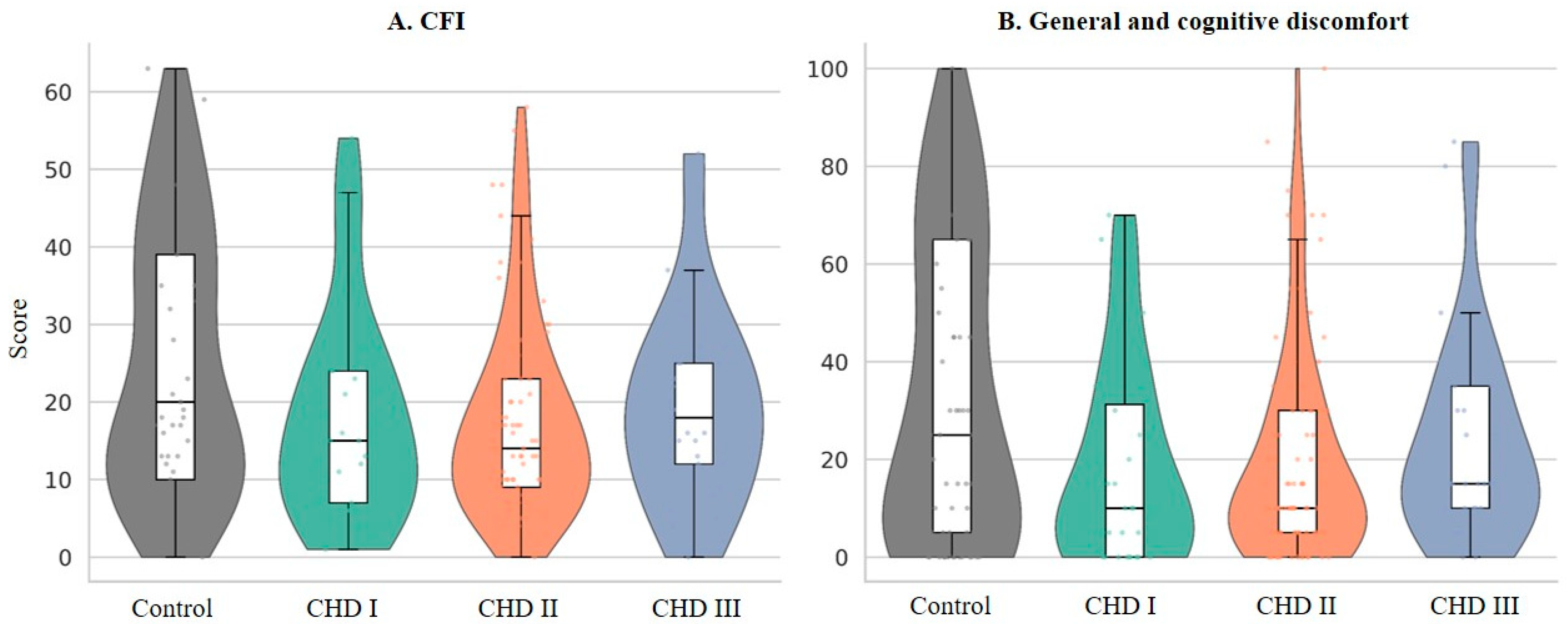
Figure 3: Distribution of chronic fatigue scores across the four groups. Panel (A)—total CFI; Panel (B)—% general and cognitive discomfort. Violin plot with embedded boxplot.
To rigorously verify the null result, post-hoc power analysis, Bayesian model comparison, and equivalence testing were performed. The observed effect of CHD anatomical complexity on fatigue was negligible (ε2 ≈ 0.000; η2 = 0.003; Cohen’s f = 0.058), and post-hoc power for this effect was 9.0%. The sample nonetheless had adequate power to detect a medium-sized effect (minimum detectable η2 = 0.060 at 80% power). Bayesian analysis provided positive evidence supporting the null hypothesis: the data were 8.91–108.37 times more compatible with a model without a CHD complexity effect than with one including it (BF01 = 8.91 for ordinal coding and 108.37 for categorical coding). Equivalence testing (TOST, boundary ±5 points) confirmed clinical equivalence of CHD I and CHD II groups; the CHD III group did not reach the formal equivalence threshold, owing in part to the comparatively small subgroup size (n = 29).
3.5 Predictors of Chronic Fatigue within the CHD Group
To identify clinical determinants of fatigue severity within the CHD cohort, Spearman correlation analysis (with FDR correction) and sequential linear regression models with HC3 robust standard errors were conducted. After FDR correction, none of the clinical variables-CHD anatomical complexity, NYHA functional class, heart failure stage, oxygen saturation, LVEF, estimated right ventricular systolic pressure, tricuspid and pulmonary regurgitation grades, aortic and pulmonary valve pressure gradients, number of previous cardiac surgeries, age at first surgical intervention, or time elapsed since the most recent surgical procedure-demonstrated a statistically significant correlation with any fatigue scale.
Physical work capacity demonstrated a weak inverse unadjusted association with physical discomfort (rs = −0.175, p = 0.030) and a weaker association with the CFI (rs = −0.142, p = 0.079). Neither association remained statistically significant after correction for multiple testing. The unadjusted relationship between physical work capacity and the CFI is illustrated in Fig. 4.
A multivariable OLS model with HC3 heteroscedasticity-robust standard errors was fitted to examine predictors of the CFI within the CHD group, including physical work capacity, sex, age, BMI, and anatomical CHD complexity (Table 8). Higher physical work capacity was associated with lower CFI scores; however, this association did not reach statistical significance (β = −4.52 per 1 W/kg, 95% CI −9.49 to 0.44, p = 0.074; standardized β = −0.146). Male sex was also associated with a lower estimated CFI score (β = −3.80, 95% CI −8.20 to 0.60, p = 0.090), as was older age (β = −0.79 per year, 95% CI −1.86 to 0.29, p = 0.150), although confidence intervals for both included the null value. BMI (β = −0.06, p = 0.880) and anatomical CHD complexity (β = 0.43, p = 0.797) showed minimal evidence of association with the total fatigue score. The model explained 7.1% of the variance in the CFI (R2 = 0.071; adjusted R2 = 0.039).
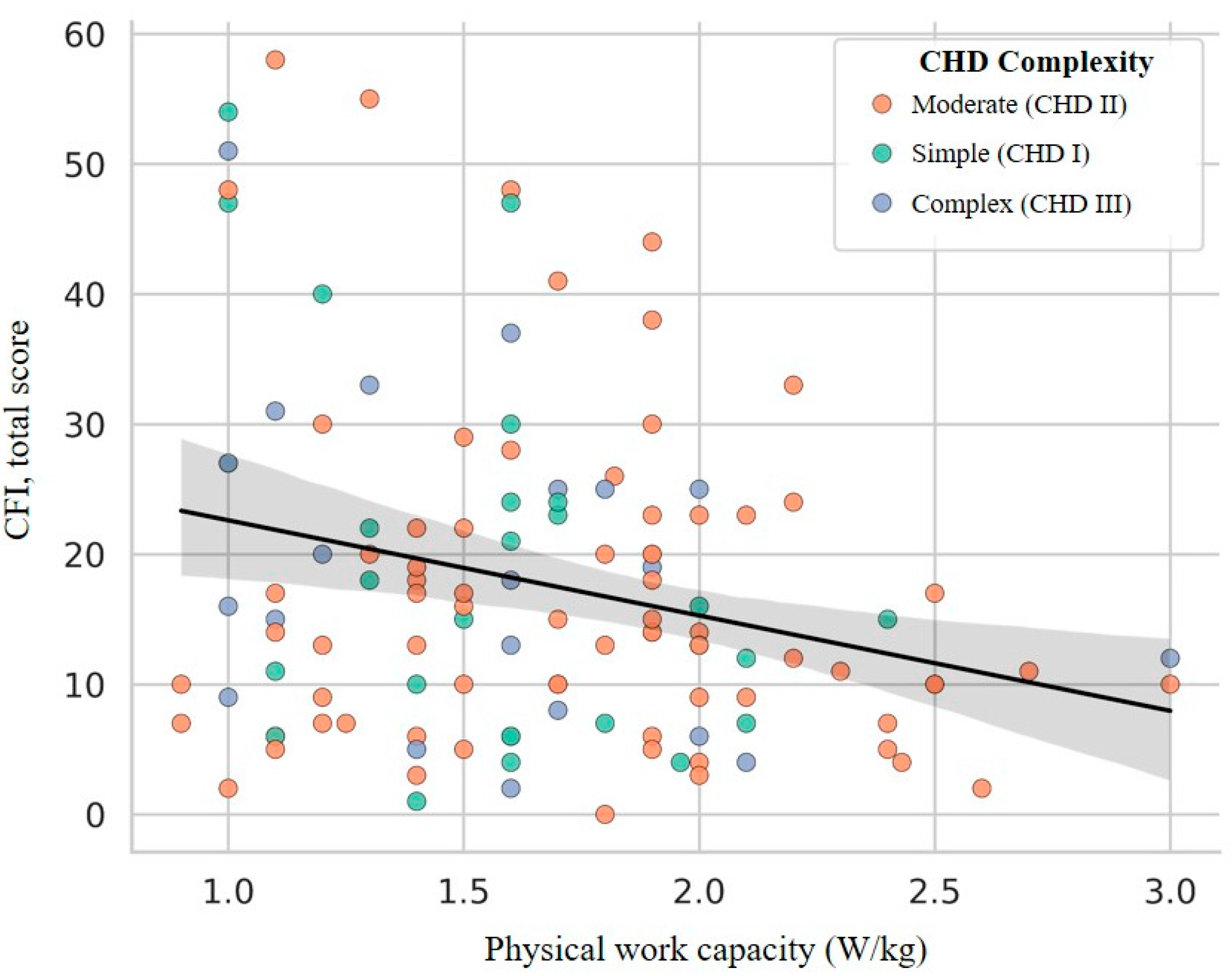
Figure 4: Relationship between physical work capacity (W/kg) and total CFI. Points are color-coded by complexity: green—CHD I, orange—CHD II, blue—CHD III. Line—linear regression; grey band—95% CI. Spearman ρ = −0.194, p = 0.034; β (W/kg) = −5.18; 95% CI [−10.27; −0.09]; p = 0.046; adj R2 = 0.113.
Table 8: Predictors of total chronic fatigue score within the CHD group (OLS HC3).
| Predictor | β | SE (HC3) | 95% CI | p | β* | LASSO/EN |
|---|---|---|---|---|---|---|
| Physical work capacity (W/kg) | −4.52 | 2.53 | [−9.49; 0.44] | 0.074 | −0.146 | ✓/✓ |
| Sex (male) | −3.80 | 2.25 | [−8.20; 0.60] | 0.090 | −0.150 | ✓/✓ |
| Age (years) | −0.79 | 0.55 | [−1.86; 0.29] | 0.150 | −0.108 | ✓/✓ |
| BMI (kg/m2) | −0.06 | 0.38 | [−0.80; 0.68] | 0.880 | −0.018 | −/− |
| CHD complexity | 0.43 | 1.69 | [−2.87; 3.74] | 0.797 | 0.022 | −/− |
Exploratory interaction analyses were additionally performed to assess whether clinically relevant associations with the CFI varied across patient subgroups. No statistically significant interaction was observed between anatomical CHD complexity and PWC (β_interaction = 4.62, 95% CI −4.17 to 13.41, p = 0.303). Likewise, the interaction between NYHA functional class and sex was not statistically significant (β_interaction = 11.07, 95% CI −3.20 to 25.35, p = 0.128). These analyses were exploratory and should be interpreted cautiously because interaction effects generally require larger samples and the highest-severity categories were sparsely represented, particularly NYHA class III.
3.6 Verification of Null Results
To rigorously establish that the absence of association between CHD complexity and fatigue severity was not due to insufficient statistical power or model misspecification, three verification approaches were undertaken:
Post-Hoc Power Analysis
The observed effect of CHD anatomical complexity on fatigue was negligible (ε2 ≈ 0.000; η2 = 0.003; Cohen’s f = 0.058), and post-hoc power for detecting this effect was 9.0%. However, the sample nonetheless had adequate power to detect a medium-sized effect (minimum detectable η2 = 0.060 at 80% power). This discrepancy indicates that the null result cannot be attributed to inadequate sample size for medium-effect detection; rather, if a true effect exists, it is likely to be very small (smaller than conventional medium effects).
Bayesian Model Comparison
Bayesian analysis provided positive evidence in support of the null hypothesis: the data were 8.91–108.37 times more compatible with a model without a CHD complexity effect than with one including it (BF01 = 8.91 for ordinal coding and 108.37 for categorical coding). Bayes factors exceeding 3.0 constitute moderate-to-strong evidence supporting the null model, and these results substantially exceed that threshold. This analysis provides robust evidence that the data are more likely under a model of no relationship between CHD complexity and fatigue.
Equivalence Testing
Equivalence testing (TOST, with boundaries of ±5 points on the CFI scale) confirmed clinical equivalence of CHD I and CHD II groups, indicating that fatigue severity in these two subgroups did not differ by more than the pre-specified clinically meaningful threshold. The CHD III group did not reach the formal equivalence threshold, owing in part to the comparatively small subgroup size (n = 29), which reduces statistical power for this comparison. These results collectively support the conclusion that measured CHD anatomical complexity does not meaningfully associate with fatigue severity in this cohort.
This study tested the conventional disease-severity hypothesis within a broader transdiagnostic framework by comparing fatigue in adolescents with CHD and school-based controls and by examining associations with anatomical complexity, functional status, and measured cardiac parameters. Contrary to the severity-specific hypothesis, the CHD group reported lower fatigue scores on four of the five DCF outcomes than the academically selective control group, and fatigue did not increase with anatomical CHD complexity. These findings therefore did not support the predicted severity gradient and were instead more compatible with the broader multifactorial framework considered a priori in the Introduction.
In pooled analyses, sex was among the most consistent predictor of fatigue across this cohort: girls reported significantly higher fatigue severity across nearly all measured domains, independent of CHD status. This pronounced sex dimorphism aligns with extensive literature documenting higher fatigue prevalence in females across clinical and population-based samples [27,28,29,30,31]. Myalgic encephalomyelitis/chronic fatigue syndrome occurs approximately three times more frequently in women than in men [32]. Sex differences in fatigue have been reported among adolescents with cancer [3], older adults without cognitive impairment [28], and patients with diverse chronic conditions [2,4,29,30,31].
The mechanisms underlying this sex difference are multifactorial. Beyond endocrine influences—particularly those related to menstrual cycle dynamics and adrenal hormone secretion [32]—documented sex differences in stress reactivity, sleep quality, and anxiety levels have been reported and partially mediate higher fatigue in females [27]. Women also demonstrate a greater predisposition to comorbid symptoms including low mood, anxiety, and emotional lability [28]. Additionally, gender socialization patterns—particularly greater reflectivity and tendency toward internalization in girls [33,34]—may amplify willingness to endorse and report fatigue symptoms.
In this cohort, measured indices of CHD anatomical complexity and cardiac dysfunction were not significantly associated with fatigue severity, a finding consistent with and complementary to recent integrative analyses [23]. Neither defect complexity, NYHA functional class, heart failure stage, nor objective echocardiographic parameters (LVEF, valvular gradients, regurgitation grades) showed significant association with fatigue across any domain. This null result was verified through multiple statistical approaches: post-hoc power analysis confirmed adequate power to detect medium-sized effects; Bayesian model comparison provided positive evidence supporting the null hypothesis; and equivalence testing confirmed clinical equivalence of CHD I and CHD II subgroups.
Importantly, the absence of significant main effects does not exclude conditional associations that may emerge within particular clinical or demographic subgroups. Exploratory interaction analyses did not provide clear evidence that the association between PWC and fatigue varied by anatomical CHD complexity or that the association between NYHA functional class and fatigue differed by sex. However, these analyses were exploratory and should not be interpreted as demonstrating the absence of effect modification. Interaction effects generally require larger samples than main effects, and precision was limited by the small numbers in the highest-severity categories. Thus, smaller or more complex conditional relationships between cardiac status, functional capacity, sex, and fatigue remain possible.
These observations suggest that the relationship between structural cardiac disease severity and subjective fatigue in adolescents with CHD is more complex than disease anatomy alone would predict. This pattern is consistent with the transdiagnostic conceptualization of fatigue, which emphasizes that fatigue arises across diverse underlying conditions and may not correspond closely to conventional indices of disease severity. At the same time, the exploratory interaction findings indicate that the present data cannot exclude the possibility that cardiac or functional factors influence fatigue differently in specific patient subgroups.
Physical work capacity (W/kg, assessed by bicycle ergometry) demonstrated a weak inverse association with fatigue within the CHD group, although this association did not attain statistical significance after correction for multiple comparisons. Importantly, PWC should not be conceptualized as an exclusively non-cardiac or purely transdiagnostic determinant. In adolescents with CHD, realized exercise capacity represents an integrative functional measure reflecting the combined influence of cardiac output and hemodynamic status, residual cardiovascular limitations, physical conditioning, habitual physical activity, and behavioral factors [35,36,37]. Accordingly, the observed association between higher PWC and lower fatigue cannot be attributed specifically to either cardiac or non-cardiac mechanisms. The CHD I/II/III groups differed significantly in PWC (p = 0.004), while LVEF and several other clinical characteristics also varied across anatomical-complexity categories. At the same time, fatigue itself did not show a corresponding anatomical-complexity gradient. This pattern suggests that subjective fatigue may relate to the integrated functional consequences of cardiovascular disease and physical conditioning rather than to anatomical complexity or individual echocardiographic parameters alone. Given the weak and analytically sensitive PWC–fatigue association observed in the present study, however, this interpretation remains exploratory and warrants confirmation in prospective studies using more comprehensive cardiopulmonary exercise assessment.
An unexpected finding was that the school-based control group reported fatigue levels equal to or exceeding those of the CHD cohort. The most parsimonious interpretation is that this pattern may reflect characteristics of the selected control sample rather than unusually low fatigue in adolescents with CHD. Control participants were recruited from a single highly selective school consistently ranked among Russia’s top 100 academic institutions for the preceding decade. Such an educational setting may be associated with substantial academic demands, while academic pressure has been linked to adverse psychological outcomes and fatigue-related factors in adolescents [19]. Because academic workload, homework volume, perceived stress, and sleep quality were not directly measured in the present study, elevated academic stress in this control group cannot be demonstrated empirically. Nevertheless, the recruitment setting represents an important source of potential selection bias, and the control group should not be assumed to represent the typical healthy adolescent population.
This interpretation is particularly important because the between-group comparison provides information about fatigue relative to this specific school-based control sample rather than about the absolute fatigue burden of adolescents with CHD versus healthy adolescents in general. In contrast, 92.9% of participants with CHD had documented state disability status that could provide access to educational accommodations. However, actual school placement, academic workload, and utilization of such accommodations were not assessed. Thus, the educational environments of the two groups cannot be directly compared, and it would be inappropriate to conclude that reduced academic demands in the CHD cohort explain the observed difference. The more defensible interpretation is that the unusually specific recruitment context of the control group may have shifted its fatigue distribution upward.
Illness adaptation and response shift represent an additional, but more speculative, explanation. Patients with chronic conditions frequently recalibrate their internal reference standards for symptom appraisal over the course of long-term disease coexistence-a phenomenon well documented in pediatric populations with cancer [38], juvenile idiopathic arthritis [39], inflammatory bowel disease [40], and other chronic conditions [41]. Adolescents with CHD who have carried their diagnosis since early childhood (median age at first surgery 0.85 years in this cohort) may normalize elevated fatigue levels as part of their habitual functional baseline, systematically underreporting relative to healthy peers who retain an uncalibrated reference standard. Social \ desirability-the desire to appear well-functioning-may additionally amplify this underreporting. However, none of these mechanisms was directly assessed in the present study, and they should therefore be regarded as secondary hypotheses rather than primary explanations for the between-group difference.
Confounding by other unmeasured psychosocial factors must also be considered. The cross-sectional design prevents definitive causal inference, and beyond health status and educational setting, the groups may differ in social support, sleep quality, perceived stress, emotional coping strategies, family functioning, and other factors capable of influencing fatigue. These characteristics were not directly assessed and therefore cannot be incorporated into the interpretation as demonstrated explanatory mechanisms.
Taken together, the observed between-group pattern should not be interpreted as evidence that adolescents with CHD intrinsically experience less fatigue than healthy adolescents. A more cautious interpretation is that fatigue was lower in the CHD cohort relative to this particular, potentially high-burden school-based comparison group. Selection of controls from an academically selective educational environment therefore represents the most immediate methodological explanation for the unexpected group difference, whereas illness adaptation, response shift, and other psychosocial mechanisms remain plausible but untested secondary explanations. Replication using controls recruited from more representative educational settings, together with direct measurement of academic workload, stress, and sleep, is required before the direction or magnitude of CHD-related differences in fatigue can be generalized to the broader adolescent population.
Overall, these findings are compatible with a multifactorial conceptualization of fatigue in adolescence. The data suggest that measured structural cardiac parameters and conventional indices of disease severity do not fully account for variability in subjective fatigue, while functional, demographic, and psychosocial influences may also be relevant. The data suggest that measured structural cardiac parameters and conventional indices of disease severity do not fully account for variability in subjective fatigue. Rather, fatigue may reflect contributions from multiple domains, including cardiac disease burden, integrated functional capacity, demographic characteristics such as sex, and psychosocial context. PWC is particularly relevant within this framework because it cannot be classified as purely cardiac or non-cardiac: it reflects the combined consequences of cardiovascular function, physical conditioning, and behavioral adaptation. The present between-group findings, however, must be interpreted in light of the specific characteristics of the control sample and should not be taken as evidence that contextual factors outweigh cardiac influences. This pattern is broadly consistent with prior integrative analyses demonstrating that transdiagnostic factors can explain substantial variance in pediatric fatigue beyond cardiology-specific variables [23].
The findings suggest that comprehensive assessment of well-being in adolescents with CHD requires consideration of structural cardiac status, functional capacity, and broader psychosocial factors. Clinicians evaluating fatigue in this population should consider not only conventional cardiac measures but also exercise capacity, sleep quality, emotional functioning, perceived stress, social support, and educational demands, while recognizing that the relative contributions and interactions among these domains were not established by the present cross-sectional study.
The control group was recruited from a single, highly selective school consistently ranked among Russia’s top 100 academic institutions. This sampling strategy, while pragmatically advantageous for accessing a healthy peer population, introduces potential selection bias. Students in elite, academically demanding schools experience well-documented elevated chronic academic stress compared to peers in typical educational settings. Consequently, the control group may not be representative of the general adolescent population with respect to fatigue risk factors and baseline fatigue severity.
Whereas control participants attended a highly selective institution with standard, full-intensity academic demands, the educational environment of the CHD cohort remains incompletely characterized. Although 92.9% of CHD participants possessed state disability status conferring access to educational accommodations and reduced academic requirements, information regarding actual school placement, academic workload, and utilization of such accommodations was not systematically collected. This asymmetry in documentation-greater certainty regarding control group context versus incomplete characterization of CHD group educational circumstances-complicates interpretation of between-group differences and warrants caution in attributing observed fatigue differences to cardiac versus psychosocial factors. Control participants were recruited from a single academically selective school, which may not represent the general adolescent population in terms of baseline fatigue risk and should be considered a potential source of selection bias.
The cross-sectional design precludes causal inference. Beyond health status, the groups may differ in unmeasured psychosocial factors-including social support, sleep quality, perceived stress, emotional coping strategies, and family functioning-that could substantially influence fatigue and were not measured. This limitation prevents definitive determination of whether fatigue differences reflect cardiac disease burden, environmental context, response shift processes, or other unmeasured confounders.
All primary fatigue outcomes rely exclusively on self-report questionnaire data without objective physiological validation. While self-report instruments are appropriate for measuring subjective fatigue severity, they cannot distinguish response shift, social desirability bias, or other reporting artifacts from genuine symptom differences.
The broad age range (11–17 years) of the sample, spanning early to mid-adolescence, constitutes a limitation. Although age was included as a covariate, systematic measurement variability associated with developmental stage cannot be fully eliminated. Future studies should consider narrower age bands and age-adapted assessment instruments.
The DCF questionnaire has not undergone formal cross-cultural validation against international instruments (MFI-20, PedsQL Multidimensional Fatigue Scale). At the item level, item 6 (“I enjoy working in a team”) showed a low item-remainder correlation (r = 0.245) within the “Reduced motivation and activity” subscale, although McDonald’s omega met the psychometric threshold (ω = 0.717). Results on this subscale should therefore be interpreted with appropriate caution. Test-retest reliability was not calculated owing to the cross-sectional design.
Complete cardiopulmonary exercise testing data, including oxygen consumption at peak exercise (VO2peak), were available for only 36 of 154 patients with CHD (23.4%), which precluded their inclusion in primary analyses. Physical work capacity (W/kg from bicycle ergometry) served as a proxy measure and does not replace complete cardiopulmonary exercise testing.
The absence of validated direct measurement of academic workload, sleep quality, and perceived stress prevents definitive disentanglement of response shift and illness adaptation mechanisms from psychosocial context explanations. Future studies should include these variables as primary measurement domains and covariates.
While the sample had adequate power to detect effects of at least moderate magnitude (minimum detectable η2 = 0.060 at 80% power), it was insufficient to exclude very small main effects. The CHD III subgroup (n = 29) is comparatively small, accurately reflecting the epidemiological rarity of complex defects. Nevertheless, future studies should consider enrolling larger samples of patients with complex CHD to increase statistical precision for this subgroup.
The study was primarily designed to evaluate main associations rather than interaction effects. Although exploratory analyses of CHD complexity × PWC and NYHA functional class × sex did not identify statistically significant effect modification, these analyses had limited power, particularly because of the small numbers in the highest-severity categories. Consequently, Type III error arising from small, subgroup-specific, or otherwise insufficiently powered conditional associations cannot be excluded. Larger studies specifically powered to examine interactions between cardiac severity, functional capacity, sex, and other biopsychosocial factors are required to determine whether clinically relevant effect modification is present.
The NYHA functional classification was based on clinician documentation rather than standardized functional testing. This classification reflects clinician assessment of exercise tolerance rather than objective cardiopulmonary exercise capacity and may be subject to inter-rater variability.
In this cross-sectional cohort, cardiac disease severity did not predict fatigue in adolescents with CHD. Sex was the strongest and most consistent factor: girls reported significantly higher fatigue than boys, regardless of cardiac status. Interestingly, healthy controls from a high-performing school reported fatigue levels similar to or higher than those with CHD—likely reflecting academic stress rather than lower fatigue in the CHD group. These findings suggest that fatigue in this population is multifactorial, shaped by functional capacity, sex, and psychosocial context rather than by cardiac anatomy alone. Clinicians should look beyond cardiac metrics when evaluating fatigue. Future studies with representative controls and direct measures of stress and sleep are needed to confirm these observations.
Acknowledgement:
Funding Statement: This work was supported by the state assignments of Federal Research Center for Innovator and Emerging Biomedical and Pharmaceutical Technologies (Project No. 122040500027-7) and A.N. Bakulev National Medical Research Center for Cardiovascular Surgery of the Russian Ministry of Health (Project No. 123020300024-9).
Author Contributions: The authors confirm contribution to the paper as follows: Elena Likhomanova: conceptualization, study design, data collection, manuscript writing. Olga Shevaldova: conceptualization, data collection, statistical analysis, manuscript writing. Anastasia Kovaleva: project supervision, methodology, manuscript revision. Anna Zavarina: clinical expertise, medical data collection, manuscript revision, resources. All authors reviewed and approved the final version of the manuscript.
Availability of Data and Materials: The raw data used for the analyses in this study are publicly available in the Zenodo repository: Shevaldova O, Likhomanova E, Kovaleva A. (2026). Chronic fatigue in adolescents with congenital heart disease and healthy peers: raw data supporting the biopsychosocial comparative analysis [Dataset]. Zenodo. https://doi.org/10.5281/zenodo.20703353.
Ethics Approval: The study was approved by the Ethics Committee of the Federal Research Center for Innovator and Emerging Biomedical and Pharmaceutical Technologies (P.K. Anokhin Research Institute of Normal Physiology), Moscow, Russian Federation (Protocol No. 18/1, 15 February 2023), and was conducted in accordance with the Declaration of Helsinki. Written informed consent for study participation and publication of results was obtained from legal guardians of all participants; adolescents provided written assent where required by the approved protocol.
Conflicts of Interest: The authors declare no conflicts of interest.
References
1. Ternrud L , Johansson B , Sparv D , Mandalenakis Z , Christersson C , Van Bulck L , et al. Fatigue in adults with congenital heart disease aged over 40 years. Int J Cardiol Congenit Heart Dis. 2025; 21: 100601. doi:10.1016/j.ijcchd.2025.100601. [Google Scholar] [CrossRef]
2. Roman-Juan J , Ceniza-Bordallo G , Sánchez-Rodríguez E , Jensen MP , Miró J . Fatigue, sleep disturbance, and pain interference in children and adolescents with chronic pain: A longitudinal study. Pain. 2025; 166( 4): 927– 35. doi:10.1097/j.pain.0000000000003432. [Google Scholar] [CrossRef]
3. Likhomanova EN , Kovaleva AV . Associations of cancer-related fatigue with autonomic and sensorimotor functions in children treated for brain tumors: An observational study. Bull Rehabil Med. 2022; 21( 4): 60– 70. doi:10.38025/2078-1962-2022-21-4-60-70. [Google Scholar] [CrossRef]
4. Nap-van der Vlist MM , Dalmeijer GW , Grootenhuis MA , van der Ent K , van den Heuvel-Eibrink MM , Swart JF , et al. Fatigue among children with a chronic disease: A cross-sectional study. BMJ Paediatr Open. 2021; 5( 1): e000958. doi:10.1136/bmjpo-2020-000958. [Google Scholar] [CrossRef]
5. Nacul L , Authier FJ , Scheibenbogen C , Lorusso L , Helland IB , Martin JA , et al. European network on myalgic encephalomyelitis/chronic fatigue syndrome (EUROMENE): Expert consensus on the diagnosis, service provision, and care of people with ME/CFS in Europe. Medicina. 2021; 57( 5): 510. doi:10.3390/medicina57050510. [Google Scholar] [CrossRef]
6. Taillefer SS , Kirmayer LJ , Robbins JM , Lasry JC . Psychological correlates of functional status in chronic fatigue syndrome. J Psychosom Res. 2002; 53( 6): 1097– 106. doi:10.1016/s0022-3999(02)00566-4. [Google Scholar] [CrossRef]
7. Murga Gandasegui I , Aranburu Laka L , Gargiulo PÁ , Gómez-Esteban JC , Lafuente Sánchez JV . Myalgic encephalomyelitis/chronic fatigue syndrome: A neurological entity? Medicina. 2021; 57( 10): 1030. doi:10.3390/medicina57101030. [Google Scholar] [CrossRef]
8. Kant IJ , Bültmann U , Schröer KA , Beurskens AJ , Van Amelsvoort LG , Swaen GM . An epidemiological approach to study fatigue in the working population: The Maastricht Cohort Study. Occup Environ Med. 2003; 60( Suppl 1): i32– 9. doi:10.1136/oem.60.suppl_1.i32. [Google Scholar] [CrossRef]
9. van’t Leven M , Zielhuis GA , van der Meer JW , Verbeek AL , Bleijenberg G . Fatigue and chronic fatigue syndrome-like complaints in the general population. Eur J Public Health. 2010; 20( 3): 251– 7. doi:10.1093/eurpub/ckp113. [Google Scholar] [CrossRef]
10. Lim EJ , Ahn YC , Jang ES , Lee SW , Lee SH , Son CG . Systematic review and meta-analysis of the prevalence of chronic fatigue syndrome/myalgic encephalomyelitis (CFS/ME). J Transl Med. 2020; 18( 1): 100. doi:10.1186/s12967-020-02269-0. [Google Scholar] [CrossRef]
11. Menting J , Tack CJ , Bleijenberg G , Donders R , Droogleever Fortuyn HA , Fransen J , et al. Is fatigue a disease-specific or generic symptom in chronic medical conditions? Health Psychol. 2018; 37( 6): 530– 43. doi:10.1037/hea0000598. [Google Scholar] [CrossRef]
12. Knight SJ , Politis J , Garnham C , Scheinberg A , Tollit MA . School functioning in adolescents with chronic fatigue syndrome. Front Pediatr. 2018; 6: 302. doi:10.3389/fped.2018.00302. [Google Scholar] [CrossRef]
13. Parslow RM , Harris S , Broughton J , Alattas A , Crawley E , Haywood K , et al. Children’s experiences of chronic fatigue syndrome/myalgic encephalomyelitis (CFS/ME): A systematic review and meta-ethnography of qualitative studies. BMJ Open. 2017; 7( 1): e012633. doi:10.1136/bmjopen-2016-012633. [Google Scholar] [CrossRef]
14. Solomon-Moore E , Jago R , Beasant L , Brigden A , Crawley E . Physical activity patterns among children and adolescents with mild-to-moderate chronic fatigue syndrome/myalgic encephalomyelitis. BMJ Paediatr Open. 2019; 3( 1): e000425. doi:10.1136/bmjpo-2018-000425. [Google Scholar] [CrossRef]
15. Herring MP , Monroe DC , Kline CE , O’Connor PJ , MacDonncha C . Sleep quality moderates the association between physical activity frequency and feelings of energy and fatigue in adolescents. Eur Child Adolesc Psychiatry. 2018; 27( 11): 1425– 32. doi:10.1007/s00787-018-1134-z. [Google Scholar] [CrossRef]
16. Yildiz E , Kovács E , Moreau S . Ranking adolescent stressors linked to urban lifestyle and overexposure. J Adolesc Youth Psychol Stud. 2025; 6( 11): 1– 8. doi:10.61838/kman.jayps.4741. [Google Scholar] [CrossRef]
17. Walburg V . Burnout among high school students: A literature review. Child Youth Serv Rev. 2014; 42: 28– 33. doi:10.1016/j.childyouth.2014.03.020. [Google Scholar] [CrossRef]
18. Chen TY , Chou YC , Tzeng NS , Chang HA , Kuo SC , Pan PY , et al. Effects of a selective educational system on fatigue, sleep problems, daytime sleepiness, and depression among senior high school adolescents in Taiwan. Neuropsychiatr Dis Treat. 2015; 11: 741– 50. doi:10.2147/NDT.S77179. [Google Scholar] [CrossRef]
19. Steare T , Gutiérrez Muñoz C , Sullivan A , Lewis G . The association between academic pressure and adolescent mental health problems: A systematic review. J Affect Disord. 2023; 339: 302– 17. doi:10.1016/j.jad.2023.07.028. [Google Scholar] [CrossRef]
20. Babaeva VA , Priboychenko AS , Zavarina AY , Engenova MS , Mitchenko EA , Kokshina AV , et al. Higher mental functions in children with congenital heart disease. Child Heart Vasc Dis. 2023; 20( 3): 191– 202. (In Russian). doi:10.24022/1810-0686-2023-20-3-191-202. [Google Scholar] [CrossRef]
21. Gorbunov GE , Solovev NA , Volodina MS , Karakhan NV , Diasamidze KE , Rybka MM . Comparison of inhalation and total intravenous methods of anesthetic support for endovascular surgeries for congenital heart defects in children and adolescents. Child Heart Vasc Dis. 2024; 21( 3): 211– 21. (In Russian). doi:10.24022/1810-0686-2024-21-3-211-221. [Google Scholar] [CrossRef]
22. De Vos DRH , Hoefnagels JW , Nap-van der Vlist MM , Breur JMPJ , Nijhof SL . Prevalence of fatigue in children with congenital heart disease and correlations with disease-specific factors. Res Sq. 2022. doi:10.21203/rs.3.rs-2280678/v1. [Google Scholar] [CrossRef]
23. Stutvoet MD , Vroegindeweij A , Nap-Van der Vlist MM , Hoefnagels JW , de Vos DRH , Weijer L , et al. Fatigue in pediatric congenital heart disease: Integrative insight into cardiology-focused, lifestyle, psychological and social factors. Fatigue. 2026; 14( 1): 1– 14. doi:10.1080/21641846.2025.2561520. [Google Scholar] [CrossRef]
24. Ternrud L , Hlebowicz J , Sandberg C , Johansson B , Sparv D . Prevalence of fatigue in adults with congenital heart disease. Cardiol Young. 2022; 32( 9): 1432– 9. doi:10.1017/S1047951121004297. [Google Scholar] [CrossRef]
25. Stout KK , Daniels CJ , Aboulhosn JA , Bozkurt B , Broberg CS , Colman JM , et al. 2018 AHA/ACC guideline for the management of adults with congenital heart disease: A report of the American College of Cardiology/American Heart Association task force on clinical practice guidelines. Circulation. 2019; 139( 14): e698– 800. doi:10.1161/cir.0000000000000603. [Google Scholar] [CrossRef]
26. Leonova AB , Kuznetsova AS , Barabanshchikova VV . Self-regulation training and prevention of negative human functional states at work: Traditions and recent issues in Russian applied research. Psych Rus. 2010; 3( 1): 482– 507. doi:10.11621/pir.2010.0023. [Google Scholar] [CrossRef]
27. Faro M , Sàez-Francás N , Castro-Marrero J , Aliste L , Fernández de Sevilla T , Alegre J . Gender differences in chronic fatigue syndrome. Reumatol Clin. 2016; 12( 2): 72– 7. doi:10.1016/j.reuma.2015.05.007. [Google Scholar] [CrossRef]
28. Toccaceli Blasi M , Alfano AR , Salzillo M , Buscarnera S , Raparelli V , Cesari M , et al. Sex-specific clinical and neurobiological correlates of fatigue in older adults. GeroScience. 2025; 47( 1): 1151– 60. doi:10.1007/s11357-024-01259-0. [Google Scholar] [CrossRef]
29. Ma Y , He B , Jiang M , Yang Y , Wang C , Huang C , et al. Prevalence and risk factors of cancer-related fatigue: A systematic review and meta-analysis. Int J Nurs Stud. 2020; 111: 103707. doi:10.1016/j.ijnurstu.2020.103707. [Google Scholar] [CrossRef]
30. Erol K , Ertaş ŞK , Ertaş R . Fatigue is common and predicted by female gender and sleep disturbance in patients with chronic spontaneous urticaria. J Allergy Clin Immunol Pract. 2021; 9( 1): 469– 76. doi:10.1016/j.jaip.2020.08.020. [Google Scholar] [CrossRef]
31. Lagogianni C , Gatzonis S , Patrikelis P . Fatigue and cognitive functions in epilepsy: A review of the literature. Epilepsy Behav. 2021; 114: 107541. doi:10.1016/j.yebeh.2020.107541. [Google Scholar] [CrossRef]
32. Thomas N , Gurvich C , Huang K , Gooley PR , Armstrong CW . The underlying sex differences in neuroendocrine adaptations relevant to Myalgic Encephalomyelitis Chronic Fatigue Syndrome. Front Neuroendocrinol. 2022; 66: 100995. doi:10.1016/j.yfrne.2022.100995. [Google Scholar] [CrossRef]
33. Barsky AJ , Peekna HM , Borus JF . Somatic symptom reporting in women and men. J Gen Intern Med. 2001; 16( 4): 266– 75. doi:10.1046/j.1525-1497.2001.016004266.x. [Google Scholar] [CrossRef]
34. Hausteiner-Wiehle C , Schneider G , Lee S , Sumathipala A , Creed F . Gender, lifespan and cultural aspects. In: Creed F , Henningsen P , Fink P , editors. Medically unexplained symptoms, somatisation and bodily distress: Developing better clinical services. Cambridge, UK: Cambridge University Press; 2011. p. 132– 57. doi:10.1017/cbo9780511977862.007. [Google Scholar] [CrossRef]
35. Villaseca-Rojas Y , Varela-Melo J , Torres-Castro R , Vasconcello-Castillo L , Mazzucco G , Vilaró J , et al. Exercise capacity in children and adolescents with congenital heart disease: A systematic review and meta-analysis. Front Cardiovasc Med. 2022; 9: 874700. doi:10.3389/fcvm.2022.874700. [Google Scholar] [CrossRef]
36. Soares A , Park LK , Mansour E , Deych E , Puritz A , Zhao M , et al. Predictive value of
37. Dzhitava TG , Filaretova OV , Filatova AG , Danilov TY , Kovalev DV . Cardiac rehabilitation after surgical correction of congenital heart defects in adult patients. Important issues. Creat Cardiol. 2024; 18: S17– 33. (In Russian). doi:10.24022/1997-3187-2024-18S-S17-S33. [Google Scholar] [CrossRef]
38. Sprangers MA , Schwartz CE . Integrating response shift into health-related quality of life research: A theoretical model. Soc Sci Med. 1999; 48( 11): 1507– 15. doi:10.1016/s0277-9536(99)00045-3. [Google Scholar] [CrossRef]
39. Arnstad ED , Glerup M , Rypdal V , Peltoniemi S , Fasth A , Nielsen S , et al. Fatigue in young adults with juvenile idiopathic arthritis 18 years after disease onset: Data from the prospective Nordic JIA cohort. Pediatr Rheumatol Online J. 2021; 19( 1): 33. doi:10.1186/s12969-021-00499-0. [Google Scholar] [CrossRef]
40. Martin-Masot R , Rodríguez-Belvís MV , Pujol Muncunill G , Palomino L , Sánchez Sánchez C , Martín de Carpi J , et al. Fatigue in pediatric patients with inflammatory bowel disease: A multicenter study by the SEGHNP. Crohn’s Colitis 360. 2026; 8( 1): otag005. doi:10.1093/crocol/otag005. [Google Scholar] [CrossRef]
41. Berkelbach van der Sprenkel EE , van der Laan SEI , Finkenauer C , Lenters VC , van de Putte EM , Bont LJ , et al. Chronic conditions and adolescents’ psychosocial wellbeing: The impact of self-reporting. Eur J Pediatr. 2025; 185( 1): 15. doi:10.1007/s00431-025-06616-5. [Google Scholar] [CrossRef]
Cite This Article
 Copyright © 2026 The Author(s). Published by Tech Science Press.
This work is licensed under a Creative Commons Attribution 4.0 International License , which permits unrestricted use, distribution, and reproduction in any medium, provided the original work is properly cited.
 Downloads
 Citation Tools
