
 Login
Login Register
 Submit a Paper
 Propose a Special lssue
 Open Access
ARTICLE
Procedural Performance and Safety of VACS® Balloon Catheters in Congenital Heart Interventions
1 Department of Pediatric Cardiology and Pediatric Intensive Care, University Hospital of Munich, LMU, Munich, Germany
2 Center for Congenital Heart Defects Stuttgart, Department of Pediatric Intensive Care, Pneumology and Allergology, Klinikum Stuttgart, Olgahospital/Women’s Clinic, Stuttgart, Germany
* Corresponding Author: Guido Mandilaras. Email:
Structural and Congenital Heart Disease 2026, 21(4), 3 https://doi.org/10.32604/schd.2026.080384
Received 08 February 2026; Accepted 18 August 2026; Issue published 30 September 2026
 View Full Text
 Download PDF
Abstract
Background: Balloon catheters are essential tools in congenital cardiac catheterization and are used for therapeutic balloon dilatation, anatomical balloon interrogation, and stent implantation. Clinical performance data for established devices have become increasingly important under the European Medical Device Regulation (MDR). We evaluated the procedural performance and safety of the Osypka VACS® (Valvuloplasty Catheter System; Osypka AG, Rheinfelden, Germany) II and VACS® III balloon catheters across a broad spectrum of congenital heart interventions. Methods: This retrospective single-center study reviewed the institutional experience with VACS® II and VACS® III balloon catheters over 10 years. Consecutive procedures from predefined indication groups were analyzed descriptively to assess balloon performance across therapeutic and diagnostic applications. The primary endpoint was technical device success, defined as successful positioning, intended balloon inflation according to the procedural objective, and uncomplicated retrieval of the balloon catheter. Device-related complications and indication-specific procedural outcomes were recorded. Results: For the present indication-based analysis, consecutive procedures were sampled as described in the Methods. The predefined criteria for technical device success were fulfilled in all analyzed procedures. Balloon rupture occurred in two of the 25 right ventricular outflow tract dilatation procedures (n = 2/25, 8%), both during high-pressure dilatation of severely calcified postoperative conduits, without vascular injury or retrieval difficulties. Therapeutic balloon dilatation resulted in substantial improvements in indication-specific hemodynamic parameters, while balloon interrogation and stent implantation procedures were successfully completed in all intended cases. Seven patients developed moderate aortic regurgitation following maximal balloon aortic valvuloplasty, representing a recognized procedural complication rather than device malfunction. No procedure-related mortality or serious device-related adverse events were observed. Conclusions: The Osypka VACS® II and VACS® III balloon catheters demonstrated reliable procedural performance, broad applicability, and a low rate of device-related complications across diverse congenital heart interventions. These real-world data provide clinical performance evidence supporting the continued use and regulatory evaluation of established balloon catheter systems under the current European MDR framework.Keywords
Congenital heart defects occur in approximately 11 per 1000 live births, and many patients require surgical and/or transcatheter interventions to correct structural abnormalities or improve hemodynamic function [1]. In addition, residual lesions, postoperative scar formation, and inadequate growth of cardiovascular structures during childhood frequently necessitate repeat catheter-based or surgical interventions. Balloon-based interventions are fundamental techniques in congenital cardiology and are routinely used to treat valvular stenosis, branch pulmonary artery stenosis, right ventricular outflow tract (RVOT) obstruction, coarctation of the aorta (CoA), and other vascular lesions.
Although many pediatric catheter interventions continue to rely on off-label devices, dedicated balloon catheter systems specifically developed for congenital cardiology have become an important component of contemporary practice. Nevertheless, published clinical performance data for these well-established devices remain limited [2].
Selection of the appropriate balloon catheter depends on lesion characteristics, procedural objectives, and device-specific properties, including balloon diameter, length, compliance, and rated burst pressure. Depending on the clinical indication, balloon catheters may be used for therapeutic dilatation, anatomical interrogation, or stent implantation.
Under the European Medical Device Regulation (MDR), manufacturers of established medical devices are increasingly required to provide contemporary clinical performance and safety data throughout the device life cycle [3]. Generating such evidence is particularly challenging for devices that have become part of routine clinical practice, as prospective clinical studies are often impractical despite extensive real-world use.
The VACS® (Valvuloplasty Catheter System; Osypka AG, Rheinfelden, Germany) balloon catheters have been available in Europe since 1995 and are widely used for a broad range of congenital cardiac interventions. Therefore, we performed a retrospective single-center analysis of congenital cardiac catheterization procedures using VACS® II and VACS® III balloon catheters over 10 years. The primary objective was to evaluate procedural performance and technical device safety across a broad spectrum of therapeutic and diagnostic congenital heart interventions and to provide contemporary clinical performance data relevant to both clinical practice and regulatory evaluation under the MDR.
2.1 Study Design and Patient Population
This retrospective single-center study evaluated the procedural performance and safety of the Osypka VACS® II and VACS® III balloon catheters in congenital cardiac catheterization procedures performed at the University Hospital of Munich, LMU, Department of Pediatric Cardiology and Pediatric Intensive Care (Munich, Germany), between January 2015 and December 2024, a 10-year period. A fixed sample size of 25 consecutive procedures per predefined indication group was selected a priori to obtain a balanced representation of the principal clinical applications of the VACS® balloon catheters while allowing feasible, systematic chart review within the study period. Cases were identified strictly in reverse chronological order from the institutional catheterization database, beginning with the most recent procedures, with no exclusion based on procedural outcome, complication status, or case difficulty; sampling stopped as soon as 25 consecutive eligible cases per indication had been reached, without any element of case selection that could introduce bias. Complete data were available for all analyzed variables; no missing data required imputation.
The predefined indication groups comprised pulmonary valve valvuloplasty, aortic valve valvuloplasty, right ventricular outflow tract interventions, peripheral pulmonary artery angioplasty, coarctation angioplasty, balloon interrogation before percutaneous pulmonary valve implantation, stent implantation, and miscellaneous congenital cardiac interventions.
Because VACS® balloon catheters were routinely used for different procedural purposes—including therapeutic balloon dilatation, anatomical balloon interrogation, and stent implantation—individual patients could contribute to more than one procedural category during the same or staged catheterization procedures. Consequently, the indication groups were not mutually exclusive, and comparisons between groups were neither intended nor performed.
The primary objective of the study was to evaluate technical device performance and procedural safety according to the intended clinical application rather than to compare clinical outcomes between treatment groups. This retrospective study was conducted in accordance with the Declaration of Helsinki and institutional guidelines. It was reviewed by the Ethics Committee of the Ludwig-Maximilians-Universität München (LMU Munich), which confirmed no objection to the project (Project No. 24-1128 kB, dated 23 December 2024); informed consent was waived because data collection and analysis were conducted exclusively on anonymized data.
2.2 Balloon Catheter Characteristics
The Osypka VACS® balloon catheters have been available in the European market since 1995 and are designed for percutaneous transluminal valvuloplasty (PTV) in both pediatric and adult interventional cardiology. The VACS® dilatation catheter is a PTV balloon catheter with a coaxial shaft construction and a non-compliant balloon that can be easily guided over a wire. The VACS® II balloon catheter features a low-profile balloon that allows the use of smaller introducers, while the VACS® III is a high-pressure balloon catheter due to its reinforced balloon segment. It is equipped with radiopaque gold markers, allows for rapid inflation and deflation, and is available in a comprehensive range of sizes for maximum flexibility. The VACS® II balloons are available in diameters ranging from 4 to 30 mm, lengths of 20 to 60 mm, and rated burst inflation pressures of 6 to 15 atm. The VACS® III balloons have diameters ranging from 5 to 30 mm, lengths of 20 to 60 mm, and rated burst pressures of 4 to 14 atm [4].
General: In all treatments, following institutional protocols, the balloons were filled with a mixture of 30–50% contrast and 50–70% normal saline. Balloon inflation and deflation were performed using mechanical inflators and deflators. Slow manual pressure inflation was initiated and completed with controlled indeflator pressures, followed by rapid deflation after maximal inflation through the application of maximum vacuum. A stiffer guiding wire was used to position the balloons in the target region. Balloons were inserted after appropriate de-airing and under maximum vacuum to ensure stability. The appropriate short sheath was used for balloon insertion according to the instructions for use (IFU) [5].
Pulmonic Valve Valvuloplasty: In neonates with subatretic and/or critical pulmonary stenosis (PS), initial predilatation with an appropriately sized coronary balloon (approximately 3 mm) was performed before inserting the proper balloon. In other patients, the balloon was advanced directly, with the diameter chosen to match 100% to 130% of the valve ring diameter. As a standard, a VACS® II balloon was used. Successful inflation was defined as complete inflation up to the rated burst pressure. If a residual gradient was present after complete inflation, a repeat dilatation was performed with a balloon 1–2 mm larger [4,6,7].
Dilatation of the Right Ventricular Outflow Tract: Native balloon dilatation (e.g., in tetralogy of Fallot) was excluded from this analysis. RVOT dilatation was performed in stenotic postoperative RVOTs, with or without calcifications, using VACS® III balloons. The balloon diameter was chosen based on the pre- and post-stenotic diameters of the pulmonary artery bifurcation and the RVOT. For patients with predominant pulmonary regurgitation, VACS® II or VACS® III balloons were used for sizing the RVOT in preparation for potential stent and valve implantation. In these cases, incomplete inflation was often intended and considered successful [8].
Peripheral Pulmonary Artery Balloon Angioplasty: Balloon dilatation was performed based on both pressure gradients and the degree of stenosis to optimize lung perfusion post-dilatation. VACS® III balloons were preferred for dilating rigid tissue stenoses and for stent redilatation. Successful inflation was defined as complete inflation up to the rated burst pressure. VACS® II balloons were used for sizing the stenosis and delineating the anatomy for potential stent implantation, with incomplete inflation sometimes being the intended outcome [9].
Aortic Valve Valvuloplasty: In neonates with subatretic and/or critical aortic stenosis (AS), initial predilatation with a coronary balloon (approximately 3 mm) was sometimes performed before inserting the appropriate balloon. Typically, a VACS® II balloon was used. A balloon-to-valve annulus ratio of 0.8 was initially employed, with gradient assessment by echocardiography (ECHO) afterward. If the gradient was less than 30 mmHg with acceptable left ventricular (LV) function, the procedure was considered complete. In patients with impaired LV function, inflation up to the valve annulus diameter was intended to maximize ventricular unloading. For other patients, a 0.8 balloon-to-annulus diameter ratio was used. ECHO was performed post-dilatation, and if moderate aortic regurgitation or a gradient of <30 mmHg was observed, the procedure was finished. Repeat dilatation with a balloon 1–2 mm larger was performed if a residual gradient >40 mmHg persisted after complete balloon inflation, up to a maximum balloon-to-annulus ratio of 1.0 [10,11].
Coarctation Balloon Angioplasty: Balloon dilatation was performed based on both pressure gradients and the degree of stenosis to optimize perfusion post-dilatation. VACS® II balloons were used in children under 1 year of age, while VACS® III balloons were preferred for more rigid stenoses and for stent redilatation. Complete inflation was defined as successful inflation up to the rated burst pressure. Some VACS® II balloons were used for sizing the stenosis and delineating the anatomy for potential stent implantation, with incomplete inflation sometimes being the intended outcome [12].
Stent Implantation: In cases where vascular stenosis showed recoil or when RVOT management was scheduled with the potential for percutaneous pulmonary valve implantation (PPVI), stent implantation was performed using hand-crimped large-caliber stents and VACS® III balloons. Stents included bare metal, covered Cheatham-Platinum (CP®) stents (NuMed, Canada), Andra® Stents (AndraMed, Germany), or Optimus Stents (AndraTEC, Germany), and Bentley stents (Bentley, Germany). Stents were implanted in patients with coarctation of the aorta, aortic arch stenosis, peripheral pulmonary stenosis, and RVOT stenosis. Successful balloon performance was assessed by complete or incomplete balloon inflation and successful stent implantation according to the anatomy [9,12,13,14,15].
Miscellaneous Indications: Beyond the well-defined congenital heart defects and treatment procedures, 24 additional indications for VACS® balloon use were identified over the 10-year study period—fewer than the fixed sample of 25 applied to the other indication groups, since this was the total number of eligible cases available for this heterogeneous category rather than a capped sample. These included balloon sizing of large veins before stent implantation, balloon dilatation of the interatrial septum, balloon dilatation of implanted biological valves or valved conduits to reduce pressure gradients or define anatomy before stent or valve implantation, and pulmonary vein dilatation.
2.4 Study Endpoints and Definitions
The primary endpoint was technical device success, defined as successful positioning of the balloon catheter, intended balloon inflation according to the procedural objective, successful balloon deflation, and uncomplicated retrieval through the recommended sheath.
Because balloon catheters were used for different procedural purposes, procedural success depended on the clinical indication. For therapeutic balloon dilatation, successful performance included achievement of the intended balloon expansion together with indication-specific hemodynamic improvement. For balloon interrogation, successful performance was defined as adequate anatomical delineation without device-related complications. For stent implantation, successful performance required appropriate balloon expansion and successful stent deployment.
Device-related adverse events included inadequate balloon inflation, balloon rupture, failure of balloon deflation, difficulties during catheter retrieval, or other device-related procedural complications.
Continuous variables are presented as mean ± standard deviation (SD), and categorical variables as frequencies and percentages. For the pulmonary valve stenosis, peripheral pulmonary artery stenosis, aortic valve stenosis, and coarctation of the aorta groups, paired Student’s t-tests were used to compare pre- and post-procedural values; a two-sided p value < 0.05 was considered significant. For the RVOT dilatation and stent implantation groups, pre- and post-procedural values are reported descriptively without formal significance testing. Because patients could contribute to more than one procedural category, all analyses were performed within, not between, indication groups, and no adjustment for multiple testing was performed.
During the study period, VACS® II and VACS® III balloon catheters were used across eight predefined indication groups, comprising 25 consecutive procedures each (24 for the miscellaneous procedures group), to evaluate device performance across the principal clinical applications of the balloon catheters. Because individual patients—and, in some cases, individual procedures—could satisfy more than one indication, the indication groups were not mutually exclusive. Cross-referencing patient identifiers across all eight groups identified 166 unique patients in the overall cohort. Data are therefore reported separately by indication group, and no comparisons between groups were performed.
3.1 Valvular Pulmonic Stenosis
Twenty-five consecutive patients with isolated valvular pulmonary stenosis were included. Patients with Noonan syndrome, echocardiographic evidence of supravalvular pulmonary stenosis, or significant infundibular obstruction were excluded from the analysis. Technical device success was achieved in all procedures, with successful balloon positioning, inflation, deflation, and retrieval without device-related complications.
The mean age of the patients was 1.89 ± 3.35 years (range, 0.1–14.5 years) (Table 1). The mean right ventricular systolic pressure decreased from 86.3 ± 23.7 mmHg before intervention to 40.3 ± 6.4 mmHg after balloon valvuloplasty, while the mean transvalvular gradient decreased from 62.9 ± 28.3 mmHg to 16.8 ± 7.3 mmHg (both p < 0.001).
Table 1: Details of the patients and procedures of pulmonary valve dilatation.
| Mean | SD | Min | Max | |
|---|---|---|---|---|
| Patient details (n = 25) | ||||
| Age [years] | 1.89 | 3.35 | 0.10 | 14.50 |
| Length [cm] | 72.52 | 28.56 | 46.00 | 160.00 |
| Weight [kg] | 10.29 | 10.85 | 2.40 | 55.00 |
| Stenosis pre-dilatation | ||||
| Valve diameter [mm] | 12.22 | 3.41 | 7.00 | 21.80 |
| RVSP [mmHg] | 86.28 | 23.70 | 57.00 | 163.00 |
| Gradient [mmHg] | 62.88 | 28.29 | 11.00 | 135.00 |
| Balloon dilatation | ||||
| Balloon size [mm] | 13.52 | 3.57 | 8.00 | 22.00 |
| Balloon Valve Ratio | 1.13 | 0.09 | 0.94 | 1.35 |
| Result | ||||
| RVSP [mmHg] | 40.28 | 6.41 | 32.00 | 60.00 |
| Gradient [mmHg] | 16.76 | 7.34 | 5.00 | 30.00 |
Twenty-five consecutive patients underwent balloon dilatation of a stenotic postoperative RVOT. The mean age was 10.9 ± 4.5 years (range, 3.6–17.5 years), with a mean body weight of 39.0 ± 20.9 kg and a mean height of 142.3 ± 27.2 cm (Table 2). The minimum RVOT diameter before intervention was 12.2 ± 4.0 mm.
Technical device success was achieved in all procedures. Balloon positioning, inflation, deflation, and retrieval were successful in every case. Severe conduit calcification was present in six patients. Balloon rupture occurred in two procedures during inflation to the rated burst pressure in heavily calcified postoperative RVOT conduits. In both cases, the balloons were retrieved without difficulty, no vascular injury or other device-related complications occurred, and the procedures were successfully completed by subsequent stent implantation.
The mean balloon diameter was 21 mm (range, 18–28 mm), corresponding to a balloon-to-stenosis ratio of 1.85 ± 0.47. Balloon dilatation increased the minimum RVOT diameter from 12.2 ± 4.0 mm to 16.2 ± 3.4 mm and reduced the mean transstenotic gradient from 58.1 ± 15.5 mmHg to 29.8 ± 16.1 mmHg. However, marked elastic recoil of the surrounding tissue limited the immediate anatomical gain, with a mean reduction in stenosis of only 38 ± 28% (Fig. 1). Consequently, nine patients underwent stent implantation during the same procedure to achieve a more durable hemodynamic result.
Table 2: Details of the patients and procedures of the RVOT dilatation.
| Mean | SD | Min | Max | |
|---|---|---|---|---|
| Patient details (n = 25) | ||||
| Age [years] | 10.88 | 4.50 | 3.60 | 17.50 |
| Length [cm] | 142.32 | 27.20 | 91.00 | 191.00 |
| Weight [kg] | 39.04 | 20.91 | 15.60 | 82.00 |
| Stenosis pre-dilatation | ||||
| Diameter [mm] | 12.22 | 3.98 | 6.60 | 21.00 |
| RVSP [mmHg] | 76.12 | 13.93 | 49.00 | 117.00 |
| Gradient [mmHg] | 58.12 | 15.50 | 26.00 | 108.00 |
| Balloon dilatation | ||||
| Balloon size [mm] | 21.04 | 3.06 | 18.00 | 28.00 |
| Max. balloon diameter [mm] | 20.43 | 2.98 | 16.50 | 27.00 |
| Result | ||||
| Diameter [mm] | 16.18 | 3.41 | 11.50 | 23.60 |
| Gradient [mmHg] | 29.84 | 16.09 | 11.00 | 90.00 |
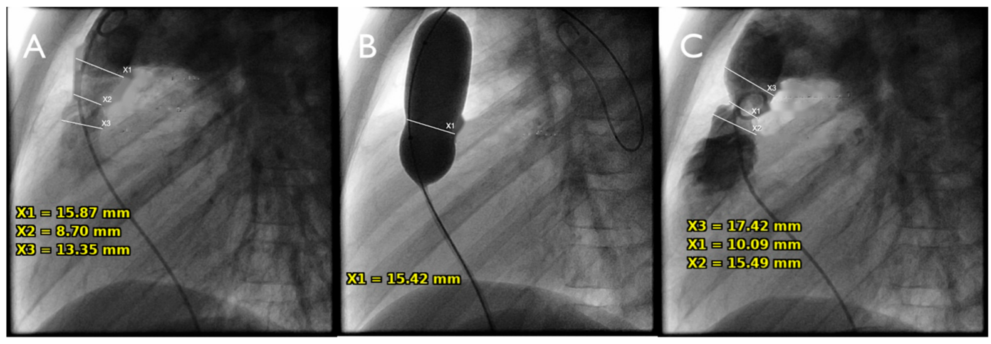
Figure 1: Balloon dilatation of a postoperative RVOT. (90° lateral view). (A) The stenosis measures 8.7 mm (gradient 50 mmHg), the RVOT before and the PA thereafter about 16 mm. (B) A 18 mm × 40 mm VACS® III balloon was inflated up to 7 atm, and a minimal waist of 15.6 mm was measured at the level of the valve annulus (stenosis/balloon ratio of 87%). (C) Recoil after multiple balloon inflations resulted in a minimal increase of the stenosis (new diameter 10 mm, gradient 40 mmHg). Balloon inflation was successful, but hemodynamically the result is insufficient due to recoil of the anatomic structure and not balloon failure.
3.3 Balloon Interrogation of the RVOT before Percutaneous Pulmonary Valve Implantation (PPVI) [16]
Twenty-five consecutive patients with dysfunctional right ventricular outflow tracts underwent balloon interrogation as part of the preprocedural assessment for potential PPVI. The mean age was 17.7 ± 7.1 years (range, 9.3–41.4 years), with a mean body weight of 62.5 ± 20.3 kg and a mean height of 159.2 ± 21.2 cm.
Technical device success was achieved in all procedures. VACS® II balloon catheters were successfully positioned within the RVOT and inflated to the intended diameter to delineate the landing zone and assess RVOT morphology (Fig. 2). In accordance with the procedural objective, complete balloon expansion to the rated burst pressure was neither required nor intended. Simultaneous selective coronary angiography was performed in all patients to exclude coronary artery compression, myocardial malperfusion, and aortic root distortion during simulated valve implantation.
Balloon diameters ranged from 22 to 30 mm (mean, 25.5 ± 2.1 mm). Adequate anatomical assessment for procedural planning was achieved in all patients without balloon rupture or other device-related complications. Based on the balloon interrogation findings, patients subsequently underwent either PPVI or alternative management according to the anatomical suitability for transcatheter valve implantation.
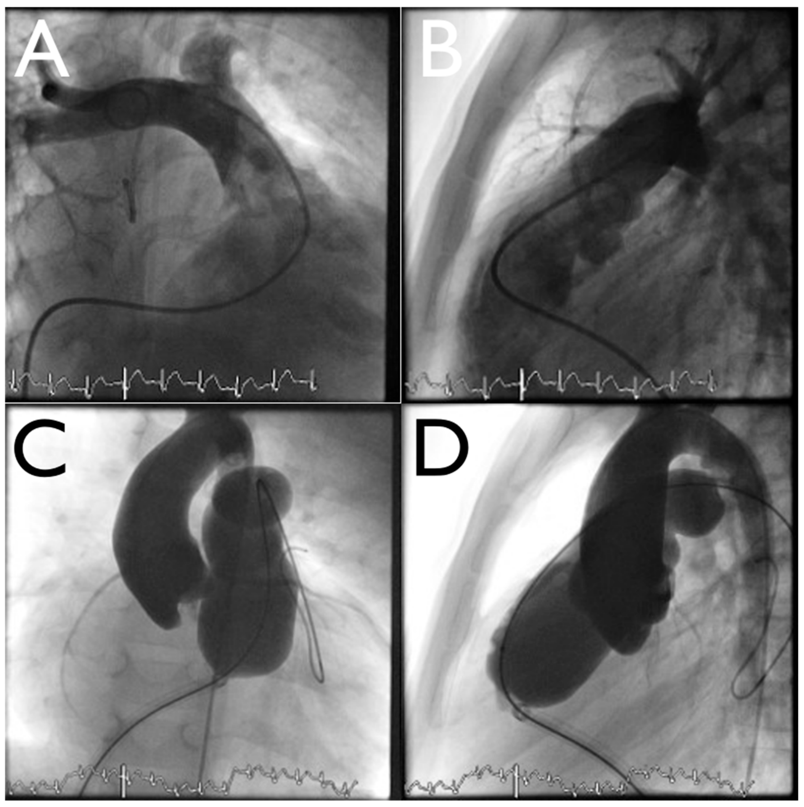
Figure 2: Balloon interrogation of the RVOT prior to PPVI. In (A) frontal view and (B) lateral view, the pulmonary artery is documented with contrast, and there is free regurgitation and no residual valve is documented. In (C) frontal view and (D) lateral view a 28 mm × 60 mm Osypka VACS®II balloon is inflated to measure the size of the pulmonary artery before stent implantation. No complete balloon expansion is required.
3.4 Peripheral Balloon Angioplasty
Twenty-five consecutive patients underwent balloon angioplasty for peripheral pulmonary artery stenosis, including eight patients with previous stent implantation. The mean age was 10.8 ± 7.4 years (range, 1–31 years), with a mean body weight of 39.2 ± 24.6 kg and a mean height of 133.0 ± 33.8 cm (Table 3). The minimum vessel diameter at the stenotic segment before intervention was 9.7 ± 3.2 mm.
Table 3: Details of the patients and procedures of the balloon dilatation of peripheral pulmonary stenosis.
| Mean | SD | Min | Max | |
|---|---|---|---|---|
| Patient details (n = 25) | ||||
| Age [years] | 10.80 | 7.37 | 1.00 | 31.00 |
| Length [cm] | 133.04 | 33.82 | 75.00 | 187.00 |
| Weight [kg] | 39.22 | 24.59 | 11.20 | 94.70 |
| Stenosis pre dilatation | ||||
| Diameter [mm] | 9.72 | 3.18 | 4.20 | 16.10 |
| Gradient [mmHg] | 18.48 | 8.68 | 4.00 | 40.00 |
| Area [mm2] | 82.12 | 52.94 | 58.20 | 204.13 |
| Stenosis post dilatation | ||||
| Diameter [mm] | 13.28 | 2.75 | 8.60 | 19.00 |
| Gradient [mmHg] | 9.32 | 4.35 | 4.00 | 18.00 |
| Area [mm2] | 144.60 | 59.48 | 58.20 | 284.30 |
| Changes | ||||
| Diameter [mm] | 3.56 | 1.73 | 0.60 | 7.30 |
| Gradient [mmHg] | 9.16 | 8.84 | 3.00 | 36.00 |
| Area [mm2] | 62.64 | 27.58 | 8.70 | 122.50 |
| Area [%] | 122.80 | 136.78 | 13.94 | 649.70 |
Technical device success was achieved in all procedures. Balloon positioning, inflation, deflation, and retrieval were successful in every patient without balloon rupture or other device-related complications. Balloon diameter was selected according to the reference diameter of the distal pulmonary artery, with balloon diameters ranging from 10 to 24 mm (mean, 17.4 ± 3.5 mm).
Balloon angioplasty increased the minimum vessel diameter by a mean of 3.6 mm, corresponding to a 122% increase in the calculated luminal cross-sectional area (p < 0.001). Despite successful balloon inflation, the immediate angiographic result was frequently limited by elastic recoil of the surrounding vessel wall, particularly in non-stented lesions (Fig. 3). Patients undergoing stent redilatation demonstrated the greatest increase in vessel diameter, with the mean minimum diameter increasing from 10.1 ± 4.2 mm to 14.1 ± 2.7 mm. Because of persistent residual stenosis after balloon angioplasty, six patients subsequently underwent stent implantation during follow-up.
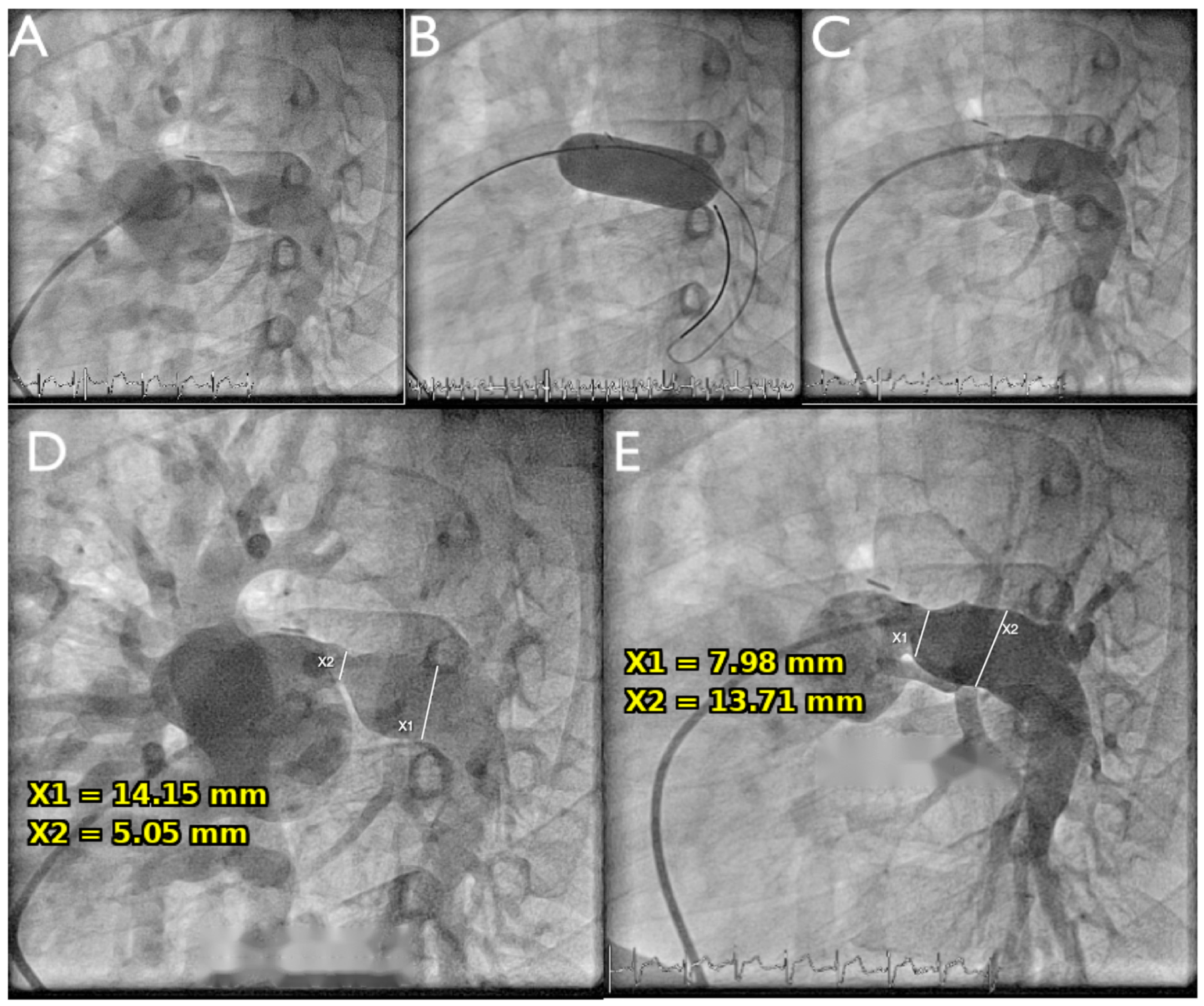
Figure 3: LPA dilatation. (A) There is a short tubular LPA stenosis (lateral view) of 5 mm, the LPA has 14 mm distally. (B) Complete balloon inflation at 14 mm with a VACS®II balloon at 2 atm (=300% balloon size). (C) Despite complete balloon inflation there is a significant residual stenosis caused by recoiling of the vessel wall. (D) Initial measurements, stenosis is 5/14 mm = 36%. (E) Increase to only 8/14 mm = 57%.
Twenty-five consecutive patients with isolated valvular aortic stenosis were included. Patients with dysplastic aortic valves or associated left ventricular outflow tract obstruction were excluded from the analysis. The mean age was 2.1 ± 4.0 years (range, 0.1–16 years), with a mean body weight of 11.2 ± 12.9 kg and a mean height of 74.3 ± 31.3 cm (Table 4).
Technical device success was achieved in all procedures. Balloon positioning, inflation, deflation, and retrieval were successful in every patient without balloon rupture or other device-related complications. The mean left ventricular systolic pressure decreased from 122.8 ± 33.9 mmHg before intervention to 93.0 ± 26.9 mmHg after balloon valvuloplasty, while the mean transvalvular gradient decreased from 53.1 ± 19.0 mmHg to 23.5 ± 12.3 mmHg (both p < 0.001).
In ten patients, sequential balloon dilatation using a balloon 1–2 mm larger was performed because of persistent residual gradients after the initial inflation. This resulted in additional hemodynamic improvement in selected patients (Table 4). Moderate aortic regurgitation was observed in seven patients after maximal balloon dilatation using the largest selected balloon diameter. This represents a recognized procedural complication of balloon aortic valvuloplasty rather than device malfunction.
Table 4: Details of the patients and procedures of aortic valve dilatation.
| Mean | SD | Min | Max | |
|---|---|---|---|---|
| Patient details (n = 25) | ||||
| Age [years] | 2.13 | 3.98 | 0.10 | 16.00 |
| Length [cm] | 74.32 | 31.26 | 47.00 | 168.00 |
| Weight [kg] | 11.19 | 12.92 | 2.60 | 62.00 |
| Stenosis pre dilatation—all procedures including two step procedures* | ||||
| Valve diameter [mm] | 12.73 | 3.83 | 7.20 | 22.50 |
| LVSP [mmHg] | 122.77 | 33.90 | 58.00 | 171.00 |
| Gradient [mmHg] | 53.09 | 18.98 | 23.00 | 100.00 |
| Balloon dilatation | ||||
| Balloon size [mm] | 11.91 | 3.65 | 6.00 | 22.00 |
| Balloon/Valve Ratio | 0.96 | 0.10 | 0.72 | 1.18 |
| Result | ||||
| LVSP [mmHg] | 93.03 | 26.92 | 50.00 | 156.00 |
| Gradient [mmHg] | 23.54 | 12.29 | 5.00 | 61.00 |
| Stenosis pre dilatation—all procedures excluding first balloon# | ||||
| Valve diameter [mm] | 12.73 | 3.83 | 7.20 | 22.50 |
| LVSP [mmHg] | 128.92 | 31.98 | 68.00 | 171.00 |
| Gradient [mmHg] | 58.76 | 17.40 | 29.00 | 100.00 |
| Balloon dilatation | ||||
| Balloon size [mm] | 12.20 | 3.44 | 7.00 | 22.00 |
| Balloon/Valve Ratio | 1.00 | 0.08 | 0.86 | 1.18 |
| Result | ||||
| LVSP [mmHg] | 87.28 | 20.93 | 50.00 | 120.00 |
| Gradient [mmHg] | 18.12 | 6.36 | 5.00 | 31.00 |
Unlike the other seven indication groups, which were each capped at a fixed sample of 25 procedures, the miscellaneous procedures group comprised all eligible cases identified over the 10-year study period, since only 24 procedures meeting the inclusion criteria for this heterogeneous category were performed during that time. These 24 consecutive patients underwent balloon-assisted interventions for miscellaneous congenital cardiac indications that were not included in the predefined treatment groups. These procedures comprised balloon atrial septostomy or atrial septal dilatation to facilitate atrial decompression (n = 10) [17], pulmonary vein angioplasty (n = 3), balloon dilatation or interrogation of superior or inferior vena cava stenoses before stent implantation (n = 4), balloon dilatation of valved conduits or bioprosthetic valves to reduce pressure gradients or assess suitability for subsequent stent implantation or percutaneous pulmonary valve implantation (n = 6), and balloon interrogation of a bioprosthetic mitral valve before transcatheter valve-in-valve implantation (n = 1).
Technical device success was achieved in all procedures. Balloon positioning, inflation according to the intended procedural objective, deflation, and retrieval were successful in every patient without balloon rupture or other device-related complications. Balloon diameters ranged from 6 to 26 mm (mean, 18 mm). The intended procedural objective—whether therapeutic balloon dilatation or anatomical interrogation—was achieved in all cases.
Twenty-five consecutive patients with native, recurrent, or stented CoA underwent balloon angioplasty. The cohort included 10 patients with previous stent implantation, 13 patients with recurrent coarctation following surgical repair, and 2 patients with native coarctation. The mean age was 7.5 ± 7.5 years (range, 0.3–24.5 years), with a mean body weight of 27.9 ± 24.9 kg and a mean height of 112.9 ± 46.3 cm (Table 5). The mean transcoarctation gradient before intervention was 37.0 ± 10.9 mmHg, and the mean minimum coarctation diameter was 8.6 ± 4.0 mm.
Technical device success was achieved in all procedures. Balloon positioning, inflation, deflation, and retrieval were successful in every patient without balloon rupture or other device-related complications. Balloon diameters ranged from 7 to 20 mm, corresponding to a mean balloon-to-stenosis ratio of 1.58 ± 0.30.
Balloon angioplasty resulted in a significant reduction in the mean transcoarctation gradient from 37.0 ± 10.9 mmHg to 16.9 ± 7.2 mmHg (p < 0.001). The minimum coarctation diameter increased from 8.6 ± 4.0 mm to 11.3 ± 4.5 mm, corresponding to an 88% increase in the calculated luminal cross-sectional area and an improvement in the coarctation index from 0.60 ± 0.10 to 0.80 ± 0.10 (p < 0.001) (Fig. 4).
Table 5: Details of the patients and procedures of the balloon dilatation of coarctation of the aorta.
| Mean | SD | Min | Max | |
|---|---|---|---|---|
| Patient details (n = 25) | ||||
| Age [years] | 7.54 | 7.50 | 0.30 | 24.50 |
| Length [cm] | 112.92 | 46.30 | 57.00 | 189.00 |
| Weight [kg] | 27.86 | 24.90 | 4.50 | 81.80 |
| Stenosis pre dilatation | ||||
| Diameter [mm] | 8.57 | 4.00 | 3.70 | 16.80 |
| Gradient [mmHg] | 37.00 | 10.90 | 21.00 | 65.00 |
| Area [mm2] | 69.60 | 61.10 | 10.70 | 221.60 |
| CoA-Index | 0.60 | 0.10 | 0.40 | 0.90 |
| Stenosis post dilatation | ||||
| Diameter [mm] | 11.28 | 4.50 | 5.20 | 18.70 |
| Gradient [mmHg] | 16.88 | 7.20 | 5.00 | 31.00 |
| Area [mm2] | 114.90 | 82.90 | 21.20 | 274.50 |
| CoA-Index | 0.80 | 0.10 | 0.60 | 1.10 |
| Changes | ||||
| Area [%] | 88.83 | 57.10 | 23.90 | 260.00 |
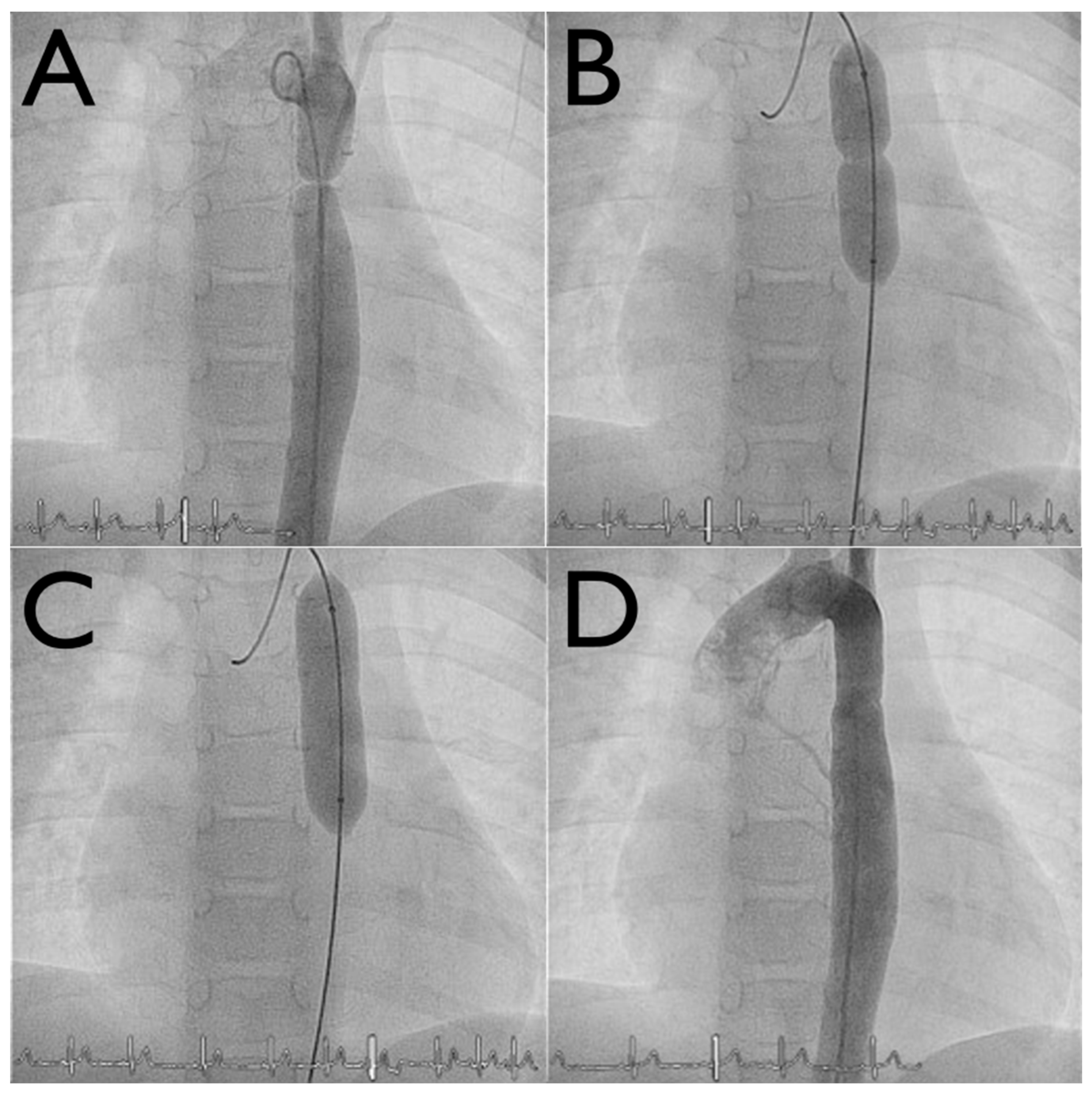
Figure 4: Balloon dilatation of a short membranous coarctation of the aorta (anterior view). (A) The short membrane is clearly detectable. (B) The VACS®II balloon is inflated at the site of the coarctation. The balloon diameter equals the diameter of the aorta. (C) Complete balloon inflation. (D) Result without any significant residual stenosis and no complication.
Twenty-five consecutive stent implantation procedures requiring VACS® III balloon catheters for stent delivery were analyzed. Stents were implanted in the right ventricular outflow tract (n = 14), the aorta for coarctation or aortic arch obstruction (n = 6), and the branch pulmonary arteries (n = 5) (Fig. 5). The dog-bone technique was used for all procedures with appropriately sized long delivery sheaths, as previously described [18].
Technical device success was achieved in all procedures. Balloon positioning, stent delivery, inflation, deflation, and retrieval were successful in every case without balloon rupture or other device-related complications (Table 6). A total of 26 stents were successfully implanted during 25 procedures using VACS® III balloon catheters, including 12 Cheatham-Platinum (CP®) stents, 13 AndraStents®, and one Bentley covered stent.
The mean diameter of the target lesion increased from 11.9 mm to 17.2 mm, representing a mean increase of 59%. The greatest relative increase in vessel diameter was observed in patients undergoing treatment for coarctation of the aorta (87.9%) and peripheral pulmonary artery stenosis (88.2%).
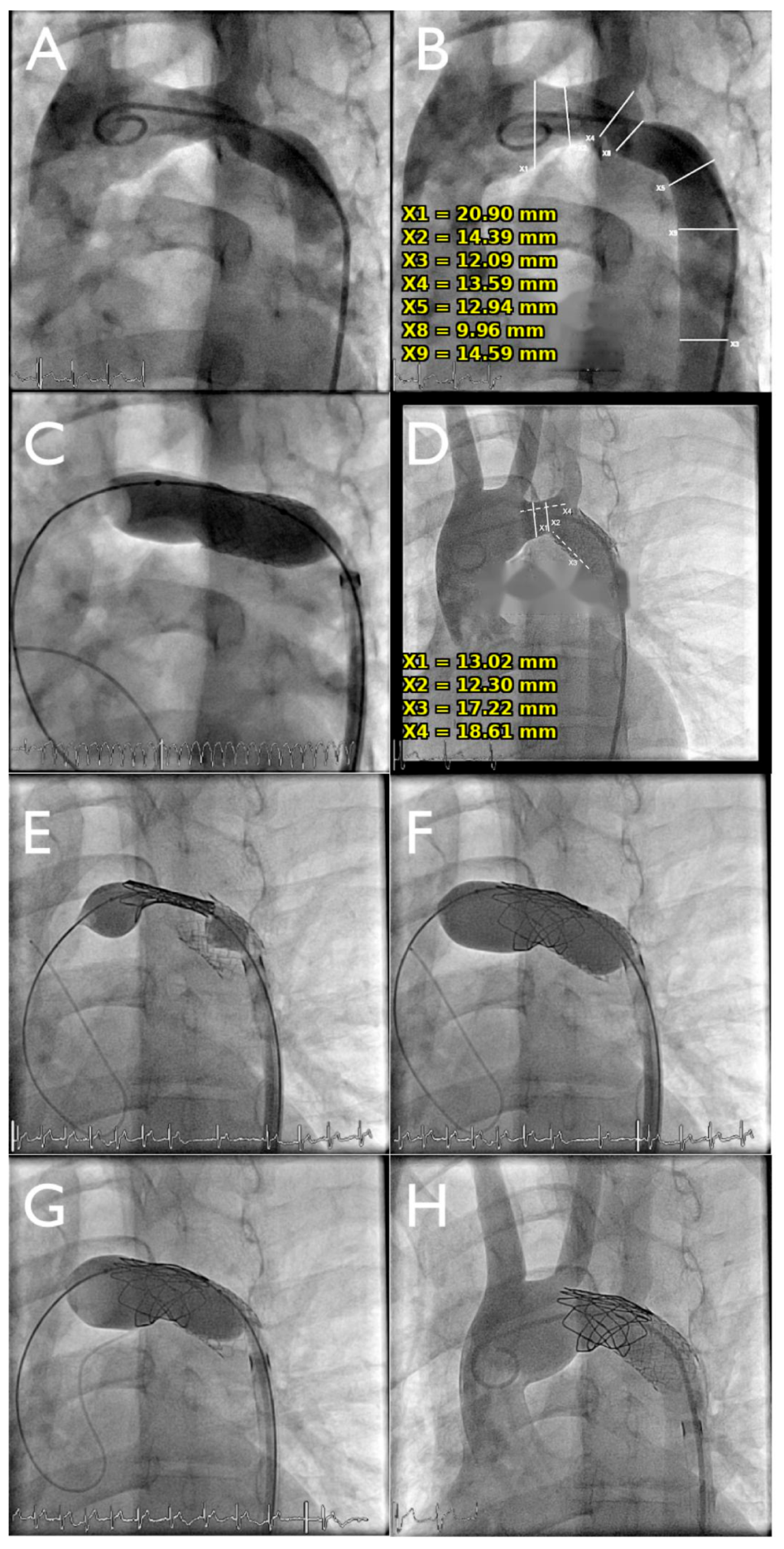
Figure 5: Various indications for balloon use in coarctation of the aorta (lateral view). (A) After surgical arch augmentation, there is multistage arch stenosis. (B) The smallest stenosis is 9 mm, the arch thereafter 14 mm. (C) Stent implantation with a 14 mm balloon, complete inflation is required and achieved. (D) Result with remaining transverse arch stenosis, minimal diameter 12 mm. (E) Stent implantation with a 16 mm oversized balloon to enable adequate aortic wall impression. (F) Incomplete balloon expansion is expected. (G) Expansion of the proximal end with an 18 mm balloon to optimize stent shaping at the entry to the transverse arch. Inadequate expansion inside the stent is required. (H) Final result without arch obstruction and excellent alignment to the enlarged proximal aorta.
Table 6: Characteristics of the patients with stent implantation.
| Mean | SD | Min | Max | |
|---|---|---|---|---|
| Patient details (n = 25) | ||||
| Age [years] | 12.38 | 6.70 | 2.60 | 31.50 |
| Stenosis pre dilatation—all patients | ||||
| Diameter [mm] | 11.85 | 5.30 | 3.70 | 21.00 |
| Balloon size [mm] | 18.56 | 5.00 | 10.00 | 26.00 |
| Stenosis post dilatation—all patients | ||||
| Diameter [mm] | 17.23 | 5.20 | 8.00 | 25.30 |
| Change [%] | 59.63 | 48.50 | 17.10 | 212.50 |
| Stenosis pre dilatation—Coarctation (n = 6) | ||||
| Diameter [mm] | 8.08 | 2.80 | 4.80 | 12.40 |
| Balloon size [mm] | 15.33 | 3.50 | 10.00 | 20.00 |
| Stenosis post dilatation—Coarctation | ||||
| Diameter [mm] | 14.27 | 3.20 | 9.50 | 18.40 |
| Change [%] | 87.91 | 62.10 | 48.40 | 212.50 |
| Stenosis pre dilatation—peripheral PS (n = 5) | ||||
| Diameter [mm] | 6.00 | 2.00 | 3.70 | 8.90 |
| Balloon size [mm] | 12.80 | 1.80 | 10.00 | 14.00 |
| Stenosis post dilatation—peripheral PS | ||||
| Diameter [mm] | 10.52 | 2.00 | 8.00 | 12.60 |
| Change [%] | 88.24 | 66.30 | 35.96 | 200.00 |
The present study evaluated the procedural performance and safety of the Osypka VACS® II and VACS® III balloon catheter systems across a broad spectrum of congenital cardiac interventions. Technical device success was achieved in all analyzed procedures, while device-related complications were rare and limited to two balloon ruptures during high-pressure dilatation of heavily calcified postoperative RVOT conduits. Overall, these findings demonstrate reliable device performance across therapeutic and diagnostic applications and highlight that clinical outcomes are determined primarily by lesion characteristics rather than by balloon catheter performance [19].
Pulmonary Valve Stenosis:
Balloon pulmonary valvuloplasty remains the treatment of choice for isolated congenital pulmonary valve stenosis. In our cohort, technical device success was achieved in all procedures without balloon-related complications, while substantial reductions in right ventricular pressure and transvalvular gradients confirmed effective procedural performance consistent with previous reports [20].
RVOT Stenosis:
Balloon dilatation of postoperative RVOT obstruction presents unique mechanical challenges because homografts and valved conduits frequently exhibit advanced fibrosis, calcification, and marked elastic recoil. In contrast to native valvular stenosis, the immediate procedural result is often limited by the intrinsic mechanical properties of the treated tissue rather than by the performance of the balloon catheter itself. Consequently, balloon dilatation is commonly performed as an initial therapeutic step before definitive stent implantation or as part of the assessment for percutaneous pulmonary valve implantation (PPVI) [8,13,14,15].
In the present study, technical device success was achieved in all RVOT interventions. Two balloon ruptures occurred during inflation to the rated burst pressure in severely calcified postoperative conduits. In both cases, the balloons were retrieved without difficulty, no vascular injury or other device-related complications occurred, and the procedures were successfully completed by subsequent stent implantation. These findings suggest that balloon rupture reflected the extreme mechanical resistance of heavily calcified conduits and occurred within the specified mechanical limits of the device, rather than representing unexpected device malfunction.
Consistent with previous reports, balloon dilatation alone frequently resulted in only modest anatomical improvement because of immediate elastic recoil of the surrounding tissue. Consequently, nine patients required additional stent implantation to achieve a more durable hemodynamic result. These findings emphasize that procedural success in postoperative RVOT interventions depends not only on reliable technical balloon performance but also on the biological and mechanical characteristics of the treated lesion.
In patients undergoing balloon interrogation before PPVI, the procedural objective differed fundamentally from therapeutic balloon dilatation. In these cases, successful balloon performance was defined by accurate delineation of the RVOT landing zone and exclusion of coronary artery compression rather than by anatomical enlargement or gradient reduction. Balloon interrogation provided sufficient anatomical information for procedural planning in all patients, enabling appropriate selection of candidates for transcatheter pulmonary valve implantation while identifying patients in whom alternative management was indicated.
Peripheral Pulmonary Artery Stenosis (PPS):
PPS represents one of the most challenging indications for balloon angioplasty because the immediate procedural result is frequently limited by the elastic properties of the vessel wall and the often low pre-interventional pressure gradient. Consequently, angiographic or hemodynamic improvement may underestimate the technical performance of the balloon catheter, particularly in native lesions without stent support [10].
In the present study, technical device success was achieved in all procedures, with successful balloon positioning, inflation, deflation, and retrieval and no balloon-related complications. Although the immediate increase in vessel diameter was modest in some patients, balloon angioplasty resulted in a substantial increase in the calculated luminal cross-sectional area. The limited anatomical gain observed in selected lesions was primarily attributable to immediate elastic recoil rather than inadequate balloon performance. In contrast, patients undergoing stent redilatation demonstrated the greatest increase in vessel diameter, reflecting the mechanical support provided by the implanted stent.
Six patients subsequently underwent stent implantation because of persistent residual stenosis during follow-up. These findings are consistent with previous reports demonstrating that the durability of balloon angioplasty in peripheral pulmonary artery stenosis is determined predominantly by lesion morphology and vascular remodeling rather than by the technical characteristics of the balloon catheter itself [21]. These findings demonstrate reliable technical device performance despite lesion-dependent variability in anatomical outcome.
Aortic Valve Stenosis (AS):
Balloon aortic valvuloplasty remains the treatment of choice for congenital valvular AS in infants, children, and adolescents, providing effective relief of left ventricular outflow tract obstruction while delaying or avoiding surgical intervention [10,11].
In the present study, technical device success was achieved in all procedures, with successful balloon positioning, inflation, deflation, and retrieval without balloon-related complications. Significant reductions in left ventricular systolic pressure and transvalvular gradients demonstrated reliable procedural performance of the VACS® balloon catheter system. A stepwise balloon-sizing strategy was employed, beginning with a conservative balloon-to-annulus ratio and increasing the balloon diameter only in patients with persistent residual gradients, consistent with current recommendations for congenital balloon aortic valvuloplasty [10,11].
Moderate aortic regurgitation developed in seven patients following maximal balloon dilatation. This represents a well-recognized procedural complication of balloon aortic valvuloplasty and reflects the inherent balance between maximizing gradient reduction and preserving valve integrity. The occurrence of post-procedural aortic regurgitation is primarily influenced by valve morphology, balloon-to-annulus ratio, and procedural strategy rather than by the technical performance of the balloon catheter itself. Despite this recognized complication, technical device success was achieved in all procedures. Despite these recognized procedural limitations, technical device performance was uniformly reliable.
Coarctation of the Aorta:
Coarctation angioplasty remains an important indication for balloon catheter intervention in congenital cardiology [12]. Although significant gradient reduction and vessel enlargement can generally be achieved, the immediate procedural outcome is influenced not only by successful balloon expansion but also by the anatomical characteristics and elastic properties of the coarctation segment.
In the present study, technical device success was achieved in all procedures, with successful balloon positioning, inflation, deflation, and retrieval without balloon-related complications. Balloon angioplasty resulted in significant reductions in transcoarctation gradients together with an increase in vessel diameter and coarctation index, demonstrating reliable procedural performance of the VACS® II and VACS® III balloon catheter systems.
The extent of anatomical improvement varied according to the underlying lesion morphology. Patients undergoing redilatation of previously implanted stents demonstrated the greatest increase in vessel diameter, reflecting the mechanical support provided by the existing stent and the reduced tendency for elastic recoil. In contrast, native and postoperative coarctation lesions remained more susceptible to vascular recoil despite technically successful balloon dilatation [12].
Overall, the present findings demonstrate reliable technical performance of the VACS® II and VACS® III balloon catheter systems across the full spectrum of coarctation interventions. While the balloon catheters consistently performed according to their intended procedural purpose, the final anatomical and hemodynamic outcome was predominantly influenced by the characteristics of the underlying lesion rather than by the technical performance of the device itself.
Stent Implantation:
Large-caliber balloon-expandable stents have become an integral component of transcatheter treatment for congenital heart disease and are routinely used for the management of CoA, branch pulmonary artery stenosis, and RVOT obstruction. Successful stent implantation depends not only on accurate positioning but also on reliable balloon performance during stent delivery, expansion, deflation, and retrieval [13,14,15].
In the present study, technical device success was achieved in all stent implantation procedures. VACS® III balloon catheters provided reliable stent delivery and expansion across a broad spectrum of congenital interventions without balloon rupture, inadequate inflation, stent displacement, or retrieval difficulties. These findings demonstrate the mechanical stability of the balloon catheter system even during large-caliber, high-pressure interventions.
Although the final procedural result was influenced by the underlying lesion and the implanted stent, successful deployment depended on predictable balloon performance throughout the procedure. The absence of balloon-related complications in this cohort supports the suitability of the VACS® III balloon catheter as a reliable platform for complex congenital stent implantation.
Miscellaneous Indications:
Beyond the predefined therapeutic indications, the VACS® II and VACS® III balloon catheters were successfully used in a variety of less common congenital cardiac interventions, including atrial septal dilatation, pulmonary and systemic venous angioplasty, valved conduit and bioprosthetic valve dilatation, and anatomical interrogation before subsequent transcatheter interventions. These procedures represent diverse clinical scenarios with distinct therapeutic objectives, further illustrating the versatility of the balloon catheter system.
Technical device success was achieved in all procedures, with successful balloon positioning, inflation according to the intended procedural objective, deflation, and retrieval without balloon-related complications. Importantly, procedural success in this heterogeneous cohort was defined according to the intended clinical application rather than solely by hemodynamic improvement. In several procedures, the primary objective was anatomical assessment or procedural planning rather than therapeutic dilatation.
These findings demonstrate that the clinical utility of the VACS® balloon catheter system extends beyond conventional balloon angioplasty and valvuloplasty, providing reliable performance across a wide spectrum of therapeutic and diagnostic applications encountered in congenital interventional cardiology.
Long-Term Outcomes and Study Limitations:
The present study was designed to evaluate the procedural performance and technical safety of the VACS® II and VACS® III balloon catheter systems across a broad spectrum of congenital cardiac interventions. Although long-term clinical outcomes are well established for many of the procedures included in this study, they are determined predominantly by the underlying congenital lesion, procedural strategy, associated surgical or transcatheter interventions, and long-term patient management rather than by the balloon catheter itself. Consequently, long-term follow-up was beyond the scope of the present analysis, which focused on immediate procedural performance and device-related safety.
Several limitations should be acknowledged. First, this was a retrospective, single-center study and is therefore subject to the inherent limitations of observational analyses. Second, the study was designed to evaluate the performance of technical devices across predefined clinical indications rather than to compare outcomes between interventions or treatment strategies. To obtain a balanced representation of the principal applications of the VACS® balloon catheter systems, 25 consecutive procedures were analyzed for each indication group (24 for the miscellaneous procedures group, reflecting the total number of eligible cases available for this heterogeneous category). Because individual patients could contribute to more than one procedural category, the indication groups were not mutually exclusive; cross-referencing patient identifiers across all eight groups identified 166 unique patients in the overall cohort, although the specific overlap structure between individual indication group pairs was not systematically tracked and should be considered when interpreting the reported group sizes. Additionally, reverse chronological sampling may introduce time-related bias, as more recent cases may reflect evolving techniques and doctor experience. Finally, because all procedures were performed by experienced doctors at a single high-volume center, the findings may not be generalizable to lower-volume centers or less experienced doctors. Finally, statistical analyses were performed within each indication group, and comparisons between groups were not intended.
Despite these limitations, the present study provides comprehensive real-world clinical performance data across a broad range of therapeutic and diagnostic congenital cardiac interventions. The consistent technical success and low rate of device-related complications observed across all indications support the safety, reliability, and versatility of the VACS® II and VACS® III balloon catheter systems in routine congenital interventional cardiology.
In this retrospective single-center study, the VACS® II and VACS® III balloon catheter systems demonstrated reliable procedural performance and a favorable safety profile across a broad spectrum of therapeutic and diagnostic congenital cardiac interventions. Technical device success was achieved in all analyzed procedures, with a low rate of device-related complications despite application in some of the most mechanically demanding lesions encountered in congenital interventional cardiology.
The present findings demonstrate that the clinical performance of balloon catheters should be evaluated according to their intended procedural application. While technical device performance was consistently reliable across all indications, the final anatomical and hemodynamic outcome was predominantly influenced by the characteristics of the underlying lesion and the therapeutic strategy rather than by the balloon catheter itself.
These real-world clinical performance data contribute to the Post-Market Clinical Follow-up (PMCF) evidence required under the European MDR and support the continued use of the VACS® II and VACS® III balloon catheter systems across a wide range of congenital cardiac interventions.
Acknowledgement:
Funding Statement: The authors received no specific funding for this study.
Author Contributions: Guido Mandilaras and Nikolaus A. Haas conceptualized and supervised the study; Nikolaus A. Haas critically revised the manuscript. All authors performed the interventional procedures. Anja Tengler, Andre Jakob, and Robert Dalla Pozza contributed to data acquisition and analysis. All authors reviewed and approved the final version of the manuscript.
Availability of Data and Materials: The datasets generated and/or analyzed during the current study are available from the corresponding author upon reasonable request.
Ethics Approval: This retrospective study was reviewed by the Ethics Committee of the Ludwig-Maximilians-Universität München (LMU Munich), which confirmed no objection to the project (Project No. 24-1128 kB, dated 23 December 2024). Because data collection and analysis were conducted exclusively on anonymized data, informed consent and a GDPR-compliant patient information document were not required.
Conflicts of Interest: The authors declare that they have no competing interests. In particular, the authors have no financial or personal relationships with the manufacturer of the VACS® II and VACS® III balloon catheters (Osypka AG, Rheinfelden, Germany).
References
1. Hoffman JIE , Kaplan S . The incidence of congenital heart disease. J Am Coll Cardiol. 2002; 39( 12): 1890– 900. doi:10.1016/S0735-1097(02)01886-7. [Google Scholar] [CrossRef]
2. Sutherell JS , Hirsch R , Beekman RH . Pediatric interventional cardiology in the United States is dependent on the off-label use of medical devices. Congenit Heart Dis. 2010; 5( 1): 2– 7. doi:10.1111/j.1747-0803.2009.00364.x. [Google Scholar] [CrossRef]
3. CELEX No. 32017R0745. Regulation (EU) 2017/745. Brussels, Belgium: EU; 2017. [Google Scholar]
4. Rao PS . Balloon pulmonary valvuloplasty: a systematic review. Indian Heart J. 2024; 76( 2): 86– 93. doi:10.1016/j.ihj.2023.12.007. [Google Scholar] [CrossRef]
5. VACS® II/VACS® III [Internet]. OSYPKA. [cited 2026 Aug 1]. Available from: https://osypka.de/produkte/valvuloplastie/valvuloplastie-ballonkatheter/. [Google Scholar]
6. Cuypers JA , Witsenburg M , van der Linde D , Roos-Hesselink JW . Pulmonary stenosis: update on diagnosis and therapeutic options. Heart. 2013; 99( 5): 339– 47. doi:10.1136/heartjnl-2012-301964. [Google Scholar] [CrossRef]
7. Jux C , Weil J , Oberhoffer R , Bennink G . Deutsche gesellschaft für pädiatrische kardiologie. [cited 2026 Apr 13]. Available from: https://www.dgpk.org/wp-content/uploads/LL_valvPstnachVSS.pdf. [Google Scholar]
8. Zeevi B , Keane JF , Perry SB , Lock JE . Balloon dilation of postoperative right ventricular outflow obstructions. J Am Coll Cardiol. 1989; 14( 2): 401– 8. doi:10.1016/0735-1097(89)90193-9. [Google Scholar] [CrossRef]
9. Bacha EA , Kreutzer J . Comprehensive management of branch pulmonary artery stenosis. J Interv Cardiol. 2001; 14( 3): 367– 75. doi:10.1111/j.1540-8183.2001.tb00346.x. [Google Scholar] [CrossRef]
10. Michel-Behnke I , Murin P , Sarikouch S . Deutsche gesellschaft für pädiatrische kardiologie und angeborene herzfehler e.V. S2k-Leitlinie. [cited 2026 Apr 13]. Available from: https://www.dgpk.org/wp-content/uploads/LL_vAS_2020_11_25_final.pdf. [Google Scholar]
11. Kallio M , Rahkonen O , Mattila I , Pihkala J . Congenital aortic stenosis: treatment outcomes in a nationwide survey. Scand Cardiovasc J. 2017; 51( 5): 277– 83. doi:10.1080/14017431.2017.1355069. [Google Scholar] [CrossRef]
12. Haas NA , Rickers C , Kozlik R , Cesnjevar R . Deutsche gesellschaft für pädiatrische kardiologie leitlinien. [cited 2026 Apr 13]. Available from: https://www.dgpk.org/wp-content/uploads/2018_06_20_LL__Aortenisthmusstenose.doc.pdf. [Google Scholar]
13. Stumper O , Ramchandani B , Noonan P , Mehta C , Bhole V , Reinhardt Z , et al. Stenting of the right ventricular outflow tract. Heart. 2013; 99( 21): 1603– 8. doi:10.1136/heartjnl-2013-304155. [Google Scholar] [CrossRef]
14. Bertram H , Eicken A , Rickers C , Rüffer A . Pulmonalklappeninsuffizienz und Pulmonalklappenersatz. [cited 2026 Apr 13]. Available from: https://www.dgpk.org/wp-content/uploads/Leitlinie_PI-PKE_final.pdf. [Google Scholar]
15. Haas NA , Vcasna R , Laser KT , Blanz U , Herrmann FE , Jakob A , et al. The standing of percutaneous pulmonary valve implantation compared to surgery in a non-preselected cohort with dysfunctional right ventricular outflow tract—reasons for failure and contraindications. J Cardiol. 2019; 74( 3): 217– 22. doi:10.1016/j.jjcc.2019.03.021. [Google Scholar] [CrossRef]
16. Zamorano JL , Bax J , Knuuti J , Lancellotti P , Pinto F , Popescu BA , et al. The ESC textbook of cardiovascular imaging. Oxford, UK: Oxford University Press; 2021. doi:10.1093/med/9780198849353.001.0001. [Google Scholar] [CrossRef]
17. Dalla Pozza R , Hermann M , Haas NA . Balloon-atrioseptostomy in small children using an embolectomy catheter: preliminary data. Case Rep Cardiol. 2023; 2023: 9920336. doi:10.1155/2023/9920336. [Google Scholar] [CrossRef]
18. Kantzis M , Lehner A , Laser KT , Racolta A , Vcasna R , Fischer M , et al. The “dog bone technique” a novel easy and safe catheter maneuver for aortic arch and coarctation stenting. J Interv Cardiol. 2017; 30( 1): 85– 92. doi:10.1111/joic.12358. [Google Scholar] [CrossRef]
19. Hasan BS , Barry OM , Ali F , Armstrong AK , Batlivala SP , Crystal MA , et al. Evaluating procedural performance: a composite outcome for aortic and pulmonary valvuloplasty in congenital cardiac catheterization. J Soc Cardiovasc Angiogr Interv. 2023; 2( 6 Pt A): 101119. doi:10.1016/j.jscai.2023.101119. [Google Scholar] [CrossRef]
20. Aggarwal N , Agarwal M , Lale P , Relan J , Rudrappa S , Kumar A , et al. Immediate and short-term outcome of balloon pulmonary valvuloplasty in neonatal critical pulmonary stenosis. Ann Pediatr Cardiol. 2024; 17( 6): 425– 30. doi:10.4103/apc.apc_162_24. [Google Scholar] [CrossRef]
21. Kondo M , Kurita Y , Fukushima Y , Shigemitsu Y , Hirai K , Kawamoto Y , et al. Outcomes of ultra-high-pressure balloon angioplasty for congenital heart disease in single-center experience. Heart Vessels. 2025; 40( 10): 953– 60. doi:10.1007/s00380-025-02547-1. [Google Scholar] [CrossRef]
Cite This Article
 Copyright © 2026 The Author(s). Published by Tech Science Press.
This work is licensed under a Creative Commons Attribution 4.0 International License , which permits unrestricted use, distribution, and reproduction in any medium, provided the original work is properly cited.
 Downloads
 Citation Tools
