
 Login
Login Register
 Submit a Paper
 Propose a Special lssue
 Open Access
ARTICLE
Early to Mid-Term Cardiac Magnetic Resonance Findings after Transcatheter Pulmonary Valve Replacement in Patients with Repaired Tetralogy of Fallot
Pediatric Cardiology, Ankara Bilkent City Hospital, Üniversiteler Neighborhood, 1604 th Street No. 9, Çankaya, Ankara, Türkiye
* Corresponding Author: Meryem Beyazal. Email:
Structural and Congenital Heart Disease 2026, 21(4), 4 https://doi.org/10.32604/schd.2026.080538
Received 11 February 2026; Accepted 01 July 2026; Issue published 30 September 2026
 View Full Text
 Download PDF
Abstract
Background: Cardiac magnetic resonance (CMR) imaging following transcatheter pulmonary valve replacement (TPVR) allows for the assessment of right ventricular (RV) volume. This study aimed to evaluate early- to mid-term findings after the procedure. Methods: A total of 21 patients with repaired tetralogy of Fallot who underwent TPVR were enrolled in the study. CMR data before and after the procedure were compared. The median interval between the procedure and the post-TPVR CMR was 9 months (range: 6–18 months). Results: RV volumes and RV end-diastolic volume indices were significantly lower after TPVR compared to before the procedure. RV volume parameters returned to normal limits in only one-third of the patients. There were no significant differences in left ventricular volume parameters or in RV ejection fraction between the pre- and post-TPVR imaging. Additionally, the distal diameters of the left and right pulmonary arteries were significantly greater after TPVR than before. A moderate positive correlation was found between the time elapsed from TPVR to CMR imaging and RV volumes. Furthermore, there was a moderate positive correlation between age and aortic root diameter. Conclusions: Our results suggest that although RV volumes decreased—with normalization in one-third of the cases—there was no significant improvement in RV function. As RV volumes tend to increase again over time, regular follow-up with CMR imaging is recommended.Keywords
Cardiac magnetic resonance (CMR) imaging is recommended in patients with repaired tetralogy of Fallot (TOF) if there is right ventricular (RV) dilatation before the age of 8–10 years, and as a routine study for patients above this age group [1]. The decision regarding the timing of pulmonary valve replacement (PVR) is made according to the American Heart Association (AHA) adult congenital heart disease (ACHD) guidelines and The Journal of the American College of Cardiology (JACC) publications based on CMR image measurements [2,3]. Moreover, CMR imaging is a good choice to quantitatively assess the right ventricular volumes and function after PVR. The RV end-diastolic volume typically normalizes in the early period after PVR, while improvements in ventricular function occur later in the recovery process [4].
PVR can be performed by surgical or transcatheter methods in patients with repaired TOF. Surgical PVR was reported first in the 1960s by Lillehei et al. [5], and since 2000, transcatheter PVR (TPVR) has been developed following the pioneering work of Philipp Bonhoeffer and Younes Boudjemline [6]. CMR studies following TPVR are limited because it is a relatively newer technique compared to surgery. Also, performing CMR after the procedure has some difficulties, such as magnetic resonance imaging (MRI) safety considerations and metal artifacts. Since patient safety is our priority, prior to conducting CMR imaging after TPVR, confirmation must be obtained from the manufacturer that the materials used during the stenting procedure are MRI compatible. Additionally, metal artifacts can reduce image quality, which may prevent accurate volumetric measurements. To address this, some sequence changes can be made if necessary (such as using a gradient echo sequence instead of a balanced steady-state free precession) [7].
There are many purposes for CMR imaging in congenital heart disease. These are primarily volumetric studies, angiographic studies (three-dimensional (3D) whole-heart sequences) to measure vascular diameters, and flow studies. In some patients, tissue characterisation (late gadolinium enhancement and mapping) can also be performed if necessary. Non-contrast 3D whole-heart sequences can prevent unnecessary contrast exposure. Flow studies also reveal the distribution of flow to the pulmonary branch arteries and the shunt fraction Qp/QS ratio (for the assessment of significant residual shunts) [8,9].
Monitoring and course of associated cardiovascular anomalies, such as supravalvar pulmonary stenosis, pulmonary artery branch stenosis, and hypoplastic pulmonary artery branch stenosis, which are seen in approximately 40% of patients with TOF, are important as they may influence the clinical course [10]. Also, approximately one-third of patients undergoing TOF repair develop aortic root dilation [11]. Therefore, patients should be carefully monitored with imaging methods for pulmonary and aortic root measurement and their course.
We aimed to compare CMR volumetric, flow, and angiographic findings before and after TPVR in the early to mid-term period and to investigate potential associations between CMR findings and time elapsed in this study.
During the study period, 28 patients with repaired TOF who underwent TPVR were followed. Inclusion criteria included having undergone TPVR following TOF repair and a minimum follow-up period of 6 months after the procedure. Four patients with a follow-up period of less than six months, one patient with incomplete medical records, and two patients who were lost to follow-up were excluded. Consequently, the study population consisted of 21 patients.
Transcatheter PVR procedure was performed in patients who met the above-mentioned criteria [2,3]. The Myval transcatheter valve (Meril Life Sciences, India) was used, with the stent size selected based on measurements obtained from computed tomography (CT) and angiography at the time of the procedure. Pre-stenting was performed in all patients. All patients underwent a CMR study within one month prior to TPVR, and these studies were obtained through electronic records. The CMR studies following the procedure (within a 6- to 18-month timeframe) were performed in our institution.
This study was reviewed and approved by the Ethics Committee of Ankara Bilkent City Hospital (approval no: TABED 2-24-54), while informed consent was obtained from patients or their guardians. The authors assert that all procedures contributing to this work comply with the ethical standards of the relevant national guidelines on human experimentation and with the Helsinki Declaration of 1975, as revised in 2024.
The patients’ data were obtained from electronic medical records. Clinical and laboratory data included the patients’ age at three important points: TOF repair, TPVR, and the second CMR, gender, body surface area (BSA), blood pressure (BP), oxygen saturation (SpO2), and heart rate (HR) at the time of CMR. In addition, symptoms, medication use, current New York Heart Association (NYHA) class, presence, and type of arrhythmia were noted to give an idea about each patient’s condition before the time of TPVR.
2.3 Cardiac Magnetic Resonance
All CMR studies were performed using a 1.5-Tesla system (GE Healthcare, Chicago, IL, USA). All measurements were done by the same reader. Data were acquired during breath-hold. Balanced Steady-state free precession (bSSFP) sequence parameters were as follows: repetition time (TR) 2.9–3.0 ms, echo time (TE) 1.5 ms, flip angle 60°, slice thickness 6 mm, in-plane image resolution 1.3 mm × 1.3 mm × 6.0 mm, temporal resolution 25–30 phases. Short-axis cine images were used for volumetric analysis. The endocardial borders of both ventricles were traced manually at end-systole and end-diastole, including the papillary muscles, in the left ventricular mass. Then, the end-diastolic volume (EDV) and end-systolic volume (ESV), stroke volume (SV), and ejection fractions (EF) were calculated by the dedicated software CardiacVX (GE Healthcare, Chicago, IL, USA). In addition, the volume ratio was calculated by dividing RV EDVi by left ventricular (LV) EDVi. All volumetric parameters were indexed to body surface area (BSA). The upper limits for RVEDVi and RVESVi were determined as 123 mL/m2 and 59 mL/m2, respectively, in male patients and 104 mL/m2 and 48 mL/m2, respectively, in females [12].
Two-dimensional (2D) through-plane phase-contrast flow measurements were obtained from the ascending aorta (Ao), main pulmonary artery (MPA), and right and left pulmonary arteries (RPA and LPA). Flow measurements were performed perpendicular to each targeted vessel using the double oblique technique. In all cases, encoding velocity was adjusted to avoid aliasing. If aliasing was noted, the velocity was progressively raised in 30-cm/s steps until aliasing disappeared. Patients were encouraged to hold their breath during the whole acquisition. Aortic and pulmonary regurgitation fractions (AoRF and PRF) and the Qp/Qs (MPA/Ao) flow ratio were obtained through 2D-phase contrast images.
Respiratory navigator-gated, Electrocardiogram (ECG)-triggered, 3D whole-heart balanced Steady-state free precession (SSFP) MRI was acquired during free breathing. Imaging parameters that were used for the 3D whole-heart imaging were as follows: TR, 4.9 ms; TE, minimum; flip angle, 65; auto preparation time, 57; temporal resolution, 190 ms; the number of signal averages, 1; voxel size, 1 × 1.7 × 1.8 mm; navigator window, 3–5 mm; 122 slices; slice thickness 1–1.4 mm. The field of view was adjusted individually according to patient size. Trigger delay was selected as ‘recommended’, which means diastole. The number of R-R intervals (the time elapsed between two consecutive R-wave peaks of the QRS complexes) was adapted based on the individual heart rate between 2 and 4. Measurements of the great vessels, such as Ao, MPA, RPA, and LPA, were made in two dimensions using 3D whole-heart sequence images. The LPA and RPA proximal diameters were measured just after the branch, and the LPA distal diameter was measured at the narrowest point. Two separate measurements were taken from oblique axial and oblique coronal sections, and the widest one was used.
All data were analysed by using the SPSS (Statistical Package for Social Sciences) for Windows 25.0 program (IBM Corp., Armonk, NY, USA). After descriptive statistics and normality analysis by the Shapiro-Wilk test, normally distributed continuous variables were reported as mean ± standard deviation, non-normally distributed continuous variables were reported as median with range, and categorical variables were reported as count with percentage of total. The Paired Test or the Wilcoxon test was applied to compare the numerical data of two dependent groups. Correlation analysis was used to determine the relationship between two numerical variables. Multivariable linear regression analysis was performed to identify independent predictors of the outcome variable. A p < 0.05 was considered statistically significant.
Among 25 patients who underwent TPVR, a total of four patients were excluded: two of them due to follow-up periods of less than six months, one because of incomplete data, and one due to low image quality. The remaining 21 patients were included in the study. Of the patients, 11 (52.4%) were female, and the median age was 20 (14–54) years. Table 1 shows the patients’ demographic characteristics. Before TPVR, 13 (61.9%) of the patients had at least one of the following symptoms: palpitations, dyspnea, or chest pain. Most of the patients were in NYHA classes 2 and 3, while only eight (38%) were in NYHA class 1. Four (19%) patients had RVOT obstruction before TPVR. Ventricular extrasystoles were observed in four (21%) patients, and supraventricular extrasystoles in one (5%) patient. One patient underwent ablation due to ventricular tachycardia. Seven (33%) patients were using either angiotensin-converting enzyme inhibitors or beta-blockers. No patient had a significant residual ventricular septal defect (VSD) or pulmonary artery branch stenosis.
Table 1: Patients’ demographic characteristics.
| Data* | n = 21 (%) |
|---|---|
| Gender (months) | Female 11 (52.4%) |
| Male 10 (47.6%) | |
| Age (years) | 20 (14–54) |
| Age at TOF repair (months) | 16 (2–101) |
| Age at TPVR | 19 (12–53) |
| Interval from TOF repair to PVR (years) | 15 (8–33) |
| Age at the first CMR (years) | 16.5 (13–52) |
| Age at the second CMR (years) | 18 (13–53) |
| Interval from TPVR to CMR (years) | |
| Ethnicity | |
| Turkish | 19 (90.5%) |
| Syrian | 2 (9.5%) |
The anthropometric measurements and clinical findings of the patients were not different at both imaging time points (pre-and post TPVR). In our patient group, pulse repetition frequency (PRF) levels decreased significantly after TPVR (p = 0.005), and no significant residual gradient was detected in any of the patients.
According to the CMR study, RVEDVi, RVESVi, and RVEDVi/LVEDVi ratios were lower at the post-TPVR measurement than pre-TPVR, with p values of 0.015, 0.008, and 0.026, respectively. More specifically, the post-TPVR measurements revealed that RVEDVi in seven patients (33%) and RVESVi in six patients (29%) were found to regress to normal limits. A positive correlation was observed in the analysis of pre-TPVR and post-TPVR RVEDVi measurements (r = 0.560, p = 0.030). Those with lower post-TPVR RVEDVi had lower pre-procedure RVEDVi values. In the comparison between those who underwent complete TOF repair before and after reaching the age of one, there was no significant difference found in RV volume parameters. In addition, PRF was significantly lower in the post-TPVR measurement than in the pre-TPVR measurement (p = 0.005). Moreover, RPA and LPA distal diameters were significantly greater at post-TPVR compared to baseline, with p values of 0.011 and 0.018, respectively. However, no significant difference was found in LV volumetric parameters, RV ejection fraction (RVEF), and RV stroke volume index (RVSVi) between the pre- and post-TPVR imaging. Table 2 shows clinical data and CMR findings at pre- and post-transcatheter valve replacement.
Table 2: Clinical data and CMR findings at pre- and post-transcatheter valve replacement.
| Data* | Pre-TPVR | Post-TPVR | p Value |
|---|---|---|---|
| Height (cm) | 161 ± 15.6 | 159.6 ± 14.1 | 0.876 |
| Weight (kg) | 60.2 ± 15.6 | 62.31 ± 17.0 | 0.987 |
| BSA (m2) | 1.56 ± 0.21 | 1.60 ± 0.25 | 0.954 |
| HR (bpm) | 70.3 ± 11.0 | 68.9 ± 12.3 | 0.798 |
| BP | |||
| Systolic (mmHg) | 112 ± 11.5 | 110.1 ± 12.3 | 0.896 |
| Diastolic (mmHg) | 59 ± 9.6 | 63.5 ± 8.7 | 0.695 |
| SpO2 (%) | 95.6 ± 2.7 | 97.5 ± 1.2 | 0.322 |
| LV volumetry | |||
| LVEF (%) | 58.2 ± 7.8 | 61.8 ± 9.2 | 0.193 |
| LVEDVi (mL/m2) | 111.9 ± 35.9 | 108.1 ± 26.1 | 0.639 |
| LVESVi (mL/m2) | 48.5 ± 23.4 | 39.6 ± 18.4 | 0.374 |
| LVSVi (mL/m2) | 63.3 ± 14.9 | 65.5 ± 15.9 | 0.799 |
| COI (L/min/m2) | 4.8 ± 0.9 | 4.7 ± 1.5 | 0.646 |
| RV volumetry | |||
| RVEF (%) | 47.4 ± 11.3 | 51.9 ± 12.6 | 0.330 |
| RVEDVi (mL/m2) | 192.7 ± 74.5 | 148.4 ± 41.6 | 0.015 |
| RVESVi (mL/m2) | 114.2 ± 50.5 | 73.4 ± 33.9 | 0.008 |
| RVSVi (mL/m2) | 97.3 ± 27.8 | 77.9 ± 23.6 | 0.053 |
| RVEDVi/LVEDVi | 2.19 ± 0.79 | 1.46 ± 0.49 | 0.026 |
| Flow study | |||
| Qp/Qs | 0.8 ± 0.1 | 1.1 ± 0.2 | 0.180 |
| AoRF (%) | 4.0 ± 2.8 | 7.0 ± 4.2 | 0.180 |
| PRF (%) | 40.1 ± 12.6 | 7.7 ± 7.3 | 0.005 |
| Diameters (mm) | |||
| MPA | 24.0 ± 5.7 | 24.9 ± 1.7 | 0.720 |
| RPA | 14.4 ± 4.9 | 18.6 ± 5.6 | 0.011 |
| LPA proximal (at the narrowest) | 8.8 ± 2.1 | 11.5 ± 4.9 | 0.285 |
| LPA distal | 16.0 ± 6.3 | 19.3 ± 5.7 | 0.018 |
| RPA/LPA ratio | 0.8 ± 0.3 | 1.1 ± 0.2 | 0.069 |
Correlation analysis of post-TPVR CMR measurements showed a moderate positive correlation between time elapsed from TPVR to CMR imaging and RVEDVi and RVESVi, as shown in Fig. 1 and Fig. 2 (r = 0.516, p = 0.024, r = 0.520, p = 0.022, respectively).
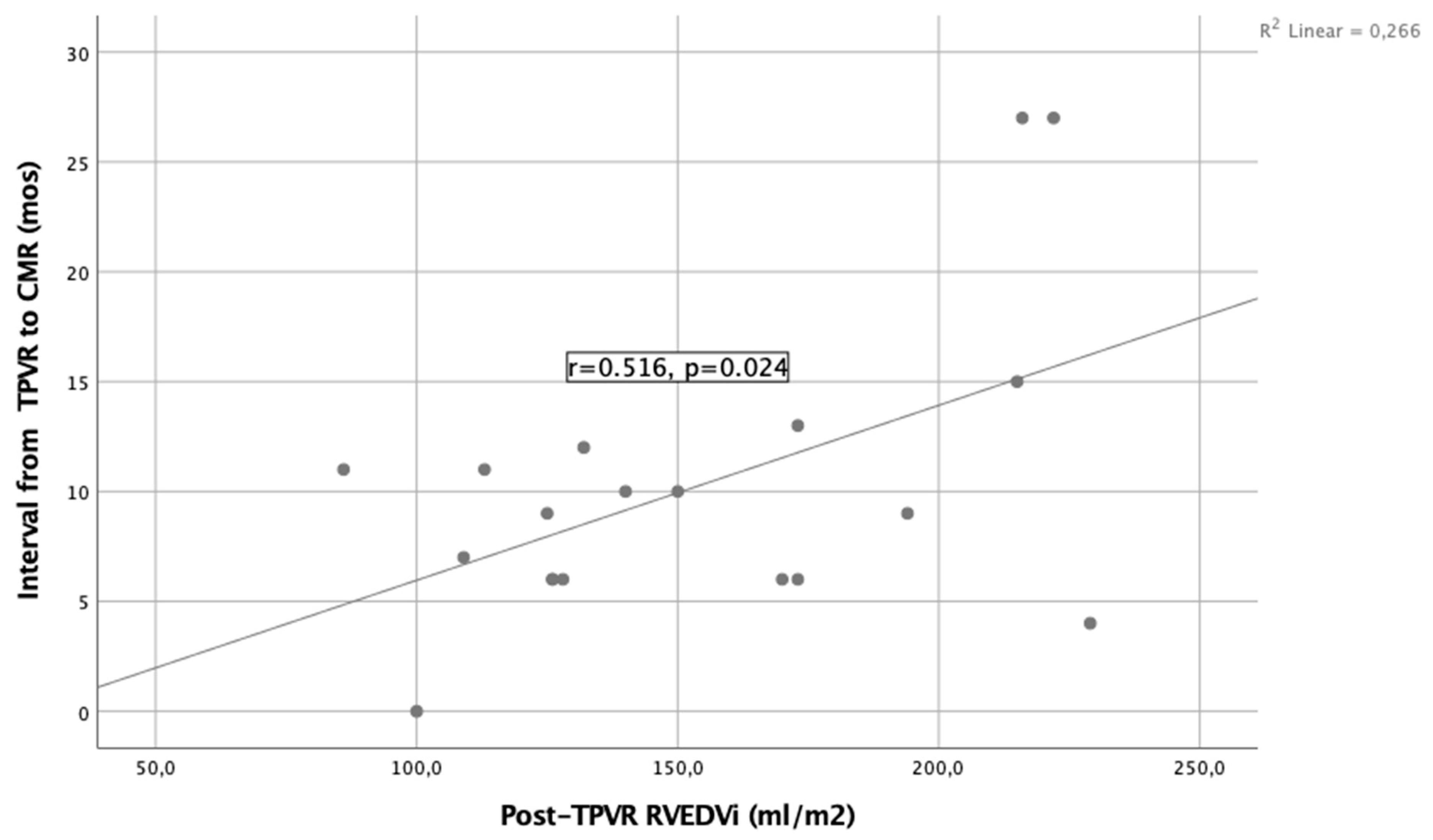
Figure 1: A moderate positive correlation between time elapsed from TPVR to CMR and RVEDVi (TPVR = transcatheter pulmonary valve replacement; CMR = cardiac magnetic resonance; RVEDVi = right ventricular end-diastolic volume index).
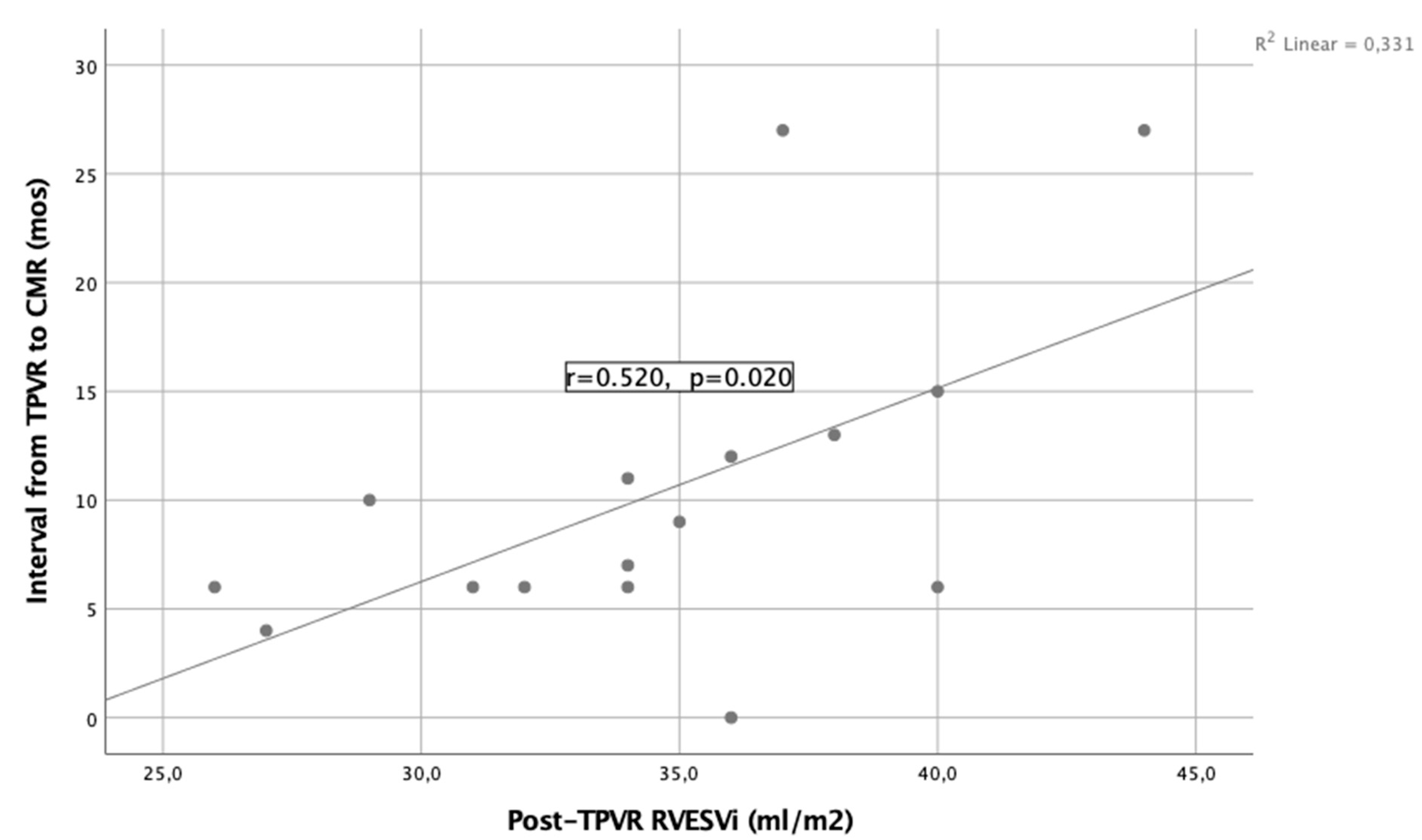
Figure 2: A moderate positive correlation between time elapsed from TPVR to CMR imaging and RVESVi (TPVR = transcatheter pulmonary valve replacement; CMR = cardiac magnetic resonance; RVESVi = right ventricular end-systolic volume index).
In addition, there was a moderate positive correlation between age and aortic sinus diameter (Fig. 3, r = 0.576, p = 0.020), whereas no correlation was observed between aortic sinus diameter and blood pressure, Qp/Qs, and BNP level. Furthermore, in the comparison between those who underwent complete TOF repair before (n = 9) and after reaching the age of one (n = 12), it was found that aortic sinus diameters were significantly smaller in the former group (p = 0.038). The median values were 31.5 (26–36) and 36.5 (27–44), respectively. However, although patients repaired after one year of age had larger aortic sinus diameters in the univariate comparison, multivariable linear regression analysis did not demonstrate an independent association between aortic sinus diameter and either age at repair (β = 0.079, p = 0.788) or current age (β = 0.393, p = 0.198). Fig. 4 shows an image of a patient with TPVR in axial and sagittal views.
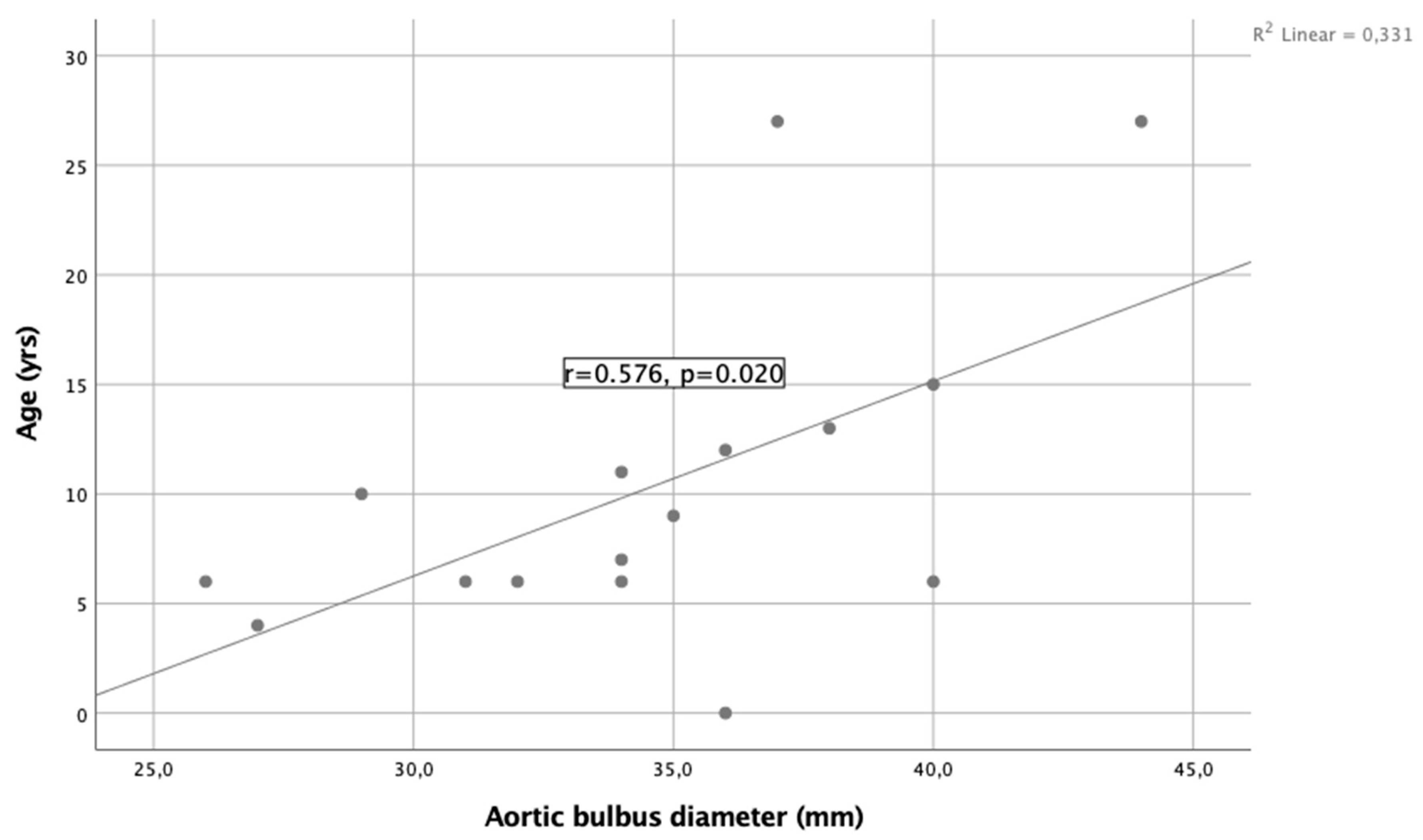
Figure 3: A significant moderate positive correlation between age and aortic bulbus diameter.
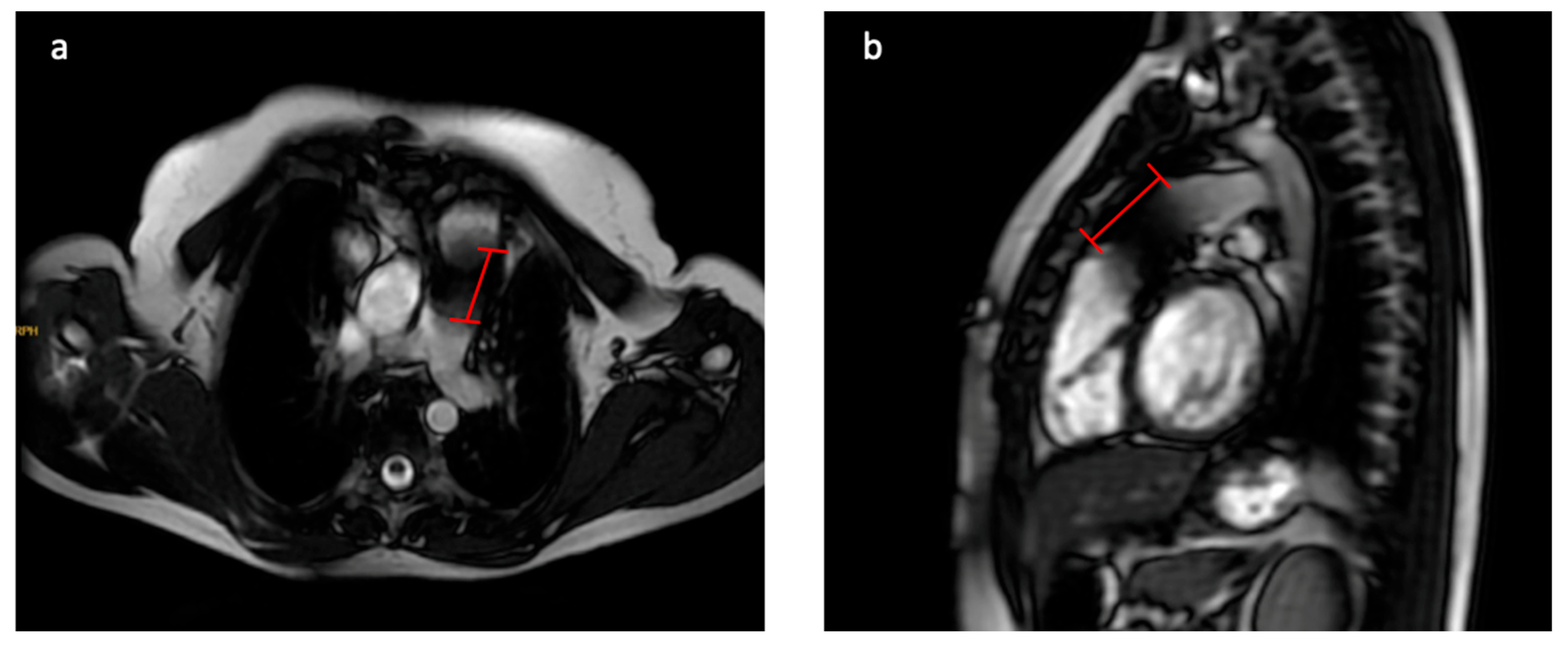
Figure 4: An image of a patient with TPVR in axial and sagittal views. (a,b) show axial and sagittal SSFP images, respectively, in a patient after TPVR (red lines indicate the stent).
According to this hypothesis-generating study, a significant decrease in RVEDVi and RVESVi indices was observed in the first year after TPVR in patients, while no change was detected in RVEF and RVSVi. The RPA and distal LPA diameter significantly increased after TPVR. A positive correlation was observed between the time from TPVR to CMR and RV volume parameters, as well as age and aortic sinus diameter. However, given the retrospective single-center design and the relatively small sample size (n = 21), these findings should be interpreted with caution due to potential selection bias and limited statistical power.
Similar to our results, RV volumes have been reported to decrease significantly in the first year after PVR, regardless of surgical or transcatheter intervention [4,13]. However, in the absence of a surgical PVR (SPVR) control group in our study, it is not possible to directly compare the magnitude or clinical significance of these changes between TPVR and SPVR. The reported rate of these volumes returning to normal limits is lower. In our study, the median age at TPVR was 19 years, and the median interval from TOF repair to TPVR was 15 years. We performed CMR scans between six and 18 months after the procedure. Accordingly, we observed that RV volumes normalized in approximately one-third of the patients. Kim et al. reported that their patients had a median age of 23.4 years at PVR, the median time elapsed from TOF repair to PVR was 20.5 years, and the rate of RV volume normalization according to CMR performed five years after the procedure was 28% [14]. According to another study, RV volume increase progresses with age in repaired TOF patients, and the possibility of RV dilatation returning to normal after PVR is lower in patients with high RV volume [15]. We also observed a significant positive correlation between RVEDVi values before and after TPVR, supporting this study. This raises the question of whether the current volume criteria should be lowered. On the other hand, continued RV dilatation after PVR has not been consistently associated with clinical conditions and mortality [16]. Therefore, the question arises whether different, more relevant threshold values for RV volume can be determined for patients with repaired TOF.
A study reported that in patients with surgical PVR, early reduction in RV size showed a gradual return to preoperative values 7 to 10 years after PVR [17]. In the current study, although we did not see any significant valvular lesions, a positive correlation was found between the time from TPVR to CMR and RV volume parameters, indicating that RV volume tends to increase over time, as in surgical PVR patients. Why this re-dilatation occurs remains unclear. We believe that longer-term CMR volume studies are needed in post-PVR patients to determine which patients develop re-dilatation and to identify predictive factors. These observations should be considered exploratory and require confirmation in larger, prospective cohorts.
In a retrospective study, improvement in RVEF in the mid-term period (at the median age of 4.2 years) was observed in 42 patients regardless of whether they underwent surgical (n = 24) or transcatheter PVR (n = 18), with a median time interval between TOF repair and PVR of 16.4 years [4]. In another study comparing surgical and transcatheter groups, no significant increase in RVEF and RVSV was observed in TPVR patients in CMR taken at the latest 1.5 years after the procedure, while there was a significant increase in RVEF in the surgical group [18]. As reported in these studies, ventricular remodeling patterns and functional recovery, including changes in RVEF, may differ between TPVR and SPVR depending on the intervention type and follow-up duration. Similar to these two studies, we also performed CMR at a median of 9 months (6–18 months) post-TPVR. Although there was an increase in the RVEF after TPVR; however this change was not significant. Therefore, our theory is that a significant increase in RVEF is not likely to be seen in the early to mid-term period in patients who undergo TPVR, although this study is not adequately powered to definitively assess this outcome. Analysis of mid- to late-term CMR results in a larger patient population would be useful to understand the change in RVEF over time. In addition, it should be emphasized that, due to the lack of a direct SPVR comparator group, this observation cannot be interpreted as evidence of a differential effect between TPVR and SPVR.
As is often reported, RV outflow tract stenting of symptomatic TOF with small pulmonary arteries allows pulmonary arterial growth [19]. Despite the differing patient population and clinical scenario, an increase in distal LPA and RPA measurements after TPVR may likely be seen due to pre-stenting through the TPVR procedure. Although there was an increase in the proximal part of LPA after TPVR, this was not statistically significant. The difference in growth between the proximal and distal LPA after TPVR that we found in our study is remarkable. This result may be due to the surgical procedure performed with a patch to relieve LPA stenosis, which is frequently seen in TOF patients. Nevertheless, these findings should be interpreted cautiously, given the limited sample size. In addition, without a surgical comparator, it remains unclear whether these vascular changes are specific to TPVR or reflect a general response to pulmonary valve replacement.
Aortic dilatation is common in patients with repaired TOF and increases with age. Older age, larger aortic size at the time of repair, and a history of an aorto-pulmonary shunt have been identified as contributing factors to increased aortic diameter [20]. Aortic dilatation is also a well-recognized phenotype in TOF patients [21]. In our study, aortic sinus diameter increased with age in the absence of a significant shunt (mean Qp: Qs 1.1), consistent with previous reports. We were unable to compare pre- and post-TPVR aortic dimensions due to the lack of pre-procedural CMR data. Although congenital aortic pathology may contribute to aortic root dilatation, late complete repair has also been identified as an important risk factor [22], supporting our finding of larger aortic sinus diameters in patients undergoing repair after one year of age in univariate analysis. However, in multivariable linear regression analysis, neither age at repair nor current age remained independently associated with aortic sinus diameter, suggesting that this relationship is likely influenced by other hemodynamic and anatomical factors. Regular imaging follow-up may be useful to monitor progressive aortic root dilatation in TOF patients, particularly in those undergoing late repair.
This is a single-centre study; the study population is limited. The power analysis result with an effect size of 0.5 and a total sample size of 21 was found to be 70%. Since most patients were referred from regional hospitals, some parameters, like the aorta measurement, were missing from pre-TPVR CMR data. In this study, only early- to mid-term post-TPVR single measurement data are discussed. Long-term repeated measurements would be useful to understand how changes occur over time, especially in terms of RV volume parameters, RVEF, and aortic measurements. Because of a lack of a surgical PVR comparative group, it is unclear whether findings are specific to TPVR or reflect broader post-PVR patterns. Lastly, since most of the patients were referred to our clinic from different regional hospitals, there was no surgeon/center consistency, and data on operative details such as previous shunt, valve-sparing approach or patch, and early postoperative outcomes were missing. In particular, the absence of detailed surgical data from the initial TOF repair (e.g., use of a transannular patch) represents a major limitation, as these factors are known to significantly influence long-term right ventricular remodeling. Furthermore, the heterogeneity in patient age, the interval between TOF repair and TPVR, and the relatively wide follow-up window (6–18 months) may have affected RV remodeling potential and functional outcomes. Due to the limited sample size, we were unable to perform subgroup or robust correlation analyses to evaluate the impact of these variables on primary outcomes such as changes in RVEF; therefore, these findings should be considered exploratory and hypothesis-generating, and multicenter, prospective studies with longer follow-up and larger patient populations are needed. On the other hand, a key strength of this study lies in the comprehensive presentation of pre-, post-, and early- to mid-term CMR findings, which is particularly valuable given the rarity of TPVR.
In the evaluation performed 6 to 18 months after the TPVR procedure, we observed that only RV volume parameters decreased significantly, while no significant improvement in RVEF was observed. On the other hand, RV volume parameters returned to normal limits only in one-third of patients. Given the small sample size and retrospective single-center design, these findings should be considered hypothesis-generating rather than definitive. The increase in RV volume over time after TPVR and the tendency for the aortic root to expand with age may suggest the need for longitudinal follow-up with repeated CMR assessments.
Acknowledgement:
Funding Statement: The authors received no specific funding for this study.
Author Contributions: The authors confirm their contributions to the paper as follows: study conception and design: Meryem Beyazal; data collection: Emine Gulsah Torun, Ibrahim Ece and Merter Keceli; analysis and interpretation of results: Meryem Beyazal; draft manuscript preparation: Meryem Beyazal. All authors reviewed and approved the final version of the manuscript.
Availability of Data and Materials: The datasets generated or analyzed during the current study are available from the corresponding author upon reasonable request.
Ethics Approval: This study was reviewed and approved by the Ethics Committee of Ankara Bilkent City Hospital (approval no.: TABED 2-24-54), while informed consent was obtained from patients or their guardians. The authors assert that all procedures contributing to this work comply with the ethical standards of the relevant national guidelines on human experimentation and with the Helsinki Declaration of 1975, as revised in 2024.
Conflicts of Interest: The authors declare no conflicts of interest.
References
1. Valente AM , Cook S , Festa P , Ko HH , Krishnamurthy R , Taylor AM , et al. Multimodality imaging guidelines for patients with repaired tetralogy of fallot: A report from the American society of echocardiography developed in collaboration with the society for cardiovascular magnetic resonance and the society for pediatric radiology. J Am Soc Echocardiogr. 2014; 27( 2): 111– 41. doi:10.1016/j.echo.2013.11.009. [Google Scholar] [CrossRef]
2. Egidy Assenza G , Krieger EV , Baumgartner H , Cupido B , Dimopoulos K , Louis C , et al. AHA/ACC vs. ESC guidelines for management of adults with congenital heart disease: JACC guideline comparison. J Am Coll Cardiol. 2021; 78( 19): 1904– 18. doi:10.1016/j.jacc.2021.09.010. [Google Scholar] [CrossRef]
3. Bokma JP , Geva T , Sleeper LA , Lee JH , Lu M , Sompolinsky T , et al. Improved outcomes after pulmonary valve replacement in repaired tetralogy of fallot. J Am Coll Cardiol. 2023; 81( 21): 2075– 85. doi:10.1016/j.jacc.2023.02.052. [Google Scholar] [CrossRef]
4. He F , Feng Z , Yuan J , Ma K , Yang K , Lu M , et al. Pulmonary valve replacement in repaired tetralogy of fallot: Midterm impact on biventricular response and adverse clinical outcomes. Front Pediatr. 2022; 10: 864455. doi:10.3389/fped.2022.864455. [Google Scholar] [CrossRef]
5. Lillehei CW , Levy MJ , Adams P , Anderson RC . Corrective surgery for tetralogy of fallot: Long-term follow-up by postoperative recatheterization in 69 cases and certain surgical considerations. J Thorac Cardiovasc Surg. 1964; 48: 556– 76. doi:10.1016/S0022-5223(19)33380-X. [Google Scholar] [CrossRef]
6. Bonhoeffer P , Boudjemline Y , Saliba Z , Merckx J , Aggoun Y , Bonnet D , et al. Percutaneous replacement of pulmonary valve in a right-ventricle to pulmonary-artery prosthetic conduit with valve dysfunction. Lancet. 2000; 356( 9239): 1403– 5. doi:10.1016/S0140-6736(00)02844-0. [Google Scholar] [CrossRef]
7. Rajiah PS , Sundaram B , Ng MY , Ranganath P , Araoz PA , Bolen MA . Artifacts at cardiac MRI: Imaging appearances and solutions. RadioGraphics. 2025; 45: e230200. doi:10.1148/rg.230200. [Google Scholar] [CrossRef]
8. Dorfman AL , Geva T , Samyn MM , Greil G , Krishnamurthy R , Messroghli D , et al. SCMR expert consensus statement for cardiovascular magnetic resonance of acquired and non-structural pediatric heart disease. J Cardiovasc Magn Reson. 2022; 24( 1): 44. doi:10.1186/s12968-022-00873-1. [Google Scholar] [CrossRef]
9. Ferrari V . The EACVI Textbook of Cardiovascular Magnetic Resonance. Oxford, UK: Oxford University Press; 2018. p. 432– 33. doi:10.1093/med/9780198779735.001.0001. [Google Scholar] [CrossRef]
10. Karl TR , Stocker C . Tetralogy of fallot and its variants. Pediatr Crit Care Med. 2016; 17( 8): S330– 6. doi:10.1097/pcc.0000000000000831. [Google Scholar] [CrossRef]
11. Woudstra OI , Ghanam A , Vliegen HW , van Dijk APJ , van Melle JP , Groenink M , et al. The aortic root in repaired tetralogy of Fallot: Serial measurements and impact of losartan treatment. Int J Cardiol. 2021; 326: 88– 91. doi:10.1016/j.ijcard.2020.10.037. [Google Scholar] [CrossRef]
12. Kawel-Boehm N , Hetzel SJ , Ambale-Venkatesh B , Captur G , Francois CJ , Jerosch-Herold M , et al. Reference ranges (“normal values”) for cardiovascular magnetic resonance (CMR) in adults and children: 2020 update. J Cardiovasc Magn Reson. 2020; 22( 1): 87. doi:10.1186/s12968-020-00683-3. [Google Scholar] [CrossRef]
13. Zablah JE , Misra N , Gruber D , Kholwadwala D , Epstein S . Comparison of patients undergoing surgical versus transcatheter pulmonary valve replacement: Criteria for referral and mid-term outcome. Pediatr Cardiol. 2017; 38( 3): 603– 7. doi:10.1007/s00246-016-1554-9. [Google Scholar] [CrossRef]
14. Kim W , Kwak JG , Kwon HW , Cho S , Kim WH , Lee JR , et al. Pulmonary valve replacement may not restore ventricular volume and functional status in patients with pulmonary regurgitation after late tetralogy of Fallot repair. Eur J Cardio Thorac Surg. 2021; 61( 1): 64– 72. doi:10.1093/ejcts/ezab277. [Google Scholar] [CrossRef]
15. Van den Eynde J , Sá MPBO , Vervoort D , Roever L , Meyns B , Budts W , et al. Pulmonary valve replacement in tetralogy of fallot: An updated meta-analysis. Ann Thorac Surg. 2022; 113( 3): 1036– 46. doi:10.1016/j.athoracsur.2020.11.040. [Google Scholar] [CrossRef]
16. Pastor TA , Geva T , Lu M , Duarte VE , Drakeley S , Sleeper LA , et al. Relation of right ventricular dilation after pulmonary valve replacement to outcomes in patients with repaired tetralogy of fallot. Am J Cardiol. 2020; 125( 6): 977– 81. doi:10.1016/j.amjcard.2019.12.017. [Google Scholar] [CrossRef]
17. Hallbergson A , Gauvreau K , Powell AJ , Geva T . Right ventricular remodeling after pulmonary valve replacement: Early gains, late losses. Ann Thorac Surg. 2015; 99( 2): 660– 6. doi:10.1016/j.athoracsur.2014.09.015. [Google Scholar] [CrossRef]
18. Secchi F , Resta EC , Cannaò PM , Pluchinotta F , Piazza L , Butera G , et al. Biventricular heart remodeling after percutaneous or surgical pulmonary valve implantation: Evaluation by cardiac magnetic resonance. J Thorac Imaging. 2017; 32( 6): 358– 64. doi:10.1097/rti.0000000000000272. [Google Scholar] [CrossRef]
19. Sandoval JP , Chaturvedi RR , Benson L , Morgan G , Van Arsdell G , Honjo O , et al. Right ventricular outflow tract stenting in tetralogy of fallot infants with risk factors for early primary repair. Circ Cardiovasc Interv. 2016; 9( 12): e003979. doi:10.1161/CIRCINTERVENTIONS.116.003979. [Google Scholar] [CrossRef]
20. Niwa K , Siu SC , Webb GD , Gatzoulis MA . Progressive aortic root dilatation in adults late after repair of tetralogy of fallot. Circulation. 2002; 106( 11): 1374– 8. doi:10.1161/01.cir.0000028462.88597.ad. [Google Scholar] [CrossRef]
21. John AS , Rychik J , Khan M , Yang W , Goldmuntz E . 22q11.2 deletion syndrome as a risk factor for aortic root dilation in tetralogy of Fallot. Cardiol Young. 2014; 24( 2): 303– 10. doi:10.1017/S1047951113000309. [Google Scholar] [CrossRef]
22. Sim HT , Kim JW , Kim SH , Park SJ , Jang SI , Lee CH . Correlation between total repair timing and late aortic root dilatation in repaired tetralogy of fallot. Pediatr Cardiol. 2020; 41( 7): 1501– 8. doi:10.1007/s00246-020-02394-z. [Google Scholar] [CrossRef]
Cite This Article
 Copyright © 2026 The Author(s). Published by Tech Science Press.
This work is licensed under a Creative Commons Attribution 4.0 International License , which permits unrestricted use, distribution, and reproduction in any medium, provided the original work is properly cited.
 Downloads
 Citation Tools
