
 Login
Login Register
 Submit a Paper
 Propose a Special lssue
 Open Access
ARTICLE
Risk Factors of Decreased Abdominal Tissue Oxygen Saturation in Infants with Congenital Heart Disease after Cardiac Surgery
Department of Pediatric Cardiac Intensive Care Unit, Fuwai Hospital, National Center for Cardiovascular Disease, Chinese Academy of Medical Sciences, Beijing, China
* Corresponding Author: Xia Li. Email:
Structural and Congenital Heart Disease 2026, 21(4), 9 https://doi.org/10.32604/schd.2026.081407
Received 01 March 2026; Accepted 23 July 2026; Issue published 30 September 2026
 View Full Text
 Download PDF
Abstract
Background: Infants undergoing surgery for congenital heart disease (CHD) are at high risk of developing gastrointestinal dysfunction (GID). Although abdominal near-infrared spectroscopy (NIRS) for monitoring tissue oxygen saturation (StO2) has shown clinical promise, its prognostic value remains to be fully elucidated. This study aimed to identify the risk factors associated with postoperative declines in abdominal StO2 and to evaluate its correlation with clinical outcomes. Methods: A total of 439 infants undergoing cardiac surgery with cardiopulmonary bypass between January 2024 and January 2025 were retrospectively enrolled. Continuous postoperative NIRS monitoring was performed during the first 72 h of PICU admission, with the minimum abdominal StO2 across four predefined time points recorded. Clinical outcomes were compared between the low StO2 group (<70%, n = 124) and the adequate StO2 group (≥70%, n = 315). Binary logistic regression analysis was conducted to identify independent risk factors for postoperative StO2 decline, adjusting for covariates including prematurity, body weight, lactate, mean arterial pressure, and oxygenation. Results: Compared with the StO2 ≥ 70% group, patients in the StO2 < 70% group were younger (with more neonates and preterm infants) and had lower body weights, lower oxygen saturation during induction and immediately post-surgery, lower mean arterial pressure, lower PaO2 and SpO2, and higher postoperative arterial lactate (all p < 0.05). Multivariate logistic regression identified lower body weight (OR = 0.46, 95% CI: 0.34–0.62, p < 0.01) and prematurity (OR = 2.96, 95% CI: 1.10–7.98, p = 0.03) as independent risk factors for postoperative abdominal StO2 decline. Although the overall incidence of early GID was 18.7% with no intergroup difference, patients with reduced abdominal StO2 experienced significantly prolonged mechanical ventilation, longer PICU and hospital stays, and higher hospital costs (all p < 0.05). Conclusions: Low birth weight and prematurity predispose infants to postoperative abdominal StO2 decline after CHD surgery; this decline is associated with prolonged recovery times and higher costs, though not significantly correlated with early GID.Keywords
Gastrointestinal dysfunction (GID) is common in critically ill pediatric patients. Compared with those without gastrointestinal complications, they not only require extended intensive care unit (ICU) care but also have higher mortality [1]. Congenital heart disease (CHD) exhibits unique anatomical abnormalities and hemodynamic characteristics, often combining with the ischemia-reperfusion injury induced by cardiopulmonary bypass (CPB) during cardiac surgery, potentially leading to hypoxic changes in local or systemic tissues [2]. Newborns and infants with CHD are at an increased risk of developing necrotising enterocolitis (NEC), which is a severe complication of gastrointestinal dysfunction [3]. However, the epidemiological data on GID in CHD and the association between GID and gastrointestinal ischemia-hypoxia are scarce [4].
The lack of reliable gastrointestinal blood flow monitoring methods has long hindered evidence-based management of neonatal circulatory failure related to the gut. With the advent of near-infrared spectroscopy (NIRS) monitoring technology, increasing evidence suggests its potential for splanchnic NIRS monitoring of abdominal oxygen saturation in both full-term and preterm infants [5]. This provides strong support for NIRS’s critical role in elucidating intestinal hemodynamic physiological responses and detecting intestinal hypoxia in infants [6,7]. However, the application of NIRS for monitoring intestinal tissue oxygen saturation began relatively recently, with limited clinical trials investigating abdominal oxygen saturation [8,9,10]. To date, most studies have focused on premature infants and newborns with NEC intestinal disorders, while research on infants with CHD following GID remains scarce [11,12].
Our study proposes this innovative hypothesis: if monitoring for ischemia and hypoxia in the abdominal mesenteric blood supply can predict the occurrence of mild GID, it may help avoid the development of more severe NEC. The aim of this study was to identify high-risk factors for decreased abdominal tissue oxygen saturation (StO2) in infants following CHD surgery and explore their correlation with GID and other clinical outcomes.
2.1 Study Design and Patient Selection
This retrospective cohort study was reviewed and approved by the Local Ethics Committee of Fuwai Hospital (approval number: 2023–2041) and conducted in accordance with the Helsinki Declaration. Informed consent has been obtained from the patient’s parents or guardians.
All neonates and infants who underwent cardiac surgery between January 2024 and January 2025 were identified. Among them, only double ventricle patients who underwent cardiac surgery on CPB and were younger than one year old were included for analysis. The exclusion criteria for the study were as follows: patients with abdominal wall defects, intra-abdominal malformations, or other severe conditions that make NIRS measurements inadvisable; those with pre-existing severe GID or NEC; requiring peritoneal dialysis for 3 days after surgery; cases where crying or movement compromised data collection for more than 72 h of NIRS measuring; Patients for whom parental or guardian consent cannot be obtained.
Clinical and social demographic data were collected from the patients’ medical records upon admission to the unit. The preoperative variables included age, weight, preterm, primary diagnosis, whether ventilator support was required, and cardiac function. In addition to the operative data, the study included emergency surgery, CPB time, aorta cross clamp (ACC) time, lactate, and pulse oximetry saturation (SpO2). Variables that may cause a decrease in StO2 include hemodynamic parameters such as heart rate (HR), mean blood pressure (MBP), partial arterial oxygen pressure (PaO2), the PaO2/FiO2 (P/F) ratio, and laboratory data. Information related to vasoactive medications was collected within 72 h after surgery. Postoperative death, delayed chest closure, mechanical ventilation time, pediatric ICU (PICU) stay duration, hospitalization time, and costs were recorded. GID was defined as the presence of at least one of the following gastrointestinal problems documented during the first week of PICU stay: vomiting, high gastric residue volume (GRV, which is >50% of the previous 4 h of feeding), abdominal distention, gastrointestinal haemorrhage, and severe complications, including ileus and NEC [13]. All GIDs were recorded, including the number of episodes and the day of presentation.
2.3 NIRS Measurements and Protocol
On arrival to the PICU from the operating room, a pediatric NIRS probe (Nonin Senmart) was placed on the mid-abdomen below the umbilicus to avoid interference from the liver, and NIRS values were recorded every 30 s in a blinded fashion. All pediatric patients admitted to the PICU within 72 h after cardiac surgery underwent continuous NIRS monitoring. Four fixed time points for postoperative monitoring at 6 h, 12 h, 48 h, and 72 h after the surgery were selected for multiple data collection, with each measurement lasting 15 min. The average of stable values during each measurement period was recorded as the abdominal tissue oxygen saturation level. The lowest value was selected as this patient’s minimum StO2. Subsequently, patients were divided into two groups depending on whether they were below 70%. This data was extracted retrospectively from charts.
Quantitative variables are presented as mean ± SD or median (IQR, Q1–Q3) in non-normally distributed variables (Shapiro-Wilk test), and qualitative data are given as number and percentage. Normally distributed data were analyzed using the 2-tailed Student’s t test, and non-normally distributed data were analyzed using the Mann-Whitney U test and the Wilcoxon signed rank tests. Categorical data were presented as numbers (percentages) and analyzed using the Pearson χ2 and Fisher’s exact test as appropriate. Binary logistic regression analysis was performed for the assessment of factors associated with decreased StO2. Combined with previous studies, prematurity, weight, and specific CHD that affect abdominal blood perfusion were included as covariates. Given that systemic circulation and oxygenation significantly impact postoperative abdominal oxygen saturation, intraoperative oxygen saturation, which reflects preoperative systemic oxygen supply, and the immediate postoperative lactate level, which indicates the influence of CPB, were included. Mean blood pressure (MBP), lactate level, and the P/F ratio on postoperative day were also selected to assess early postoperative effects. All the above variables were incorporated into a multivariate logistic regression analysis. Considering potential strong correlations among variables, the variance inflation factor (VIF) was applied to evaluate multicollinearity, with all less than 5 and ensuring stable odds ratio estimation. All data were entered and analyzed using Statistical Packages for the Social Sciences version 26 (SPSS, Chicago, IL, USA). The level of significance was set at p < 0.05.
A total of 439 infants and neonates were enrolled in the clinical study. Of these, 124 cases (28.2%) exhibited a decrease in abdominal oxygen saturation following cardiac surgery for CHD. The patients were divided into two groups based on whether their abdominal tissue oxygen saturation was less than 70%. Perioperative variables that could potentially affect abdominal StO2 were statistically analyzed. The baseline and clinical data for both groups during the perioperative period are presented in Table 1.
Table 1: The differences in preoperative and perioperative indicators between CHD children with abdominal StO2 ≥ 70% and StO2 < 70%.
| Variables | StO2 ≥ 70% (n = 315) | StO2 < 70% (n = 124) | p-Value |
|---|---|---|---|
| Minimum StO2 (%) | 81.6 ± 6.8 | 61.4 ± 6.9 | <0.01 |
| Pre-operative data | |||
| Age at surgery (m) | 5 (3–7) | 1.5 (0.4–4.5) | <0.01 |
| Weight (kg) | 6.1 ± 1.6 | 4.2 ± 1.3 | <0.01 |
| Newborn | 27 (8.6%) | 46 (37.1%) | <0.01 |
| Premature | 25 (7.9%) | 22 (17.7%) | <0.01 |
| Type of congenital heart disease | — | ||
| Cyanotic | 71 (22.5%) | 39 (31.5%) | 0.16 |
| COA/IAA | 24 (7.6%) | 16 (12.9%) | 0.16 |
| STS-EACTS category | 0.32 | ||
| 1 | 130 (41.3%) | 48 (38.7%) | |
| 2 | 81 (25.7%) | 34 (27.4%) | |
| 3 | 75 (23.8%) | 24 (19.4%) | |
| 4 | 29 (9.2%) | 18 (14.5%) | |
| 5 | 0 | 0 | |
| Ventilator support | 12 (3.8%) | 27 (21.8%) | <0.01 |
| LV EF (%) | 67 ± 8 | 68 ± 10 | 0.80 |
| Operative data | |||
| CPB (min) | 114 ± 47 | 122 ± 45 | 0.23 |
| ACC (min) | 74 ± 32 | 78 ± 32 | 0.43 |
| Arterial lactate (mmol/L) | |||
| before CPB | 1.1 (0.9–1.9) | 1.4 (0.9–2.3) | <0.01 |
| after CPB | 1.8 (1.2–2.4) | 1.9 (1.2–3.8) | <0.01 |
| SpO2 | |||
| before anesthesia (%) | 97 ± 6 | 91 ± 13 | <0.01 |
| after surgery (%) | 99 ± 3 | 95 ± 8 | <0.01 |
3.1 Preoperative and Operative Characteristics of Infants and Neonates Undergoing Cardiac Surgery on Cardiopulmonary Bypass
The preoperative variables listed in Table 1 were documented, including age at surgery, weight, and whether the patient was a newborn or preterm. These were identified as highly predictive (p < 0.05) risk factors for decreased StO2 development. The median age of the StO2 < 70% population was 1.5 months (IQR 0.4–4.5), with a mean weight of 4.2 ± 1.3 kg. In contrast, the median age of the 315 patients with StO2 ≥ 70% was 5 months (IQR 3–7), and their mean weight was 6.1 ± 1.6 kg. The StO2 < 70% group consisted of 46 (37.1%) newborns and 22 (17.7%) preterm infants, with rates significantly higher than those in the control group (8.6% and 7.9%, respectively; p < 0.05). Special anatomical anomalies, such as cyanotic CHD (CCHD) and aortic arch abnormalities, showed a higher incidence in the decreased StO2 group compared to the normal group, although no statistically significant difference was observed (31.5% vs. 22.5%, 12.9% vs. 7.6%, p = 0.16). No significant differences in left ventricular systolic function were found between the two groups. However, when assisted ventilation, including continuous positive airway pressure (CPAP) and tracheal intubation, was applied, the incidence significantly increased in the decreased StO2 group (21.8% vs. 3.8%, p < 0.01).
The operative data of pediatric patients revealed that there was no significant difference in CPB/ACC time between the groups. During the operation, anesthesiologists dynamically monitored lactate levels and transcutaneous oxygen saturation in the upper limbs. Both before CPB and after CPB, lactate levels were higher in the StO2 < 70% group (p < 0.01). The StO2 < 70% group exhibited a significantly lower preoperative SpO2 of 91 ± 13% compared to the normal group’s 97 ± 6% (p < 0.01). Even after cardiac corrective surgery, StO2 levels between the two groups remained statistically significant (95 ± 8% vs. 99 ± 3%, p < 0.01).
3.2 Hemodynamic Parameters within 72 h after Surgery
All pediatric patients admitted to the PICU within 72 h after cardiac surgery underwent continuous NIRS monitoring. During this period, hemodynamic changes in the postoperative three days were shown in Table 2. No significant statistical differences in cardiac function were observed between the two groups (55 ± 11% vs. 58 ± 10%, p = 0.05). The results indicated that children in the StO2 < 70% group at POD1 and POD2 had significantly higher heart rates, lower MBP, and decreased SpO2 compared to those with StO2 ≥ 70% (p < 0.01). After treatment and intervention adjustments, the between-group differences in HR and SpO2 disappeared on POD3. In contrast, the differences in MBP and P/F ratio persisted throughout the first three postoperative days. Notably, lactate levels in the hypoxemia group increased at postoperative day 1 (POD1) (1.5 vs. 1.0, p = 0.03), but the difference disappeared on the second and third postoperative days. No significant differences in VIS score were observed at various time points between the groups. However, within 72 h postoperatively, the StO2 ≥ 70% group showed a rapid decline in VIS score, while the other group demonstrated a slight decline in VIS.
Table 2: Hemodynamic parameters within 72 h after surgery between CHD children with abdominal StO2 ≥ 70% and StO2 < 70%.
| Variables | StO2 ≥ 70% (n = 315) | StO2 < 70% (n = 124) | p-Value |
|---|---|---|---|
| LV EF% | 55 ± 11 | 58 ± 10 | 0.05 |
| Hemodynamic parameters | |||
| HR | |||
| POD1 | 139 ± 18 | 149 ± 16 | <0.01 |
| POD2 | 139 ± 17 | 148 ± 14 | <0.01 |
| POD3 | 140 ± 20 | 145 ± 16 | 0.06 |
| MBP | |||
| POD1 | 63 ± 9 | 57 ± 9 | <0.01 |
| POD2 | 64 ± 8 | 57 ± 10 | <0.01 |
| POD3 | 64 ± 9 | 59 ± 7 | <0.01 |
| SpO2 | |||
| POD1 | 97 ± 7 | 94 ± 7 | <0.01 |
| POD2 | 97 ± 6 | 93 ± 12 | <0.01 |
| POD3 | 95 ± 9 | 93 ± 12 | 0.14 |
| P/F ratio | |||
| POD1 | 272 ± 67 | 176 ± 49 | <0.01 |
| POD2 | 264 ± 55 | 219 ± 43 | 0.04 |
| POD3 | 270 ± 69 | 223 ± 58 | 0.03 |
| Arterial lactate (maximum, mmol/L) | |||
| POD1 | 1 (0.8–1.4) | 1.5 (1.1–2) | 0.03 |
| POD2 | 1.3 (0.9–1.9) | 1.4 (1–2) | 0.20 |
| POD3 | 0.9 (0.7–1.2) | 0.9 (0.8–1.4) | 0.08 |
| VIS (maximum) | |||
| 12 h | 8.8 ± 4.1 | 8.9 ± 4.3 | 0.8 |
| 24 h | 8.4 ± 4.1 | 8.7 ± 4.0 | 0.62 |
| 48 h | 7.4 ± 3.8 | 8.3 ± 4.2 | 0.10 |
| 72 h | 7.3 ± 3.5 | 8.1 ± 4.1 | 0.20 |
3.3 Multivariable Predictor Variables of Decreased StO2
Given the multiple factors affecting postoperative StO2 decline in univariate analysis, we selected representative reference variables from a clinical perspective and incorporated statistically significant univariate variables into multivariate analysis. Given that patients on POD1 were generally in a more critical condition with overall higher VIS scores, we selected MBP, lactate, and P/F ratio on postoperative day 1 for inclusion in the multivariate regression model. Binary logistic regression analysis revealed that low body weight (OR = 0.46, 95% CI: 0.34–0.62, p < 0.01) and preterm (OR = 2.96, 95% CI: 1.1–7.98, p = 0.03) were independent risk factors for decreased StO2 in CHD patients (Fig. 1).
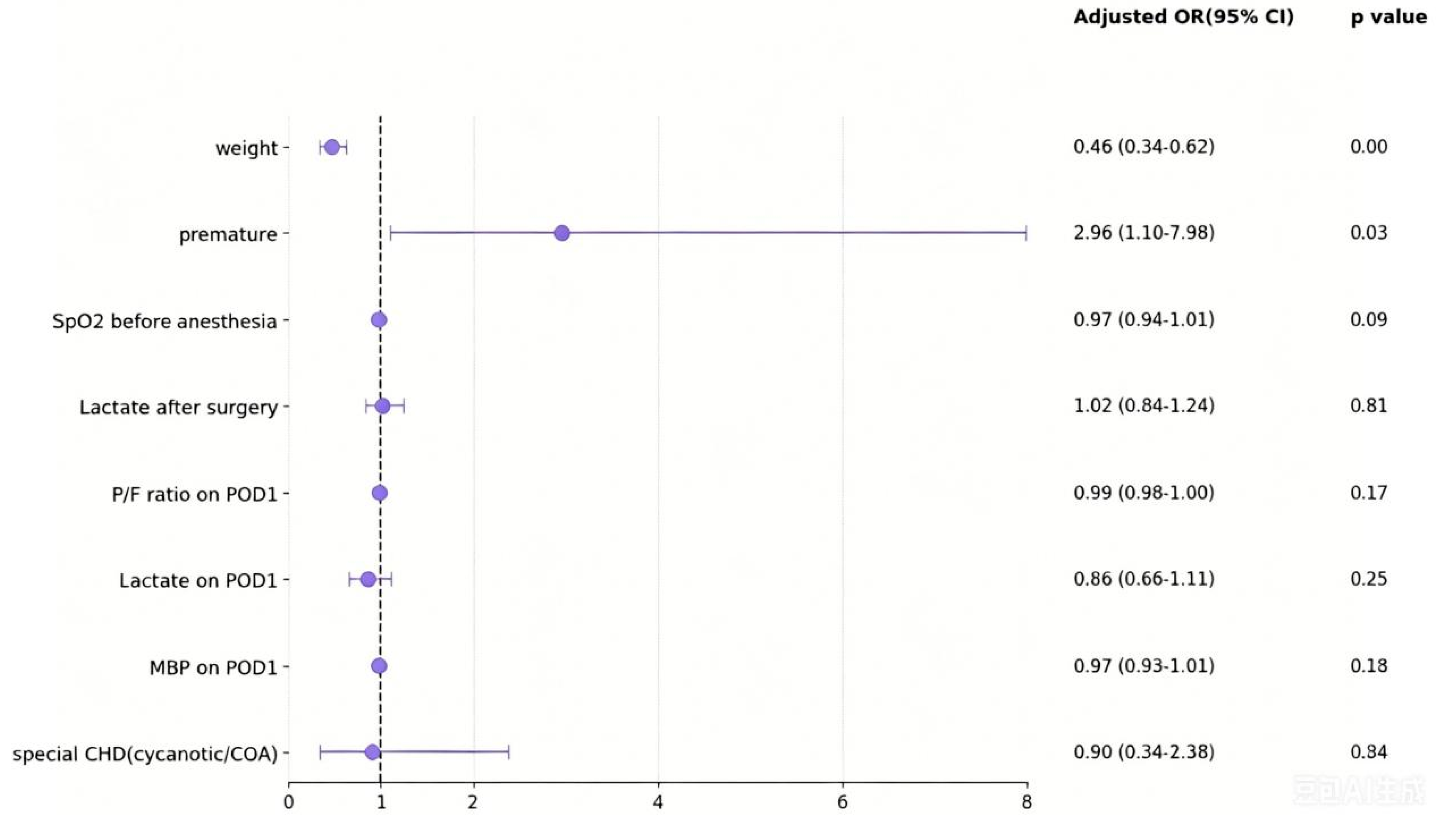
Figure 1: Multivariable predictors of decreased abdominal tissue StO2 in CHD patients after cardiac surgery.
3.4 Relation with StO2 and Clinical Outcomes in CHD Patients
A total of 82 patients (18.7%) developed one or more GIDs during the first week of their PICU admission. Of the patients who developed GIDs, 38 (46.3%) developed gastrointestinal haemorrhage, 24 (29.3%) abdominal distention, 11 (13.4%) GRV, 7 (8.5%) patients developed vomiting, and 2 cases in the StO2 < 70% group developed NEC, which improved with conservative medical treatment. Variables such as anticoagulants, analgesics, and sedatives, combined with antibiotics and vasoactive drugs, which are considered to affect GID, were compared, but no significant difference. However, the incidence of GID was also not significantly different between the two groups (18.5% vs. 18.7%, p = 0.84) (Table 3).
Table 3: Postoperative complications and clinical outcomes between CHD children with abdominal StO2 ≥ 70% and StO2 < 70%.
| StO2 ≥ 70% (n = 315) | StO2 < 70% (n = 124) | p-Value | |
|---|---|---|---|
| Gastrointestinal dysfunction | 59 (18.7%) | 23 (18.5%) | 0.84 |
| gastrointestinal haemorrhage | 27 (8.6%) | 11 (8.9%) | - |
| abdominal distention | 19 (6.0%) | 5 (4.0%) | - |
| GRV | 8 (2.5%) | 3 (2.4%) | - |
| Vomiting | 5 (1.6%) | 2 (1.6%) | - |
| NEC | 0 (0) | 2 (1.6%) | - |
| Death | 3 (1%) | 1 (0.8%) | 0.78 |
| ECMO | 3 (1%) | 3 (2.4%) | 0.12 |
| Mechanical ventilation time (hours) | 22 (14.5–26.2) | 35 (24.3–72.8) | 0.02 |
| ICU stay time (days) | 5 (4–10.5) | 11.5 (5.3–16.9) | <0.01 |
| Hospital time (days) | 15 (11.2–20.6) | 20 (14.4–26.3) | <0.01 |
| Hospitalization costs (10,000 CNY) | 10.5 (8.5–13.8) | 13.5 (10.8–18.9) | <0.01 |
Intensive care mortality and use of ECMO were not different between the two groups (p > 0.05). Decreased StO2 was associated with a significantly longer duration of mechanical ventilation (22 h vs. 35 h, p = 0.02), prolonged the ICU stay (5 days vs. 11.5 days, p < 0.01), hospital stay (15 days vs. 20 days, p < 0.01), and much higher costs (10.5 vs. 13.5 10,000 CNY, p < 0.01) (Table 3, Fig. 2).
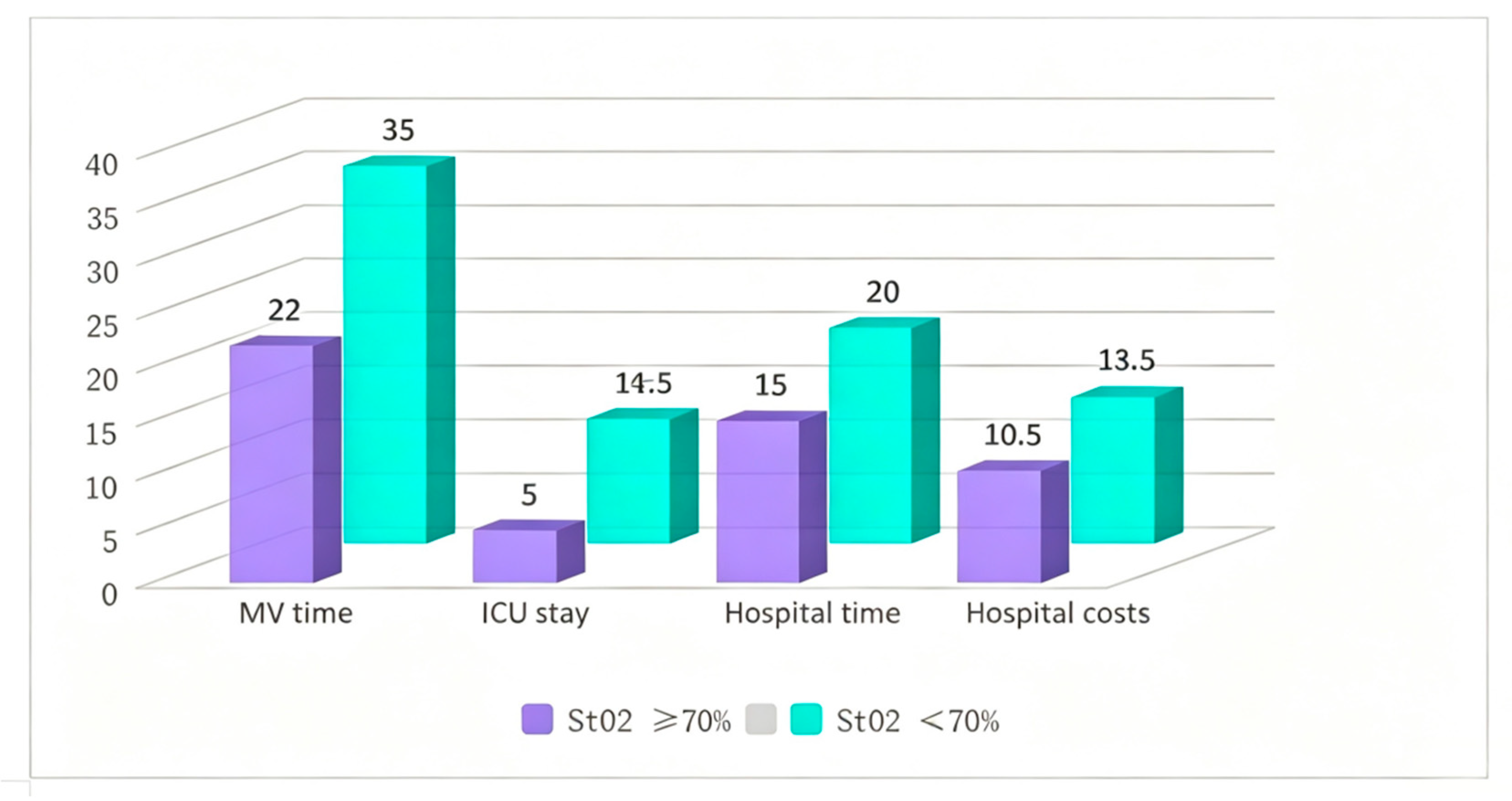
Figure 2: Postoperative outcomes and complications of the two patient groups were presented, including mechanical ventilator time, ICU time, hospital stay duration, and hospitalization costs. The numerical values represent the median of the variables in each group.
Here, the study describes changes in postoperative abdominal StO2 for neonates and infants who underwent CHD surgery on CPB. Our study shows that the incidence of decreased StO2 lower than 70% was higher than that of CHD, which was about 28.2%. Similar to other research populations, we found that lower weight and preterm infants in this special population of CHD often experienced a significant decrease in abdominal oxygen saturation in the early postoperative period. Our findings demonstrate that decreased abdominal StO2 represents a relevant clinical predictor of poor clinical prognosis, as evidenced by longer mechanical ventilation, hospitalization, and PICU stay, along with higher costs. Additionally, the group showed higher VIS and a slow declining trend within 72 h post-surgery, although no statistically significant differences were observed between the two groups.
Given the possible role of impaired intestinal perfusion, especially in infants with CHD, NIRS may be a promising tool for the early detection of hypo-perfused states leading to a greater risk of NEC after CHD repair [14]. To date, only small studies have trialed the use of splanchnic NIRS as a surrogate marker of impaired intestinal oxygenation and assessed the relationship between regional splanchnic oximetry (rSO2) values and NEC or the clinical prognosis [15,16]. However, few studies have explored the risk factors for decreased abdominal oxygen saturation after CHD surgery. Besides, detecting real splanchnic oxygenation using NIRS can be challenging due to the potential variability in recordings and low reproducibility.
Robust reference values and their associated variations for regional tissue oxygen saturation measured by NIRS must be firmly established before routine clinical adoption can be implemented in patients. Several studies have defined normal values or established centile charts [17,18]. However, these values may vary depending on the device, sensor, study population, and/or measured body sites. Coupled with the relatively small existing cohorts and the use of a wide range of epochs for StO2 averaging, the possible influencing factors listed above represent a common limitation in current literature on abdominal NIRS monitoring in the neonatal population. This could have contributed to the challenge of defining normal StO2 values. Bailey et al. measured the normal values of visceral oxygen saturation in full-term newborns, and exhibited a mean intestinal tissue oxygen saturation of 69.9 ± 12.1 on the first postnatal day [19]. Our earlier dataset, a preliminary experimental analysis involving 300 infants under 1 year old with CHD, revealed a mean preoperative abdominal StO2 of 75.2 ± 11.4. Based on these findings, we define decreased StO2 in CHD infants as less than 70%. Previous studies have highlighted that monitoring visceral oxygen reveals significant inter-individual variability in newborns, in contrast to the low variability of cerebral oxygen saturation. This fluctuation pattern can complicate the establishment of reference values. To minimize the impact of StO2 variability on data analysis and interpretation, we adopted the shorter data averaging interval proposed for continuous StO2 monitoring and measurements over the anterior abdominal wall, rather than flank rSO2.
In our patients, 18.7% developed GID, of whom only two detected NEC in the decreased StO2 group. There was no significant correlation between GID and abdominal oxygen saturation. We believe this may be due to substantial differences between our study population and previous research: the age range was expanded from newborns to 1-year-olds. Secondly, in outcome variables, postoperative gastrointestinal dysfunction symptoms occurred one week later, with infants exhibiting mild manifestations such as simple gastrointestinal bleeding, vomiting, and abdominal distension. The incidence of postoperative NEC is relatively rare, suggesting that gastrointestinal ischemia may not have caused significant local blood flow impairment detectable through StO2 monitoring. Preliminary evidence from existing neonatal studies indicates that mesenteric oxygenation monitoring may hold greater potential value for early detection of intestinal ischemia and NEC. Therefore, for simple feeding intolerance in older infants, NIRS monitoring of abdominal oxygen saturation may have certain limitations in predicting mild gastrointestinal dysfunction. Finally, while some studies have found that abdominal oxygen saturation values fluctuate between 32–66% after NEC in preterm infants, data on abdominal oxygen saturation in other populations, such as those with CHD and full-term infants with NEC, remain relatively scarce. In our study, the normal value of abdominal oxygen saturation in CHD patients after cardiac surgery was defined as 70%. This present study suggests the need for further investigation and discussion in order to explore the use of StO2 measurement by NIRS to assist patient status and monitor patient trends for early prediction of cardiac GID.
The aim of neonatal intensive care and monitoring is to ensure adequate organ perfusion and oxygenation, prevent injury, and promote normal development in critically ill newborns. However, standard non-invasive monitoring techniques do not offer insights into regional tissue oxygen saturation, oxygen delivery, or oxygen consumption. NIRS measurement of rSO2 reflects the local hemodynamic status, but this metric alone cannot directly assess regional perfusion. To accurately interpret changes in rSO2, it must be combined with arterial Oxygen Saturation (SaO2) and NIRS monitoring data. Consequently, we meticulously documented hemodynamic parameters in the early postoperative period to aid in the rational interpretation of regional perfusion and hemodynamic status in pediatric abdominal intestinal tissues following cardiac surgery.
Infants exhibiting reduced StO2 in the early postoperative phase frequently displayed tachycardia and hypotension, suggesting potential systemic hypovolemia or cardiac insufficiency. Furthermore, within 72 h post-surgery, both SpO2 and P/F ratio were lower than in the control group, indicating a possible systemic oxygen supply deficiency. The lactate level on POD1 was significantly higher, suggesting a marked increase in tissue oxygen consumption. Additionally, the VIS score in the early postoperative period of this group was higher and exhibited a slower decline, indicating a greater reliance on vasoactive drugs and volume distribution adjustments. However, after treatment, lactate levels normalized by the second and third postoperative days without significant differences. Researchers discovered that abdominal NIRS monitoring facilitates early detection of low cardiac output syndrome (LCOS) following cardiac surgery, with persistent NIRS values below 58% serving as a reliable predictor of LCOS [20]. Thus, our study findings suggest that postoperative decreased abdominal oxygen saturation can reflect and predict local tissue perfusion and systemic oxygen supply in pediatric patients. PICU physicians should be fully attentive to this and intervene early for diagnosis and treatment.
There are several limitations that merit discussion. First, this was a retrospective single-center study with a relatively small sample size, which inherently carries a risk of selection bias and limits the generalizability of our findings. Second, unlike brain tissue, the mesenteric region contains multiple structures (such as the large and small intestines, ureters, and bladder). Consequently, conditions like urinary retention and meconium, which is rich in bilirubin and interferes with light absorption, may affect StO2 measurements and introduce bias. Third, we grouped patients based on the lowest StO2 value recorded within 72 h post-surgery, rather than employing time-weighted trend analysis. Given that the lowest StO2 may coincide with specific interventions (e.g., fluid boluses or vasopressor adjustments), this approach does not fully capture the dynamic nature of splanchnic oxygenation; longitudinal trend monitoring might offer more clinically actionable insights. Fourth, we did not include patients with single-ventricle physiology, a population at particularly high risk for gastrointestinal complications, which restricts the applicability of our results to broader CHD cohorts. Fifth, enteral feeding status, an important confounder that directly influences splanchnic blood flow, was not consistently recorded or adjusted for in our multivariate models. Sixth, the definition of GID used in this study was intentionally broad and included subjective variables such as vomiting and high gastric residual volume. This may have diluted the association between regional hypoxia and clinically significant gastrointestinal injury, and the low incidence of NEC in our cohort further limited statistical power to detect meaningful subgroup differences. Finally, the selected cut-off of 70% for defining decreased StO2 was derived from a preliminary internal analysis and requires external validation across different NIRS devices and diverse patient populations before routine clinical adoption can be recommended.
In infants with CHD after cardiac surgery, lower body weight and prematurity are independent risk factors for decreased abdominal StO2. Reduced StO2 is significantly associated with prolonged mechanical ventilation, extended ICU and hospital stays, and increased costs. However, we found no statistically significant correlation between StO2 < 70% and the occurrence of GID, suggesting that abdominal NIRS may better reflect systemic hemodynamic instability than predict mild gastrointestinal dysfunction in this population.
Acknowledgement:
Funding Statement: This research was supported by the following aspects: the National High-Level Hospital Clinical Research Funding (Grant No. 2023-GSP-QN-5) and the National High-Level Hospital Clinical Research Funding (Grant No. 2024-GSP-TJ-13).
Author Contributions: The authors confirm their contributions to the paper as follows: Conceptualization, Rongyuan Zhang, Xu Wang and Xia Li; methodology, Dan Li; data curation, Yuzi Zhou and Yazhou Jiang; writing—original draft preparation, Dan Li; writing—review and editing, Rongyuan Zhang and Xia Li; supervision, Xu Wang and Xia Li. All authors reviewed and approved the final version of the manuscript.
Availability of Data and Materials: The data that support the findings of this study are available from the corresponding author upon reasonable request.
Ethics Approval: This retrospective cohort study was reviewed and approved by the Fuwai Hospital Ethics Committee (approval number: 2023–2041) and conducted in accordance with the Helsinki Declaration. Informed consent has been obtained from the patient’s parents or guardians.
Conflicts of Interest: The authors declare no conflicts of interest.
References
1. Ishaque S , Shakir M , Ladak A , Haque AU . Gastrointestinal complications in critically ill children: Experience from a resource-limited country. Pak J Med Sci. 2021; 37( 3): 657– 62. doi:10.12669/pjms.37.3.3493. [Google Scholar] [CrossRef]
2. Braun JP , Schroeder T , Buehner S , Dohmen P , Moshirzadeh M , Grosse J , et al. Splanchnic oxygen transport, hepatic function and gastrointestinal barrier after normothermic cardiopulmonary bypass. Acta Anaesthesiol Scand. 2004; 48( 6): 697– 703. doi:10.1111/j.1399-6576.2004.00392.x. [Google Scholar] [CrossRef]
3. Spinner JA , Morris SA , Nandi D , Costarino AT , Marino BS , Rossano JW , et al. Necrotizing enterocolitis and associated mortality in neonates with congenital heart disease: A multi-institutional study. Pediatr Crit Care Med. 2020; 21( 3): 228– 34. doi:10.1097/PCC.0000000000002133. [Google Scholar] [CrossRef]
4. Ferguson LP , Gandiya T , Kaselas C , Sheth J , Hasan A , Gabra HOS . Gastrointestinal complications associated with the surgical treatment of heart disease in children. J Pediatr Surg. 2017; 52( 3): 414– 9. doi:10.1016/j.jpedsurg.2016.10.052. [Google Scholar] [CrossRef]
5. Martini S , Corvaglia L . Splanchnic NIRS monitoring in neonatal care: Rationale, current applications and future perspectives. J Perinatol. 2018; 38( 5): 431– 43. doi:10.1038/s41372-018-0075-1. [Google Scholar] [CrossRef]
6. Kooi EMW , Mintzer JP , Rhee CJ , Ergenekon E , Schwarz CE , Pichler G , et al. Neonatal somatic oxygenation and perfusion assessment using near-infrared spectroscopy: Part of the series on near-infrared spectroscopy by the European Society of Paediatric Research Special Interest Group “Near-Infrared Spectroscopy”. Pediatr Res. 2024; 96( 5): 1180– 94. doi:10.1038/s41390-024-03226-z. [Google Scholar] [CrossRef]
7. Nuzum TA , Bailey SM , Caprio M , Wachtel EV . A prospective study describing splanchnic NIRS and clinical outcomes in encephalopathic neonates receiving minimal enteral nutrition during therapeutic hypothermia. J Perinatol. 2025; 45( 8): 1087– 92. doi:10.1038/s41372-025-02270-9. [Google Scholar] [CrossRef]
8. Kim MJ , Baek JS , Kim JA , Cha SG , Yu JJ . Cerebral and somatic oxygen saturation in neonates with congenital heart disease before surgery. J Clin Med. 2021; 10( 11): 2455. doi:10.3390/jcm10112455. [Google Scholar] [CrossRef]
9. Moschino L , Guiducci S , Duci M , Meggiolaro L , Nardo D , Bonadies L , et al. Noninvasive tools to predict necrotizing enterocolitis in infants with congenital heart diseases: A narrative review. Children. 2024; 11( 11): 1343. doi:10.3390/children11111343. [Google Scholar] [CrossRef]
10. Holleran EM , Brown MD , Sassano C , Musa N , Colyer J , Sagiv E . Monitoring abdominal near-infrared spectroscopy during feeds in neonates with CHD recovering from surgery: A feasibility study. Cardiol Young. 2024; 34( 11): 2355– 61. doi:10.1017/S1047951124036114. [Google Scholar] [CrossRef]
11. Kaufman J , Almodovar MC , Zuk J , Friesen RH . Correlation of abdominal site near-infrared spectroscopy with gastric tonometry in infants following surgery for congenital heart disease. Pediatr Crit Care Med. 2008; 9( 1): 62– 8. doi:10.1097/01.pcc.0000298640.47574.da. [Google Scholar] [CrossRef]
12. Iliopoulos I , Branco RG , Brinkhuis N , Furck A , LaRovere J , Cooper DS , et al. Mesenteric near-infrared spectroscopy and risk of gastrointestinal complications in infants undergoing surgery for congenital heart disease. Cardiol Young. 2016; 26( 4): 772– 80. doi:10.1017/s1047951115001365. [Google Scholar] [CrossRef]
13. Reintam Blaser A , Malbrain ML , Starkopf J , Fruhwald S , Jakob SM , de Waele J , et al. Gastrointestinal function in intensive care patients: Terminology, definitions and management. Recommendations of the ESICM Working Group on Abdominal Problems. Intensive Care Med. 2012; 38( 3): 384– 94. doi:10.1007/s00134-011-2459-y. [Google Scholar] [CrossRef]
14. Seager E , Longley C , Aladangady N , Banerjee J . Measurement of gut oxygenation in the neonatal population using near-infrared spectroscopy: A clinical tool? Arch Dis Child Fetal Neonatal Ed. 2020; 105( 1): 76– 86. doi:10.1136/archdischild-2018-316750. [Google Scholar] [CrossRef]
15. Baserga M , Reich B , Braski K . Abnormal splanchnic regional saturations in a preterm infant that developed necrotizing enterocolitis following a red blood cell transfusion. Adv Neonatal Care. 2020; 20( 5): 401– 5. doi:10.1097/ANC.0000000000000711. [Google Scholar] [CrossRef]
16. Howarth C , Banerjee J , Leung T , Aladangady N . Could Near Infrared Spectroscopy (NIRS) be the new weapon in our fight against Necrotising Enterocolitis? Front Pediatr. 2022; 10: 1024566. doi:10.3389/fped.2022.1024566. [Google Scholar] [CrossRef]
17. van der Heide M , Dotinga BM , Stewart RE , Kalteren WS , Hulscher JBF , Reijneveld SA , et al. Regional splanchnic oxygen saturation for preterm infants in the first week after birth: Reference values. Pediatr Res. 2021; 90( 4): 882– 7. doi:10.1038/s41390-020-01323-3. [Google Scholar] [CrossRef]
18. Bruckner M , Wolfsberger CH , Dempsey EM , Liem KD , Lemmers P , Alderliesten T , et al. Normal regional tissue oxygen saturation in neonates: A systematic qualitative review. Pediatr Res. 2024; 96( 4): 844– 55. doi:10.1038/s41390-021-01786-y. [Google Scholar] [CrossRef]
19. Bailey SM , Hendricks-Munoz KD , Mally P . Cerebral, renal, and splanchnic tissue oxygen saturation values in healthy term newborns. Am J Perinatol. 2014; 31( 4): 339– 44. doi:10.1055/s-0033-1349894. [Google Scholar] [CrossRef]
20. Hickok RL , Spaeder MC , Berger JT , Schuette JJ , Klugman D . Postoperative abdominal NIRS values predict low cardiac output syndrome in neonates. World J Pediatr Congenit Heart Surg. 2016; 7( 2): 180– 4. doi:10.1177/2150135115618939. [Google Scholar] [CrossRef]
Cite This Article
 Copyright © 2026 The Author(s). Published by Tech Science Press.
This work is licensed under a Creative Commons Attribution 4.0 International License , which permits unrestricted use, distribution, and reproduction in any medium, provided the original work is properly cited.
 Downloads
 Citation Tools
