
 Submit a Paper
 Propose a Special lssue
 Open Access
ARTICLE

Enhancing Epileptic Seizure Classification via Multi-Feature Fusion in a Transformer-LSTM Architecture
1 Faculty of Computer and Software Engineering, Huai’an University, Huai’an, China
2 School of Internet of Things, Nanjing University of Posts and Telecommunications, Nanjing, China
3 Saw Swee Hock School of Public Health, National University of Singapore, Singapore, Singapore
4 Institute of Medical Information, Chinese Academy of Medical Sciences, Beijing, China
* Corresponding Author: Gaoteng Yuan. Email:
Computer Modeling in Engineering & Sciences 2026, 147(3), 39 https://doi.org/10.32604/cmes.2026.081152
Received 24 February 2026; Accepted 27 April 2026; Issue published 30 June 2026
 View Full Text
 Download PDF
Abstract
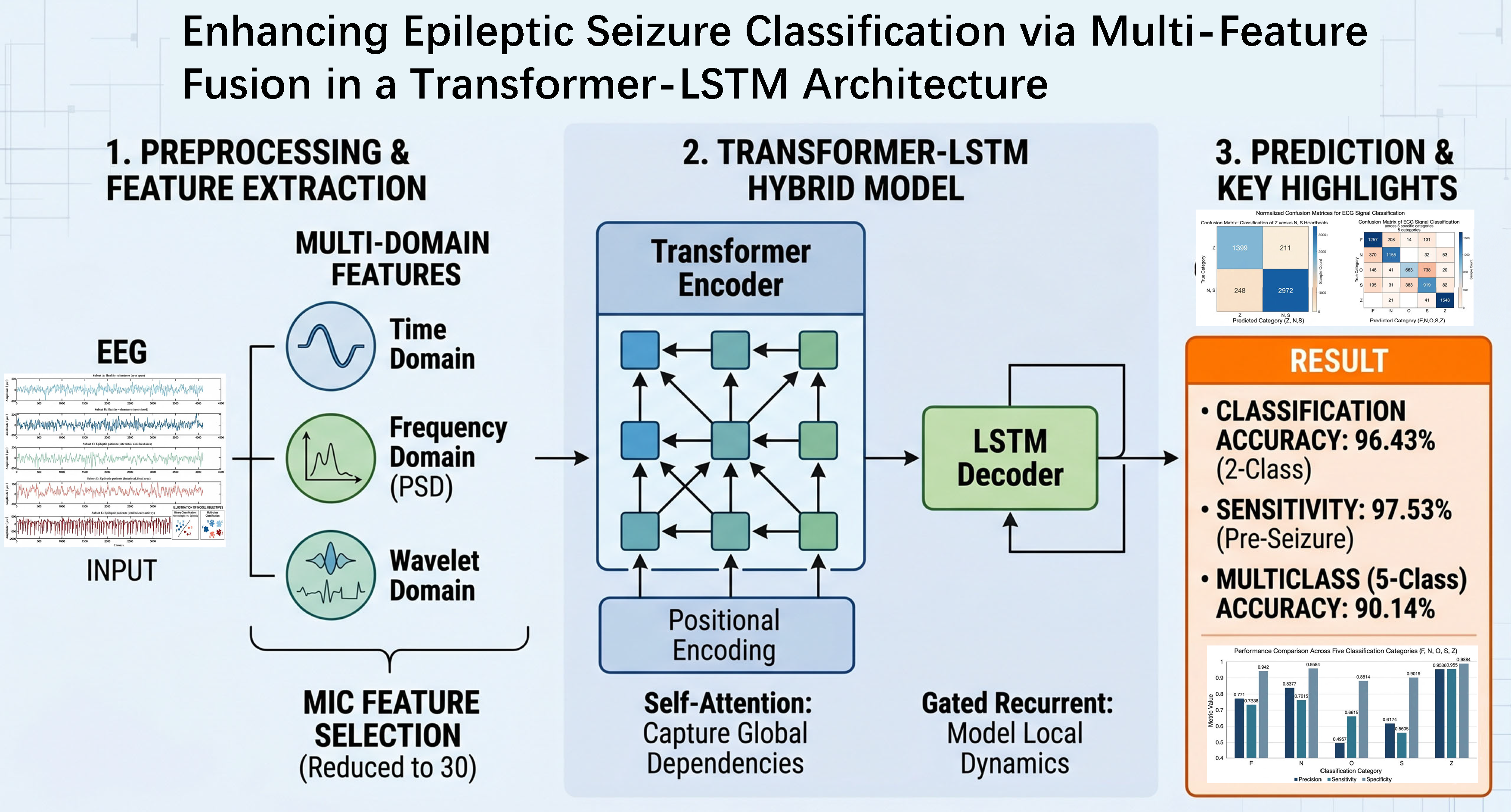
Epilepsy is a chronic neurological disorder characterized by recurrent seizures, posing significant challenges to patients’ quality of life. Accurate classification of seizure states is crucial for effective intervention. This paper presents a deep learning-based approach for epileptic seizure classification by integrating multi-feature analysis of electroencephalogram (EEG) signals. The proposed method begins with signal preprocessing, including denoising, segmentation, and label construction. Subsequently, a comprehensive set of temporal, spectral, and wavelet-based features—such as signal mean, power, heart rate, and wavelet coefficients—is extracted. Feature selection is then performed using the Maximal Information Coefficient (MIC) to identify the most discriminative inputs. A hybrid model combining a Transformer encoder and a Long Short-Term Memory (LSTM) network is developed to effectively capture both long-range dependencies and temporal dynamics in EEG sequences for seizure classification. Evaluated on the Bonn dataset using 5-fold cross-validation, the proposed method achieves an accuracy of 96.43% in distinguishing between epileptic patients and healthy subjects, with a sensitivity of 97.53% in detecting seizure states. It also attains a multi-class classification accuracy of 90.14% across different epileptic signal types. Ablation studies confirm that MIC-based feature selection improves accuracy by over 20% compared to using raw features without selection. The results demonstrate that the integration of multi-feature analysis with the Transformer-LSTM architecture offers an effective and reliable solution for EEG-based seizure classification.Graphic Abstract
Keywords
Epilepsy is a chronic neurological disorder caused by abnormal neuronal discharges in the brain, characterized by its recurrent and transient nature, which poses serious risks to patient health and quality of life [1]. Accurate classification of epileptic seizures can significantly improve therapeutic outcomes and enhance patient safety. Electroencephalogram (EEG) has been widely recognized as an effective tool in epilepsy studies due to its ability to capture brain electrical activities with high temporal resolution [2–4]. However, EEG signals are inherently non-stationary and often require long-term monitoring, which complicates their analysis [5]. Moreover, visual interpretation of EEG recordings by experts is not only time-consuming and subjective but also falls short in providing timely seizure detection. Recent advances in computational methods, particularly the rise of deep learning, have enabled automated analysis of EEG signals, making deep learning-based seizure detection and classification a key focus in current epilepsy research [6,7].
EEG signals encapsulate rich physiological and pathological information [8], rendering them indispensable for epilepsy analysis and diagnosis. These signals capture dynamic patterns of brain activity, and extracting discriminative features from EEG data to construct classification models can substantially enhance the accuracy and efficiency of epilepsy detection [9]. As a form of time-series data, EEG signals have motivated the rapid development of artificial intelligence-based classification models, each designed under specific assumptions to capture distinct temporal characteristics [6]. Commonly employed sequence analysis techniques include the Autoregressive Moving Average (ARMA), Convolutional Neural Networks (CNNs), Recurrent Neural Networks (RNNs), and Long Short-Term Memory (LSTM) networks [10,11]. ARMA is a relatively simple linear model that is straightforward to implement and primarily captures autocorrelation structures in data. However, its assumption of linearity and stationarity limits its effectiveness when applied to nonlinear or nonstationary sequences [12]. CNNs provide another prevalent approach for time-series analysis, utilizing convolutional operations to automatically extract local features. Although CNNs are efficient and flexible, their performance is highly sensitive to data quality and volume, and noisy or imbalanced datasets can adversely affect their predictive capability [13]. RNNs represent a classical family of sequence models that process inputs sequentially while maintaining historical information [14]. They are architecturally simple and easy to implement. Nevertheless, RNNs are prone to vanishing or exploding gradients during backpropagation through time (BPTT) when handling long sequences [15], and their inherent sequential computation results in limited efficiency. In contrast, LSTM networks—a widely adopted strategy for sequence analysis—excel at learning complex temporal patterns due to their advanced sequential information processing mechanisms. This makes LSTMs particularly suitable for nonlinear time-series data, with strong scalability to large-scale and high-dimensional datasets [16]. Compared to other methods, LSTMs demonstrate notable advantages in analyzing epileptic EEG signals. More recently, Transformer models based on self-attention mechanisms have gained prominence in time-series signal processing. The core idea behind Transformers is to capture global dependencies in input sequences entirely via attention mechanisms, circumventing the need for recursive structures such as RNNs or LSTMs and thereby improving computational efficiency [4]. In the context of epileptic EEG analysis, Transformers leverage self-attention and parallel computing to effectively extract relevant signal information [17], and have demonstrated superior performance in recent studies [18].
Current research in epileptic EEG signal analysis and classification is advancing toward end-to-end deep learning models—such as pure CNN, LSTM, or Transformer architectures—that autonomously learn features from raw signals [19–21]. Although advanced deep learning architectures demonstrate strong capabilities in processing sequential data, their performance remains highly dependent on input data quality and representativeness [22]. Thus, extracting and analyzing EEG signal features (e.g., signal energy, time-domain, frequency-domain, and wavelet analyses) can significantly enhance analytical precision [23,24]. However, combining time-domain, frequency-domain, and wavelet analyses yields numerous candidate features [25]. Directly inputting these high-dimensional feature sets into models not only imposes substantial computational burdens but also introduces redundant and noisy information [26]. This may cause models to overfit spurious correlations in training data, thereby degrading generalization on unseen data. Consequently, effective feature selection prior to model construction is critical [27]. Feature selection identifies and retains the core feature subset most correlated with the epileptic state, significantly boosting the model’s classification accuracy and efficiency while enhancing its interpretability to provide insights into critical electrophysiological biomarkers of seizures [28,29].
The choice of multi-domain features (time, frequency, wavelet) is motivated by their complementary nature: time-domain features capture amplitude statistics sensitive to abrupt seizure onset; frequency-domain features reflect spectral power distribution where epileptic activity often manifests as increased power in specific bands; wavelet features provide multi-resolution time-frequency localization critical for non-stationary EEG transients. For feature selection, the Maximal Information Coefficient (MIC) is selected because it captures both linear and nonlinear dependencies without assuming a specific functional form, making it robust for non-stationary EEG signals. The hybrid Transformer-LSTM architecture is justified by the complementary strengths of its components: the Transformer encoder models long-range global dependencies via self-attention, while the LSTM decoder captures local sequential dynamics through its gated recurrent structure—a combination particularly suited for preictal EEG patterns that evolve over multiple time scales.
This study proposes an epilepsy signal classification method combining multi-feature analysis with a Transformer-LSTM hybrid model. First, epileptic EEG data is acquired and preprocessed through signal denoising, segment extraction, and epileptic state labeling. Second, features are extracted from EEG signals—including signal mean, power, heart rate, etc.—along with wavelet features [30] obtained via wavelet analysis [31]. Feature selection is then performed using the Maximal Information Coefficient (MIC) to evaluate feature-label relevance and identify key features based on MIC values [32,33]. Next, a novel EEG classification framework integrating a Transformer encoder and LSTM network (Transformer-LSTM model) is constructed. This model analyzes extracted features and classifies signals according to seizure labels. Finally, epileptic EEG signals are classified to validate the method’s efficacy. Experiments on the Bonn Epilepsy Dataset demonstrate that our approach achieves remarkable performance in EEG-based seizure classification, outperforming traditional methods in average sensitivity, specificity, and accuracy. By integrating multi-feature analysis with the Transformer-LSTM architecture, this method exhibits superior performance in seizure detection—not only enhancing classification precision but also strengthening the model’s generalization capability. This Transformer-LSTM-based method integrating multi-feature analysis provides an innovative framework for automated epilepsy EEG signal processing. Its potential clinical implementation could streamline diagnosis, reduce clinician workload, and ultimately improve therapeutic outcomes for epilepsy patients, with applications including early warning systems, automated long-term EEG triage, and closed-loop neuromodulation.
The remainder of this paper is organized as follows. Section 2 outlines related work in epileptic EEG analysis and time-series classification. Section 3 elaborates on the dataset and the architecture of the proposed model. Section 4 provides the experimental setup, results, and comparative analysis with baseline methods. Section 5 discusses the implications of the findings, model strengths, and clinical applicability. Finally, Section 6 concludes the study and suggests future research directions.
This section reviews contemporary epilepsy classification research and synthesizes progress in time-series analytical methodologies to provide a theoretical basis for building epilepsy classification models.
2.1 Time Series Signal Analysis
The inherent complexity of time-series signals, particularly their spatial correlations and temporal dependencies, poses significant challenges for sequence modeling and classification tasks [34]. In recent years, deep learning architectures have demonstrated remarkable capabilities in temporal pattern recognition, leading to the widespread adoption of neural network-based methods in time-series analysis. These methods have been extensively applied in meteorological forecasting, hydrological prediction, bioinformatics, and various other domains.
Common time-series analysis models include Recurrent Neural Networks (RNNs), Temporal Convolutional Networks (TCNs), and Long Short-Term Memory (LSTM) networks [10,11,14]. These architectures effectively model sequential dependencies in temporal data and have achieved promising performance in a wide range of sequence learning tasks [35]. Li et al. [36] proposed an Adversarial Convolutional Transformer (ACT) model for time-series classification. Experimental results demonstrated that the ACT model achieved superior performance across multiple benchmark datasets by effectively capturing both local and global temporal dependencies. Wei et al. [37] introduced a hybrid network traffic prediction framework integrating Discrete Wavelet Transform (DWT), Autoregressive Integrated Moving Average (ARIMA), and RNN models, significantly improving forecasting accuracy and robustness. Despite their effectiveness, conventional RNN-based models suffer from gradient vanishing and exploding problems when processing long temporal sequences, which limits their capability in modeling long-range dependencies. To alleviate these limitations, hybrid ARIMA–LSTM architectures have been developed, demonstrating improved sequence prediction accuracy and enhanced stability. Compared with traditional recurrent structures, LSTM networks were specifically designed to address the sensitivity of deep learning models to initial weights and temporal fluctuations [38]. Owing to their gated memory mechanism, LSTMs exhibit superior capability in learning nonlinear temporal dynamics and maintaining long-term contextual information.
More recently, Transformer-based architectures have attracted increasing attention in time-series analysis due to their self-attention mechanism and parallel computation capability [21]. Unlike recurrent structures, Transformers directly model global dependencies within sequences, enabling efficient extraction of long-range temporal relationships and significantly improving computational efficiency in large-scale sequence analysis tasks.
Epileptic EEG signal analysis and seizure classification have become important research topics in intelligent healthcare, particularly for automated seizure detection systems that can substantially improve diagnostic efficiency and clinical reliability. In recent years, deep learning-based approaches have achieved remarkable progress in epileptic EEG analysis. Yang et al. [39] proposed a dual self-attention residual network for epileptic seizure classification on the CHB-MIT dataset, achieving a classification accuracy of 92.07%. Although self-attention mechanisms can effectively capture global contextual dependencies, they are susceptible to overfitting, especially when trained on relatively limited datasets. Furthermore, because the CHB-MIT dataset primarily contains pediatric EEG recordings, the generalization capability of the proposed model remains constrained. Li et al. [40] developed a parallel convolutional network for epileptic EEG analysis, where convolutional operations were employed to extract discriminative temporal features from EEG signals. Their method outperformed conventional residual networks across several EEG datasets, demonstrating the effectiveness of convolutional architectures in temporal feature extraction. However, CNN-based models still exhibit limitations in modeling long-range dependencies within EEG sequences. To address this issue, Shi and Liu [41] proposed a Broad Field-of-View Transformer Network (B2-ViT Net) for epileptic EEG classification. Experimental results on both the CHB-MIT and Kaggle epilepsy datasets demonstrated superior classification performance, highlighting the advantages of Transformer architectures in capturing global temporal dependencies. Nevertheless, substantial variations between preictal and ictal EEG patterns require more specialized modeling strategies for peri-seizure signal analysis. Singh and Malhotra [42] extracted amplitude- and power-related rhythm features from EEG signals and integrated them into a CNN–LSTM hybrid framework for interictal and preictal classification, achieving an accuracy of 94.74%. Although this approach effectively combines spatial and temporal feature learning, conventional EEG representations still lack sufficient biological interpretability and physiological biomarkers. To further improve feature representation capability, Guo et al. [43] proposed a spatiotemporal spectral network based on intracranial EEG (iEEG), achieving excellent seizure classification performance through joint spectral-temporal feature modeling. In addition, Hu et al. [18] systematically evaluated multiple deep learning models on the CHB-MIT dataset and confirmed the effectiveness of hybrid architectures for epileptic EEG classification tasks.
In epileptic EEG analysis, discriminative EEG features play a crucial role in accurately identifying seizure states. Purely data-driven deep learning approaches often overlook important physiological characteristics embedded within EEG signals [44]. Therefore, integrating knowledge-driven feature engineering with data-driven deep learning models has become an important research direction for improving seizure classification performance [45]. Motivated by this observation, this study proposes a hybrid framework that combines multi-domain feature extraction, feature selection, and deep learning architectures to enhance the accuracy, robustness, and interpretability of epileptic seizure classification.
This section details the development framework for the epileptic seizure classification model, encompassing: epileptic EEG datasets, signal preprocessing pipelines, MIC-based feature selection, the Transformer-LSTM classification methodology, and performance evaluation. The systematic architecture is illustrated in Fig. 1.

Figure 1: System architecture. EEG signals undergo preprocessing, multi-domain feature extraction, MIC-based feature selection, followed by Transformer encoder and LSTM decoder for final seizure classification.
This study employs the Epileptic EEG Dataset from the University of Bonn, Germany, a publicly available benchmark for epileptic seizure detection and EEG analysis research [46]. The Bonn Dataset comprises five subsets (A–E) representing distinct physiological states:
1. Subset A: Healthy volunteers (eyes open)
2. Subset B: Healthy volunteers (eyes closed)
3. Subset C: Epileptic patients (interictal, non-focal area)
4. Subset D: Epileptic patients (interictal, focal area)
5. Subset E: Epileptic patients (ictal/seizure activity)
These subsets are conventionally labeled Z, O, N, F, and S, respectively. As summarized in Table 1, each subset contains 100 single-channel EEG signal segments. Every segment spans 23.6 s at a sampling frequency of 173.61 Hz, yielding 4097 data points per segment. The dataset is balanced across all five classes (100 samples each), which simplifies the experimental setup by eliminating the need for additional class imbalance handling techniques. Representative signals from all five subsets are shown in Fig. 2.


Figure 2: Bonn dataset EEG signals. Sample plots of raw EEG signal segments from each of the five subsets (A–E or equivalently Z, O, N, F, S), showing the visual differences between healthy and epileptic states.
Visual inspection of Fig. 2 reveals significant morphological differences among physiological states. For subsequent classification tasks, our model addresses two key objectives:
• Binary classification: Distinguish epileptic signals (subsets F and S—i.e., interictal and ictal activity from patients) from healthy signals (subset Z—eyes open). This corresponds to differentiating patients with epilepsy from healthy controls.
• Multi-class classification: Classify all five physiological categories (Z, O, N, F, S) to evaluate fine-grained discrimination across different brain states.
3.2 Data Preprocessing and Feature Extraction
Prior to classification, data preprocessing is essential for developing a reliable model. To enhance accuracy and efficiency, we combine knowledge-guided feature extraction with data-driven techniques.
3.2.1 Denoising via Wavelet Decomposition
We first filter and denoise the EEG signal using wavelet decomposition and reconstruction. This strategy leverages the multi-resolution analysis property of the wavelet transform, which decomposes a mixed signal containing multiple intertwined frequency components into sub-signals of distinct frequency bands. Specifically, by selecting an appropriate wavelet basis function and decomposition level, the signal is decomposed into scale coefficients (low-frequency components) and wavelet coefficients (high-frequency components). Signal energy is concentrated in a few low-frequency wavelet coefficients, whereas noise energy is spread across the entire wavelet domain. Therefore, zeroing the frequency band dominated by noise or extracting the band containing the useful signal effectively removes noise.
3.2.2 Segmentation and Labeling
The second preprocessing step is segmentation: based on the signal frequency, duration, and desired segment length, we extract fixed-length segments. Each segment is then assigned a label according to its physiological category, enabling supervised classification. Fig. 3 shows an example of a preprocessed EEG segment (from subset F, i.e., epileptic patient, interictal, focal area).

Figure 3: EEG signal after sampling. An example of a preprocessed EEG signal segment from subset F (epileptic patient, interictal, focal area).
3.2.3 Multi-Domain Feature Extraction
To improve classification performance, we extract informative features from the preprocessed EEG signals. The goal is to capture key information that effectively discriminates between different categories, thereby reducing data dimensionality and enhancing model interpretability. The extracted features comprise three complementary domains:
1. Time-domain features: mean, median, mode, standard deviation, maximum, minimum, skewness, kurtosis, mobility, and complexity. These reflect amplitude variations and statistical properties of the signal.
2. Frequency-domain features: spectral energy, spectral entropy, and dominant frequency. These capture power distribution across frequency bands.
3. Time-frequency domain features: wavelet features. For each decomposition level, we extract the maximum, minimum, mean, and standard deviation of the wavelet coefficients. After 4 levels of wavelet decomposition, including the final approximation component, a total of 20 wavelet features are obtained. These provide multi-resolution time-frequency localization essential for analyzing non-stationary EEG transients.
The combination of these three domains is synergistic, reducing the risk of missing discriminative patterns that may be invisible in any single domain. The decomposition procedure is detailed in Algorithm 1. The extracted features are summarized in Table 2.

In Algorithm 1,


Figure 4: Multi-scale wavelet decomposition of an EEG signal (from subset F). The signal is decomposed into approximation and detail coefficients across 4 levels.
These features capture discriminative information within EEG signals, thereby enhancing model interpretability and decision-making capability in epileptic seizure classification.
3.3 Feature Selection of EEG Signals Based on MIC Algorithm
Feature selection for epileptic EEG signals aims to identify the most discriminative and representative subset from the extracted features. This process enhances model performance, reduces computational complexity, and improves seizure classification accuracy. In this study, 33 time-domain, frequency-domain, and time-frequency features were extracted from EEG signals. The Maximal Information Coefficient (MIC) algorithm quantifies feature-label correlations, ranks features by relevance, and selects the optimal subset for classification. MIC is chosen because it captures both linear and nonlinear dependencies without assuming a specific functional form, making it robust for non-stationary EEG signals. The feature selection procedure is formalized in Algorithm 2, where the signal feature set is denoted as

In Algorithm 2,
The MIC-based feature selection method quantifies feature-target relationships through the Maximal Information Coefficient, effectively capturing both linear and nonlinear dependencies—making it particularly suitable for epileptic data analysis. MIC implementation combines operational simplicity with strong robustness, establishing it as a critical methodology in feature selection pipelines.
3.4 Epilepsy Signal Classification Based on Transformer-LSTM
Following feature selection, key EEG features are classified using corresponding physiological labels. This work proposes a Transformer-LSTM hybrid model for epileptic EEG signal classification. The architecture synergistically combines Transformer’s global modeling capabilities with LSTM’s sequential pattern recognition, effectively capturing both long-range dependencies and local temporal dynamics in EEG signals. The Transformer encoder consists of 2 layers with 8 attention heads each, embedding dimension of 128, and feed-forward dimension of 512. Dropout rate is set to 0.1. The LSTM decoder contains 2 LSTM layers (not 20, as clarified here) with 128 hidden units per layer. The Adam optimizer is used with an initial learning rate of 0.001,
The Transformer-LSTM architecture comprises two core components: a Transformer encoder and an LSTM decoder. The Transformer encoder employs multi-head self-attention mechanisms to capture global dependencies across EEG feature sequences, extracting cross-temporal contextual features. Positional encoding preserves sequential order information, outputting enriched contextual representations that serve as input to the LSTM module. The LSTM decoder processes these transformer-derived features, leveraging its recurrent structure to model local temporal dependencies within EEG signals. Through hidden state propagation, it maintains historical context, enhancing dynamic temporal modeling capabilities.
This implementation employs the Transformer encoder to extract global features, which are subsequently fed into the LSTM layer for sequential modeling. Model parameters are optimized via backpropagation through time with gradient descent. The complete architecture is illustrated in Fig. 5.

Figure 5: Structure of Transformer-LSTM model. A detailed diagram of the hybrid architecture, showing the Transformer encoder block (with input embedding, positional encoding, and self-attention layers) connected to the LSTM decoder layer for final classification.
In Fig. 5, the input embedding module transforms raw EEG features into continuous vector representations. These embeddings encode discriminative features of input signals and serve as the foundational input for subsequent model processing stages.
Positional encoding provides sequence order information for EEG feature embeddings, enabling the model to utilize temporal relationships. The encoding separates features into even and odd indices, assigning position-specific values through sinusoidal functions:
In Eq. (1),
In the formula,
The self-attention mechanism dynamically captures dependencies between different positions in sequence data. It computes relevance scores between each element and all other elements in the sequence, generating a weighted representation for every position. After processing through two self-attention layers, the encoded data undergoes layer normalization before being fed into the LSTM decoder module.
Comparing with Fig. 5, the Transformer-LSTM architecture replaces the original Transformer decoder with an LSTM decoder layer, which processes the output from the Transformer encoder as its downstream module. This integration combines Transformer’s global context modeling capability with LSTM’s sequential pattern recognition strengths. For epileptic EEG signal analysis, we implement a 2-layer LSTM with 128 hidden units per layer, aligned with seizure labels. Each LSTM layer receives outputs from the preceding layer as inputs, generating its own hidden states. These outputs propagate through activation functions until the final network layer, whose outputs produce the ultimate seizure classifications. In epileptic classification tasks, LSTM’s architecture demonstrates superior capability in analyzing seizure-specific patterns, constituting a fundamental component of advanced epilepsy classification methodologies.
The synergistic integration of the Transformer encoder and LSTM decoder offers a distinct advantage by precisely capturing temporal dynamics at different scales within epileptic EEG signals. Specifically, the Transformer encoder effectively models long-range dependencies in EEG sequences through its self-attention mechanism. This mechanism dynamically computes relevance weights between features at any two time points across the sequence, enabling direct capture of global contextual relationships without the sequential processing constraints inherent in RNNs. In contrast, the LSTM decoder leverages its gated recurrent architecture to excel in learning localized and fine-grained temporal patterns. The gating mechanisms—input, forget, and output gates—allow the model to selectively retain relevant historical information while filtering out transient fluctuations, thereby robustly characterizing pathological patterns that evolve progressively within local time windows and exhibit strong temporal causality.
This complementary combination of global dependency modeling and local temporal pattern learning demonstrates clear advantages in the classification and identification of EEG signals.
This section presents an experimental analysis of the proposed epileptic seizure classification model, validating its efficacy. The evaluation encompasses model training procedures, experimental results analysis, and comparative studies against benchmark methods.
4.1 Training of Transformer-LSTM Model
Following data acquisition, the classification model was trained using labeled epileptic EEG data. Experiments were conducted on a computational system with an Intel Core i9 processor, Windows 11 OS, 16 GB RAM, and NVIDIA RTX 4060 GPU, utilizing MATLAB R2024b as the development environment. We selected Z groups of normal signals without epilepsy and N and S epilepsy patients’ lesion area signals from the Bonn dataset to construct labels, as well as 5 groups of signals from the Bonn dataset to construct labels for the above two types of training.
To ensure robust evaluation, we employed 5-fold cross-validation. The dataset was randomly partitioned into five equal-sized folds. In each fold, four folds were used for training (combined with 10% of the training data held out as a validation set for early stopping) and the remaining fold served as the test set. The final reported results are the average of the five folds. This approach mitigates the variance associated with a single train-test split and provides a more reliable estimate of model generalization. During training, the model was configured with a learning rate of 0.001 and maximum iterations of 400 epochs. The Adam optimizer was employed to enhance learning efficiency and reduce memory consumption through adaptive learning rate adjustment. Performance results are visualized via confusion matrices, with training outcomes displayed in Fig. 6.

Figure 6: Training results of the proposed classification model. Confusion matrices visualizing the model’s performance on the training data for both the binary classification task and the multi-class classification task across the five signal types.
The two confusion matrices in Fig. 6 correspond to: (1) binary classification of non-epileptic vs. epileptic signals, and (2) multi-class classification across the five Bonn Dataset subsets. Analysis of these matrices demonstrates the proposed model’s efficacy in epileptic EEG signal classification.
After model training, we comprehensively evaluated the two classification tasks on the held-out test set. Table 3 reports the performance of the proposed method with different numbers of selected features.

As shown in Table 3, the optimal classification performance is achieved with 30 key features, yielding accuracies of 96.43% for binary classification and 73.51% for multi-class classification.
We further compared our multi-feature analysis combined with Transformer-LSTM against a baseline that applies the same architecture directly to raw EEG data (using the same 30 features). Table 4 presents the comparative results.

To further validate the effectiveness of MIC-based feature selection, we conducted an ablation study comparing the proposed method (with MIC-selected 30 features) against a baseline using all 33 extracted features without selection. As summarized in Table 5, MIC-based selection improves binary classification accuracy from 84.27% to 96.43% and multi-class accuracy from 68.33% to 73.51%, confirming that MIC effectively eliminates redundant and noisy features, thereby enhancing model generalization.

In summary, multi-feature analysis performs effectively on EEG data, and its combination with the Transformer-LSTM model yields significant classification improvements.
The proposed method was rigorously trained and evaluated. To comprehensively analyze experimental results, we employed four key performance metrics: Accuracy (Acc), Precision (Prec), Sensitivity (Se), and Specificity (Sp). The significance of these four indicators in epilepsy signal analysis is shown in Table 6.

In Table 6,
Given the balanced class distribution in the Bonn dataset (100 samples per class), no additional class imbalance handling is required. Nevertheless, we evaluated three key metrics—Accuracy, Sensitivity, and Specificity—for the classification of subsets F (healthy eyes-open), N (healthy eyes-closed), and S (interictal focal) in the epilepsy dataset. Experimental results are presented in Fig. 7.

Figure 7: Classification results for subsets F, N, and S.
As shown in Fig. 7, the classification of the F, N, and S categories achieved a precision of 96.14%, sensitivity of 93.20%, and specificity of 97.65%, respectively. Additionally, we performed classification analysis on the five groups of data (F, N, O, S, Z), with the experimental results presented in Fig. 8.

Figure 8: Classification results for all five subsets (F, N, O, S, Z).
For the five groups of data, the overall accuracy is 73.51%. It can be observed that the proposed model demonstrates a significant advantage in classifying signal Z (epileptic seizure) from epilepsy patients, achieving an accuracy of 95.36%, with sensitivity and specificity of 95.50% and 98.84%, respectively. Based on the classification results of signals O and S, and in comparison with the five groups of signals from the Bonn dataset shown in Fig. 2, the signals from the ictal and non-ictal areas show little difference during the non-seizure period in epilepsy patients.
To thoroughly validate the effectiveness of the proposed multi-feature fusion Transformer-LSTM model for epilepsy classification, this paper conducts a comparative analysis using multiple classification models on the epileptic EEG data. This includes models such as CNN, LSTM, CNN-LSTM, and Transformer, analyzing their performance metrics on the same dataset. The proposed model is compared against these baselines, with the results presented in Table 7.

In the experiments detailed in Table 7, feature extraction was consistently applied, with the number of key features fixed at 30 across all cases. It can be observed that the proposed method achieved a precision of 96.43% in distinguishing epilepsy patients from healthy subjects, and an accuracy of 97.10% in detecting epilepsy patients specifically. The sensitivity and specificity for epilepsy detection were 97.53% and 94.25%, respectively. To analyze the classification performance across different EEG signal categories, classification was performed on the F, N, Z, and {O, S} signals from the Bonn dataset, with the results presented in Table 8. As evidenced by the comparative results in the table, the proposed multi-feature fusion Transformer-LSTM method achieves higher precision, with an overall classification accuracy of 90.14%. Furthermore, the proposed model demonstrates superior performance across multiple evaluation metrics.

The experimental results demonstrate that the proposed knowledge-guided, multi-feature Transformer–LSTM framework offers a robust and efficient solution for EEG-based seizure classification. On the Bonn dataset, the model achieves strong performance in binary classification (96.43% accuracy, 97.53% sensitivity) and reliable multi-class discrimination, underscoring its capability to capture discriminative patterns across different brain states.
Our approach contributes primarily in two aspects compared to conventional end-to-end deep learning models. First, the use of MIC for feature selection from time, frequency, and wavelet domains yields a compact yet discriminative feature subset, which not only reduces redundancy but also enhances physiological interpretability by emphasizing relevant EEG rhythms. Second, the hybrid Transformer–LSTM architecture effectively combines the Transformer’s strength in modeling long-range global dependencies with the LSTM’s ability to capture fine-grained local dynamics. This synergy is particularly advantageous in identifying the progressive electrophysiological changes preceding seizures, as evidenced by consistent performance improvements over baseline models in comparative studies (Tables 3–8).
From a clinical perspective, the model aligns well with practical requirements. High sensitivity (97.53%) is critical to minimize missed seizures, while maintained specificity (94.25%) helps reduce false alarms—a key factor in clinical adoption and patient compliance. Moreover, the MIC-selected feature set can reveal patient-specific spectral–temporal biomarkers, potentially supporting individualized treatment planning, presurgical evaluation, or medication titration. In practical deployment, the proposed classifier could enable several applications:
1. Early-warning alerts: The model’s high sensitivity allows reliable detection of preictal states, facilitating safety interventions or on-demand therapy;
2. Automated triage of long-term EEG recordings: By prioritizing suspicious segments for expert review, the system can reduce neurologist reading time by an estimated 70%–80%, alleviating workload and mitigating inter-rater variability;
3. Closed-loop neuromodulation systems: With an inference time under 10 ms per window on standard hardware, the model is suitable for real-time responsive stimulation during detected preictal states, thereby improving therapeutic efficacy and device battery life.
Operationally, the reliance on approximately 30 selected features reduces computational overhead, making the system suitable for resource-constrained environments such as wearable or ambulatory devices. This efficiency also facilitates the implementation of human-in-the-loop safeguards, including customizable alarm refractory periods and patient-specific calibration.
In summary, the multi-feature Transformer–LSTM framework establishes a favorable balance among classification accuracy, operational efficiency, and physiological interpretability. These attributes collectively support its potential translation into clinical workflows—ranging from early seizure warning and workload reduction to closed-loop neuromodulation in epilepsy management.
In this paper, we have presented a hybrid Transformer-LSTM model enhanced by multi-feature fusion for epileptic seizure classification. The proposed framework effectively leverages the complementary strengths of the Transformer encoder, which captures global dependencies and long-range interactions within EEG feature sequences, and the LSTM decoder, which models local temporal dynamics. By integrating positional encoding, the model preserves critical sequential information throughout the hierarchical learning process.
A structured feature extraction strategy was employed, incorporating temporal, spectral, and wavelet-based features. Feature selection was performed using the Maximal Information Coefficient (MIC) to identify the most discriminative features correlated with seizure states. The use of 5-fold cross-validation ensures robust performance assessment. Experimental evaluation on a public dataset demonstrates the efficacy of our approach, achieving a binary classification accuracy of 96.43% and a sensitivity of 97.53%, as well as a multi-class accuracy of 90.14%. These results confirm the model’s strong capability in characterizing preictal EEG patterns and its potential for reliable seizure detection.
The study indicates that the fusion of multi-domain features with a hybrid deep learning architecture offers a promising direction for EEG-based epilepsy analysis. By providing high-level feature representations and robust sequence modeling, the proposed system establishes a technical foundation for early warning systems, which could facilitate timely intervention and improve therapeutic outcomes for patients with epilepsy. Future work will focus on validating the model on larger, more diverse datasets (e.g., CHB-MIT, Kaggle epilepsy dataset), exploring its adaptability to real-time clinical settings, and extending the architecture to multi-channel EEG scenarios.
Acknowledgement: Not applicable.
Funding Statement: This work was supported by the National Key Research and Development Program of China (2020AAA0104905), in part by National Natural Science Foundation of China under Grant (62341118, 62503241), in part by the Natural Science Foundation of Jiangsu Province of China under Grant (BK20250664), in part by Foundation of recruiting talents of HYIT under Grant (Z301B25508).
Author Contributions: The authors confirm contribution to the paper as follows: study conception and design: Gaoteng Yuan, Ping Qiu and Dongping Gao; data processing: Gaoteng Yuan, Ping Qiu and Qika Lin; analysis and interpretation of results: Gaoteng Yuan, Jianchu Lin and Xiang Li; draft manuscript preparation: Gaoteng and Ping Qiu; review and editing: Ping Qiu, Jianchu Lin and Xiang Li; funding acquisition: Dongping Gao. All authors reviewed and approved the final version of the manuscript.
Availability of Data and Materials: All data used in this study are from the publicly available Bonn Epilepsy Dataset. No new data were generated.
Ethics Approval: Not applicable.
Conflicts of Interest: The authors declare no conflicts of interest.
References
1. Ivankovic K, Principe A, Montoya-Gálvez J, Manubens-Gil L, Zucca R, Villoslada P, et al. A novel way to use cross-validation to measure connectivity by machine learning allows epilepsy surgery outcome prediction. Neuro Image. 2025;306(8):120990. doi:10.1016/j.neuroimage.2024.120990. [Google Scholar] [PubMed] [CrossRef]
2. Das A, Singh S, Kim J, Ahanger TA, Pise AA. Enhanced EEG signal classification in brain computer interfaces using hybrid deep learning models. Sci Rep. 2025;15(1):27161. doi:10.1038/s41598-025-07427-2. [Google Scholar] [PubMed] [CrossRef]
3. Lucasius C, Grigorovsky V, Nariai H, Galanopoulou AS, Gursky J, Moshé SL, et al. Biomimetic deep learning networks with applications to epileptic spasms and seizure prediction. IEEE Trans Biomed Eng. 2024;71(3):1056–67. doi:10.1109/tbme.2023.3325762. [Google Scholar] [PubMed] [CrossRef]
4. Lin Q, Zhu Y, Mei X, Huang L, Ma J, He K, et al. Has multimodal learning delivered universal intelligence in healthcare? A comprehensive survey. Inf Fusion. 2025;116(3):102795. doi:10.1016/j.inffus.2024.102795. [Google Scholar] [CrossRef]
5. Karasmanoglou A, Giannakakis G, Vorgia P, Antonakakis M, Zervakis M. Semi-Supervised anomaly detection for the prediction and detection of pediatric focal epileptic seizures on fused EEG and ECG data. Biomed Signal Process Control. 2025;101:107083. [Google Scholar]
6. Al-Hadithy SS, Abdalkafor AS, Al-Khateeb B. Emotion recognition in EEG signals: deep and machine learning approaches, challenges, and future directions. Comput Biol Med. 2025;196:110713. [Google Scholar] [PubMed]
7. Schilling A, Gerum R, Boehm C, Rasheed J, Metzner C, Maier A, et al. Deep learning based decoding of single local field potential events. NeuroImage. 2024;297(6):120696. doi:10.1101/2022.10.14.512209. [Google Scholar] [CrossRef]
8. Acharya UR, Vinitha Sree S, Swapna G, Martis RJ, Suri JS. Automated EEG analysis of epilepsy: a review. Knowl Based Syst. 2013;45(1):147–65. doi:10.1016/j.knosys.2013.02.014. [Google Scholar] [CrossRef]
9. Liu T, Shah MZH, Yan X, Yang D. Unsupervised feature representation based on deep boltzmann machine for seizure detection. IEEE Trans Neural Syst Rehabil Eng. 2023;31(11):1624–34. doi:10.1109/tnsre.2023.3253821. [Google Scholar] [PubMed] [CrossRef]
10. Assim OM, Mahmood AF. A novel universal deep learning approach for accurate detection of epilepsy. Med Eng Phys. 2024;131(1):104219. doi:10.1016/j.medengphy.2024.104219. [Google Scholar] [PubMed] [CrossRef]
11. Jaffino G, Sundaram M, Prabin Jose J. Weighted 1D-local binary pattern features and Taylor-Henry gas solubility optimization based Deep Maxout network for discovering epileptic seizure using EEG. Digit Signal Process. 2022;122(8):103349. doi:10.1016/j.dsp.2021.103349. [Google Scholar] [CrossRef]
12. Ma Z, Yang X, Meng J, Wang K, Xu M, Ming D. Decoding arm movement direction using ultra-high-density EEG. IEEE J Biomed Health Inform. 2025;29(6):4035–45. doi:10.1109/jbhi.2025.3545856. [Google Scholar] [PubMed] [CrossRef]
13. Quadri ZF, Saqib Akhoon M, Loan SA. Epileptic seizure prediction using stacked CNN-BiLSTM: a novel approach. IEEE Trans Artif Intell. 2024;5(11):5553–60. doi:10.1109/tai.2024.3410928. [Google Scholar] [CrossRef]
14. Yu W, Kim IY, Mechefske C. Analysis of different RNN autoencoder variants for time series classification and machine prognostics. Mech Syst Signal Process. 2021;149:107322. doi:10.1016/j.ymssp.2020.107322. [Google Scholar] [CrossRef]
15. Zhang F, Zhang B, Guo S, Zhang X. MFCC-CNN: a patient-independent seizure prediction model. Neurol Sci. 2024;45:1–12. [Google Scholar]
16. Anita M, Kowshalya AM. Automatic epileptic seizure detection using MSA-DCNN and LSTM techniques with EEG signals. Expert Syst Appl. 2024;238(12):121727. doi:10.1016/j.eswa.2023.121727. [Google Scholar] [CrossRef]
17. Li C, Meng W, Li W. Enhancing EEG-based authentication with transformer in internet of things. Trans Inf Forensics Secur. 2025;20:7197–210. doi:10.1109/tifs.2025.3586486. [Google Scholar] [CrossRef]
18. Hu S, Liu J, Yang R, Wang Y, Wang A, Li K, et al. Exploring the applicability of transfer learning and feature engineering in epilepsy prediction using hybrid transformer model. IEEE Trans Neural Syst Rehabil Eng. 2023;31:1321–32. doi:10.1109/tnsre.2023.3244045. [Google Scholar] [PubMed] [CrossRef]
19. Huang C, Xiao Y, Xu G. Predicting human intention-behavior through EEG signal analysis using multi-scale CNN. IEEE ACM Trans Comput Biol Bioinform. 2021;18(5):1722–9. doi:10.1109/TCBB.2020.3039834. [Google Scholar] [PubMed] [CrossRef]
20. Tang Y, Huang W, Chen C, Chen D. CT-DCENet: deep EEG denoising via CNN-transformer-based dual-stage collaborative ensemble learning. IEEE J Biomed Health Inform. 2025;29(6):4095–108. [Google Scholar] [PubMed]
21. Pei Y, Xu J, Chen Q, Wang C, Yu F, Zhang L, et al. DTP-Net: learning to reconstruct EEG signals in time–frequency domain by multi-scale feature reuse. IEEE J Biomed Health Inform. 2024;28(5):2662–73. [Google Scholar]
22. Jawed S, Faye I, Malik AS. Deep learning-based assessment model for real-time identification of visual learners using raw EEG. IEEE Trans Neural Syst Rehabil Eng. 2024;32(4):378–90. doi:10.1109/tnsre.2024.3351694. [Google Scholar] [PubMed] [CrossRef]
23. Nalwaya A, Pachori RB. Fourier–bessel domain adaptive wavelet transform-based method for emotion identification from EEG signals. IEEE Sens Lett. 2024;8(2):1–4. doi:10.1109/lsens.2023.3347648. [Google Scholar] [CrossRef]
24. Zandi AS, Javidan M, Dumont GA, Tafreshi R. Automated real-time epileptic seizure detection in scalp EEG recordings using an algorithm based on wavelet packet transform. IEEE Trans Biomed Eng. 2010;57(7):1639–51. doi:10.1109/tbme.2010.2046417. [Google Scholar] [PubMed] [CrossRef]
25. Wen D, Liang B, Li J, Wu L, Wan X, Dong X, et al. Feature extraction method of EEG signals evaluating spatial cognition of community elderly with permutation conditional mutual information common space model. IEEE Trans Neural Syst Rehabil Eng. 2023;31:2370–80. doi:10.1109/tnsre.2023.3273119. [Google Scholar] [PubMed] [CrossRef]
26. Hsu WY, Cheng YW. EEG-channel-temporal-spectral-attention correlation for motor imagery EEG classification. IEEE Trans Neural Syst Rehabil Eng. 2023;31:1659–69. doi:10.1109/TNSRE.2023.3255233. [Google Scholar] [PubMed] [CrossRef]
27. Saadatmand H, Akbarzadeh-T MR. Multiobjective evolutionary sequential channel/feature selection for EEG motor imagery analysis. IEEE J Biomed Health Inform. 2025;29(4):2546–56. doi:10.1109/jbhi.2024.3508277. [Google Scholar] [PubMed] [CrossRef]
28. Xu X, Jia T, Li Q, Wei F, Ye L, Wu X. EEG feature selection via global redundancy minimization for emotion recognition. IEEE Trans Affect Comput. 2023;14(1):421–35. [Google Scholar]
29. Li J, De Ridder D, Adhia D, Hall M, Mani R, Deng JD. Modified feature selection for improved classification of resting-state raw EEG signals in chronic knee pain. IEEE Trans Biomed Eng. 2025;72(5):1688–96. doi:10.1109/tbme.2024.3517659. [Google Scholar] [PubMed] [CrossRef]
30. Rezaee K, Zhu M. Diagnose Alzheimer’s disease and mild cognitive impairment using deep CascadeNet and handcrafted features from EEG signals. Biomed Signal Process Control. 2025;99:106895. [Google Scholar]
31. Alharbi NS, Bekiros S, Jahanshahi H, Mou J, Yao Q. Spatiotemporal wavelet-domain neuroimaging of chaotic EEG seizure signals in epilepsy diagnosis and prognosis with the use of graph convolutional LSTM networks. Chaos Solitons Fractals. 2024;181:114675. doi:10.1016/j.chaos.2024.114675. [Google Scholar] [CrossRef]
32. Jiao X, Zhang D, Zhang Z, Yin R, Wang L, Zhu C, et al. A hybrid deep and broad learning architecture for wind power forecasting based on spatial–temporal feature selection. IEEE Trans Instrum Meas. 2025;74(1):1–16. doi:10.1109/tim.2025.3545992. [Google Scholar] [CrossRef]
33. Liang T, Zhang Q, Liu X, Lou C, Liu X, Wang H. Time-frequency maximal information coefficient method and its application to functional corticomuscular coupling. IEEE Trans Neural Syst Rehabil Eng. 2020;28(11):2515–24. doi:10.1109/tnsre.2020.3028199. [Google Scholar] [PubMed] [CrossRef]
34. Gupta V, Mittal M, Mittal V, Diwania S, Singh R, Gupta V. A firefly based deep belief signal specification based novel hybrid technique for EEG signal analysis. IETE J Res. 2024;70(5):5263–9. doi:10.1080/03772063.2023.2220698. [Google Scholar] [CrossRef]
35. Pham TD. Time-frequency time-space LSTM for robust classification of physiological signals. Sci Rep. 2021;11(1):6936. doi:10.1038/s41598-021-86432-7. [Google Scholar] [PubMed] [CrossRef]
36. Li Y, Wang H, Li J, Liu C, Tan J. ACT: adversarial convolutional transformer for time series forecasting. In: Proceedings of the 2022 International Joint Conference on Neural Networks (IJCNN); 2022 Jul 18–23; Padoua, Italy. p. 1–8. [Google Scholar]
37. Wei W, Wang Z, Pang B, Wang J, Liu X. Wavelet transformer: an effective method on multiple periodic decomposition for time series forecasting. IEEE Trans Neural Netw Learn Syst. 2025;36(8):14063–77. [Google Scholar] [PubMed]
38. Erazo A, Ko SB. A long short-term memory-based interconnected architecture for classification of grasp types using surface-electromyography signals. IEEE Trans Artif Intell. 2024;5(1):434–45. doi:10.1109/tai.2023.3244177. [Google Scholar] [CrossRef]
39. Yang X, Zhao J, Sun Q, Lu J, Ma X. An effective dual self-attention residual network for seizure prediction. IEEE Trans Neural Syst Rehabil Eng. 2021;29:1604–13. doi:10.1109/tnsre.2021.3103210. [Google Scholar] [PubMed] [CrossRef]
40. Li C, Lammie C, Dong X, Amirsoleimani A, Azghadi MR, Genov R. Seizure detection and prediction by parallel memristive convolutional neural networks. IEEE Trans Biomed Circuits Syst. 2022;16(4):609–25. doi:10.1016/j.simpa.2023.100473. [Google Scholar] [CrossRef]
41. Shi S, Liu W. B2-ViT Net: broad vision transformer network with broad attention for seizure prediction. IEEE Trans Neural Syst Rehabil Eng. 2024;32:178–88. [Google Scholar] [PubMed]
42. Singh K, Malhotra J. Prediction of epileptic seizures from spectral features of intracranial EEG recordings using deep learning approach. Multimed Tools Appl. 2022;81(20):28875–98. doi:10.1007/s11042-022-12611-x. [Google Scholar] [CrossRef]
43. Guo L, Yu T, Zhao S, Li X, Liao X, Li Y. CLEP: contrastive learning for epileptic seizure prediction using a Spatio-temporal-spectral network. IEEE Trans Neural Syst Rehabil Eng. 2023;31:3915–26. [Google Scholar] [PubMed]
44. Xu T, Wu Y, Tang Y, Zhang W, Cui Z. Dynamic functional connectivity neural network for epileptic seizure prediction using multi-channel EEG signal. IEEE Signal Process Lett. 2024;31:1499–503. doi:10.1109/lsp.2024.3400037. [Google Scholar] [CrossRef]
45. Xin Q, Hu S, Liu S, Zhao L, Zhang YD. An attention-based wavelet convolution Neural network for epilepsy EEG classification. IEEE Trans Neural Syst Rehabil Eng. 2022;30:957–66. doi:10.1109/tnsre.2022.3166181. [Google Scholar] [PubMed] [CrossRef]
46. Deng Z, Xu P, Xie L, Choi KS, Wang S. Transductive joint-knowledge-transfer TSK FS for recognition of epileptic EEG signals. IEEE Trans Neural Syst Rehabil Eng. 2018;26(8):1481–94. doi:10.1109/tnsre.2018.2850308. [Google Scholar] [PubMed] [CrossRef]
Cite This Article
 Copyright © 2026 The Author(s). Published by Tech Science Press.
This work is licensed under a Creative Commons Attribution 4.0 International License , which permits unrestricted use, distribution, and reproduction in any medium, provided the original work is properly cited.
 Downloads
 Citation Tools