
 Login
Login Register
 Submit a Paper
 Propose a Special lssue
 Open Access
ARTICLE
Oracle-Conditioned Deep Q-Network Refinement of Small Brain Tumor Components in Axial Magnetic Resonance Imaging
1 School of Physical Education and Sports, International University of Tourism and Hospitality, Turkistan, Kazakhstan
2 School of Languages, International University of Tourism and Hospitality, Turkistan, Kazakhstan
3 School of Digital Technologies, Narxoz University, Almaty, Kazakhstan
* Corresponding Authors: Balnur Kenjayeva. Email: ; Daniyar Sultan. Email:
Computer Modeling in Engineering & Sciences 2026, 148(3), 46 https://doi.org/10.32604/cmes.2026.088762
Received 09 July 2026; Accepted 10 September 2026; Issue published 28 September 2026
 View Full Text
 Download PDF
Abstract
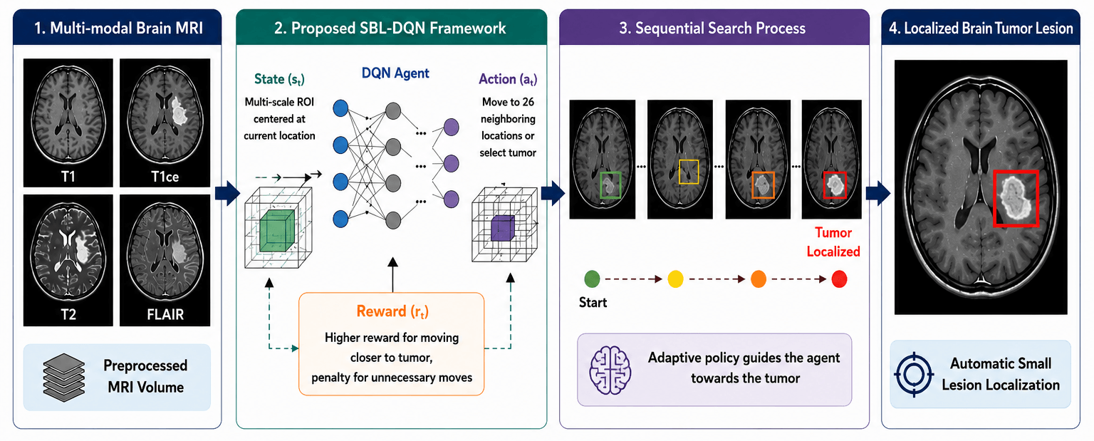
Brain tumor localization in magnetic resonance imaging remains challenging for small, low-contrast lesions. This study presents Small Brain Lesion Deep Q Network (SBL-DQN), a two-dimensional lesion-wise reinforcement-learning framework that refines an axis-aligned region of interest on a co-registered axial multi-channel MRI slice. In the current evaluation protocol, voxel reference masks are resampled, processed by 26-connected-component analysis, and filtered using a maximum in-plane diameter of 10 mm; one axial episode is then executed for each reference-enumerated component. The mask and component coordinates are not included in the DQN state, but ground-truth annotations are required to generate the evaluation candidate list and to compute reward traces and localization metrics. Accordingly, the reported experiment is oracle-conditioned lesion-wise localization refinement rather than autonomous lesion detection. The agent uses seven actions (up, down, left, right, zoom in, zoom out, and stop) and does not move through the anatomical z-axis. On the internal BraTS 2025 evaluation, the complete configuration yielded 97.46% localization accuracy, 97.23% precision, 96.88% recall, 97.05% F1-score, mean Intersection over Union of 0.834, AP@0.5 of 0.961, and center localization error of 0.91 mm. BraTS-METS 2025 and QIN-GBM-TREATMENT-RESPONSE remain qualitative transfer examples. Component-wise ablation, eligible-lesion accounting, and a balanced failure-case archive were not retained in the available experimental record; therefore, no individual component contribution, autonomous detection performance, or comprehensive robustness claim is made.Graphic Abstract
Keywords
Brain tumors remain one of the most challenging neurological disorders because their early detection and accurate localization are essential for diagnosis, treatment planning, surgical navigation, and post-treatment monitoring [1]. Magnetic resonance imaging (MRI) is the primary imaging modality for brain tumor assessment owing to its excellent soft-tissue contrast and ability to visualize anatomical structures using multiple imaging sequences [2]. Nevertheless, the localization of small brain tumor lesions remains particularly difficult due to their limited size, irregular morphology, heterogeneous intensity distribution, and similarity to surrounding healthy tissues. These characteristics often reduce the sensitivity of conventional computer-aided diagnosis systems and increase the likelihood of missed detections, especially during the early stages of disease progression [3]. Consequently, the development of intelligent localization approaches capable of accurately identifying small lesions has become an important research direction in medical image analysis and clinical decision support systems [4]. Recent advances in artificial intelligence have demonstrated considerable potential for improving the efficiency and reliability of automated brain tumor analysis, thereby supporting radiologists in routine clinical practice.
Deep learning has substantially advanced medical image interpretation by enabling automatic extraction of hierarchical image representations without the need for handcrafted features. Convolutional neural networks, encoder-decoder architectures, attention mechanisms, and transformer-based models have achieved remarkable performance in various brain tumor segmentation and classification tasks [5,6]. Despite these successes, many existing approaches rely on exhaustive dense prediction or sliding-window strategies that require significant computational resources and frequently struggle to localize very small lesions with high precision [7]. Moreover, these supervised models generally treat localization as a static prediction problem and do not explicitly learn sequential search behaviors that progressively refine lesion positions. Reinforcement learning provides an attractive alternative by formulating localization as a sequential decision-making process in which an intelligent agent continuously interacts with an imaging environment to identify the optimal lesion location through a sequence of actions [8]. Such an adaptive search strategy can substantially reduce unnecessary image exploration while improving localization efficiency and robustness.
Deep Q Networks (DQNs) integrate deep neural function approximation with Q-learning, providing a principled framework for learning sequential localization policies directly from high-dimensional medical images. The application of DQN-based strategies to brain tumor localization, however, predates the present study. Stember and Shalu [9] investigated a DQN/Q-learning formulation using 30 two-dimensional contrast-enhanced BraTS slices for training and an independent set of 30 slices for evaluation, with each image containing a single lesion. Their formulation represented localization as navigation within a 60 × 60-pixel gridworld using three discrete actions, namely stay, move down, and move right, and considered localization successful when the agent reached a grid cell overlapping the lesion. In contrast, the proposed SBL-DQN formulates the task as variable-size ROI refinement over co-registered axial multi-channel MRI and employs seven actions for bidirectional translation, zooming, and termination. Its state representation combines the current ROI patch with ROI geometry, normalized episode progress, and previous-action information, while the final prediction comprises a two-dimensional bounding box and lesion centroid. Importantly, the evaluation protocol uses reference masks to enumerate lesion components and identify component-intersecting axial slices before individual episodes are initialized. Accordingly, SBL-DQN is evaluated as an oracle-conditioned lesion-wise localization and refinement framework rather than an autonomous lesion detection system. Its contribution therefore lies in the proposed sequential ROI refinement formulation and its controlled internal evaluation.
Automatic analysis of brain tumors from magnetic resonance imaging has received significant attention due to its potential to improve diagnostic accuracy and reduce the workload of radiologists. Early computer-aided diagnosis systems primarily relied on handcrafted features describing texture, intensity, edge information, and shape characteristics, followed by conventional machine learning classifiers. Although these approaches achieved reasonable performance under controlled conditions, their ability to generalize across different imaging protocols and tumor characteristics remained limited [10]. The emergence of deep learning has fundamentally transformed brain tumor analysis by enabling end-to-end feature learning directly from medical images [11]. Convolutional neural networks have become the dominant architecture for tumor detection, classification, and segmentation because of their strong capability to extract hierarchical spatial representations from MRI data [12]. Multi-scale feature extraction strategies have further enhanced the detection of lesions exhibiting substantial variations in size and appearance [13]. More recently, transformer-based architectures have demonstrated promising performance by modeling long-range contextual dependencies that complement conventional convolutional operations.
Brain tumor localization has traditionally been addressed using object detection and semantic segmentation frameworks. Region proposal networks, two-stage detectors, and single-stage detection algorithms have been successfully adapted to identify tumor regions within MRI scans [14]. Encoder-decoder segmentation networks have also been widely employed to generate dense lesion masks while simultaneously providing localization information [15]. Several studies have incorporated attention mechanisms to emphasize diagnostically relevant regions and suppress background structures, thereby improving localization precision [16–18]. Hybrid convolution-transformer architectures have further improved feature representation by combining local spatial information with global contextual modeling [19]. Despite these advances, accurate localization of small brain tumor lesions remains challenging because of low lesion contrast, irregular boundaries, substantial anatomical variability, and severe class imbalance between tumor and healthy tissues [20]. Consequently, existing supervised learning approaches often experience reduced sensitivity when detecting small lesions occupying only a limited portion of the brain volume.
Reinforcement learning has been used for anatomical landmark localization, lesion search, and medical-image navigation [8,21,22]. Direct precedent exists for brain tumor localization: Stember and Shalu [9] showed that a DQN/Q-learning agent could localize one lesion on a two-dimensional contrast-enhanced MRI slice using a coarse gridworld and a very small training set. That study output a lesion-overlapping grid location, whereas SBL-DQN refines a variable-size 2D bounding box by translation and zoom actions. SBL-DQN further includes ROI geometry and action history in the state and combines overlap, centroid-distance, size, step, and terminal terms in the reward. These are formulation-level distinctions, not evidence that any individual element independently causes the reported improvement.
Although dense segmentation and detection networks provide comprehensive spatial predictions, they process large portions of the image and optimize a different output from a sequential localization policy [6,13,23,24]. The present study therefore evaluates SBL-DQN primarily against reinforcement learning baselines on the same lesion-wise localization task. The intended contribution is a reproducible 2D ROI-refinement formulation for small-lesion localization, with explicit limits on dimensionality, multi-lesion inference, and external validation.
This study presents SBL-DQN (Small Brain Lesion Deep Q Network), a two-dimensional reinforcement-learning framework for oracle-conditioned lesion-wise localization on axial MRI slices. The input is a co-registered multi-channel axial slice, represented as C × H × W, with T1, T1ce, T2, and FLAIR channels when available. Each episode operates within one axial plane; neither the state transition nor the action space changes the anatomical slice index. MRI volumes organize subjects and supply reference lesion components, whereas the DQN observation and predicted bounding box are two-dimensional. At evaluation, the candidate list is generated from the ground-truth mask rather than from an image-only proposal model; the method must therefore not be interpreted as autonomous lesion detection.
Fig. 1 separates candidate construction from ROI refinement. For training and internal evaluation, each resampled voxel reference mask is converted to the whole-tumor union, decomposed using three-dimensional 26-connectivity, and filtered by the prespecified maximum in-plane diameter criterion of 10 mm. An axial slice intersecting each eligible component is selected, and one brain-field-initialized episode is created for that assigned component. No learned proposal network, thresholded image-only detector, or unsupervised connected-component generator is used at test time. The DQN state excludes the mask and component coordinates, but the evaluation harness requires the reference annotation to enumerate targets and compute the reward trace and outcome metrics. Consequently, all quantitative results in this manuscript describe oracle-conditioned lesion-wise refinement, not autonomous examination-level detection.
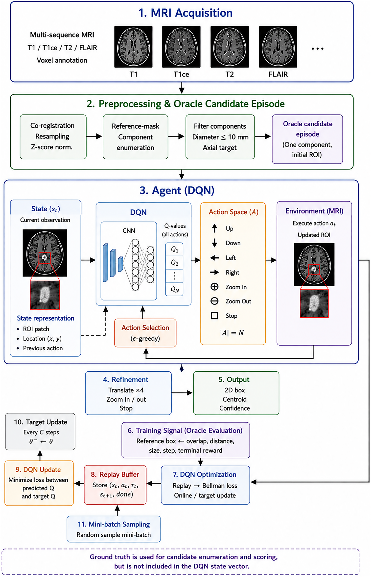
Figure 1: Oracle-conditioned workflow of SBL-DQN for reference-defined axial brain tumor component localization.
Prior to reinforcement learning, all MRI volumes underwent identical preprocessing procedures to reduce inter-subject variability. Skull stripping was first performed to eliminate non-brain tissues, followed by N4 bias field correction to compensate for low-frequency intensity inhomogeneity. Each MRI volume was subsequently registered to a common anatomical space and resampled to an isotropic voxel spacing of 1 mm × 1 mm × 1 mm. Intensity normalization was then applied using Z-score normalization
where
Unlike conventional supervised localization methods, SBL-DQN formulates lesion localization as a Markov Decision Process (MDP)
where
The state representation preserves local appearance, two-dimensional ROI geometry, normalized episode progress, and action history. Each state is defined as
where I(ROI) ∈ RC×H×W is the current multi-channel axial ROI patch; (xt, yt) is the in-plane ROI center; wt and ht are its width and height; τt = t/T is normalized episode progress; and at−1 is the previous action. No z-coordinate or ROI depth is included because the environment is two-dimensional.
The action space contains seven discrete actions: Move Up, Move Down, Move Left, Move Right, Zoom In, Zoom Out, and Stop. Move Forward and Move Backward are not implemented.
Translation actions update the in-plane ROI center, and zoom actions change its width and height while preserving the axial slice. Coordinates are clipped to the brain-image boundary. Stop terminates the episode and returns the final 2D bounding box (x, y, w, h), centroid (x, y), and confidence score P*.
Fig. 2 details the interaction loop for one reference-enumerated target. The episode begins on the target-intersecting axial slice with a standardized ROI covering the brain field. The agent observes the multi-channel patch, 2D geometry, normalized step index, and previous-action code; it then estimates seven Q-values and executes an in-plane translation, zoom, or stop action. During evaluation, network weights are frozen and actions are selected greedily. The reference target is not an observed state feature, but the evaluation harness uses it to calculate the reported reward trace and success metrics. The episode ends when Stop is selected or 300 actions have been executed.
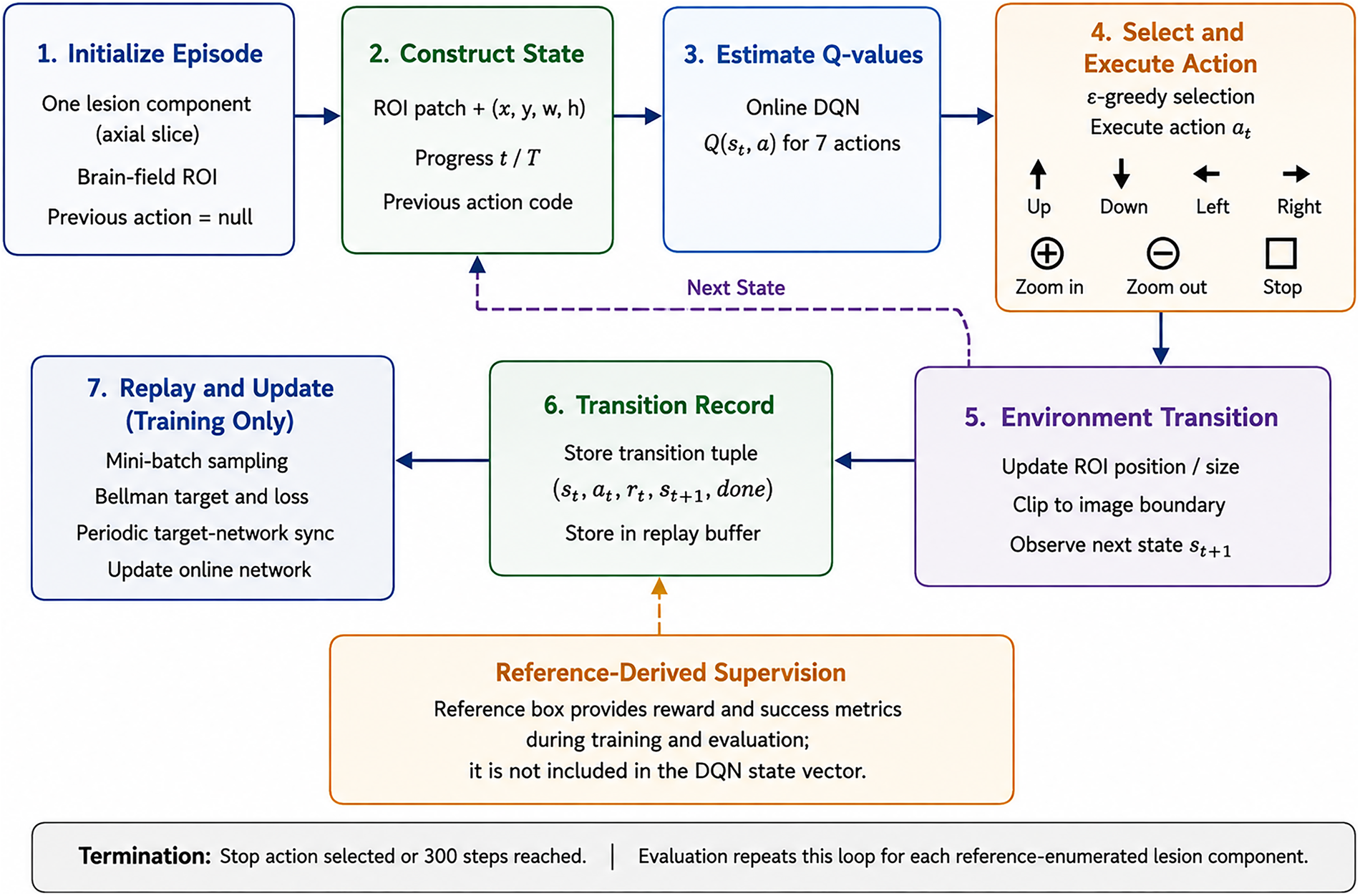
Figure 2: Oracle-conditioned lesion-wise agent–environment interaction on one axial MRI slice.
The reward function is one of the key components of the proposed SBL-DQN framework because it directly guides the search behavior of the reinforcement learning agent. Rather than relying on a single localization criterion, a composite reward was designed to simultaneously optimize localization accuracy, convergence speed, and search efficiency. The reward at each interaction step is defined as
where α, β, κ, δ, and η are reward weights. The symbol κ replaces the earlier use of γ as a reward coefficient; γ is reserved exclusively for the MDP discount factor.
The Intersection-over-Union reward encourages progressive overlap between the predicted search window
where,
To further improve localization precision, a centroid distance reward is introduced
where
To encourage efficient exploration, each localization step receives a small penalty
which discourages unnecessarily long trajectories. The terminal term is RStop = +1 when IoU ≥ 0.50 and CLE ≤ 5 mm and RStop = −1 otherwise. The numerical reward weights, step penalty, translation increment, zoom factor, and oracle candidate source are reported in Table 1.
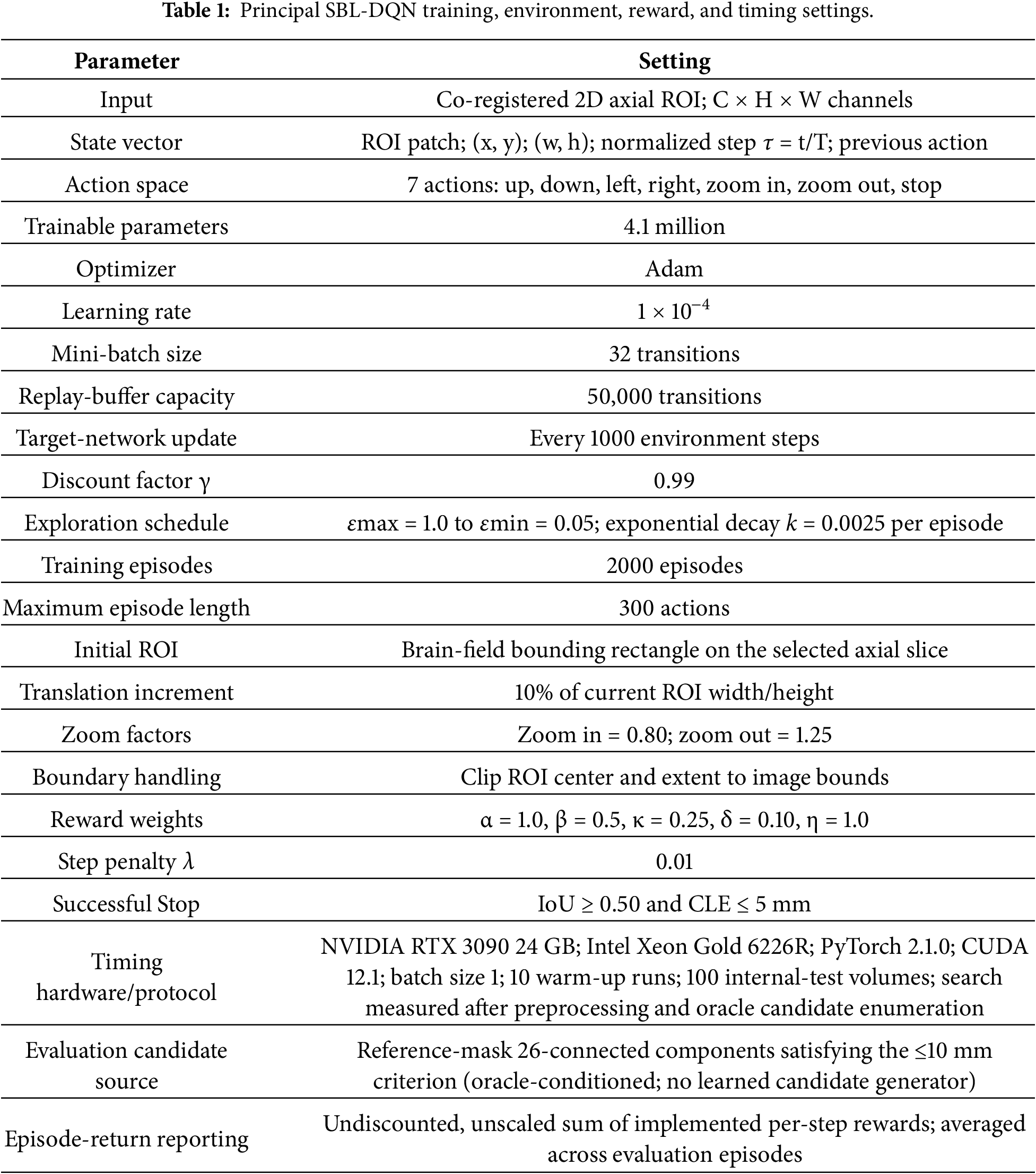
Reward-scale verification. Because R(IoU) is in [0, 1], R(Dist) is in [−1, 0], R(Size) is in [−1, 0], and R(Step) = −0.01, the weighted nonterminal reward is bounded by −0.751 ≤ R_t ≤ 0.999 for the coefficients in Table 1. When the terminal Stop term is applied once, the terminal-step range is −1.751 ≤ R_t ≤ 1.999. Therefore, for an episode of at most 300 actions, the undiscounted cumulative episode return cannot exceed 300.700 (and is no lower than −226.300 under these bounds). Average Episode Reward is calculated from the unscaled sum of the implemented per-step rewards; no multiplier, percentage conversion, or reward rescaling is applied.
Fig. 3 depicts the DQN used for the two-dimensional implementation. The network receives a C × H × W axial ROI patch, extracts convolutional features, applies global average pooling, and concatenates the image vector with ROI geometry, normalized episode progress, and previous-action encoding. Fully connected layers produce one Q-value for each of the seven actions. The lesion mask, assigned component coordinates, and through-plane position are not network inputs. A separate target network is used only to construct the Bellman target during training.
where
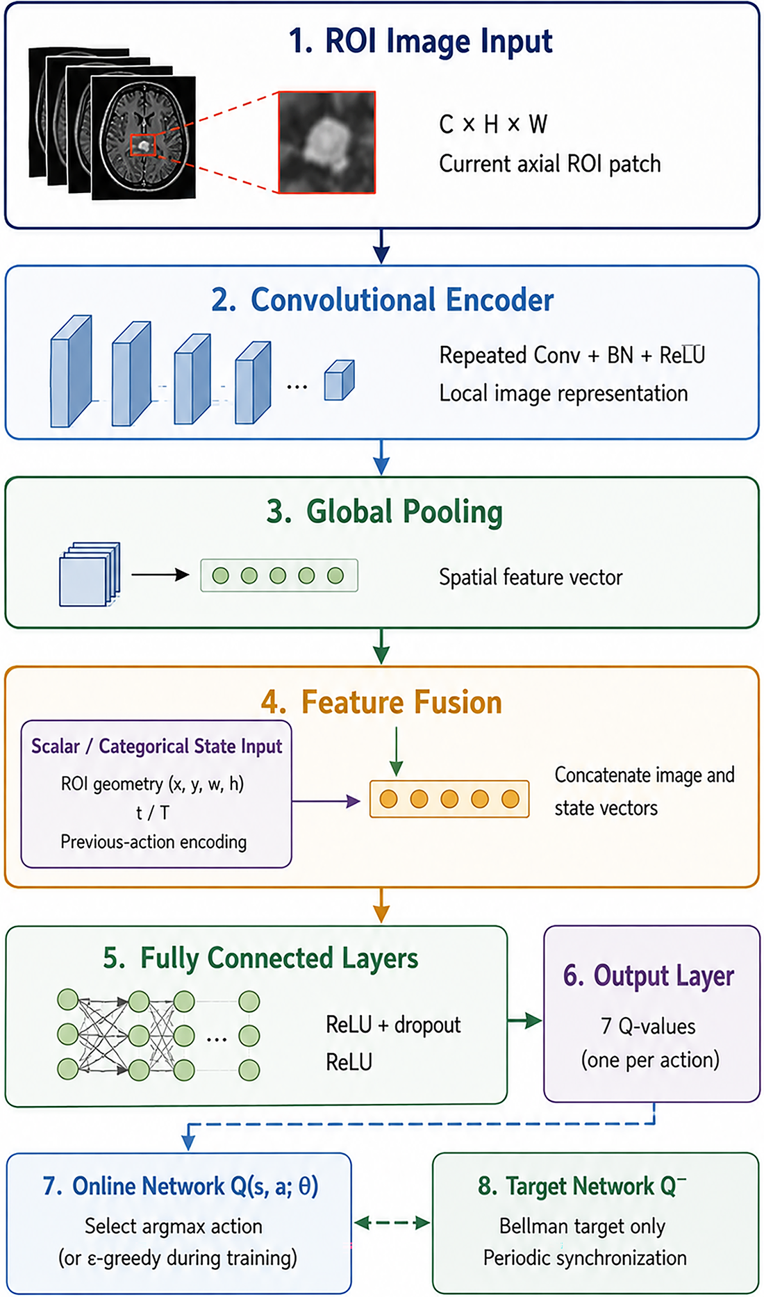
Figure 3: DQN architecture for two-dimensional multi-channel ROI localization without reference-mask state input.
To balance exploration and exploitation during training, an ε-greedy policy is adopted. The probability of selecting a random exploratory action is given by
where
The proposed SBL-DQN utilizes experience replay to improve sample efficiency and reduce temporal correlations among consecutive observations. Each transition
while the network parameters are optimized by minimizing the Bellman loss.
The target network parameters are periodically synchronized with the online network, thereby reducing oscillations during training and improving convergence stability. Algorithm 1 summarizes the learning and evaluation procedures. Each training episode samples one reference lesion component and one axial slice intersecting that component; the environment exposes only the current ROI patch and two-dimensional state variables to the agent, while the selected reference box is used to compute the training reward. For internal evaluation, the reference mask is again processed to enumerate eligible components and select target-intersecting slices before the frozen policy is executed. One greedy episode is run for each reference-enumerated component and one prediction is matched to that assigned target. Thus, ground truth is not a DQN input but is required by the evaluation harness. The protocol evaluates refinement of known lesion targets and cannot estimate how many lesions are present in an unseen volume.
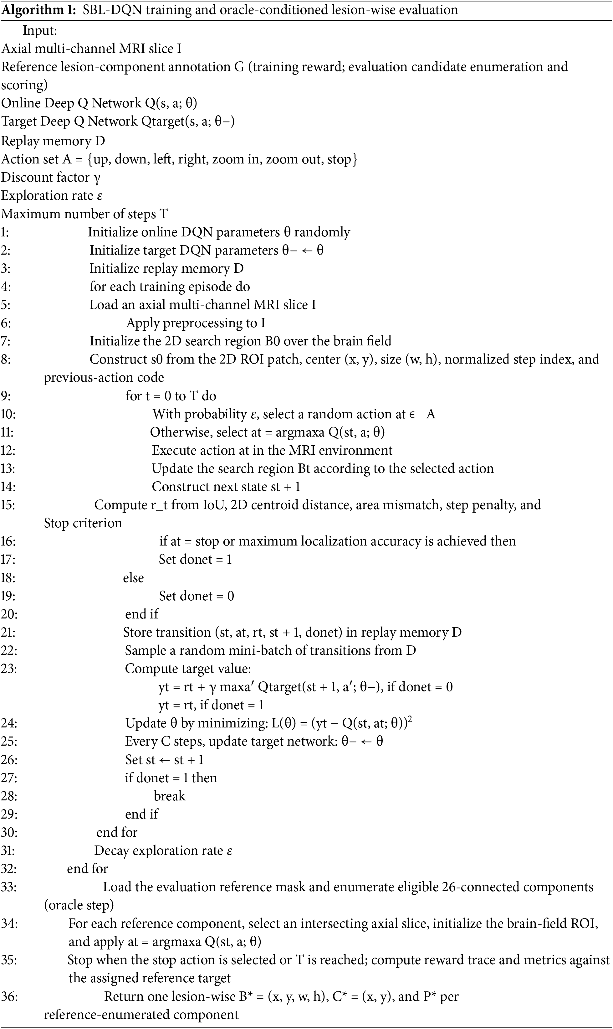
Table 1 consolidates the DQN, environment, reward, timing, and candidate-source settings used in the reported protocol. Candidate enumeration from reference masks is completed before the timed DQN search; the 0.18 s value therefore excludes preprocessing and oracle target construction.
Three public collections were used. BraTS 2025 adult glioma [25] was the development source for training and internal evaluation. BraTS-METS 2025 [26] and QIN-GBM-TREATMENT-RESPONSE [27] were used for qualitative transfer examples and were not used for hyperparameter selection. This wording distinguishes the internal quantitative evidence from the external qualitative evidence and avoids an unsupported claim of strong cross-dataset generalization.
Fig. 4 illustrates representative axial MRI samples and the construction of lesion localization targets across the three datasets employed in this study. The BraTS 2025 examples demonstrate complementary T1ce and FLAIR appearances of adult glioma, with expert voxel-level annotations converted into lesion contours, two-dimensional bounding boxes, and centroid targets. BraTS-METS 2025 presents a more complex multi-lesion scenario in which spatially separated brain metastases are represented as distinct connected components, allowing an individual bounding box and centroid to be defined for each lesion. The QIN-GBM-TREATMENT-RESPONSE example further demonstrates the variability of lesion appearance under a different MRI acquisition setting. Across the datasets, green contours denote reference lesion annotations, yellow rectangles represent the corresponding localization targets, and red points identify lesion centroids. Thus, Fig. 4 visually demonstrates how heterogeneous voxel-level or reference-region annotations are transformed into standardized lesion-wise localization targets while preserving differences in MRI modality, lesion morphology, size, and multiplicity across the evaluated datasets.
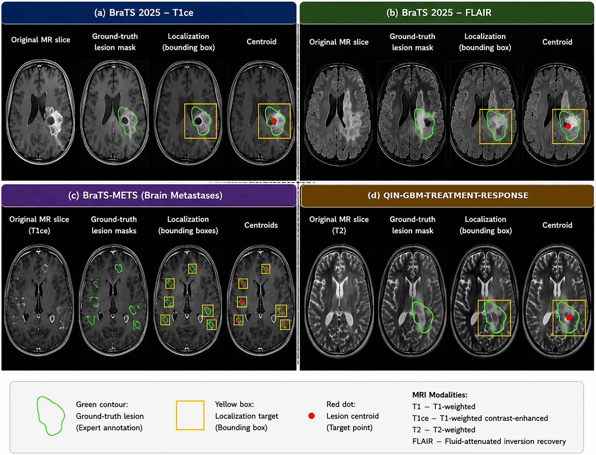
Figure 4: Representative axial MRI localization targets from BraTS 2025, BraTS-METS 2025, and QIN-GBM-TREATMENT-RESPONSE.
Table 2 summarizes the three public MRI collections, their imaging modalities, cohort characteristics, lesion types, annotation strategies, spatial resolution, and specific roles within the present evaluation protocol. BraTS 2025 Task 1 contains 2877 multi-institutional pre- and post-treatment adult glioma cases with co-registered T1, T1ce, T2, and FLAIR images and voxel-level labels [25]. For this localization study, the whole-tumor union of nonzero tumor labels was used. Three-dimensional 26-connected components were generated directly from the reference masks after the patient-level split; each intersecting two-dimensional component was converted to a minimum enclosing 2D bounding box and centroid. This reference-derived procedure was used during internal testing and constitutes oracle candidate enumeration. Patient identifiers, not slices or components, were partitioned before augmentation to prevent leakage.
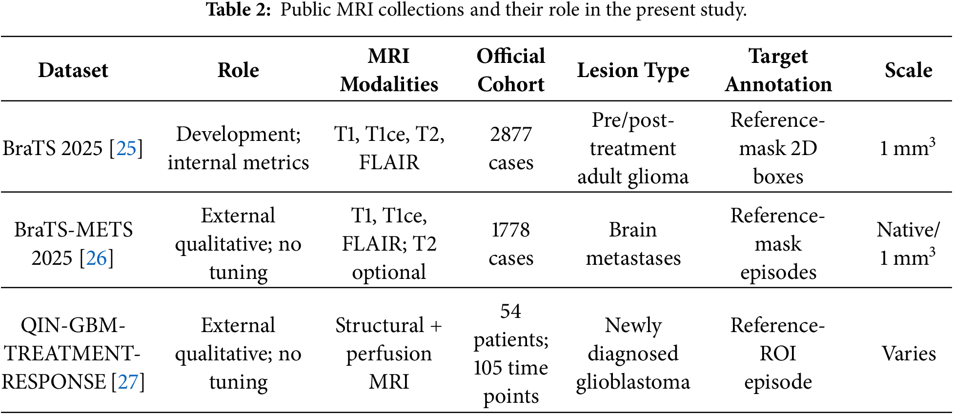
BraTS-METS 2025 includes 1778 pre- and post-treatment brain-metastasis cases (1296 training, 179 validation, and 303 hidden testing cases in the official challenge organization) and requires T1, post-contrast T1, and FLAIR, with T2 optional [26]. Individual enhancing-tumor connected components were treated as distinct targets. QIN-GBM-TREATMENT-RESPONSE contains multiparametric MRI from 54 patients across 105 acquisition time points [27].
A small lesion was operationalized as a connected target with maximum in-plane diameter ≤10 mm after resampling. Eligible targets were identified from the reference-mask connected-component manifest after resampling. Each positive episode contains one assigned component and one intersecting axial slice; other disconnected components are excluded from that episode’s reward target. Multi-lesion volumes are evaluated by running one episode per reference-enumerated component and matching one prediction to the assigned component.
Table 3 summarizes the study-specific patient-level partitioning and evidence roles across the three datasets, with BraTS 2025 divided into 2014 training (70%), 432 validation (15%), and 431 internal test subjects (15%) before oracle candidate enumeration, thereby preventing patient-level leakage. In contrast, BraTS-METS 2025 and QIN-GBM-TREATMENT-RESPONSE were excluded from model training and hyperparameter tuning and were used only to provide selected qualitative external examples. For BraTS-METS 2025, evaluation was conducted through lesion-wise target-conditioned episodes, whereas QIN-GBM-TREATMENT-RESPONSE was similarly examined without adaptation or parameter optimization. This partitioning strategy clearly separates model-development evidence from external qualitative evidence and ensures that the reported internal quantitative metrics originate exclusively from the held-out BraTS 2025 test partition.
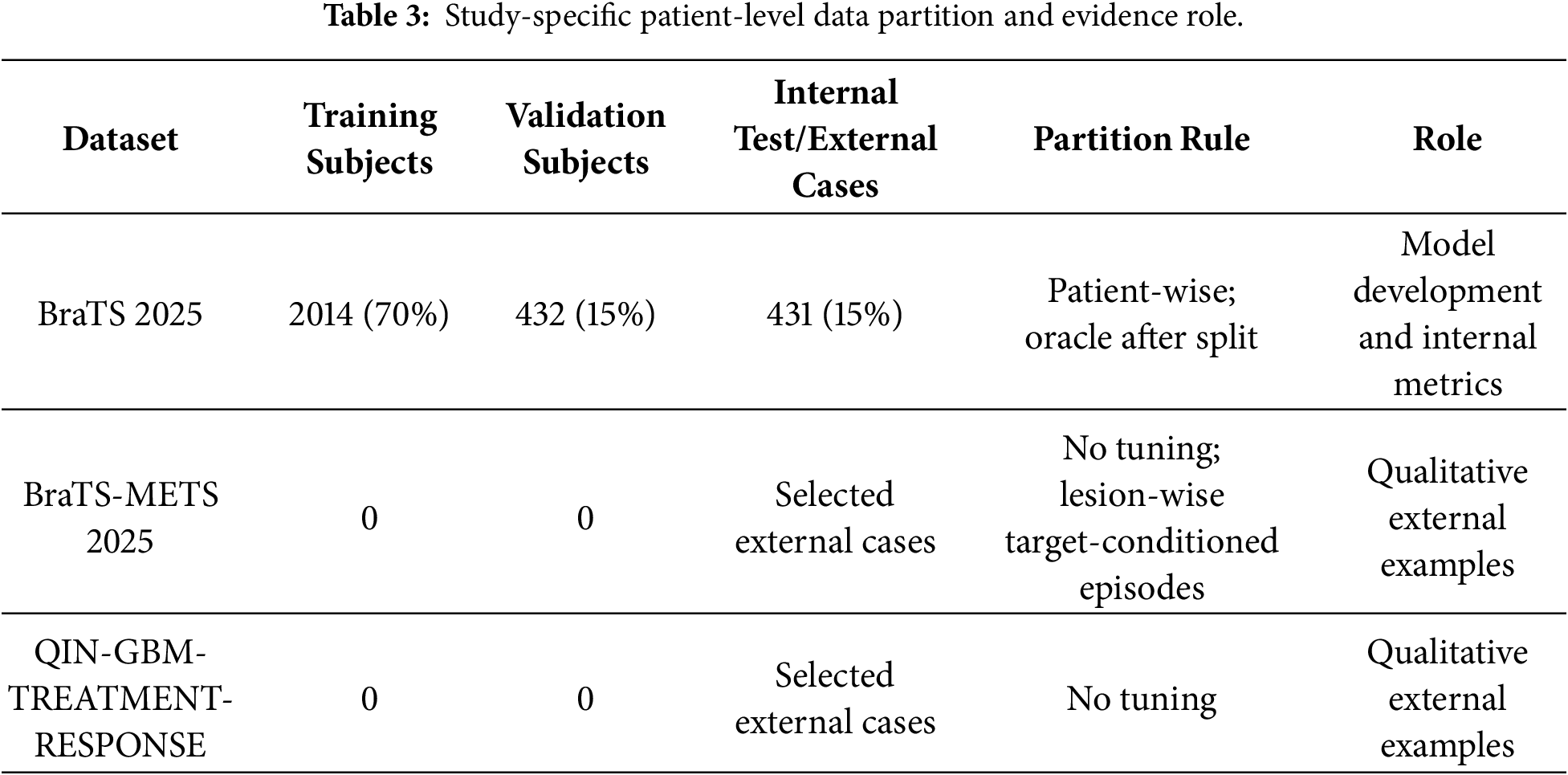
The evaluation is organized around the evidence available for the oracle-conditioned lesion-wise two-dimensional protocol. Quantitative localization, convergence, and computational results are reported for the internal BraTS 2025 evaluation. Selected BraTS-METS 2025 and QIN-GBM-TREATMENT-RESPONSE cases are qualitative only. Reinforcement-learning baselines are compared under the same reference-mask candidate-episode protocol, whereas segmentation studies are retained only for task positioning because their voxel-mask outputs and metrics are not directly commensurate.
The evaluation unit is one oracle-conditioned axial candidate episode. A positive episode is assigned one reference lesion component; a background-only episode is negative. A prediction is a true positive when its final box satisfies both IoU ≥ 0.50 and CLE ≤ 5 mm with the assigned lesion. A positive episode that fails either criterion is a false negative. A terminated box on a background episode, or an unmatched duplicate box, is a false positive. A background episode that ends without a box above the confidence threshold P* = 0.50 is a true negative. One-to-one matching prevents a reference component from generating more than one true positive. These are conditional episode-level refinement statistics; they are not examination-level sensitivity or specificity for autonomous lesion discovery.
Accuracy, Precision, Recall, and F1-score were computed from the episode-level TP, TN, FP, and FN definitions above [28]. Accuracy is
where TP, TN, FP, and FN are counts of correctly localized positive episodes, correctly rejected background episodes, false localizations, and missed positive episodes, respectively. Precision and Recall are
whereas Recall measures the proportion of correctly localized lesions among all actual lesions,
To balance both precision and recall, the harmonic mean of these measures was calculated using the F1-score,
These metrics collectively evaluate the capability of the proposed SBL-DQN framework to accurately identify lesion locations while minimizing both missed detections and false localization results.
Localization geometry was assessed with two-dimensional Intersection over Union (IoU) and Center Localization Error (CLE) [29]. IoU is the overlap between predicted box Bp and ground-truth box Bg box on the axial plane,
Higher IoU indicates better in-plane overlap. CLE is the Euclidean distance between predicted centroid Cp and reference centroid Cg in millimeters,
where (xp, yp) and (xg, yg) are the predicted and reference in-plane centroid coordinates after conversion to millimeters. Average precision is reported at a single IoU threshold of 0.50, not as mean AP across thresholds,
where
Since SBL-DQN is based on reinforcement learning, several reinforcement learning-specific evaluation measures were additionally employed. The Episode Success Rate (ESR) quantifies the proportion of localization episodes that successfully reached the lesion before the maximum episode length,
The Average Episode Reward (AER) evaluates the overall effectiveness of the learned policy by averaging cumulative rewards over all testing episodes [30],
where
where
Computational efficiency was assessed with search latency and model complexity. Search latency is the elapsed time required by the DQN to process a predefined volume-level candidate set after preprocessing and candidate enumeration; it is not end-to-end examination time. Model complexity is the number of trainable parameters.
where
Quantitative convergence, localization, search-efficiency, and computational results were obtained on the internal BraTS 2025 oracle-conditioned evaluation. BraTS-METS 2025 and QIN-GBM-TREATMENT-RESPONSE are represented by selected qualitative examples only; no dataset-level external metric table is claimed. Comparisons with Vanilla DQN, Double DQN, Dueling DQN, and PPO use the same reference-enumerated lesion-wise protocol. Segmentation methods are discussed only to position the output task and are not treated as commensurate baselines.
Fig. 5 illustrates the evolution of average episode return for SBL-DQN and four baseline methods across 1800 training episodes. All approaches begin with strongly negative returns, indicating inefficient exploratory actions, limited localization accuracy, and frequent accumulation of distance, size, and step penalties during early training. SBL-DQN improves most rapidly, crosses the zero-return threshold at approximately 150 episodes, and reaches about 40 by 500 episodes. Its return subsequently increases more gradually, stabilizing around 50–55 after roughly 1000 episodes, with only small oscillations thereafter. Double DQN exhibits the second-fastest improvement, becomes positive near 300 episodes, and converges around 20–25. Dueling DQN crosses zero later, at approximately 400 episodes, before plateauing near 15. PPO requires about 700–750 episodes to obtain positive returns and ultimately stabilizes between 8 and 10. Vanilla DQN improves steadily but remains slightly negative at the end of training, suggesting less efficient search behavior and weaker terminal localization performance. The widening separation between SBL-DQN and the baselines during the first 800 episodes demonstrates superior learning efficiency, while the flatter trajectories after approximately 1200 episodes indicate convergence. Moreover, the limited late-stage fluctuations suggest stable policy updates rather than reward divergence. Overall, SBL-DQN achieves the highest final return and the most favorable balance between localization quality, trajectory efficiency, and successful stopping behavior under the common 300-action protocol for small brain lesion localization.
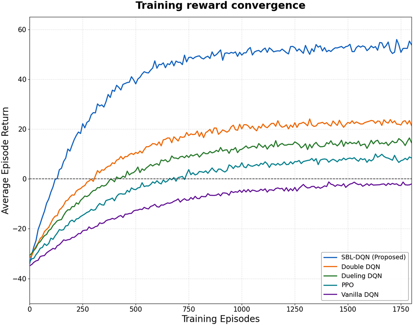
Figure 5: Training reward convergence of the proposed SBL-DQN and baseline reinforcement learning methods.
Fig. 6 demonstrates the progressive improvement in localization accuracy achieved by SBL-DQN and the four baseline reinforcement learning methods on the BraTS 2025 validation set as training proceeds. The proposed SBL-DQN exhibits the fastest convergence, increasing from approximately 7% during the initial stage to 50% at 100 episodes and approximately 69% at 280 episodes. Its localization accuracy subsequently reaches approximately 85% at 600 episodes and approaches 90% by 800 episodes. Continued training produces smaller but consistent improvements, with accuracy exceeding 91% at 1000 episodes and ultimately reaching approximately 94.5% after 1800 episodes. Double DQN represents the strongest baseline, attaining approximately 86.5% at the end of training, corresponding to an approximately eight-percentage-point difference from SBL-DQN. Dueling DQN converges at approximately 78%, while PPO reaches approximately 65.7%. Vanilla DQN exhibits the weakest localization performance, plateauing near 47.5% despite continued training. Notably, the performance curves progressively flatten after approximately 1000–1200 episodes, indicating convergence toward stable localization policies. The consistently higher SBL-DQN trajectory across all training stages demonstrates superior learning efficiency and final localization performance under the common validation protocol, although these results characterize the complete configuration rather than the isolated effects of individual model components.
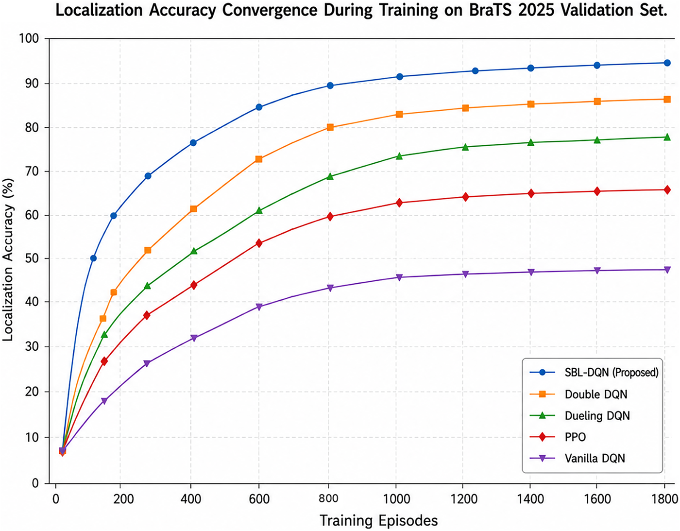
Figure 6: Localization accuracy convergence of the proposed SBL-DQN and baseline reinforcement learning methods on the BraTS 2025 validation set.
Fig. 7 demonstrates the evolution of mean Intersection-over-Union (IoU) during training on the BraTS 2025 validation set, revealing substantial differences in localization quality among the evaluated reinforcement learning methods. The proposed SBL-DQN exhibits the fastest initial improvement, achieving a mean IoU of approximately 0.38 after 100 episodes and 0.53 after 200 episodes. Performance continues to increase steadily, reaching approximately 0.74 at 600 episodes and 0.80 at approximately 1100 episodes. Beyond this stage, the curve gradually approaches a stable plateau, ultimately attaining a mean IoU of approximately 0.83. Double DQN provides the strongest baseline performance, converging near 0.73, approximately 0.10 below SBL-DQN. Dueling DQN reaches approximately 0.61, whereas PPO stabilizes near 0.49. Vanilla DQN demonstrates substantially weaker spatial localization, with its mean IoU plateauing at approximately 0.33. The progressive flattening of all curves after approximately 1200–1400 episodes indicates that additional training produces increasingly marginal improvements. Importantly, SBL-DQN maintains the highest mean IoU throughout the training process, demonstrating more accurate overlap between predicted and reference lesion regions under the common validation protocol. Nevertheless, the observed advantage represents the performance of the complete SBL-DQN configuration and cannot independently establish the contribution of its state representation, zoom operations, or individual reward components.
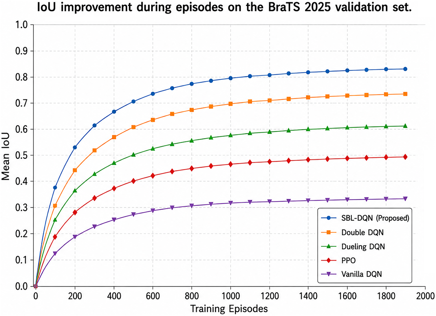
Figure 7: Mean intersection-over-union (IoU) convergence during training on the BraTS 2025 validation set.
Fig. 8 presents representative search trajectories generated by the proposed SBL-DQN for three lesion examples drawn from BraTS 2025, BraTS-METS 2025, and QIN-GBM-TREATMENT-RESPONSE, providing qualitative evidence of the learned sequential localization behavior. In each example, the agent begins with a relatively broad region of interest and progressively adjusts its spatial position and dimensions through successive actions. The intermediate steps demonstrate simultaneous translation toward the target and contraction of the search region, ultimately producing a predicted localization closely aligned with the ground-truth lesion. The corresponding trajectory plots further characterize this process in terms of the x- and y-coordinates and ROI area. Importantly, the third axis represents the changing two-dimensional ROI area rather than anatomical depth or movement between MRI slices. Across the three examples, ROI area generally decreases as the trajectory approaches the final localization, indicating progressive spatial refinement rather than exhaustive exploration of the image. The examples additionally suggest that the learned policy can accommodate differences in lesion position, appearance, and size across glioma, metastatic, and glioblastoma cases. Nevertheless, the BraTS-METS 2025 and QIN-GBM-TREATMENT-RESPONSE trajectories constitute selected qualitative examples. Consequently, they demonstrate the feasibility of transferring the learned search behavior to heterogeneous MRI cases but should not be interpreted as quantitative evidence of cross-dataset generalization.
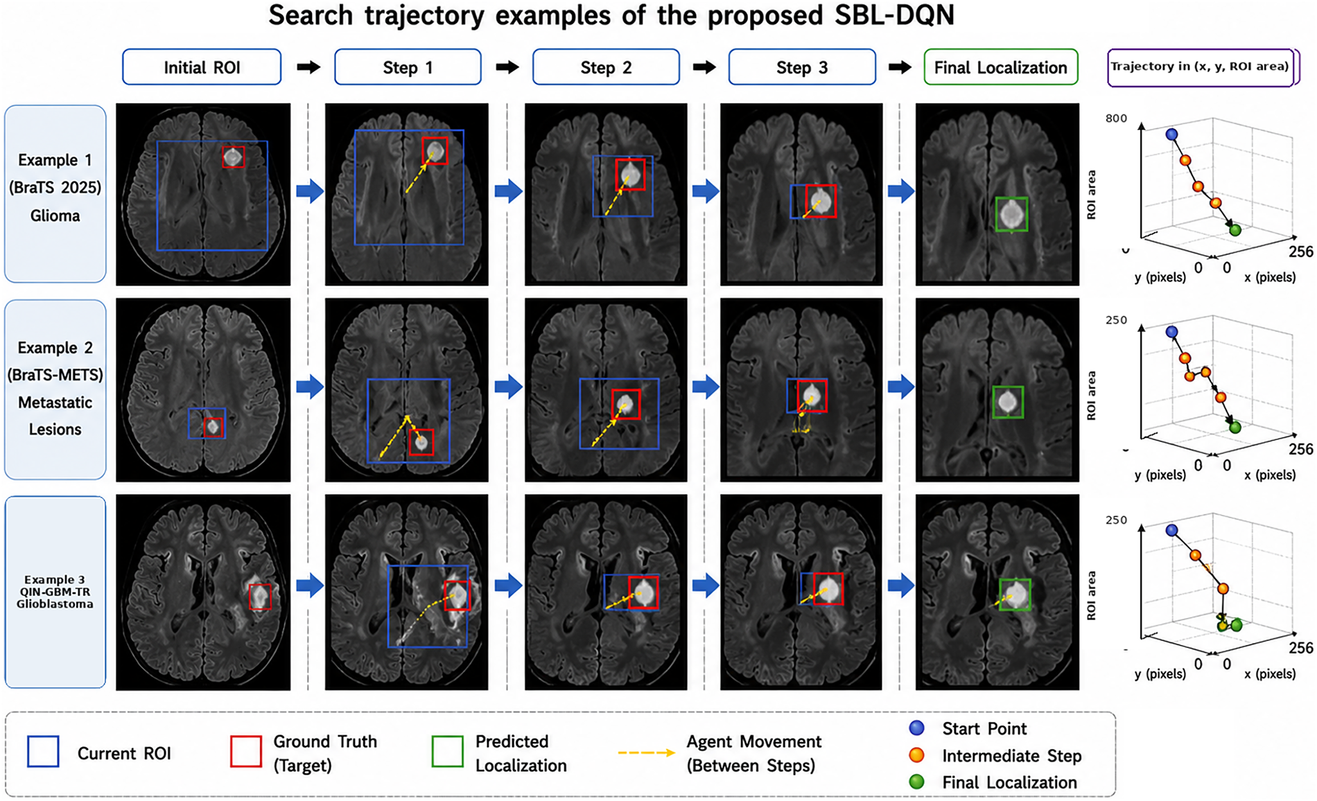
Figure 8: Representative two-dimensional search trajectories; the third axis denotes ROI area rather than anatomical depth.
Fig. 9 presents selected successful examples and Grad-CAM maps. Grad-CAM highlights image regions whose final-layer activations are associated with the selected action value [31]; it does not prove clinical interpretability, causal reasoning, or clinical relevance. The shown maps overlap the visible lesion in these examples, but no reader study or attribution-faithfulness analysis was performed. These cases should therefore be interpreted as model-inspection aids rather than clinical validation.
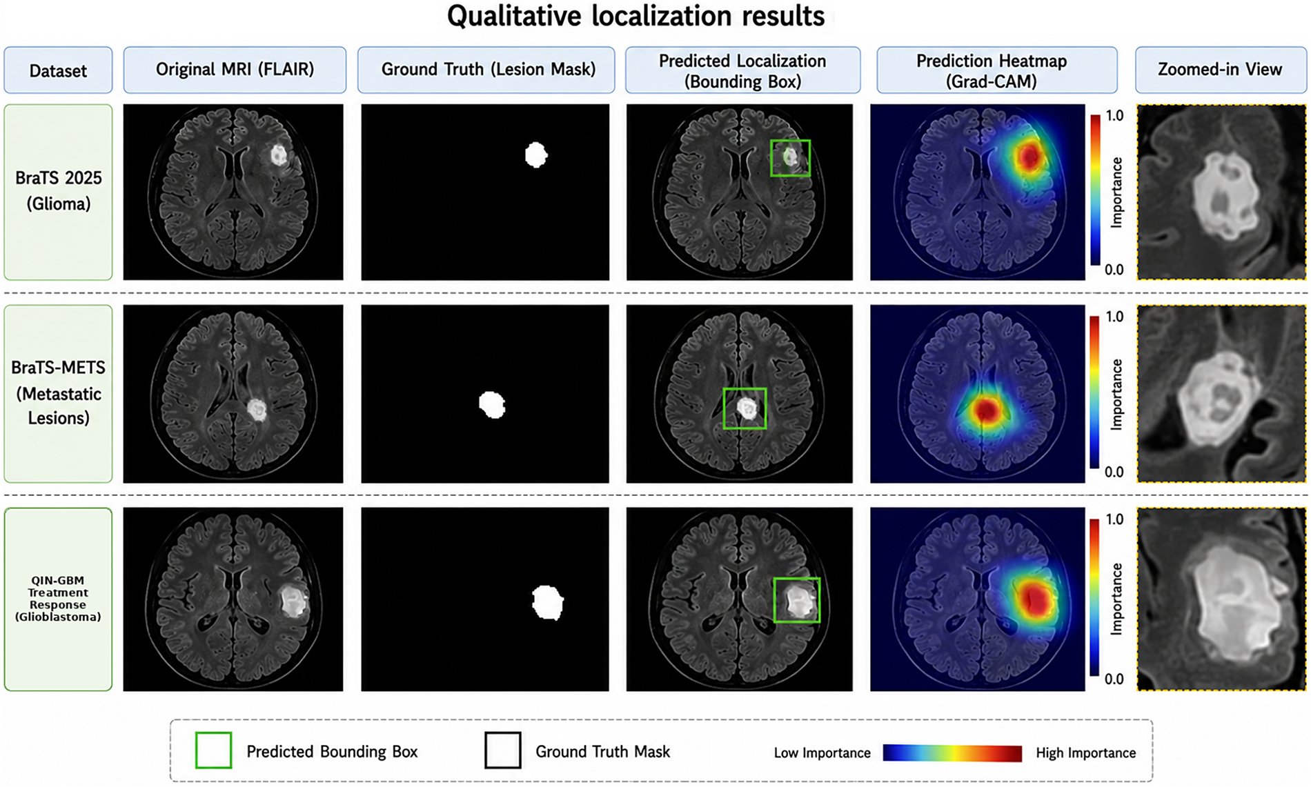
Figure 9: Illustrative localization and Grad-CAM maps on BraTS 2025, BraTS-METS 2025, and QIN-GBM-TREATMENT-RESPONSE.
5.3 Component-Wise Ablation and Failure-Case Evidence
The retained experiment record contains the complete SBL-DQN configuration and algorithm-level baselines, but no controlled component-wise reruns. The baseline curves cannot be re-labeled as ablations because they change the reinforcement-learning algorithm rather than isolating the proposed state representation, adaptive zoom strategy, or composite reward. A valid ablation must hold the patient split, initialization, training budget, random seeds, and evaluation candidate list fixed while comparing at minimum: (i) the full model; (ii) a state variant without ROI geometry, episode progress, and action history; (iii) a translation-and-stop variant without zoom actions; and (iv) an IoU-only reward variant. Until those runs are performed, the integrated results do not establish the independent contribution of any one component.
The available prediction archive contains selected successful cases only and does not retain the case-level boxes, masks, confidence scores, or residuals needed to sample false-positive, false-negative, low-IoU, and large-center-error examples. For a balanced audit, false positive is defined as a confident localization on a background episode or an unmatched duplicate; false negative is an assigned positive episode failing IoU ≥ 0.50 or CLE ≤ 5 mm; low IoU is 0 < IoU < 0.50; and large center error is CLE > 5 mm. A complete qualitative panel should sample equal numbers from these four categories together with successful controls from the locked internal test set. Fig. 9 is therefore identified only as a selected success panel. No missing cases or per-dataset counts have been manufactured, and claims of comprehensive robustness are withdrawn pending regeneration of the component manifest and per-case prediction archive.
Fig. 10 compares the complete SBL-DQN configuration with four reinforcement learning baselines. Fig. 10a reports AP@0.5 = 0.961 for SBL-DQN, followed by 0.913, 0.842, 0.741, and 0.576 for Double DQN, Dueling DQN, PPO, and Vanilla DQN. Fig. 10b reports final CLE near 0.9 mm, and Fig. 10c shows episode success approaching 95% at the 300-step limit. These values are internal results under the common lesion-wise protocol.
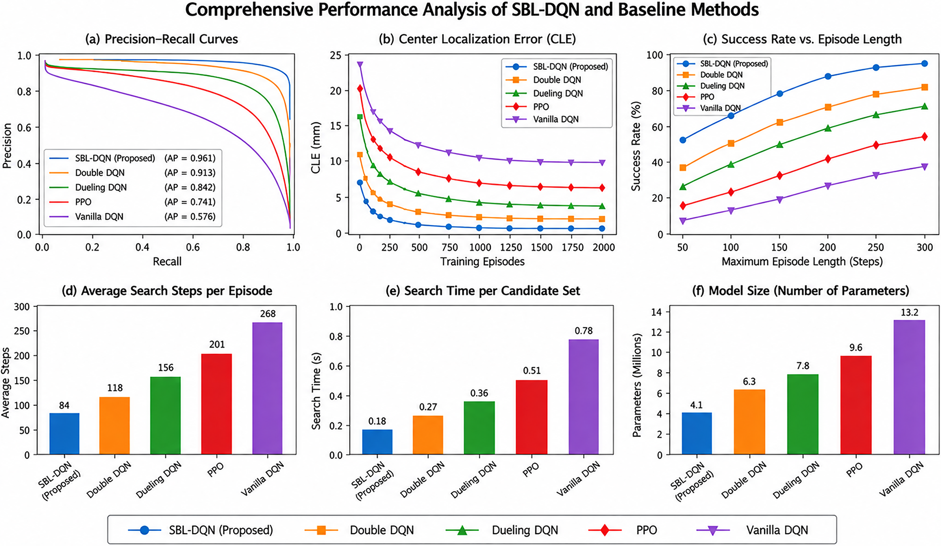
Figure 10: Comprehensive performance comparison of the proposed SBL-DQN and baseline reinforcement learning methods.
Fig. 10d–f reports 84 average search steps, 0.18 s search time per predefined volume-level candidate set, and 4.1 million trainable parameters for SBL-DQN. The 0.18 s value excludes image loading, registration, resampling, normalization, candidate enumeration, and visualization; it was measured with batch size 1 over 100 BraTS 2025 internal-test volumes on an NVIDIA RTX 3090 24 GB GPU with an Intel Xeon Gold 6226R CPU, using PyTorch 2.1.0 and CUDA 12.1 after 10 warm-up runs. It is therefore described as search latency rather than real-time or end-to-end clinical performance.
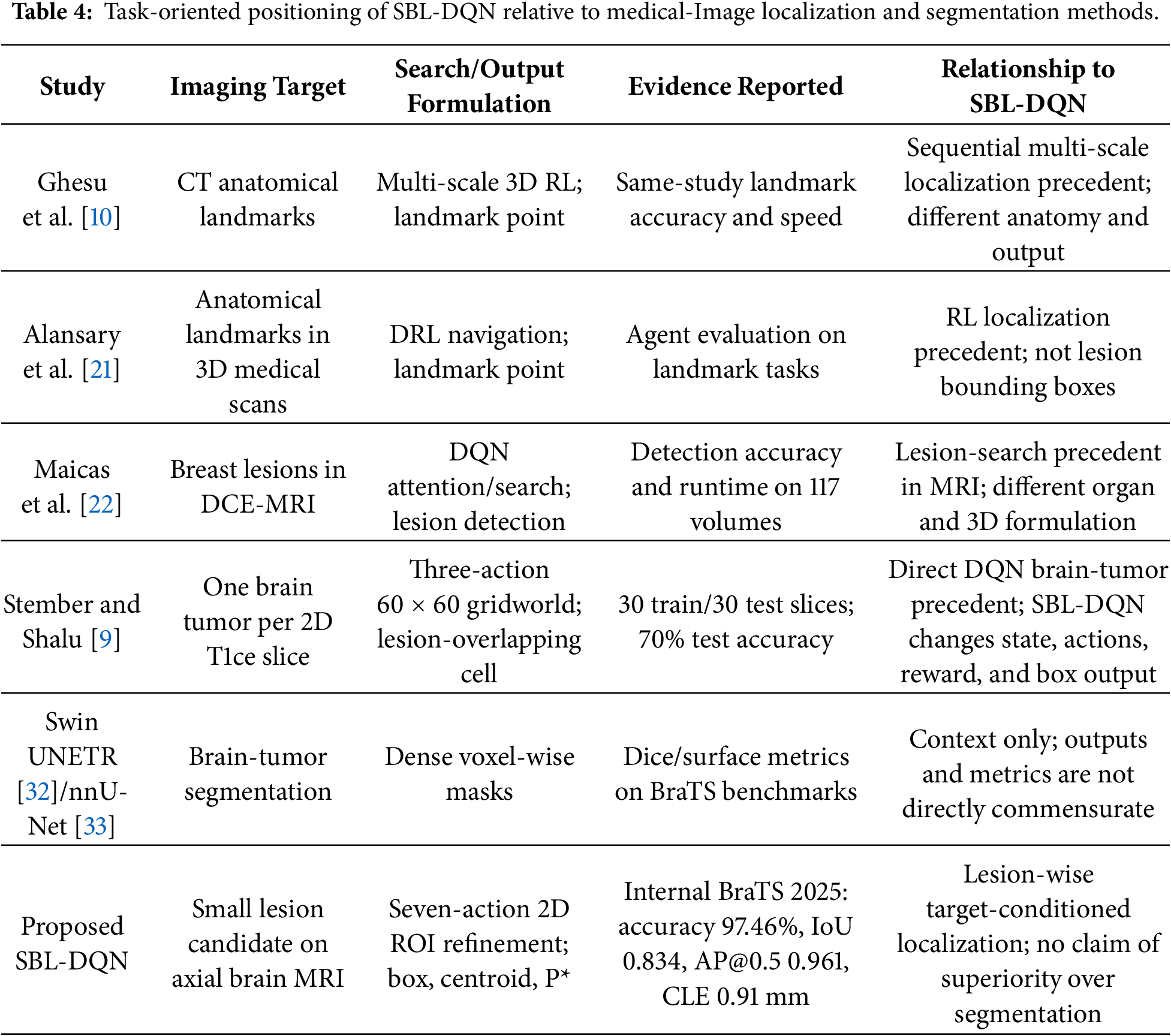
Table 4 positions SBL-DQN by task and output rather than combining non-commensurate numerical metrics. Stember and Shalu [9] are the closest direct precedent because they also use DQN/Q-learning for 2D brain tumor localization. Dense segmentation methods predict voxel masks and are therefore not head-to-head baselines for the sequential bounding-box task. No superiority over those segmentation methods is inferred from the comparison.
Overall, the internal experiments show that the complete SBL-DQN configuration converges faster and yields better oracle-conditioned lesion-wise localization metrics than the evaluated reinforcement-learning baselines. The present evidence does not isolate the effects of the state representation, zoom actions, or reward terms; it does not quantify autonomous candidate generation; and it does not provide dataset-level external validation or a balanced failure taxonomy. The conclusions are restricted accordingly.
The internal results support sequential two-dimensional ROI refinement for a reference-defined lesion target. SBL-DQN achieved higher reward, localization accuracy, and IoU than four reinforcement-learning baselines under the same oracle-conditioned protocol. These observations apply only to the integrated configuration. Because controlled component-wise runs are absent, the differences cannot be assigned independently to the expanded state, zoom actions, or composite reward.
The experiment is explicitly oracle-conditioned. Ground-truth masks are processed at test time to enumerate eligible connected components and select target-intersecting axial slices; one episode is then constructed for each assigned component. Although the agent state excludes the reference mask and target coordinates, the evaluation cannot be initiated without these annotations. The method therefore does not infer how many lesions are present in an unseen volume and is not an autonomous multiple-metastasis detector. A separately validated image-only candidate generator, duplicate suppression, and volume-level matching are required before autonomous inference can be claimed.
The external figures show that the trained policy can produce plausible boxes on selected BraTS-METS 2025 and QIN-GBM-TREATMENT-RESPONSE examples. Because dataset-specific quantitative estimates, confidence intervals, and balanced failure cases are not available, these examples are evidence of feasibility rather than strong generalization. Grad-CAM maps likewise indicate gradient-associated regions only and do not establish clinical interpretability or relevance [31].
Additional limitations include the two-dimensional axial formulation, oracle target enumeration, unavailable eligible-lesion counts, missing controlled component-wise ablation, absence of a retained balanced failure-case archive, and search-only rather than end-to-end timing. Future work should operate directly on three-dimensional volumes, discover and match multiple lesions without ground-truth enumeration, regenerate per-dataset component manifests, run fixed-protocol ablations across independent seeds, report confidence intervals, and include systematic failure cases before clinical translation is considered.
This study presented SBL-DQN, a two-dimensional deep reinforcement learning framework for iterative bounding-box refinement of reference-defined lesion components on axial multi-channel MRI. The agent operates through four translation actions, two scale-adjustment actions, and a termination action to progressively localize each assigned target. Under the oracle-conditioned BraTS 2025 internal evaluation protocol, SBL-DQN achieved 97.46% localization accuracy, 0.834 mean IoU, 0.961 AP@0.5, and 0.91 mm center localization error, outperforming the evaluated reinforcement-learning baselines. The framework integrates ROI-based image information, spatial state variables, adaptive action selection, experience replay, and composite reward optimization. Nevertheless, these results should be interpreted within the defined experimental setting. Reference masks are used to enumerate lesion candidates before individual localization episodes; therefore, the reported performance represents oracle-conditioned lesion-wise refinement rather than autonomous lesion discovery. Moreover, the independent contributions of the state representation, action strategy, and reward components require controlled component-wise ablation. External-dataset results currently provide qualitative evidence only, while comprehensive eligible-lesion counts, systematic failure analysis, and end-to-end computational profiling remain necessary. Future work will consequently focus on autonomous candidate generation, rigorous ablation studies, quantitative multi-center external validation, failure-mode characterization, and complete end-to-end efficiency assessment to establish the framework’s generalizability and practical applicability.
Acknowledgement: Not applicable.
Funding Statement: This study was supported by the Science Committee of the Ministry of Higher Education and Science of the Republic of Kazakhstan within the framework of grant AP23489899 “Applying Deep Learning and Neuroimaging Methods for Brain Stroke Diagnosis”.
Author Contributions: The authors confirm contribution to the paper as follows: conceptualization, Bakhytzhan Omarov and Balnur Kenjayeva; methodology, Bakhytzhan Omarov and Daniyar Sultan; software, Bakhytzhan Omarov and Zhanseri Ikram; validation, Bakhytzhan Omarov, Balnur Kenjayeva, Daniyar Sultan, and Zhanseri Ikram; formal analysis, Bakhytzhan Omarov and Daniyar Sultan; investigation, Bakhytzhan Omarov and Balnur Kenjayeva; resources, Balnur Kenjayeva; data curation, Bakhytzhan Omarov and Zhanseri Ikram; writing—original draft preparation, Bakhytzhan Omarov; writing—review and editing, Balnur Kenjayeva, Daniyar Sultan, and Zhanseri Ikram; visualization, Bakhytzhan Omarov and Zhanseri Ikram; supervision, Balnur Kenjayeva and Daniyar Sultan; project administration, Balnur Kenjayeva; funding acquisition, Balnur Kenjayeva. All authors reviewed and approved the final version of the manuscript.
Availability of Data and Materials: BraTS 2025 and BraTS-METS 2025 are available through the BraTS 2025 Synapse challenge workspace (Synapse ID: syn64153130; access subject to registration and challenge terms). QIN-GBM-TREATMENT-RESPONSE is hosted by The Cancer Imaging Archive under DOI 10.7937/K9/TCIA.2016.nQF4gpn2.
Ethics Approval: This study used publicly available, de-identified cancer imaging data. No new human participant recruitment or data collection was conducted, and no identifiable patient information was accessed. The study was determined to be exempt from ethics approval.
Conflicts of Interest: Given his role as Guest Editor of this Special Issue, Bakhytzhan Omarov had no involvement in the peer review of this article and had no access to information regarding its peer review. Full responsibility for the editorial process for this article was delegated to another journal editor. The authors declare no other conflicts of interest.
Abbreviations
The following abbreviations are used in this manuscript:
| AER | Average Episode Reward |
| AP | Average Precision |
| ASS | Average Search Steps |
| BraTS | Brain Tumor Segmentation Challenge |
| CLE | Center Localization Error |
| CNN | Convolutional Neural Network |
| DCE-MRI | Dynamic Contrast-Enhanced Magnetic Resonance Imaging |
| DRL | Deep Reinforcement Learning |
| DQN | Deep Q Network |
| ESR | Episode Success Rate |
| FLAIR | Fluid-Attenuated Inversion Recovery |
| IoU | Intersection over Union |
| MDP | Markov Decision Process |
| MRI | Magnetic Resonance Imaging |
| PPO | Proximal Policy Optimization |
| QIN | Quantitative Imaging Network |
| ReLU | Rectified Linear Unit |
| RL | Reinforcement Learning |
| ROI | Region of Interest |
| SBL-DQN | Small Brain Lesion Deep Q Network |
| T1 | T1-Weighted MRI |
| T1ce | Contrast-Enhanced T1-Weighted MRI |
| T2 | T2-Weighted MRI |
| TCIA | The Cancer Imaging Archive |
References
1. Khalighi S, Reddy K, Midya A, Pandav KB, Madabhushi A, Abedalthagafi M. Artificial intelligence in neuro-oncology: advances and challenges in brain tumor diagnosis, prognosis, and precision treatment. NPJ Precis Oncol. 2024;8(1):80. doi:10.1038/s41698-024-00575-0. [Google Scholar] [CrossRef]
2. Rastogi D, Johri P, Tiwari V, Elngar AA. Multi-class classification of brain tumour magnetic resonance images using multi-branch network with inception block and five-fold cross validation deep learning framework. Biomed Signal Process Control. 2024;88:105602. doi:10.1016/j.bspc.2023.105602. [Google Scholar] [CrossRef]
3. Rasool N, Bhat JI. A critical review on segmentation of glioma brain tumor and prediction of overall survival. Arch Comput Meth Eng. 2025;32(3):1525–69. doi:10.1007/s11831-024-10188-2. [Google Scholar] [CrossRef]
4. Tosee ZR, Jalali-Zefrei F, Chaboki BG, Karimian P, Mohammadi-Vajari E, Souri Z. Integration of conventional MRI and diffusion-weighted imaging for differential diagnosis of high-grade gliomas and metastatic brain tumors. Curr Radiopharm. 2026;19(2):100022. doi:10.1016/j.craph.2025.100022. [Google Scholar] [CrossRef]
5. Omarov B. Deep learning in biomedical image and signal processing: a survey. Comput Mater Contin. 2025;85(2):2195–253. doi:10.32604/cmc.2025.064799. [Google Scholar] [CrossRef]
6. Rahman MA, Masum MI, Hasib KM, Mridha MF, Alfarhood S, Safran M, et al. GliomaCNN: an effective lightweight CNN model in assessment of classifying brain tumor from magnetic resonance images using explainable AI. Comput Model Eng Sci. 2024;140(3):2425–48. doi:10.32604/cmes.2024.050760. [Google Scholar] [CrossRef]
7. Kamnitsas K, Ledig C, Newcombe VFJ, Simpson JP, Kane AD, Menon DK, et al. Efficient multi-scale 3D CNN with fully connected CRF for accurate brain lesion segmentation. Med Image Anal. 2017;36:61–78. doi:10.1016/j.media.2016.10.004. [Google Scholar] [CrossRef]
8. Sampa MB, Abdul Aziz NH, Rahman MS, Ab Aziz NA, Besar R, Ghazali AK. Reinforcement learning for medical image analysis: a systematic review of algorithms, engineering challenges, and clinical deployment. Comput Assist Surg. 2026;31(1):2597553. doi:10.1080/24699322.2025.2597553. [Google Scholar] [CrossRef]
9. Stember JN, Shalu H. Reinforcement learning using deep Q networks and Q learning accurately localizes brain tumors on MRI with very small training sets. BMC Med Imaging. 2022;22(1):224. doi:10.1186/s12880-022-00919-x. [Google Scholar] [CrossRef]
10. Ghesu FC, Georgescu B, Zheng Y, Grbic S, Maier A, Hornegger J, et al. Multi-scale deep reinforcement learning for real-time 3D-landmark detection in CT scans. IEEE Trans Pattern Anal Mach Intell. 2019;41(1):176–89. doi:10.1109/TPAMI.2017.2782687. [Google Scholar] [CrossRef]
11. Abu Owida H, AlMahadin G, Al-Nabulsi JI, Turab N, Abuowaida S, Alshdaifat N. Automated classification of brain tumor-based magnetic resonance imaging using deep learning approach. Int J Electr Comput Eng. 2024;14(3):3150. doi:10.11591/ijece.v14i3.pp3150-3158. [Google Scholar] [CrossRef]
12. Das S, Goswami RS. Review, limitations, and future prospects of neural network approaches for brain tumor classification. Multimed Tools Appl. 2024;83(15):45799–841. doi:10.1007/s11042-023-17215-7. [Google Scholar] [CrossRef]
13. Ghosh S, Das S. Multi-scale morphology-aided deep medical image segmentation. Eng Appl Artif Intell. 2024;137:109047. doi:10.1016/j.engappai.2024.109047. [Google Scholar] [CrossRef]
14. Baumgartner M, Jäger PF, Isensee F, Maier-Hein KH. nnDetection: a self-configuring method for medical object detection. In: Medical image computing and computer assisted intervention—MICCAI 2021. Cham, Switzerland: Springer International Publishing; 2021. p. 530–9. doi:10.1007/978-3-030-87240-3_51. [Google Scholar] [CrossRef]
15. Ghosal P, Roy A, Agarwal R, Purkayastha K, Sharma AL, Kumar A. Compound attention embedded dual channel encoder-decoder for ms lesion segmentation from brain MRI. Multimed Tools Appl. 2025;84(26):31139–71. doi:10.1007/s11042-024-20416-3. [Google Scholar] [CrossRef]
16. Abbas J, Soomro DB, Huang S, Liu L. DualAttendMed: a coarse-to-fine dual-stage attention framework for interpretable disease localization and classification. Expert Syst Appl. 2026;305:130886. doi:10.1016/j.eswa.2025.130886. [Google Scholar] [CrossRef]
17. Aiya AJ, Wani N, Ramani M, Kumar A, Pant S, Kotecha K, et al. Optimized deep learning for brain tumor detection: a hybrid approach with attention mechanisms and clinical explainability. Sci Rep. 2025;15(1):31386. doi:10.1038/s41598-025-04591-3. [Google Scholar] [CrossRef]
18. Chen YT, Ahmad N, Aurangzeb K. Enhancing 3D U-Net with residual and squeeze-and-excitation attention mechanisms for improved brain tumor segmentation in multimodal MRI. Comput Model Eng Sci. 2025;144(1):1197–224. doi:10.32604/cmes.2025.066580. [Google Scholar] [CrossRef]
19. Wang W, Chen C, Ding M, Yu H, Zha S, Li J. TransBTS: multimodal brain tumor segmentation using transformer. In: Medical image computing and computer assisted intervention—MICCAI 2021. Cham, Switzerland: Springer International Publishing; 2021. p. 109–19. doi:10.1007/978-3-030-87193-2_11. [Google Scholar] [CrossRef]
20. Alshomrani F. Challenges and advances in classifying brain tumors: an overview of machine, deep learning, and hybrid approaches with future perspectives in medical imaging. Curr Med Imaging. 2025;21:e15734056365191. doi:10.2174/0115734056365191250602124819. [Google Scholar] [CrossRef]
21. Alansary A, Oktay O, Li Y, Folgoc LL, Hou B, Vaillant G, et al. Evaluating reinforcement learning agents for anatomical landmark detection. Med Image Anal. 2019;53:156–64. doi:10.1016/j.media.2019.02.007. [Google Scholar] [CrossRef]
22. Maicas G, Carneiro G, Bradley AP, Nascimento JC, Reid I. Deep reinforcement learning for active breast lesion detection from DCE-MRI. In: Medical image computing and computer assisted intervention—MICCAI 2017. Cham, Switzerland: Springer International Publishing; 2017. p. 665–73. doi:10.1007/978-3-319-66179-7_76. [Google Scholar] [CrossRef]
23. Milletari F, Navab N, Ahmadi SA. V-Net: fully convolutional neural networks for volumetric medical image segmentation. In: Proceedings of the 2016 Fourth International Conference on 3D Vision (3DV); 2016 Oct 25–28; Stanford, CA, USA. p. 565–71. doi:10.1109/3DV.2016.79. [Google Scholar] [CrossRef]
24. Khajuria R, Sarwar A. Active reinforcement learning based approach for localization of target ROI (region of interest) in cervical cell images. Multimed Tools Appl. 2025;84(17):18467–79. doi:10.1007/s11042-024-19416-0. [Google Scholar] [CrossRef]
25. Brain Tumor Segmentation (BraTS) Cluster of Challenges. BraTS 2025 lighthouse challenge: adult glioma segmentation on pre- and post-treatment MRI. Synapse ID: syn64153130. 2025 [cited 2026 Aug 20]. Available from: https://www.synapse.org/brats2025. [Google Scholar]
26. Maleki N, Amiruddin R, Moawad AW, Yordanov N, Gkampenis A, Fehringer P, et al. Analysis of the MICCAI brain tumor segmentation—metastases (BraTS-METS) 2025 lighthouse challenge: brain metastasis segmentation on pre- and post-treatment MRI. arXiv:2504.12527. 2025. [Google Scholar]
27. Mamonov AB, Kalpathy-Cramer J. Data from QIN-GBM-TREATMENT-RESPONSE [Dataset]. The cancer imaging archive. 2016. doi:10.7937/K9/TCIA.2016.nQF4gpn2. [Google Scholar] [CrossRef]
28. Padilla R, Passos WL, Dias TLB, Netto SL, da Silva EAB. A comparative analysis of object detection metrics with a companion open-source toolkit. Electronics. 2021;10(3):279. doi:10.3390/electronics10030279. [Google Scholar] [CrossRef]
29. Rezatofighi H, Tsoi N, Gwak J, Sadeghian A, Reid I, Savarese S. Generalized intersection over union: a metric and a loss for bounding box regression. In: Proceedings of the 2019 IEEE/CVF Conference on Computer Vision and Pattern Recognition (CVPR); 2019 Jun 15–20; Long Beach, CA, USA. p. 658–66. doi:10.1109/CVPR.2019.00075. [Google Scholar] [CrossRef]
30. Zhang Y, Ross K. On-policy deep reinforcement learning for the average-reward criterion. In: Proceedings of the 38th International Conference on Machine Learning (ICML 2021); 2021 Jul 18–24; Online. p. 5925–35. [Google Scholar]
31. Selvaraju RR, Cogswell M, Das A, Vedantam R, Parikh D, Batra D. Grad-CAM: visual explanations from deep networks via gradient-based localization. In: Proceedings of the IEEE International Conference on Computer Vision; 2017 Oct 22–19; Venice, Italy. p. 12535–45. [Google Scholar]
32. Hatamizadeh A, Nath V, Tang Y, Yang D, Roth HR, Xu D. Swin UNETR: swin transformers for semantic segmentation of brain tumors in MRI images. In: International MICCAI brainlesion workshop. Cham, Switzerland: Springer International Publishing; 2021. p. 272–84. doi:10.1007/978-3-031-08999-2_22. [Google Scholar] [CrossRef]
33. Isensee F, Jaeger PF, Kohl SAA, Petersen J, Maier-Hein KH. nnU-Net: a self-configuring method for deep learning-based biomedical image segmentation. Nat Meth. 2021;18(2):203–11. doi:10.1038/s41592-020-01008-z. [Google Scholar] [CrossRef]
Cite This Article
 Copyright © 2026 The Author(s). Published by Tech Science Press.
This work is licensed under a Creative Commons Attribution 4.0 International License , which permits unrestricted use, distribution, and reproduction in any medium, provided the original work is properly cited.
 Downloads
 Citation Tools
