
 Login
Login Register
 Submit a Paper
 Propose a Special lssue
 Open Access
ARTICLE
Assessment of T helper 17 cells/regulatory T cells balance and serum cytokine levels in patients with Crohn’s disease
1 Department of Medical Immunology, Beni Messous University Hospital Center, Algiers, Algeria
2 Faculty of Pharmacy, The University of Health Sciences, Algiers, Algeria
3 Department of Gastroenterology & Hepatology, Beni Messous University Hospital Center, Algiers, Algeria
4 Faculty of Medicine, The University of Health Sciences, Algiers, Algeria
* Corresponding Author: Reda Djidjik. Email:
# These authors contributed equally to this work as the first author
European Cytokine Network 2026, 37(3), 349-356. https://doi.org/10.32604/ecn.2026.082748
Received 22 March 2026; Accepted 24 June 2026; Issue published 28 September 2026
 View Full Text
 Download PDF
Abstract
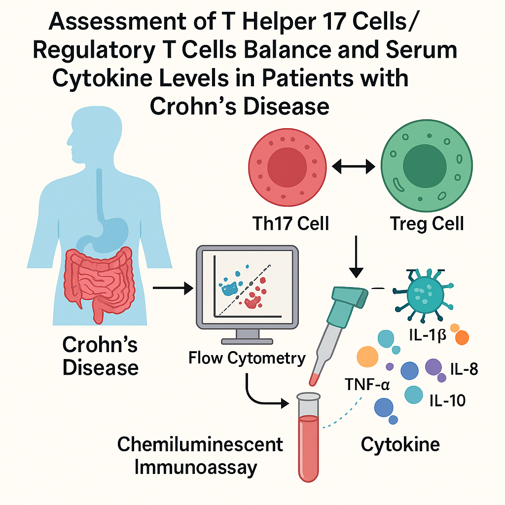
Background: Crohn’s disease (CD) is a chronic inflammatory disorder resulting from the interaction between genetic susceptibility, environmental factors, intestinal microbiota, and dysregulated immune responses. Despite major advances, the precise mechanisms underlying disease development remain incompletely understood. This study aimed to evaluate the proportions of Th17 and regulatory T (Treg) cells, as well as serum cytokine levels, in the peripheral blood of patients with CD. Methods: We enrolled 46 patients with active CD (median age: 31.5 years) and 30 healthy subjects (median age: 30.0 years). Th17 and Treg cell populations were analyzed by flow cytometry, while serum concentrations of TNF-α, IL-1β, IL-6, IL-8, and IL-10 were measured using a chemiluminescent immunoassay. Results: Our results showed a significantly higher proportion of Th17 cells (3.62 ± 1.65% vs. 1.83 ± 1.00%, p = 0.007) and a significantly lower proportion of Treg cells (1.03 ± 0.96% vs. 2.59 ± 2.04%, p < 0.001) in CD patients compared with healthy controls. In addition, serum levels of TNF-α, IL-1β, IL-6, and IL-8 were significantly increased in CD patients (p = 0.011, p = 0.012, p < 0.0001, p < 0.0001, respectively), whereas IL-10 levels did not differ significantly between the two groups (p > 0.05). Conclusion: The imbalance in the Th17/Treg ratio and the elevated levels of inflammatory cytokines support the presence of an active inflammatory process in CD. These findings suggest that such immunological alterations may represent potential biomarkers of disease activity, although further validation in larger longitudinal studies, including mucosal investigations, is required.Graphic Abstract
Keywords
Crohn’s disease (CD) is a chronic inflammatory condition that, together with ulcerative colitis, is classified among inflammatory bowel disease (IBD). Crohn’s disease was first described as regional ileitis by Crohn, Ginzburg, and Oppenheimer in 1932 [1]. It affects the intestinal mucosa in a discontinuous manner and can involve any part of the digestive tract, from the mouth to the anus. The main symptoms include abdominal pain, fever, and altered bowel habits. The disease may lead to complications such as fistulas, stenosis, and abscesses. In addition to intestinal manifestations, CD can also cause several extra-intestinal manifestations, including arthritis, skin disorders, ocular inflammation, and hepatobiliary diseases such as primary sclerosing cholangitis. The disease is characterized by alternating periods of relapse and remission [2]. Treatment is multidisciplinary, involving medical interventions to reduce inflammation and manage symptoms. This approach typically focuses on immunosuppressive drugs, such as corticosteroids and thiopurines. In cases of inadequate response, an anti-TNFα agent is introduced [3].
Although the exact cause of CD is not fully understood, several factors seem to contribute, including the environment, genetics, intestinal microbiota, and immune dysregulation of both the innate and adaptive immune systems [4]. The innate immune system, which serves as the body’s first line of defense, may be impaired in the intestinal mucosal barrier. These defects can allow bacteria and other pathogens to penetrate the intestinal lining, triggering an inflammatory response. The adaptive immune system, responsible for antigen-specific immune responses, plays a crucial role in CD. An imbalance between different types of T cells, particularly T helper 17 cells (Th17) and regulatory T cells (Treg), has been observed. Th17 cells promote inflammation, while Treg cells help regulate and suppress immune responses. An exaggerated Th17 response, combined with insufficient Treg activity, can lead to the chronic inflammation characteristic of CD [5,6].
Th17 cells, a subset of CD4+ T cells, produce IL-17A and have been implicated in both anti-infection responses and inflammatory processes. Genome-wide association studies support the role of Th17 cells in the development of CD by identifying multiple genes associated with inflammatory bowel disease susceptibility and the differentiation of Th17 cells [7]. While regulatory T cells modulate immune cell activation and proliferation, they also play a crucial role in tolerance and anti-inflammatory responses [8]. In Crohn’s disease, intestinal macrophages secrete IL-12 and IL-18, promoting the gut immune response toward a Th1 phenotype and subsequent production of pro-inflammatory cytokines, such as TNF-α, IL-1β, and IL-6, which contribute to chronic intestinal inflammation [9]. Improved understanding of the mechanisms regulating Th1 and Th17-mediated inflammation may contribute to the development of novel therapeutic strategies.
This study aimed to assess the Th17/Treg balance using FOXP3 and IL-17A as defining markers and to investigate the profiles of pro-inflammatory cytokines (TNF-α, IL-1β, IL-6, and IL-8), as well as the anti-inflammatory cytokine IL-10, in the peripheral blood of patients with CD.
2.1 Patients and Healthy Donors
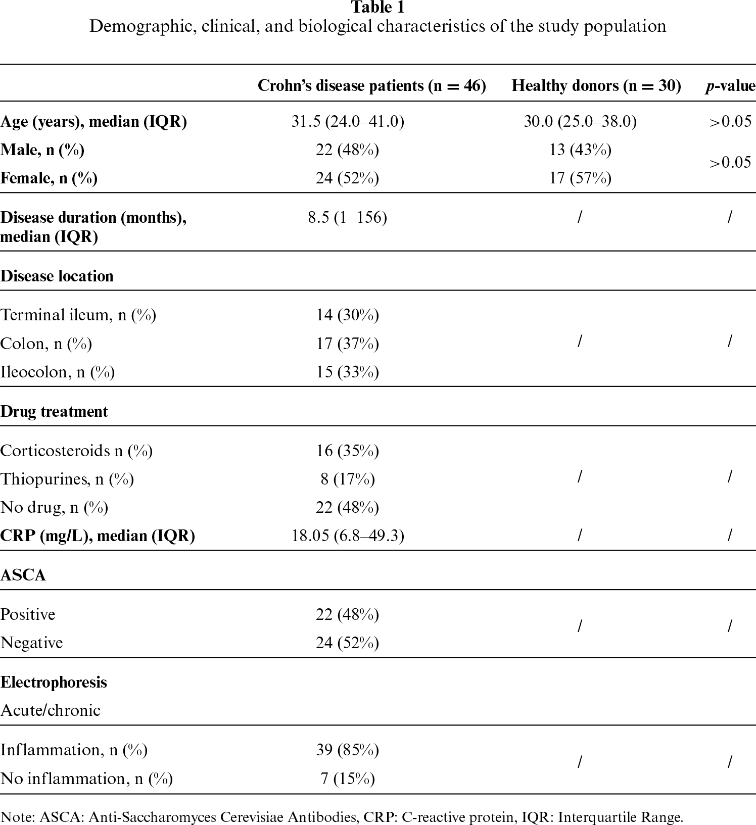
In our study, we recruited 46 patients with active CD, from the department of Gastroenterology and Hepatology at the University Hospital Center of Beni Messous. The study cohort comprised patients aged 18 to 58 years, with a median age of 31.5 years, including 24 women and 22 men. Additionally, we enrolled 30 healthy subjects from the neighboring community and with no history of inflammatory disorders, aged 20 to 58 years, with a median age of 30.0 years, including 17 women and 13 men.
All patients were adults and were in clinical relapse at the time of blood sampling, as determined by clinical assessments and blood tests. The diagnosis of CD was established based on clinical presentations, supported by laboratory, radiological, endoscopic, and histological examinations. The diagnosis was established according to the ECCO-ESGAR guidelines for the diagnostic assessment of IBD, which were updated in 2019 by the European Crohn’s and Colitis Organization (ECCO) and the European Society of Gastrointestinal and Abdominal Radiology (ESGAR).
For all subjects, the exclusion criteria included ongoing gastrointestinal infections, a history of cancer, pregnancy and breastfeeding, treatment with biological agents within the three months prior to recruitment, and psychiatric disorders. In addition, healthy controls with ongoing autoimmune/inflammatory diseases were excluded. Patient characteristics are listed in table 1.
The study was approved by the institutional ethics committee of our hospital, The Ethics and Deontology Committee of the University Hospital Center of Beni Messous (Approval No. 19/CE/2025). All participants provided written informed consent for the potential subsequent use of their samples in research.
2.2 Blood Sample Collection and Stimulation
Venous blood samples were collected from each subject using standard phlebotomy techniques into two separate tubes. Following centrifugation, serum obtained was carefully separated and stored at −80°C for subsequent cytokine measurement. The heparinized blood fraction was used for flow cytometric analysis of Th17 and Treg cell populations.
To assess the percentages of Th17 and Treg cells, 500 μL of peripheral blood was incubated with 2 μL of a polyclonal activation mixture (Leukocyte Activation Cocktail with BD GolgiPlug™, BD Biosciences, San Jose, CA, USA, Cat. No.550583), containing phorbol 12-myristate-13-acetate (PMA), ionomycin, and the protein transport inhibitor Brefeldin A, according to the manufacturer’s instructions. Following 5 h of stimulation (37°C, 5% CO2), 100 μL of the cell suspension was transferred into a 5 mL BD Falcon tube, and cells were washed twice with 1x Phosphate–buffered saline (PBS; pH 7.4) prior to flow cytometry analysis.
In brief, Th17 cells were defined as CD3+CD4+IL-17+ cells, and Treg cells as CD3+CD4+FoxP3+ cells. For their analysis, cells were first incubated with 10 μL/test of FITC-conjugated anti-human CD3 (clone UCHT1, BD Biosciences, Cat. No. 555332), and 5 μL/test PE-Cy7–conjugated anti-human CD8 (clone RPA-T8, BD Biosciences, Cat. No. 557746) antibodies for 20 min at 4°C in the dark. CD8 staining was included because PMA/ionomycin stimulation is known to induce partial downregulation of CD4 surface expression, thereby facilitating the exclusion of CD8+ T cells and the accurate delineation of the CD4+ T-cell population during subsequent gating. Cells were resuspended in fixation and permeabilization solution according to the manufacturer’s instructions. Following fixation/permeabilization, cells were incubated for 40 min with the antibody cocktail from the Human Th17/Treg Phenotyping Kit (BD Pharmingen™, San Jose, CA, USA; Cat. No. 560762), which includes PE-labelled anti-IL-17A (clone N49-653), PerCP-Cy5.5–labelled anti-human CD4 (clone RPA-T4), and APC-labelled anti-FoxP3 (clone 259D/C7) antibodies. Th17 and Treg subsets were subsequently identified based on the expression of CD4, IL-17A, and FoxP3 (table A1). Intracellular IL-17A staining after PMA/ionomycin stimulation was used to assess the inducible cytokine-producing capacity of Th17 cells rather than their spontaneous basal circulating activity. Although live/dead discrimination staining was not incorporated into the present protocol, all samples were processed under standardized conditions with rapid acquisition to minimize potential viability-related artifacts and maintain data reliability.
Data acquisition was performed on a BD FACS Lyric™ flow cytometer (BD Biosciences). Data were analyzed using BD FACSuite™ clinical software v1.2.1.5657 and FlowJo™ v10 (BD Biosciences), and results were expressed as the percentage of positive cells (figure A1).
2.4 Measurement of Cytokine Concentrations: TNF-α, IL-1β, IL-6, IL-8 and IL-10
The measurement of selected cytokines (TNF-α, IL-1β, IL-6, IL-8, and IL-10) was performed in serum samples using chemiluminescent enzyme immunoassay (CLEIA) on the IMMULITE system (Siemens Healthineers, Erlangen, Germany), according to the manufacturer’s instructions.
The assay kits used were as follows: TNF-α (catalog no. LKTA1; Limit of detection (LOD) 4 pg/mL), IL-1β (catalog no. L2KLI2; LOD 5 pg/mL), IL-6 (catalog no. L2K6P2; LOD 2 pg/mL), IL-8 (catalog no. LK8P1; LOD 4 pg/mL), and IL-10 (catalog no. L2KI10; LOD 5 pg/mL). Values below the assay detection limit were considered left-censored and were assigned a value equal to one-half of the corresponding LOD (LOD/2) for statistical analyses. All patient and control samples were analyzed as single measurements (singleton). Cytokine concentrations were expressed in pg/mL.
All data were analyzed using SPSS 26.0 (IBM Corp., Armonk, NY, USA). Continuous variables are presented as mean ± standard deviation (SD). For variables showing substantial dispersion and/or a skewed distribution, median and interquartile range (IQR) were additionally reported to provide a more appropriate description of data central tendency and variability. Data with a normal distribution were analyzed using the parametric Student’s t-test, while variables that deviated from normality were analyzed using the non-parametric Mann-Whitney U test. Correlation analysis was performed using Spearman’s correlation. A p-value of less than 0.05 was considered statistically significant.
3.1 The Proportion of Th17 Cells in the Peripheral Blood of CD Patients and Controls
Flow cytometry was performed to evaluate Th17 cells in all subjects. The proportion of CD4+IL-17+ Th17 cells was determined in active CD patients and healthy controls. The analysis revealed significantly higher percentages of Th17 cells in the peripheral blood of active CD patients compared with controls (3.62 ± 1.65% vs. 1.83 ± 1.00%, p = 0.007) (figure 1A,B).
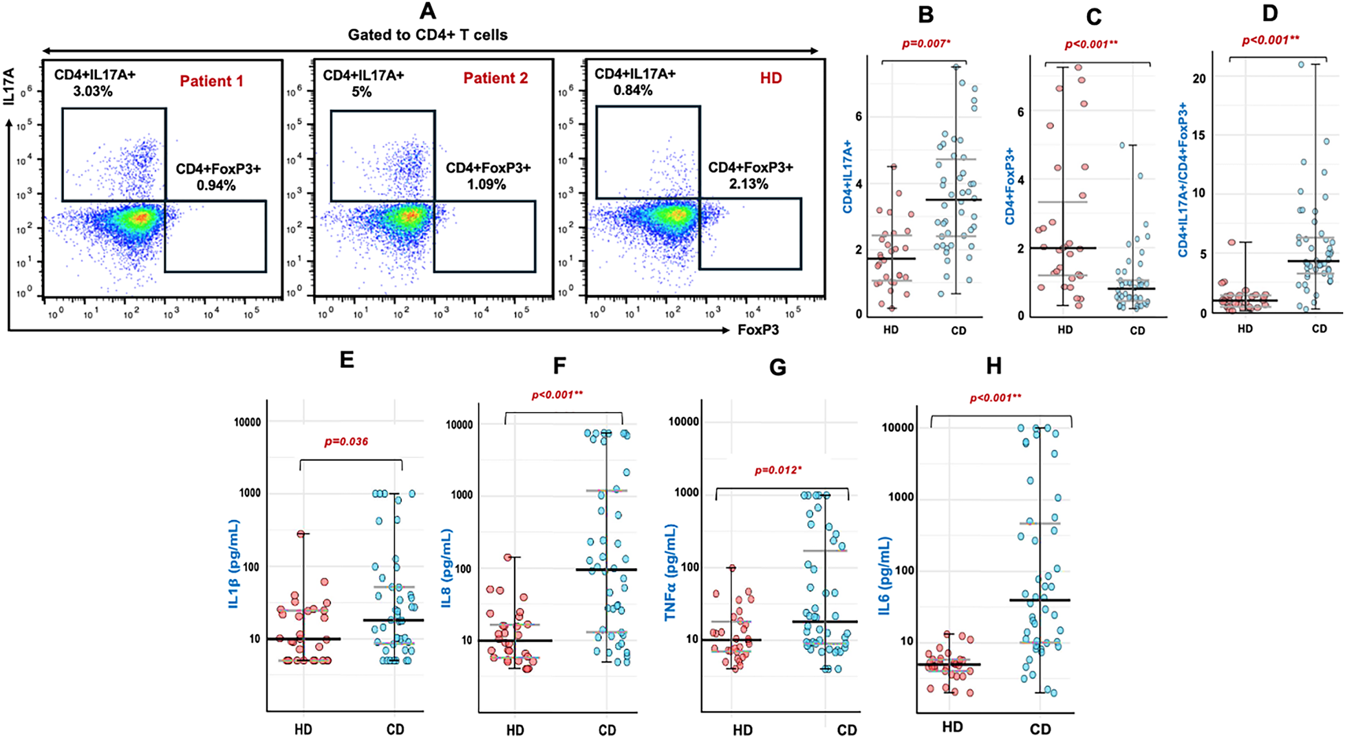
Figure 1: Comparison of CD4+IL-17A+ (Th17) and CD4+FoxP3+ (Treg) lymphocyte subpopulations, along with proinflammatory cytokines (IL-1β, IL-6, TNF-α, and IL-8), between patients with Crohn’s disease (CD) and healthy donors (HD). (A) Representative flow cytometry density plots illustrating Th17 and Treg subpopulations in two Crohn’s disease patients and one healthy donor. Dot plots depicting the percentages of Th17 (CD4+IL-17+) cells (B), Treg (CD4+FoxP3+) cells (C), and the Th17/Treg ratio (D), and the interleukin levels IL-1β (E), IL-8 (F), TNF-α (G), and IL-6 (H) in CD patients (n = 46) compared with HD (n = 30). Treg, regulatory T cells; FOXP3, Forkhead box P3; *Student’s t-test, **Mann-Whitney U test.
3.2 The Proportion of Treg Cells
Considering the crucial role of regulatory T cells in maintaining immune homeostasis, their frequency was assessed by evaluating FOXP3 expression within the total CD4+ cell population. The percentage of circulating Treg cells was significantly reduced in patients with active CD compared with controls (1.03 ± 0.96% vs. 2.59 ± 2.04%, p < 0.001, figure 1C).
The Th17/Treg cell ratio was significantly higher in CD patients compared to controls (5.45 ± 3.89 vs. 1.14 ± 1.09, p < 0.001) (figure 1D).
3.4 Comparison of Cytokines Levels
The analysis of serum cytokines revealed significantly higher levels of TNF-α, IL-1β, IL-6, and IL-8 in patients with active CD compared with healthy subjects [18.10 pg/mL (9.02–176.00) vs. 9.96 pg/mL (6.96–17.90), p = 0.011; 18.00 pg/mL (8.58–51.75) vs. 9.88 pg/mL (2.50–24.38), p = 0.012; 39.35 pg/mL (10.08–467.25) vs. 4.93 pg/mL (4.02–5.83), p < 0.0001, and 95.80 pg/mL (13.08–1201.00) vs. 9.74 pg/mL (5.71–16.43), p < 0.0001, respectively]. (figure 1E–H).
In contrast, IL-10 levels did not differ significantly between patients and controls [2.50 pg/mL (2.50–5.60) vs. 2.50 pg/mL (2.50–2.50), p > 0.05].
To further explore the relationship between pro-inflammatory mediators, correlation analyses were performed among cytokine levels. IL-1β showed strong positive associations with TNF-α (Rho = 0.59, p < 0.001) and IL-6 (Rho = 0.51, p < 0.001), while IL-8 levels were positively correlated with IL-1β (Rho = 0.75, p < 0.001), TNF-α (Rho = 0.60, p < 0.001), and IL-6 (Rho = 0.47, p < 0.001).
3.5 Comparison of Th17/Treg Cell Proportion and Serum Cytokine Levels According to Treatment
In this study, no significant differences were observed in Th17/Treg cell subsets or cytokine levels according to treatment status. The proportions of Th17 cells (3.70 ± 1.37% vs. 3.54 ± 1.27%, p > 0.05) and Treg cells (0.97 ± 0.64% vs. 1.10 ± 0.62%, p > 0.05) were comparable between patients receiving corticosteroids or thiopurines and untreated patients. Similarly, the Th17/Treg ratio did not differ significantly between the two groups (6.17 ± 3.19 vs. 4.67 ± 2.09, p > 0.05). Regarding serum cytokines, TNF-α, IL-1β, IL-6, IL-8, and IL-10 levels were 22.50 (10.61–144.00), 24.50 (12.60–56.27), 24.00 (8.70–174.00) pg/mL, 182.50 (13.62–3152.50) and 2.50 (2.50–5.78) in treated patients, compared with 11.20 (7.46–176.00), 10.30 (5.00–36.12), 43.25 (16.35–548.75), 68.10 (15.03–443.25), and 5.00 (2.50–5.32) pg/mL in untreated patients, respectively. None of these differences reached statistical significance (p = 0.206, 0.120, 0.350, 0.247 and 0.864, respectively).
Crohn’s disease is characterized by a dysregulated immune response involving both innate and adaptive immunity. In the present study, we investigated the balance between Th17 and Treg cells, with particular emphasis on the Th17/Treg ratio, as well as the associated cytokine profile in patients with CD.
Our analysis demonstrated a significantly higher percentage of Th17 cells in the peripheral blood of active CD patients (p = 0.007), highlighting the potential involvement of these cells in the pathogenesis of CD. Over the past several years, multiple studies have consistently reported a similar increase in Th17 cells in patients with IBD [10–13]. This consistent increase in Th17 cell frequency in IBD patients has become increasingly evident in recent years, highlighting their potential contribution to disease pathogenesis [14,15]. Indeed, the elevated frequency of Th17 cells in CD suggests that these cells may exacerbate the intestinal inflammatory response through their pro-inflammatory cytokines. Moreover, the expansion of Th17 cells could be associated with an impaired suppressive function of regulatory T cells. However, clinical trials evaluating anti-IL-17 therapies for CD have paradoxically led to worsening of symptoms in some patients, suggesting a complex role for IL-17 in maintaining gut homeostasis. Although IL-17 contributes to inflammation, it may also play a protective role in preserving mucosal barrier integrity [16], which could explain these seemingly contradictory findings. It is worth noting that, in addition to Th17 cells, other T helper subsets such as Th1 cells also play a crucial role in the pathophysiology of CD. In this context, an excessive Th1 response is observed, characterized by elevated IL-12, IL-18, IFN-γ, and TNF-α levels, which promote epithelial apoptosis, immune cell recruitment, and chronic mucosal inflammation [17,18].
On the other hand, regulatory T cells, a subset of CD4+ T cells, play a crucial immunomodulatory role despite their relatively low numbers. These lymphocytes maintain immune tolerance and regulate both physiological and pathological immune responses. Tregs cells are known to secrete anti-inflammatory cytokines such as TGF-β and IL-10, which suppress immune cell activity and help control inflammation. In our study, we found that the frequency of Tregs in CD patients was significantly lower than in healthy controls (p < 0.001). Other studies have similarly documented a decrease in Treg populations in CD patients [12,13,19], suggesting a potential impairment in the regulatory mechanisms that normally help control inflammation. These insights have fostered the development of therapeutic approaches aimed at restoring Treg function, including small molecules, antibody-based strategies, and cell therapies, with emerging clinical trials and precision medicine approaches highlighting Treg modulation as a promising avenue in IBD management [20]. Taken together, these findings strongly indicate an imbalanced Th17/Treg ratio in patients with CD, contributing to disease progression. This imbalance could potentially serve as a future biomarker for monitoring disease progression and response to therapy in CD patients.
In addition to the observed alterations in Th17 cells, our findings revealed significantly increased serum levels of TNF-α, IL-1β, IL-6, and IL-8 in patients with CD compared with healthy controls. These results further support the presence of an enhanced pro-inflammatory immune response in CD. The elevated cytokine levels may contribute to the recruitment and activation of inflammatory cells, thereby sustaining intestinal inflammation and tissue damage. Given their central role in the inflammatory process, these cytokines represent promising targets for current and future therapeutic strategies in CD [21].
In addition to increased serum levels observed in our study, other human studies have reported elevated TNF-α levels in the serum, stool, and intestinal mucosal tissue of patients with CD [12,22,23]. This cytokine has been extensively investigated due to its central role in CD pathophysiology and its strong therapeutic relevance. Indeed, TNF-α represents a key target in biologic therapy, and anti-TNF agents, including infliximab, adalimumab, certolizumab, and golimumab, have demonstrated significant efficacy in reducing inflammatory activity and inducing clinical remission in CD patients [24,25].
Elevated concentrations of IL-1β have been reported in both serum and intestinal lesions of patients with CD and ulcerative colitis [26]. IL-1β is a key pro-inflammatory cytokine involved in the pathogenesis of inflammatory bowel disease. It contributes to intestinal inflammation by promoting immune cell recruitment, stimulating other pro-inflammatory mediators, such as Prostaglandin E2, and supporting Th17 cell differentiation. Dysregulation of IL-1β disrupts mucosal homeostasis and may exacerbate disease activity, highlighting its central role in CD and its potential as a therapeutic target [27].
Among pleiotropic cytokines, IL-6 is particularly relevant due to its dual role in amplifying inflammation and promoting Th17 cell differentiation in the presence of TGF-β. In agreement with our findings, increased IL-6 levels have been reported in the serum of patients with CD [28,29], and polymorphisms in the IL-6 gene have been linked to specific disease phenotypes [30]. Given its pro-inflammatory role, IL-6 has also emerged as a therapeutic target, with clinical trials investigating anti-IL-6 therapies showing promising results in CD patients [31].
In addition, IL-8, a potent chemotactic cytokine, was significantly elevated in our cohort. Previous studies have shown that increased serum IL-8 levels correlate with disease activity and colonic inflammation [32,33]. As a strong neutrophil chemoattractant, IL-8 likely contributes to the chronic inflammation characteristic of CD. The elevated levels of IL-8, together with other pro-inflammatory cytokines, highlight their potential utility as biomarkers for assessing disease activity and guiding therapeutic strategies.
In our study, IL-10 levels were not significantly decreased in patients with CD, in contrast to previous reports that associated reduced IL-10 levels with impaired regulatory T cell function [13,34]. Moreover, polymorphisms in the IL-10 gene have been associated with increased susceptibility to CD, as evidenced by genome-wide association studies (GWAS) [35]. IL-10, a key anti-inflammatory cytokine, is primarily produced by Treg cells and plays a central role in controlling excessive immune responses. Given the observed reduction in Treg cells in our cohort, a corresponding decrease in IL-10 was expected, however, this was not observed. This finding may indicate that IL-10’s role in maintaining intestinal homeostasis is more pronounced locally within gut tissue, as reported by Jarry et al. [36], rather than in circulating blood. Additionally, variability in IL-10 expression may be influenced by factors such as disease activity, patient heterogeneity, and the methods used to measure cytokines, contributing to inconsistent findings.
Beyond analyzing individual cytokine levels, we examined potential correlations among various inflammatory cytokines in patients with CD. Notably, we observed a significant positive correlation between IL-1β and TNF-α and IL-6, as well as between IL-8 and IL-1β, TNF-α, and IL-6. These findings suggest a coordinated interplay among these cytokines in driving inflammation in CD. Such interactions underscore the complexity of the cytokine network in CD, where multiple pro-inflammatory mediators act synergistically. In addition to these cytokines, alternative pathways such as the IL-23/Th17 axis and IL-12/IFN-γ signaling can sustain inflammation, highlighting why blockade of a single pathway is often insufficient to fully control disease activity. Indeed, inhibition of a single cytokine in CD patients may lead to compensatory activation of alternative pro-inflammatory pathways. Therefore, a more effective therapeutic strategy could involve combined blockade of multiple cytokines or targeting common downstream signaling pathways like JAK-STAT, thereby providing broader disruption of the inflammatory network and improving clinical outcomes [37].
On the other hand, our findings indicate that treatment exposure did not markedly affect cytokine production or T-cell subsets distribution in this cohort of CD patients. Although both corticosteroids and thiopurines are known to exert immunosuppressive and anti-inflammatory effects, their impact on circulating pro-inflammatory cytokines and the Th17/Treg cell balance appeared limited. This observation may reflect the chronic nature of the disease, variability in treatment response among individuals, or incomplete immune modulation under conventional therapy. The absence of significant differences may also be influenced by the limited sample size of the stratified groups, reducing statistical power to detect subtle effects. In contrast, several studies have reported significant immunological shifts following biologic agents, including reductions in pro-inflammatory cytokines and restoration of Th17/Treg balance [38–40], suggesting that these parameters could serve as useful biomarkers for monitoring therapeutic efficacy in CD.
One of the key strengths of this study is its provision of original data on the Th17/Treg imbalance in CD patients, particularly in North Africa, where few similar studies have been conducted. These findings may lay the groundwork for further regional and international investigations into immune dysregulation in CD and other inflammatory bowel diseases. A main limitation of our study is that the results are derived solely from peripheral blood and serum measurements. Ideally, samples should be obtained from the intestinal mucosa, where inflammatory events predominantly occur, via biopsies. However, the evaluation in blood was deliberately chosen to explore potential non-invasive biomarkers.
Another important limitation is that CD25 was not included in the flow cytometry panel and no fluorescence-minus-one (FMO) control was used for this marker, which may have affected the accuracy of Treg identification and gating. In addition, objective measures of disease activity, including systematic endoscopic assessment and fecal calprotectin measurements, were not available during the study period. Methodological limitations related to the distribution of several biological variables and the absence of formal correction for multiple comparisons should also be considered when interpreting the results. Future studies incorporating a more comprehensive immunophenotyping strategy, standardized assessment of disease activity, and larger patient cohorts will be required to achieve a more comprehensive understanding of immune dysregulation in CD.
In summary, Crohn’s disease is characterized by immune system imbalance, dysregulated immune responses, a reduction in regulatory T cells, and an upregulation of inflammatory Th17 cells. These alterations drive the production of pro-inflammatory cytokines, including TNF-α, IL-1β, IL-6, and IL-8 contributing to severe intestinal inflammation. Given the disrupted Th17/Treg ratio and the elevated levels of these cytokines in CD patients, they may serve as potential biomarkers for assessing disease activity and guiding therapeutic strategies.
Acknowledgement: Not applicable.
Funding Statement: The authors received no specific funding for this study.
Author Contributions: The authors confirm contribution to the paper as follows: Study conception and design: Ines Allam, Reda Djidjik. Data collection: Ouassila Madani, Brahim Belaid, Fatma Merah, Ferial Messaoui, Linda Azzoug, Abdelmalek Balamane. Analysis and interpretation of results: Ines Allam, Ouassila Madani, Brahim Belaid, Reda Djidjik. Draft manuscript preparation: Ines Allam, Ouassila Madani, Reda Djidjik. All authors reviewed and approved the final version of the manuscript.
Availability of Data and Materials: The data underlying this article will be shared on reasonable request to the corresponding author.
Ethics Approval: The study was approved by The Ethics and Deontology Committee of the University Hospital Center of Beni Messous (Approval No. 19/CE/2025). Written informed consent was obtained from all participants prior to enrollment, including consent for the potential future use of their biological samples for research purposes.
Conflicts of Interest: The authors declare no conflicts of interest.
Appendix A
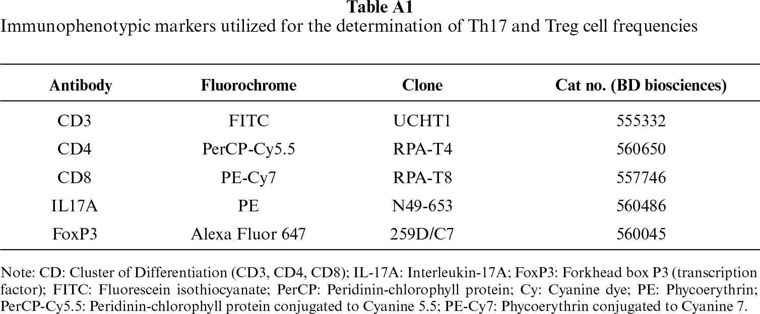
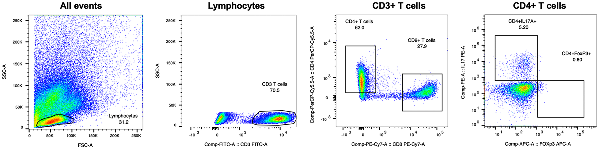
Figure A1: Gating strategy for lymphocyte subpopulations used to identify Th17 (CD4+IL-17A+) and regulatory T cells (CD4+FoxP3+).
References
1. Crohn BB, Ginzburg L, Oppenheimer GD. Regional ileitis: a pathologic and clinical entity. J Am Med Assoc 1932;99(16):1323–9. doi:10.1001/jama.1932.02740680019005. [Google Scholar] [CrossRef]
2. Dolinger M, Torres J, Vermeire S. Crohn’s disease. Lancet 2024;403(10432):1177–91. doi:10.1016/S0140-6736(23)02586-2. [Google Scholar] [CrossRef]
3. Torres J, Bonovas S, Doherty G et al. ECCO guidelines on therapeutics in Crohn’s disease: medical treatment. J Crohn’s Colitis 2020;14(1):4–22. doi:10.1093/ecco-jcc/jjz180. [Google Scholar] [CrossRef]
4. Calvez V, Puca P, Di Vincenzo F et al. Novel insights into the pathogenesis of inflammatory bowel diseases. Biomedicines 2025;13(2):305. doi:10.3390/biomedicines13020305. [Google Scholar] [PubMed] [CrossRef]
5. Neurath MF. Targeting immune cell circuits and trafficking in inflammatory bowel disease. Nat Immunol 2019;20(8):970–9. doi:10.1038/s41590-019-0415-0. [Google Scholar] [PubMed] [CrossRef]
6. Schmitt H, Neurath MF, Atreya R. Role of the IL23/IL17 pathway in Crohn’s disease. Front Immunol 2021;12:622934. doi:10.3389/fimmu.2021.622934. [Google Scholar] [CrossRef]
7. Verstockt B, Smith KG, Lee JC. Genome-wide association studies in Crohn’s disease: past, present and future. Clin Transl Immunology 2018;7(1):e1001. doi:10.1002/cti2.1001. [Google Scholar] [CrossRef]
8. Sakaguchi S, Yamaguchi T, Nomura T, Ono M. Regulatory T cells and immune tolerance. Cell 2008;133(5):775–87. doi:10.1016/j.cell.2008.05.009. [Google Scholar] [PubMed] [CrossRef]
9. Petagna L, Antonelli A, Ganini C et al. Pathophysiology of Crohn’s disease inflammation and recurrence. Biol Direct 2020;15(1):23. doi:10.1186/s13062-020-00280-5. [Google Scholar] [CrossRef]
10. Eastaff-Leung N, Mabarrack N, Barbour A, Cummins A, Barry S. Foxp3+ regulatory T cells, Th17 effector cells, and cytokine environment in inflammatory bowel disease. J Clin Immunol 2010;30(1):80–9. doi:10.1007/s10875-009-9345-1. [Google Scholar] [CrossRef]
11. Dige A, Støy S, Rasmussen TK et al. Increased levels of circulating Th17 cells in quiescent versus active Crohn’s disease. J Crohn’s Colitis 2013;7(3):248–55. doi:10.1016/j.crohns.2012.06.015. [Google Scholar] [CrossRef]
12. Chao K, Zhang S, Yao J et al. Imbalances of CD4+ T-cell subgroups in Crohn’s disease and their relationship with disease activity and prognosis. J Gastroenterol Hepatol 2014;29(10):1808–14. doi:10.1111/jgh.12592. [Google Scholar] [CrossRef]
13. Geng X, Xue J. Expression of Treg/Th17 cells as well as related cytokines in patients with inflammatory bowel disease. Pak J Med Sci 2016;32(5):1164–8. doi:10.12669/pjms.325.10902. [Google Scholar] [PubMed] [CrossRef]
14. Jiang P, Zheng C, Xiang Y et al. The involvement of TH17 cells in the pathogenesis of IBD. Cytokine Growth Factor Rev 2023;69:28–42. doi:10.1016/j.cytogfr.2022.07.005. [Google Scholar] [PubMed] [CrossRef]
15. Wen Y, Wang H, Tian D, Wang G. TH17 cell: a double-edged sword in the development of inflammatory bowel disease. Ther Adv Gastroenterol 2024;17:17562848241230896. doi:10.1177/17562848241230896. [Google Scholar] [PubMed] [CrossRef]
16. Fauny M, Moulin D, D’Amico F et al. Paradoxical gastrointestinal effects of interleukin-17 blockers. Ann Rheumatic Dis 2020;79(9):1132–8. doi:10.1136/annrheumdis-2020-217927. [Google Scholar] [PubMed] [CrossRef]
17. Imam T, Park S, Kaplan MH, Olson MR. Effector T helper cell subsets in inflammatory bowel diseases. Front Immunol 2018;9:1212. doi:10.3389/fimmu.2018.01212. [Google Scholar] [PubMed] [CrossRef]
18. Gomez-Bris R, Saez A, Herrero-Fernandez B, Rius C, Sanchez-Martinez H, Gonzalez-Granado JM. CD4 T-cell subsets and the pathophysiology of inflammatory bowel disease. Int J Mol Sci 2023;24(3):2696. doi:10.3390/ijms24032696. [Google Scholar] [PubMed] [CrossRef]
19. Wang Y, Liu XP, Zhao ZB, Chen JH, Yu CG. Expression of CD4+ forkhead box P3 (FOXP3)+ regulatory T cells in inflammatory bowel disease. J Dig Dis 2011;12(4):286–94. doi:10.1111/j.1751-2980.2011.00505.x. [Google Scholar] [CrossRef]
20. Lee C, Park YW, Park MH, Lee YJ, Rhee I. Regulatory T cells and their role in inflammatory bowel disease: molecular targets, therapeutic strategies and translational advances. Biochem Pharmacol 2025;239(3):117087. doi:10.1016/j.bcp.2025.117087. [Google Scholar] [PubMed] [CrossRef]
21. Aebisher D, Bartusik-Aebisher D, Przygórzewska A, Oleś P, Woźnicki P, Kawczyk-Krupka A. Key interleukins in inflammatory bowel disease—a review of recent studies. Int J Mol Sci 2025;26(1):121. doi:10.3390/ijms26010121. [Google Scholar] [CrossRef]
22. Friedrich M, Pohin M, Powrie F. Cytokine networks in the pathophysiology of inflammatory bowel disease. Immunity 2019;50(4):992–1006. doi:10.1016/j.immuni.2019.03.017. [Google Scholar] [PubMed] [CrossRef]
23. Murate K, Maeda K, Nakamura M et al. Endoscopic activity and serum TNF-α level at baseline are associated with clinical response to ustekinumab in Crohn’s disease patients. Inflamm Bowel Dis 2020;26(11):1669–81. doi:10.1093/ibd/izaa086. [Google Scholar] [CrossRef]
24. Shehab M, Alrashed F, Alrashidi A et al. Network meta-analysis: comparative efficacy of biologics and small molecules in the induction and maintenance of remission in Crohn’s disease. Aliment Pharmacol Ther 2025;62(5):472–82. doi:10.1111/apt.70295. [Google Scholar] [CrossRef]
25. Gebeyehu GG, Broglio G, Liu E et al. Comparative safety and effectiveness of ustekinumab and anti-TNF in elderly Crohn’s disease patients. Inflamm Bowel Dis 2025;31(5):1325–33. doi:10.1093/ibd/izae174. [Google Scholar] [CrossRef]
26. Fasching P, Stradner M, Graninger W, Dejaco C, Fessler J. Therapeutic potential of targeting the Th17/Treg axis in autoimmune disorders. Molecules 2017;22(1):134. doi:10.3390/molecules22010134. [Google Scholar] [PubMed] [CrossRef]
27. Zoeten EF, Fuss IJ. Cytokines and inflammatory bowel disease. In: Pediatric inflammatory bowel disease. Cham, Switzerland: Springer; 2023. p. 33–48. doi:10.1007/978-3-031-14744-9_3 [Google Scholar] [CrossRef]
28. Caviglia GP, Rosso C, Stalla F et al. On-treatment decrease of serum interleukin-6 as a predictor of clinical response to biologic therapy in patients with inflammatory bowel diseases. J Clin Med 2020;9(3):800. doi:10.3390/jcm9030800. [Google Scholar] [PubMed] [CrossRef]
29. Boucher G, Paradis A, Chabot-Roy G et al. Serum analyte profiles associated with Crohn’s disease and disease location. Inflamm Bowel Dis 2022;28(1):9–20. doi:10.1093/ibd/izab123. [Google Scholar] [CrossRef]
30. Magyari L, Kovesdi E, Sarlos P, Javorhazy A, Sumegi K, Melegh B. Interleukin and interleukin receptor gene polymorphisms in inflammatory bowel diseases susceptibility. World J Gastroenterol 2014;20(12):3208. doi:10.3748/wjg.v20.i12.3208. [Google Scholar] [PubMed] [CrossRef]
31. Alhendi A, Naser SA. The dual role of interleukin-6 in Crohn’s disease pathophysiology. Front Immunol 2023;14:1295230. doi:10.3389/fimmu.2023.1295230. [Google Scholar] [CrossRef]
32. Chen P, Zhou G, Lin J et al. Serum biomarkers for inflammatory bowel disease. Front Med 2020;7:123. doi:10.3389/fmed.2020.00123. [Google Scholar] [PubMed] [CrossRef]
33. Adumitrăchioaiei H, Săsăran MO, Mărginean CO. The diagnostic and prognostic role of interleukin 6 and interleukin 8 in childhood acute gastroenteritis—a review of the literature. Int J Mol Sci 2024;25(14):7655. doi:10.3390/ijms25147655. [Google Scholar] [CrossRef]
34. Zhu XM, Shi YZ, Cheng M, Wang DF, Fan JF. Serum IL-6, IL-23 profile and Treg/Th17 peripheral cell populations in pediatric patients with inflammatory bowel disease. Pharmazie 2017;72(5):283–7. doi:10.1691/ph.2017.6957. [Google Scholar] [PubMed] [CrossRef]
35. Sun A, Li W, Shang S. Association of polymorphisms in the IL-10 promoter region with Crohn’s disease. Clin Lab Anal 2022;36(12):e24780. doi:10.1002/jcla.24780. [Google Scholar] [CrossRef]
36. Jarry A, Bossard C, Bou-Hanna C et al. Mucosal IL-10 and TGF-β play crucial roles in preventing LPS-driven, IFN-γ–mediated epithelial damage in human colon explants. J Clin Investig 2008;118(3):1132–42. doi:10.1172/jci32140. [Google Scholar] [CrossRef]
37. Neurath MF. Strategies for targeting cytokines in inflammatory bowel disease. Nat Rev Immunol 2024;24(8):559–76. doi:10.1038/s41577-024-01008-6. [Google Scholar] [PubMed] [CrossRef]
38. Kwon Y, Kim ES, Kim YZ, Choe YH, Kim MJ. Cytokine profile at diagnosis affecting trough concentration of infliximab in pediatric Crohn’s disease. Biomedicines 2022;10(10):2372. doi:10.3390/biomedicines10102372. [Google Scholar] [CrossRef]
39. Chen R, Li C, Zheng J et al. Lymphocyte subsets for predicting inflammatory bowel disease progression and treatment response: a systematic review. Front Immunol 2024;15:1403420. doi:10.3389/fimmu.2024.1403420. [Google Scholar] [PubMed] [CrossRef]
40. Zapata-Cobo P, Salvador-Martín S, Gil-Manso S et al. Changes in T lymphocytes and cytokines after anti-TNF treatment in pediatric inflammatory bowel disease: association with response to pharmacologic therapy. Int J Mol Sci 2025;26(7):3323. doi:10.3390/ijms26073323. [Google Scholar] [PubMed] [CrossRef]
Cite This Article
 Copyright © 2026 The Author(s). Published by Tech Science Press.
This work is licensed under a Creative Commons Attribution 4.0 International License , which permits unrestricted use, distribution, and reproduction in any medium, provided the original work is properly cited.
 Downloads
 Citation Tools
