
 Login
Login Register
 Submit a Paper
 Propose a Special lssue
 Open Access
REVIEW
The interleukin-18/Interleukin-18 binding protein axis across allergy, systemic autoimmunity, and hyperinflammation: from total to free IL-18
1 Department of Medical Sciences, University of Turin, Corso Dogliotti, Turin, Italy
2 Immunology and Allergy Unit, AO Ordine Mauriziano di Torino, Via Magellano, Turin, Italy
* Corresponding Author: Simone Negrini. Email:
European Cytokine Network 2026, 37(3), 151-168. https://doi.org/10.32604/ecn.2026.083788
Received 10 April 2026; Accepted 13 July 2026; Issue published 28 September 2026
 View Full Text
 Download PDF
Abstract
Interleukin-18 (IL-18) is a pleiotropic IL-1-family cytokine whose clinical relevance depends strongly on context. A central reason is that IL-18 activity is constrained by interleukin-18 binding protein (IL-18BP), a high-affinity endogenous antagonist that limits the free, bioactive cytokine fraction. This review re-examines the IL-18/IL-18BP axis across a clinical gradient from barrier dysfunction and allergic disease, through systemic autoimmunity, to Still disease, macrophage activation syndrome, hemophagocytic lymphohistiocytosis, and monogenic IL-18opathies. We emphasize that total IL-18 elevation does not necessarily imply pathogenic IL-18 activity; the key translational question is whether IL-18BP-mediated restraint remains sufficient. In barrier disorders and systemic autoimmunity, IL-18 often behaves as a contextual amplifier or biomarker-amplifier axis. In hyperinflammatory and selected monogenic disorders, extreme IL-18 production may exceed endogenous antagonism, allowing free IL-18 to emerge and acquire diagnostic, mechanistic, and therapeutic relevance. Interpreting total IL-18, IL-18BP/free IL-18, and downstream IFN-γ/CXCL9 signals together may improve biomarker interpretation and guide rational targeting of the pathway.Keywords
Interleukins are a broad group of cytokines that coordinate communication between leukocytes, epithelial and stromal cells during immune surveillance, host defense, tissue repair, and inflammation. Rather than acting as isolated mediators, they function within interconnected networks in which biological output depends on receptor expression, accessory cytokines, tissue compartment, and endogenous regulatory mechanisms. Interleukin-18 (IL-18) belongs to the IL-1 cytokine family and was originally identified as an interferon-gamma (IFN-γ)-inducing factor, but it is now recognized as a pleiotropic mediator capable of shaping both innate and adaptive immunity [1,2]. Its biological meaning is often less straightforward than that of more lineage-restricted inflammatory mediators: depending on the surrounding cytokine milieu, IL-18 can reinforce IFN-γ-dominated responses or contribute to different immune outputs. This contextual nature is one reason why the IL-18 literature has often seemed heterogeneous.
A more important reason is that IL-18 cannot be interpreted on its own. Unlike many cytokines, it circulates in the presence of a high-affinity endogenous inhibitor, interleukin-18 binding protein (IL-18BP), a soluble IL-18-binding protein encoded by a separate gene rather than the extracellular domain of the IL-18 receptor [3]. Under physiological conditions, IL-18BP is present in marked molar excess and keeps bioactive free IL-18 at very low levels [2,4,5]. IL-18BP is itself induced by IFN-γ, creating a feedback loop in which IL-18 promotes IFN-γ, IFN-γ induces IL-18BP, and IL-18BP in turn restrains IL-18 activity [2,5]. This means that measuring total IL-18 alone can be misleading: it reflects cytokine production, but not necessarily cytokine bioactivity. The conceptual basis for this distinction emerged early, when the first IL-18BP ELISA made it possible to estimate free IL-18 and showed that total IL-18 and biologically available IL-18 were not interchangeable quantities [4].
This has important translational consequences. In many inflammatory diseases, total IL-18 is elevated while free IL-18 remains low or only modestly increased, suggesting that the pathway is engaged but still contained. By contrast, in hyperinflammatory syndromes such as Still disease, macrophage activation syndrome (MAS), and monogenic IL-18opathies, buffering may fail and free IL-18 becomes detectable, clinically informative, and in some cases therapeutically actionable [5–7]. This contrast helps explain why IL-18 can behave as a contextual signal in some diseases, an amplifier in others, and a dominant pathogenic driver only in a restricted set of conditions.
In this review, we re-examine IL-18 through the lens of the IL-18/IL-18BP axis. We follow a clinical gradient from barrier and allergic disease, through systemic autoimmunity, to hyperinflammatory syndromes and monogenic IL-18opathies. Our central argument is that the key translational question is not simply whether IL-18 is elevated, but when and why high-affinity endogenous antagonism by IL-18BP becomes insufficient. The distinctive contribution of this review is therefore not to re-summarize IL-18 biology alone, but to interpret the transition from total IL-18 to free, bioactive IL-18 as a clinically meaningful shift across disease categories. We integrate advances that have accumulated beyond earlier foundational reviews, including recent structural insights into IL-18 maturation and receptor competence, the expanding recognition of monogenic IL-18-related disorders, improved assessment of free versus total IL-18, and the evolving clinical and therapeutic landscape of IL-18- and IFN-γ-directed interventions. A recent dyad-focused review has comprehensively summarized IL-18 and IL-18BP in health and disease, including therapeutic strategies aimed at either inhibiting or enhancing IL-18 activity [6]. Building on that dyad framework, the present review focuses on the clinical and biomarker implications of when total IL-18 does, or does not, translate into free bioactive IL-18 across disease domains. The disease gradient that frames the review is summarized in table 1.
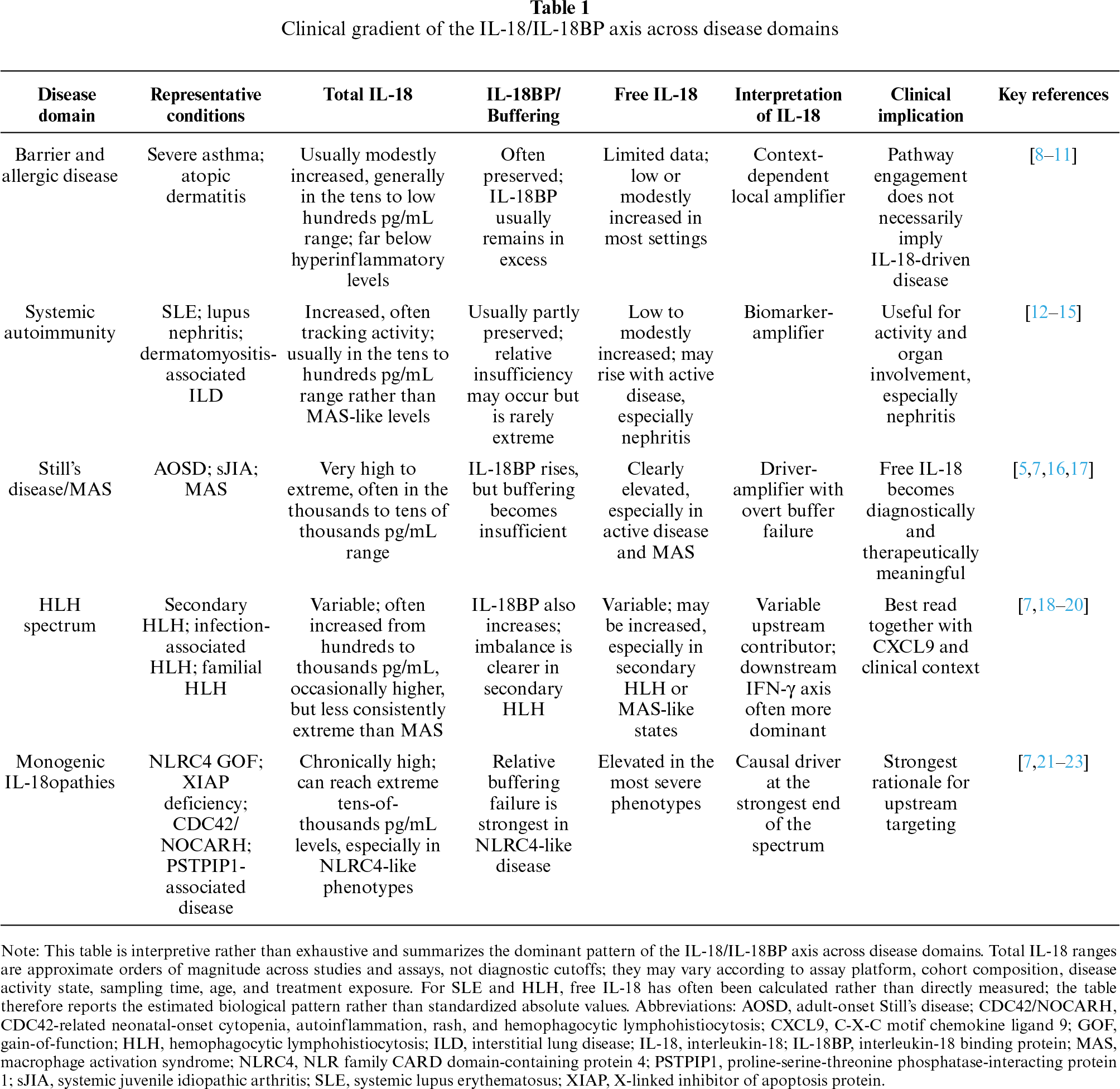
As a narrative review, this article does not aim to provide a systematic meta-analysis of all IL-18-related literature. Instead, we prioritized studies that directly inform the biology, measurement, and clinical interpretation of the IL-18/IL-18BP/free IL-18 axis. Emphasis was placed on foundational mechanistic studies, recent translational biomarker data, genetic disorders with clear pathway relevance, and clinical or preclinical studies of IL-18-, IL-18BP-, IFN-γ-, and Janus kinase (JAK)-directed interventions. Disease areas were selected to illustrate a clinical gradient: barrier and allergic diseases, in which IL-18 often acts as a contextual local amplifier; systemic autoimmunity, in which it more often behaves as a biomarker-amplifier axis; and hyperinflammatory or monogenic disorders, in which IL-18BP-mediated restraint may become quantitatively insufficient and free IL-18 becomes more directly actionable.
2 Biology of the IL-18/IL-18BP Axis
2.1 IL-18 Production, Processing, and Release
IL-18 was originally identified as an IFN-γ-inducing factor, but its biology differs from that of IL-1β in several important ways. Most notably, the IL-18 precursor is constitutively expressed in multiple cell types, including monocytes, macrophages, endothelial cells, keratinocytes, and epithelial cells at barrier sites [2,5,24]. This preformed intracellular pool helps explain why IL-18 can be mobilized rapidly once activating signals are present. It is also relevant that, even after activation, most intracellular precursor remains unprocessed, reinforcing the idea that the availability of pro-IL-18 alone does not equate with bioactive cytokine [2].
Biological activity depends on proteolytic maturation. Pro-IL-18 is cleaved by caspase-1 at the Asp36-Tyr37 site, generating the mature cytokine [25,26]. Recent structural work has clarified why this step is indispensable: in its precursor form, IL-18 adopts a compact conformation that masks the surfaces required for binding both IL-18R and IL-18 binding protein (IL-18BP), whereas cleavage exposes these interfaces and enables signaling [27]. In this sense, processing is not merely a trimming step, but the event that converts IL-18 into a receptor-competent cytokine.
Release of mature IL-18 is now thought to occur mainly through gasdermin D (GSDMD) pores [28,29]. This may accompany pyroptotic cell death, but it can also occur in hyperactivated myeloid cells that remain viable while releasing inflammatory mediators [28]. Evidence that GSDMD pores can act as conduits for the secretion of IL-1 family cytokines, including IL-18, has strengthened this model and provides a plausible mechanism linking inflammasome activation to extracellular bioactive IL-18 [29].
At the same time, canonical caspase-1 processing is not the only route by which IL-18 may become active. Non-classical processing can occur through at least two mechanistically distinct routes. First, caspase-8-mediated cleavage downstream of death-receptor signaling, including Fas/FasL-dependent pathways, may link apoptotic or inflammatory receptor engagement to IL-18 bioactivation. Second, extracellular or granule-derived proteases, including proteinase-3, chymase, meprin-β, and granzyme B, can process pro-IL-18 outside the canonical inflammasome-caspase-1 module [2,5]. These routes are particularly plausible in inflamed barrier tissues, where epithelial injury, neutrophil and mast-cell activation, and cytotoxic-cell infiltration can coexist. However, most evidence for these pathways comes from biochemical, cellular, or tissue-specific experimental contexts, and their quantitative contribution in human disease remains less well established than that of canonical caspase-1-dependent maturation.
2.2 Receptor Signaling and Context-Dependent Output
IL-18 signals through a heterodimeric receptor composed of IL-18Rα, which binds the ligand, and IL-18Rβ, which is required for signal transduction [30–32]. Downstream signaling is MyD88-dependent and proceeds through IRAK- and TRAF6-linked pathways, ultimately activating NF-κB and MAP kinases [33]. Mechanistically, IL-18 therefore belongs firmly to the IL-1 receptor/Toll-like receptor signaling family.
However, receptor expression does not automatically translate into functional responsiveness. IL-18Rα is relatively widespread, whereas IL-18Rβ is more selectively expressed and can be upregulated by IL-12, a feature that strongly influences cellular sensitivity to IL-18 [31,34]. This helps explain the best-known functional partnership of IL-18: in the presence of IL-12, it potently enhances IFN-γ production by NK cells and T cells and reinforces Th1-polarized responses [24,34].
Yet IL-18 does not have a single fixed immune output. Its biological effect depends on the surrounding cytokine milieu, the responding cell population, and the tissue context. In settings dominated by IL-12, IL-18 mainly amplifies IFN-γ-driven immunity. In the absence of IL-12, by contrast, IL-18 can promote IL-4- and IL-13-associated responses, including basophil- and mast-cell-driven programs and forms of innate-type allergy [8,35,36]. More controversially, IL-18 has also been implicated in Th17-oriented inflammation in the presence of IL-23, although this appears more context-dependent and less consistently established [5]. Overall, IL-18 is better understood as a pleiotropic amplifier whose output is dictated by context rather than as a cytokine with a single invariant functional signature.
2.3 IL-18BP as a High-Affinity Endogenous Antagonist
IL-18 binding protein (IL-18BP) is the main endogenous regulator of IL-18. In its original description, IL-18BP was identified as a distinct soluble inhibitor rather than the shed extracellular domain of the receptor, immediately setting it apart from more conventional soluble cytokine receptors [3]. Among its naturally occurring isoforms, IL-18BPa is the most abundant and has the highest affinity for IL-18; IL-18BPc retains weaker but still measurable neutralizing activity, whereas isoforms b and d lack a complete immunoglobulin domain and do not effectively bind IL-18 [37].
The importance of IL-18BP lies in the strength of this interaction. Initial studies estimated a dissociation constant of about 400 pM for IL-18BPa [37], whereas later work suggested a substantially tighter interaction, with Biacore and titration-based approaches placing the Kd in the 26–50 pM range [5,16]. This is not a trivial technical refinement. It implies that endogenous buffering of IL-18 is stronger than originally appreciated. In healthy individuals, circulating IL-18BP is present in marked molar excess relative to IL-18, so free IL-18 is expected to be very low or virtually absent under physiological conditions [2,4,5].
IL-18BP is also embedded in a negative feedback loop. IL-18 promotes IFN-γ production, whereas IFN-γ induces IL-18BP expression through a promoter region that includes GAS and IRF-responsive elements and depends on IRF-1 and C/EBPβ for full activation [38]. In human PBMCs, monocytes are a major source of IL-18BPa, while IFN-γ derived predominantly from NK cells mediates its induction in response to IL-12 [39]. This circuitry helps explain why elevated total IL-18 does not necessarily imply increased IL-18 bioactivity: in many inflammatory settings, the ligand and its endogenous buffer rise in parallel.
A further nuance is that IL-18BP is not biologically confined to IL-18 alone. At higher concentrations, it can also bind IL-37, which adds a further layer of complexity to the interpretation of elevated IL-18BP in inflammatory settings [35]. This point is not central to the present review, but it is worth keeping in mind because it means that IL-18BP cannot always be interpreted as a pure surrogate of anti-IL-18 buffering.
2.4 Translational Bridge: Total Versus Free IL-18
This is the main translational consequence of the IL-18/IL-18BP axis. Standard assays measure total IL-18, but they do not distinguish between IL-18 already bound to IL-18BP and IL-18 that remains unbound and biologically active [2,5,16]. As a result, total IL-18 alone can be misleading if interpreted as a direct readout of pathway activation.
That problem was already apparent in early studies of adult-onset Still’s disease, where IL-18 bioactivity was far lower than the concentration measured by ELISA, indicating substantial circulating inhibitory activity [40]. The distinction became clearer once free IL-18 could be assessed more directly. In adult-onset Still’s disease, free IL-18 is elevated even when other inflammatory diseases also show high total IL-18, suggesting that the clinically relevant question is not simply how much IL-18 is produced, but whether IL-18BP buffering has been overcome [16].
The added value of free IL-18 appears greatest in Still’s disease, macrophage activation syndrome, and monogenic IL-18opathies, where unbuffered IL-18 more closely reflects pathogenic activity [5,7,16]. Longitudinal observations are also informative: in NLRC4-associated disease and MAS-like states, total IL-18 may remain chronically elevated, whereas free IL-18 falls with clinical improvement, underscoring that total IL-18 and current inflammatory activity can dissociate over time [7]. By contrast, in classic autoimmune disease and in barrier disorders, elevated total IL-18 more often reflects inflammatory context than unequivocal IL-18-driven pathology [2,5].
Methodological aspects of calculated and directly measured free IL-18 are addressed in Section 6.2. The biological point here is that IL-18 should not be interpreted in isolation: its clinical meaning depends on the balance between cytokine production and high-affinity antagonism by IL-18BP.
The mechanistic and translational framework of the IL-18/IL-18BP/free IL-18 axis is summarized in figure 1.
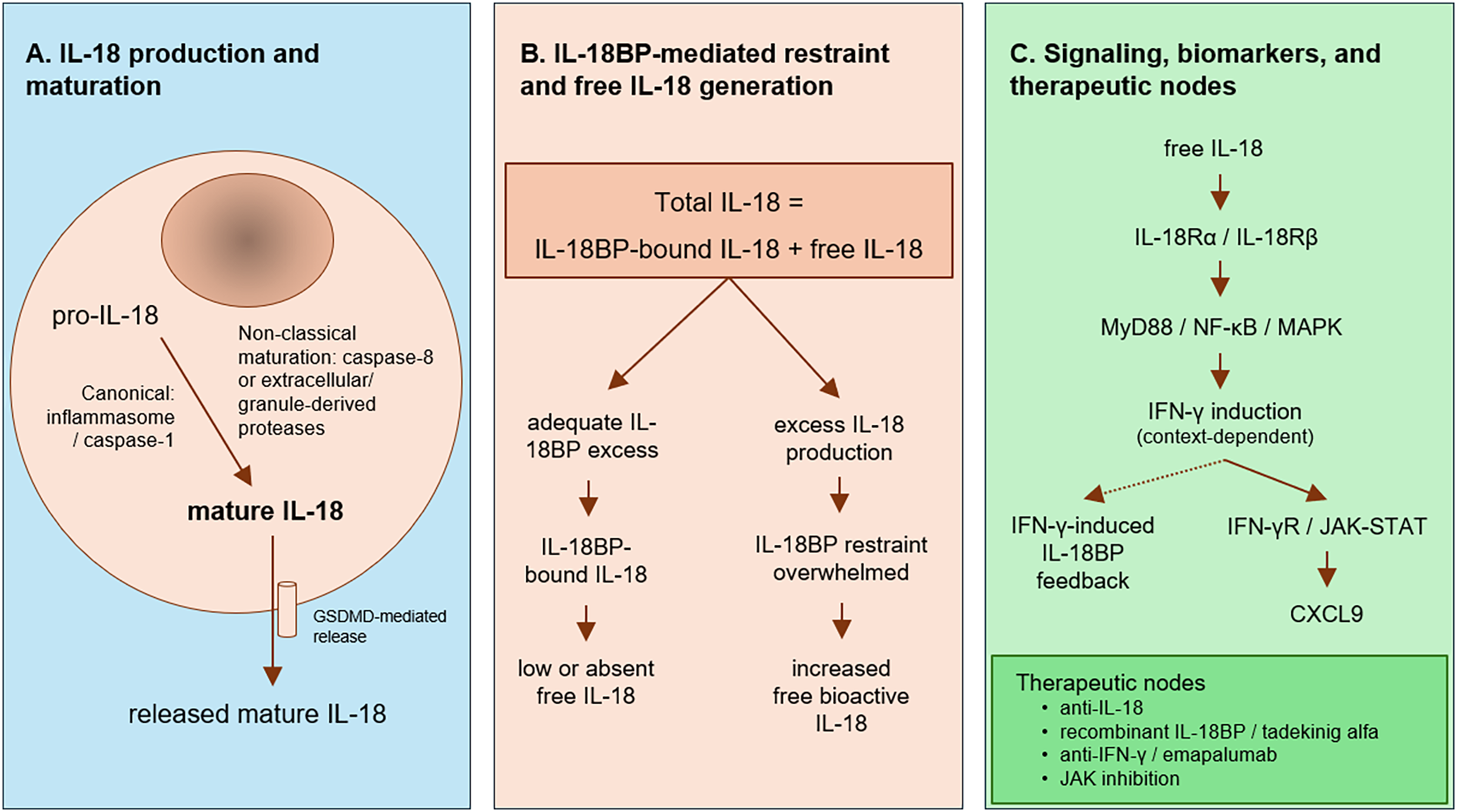
Figure 1: Mechanistic and translational framework of the IL-18/IL-18BP/free IL-18 axis. The schematic is organized into three linked levels: upstream IL-18 production and maturation (A), IL-18BP-mediated restraint and free IL-18 generation (B), and signaling, biomarkers, and therapeutic nodes (C).
IL-18 is produced as an inactive precursor and can be matured through canonical inflammasome-caspase-1-dependent processing or through non-classical routes involving caspase-8 or extracellular/granule-derived proteases. Mature IL-18 is released, including through GSDMD-mediated pathways, and may then be neutralized by the high-affinity endogenous antagonist IL-18BP or remain unbound as free bioactive IL-18 when IL-18 production exceeds IL-18BP-mediated restraint. Free IL-18 signals through the IL-18Rα/IL-18Rβ receptor complex and promotes context-dependent IFN-γ induction, followed by downstream IFN-γR/JAK-STAT signaling and CXCL9 production. IFN-γ also induces IL-18BP, forming a negative feedback loop that limits IL-18 bioactivity. Potential therapeutic nodes include IL-18 neutralization, restoration of IL-18BP-mediated antagonism with recombinant IL-18BP, IFN-γ blockade, and broader JAK-mediated pathway modulation. Abbreviations: CXCL9, C-X-C motif chemokine ligand 9; GSDMD, gasdermin D; IFN-γ, interferon-gamma; IL-18, interleukin-18; IL-18BP, interleukin-18 binding protein; IL-18R, interleukin-18 receptor; JAK, Janus kinase; MAPK, mitogen-activated protein kinase; MyD88, myeloid differentiation primary response 88; NF-κB, nuclear factor kappa B.
3 Barrier and Allergic Disease
Barrier tissues are a natural home for IL-18 biology. Unlike many inflammatory mediators that require de novo induction, IL-18 is constitutively present as a preformed precursor in epithelial compartments, including keratinocytes and intestinal epithelial cells, and can therefore be mobilized rapidly after tissue stress or injury [8,9,41,42]. That arrangement makes biological sense. At barrier surfaces, the host has to react quickly to disruption, and IL-18 is unusually well positioned to translate local damage into an immediate immune signal.
What makes this especially relevant, however, is that IL-18 does not carry a fixed functional meaning. Its output is shaped by the surrounding cytokine milieu, the responding cell population, and the tissue in which it is released. In the presence of IL-12, IL-18 strengthens IFN-γ-dominated immunity; in the absence of IL-12, it can instead support allergic and type 2-associated programs [8,9]. This flexibility is particularly important at barrier sites, where epithelial cells, mast cells, resident lymphocytes, and recruited T cells coexist in a highly contingent microenvironment.
Barrier tissues also appear to be enriched not only for cells able to release IL-18, but for cells poised to respond to it. A large population of memory CD4+ T cells co-expressing IL-18Rα and DR3 has been identified at barrier surfaces, including skin and mucosal tissues, with innate-like effector properties and antigen-independent responsiveness to IL-18 [43]. In other words, these tissues are primed at both ends of the circuit: they are ready to produce IL-18 and ready to interpret it.
This helps explain why IL-18 is so often implicated in barrier disease and yet behaves differently from the way it does in hyperinflammatory syndromes. In Still disease, MAS, and monogenic IL-18opathies, the crucial issue is often failure of IL-18BP buffering and the emergence of biologically free IL-18. In barrier disease, the main question is usually subtler: how IL-18 shapes local immune tone and whether it amplifies an inflammatory program that is already underway.
The case for IL-18 in severe asthma is biologically plausible, but still more suggestive than definitive. Mechanistically, asthma provides several opportunities for IL-18 pathway engagement: epithelial injury and inflammasome activation can promote IL-18 maturation, IL-18 receptor expression can render local immune and structural cells responsive to the cytokine, and the final inflammatory output depends on the surrounding cytokine milieu. Serum IL-18 is increased in asthma and tends to track with severity, while sputum transcriptomic studies in severe disease show upregulation of IL-1 receptor family members and inflammasome-associated genes, particularly in neutrophilic asthma [44–46]. More detailed translational work has demonstrated increased IL-18 and IL-18R1 in sputum and bronchial tissue, especially in exacerbation-prone disease, providing direct evidence of in situ airway pathway engagement rather than a signal merely reflected in the circulation [45].
These observations refine, but do not overturn, the need for caution. Severe asthma is heterogeneous, and the available evidence does not support a simple model in which IL-18 is a dominant upstream driver across all phenotypes. Rather, IL-18 appears to behave as a context-dependent amplifier. In type 2-skewed settings, it may cooperate with epithelial and Th2-associated cytokine programs, whereas in neutrophilic or mixed inflammatory asthma it may align more closely with inflammasome activation, IL-1-family signaling, and IFN-γ-related immune activation [8,9,44–46]. This dual capacity helps explain why IL-18 can be associated with severe asthma without defining a single uniform pathogenic endotype.
The behavior of IL-18BP is clinically revealing here. In atopic asthma, both IL-18 and IL-18BP are increased, but IL-18BP remains in clear molar excess, with an IL-18:IL-18BP ratio of about 1:12.8 in one study [10]. That suggests that much of the circulating cytokine remains effectively neutralized, which may explain why total IL-18 is easy to detect in asthma while its clinical meaning remains difficult to define. The limited available data on free IL-18 point in the same direction. In a small prospective conference report, higher baseline free IL-18 was associated with a subgroup of severe asthmatics who experienced more frequent exacerbations early during omalizumab treatment, followed by more gradual improvement over two years, together with a less eosinophilic and lower-periostin profile [47]. At present, therefore, the most realistic clinical application of IL-18 in asthma is not immediate pathway-directed therapy, but biomarker refinement: free IL-18, interpreted together with IL-18BP and established type 2/non-type 2 markers, may help identify exacerbation-prone or mixed inflammatory phenotypes that require prospective validation.
Taken together, severe asthma supports the idea that IL-18 matters at barrier sites, but usually as a phenotype-modifying cytokine rather than a dominant driver. The pathway is engaged, yet in most patients the evidence still points to buffered, context-shaped amplification rather than overt loss of biological restraint.
The signal is stronger in atopic dermatitis (AD) than in asthma. Serum IL-18 is elevated in patients with atopic dermatitis, correlates with disease severity, and tracks with established clinical and laboratory markers such as SCORAD, TARC, IgE, and LDH [48]. More recent syntheses of the field support the same conclusion: IL-18 is not merely present in AD, but plausibly involved in the architecture of disease, linking barrier dysfunction, type 2 inflammation, and lesion severity [49].
The skin is a particularly informative barrier compartment for IL-18 biology because keratinocytes constitute a local reservoir of constitutive pro-IL-18. Under conditions of epithelial stress, barrier injury, or inflammasome activation, this preformed pool can be processed and released as mature IL-18, providing a rapid source of local inflammatory amplification within lesional skin [49,50]. This local pathway activation has been shown not only in inflammatory skin disease more broadly, but also in keratin-associated disorders in which epidermal stress activates NLRP3 and drives IL-18 release from keratinocytes [50]. In murine AD-like inflammation, IL-18 deficiency markedly attenuates disease, reduces Th2-associated cytokines, and improves barrier injury [51]. These data support local pathogenic relevance, while still falling short of proving that IL-18 is a dominant therapeutic driver in all human atopic dermatitis.
The regulatory side of the equation is also important, although it is less completely mapped at tissue level than IL-18 itself. IL-18BP-mediated antagonism remains the main mechanism limiting bioactive IL-18, but spatially resolved data on IL-18BP expression within lesional skin are more limited than data on keratinocyte IL-18 production. In eczema, both free IL-18 and free IL-18BP are elevated, but the molar IL-18BP:IL-18 ratio is reduced relative to healthy controls, suggesting a relative deficit of restraint [11]. The clinical population in that study was broader than classic atopic dermatitis alone, so the finding should not be overextended. Still, it illustrates a key interpretive point for barrier disease: circulating total IL-18, free IL-18, and IL-18BP may provide useful systemic clues, but they are imperfect surrogates for the cutaneous microenvironment, where local epithelial injury, inflammasome activation, and tissue-level antagonist availability may determine the biological effect of IL-18.
Overall, the fairest interpretation is that IL-18 in atopic dermatitis behaves as a context-dependent amplifier with plausible local pathogenic relevance. It does not account for the whole disease, and current human evidence does not establish IL-18 as a dominant therapeutic driver. However, the convergence of lesional expression, keratinocyte biology, biomarker data, and experimental models supports the view that IL-18 may contribute to lesion severity in selected inflammatory contexts rather than merely mirroring background inflammation.
Barrier disease illustrates the contextual nature of IL-18 particularly well. At epithelial surfaces, IL-18 is poised for rapid activation, but its meaning depends heavily on the tissue and the surrounding cytokine environment. In severe asthma, the balance of evidence still favors a role as a context-dependent amplifier, and the available data suggest that circulating IL-18 often remains substantially buffered. In atopic dermatitis, the signal is more consistent, the mechanistic support stronger, and the possibility of relative buffering insufficiency more plausible.
The contrast matters for the larger argument of the review. Barrier disease shows that IL-18 can be biologically important without becoming uniformly dominant. This stands apart from the hyperinflammatory syndromes, where free IL-18 emerges more clearly and the IL-18/IL-18BP balance becomes clinically decisive.
The same barrier-centered logic may cautiously extend beyond asthma and atopic dermatitis. Eosinophilic esophagitis is an epithelial-driven allergic disease in which IL-18 has been implicated, and we mention it here only as an example of possible barrier-centered IL-18 involvement rather than as evidence for an established IL-18-driven endotype [49]. Current evidence is not sufficient to assign IL-18 the same pathogenic weight across all allergic barrier diseases; rather, epithelial stress, local cytokine context, and endogenous IL-18BP-mediated restraint may together determine whether IL-18 remains a contextual amplifier or becomes a more relevant contributor to tissue inflammation.
Systemic autoimmunity poses a different problem from hyperinflammation. Here, IL-18 is clearly involved, often elevated, and repeatedly associated with disease activity. The harder question is how to interpret that signal. In most autoimmune settings, IL-18 does not behave as it does in Still disease, MAS, or monogenic IL-18opathies, where free IL-18 can become strikingly elevated and the loss of buffering becomes part of the clinical picture. Instead, the overall weight of evidence suggests that, in systemic autoimmunity, IL-18 is usually best understood as a marker of active inflammation and an amplifier of downstream immune programs rather than as a dominant upstream driver in its own right [12,13,52,53].
That distinction matters because the same cytokine can play very different roles depending on context. In autoimmunity, IL-18 often rises with disease activity and organ involvement, especially where type 1 or mixed inflammatory programs are prominent, yet the magnitude of elevation is usually more modest than in IL-18-driven hyperinflammation. That does not make IL-18 unimportant. It means that, in this domain, the central issue is less catastrophic failure of IL-18BP buffering and more the contribution of IL-18 to an already established autoimmune network.
Systemic lupus erythematosus (SLE) is the autoimmune disease in which IL-18 has been studied most consistently. Across multiple cohorts, circulating IL-18 is elevated compared with healthy controls and correlates with disease activity, including SLEDAI [54,55]. This is one of the most reproducible findings in the field and firmly establishes IL-18 as a disease-activity biomarker in lupus.
The signal becomes more compelling in lupus nephritis. Calvani and colleagues showed that IL-18 is not only increased in serum but also expressed within nephritic renal tissue, particularly in glomerular and infiltrating cells, where it correlates positively with IFN-γ and inversely with IL-4, supporting the view that IL-18 helps sustain a Th1-skewed inflammatory milieu in the lupus kidney [14]. Clinical studies have confirmed that serum IL-18 is higher in patients with lupus nephritis than in those without renal involvement and increases with histologic severity, particularly in proliferative classes [56]. Taken together, these observations suggest that IL-18 is more than a passive marker in lupus nephritis, even if that still falls short of proving a dominant pathogenic role in human disease.
The strongest mechanistic evidence comes from experimental work. In MRL/lpr mice, exogenous IL-18 accelerates proteinuria, glomerulonephritis, vasculitis, and skin disease, while the phenotype varies with cytokine context: IL-18 alone favors a more Th2-associated pattern, whereas IL-18 plus IL-12 drives more severe nephritis [57]. More recent work has extended this picture by showing that IL-18 can also promote pathogenic Th17-associated responses through a Bhlhe40-dependent program, and that anti-IL-18 treatment improves disease in murine lupus models [58]. These findings strongly support an amplifying role for IL-18 in lupus pathogenesis, but they remain preclinical.
The question of IL-18BP and free IL-18 is especially interesting in SLE because it shows that some degree of buffering insufficiency can occur even outside classic IL-18opathies. Novick and colleagues found that patients with active SLE had increased total IL-18, increased IL-18BP, and also increased calculated free IL-18 compared with controls, with free IL-18 correlating with SLEDAI-2K [12]. Migliorini et al. independently reported similar findings, including the association of serum free IL-18 with ECLAM [53]. Italiani and colleagues later confirmed that total and free IL-18 help distinguish active from inactive disease and identified IL-18BP among the variables associated with active nephritis in multivariable analysis [13].
Even so, these results need to be read carefully. A key limitation is that, in SLE, free IL-18 has primarily been calculated from total IL-18 and IL-18BP rather than directly measured. This means that apparent increases in free IL-18 depend on assumptions about binding affinity and assay performance, and should not be interpreted as equivalent to direct demonstration of excess bioactive cytokine. Consequently, the value of free IL-18 in SLE remains more conceptual than operational: it supports the idea that IL-18BP-mediated restraint may become relatively insufficient, but it does not redefine lupus in the way free IL-18 does Still disease or MAS. The same applies clinically. IL-18 may outperform traditional serological markers such as anti-dsDNA and C3 in some models of activity [55], but no study has yet shown that calculated free IL-18 clearly outperforms total IL-18 for routine clinical use, and there is still no human therapeutic evidence showing that lupus behaves as an IL-18-dependent disease in the way hyperinflammatory syndromes do.
Taken together, in SLE—and especially in lupus nephritis—IL-18 is best interpreted as a biomarker-amplifier axis. It reflects disease activity and associates with renal inflammation, while tissue and translational data support a plausible contribution to local inflammatory polarization and injury. However, current human evidence does not establish IL-18 as a dominant, therapeutically decisive driver in SLE or lupus nephritis. This distinction is important because it prevents biomarker association from being overinterpreted as proof of IL-18 dependency.
4.2 Brief Bridge Examples Beyond SLE
A few other systemic autoimmune diseases help place lupus in context. Dermatomyositis is probably the most informative comparison. Here, IL-18 can also become strikingly elevated, particularly in patients with interstitial lung disease, and correlations with ferritin and disease severity suggest a biology that begins to approach the hyperinflammatory end of the spectrum [15,59]. This does not make dermatomyositis an IL-18opathy, but it does show that the boundary between autoimmunity and hyperinflammation is not always sharp.
Beyond lupus and dermatomyositis, the picture becomes thinner and less coherent. IL-18 often appears as a relevant inflammatory signal, but far less often as a clearly dominant one. That is the key point for this review: systemic autoimmunity repeatedly places IL-18 in the disease network, but usually in the position of biomarker or amplifier rather than master pathogenic node.
Systemic autoimmunity shows a pattern distinct from both barrier disease and hyperinflammatory syndromes. The evidence is strongest in SLE, particularly lupus nephritis, where IL-18 is consistently elevated, correlates with disease activity, and likely amplifies local renal and systemic immune injury. At the same time, the human data do not support placing lupus in the same category as Still disease, MAS, or monogenic IL-18opathies. The elevations are real, and at times substantial, but they are not usually extreme; buffering may be relatively insufficient, but not catastrophically so; and the clinical phenotype is not defined by free IL-18 in the same way.
This is why IL-18 in systemic autoimmunity is best framed as a biomarker-amplifier axis. It is clearly relevant, probably pathogenic in part, and potentially informative for organ-specific activity, especially in lupus nephritis. But it does not yet meet the threshold of evidence needed to define human systemic autoimmunity as an IL-18-dominant disease.
5 Hyperinflammation, HLH, and Monogenic IL-18opathies
This is where the IL-18/IL-18BP axis becomes most clinically informative. In barrier disease and systemic autoimmunity, IL-18 usually behaves as a contextual inflammatory signal. In hyperinflammatory syndromes, the question changes: is buffering still sufficient? Across Still disease, hemophagocytic lymphohistiocytosis (HLH), and monogenic IL-18opathies, the biology shifts from high total IL-18 to insufficiently restrained, biologically relevant IL-18, often with a strong downstream IFN-γ program.
5.1 Still Disease and Macrophage Activation Syndrome
Still disease—adult-onset Still’s disease (AOSD) and systemic juvenile idiopathic arthritis (sJIA)—is the clearest clinical setting in which the IL-18/IL-18BP axis comes into focus. In both conditions, circulating IL-18 reaches levels that dwarf those seen in most other rheumatic and autoinflammatory disorders, often by two to three orders of magnitude [17,40,60,61]. This is not just a quantitative difference. It suggests that, in Still disease, IL-18 is a central feature of disease biology rather than a generic inflammatory bystander.
Early work already hinted that total IL-18 alone did not tell the full story. In AOSD, Kawashima et al. showed that IL-18 bioactivity was more than ten-fold lower than the concentration measured by ELISA, indicating that a substantial fraction of circulating IL-18 was functionally inhibited [40]. That observation became easier to interpret once free IL-18 could be assessed more directly. In the study by Girard et al., free IL-18 was selectively elevated in active AOSD, whereas rheumatoid arthritis, systemic lupus erythematosus, ankylosing spondylitis, psoriatic arthritis, and hidradenitis suppurativa showed no comparable increase despite elevated total IL-18 levels in some cases [16]. The European ImmunAID cohort extended this point on a larger scale: total IL-18 discriminated Still disease from most systemic autoinflammatory diseases, while free IL-18 added specificity by remaining selectively elevated in Still disease but not in familial Mediterranean fever, where total IL-18 may also be high [17]. Clinically, the relevant question is therefore not simply how much IL-18 is present, but whether IL-18BP buffering has been exceeded.
The same framework helps compare—but not collapse—AOSD and sJIA. Both disorders show striking elevations of total IL-18, and in both, very high IL-18 identifies a more hyperinflammatory phenotype [60–62]. However, their clinical contexts differ. AOSD is an adult systemic autoinflammatory disease in which IL-18 elevation accompanies systemic activity and may identify patients closer to the Still/MAS end of the spectrum. sJIA begins in childhood, overlaps more directly with chronic arthritis, and carries a particularly well-recognized risk of MAS; in sJIA, total IL-18 above 47,750 pg/mL was reported to predict MAS development [61]. Direct comparison of AOSD and sJIA showed similarly marked IL-18 elevations during active disease [62], supporting shared pathway biology, but not complete clinical equivalence. Diagnostically, IL-18 should therefore be interpreted as a shared pathway marker rather than as a disease-specific discriminator between AOSD and sJIA. Therapeutically, the relevant question in both disorders remains whether extreme total IL-18 is accompanied by biologically meaningful free IL-18, especially during MAS or MAS-prone states.
MAS is where this biology becomes most consequential. Here, total IL-18 reaches extreme concentrations and free IL-18 becomes far more relevant, consistent with failure of endogenous buffering [5,7,63]. The study by Weiss et al. is central here. It showed that IL-18 distinguishes MAS from familial HLH better than most conventional inflammatory markers, and that chronic elevation of free IL-18 is closely linked to the MAS-prone state [7]. Just as important, their longitudinal data introduced the idea of an IL-18 “set-point”: in patients with NLRC4-MAS and AOSD-MAS, total IL-18 stayed high—sometimes for months to years—even after CRP, ferritin, and free IL-18 had normalized [7]. Total IL-18 therefore reflects the scale of pathway engagement, whereas free IL-18 tracks the fraction that is biologically unrestrained.
Why the buffer fails in Still disease is still not fully resolved. One plausible explanation is that IL-18BP induction becomes inadequate relative to the sheer size of the IL-18 burden. In the Girard cohort, IFN-γ and IL-12 were largely undetectable even in samples with high IL-18BP, suggesting that the classical IL-18→IFN-γ→IL-18BP feedback loop may be only partially operative in this setting [16]. Defective NK-cell responses to IL-18 may further limit compensatory IFN-γ and, in turn, IL-18BP production [63]. Whatever the mechanism, the clinical consequence is clear: MAS is not simply “more inflammation,” but a state in which the balance between ligand and buffer has shifted toward free, pathogenic IL-18.
Experimental work strengthens this interpretation. In murine MAS, loss of IL-18BP aggravates disease, whereas blockade of IL-18 or IFN-γ ameliorates the phenotype [7,63]. Crucially, Weiss et al. showed that only mice with chronic elevation of free IL-18, but not mice with high total IL-18 alone, developed more severe TLR9-induced MAS, providing direct evidence that free IL-18 is the pathogenically relevant fraction [7]. In murine MAS models, IL-18 and IL-1β also make distinct contributions: IL-18 mainly sustains the IFN-γ/CXCL9 axis, whereas IL-1β contributes more to weight loss, and both together are needed for the full cytopenic phenotype [64].
Human biomarker studies point in the same direction. In MAS, IL-18 belongs to the same inflammatory circuit as IFN-γ-related mediators, particularly CXCL9. In Weiss et al., normalizing IL-18 by CXCL9 slightly improved discrimination of MAS from familial HLH [7]. More recently, De Matteis et al. showed that high IL-18 and CXCL9 at MAS onset identify patients with a more severe and treatment-resistant course [65].
The therapeutic data are still limited, and the distinction between AOSD and sJIA is important here. In adults, the phase II open-label trial of tadekinig alfa in refractory AOSD provided the first formal proof-of-concept that restoring IL-18 buffering can translate into clinical benefit [66]. Additional support in AOSD comes from prolonged treatment in selected cases [67]. In sJIA, evidence for IL-18BP-based therapy is more limited and is mainly represented by recurrent sJIA-associated MAS responding to tadekinig alfa with reduction of free IL-18 [68]. This pattern supports a shared pathway but different evidentiary strength: AOSD has formal trial-level proof of concept, whereas sJIA currently has stronger biomarker/risk-stratification data and case-level therapeutic support. By contrast, earlier placebo-controlled phase Ib studies in rheumatoid arthritis and psoriasis had not shown efficacy, which fits with the absence of demonstrable free IL-18 excess in those diseases [5,66]. For this reason, the Still/MAS spectrum occupies a central place in this review: it is the domain in which the distinction between total and free IL-18 moves from biochemical nuance to clinical meaning.
5.2 HLH and the IL-18/IFN-γ Axis
If Still disease and MAS define the setting of maximal IL-18 excess, HLH helps clarify how that excess fits within a broader hyperinflammatory circuit. The most consistent pathogenic core of HLH is the IFN-γ axis, whereas the contribution of IL-18 varies substantially depending on the subtype. The key question is therefore not whether IL-18 is involved in HLH, but in which forms of HLH the IL-18→IFN-γ pathway acts as a dominant upstream program rather than a secondary inflammatory accompaniment.
The first indication that IL-18 might matter in HLH came from Takada et al., who showed that circulating IL-18 was markedly increased during active HLH, declined with disease control, and correlated with IFN-γ levels [18]. That study established IL-18 as a marker of hyperinflammatory activity, but it did not distinguish total from bioactive cytokine. This became important later, once it was clear that IL-18BP can buffer much of the circulating IL-18.
The clearest early evidence for a clinically relevant IL-18/IL-18BP imbalance in HLH came from Mazodier et al., who studied adults with secondary hemophagocytic syndrome [19]. In that cohort, total IL-18 was markedly increased, whereas IL-18BP rose less selectively, resulting in increased calculated free IL-18. It was calculated free IL-18—not total IL-18 or IL-12—that correlated with the hallmark biological features of hemophagocytic syndrome, including anemia, hyperferritinemia, hypertriglyceridemia, IFN-γ, and soluble markers of T-cell and macrophage activation [19]. Even though these data rely on calculated rather than directly measured free IL-18, they remain important because they show that, at least in some forms of secondary HLH, IL-18 is not simply elevated in parallel with inflammation but may contribute to an upstream cytokine program feeding into IFN-γ-driven disease.
The comparison with MAS remains especially informative. Weiss et al. showed that total IL-18 is often far higher in MAS than in familial HLH, whereas IL-18BP and CXCL9 are elevated across MAS, infection-associated HLH, and familial HLH with less dramatic separation [7]. Free IL-18 was much more consistently elevated in MAS, while familial HLH more often showed a comparable, and in some datasets slightly higher, downstream CXCL9 signature without the same degree of unbuffered IL-18 [7]. This argues against the idea that all forms of HLH are equally IL-18-driven. A more accurate reading is that MAS is often both IL-18-high and IFN-γ-high, whereas primary/familial HLH is more often IFN-γ-dominant, with IL-18 acting as a contextual amplifier rather than the main driver.
That distinction should not be made too rigidly. Weiss et al. also described a bimodal distribution of IL-18 in infection-associated HLH, with some patients showing very high levels and others much lower values [7]. This suggests that, even outside Still/MAS, excess IL-18 may define a host susceptibility pattern in a subset of HLH-like syndromes. So IL-18 is better viewed as a variable upstream determinant whose weight differs across the HLH spectrum, rather than as a binary on/off feature.
The downstream IFN-γ arm remains essential to this distinction, but is discussed in more detail as a biomarker framework in Section 6.3. Briefly, human MAS and adult HLH studies show that CXCL9 can capture downstream IFN-γ pathway activation and help distinguish inflammatory patterns across HLH triggers [20,69,70]. For the purposes of this section, the key point is that IL-18 and CXCL9 should not be treated as competing markers: IL-18 is most informative about upstream cytokine burden and loss of IL-18BP-mediated restraint, whereas CXCL9 reports downstream IFN-γ activity.
The preclinical literature supports the same layered model. In murine MAS, unopposed IL-18 signaling leads to severe disease that can be attenuated by IL-18R or IFN-γ blockade, directly demonstrating the protective role of endogenous buffering [63]. Weiss et al. refined this point further by showing that free IL-18, rather than total IL-18 alone, was required for aggravated disease in murine MAS [7]. Even more directly, Tsoukas et al. showed that transgenic overexpression of mature IL-18 caused virus-induced hyperinflammation comparable to perforin deficiency despite preserved viral control and intact cytotoxicity, while the combination of IL-18 excess and perforin deficiency produced spontaneous lethal hyperinflammation [71]. These experiments make an important point: IL-18 is not merely an incidental upstream signal. In the right setting, it can itself create susceptibility to IFN-γ-driven immunopathology and can synergize with cytotoxic dysfunction.
Therapeutic evidence fits the same logic. The clearest formal proof of pathogenic relevance in primary HLH comes from IFN-γ blockade: emapalumab induced clinical responses in children with primary HLH and established IFN-γ as a valid downstream therapeutic node [72]. In MAS-like disease, anti-IFN-γ also reverses pathology in preclinical models [73]. This does not make IL-18 irrelevant. It suggests, rather, that in many HLH settings the most reliable final common effector target lies downstream at the level of IFN-γ, even when upstream cytokines such as IL-18 contribute substantially to disease initiation or amplification.
Monogenic disorders provide a different kind of evidence. They do not simply show association; they hardwire excess IL-18 into the disease itself. For that reason, monogenic IL-18opathies provide the strongest human evidence that IL-18 can act as more than a biomarker of inflammation. In these conditions, chronically excessive IL-18 is built into the disease architecture by the underlying genetic lesion, and the clinical phenotype often includes recurrent hyperinflammation or a clear predisposition to MAS-like episodes. Their value in this review is mainly causal: they show that sustained IL-18 excess can predispose the immune system to pathological inflammation, particularly when endogenous buffering becomes insufficient.
The clearest prototype is NLRC4 gain-of-function disease. The original reports established a syndrome of recurrent inflammation with enterocolitis and MAS, characterized by strikingly elevated IL-18, often far above the levels seen in most other autoinflammatory conditions [21,74]. Subsequent work strengthened this model considerably. In the translational study by Weiss et al., patients with NLRC4-MAS showed extraordinarily high total IL-18 together with persistently elevated free IL-18, and the murine counterpart clarified an important mechanistic point: the major source of chronic IL-18 was non-hematopoietic, arising from intestinal epithelium rather than from myeloid cells [7]. This moves NLRC4 disease beyond a generic “inflammasome disorder” label and suggests that the tissue source matters for sustained systemic cytokine excess.
NLRC4 disease also shows very clearly why total and free IL-18 should not be conflated. In the murine T337S model, high total IL-18 alone was not sufficient to aggravate TLR9-induced MAS unless free IL-18 was also detectable, whereas mice with enforced circulating free IL-18 developed much more severe disease [7]. This model is strengthened further by the observation that transgenic IL-18 overexpression alone can drive virus-induced hyperinflammation comparable to perforin deficiency, even when cytotoxic function is preserved [71]. Together, these findings show that chronic IL-18 excess is not simply a bystander of inflammasome activation but can itself create susceptibility to severe hyperinflammation.
The clinical spectrum of NLRC4 disease is broader than recurrent MAS alone. Severe infantile enterocolitis with macrophage activation, milder cold-induced urticarial or arthritic phenotypes, and intermediate presentations have all been described, yet IL-18 tends to remain markedly elevated across this spectrum [75]. That suggests that extreme IL-18 excess is a stable biological trait, whereas overt phenotype depends on additional factors, including degree of buffering, trigger exposure, and downstream immune responsiveness. A recent knock-in murine model of NLRC4-V341A reinforced this point by reproducing early enterocolitis, hemophagocytosis, and neonatal lethality, while also supporting IL-18-directed intervention as a biologically rational strategy [76].
XIAP deficiency occupies a different, but still very relevant, position within the IL-18opathy spectrum. Unlike NLRC4 gain-of-function, it does not provide the same direct inflammasome-based proof of causality, yet it is characterized by chronically elevated IL-18 that persists beyond acute HLH episodes and rises again with relapse [22]. This pattern suggests that IL-18 is not merely a consequence of cytokine storm in XIAP deficiency but a constitutive part of disease predisposition. The mechanistic link remains less well resolved than in NLRC4 disease, and direct evidence for free IL-18 is still limited [22,77]. Even so, the overall phenotype supports the idea of a chronic IL-18-high state that lowers the threshold for HLH-like hyperinflammation.
CDC42 C-terminal disease, especially NOCARH syndrome, adds another layer of complexity. Here, IL-18 is clearly elevated and clinically relevant, but the biology appears more mixed. C-terminal CDC42 variants can give rise to severe hematologic and autoinflammatory disease with marked inflammatory activation, including elevated IL-18 [78]. More recent mechanistic work indicates that these variants may activate not only inflammasome-associated pathways but also STING/type I interferon signaling, making NOCARH less purely IL-18-driven than NLRC4 disease [79]. For this review, CDC42 disease is best understood as a mixed hyperinflammatory disorder in which IL-18 is important, but not necessarily the sole dominant driver.
A useful counterexample is provided by pathogenic PSTPIP1-associated disease. In these patients, serum IL-18 is chronically elevated and distinguishes pathogenic variants from benign or uncertain ones, yet overt MAS is not typically observed [23]. Importantly, the available stoichiometric data do not support a simple explanation in which preserved IL-18BP molar excess keeps free IL-18 low despite extreme total IL-18. Rather, IL-18BP was only modestly increased and free IL-18 was detectable, while CXCL9, a marker of downstream IFN-γ pathway activation, was rarely elevated [23]. This makes PSTPIP1-associated disease particularly informative: it shows that chronic total IL-18 elevation, and even detectable free IL-18, may be insufficient to produce MAS unless coupled to a permissive downstream inflammatory program. The missing step may therefore lie less in total cytokine abundance alone than in the degree of IL-18BP escape, tissue source, cytotoxic context, and especially downstream IFN-γ/CXCL9 amplification.
Taken together, the monogenic disorders support a graded rather than binary view. NLRC4 gain-of-function sits at the strongest end of the spectrum, where human genetics, translational work, and animal models all converge to show that chronically excessive and insufficiently buffered IL-18 can directly drive MAS-like disease. XIAP deficiency supports a chronic IL-18-high predisposition state, CDC42 disease points to a mixed IL-18 plus innate signaling phenotype, and PSTPIP1 shows that persistent IL-18 elevation alone does not guarantee MAS. This hierarchy helps explain why monogenic IL-18opathies matter so much in this review: they turn the IL-18/IL-18BP axis from a biomarker framework into a causal biological model.
Therapeutic observations, though still limited, reinforce this reading. Recombinant IL-18BP has shown striking benefit in selected severe NLRC4-MAS cases, with normalization of free IL-18 accompanying clinical improvement [80]. These data do not yet define a universal treatment algorithm for monogenic IL-18opathies, but they do support one key conclusion: in the right genetic and biological context, IL-18 is not simply present at high levels—it is therapeutically actionable.
Overall, the hyperinflammatory spectrum can be read in three layers. Still disease and MAS show where IL-18 becomes clinically distinctive; HLH clarifies how that signal feeds into a broader IFN-γ-centered inflammatory circuit; and monogenic IL-18opathies provide the clearest proof that chronic, insufficiently buffered IL-18 can be pathogenic in its own right. This is the part of the review in which the distinction between total and free IL-18 becomes not just conceptually useful, but biologically decisive.
6 Biomarker Interpretation in Practice
6.1 Why Total IL-18 Is Often Insufficient
Total IL-18 is easy to measure and remains a useful entry biomarker, but it does not tell us whether the pathway is actually free to signal. What it captures is the overall amount of cytokine in the sample, regardless of whether that cytokine is already neutralized by IL-18 binding protein (IL-18BP) or remains biologically available. Put simply, total IL-18 reflects cytokine burden, but not necessarily cytokine activity [2,5,7].
A useful nuance is that the relationship between total IL-18 and IL-18BP is not static. In healthy individuals, total IL-18 is very low. As inflammation drives IL-18 upward, IL-18BP usually rises in parallel, and in many settings this proportional increase is sufficient to keep a large fraction of IL-18 bound and biologically restrained [2,4,5]. Sepsis is a good example of this pattern: both total IL-18 and IL-18BP increase, yet most IL-18 remains bound despite residual free cytokine being higher than in health [4].
For this reason, moderate or even marked elevations of total IL-18 can be seen across inflammatory diseases without proving that IL-18 is acting as a dominant driver. The Still disease/MAS spectrum is different because the cytokine burden may become so extreme that buffering is no longer enough [7,16,17]. In practice, total IL-18 is therefore best viewed as a screening biomarker: it tells us that the pathway deserves attention, but not whether IL-18 is truly biologically unrestrained.
6.2 Free IL-18 and the IL-18BP Context
Free IL-18 is conceptually closer to pathway activity because it represents the fraction that has escaped endogenous buffering. For this reason, IL-18 should ideally be interpreted together with IL-18BP, not in isolation. The biological logic is straightforward: IL-18 promotes IFN-γ production, IFN-γ induces IL-18BP, and IL-18BP in turn restrains IL-18 activity [2,16]. Disease becomes particularly informative when that feedback is no longer sufficient to contain the cytokine load.
Two main strategies have been used to assess free IL-18. The first is indirect calculation from total IL-18 and IL-18BP using the law of mass action. This approach was introduced in 2001 together with the first double-antibody sandwich ELISA for IL-18BP, in which free IL-18 was conceptualized and calculated from measured total IL-18 and IL-18BP using mass-action principles [4].
This remains attractive because it is simple, but it depends heavily on the assumed dissociation constant of the IL-18/IL-18BP complex and on the reliability of IL-18BP measurement. Early work used a Kd around 400 pM, whereas later measurements suggested a much tighter interaction, in the 26–50 pM range, which substantially changes the calculated free fraction [5,16,37]. Importantly, the original IL-18BP ELISA study also showed that very high IL-18 concentrations can interfere with IL-18BP assays and lead to underestimation of IL-18BP, thereby inflating calculated free IL-18; this caveat was later re-emphasized in hyperinflammatory samples [4,7].
The second approach is the direct measurement of free IL-18. This is conceptually closer to the bioactive unbound cytokine fraction and has provided some of the strongest translational data in adult-onset Still’s disease, where free IL-18 separates active disease from remission and distinguishes Still disease from other inflammatory conditions better than total IL-18 alone [16,17]. A recently developed NanoLuc-IL-18BP-based ELISA in sJIA points in the same direction, showing that directly measured free IL-18 outperformed IL-6 and ferritin for identifying MAS [81].
However, calculated and directly measured free IL-18 should not be treated as interchangeable readouts. Calculated values are model-dependent, whereas direct assays are platform-dependent and not yet standardized across centers. In practical terms, free IL-18 is often the more biologically meaningful readout, but its absolute value should still be interpreted cautiously, preferably in relation to total IL-18, IL-18BP, CXCL9, disease context, and longitudinal change rather than as a universal standalone cutoff.
6.3 CXCL9 and Downstream Surrogates
Free IL-18 and IL-18BP help define the upstream status of the pathway. CXCL9 addresses a different question: whether the downstream IFN-γ circuit is actually engaged. This is clinically important because serum IFN-γ is often harder to interpret as a routine biomarker, whereas CXCL9 provides a more stable peripheral readout of IFN-γ activity [20,69,70].
In MAS and HLH-like states, this downstream perspective is particularly useful. IL-18 may be very high, but CXCL9 helps show whether the IL-18→IFN-γ axis is not only plausible but functionally active. In sJIA-associated MAS, elevated CXCL9 and related IFN-γ-induced chemokines define a recognizable inflammatory signature [69]. In critically ill adults, IL-18 improved diagnostic discrimination of HLH, while CXCL9 and IFN-γ were particularly informative for distinguishing malignancy-triggered from infection-triggered disease [20]. More recent work suggests that CXCL9 also carries prognostic value when combined with routine laboratory markers [65].
Importantly, CXCL9 should not be mistaken for an effector of disease. In murine primary and secondary HLH models, genetic deficiency of CXCL9 did not ameliorate disease, underscoring that its clinical value lies in reporting IFN-γ activity rather than driving pathology itself [82]. Used together, free IL-18 and CXCL9 offer a clearer picture than either marker alone: the former identifies upstream loss of buffering, the latter confirms downstream IFN-γ activation.
6.4 A Pragmatic Reading across Disease Domains
Not all IL-18 elevations carry the same meaning. In barrier and allergic disease, IL-18 is usually best read as a context-dependent amplifier. In systemic autoimmunity, it more often behaves as a biomarker of inflammatory activity than as a dominant upstream driver. It is in the Still disease/MAS/IL-18opathy spectrum that the distinction between total and free IL-18 most clearly changes clinical interpretation [7,16,17].
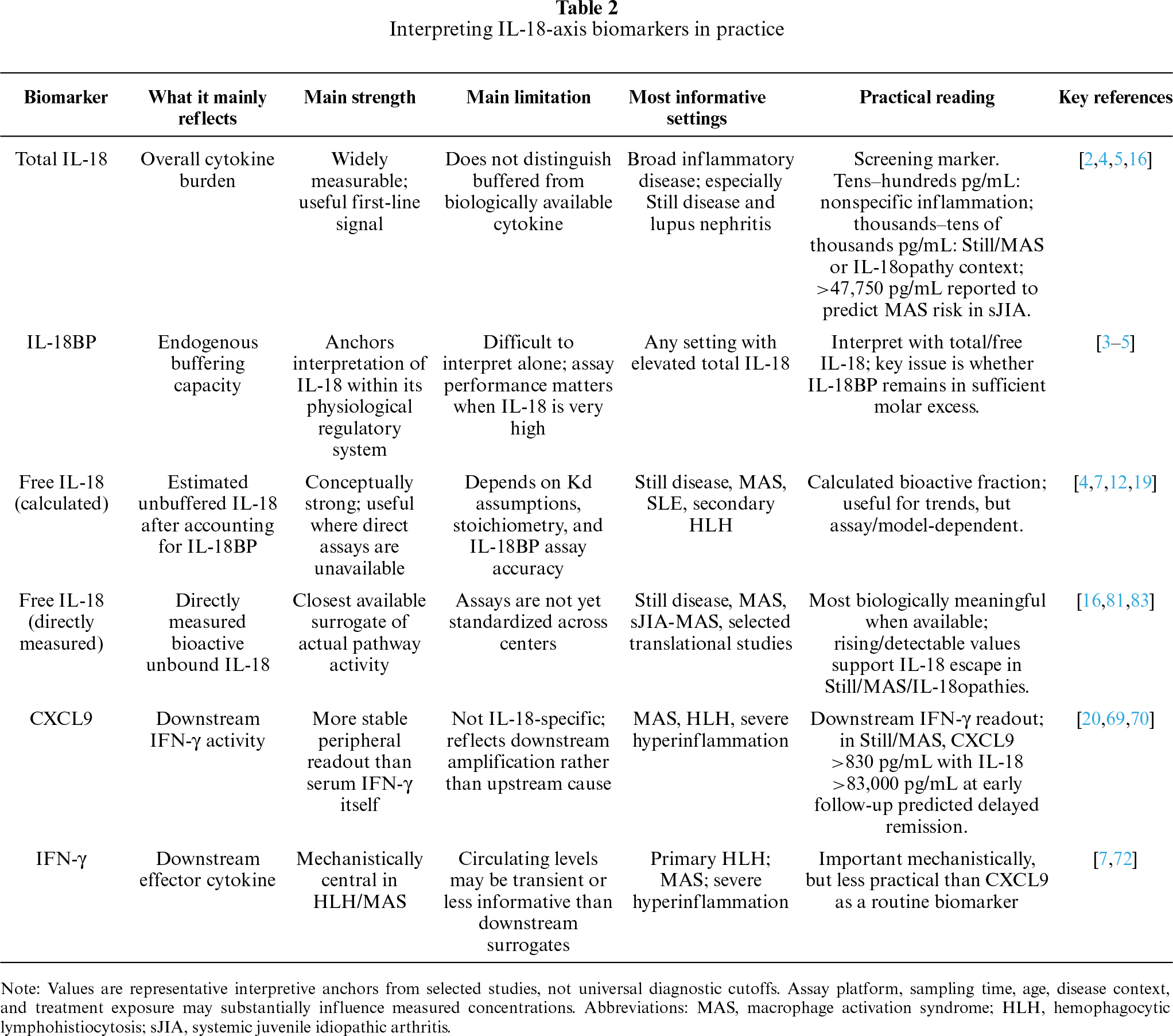
A practical three-tier approach follows from this. Total IL-18 identifies the possibility of pathway engagement. Free IL-18 identifies failure of buffering. CXCL9 shows whether the downstream IFN-γ loop is functionally active. This is not yet a standardized clinical algorithm, but it is the most biologically coherent way to interpret the axis in practice. It is also the framework on which emerging therapeutic strategies are being built. A practical summary of how to read these biomarkers is provided in table 2.
7 Therapeutic Targeting of the Axis
7.1 Restoring the Buffer: Recombinant IL-18BP
Among the therapeutic options currently available, recombinant IL-18 binding protein (IL-18BP) has the most straightforward biological rationale when disease is driven by genuinely elevated free IL-18. Rather than imposing an artificial block on the pathway, it restores a buffering mechanism that is already part of normal IL-18 physiology. This makes it particularly appealing in conditions such as adult-onset Still’s disease (AOSD), macrophage activation syndrome (MAS), and monogenic IL-18opathies, where the problem appears to be not simply excess cytokine production, but failure of endogenous restraint.
The phase II open-label study of tadekinig alfa, a recombinant IL-18BP, in refractory AOSD provided the first formal clinical proof-of-concept for this strategy [66]. Roughly half of treated patients met early response criteria, and the overall safety profile was acceptable. The study was small and uncontrolled, so it cannot be read as definitive efficacy evidence. Even so, it established an important principle: restoring IL-18 buffering can translate into measurable clinical improvement in an IL-18-high disease. That point was reinforced by a particularly informative case of sJIA with recurrent MAS, in which free IL-18 became undetectable within hours of the first tadekinig dose, remained suppressed during remission, and rose again after drug discontinuation in parallel with relapse [68,84]. Few reports make the pharmacodynamic logic of the pathway as clear as this one.
That selectivity is informative in itself. The clinical signal of tadekinig has emerged most clearly in disorders characterized by excess free IL-18, whereas the rationale is much weaker in diseases where total IL-18 may be increased but remains largely buffered. In that sense, response to recombinant IL-18BP is not only a therapeutic effect; it is also a kind of biological validation of the buffering model. More recently, the randomized-withdrawal phase III trial of tadekinig alfa in NLRC4 gain-of-function and XIAP deficiency has been listed as completed, extending the clinical development of recombinant IL-18BP into monogenic IL-18opathies (ClinicalTrials.gov identifier: NCT03113760), although peer-reviewed efficacy data remain limited [84].
7.2 Direct IL-18 Neutralization
Direct neutralization of IL-18 is conceptually attractive, but clinical evidence remains less developed than for recombinant IL-18BP. The appeal is obvious: if IL-18 is pathogenic, directly blocking the cytokine should be effective. In practice, however, the strongest human data so far have centered on restoring the endogenous decoy rather than replacing it with a separate neutralizing strategy.
That difference is probably not just a matter of how far drug development has progressed. In diseases defined by marked imbalance between IL-18 and IL-18BP, restoring the missing buffer may be the most physiologically coherent intervention. At the same time, direct anti-IL-18 strategies are now moving beyond a purely preclinical concept. Monoclonal antibodies against IL-18, including neoepitope-specific antibodies directed against the mature cleaved form, are in early development, and anti-IL-18 monoclonal antibody programs have entered clinical testing in inflammatory diseases such as atopic dermatitis. However, published efficacy data in the hyperinflammatory conditions most relevant to this review remain sparse [84,85]. At present, direct anti-IL-18 therapy therefore remains clinically promising but less established than recombinant IL-18BP or downstream IFN-γ blockade in the disease settings discussed here.
7.3 Downstream Effector Blockade: IFN-γ
If IL-18BP is the clearest upstream strategy, IFN-γ blockade is the strongest downstream one. The pivotal study of emapalumab in children with primary HLH showed that neutralizing IFN-γ can induce meaningful clinical responses and allow bridging to hematopoietic stem-cell transplantation [72]. This remains the most rigorous prospective evidence currently available for targeted intervention in severe hyperinflammation.
For this review, the biological message is important. The success of anti-IFN-γ does not make IL-18 irrelevant. Rather, it shows that in many forms of HLH the final common effector node lies downstream, at the level of IFN-γ-mediated tissue damage. Upstream and downstream targeting are therefore addressing different therapeutic problems. Blocking IL-18 is most persuasive when the central abnormality is chronic or disproportionate generation of free IL-18. Blocking IFN-γ becomes especially compelling when the inflammatory circuit is already fully engaged and organ-threatening macrophage activation is the immediate concern.
This distinction is especially helpful when comparing primary HLH with MAS or monogenic IL-18opathies. In the former, IFN-γ blockade has the strongest clinical footing. In the latter, IL-18 often remains the more proximal biological abnormality, even if downstream IFN-γ neutralization may still be clinically useful. That logic has now been extended to Still/MAS itself: a prospective study in glucocorticoid-refractory MAS secondary to sJIA or AOSD showed that emapalumab can control disease in this setting as well, reinforcing the view that IFN-γ is a meaningful downstream node across hyperinflammatory syndromes, not only in primary HLH [86].
7.4 JAK Inhibition as a Broader Downstream Strategy
JAK inhibition occupies a more pragmatic middle ground. It is not specific for the IL-18/IL-18BP axis, but it intercepts several cytokine-dependent inflammatory circuits at once, including IFN-γ signaling. Preclinical work with ruxolitinib in murine HLH showed improvement in inflammation and survival, providing much of the mechanistic basis for this approach [87]. Small clinical series in difficult-to-treat Still disease suggest that JAK inhibitors can be useful in selected refractory patients, and systematic review data support a signal of efficacy, although the available evidence remains uncontrolled and heterogeneous [88,89].
For that reason, JAK inhibition is probably best understood as a secondary downstream option rather than a pathway-defining therapy. It may be particularly useful when inflammation is clearly cytokine-driven but the dominant node is uncertain, or when broader suppression of the IFN-γ/JAK-STAT program is preferred to a more selective intervention. Its strength is breadth; its limitation is lack of specificity. In that sense, JAK inhibition complements rather than competes with the more biologically explicit strategies represented by IL-18BP or IFN-γ blockade.
7.5 A Brief Reverse Perspective: IL-18 in Oncology
Although oncology lies outside the main scope of this review, it is mentioned only briefly as a reverse-perspective counterpoint to the inflammatory diseases discussed above. In hyperinflammation, the therapeutic goal is to restrain excess free IL-18 or restore IL-18BP-mediated neutralization. In cancer, by contrast, IL-18BP may limit potentially useful antitumor IL-18 activity, and experimental strategies have therefore attempted to engineer IL-18 variants that evade IL-18BP neutralization in the tumor microenvironment [83]. This reverse perspective reinforces the central message of the review: the clinical meaning of IL-18 depends not only on total cytokine abundance, but on whether IL-18 is neutralized, free, or therapeutically redirected.
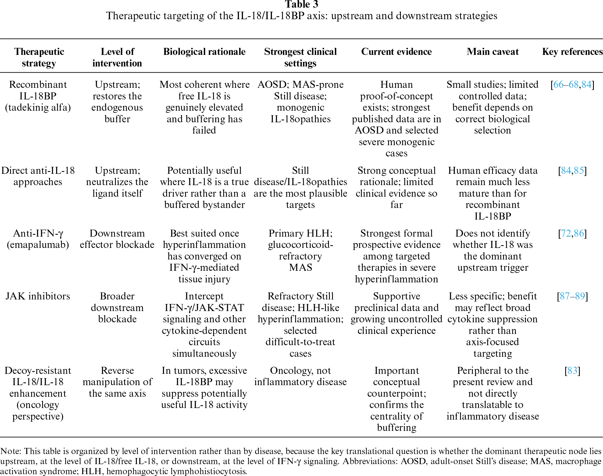
Taken together, the current therapeutic data support a layered model of intervention. In practical terms, IL-18BP-based therapy is most rational when the dominant abnormality is extreme total IL-18 together with measurable or rising free IL-18, relative insufficiency of IL-18BP-mediated restraint, and a Still/MAS-prone or monogenic IL-18opathy phenotype. Anti-IFN-γ therapy is more compelling when the clinical picture is dominated by organ-threatening macrophage activation and a downstream IFN-γ signature, reflected by high CXCL9, marked ferritin elevation, cytopenias, coagulopathy, liver involvement, or primary HLH-like biology. JAK inhibition occupies a broader downstream space and may be most useful when inflammation is clearly cytokine-driven but the dominant node is uncertain, when IFN-γ/JAK-STAT activation overlaps with other cytokine circuits, or when a less pathway-specific strategy is clinically required. These are not yet validated treatment algorithms, but provisional biological selection principles for matching the right therapeutic node to the right inflammatory context. This framework further illustrates that future treatment may involve matching therapeutic nodes to biomarker patterns and clinical phenotype rather than selecting a single universal target. The main therapeutic nodes of the axis are summarized in table 3.
8 Conclusions and Future Directions
Across the disease spectrum reviewed here, IL-18 emerges as a context-dependent cytokine whose clinical meaning is determined less by total abundance alone than by the balance between production, IL-18BP-mediated neutralization, and downstream inflammatory activation. This framework helps distinguish settings in which IL-18 behaves mainly as a biomarker or local amplifier from those in which insufficient endogenous antagonism permits free IL-18 to become diagnostically, mechanistically, and therapeutically relevant [5,7,84]. This interpretation complements recent dyad-focused syntheses of IL-18 and IL-18BP while emphasizing free IL-18 as the key translational discriminator across disease categories [6].
Several practical implications follow from this framework. At present, free IL-18 should not be treated as a stand-alone clinical decision threshold: assay standardization, multicenter validation, and disease-specific cutoffs are still needed, and the strongest numerical thresholds currently relate mainly to total IL-18, such as the >47,750 pg/mL value reported for MAS risk in sJIA [61]. For clinical translation, the more realistic near-term strategy is dynamic monitoring rather than reliance on a single cutoff. Total IL-18 may identify unusually high upstream cytokine production; IL-18BP and free IL-18 may indicate whether high-affinity endogenous antagonism has become quantitatively insufficient; and CXCL9 may capture downstream IFN-γ pathway activation [4,90]. In preliminary practical terms, a profile dominated by extreme total IL-18 together with measurable or rising free IL-18 would support consideration of IL-18-directed strategies in appropriate clinical or trial settings, whereas a CXCL9-dominant profile may point more strongly toward downstream IFN-γ- or JAK-directed approaches. Outside the Still/MAS/IL-18opathy spectrum, these applications remain investigational and require prospective validation. In parallel, monoclonal antibodies targeting IL-18 are entering early clinical development, adding a second potential modality to IL-18BP-based inhibition [84,85].
Recent reviews and translational studies published during the last five years further support the current relevance of this framework across inflammatory disease, autoimmunity, and IL-18BP-based therapeutic development [91–93].
The broader implication is that the transition from total to free IL-18 is not simply a technical refinement. It represents a shift in how IL-18 should be read biologically and clinically. If there is a unifying message to this review, it is that the relevance of IL-18 lies not in its ubiquity, but in the conditions under which endogenous buffering is no longer enough.
Acknowledgement: The authors gratefully acknowledge the National Lupus Patient Association, Gruppo LES Italiano ODV, for its continued support of our Lupus Clinic.
Funding Statement: The authors received no specific funding for this study.
Author Contributions: Simone Negrini: conceptualization, literature review, manuscript drafting. Stefania Nicola, Iuliana Badiu, Anna Quinternetto, Ilaria Vitali and Lo Sardo L: literature review, critical discussion of the evidence, manuscript revision. Luisa Brussino: conceptualization, supervision, critical revision of the manuscript. All authors contributed to interpretation of the literature, reviewed the manuscript. All authors reviewed and approved the final version of the manuscript.
Availability of Data and Materials: No new data were created or analyzed in this study. Data sharing is not applicable to this article.
Ethics Approval: Not applicable.
Conflicts of Interest: The authors declare no conflicts of interest.
References
1. Mantovani A, Dinarello CA, Molgora M, Garlanda C. Interleukin-1 and related cytokines in the regulation of inflammation and immunity. Immunity 2019;50(4):778–95. doi:10.1016/j.immuni.2019.03.012. [Google Scholar] [CrossRef]
2. Dinarello CA, Novick D, Kim S, Kaplanski G. Interleukin-18 and IL-18 binding protein. Front Immunol 2013;4:289. doi:10.3389/fimmu.2013.00289. [Google Scholar] [CrossRef]
3. Novick D, Kim SH, Fantuzzi G, Reznikov LL, Dinarello CA, Rubinstein M. Interleukin-18 binding protein: a novel modulator of the Th1 cytokine response. Immunity 1999;10(1):127–36. doi:10.1016/s1074-7613(00)80013-8. [Google Scholar] [CrossRef]
4. Novick D, Schwartsburd B, Pinkus R, et al. A novel IL-18BP ELISA shows elevated serum IL-18BP in sepsis and extensive decrease of free IL-18. Cytokine 2001;14(6):334–42. doi:10.1006/cyto.2001.0914. [Google Scholar] [CrossRef]
5. Harel M, Fauteux-Daniel S, Girard-Guyonvarc’h C, Gabay C. Balance between Interleukin-18 and Interleukin-18 binding protein in auto-inflammatory diseases. Cytokine 2022;150(1):155781. doi:10.1016/j.cyto.2021.155781. [Google Scholar] [CrossRef]
6. Novick D. IL-18 and IL-18BP: a unique dyad in health and disease. Int J Mol Sci 2024;25(24):13505. doi:10.3390/ijms252413505. [Google Scholar] [CrossRef]
7. Weiss ES, Girard-Guyonvarc’h C, Holzinger D, et al. Interleukin-18 diagnostically distinguishes and pathogenically promotes human and murine macrophage activation syndrome. Blood 2018;131(13):1442–55. doi:10.1182/blood-2017-12-820852. [Google Scholar] [CrossRef]
8. Nakanishi K, Yoshimoto T, Tsutsui H, Okamura H. Interleukin-18 is a unique cytokine that stimulates both Th1 and Th2 responses depending on its cytokine milieu. Cytokine Growth Factor Rev 2001;12(1):53–72. doi:10.1016/s1359-6101(00)00015-0. [Google Scholar] [CrossRef]
9. Sanders NL, Mishra A. Role of interleukin-18 in the pathophysiology of allergic diseases. Cytokine Growth Factor Rev 2016;32(10):31–9. doi:10.1016/j.cytogfr.2016.07.001. [Google Scholar] [CrossRef]
10. Zhang H, Wang J, Wang L, Xie H, Chen L, He S. Role of IL-18 in atopic asthma is determined by balance of IL-18/IL-18BP/IL-18R. J Cell Mol Med 2017;21(12):3550–60. doi:10.1111/jcmm.13323. [Google Scholar] [CrossRef]
11. Hu Y, Wang J, Zhang H, et al. Enhanced expression of IL-18 and IL-18BP in plasma of patients with eczema: altered expression of IL-18BP and IL-18 receptor on mast cells. Mediat Inflamm 2017;2017(3):3090782. doi:10.1155/2017/3090782. [Google Scholar] [CrossRef]
12. Novick D, Elbirt D, Miller G, Dinarello CA, Rubinstein M, Sthoeger ZM. High circulating levels of free interleukin-18 in patients with active SLE in the presence of elevated levels of interleukin-18 binding protein. J Autoimmun 2010;34(2):121–6. doi:10.1016/j.jaut.2009.08.002. [Google Scholar] [CrossRef]
13. Italiani P, Manca ML, Angelotti F, et al. IL-1 family cytokines and soluble receptors in systemic lupus erythematosus. Arthritis Res Ther 2018;20(1):27. doi:10.1186/s13075-018-1525-z. [Google Scholar] [CrossRef]
14. Calvani N, Richards HB, Tucci M, Pannarale G, Silvestris F. Up-regulation of IL-18 and predominance of a Th1 immune response is a hallmark of lupus nephritis. Clin Exp Immunol 2004;138(1):171–8. doi:10.1111/j.1365-2249.2004.02588.x. [Google Scholar] [CrossRef]
15. Gono T, Kawaguchi Y, Sugiura T, et al. Interleukin-18 is a key mediator in dermatomyositis: potential contribution to development of interstitial lung disease. Rheumatology 2010;49(10):1878–81. doi:10.1093/rheumatology/keq196. [Google Scholar] [CrossRef]
16. Girard C, Rech J, Brown M, et al. Elevated serum levels of free interleukin-18 in adult-onset Still’s disease. Rheumatology 2016;55(12):2237–47. doi:10.1093/rheumatology/kew300. [Google Scholar] [CrossRef]
17. Girard-Guyonvarc’h C, Rodriguez E, Mueller YM, et al. Elevated serum levels of interleukin-18 discriminate Still’s disease from other autoinflammatory conditions: results from the European ImmunAID cohort. RMD Open 2025;11(2):e005388. doi:10.1136/rmdopen-2024-005388. [Google Scholar] [CrossRef]
18. Takada H, Ohga S, Mizuno Y, et al. Oversecretion of IL-18 in haemophagocytic lymphohistiocytosis: a novel marker of disease activity. Br J Haematol 1999;106(1):182–9. doi:10.1046/j.1365-2141.1999.01504.x. [Google Scholar] [CrossRef]
19. Mazodier K, Marin V, Novick D, et al. Severe imbalance of IL-18/IL-18BP in patients with secondary hemophagocytic syndrome. Blood 2005;106(10):3483–9. doi:10.1182/blood-2005-05-1980. [Google Scholar] [CrossRef]
20. Debaugnies F, Mahadeb B, Nagant C, et al. Biomarkers for early diagnosis of hemophagocytic lymphohistiocytosis in critically ill patients. J Clin Immunol 2021;41(3):658–65. doi:10.1007/s10875-020-00950-z. [Google Scholar] [CrossRef]
21. Canna SW, de Jesus AA, Gouni S, et al. An activating NLRC4 inflammasome mutation causes autoinflammation with recurrent macrophage activation syndrome. Nat Genet 2014;46(10):1140–6. doi:10.1038/ng.3089. [Google Scholar] [CrossRef]
22. Wada T, Kanegane H, Ohta K, et al. Sustained elevation of serum interleukin-18 and its association with hemophagocytic lymphohistiocytosis in XIAP deficiency. Cytokine 2014;65(1):74–8. doi:10.1016/j.cyto.2013.09.007. [Google Scholar] [CrossRef]
23. Stone DL, Ombrello A, Arostegui JI, et al. Excess serum interleukin-18 distinguishes patients with pathogenic mutations in PSTPIP1. Arthritis Rheumatol 2022;74(2):353–7. doi:10.1002/art.41976. [Google Scholar] [CrossRef]
24. Okamura H, Tsutsui H, Komatsu T, et al. Cloning of a new cytokine that induces IFN-γ production by T cells. Nature 1995;378(6552):88–91. doi:10.1038/378088a0. [Google Scholar] [CrossRef]
25. Gu Y, Kuida K, Tsutsui H, et al. Activation of interferon-γ inducing factor mediated by interleukin-1β converting enzyme. Science 1997;275(5297):206–9. doi:10.1126/science.275.5297.206. [Google Scholar] [CrossRef]
26. Akita K, Ohtsuki T, Nukada Y, et al. Involvement of caspase-1 and caspase-3 in the production and processing of mature human interleukin 18 in monocytic THP.1 cells. J Biol Chem 1997;272(42):26595–603. doi:10.1074/jbc.272.42.26595. [Google Scholar] [CrossRef]
27. Dong Y, Bonin JP, Devant P, et al. Structural transitions enable interleukin-18 maturation and signaling. Immunity 2024;57(7):1533–48.e10. doi:10.1016/j.immuni.2024.04.015. [Google Scholar] [CrossRef]
28. Evavold CL, Ruan J, Tan Y, Xia S, Wu H, Kagan JC. The pore-forming protein gasdermin D regulates interleukin-1 secretion from living macrophages. Immunity 2018;48(1):35–44.e6. doi:10.1016/j.immuni.2017.11.013. [Google Scholar] [CrossRef]
29. Heilig R, Dick MS, Sborgi L, Meunier E, Hiller S, Broz P. The Gasdermin-D pore acts as a conduit for IL-1β secretion in mice. Eur J Immunol 2018;48(4):584–92. doi:10.1002/eji.201747404. [Google Scholar] [CrossRef]
30. Torigoe K, Ushio S, Okura T, et al. Purification and characterization of the human interleukin-18 receptor. J Biol Chem 1997;272(41):25737–42. doi:10.1074/jbc.272.41.25737. [Google Scholar] [CrossRef]
31. Born TL, Thomassen E, Bird TA, Sims JE. Cloning of a novel receptor subunit, AcPL, required for interleukin-18 signaling. J Biol Chem 1998;273(45):29445–50. doi:10.1074/jbc.273.45.29445. [Google Scholar] [CrossRef]
32. Hyun Kim S, Reznikov LL, Stuyt RJ, et al. Functional reconstitution and regulation of IL-18 activity by the IL-18Rβ chain. J Immunol 2001;166(1):148–54. doi:10.4049/jimmunol.166.1.148. [Google Scholar] [CrossRef]
33. Adachi O, Kawai T, Takeda K, et al. Targeted disruption of the MyD88 gene results in loss of IL-1- and IL-18-mediated function. Immunity 1998;9(1):143–50. doi:10.1016/s1074-7613(00)80596-8. [Google Scholar] [CrossRef]
34. Yoshimoto T, Takeda K, Tanaka T, et al. IL-12 up-regulates IL-18 receptor expression on T cells, Th1 cells, and B cells: synergism with IL-18 for IFN-γ production. J Immunol 1998;161(7):3400–7. doi:10.4049/jimmunol.161.7.3400. [Google Scholar] [CrossRef]
35. Dinarello CA. Overview of the IL-1 family in innate inflammation and acquired immunity. Immunol Rev 2018;281(1):8–27. doi:10.1111/imr.12621. [Google Scholar] [CrossRef]
36. Nakanishi K, Yoshimoto T, Tsutsui H, Okamura H. Interleukin-18 regulates both Th1 and Th2 responses. Annu Rev Immunol 2001;19(1):423–74. doi:10.1146/annurev.immunol.19.1.423. [Google Scholar] [CrossRef]
37. Kim SH, Eisenstein M, Reznikov L, et al. Structural requirements of six naturally occurring isoforms of the IL-18 binding protein to inhibit IL-18. Proc Natl Acad Sci U S A 2000;97(3):1190–5. doi:10.1073/pnas.97.3.1190. [Google Scholar] [CrossRef]
38. Hurgin V, Novick D, Rubinstein M. The promoter of IL-18 binding protein: activation by an IFN-γ-induced complex of IFN regulatory factor 1 and CCAAT/enhancer binding protein β. Proc Natl Acad Sci U S A 2002;99(26):16957–62. doi:10.1073/pnas.262663399. [Google Scholar] [CrossRef]
39. Veenstra KG, Jonak ZL, Trulli S, Gollob JA. IL-12 induces monocyte IL-18 binding protein expression via IFN-γ. J Immunol 2002;168(5):2282–7. doi:10.4049/jimmunol.168.5.2282. [Google Scholar] [CrossRef]
40. Kawashima M, Yamamura M, Taniai M, et al. Levels of interleukin-18 and its binding inhibitors in the blood circulation of patients with adult-onset Still’s disease. Arthritis Rheum 2001;44(3):550–60. doi:10.1002/1529-0131(200103)44:3<550::AID-ANR103>3.0.CO;2-5. [Google Scholar] [CrossRef]
41. Jarret A, Jackson R, Duizer C, et al. Enteric nervous system-derived IL-18 orchestrates mucosal barrier immunity. Cell 2020;180(4):813–4. doi:10.1016/j.cell.2020.02.004. [Google Scholar] [CrossRef]
42. Harrison OJ, Srinivasan N, Pott J, et al. Epithelial-derived IL-18 regulates Th17 cell differentiation and Foxp3+ Treg cell function in the intestine. Mucosal Immunol 2015;8(6):1226–36. doi:10.1038/mi.2015.13. [Google Scholar] [CrossRef]
43. Holmkvist P, Roepstorff K, Uronen-Hansson H, et al. A major population of mucosal memory CD4+ T cells, coexpressing IL-18Rα and DR3, display innate lymphocyte functionality. Mucosal Immunol 2015;8(3):545–58. doi:10.1038/mi.2014.87. [Google Scholar] [CrossRef]
44. Rossios C, Pavlidis S, Hoda U, et al. Sputum transcriptomics reveal upregulation of IL-1 receptor family members in patients with severe asthma. J Allergy Clin Immunol 2018;141(2):560–70. doi:10.1016/j.jaci.2017.02.045. [Google Scholar] [CrossRef]
45. Kaur D, Chachi L, Gomez E, Sylvius N, Brightling CE. Interleukin-18, IL-18 binding protein and IL-18 receptor expression in asthma: a hypothesis showing IL-18 promotes epithelial cell differentiation. Clin Transl Immunol 2021;10(6):e1301. doi:10.1002/cti2.1301. [Google Scholar] [CrossRef]
46. Thawanaphong S, Nair A, Volfson E, Nair P, Mukherjee M. IL-18 biology in severe asthma. Front Med 2024;11:1486780. doi:10.3389/fmed.2024.1486780. [Google Scholar] [CrossRef]
47. Tajiri T, Matsumoto H, Gon Y, et al. Free serum IL-18 levels and responsiveness to omalizumab in patients with severe asthma. Eur Respir J 2016;48(Suppl 60):PA575. doi:10.1183/13993003.congress-2016.PA575. [Google Scholar] [CrossRef]
48. Kou K, Aihara M, Matsunaga T, et al. Association of serum interleukin-18 and other biomarkers with disease severity in adults with atopic dermatitis. Arch Dermatol Res 2012;304(4):305–12. doi:10.1007/s00403-011-1198-9. [Google Scholar] [CrossRef]
49. Bähler L, Schärli S, Luther F, et al. IL-18 in atopic dermatitis—a multifaceted driver of skin inflammation. J Allergy Clin Immunol 2025;156(5):1160–72. doi:10.1016/j.jaci.2025.07.025. [Google Scholar] [CrossRef]
50. Ansai O, Miyauchi T, Hayashi R, et al. Interleukin-18 as a severity marker and novel potential therapeutic target for epidermolytic ichthyosis. Clin Exp Dermatol 2023;48(3):199–210. doi:10.1093/ced/llac069. [Google Scholar] [CrossRef]
51. Chen JL, Niu XL, Gao YL, et al. IL-18 knockout alleviates atopic dermatitis-like skin lesions induced by MC903 in a mouse model. Int J Mol Med 2020;46(2):880–8. doi:10.3892/ijmm.2020.4630. [Google Scholar] [CrossRef]
52. Xiang M, Feng Y, Wang Y, et al. Correlation between circulating interleukin-18 level and systemic lupus erythematosus: a meta-analysis. Sci Rep 2021;11(1):4707. doi:10.1038/s41598-021-84170-4. [Google Scholar] [CrossRef]
53. Migliorini P, Anzilotti C, Pratesi F, et al. Serum and urinary levels of IL-18 and its inhibitor IL-18BP in systemic lupus erythematosus. Eur Cytokine Netw 2010;21(4):264–71. doi:10.1684/ecn.2010.0210. [Google Scholar] [CrossRef]
54. Wong CK, Li EK, Ho CY, Lam CW. Elevation of plasma interleukin-18 concentration is correlated with disease activity in systemic lupus erythematosus. Rheumatology (Oxford) 2000;39(10):1078–81. doi:10.1093/rheumatology/39.10.1078. [Google Scholar] [CrossRef]
55. Ruchakorn N, Ngamjanyaporn P, Suangtamai T, et al. Performance of cytokine models in predicting SLE activity. Arthritis Res Ther 2019;21(1):287. doi:10.1186/s13075-019-2029-1. [Google Scholar] [CrossRef]
56. Zhou C, Li J, Zhao J, Bai XY, Shang S, Li W. Interleukin-18 in lupus nephritis: a meta-analysis of cytokine signaling dysregulation in immune-mediated nephropathy. Front Immunol 2025;16:1631728. doi:10.3389/fimmu.2025.1631728. [Google Scholar] [CrossRef]
57. Esfandiari E, McInnes IB, Lindop G, et al. A proinflammatory role of IL-18 in the development of spontaneous autoimmune disease. J Immunol 2001;167(9):5338–47. doi:10.4049/jimmunol.167.9.5338. [Google Scholar] [CrossRef]
58. Tang Y, Zhao Y, Chen Z, et al. IL-18 drives the Bhlhe40-mediated pathogenic Th17 cell response and exacerbates autoimmune disease progression. Cell Mol Immunol 2025;22(12):1581–97. doi:10.1038/s41423-025-01356-w. [Google Scholar] [CrossRef]
59. Kobayashi I. Advances in juvenile dermatomyositis: pathophysiology, diagnosis, treatment and interstitial lung diseases—a narrative review. Children 2024;11(9):1046. doi:10.3390/children11091046. [Google Scholar] [CrossRef]
60. Shimizu M, Yokoyama T, Yamada K, et al. Distinct cytokine profiles of systemic-onset juvenile idiopathic arthritis-associated macrophage activation syndrome with particular emphasis on the role of interleukin-18 in its pathogenesis. Rheumatology 2010;49(9):1645–53. doi:10.1093/rheumatology/keq133. [Google Scholar] [CrossRef]
61. Shimizu M, Nakagishi Y, Inoue N, et al. Interleukin-18 for predicting the development of macrophage activation syndrome in systemic juvenile idiopathic arthritis. Clin Immunol 2015;160(2):277–81. doi:10.1016/j.clim.2015.06.005. [Google Scholar] [CrossRef]
62. Kudela H, Drynda S, Lux A, Horneff G, Kekow J. Comparative study of Interleukin-18 (IL-18) serum levels in adult onset Still’s disease (AOSD) and systemic onset juvenile idiopathic arthritis (sJIA) and its use as a biomarker for diagnosis and evaluation of disease activity. BMC Rheumatol 2019;3(1):4. doi:10.1186/s41927-019-0053-z. [Google Scholar] [CrossRef]
63. Girard-Guyonvarc’h C, Palomo J, Martin P, et al. Unopposed IL-18 signaling leads to severe TLR9-induced macrophage activation syndrome in mice. Blood 2018;131(13):1430–41. doi:10.1182/blood-2017-06-789552. [Google Scholar] [CrossRef]
64. Mizuta M, Inoue N, Shimizu M, et al. Distinct roles of IL-18 and IL-1β in murine model of macrophage activation syndrome. J Allergy Clin Immunol 2023;152(4):940–8.e6. doi:10.1016/j.jaci.2023.05.027. [Google Scholar] [CrossRef]
65. De Matteis A, Pardeo M, Caiello I, et al. Traditional laboratory parameters and IFNγ-related biomarkers in the diagnosis and management of MAS. Semin Arthritis Rheum 2025;74(12):152812. doi:10.1016/j.semarthrit.2025.152812. [Google Scholar] [CrossRef]
66. Gabay C, Fautrel B, Rech J, et al. Open-label, multicentre, dose-escalating phase II clinical trial on the safety and efficacy of tadekinig Alfa (IL-18BP) in adult-onset Still’s disease. Ann Rheum Dis 2018;77(6):840–7. doi:10.1136/annrheumdis-2017-212608. [Google Scholar] [CrossRef]
67. Kiltz U, Kiefer D, Braun J, Schiffrin EJ, Girard-Guyonvarc’h C, Gabay C. Prolonged treatment with Tadekinig Alfa in adult-onset Still’s disease. Ann Rheum Dis 2020;79(1):e10. doi:10.1136/annrheumdis-2018-214496. [Google Scholar] [CrossRef]
68. Yasin S, Solomon K, Canna SW, et al. IL-18 as therapeutic target in a patient with resistant systemic juvenile idiopathic arthritis and recurrent macrophage activation syndrome. Rheumatology 2020;59(2):442–5. doi:10.1093/rheumatology/kez284. [Google Scholar] [CrossRef]
69. Bracaglia C, de Graaf K, Pires Marafon D, et al. Elevated circulating levels of interferon-γ and interferon-γ-induced chemokines characterise patients with macrophage activation syndrome complicating systemic juvenile idiopathic arthritis. Ann Rheumatic Dis 2017;76(1):166–72. doi:10.1136/annrheumdis-2015-209020. [Google Scholar] [CrossRef]
70. Buatois V, Chatel L, Cons L, et al. Use of a mouse model to identify a blood biomarker for IFNγ activity in pediatric secondary hemophagocytic lymphohistiocytosis. Transl Res 2017;180:37–52.e2. doi:10.1016/j.trsl.2016.07.023. [Google Scholar] [CrossRef]
71. Tsoukas P, Rapp E, Van Der Kraak L, et al. Interleukin-18 and cytotoxic impairment are independent and synergistic causes of murine virus-induced hyperinflammation. Blood 2020;136(19):2162–74. doi:10.1182/blood.2019003846. [Google Scholar] [CrossRef]
72. Locatelli F, Jordan MB, Allen C, et al. Emapalumab in children with primary hemophagocytic lymphohistiocytosis. N Engl J Med 2020;382(19):1811–22. doi:10.1056/NEJMoa1911326. [Google Scholar] [CrossRef]
73. Prencipe G, Caiello I, Pascarella A, et al. Neutralization of IFN-γ reverts clinical and laboratory features in a mouse model of macrophage activation syndrome. J Allergy Clin Immunol 2018;141(4):1439–49. doi:10.1016/j.jaci.2017.07.021. [Google Scholar] [CrossRef]
74. Romberg N, Al Moussawi K, Nelson-Williams C, et al. Mutation of NLRC4 causes a syndrome of enterocolitis and autoinflammation. Nat Genet 2014;46(10):1135–9. doi:10.1038/ng.3066. [Google Scholar] [CrossRef]
75. Wang Y, Lingappa S, Kang Z. Current understanding of the NLRC4 Inflammasome in autoinflammation and enterocolitis. Commun Biol 2026;9(1):611. doi:10.1038/s42003-026-10174-3. [Google Scholar] [CrossRef]
76. Wang Y, Gao JZ, Gurung P, et al. An animal model of NLRC4-associated autoinflammation and infantile enterocolitis reveals novel therapeutic strategies. Cell Mol Immunol 2025;22(12):1567–80. doi:10.1038/s41423-025-01355-x. [Google Scholar] [CrossRef]
77. Miyazawa H, Wada T. Immune-mediated inflammatory diseases with chronic excess of serum interleukin-18. Front Immunol 2022;13:930141. doi:10.3389/fimmu.2022.930141. [Google Scholar] [CrossRef]
78. Coppola S, Insalaco A, Zara E, et al. Mutations at the C-terminus of CDC42 cause distinct hematopoietic and autoinflammatory disorders. J Allergy Clin Immunol 2022;150(1):223–28. doi:10.1016/j.jaci.2022.01.024. [Google Scholar] [CrossRef]
79. Iannuzzo A, Delafontaine S, El Masri R, et al. Autoinflammatory patients with Golgi-trapped CDC42 exhibit intracellular trafficking defects leading to STING hyperactivation and ER stress. Nat Commun 2024;15(1):9940. doi:10.1038/s41467-024-54294-y. [Google Scholar] [CrossRef]
80. Canna SW, Girard C, Malle L, et al. Life-threatening NLRC4-associated hyperinflammation successfully treated with IL-18 inhibition. J Allergy Clin Immunol 2017;139(5):1698–701. doi:10.1016/j.jaci.2016.10.022. [Google Scholar] [CrossRef]
81. Miwa Y, Kawamoto N, Miwa T, et al. Transition of serum-free IL-18 levels in systemic juvenile idiopathic arthritis measured by a novel detection method. Cytokine 2026;201:157128. doi:10.1016/j.cyto.2026.157128. [Google Scholar] [CrossRef]
82. Diamond T, Lau M, Morrissette J, Chu N, Behrens EM. CXCL9 inhibition does not ameliorate disease in murine models of both primary and secondary hemophagocytic lymphohistiocytosis. Sci Rep 2023;13(1):12298. doi:10.1038/s41598-023-39601-9. [Google Scholar] [CrossRef]
83. Zhou T, Damsky W, Weizman OE, et al. IL-18BP is a secreted immune checkpoint and barrier to IL-18 immunotherapy. Nature 2020;583(7817):609–14. doi:10.1038/s41586-020-2422-6. [Google Scholar] [CrossRef]
84. Girard-Guyonvarc’h C, Harel M, Gabay C. The role of interleukin 18/interleukin 18-binding protein in adult-onset Still’s disease and systemic juvenile idiopathic arthritis. J Clin Med 2022;11(2):430. doi:10.3390/jcm11020430. [Google Scholar] [CrossRef]
85. Nariai Y, Kamino H, Obayashi E, et al. Generation and characterization of antagonistic anti-human interleukin (IL)-18 monoclonal antibodies with high affinity: two types of monoclonal antibodies against full-length IL-18 and the neoepitope of inflammatory caspase-cleaved active IL-18. Arch Biochem Biophys 2019;663:71–82. doi:10.1016/j.abb.2019.01.001. [Google Scholar] [CrossRef]
86. de Benedetti F, Grom AA, Brogan PA, et al. Efficacy and safety of emapalumab in macrophage activation syndrome. Ann Rheum Dis 2023;82(6):857–65. doi:10.1136/ard-2022-223739. [Google Scholar] [CrossRef]
87. Das R, Guan P, Sprague L, et al. Janus kinase inhibition lessens inflammation and ameliorates disease in murine models of hemophagocytic lymphohistiocytosis. Blood 2016;127(13):1666–75. doi:10.1182/blood-2015-12-684399. [Google Scholar] [CrossRef]
88. Gillard L, Pouchot J, Cohen-Aubart F, et al. JAK inhibitors in difficult-to-treat adult-onset Still’s disease and systemic-onset juvenile idiopathic arthritis. Rheumatology 2023;62(4):1594–604. doi:10.1093/rheumatology/keac440. [Google Scholar] [CrossRef]
89. Liao J, Tang Q, Xie X, Zhou B, Li F, Li S. The efficacy and safety of JAK inhibitors in patients with adult-onset Still’s disease: a meta-analysis and systematic review. Int Immunopharmacol 2024;137(2):112451. doi:10.1016/j.intimp.2024.112451. [Google Scholar] [CrossRef]
90. Fauteux-Daniel S, Girard-Guyonvarc’h C, Caruso A, Rodriguez E, Gabay C. Detection of free bioactive IL-18 and IL-18BP in inflammatory disorders. Methods Mol Biol 2023;2691:263–77. doi:10.1007/978-1-0716-3331-1_21. [Google Scholar] [CrossRef]
91. Landy E, Carol H, Ring A, Canna S. Biological and clinical roles of IL-18 in inflammatory diseases. Nat Rev Rheumatol 2024;20(1):33–47. doi:10.1038/s41584-023-01053-w. [Google Scholar] [CrossRef]
92. Zhao Y, Tang Y, Zhu X, et al. The roles of interleukin-18 in inflammation and autoimmune disease. Clin Rev Allergy Immunol 2026;69(1):17. doi:10.1007/s12016-026-09139-5. [Google Scholar] [CrossRef]
93. Jang YS, Lee K, Park M, et al. Albumin-binding recombinant human IL-18BP ameliorates macrophage activation syndrome and atopic dermatitis via direct IL-18 inactivation. Cytokine 2023;172(2):156413. doi:10.1016/j.cyto.2023.156413. [Google Scholar] [CrossRef]
Cite This Article
 Copyright © 2026 The Author(s). Published by Tech Science Press.
This work is licensed under a Creative Commons Attribution 4.0 International License , which permits unrestricted use, distribution, and reproduction in any medium, provided the original work is properly cited.
 Downloads
 Citation Tools
