
 Login
Login Register
 Submit a Paper
 Propose a Special lssue
 Open Access
REVIEW
The role of leptin in osteoarthritis: from pathogenesis to clinical implications
1 BIOMORF Department of Biomedical, Dental, Morphological and Functional Imaging Sciences, A.O.U. Policlinico “G.Martino”, University of Messina, Via Consolare Valeria 1, Messina, Italy
2 Istituto Clinico Polispecialistico C.O.T. Cure Ortopediche Traumatologiche s.p.a., Messina, Italy
3 BIOMORF Department of Biomedical, Dental, Morphological and Functional Imaging Sciences, Section of Orthopaedic and Trauma Surgery, A.O.U. Policlinico “G.Martino”, University of Messina, Via Consolare Valeria 1, Messina, Italy
4 Operative Research Unit of Orthopaedic and Trauma Surgery, Fondazione Policlinico Universitario Campus Bio-Medico, Via Alvaro del Portillo 200, Rome, Italy
5 Research Unit of Orthopaedic and Trauma Surgery, Department of Medicine and Surgery, Università Campus Bio-Medico Di Roma, Via Alvaro del Portillo 21, Rome, Italy
6 Foresight Consulting, Messina, Italy
7 Institute for Biomedical Research and Innovation (IRIB), National Research Council of Italy (CNR), Messina, Italy
8 Research Unit of Artificial Intelligence and Computer Systems, Department of Engineering, Università Campus Bio-Medico di Roma, Rome, Italy
9 Department of Human Pathology of Adults and Developmental Age “Gaetano Barresi,” Division of Pathology, University of Messina, Messina, Italy
* Corresponding Authors: Gabriele Ricciardi. Email: ; Maurizio Martini. Email:
# These authors contributed equally to this work
European Cytokine Network 2026, 37(3), 169-191. https://doi.org/10.32604/ecn.2026.084855
Received 30 April 2026; Accepted 25 August 2026; Issue published 28 September 2026
 View Full Text
 Download PDF
Abstract
Osteoarthritis (OA) is increasingly recognized as a multifactorial disease in which metabolic dysfunction and chronic low-grade inflammation contribute to joint degeneration. Among the mediators involved, leptin has emerged as a key adipokine linking obesity, aging, and inflammatory processes to OA development and progression. However, despite growing evidence, its role remains incompletely understood, and a comprehensive framework integrating leptin-related mechanisms across different joint tissues is still lacking. This review provides an updated overview of the role of leptin in OA pathogenesis, focusing on its effects on cartilage, synovium, subchondral bone, and the infrapatellar fat pad. We also discuss the interplay between leptin, obesity, inflammaging, and cellular senescence, together with its potential utility as a biomarker and therapeutic target. Current evidence indicates that leptin promotes inflammation, extracellular matrix degradation, pain sensitization, and tissue remodeling through multiple signaling pathways. Elevated leptin levels have been associated with disease severity and metabolically driven OA phenotypes, supporting its role as a molecular link between systemic metabolic alterations and local joint pathology. Nevertheless, important questions remain regarding its context-dependent effects and clinical applicability. A better understanding of leptin-related mechanisms may improve patient stratification and support the development of precision medicine approaches for inflammatory–metabolic OA phenotypes.Keywords
Supplementary Material
Supplementary Material FileOsteoarthritis (OA) is the most prevalent chronic joint disease and a leading cause of disability worldwide. It primarily affects load-bearing joints such as the knee and hip and is characterized by progressive cartilage degeneration, subchondral bone remodeling, osteophyte formation, and synovial inflammation [1–3]. Although traditionally considered a degenerative disorder driven by mechanical stress, OA is now widely recognized as a multifactorial disease resulting from the complex interplay of mechanical, metabolic, genetic, inflammatory, and age-related factors [4–6]. Among these, aging is considered one of the most important contributors to disease onset and progression, as it promotes structural and functional alterations across multiple joint tissues [4]. With advancing age, joint tissues undergo structural and functional changes, including extracellular matrix (ECM) remodeling, reduced cellularity, and increased oxidative stress. In particular, chondrocytes develop a senescence-associated secretory phenotype (SASP), characterized by the release of pro-inflammatory cytokines, matrix-degrading enzymes, and catabolic mediators that contribute to the disruption of cartilage homeostasis and the establishment of a catabolic microenvironment favoring cartilage degradation and OA progression [7–9]. This age-related low-grade chronic inflammation, often referred to as “inflammaging”, is further exacerbated by metabolic alterations such as increased adiposity, insulin resistance, and oxidative stress [10]. These systemic changes contribute to a pro-inflammatory microenvironment within the joint, where cytokines such as interleukin-1β (IL-1β) and tumor necrosis factor-α (TNF-α) activate key signaling pathways, including NF-κB, ultimately leading to cartilage degradation and disease progression [9–12]. In this context, increasing attention has been directed toward adipose tissue-derived mediators, known as adipokines, which link metabolic dysfunction to joint pathology [13]. Among these, leptin has emerged as a key regulator at the interface between obesity, inflammation, and OA. Initially identified as a hormone involved in energy homeostasis, leptin is now recognized as a pleiotropic molecule with roles in immune regulation, bone metabolism, and cartilage biology [6,11,14]. Leptin is produced not only by white adipose tissue but also locally within joint structures, including the infrapatellar fat pad (IPFP), synovium, and cartilage [6]. Its biological effects are mediated through specific receptors that activate multiple intracellular pathways involved in inflammation and matrix remodeling [15,16]. Elevated leptin levels have been detected in both serum and synovial fluid of OA patients and have been associated with disease severity, structural progression, and pain [17–19]. Although substantial evidence supports the involvement of leptin in OA pathogenesis, the available literature remains heterogeneous and, in some cases, conflicting. While numerous studies identify leptin as a key mediator of inflammation, cartilage degradation, and metabolic dysfunction, others suggest that its biological effects may be context-dependent and influenced by tissue type, disease stage, leptin concentration, and systemic metabolic status. Consequently, a unified understanding of the precise role of leptin in OA has not yet been fully established. It is still debated whether leptin acts as a primary driver of disease pathogenesis or rather as a secondary mediator reflecting underlying metabolic and inflammatory alterations. Furthermore, emerging evidence suggests that leptin interacts with aging-related processes, including cellular senescence and metabolic dysregulation, thereby amplifying joint degeneration [11,20,21]. Another unresolved issue concerns the dual nature of leptin activity, as both protective and deleterious effects have been reported depending on its concentration and duration of exposure. While physiological levels may contribute to tissue homeostasis, chronic elevation, such as that observed in obesity and aging, appears to promote inflammation and matrix degradation [22]. The aim of this review is to provide a comprehensive and critical overview of the current evidence regarding the role of leptin in OA pathogenesis, with particular emphasis on its involvement in aging-related mechanisms, its potential as a biomarker, and its relevance as a therapeutic target. Unlike previous reviews, which have largely focused on the metabolic and inflammatory functions of leptin in OA, the present work provides an integrated overview of leptin biology across different joint tissues and examines its role within the broader context of aging-related processes and the emerging inflammatory–metabolic OA phenotype. By combining mechanistic, biomarker, and therapeutic perspectives, this review aims to offer a translational framework that links systemic metabolic dysfunction and inflammaging to local joint degeneration. This framework also provides the biological rationale for translational approaches aimed at investigating leptin expression in joint tissues and circulation in patients with OA.
2 Leptin Biology and Signaling Pathways
Leptin is a 16-kDa adipokine encoded by the ob gene, originally identified in 1994 as a key regulator of body weight and energy homeostasis. It is primarily secreted by white adipose tissue, and its circulating levels are positively correlated with fat mass. Under physiological conditions, leptin acts as a negative feedback signal regulating appetite and energy expenditure through central mechanisms [22,23]. In the central nervous system, leptin binds to receptors in the hypothalamus, where it suppresses orexigenic pathways, such as neuropeptide Y (NPY) and agouti-related peptide, and stimulates anorexigenic signals, including pro-opiomelanocortin (POMC), via activation of the Janus kinase/signal transducer and activator of transcription (JAK/STAT) pathway [24,25]. Despite elevated circulating leptin levels in obesity, reduced responsiveness to leptin, commonly referred to as leptin resistance, is frequently observed. This condition is associated with impaired leptin transport across the blood–brain barrier and increased expression of negative regulators such as suppressor of cytokine signaling-3 (SOCS-3) [6,26]. Importantly, leptin resistance appears to be tissue-specific, predominantly affecting central pathways, while peripheral tissues may retain partial or even enhanced responsiveness to leptin signaling. This dissociation is particularly relevant in OA, where local leptin activity may persist despite systemic resistance. Beyond its central metabolic role, leptin is now recognized as a pleiotropic hormone involved in multiple physiological processes, including immune regulation, reproduction, bone metabolism, and musculoskeletal homeostasis. Importantly, leptin is also locally produced in peripheral tissues, including cartilage, synovium, subchondral bone, and the IPFP, suggesting both endocrine and paracrine/autocrine functions within the joint microenvironment [6,27]. This local production supports the concept that leptin may act as a key mediator of intra-articular crosstalk, linking metabolic alterations to tissue-specific pathological responses. The biological effects of leptin are mediated through its receptor (Ob-R), a member of the class I cytokine receptor family. Alternative splicing of the Ob-R gene generates several isoforms, including multiple short forms (Ob-Ra, Ob-Rc, Ob-Rd, Ob-Rf), a soluble form (Ob-Re), and the long signaling isoform (Ob-Rb). Among these, Ob-Rb is the primary functional receptor responsible for intracellular signal transduction, owing to its full-length cytoplasmic domain containing key motifs required for downstream signaling [28–30]. The differential expression of these isoforms across tissues may contribute to the heterogeneous biological effects of leptin observed in OA joints. Upon leptin binding, Ob-Rb activates JAK2, leading to phosphorylation of specific tyrosine residues within the receptor intracellular domain. These phosphorylation sites serve as docking platforms for signaling molecules such as STAT proteins, which translocate to the nucleus and regulate gene expression. In addition to the JAK/STAT pathway, leptin signaling also involves activation of mitogen-activated protein kinase (MAPK), phosphatidylinositol 3-kinase PI3K/Akt, and NF-κB cascades [31]. These molecular routes are critically involved in the regulation of inflammation, cell survival, and matrix remodeling, processes that are central to OA pathogenesis, and intersect at multiple levels through signaling crosstalk. A key node of pathogenic integration lies in the synergistic cooperation between NF-κB and MAPK-driven AP-1 signaling, which contributes to the transcriptional upregulation of matrix-degrading enzymes, including MMP-13 and ADAMTS-5, thereby fueling ECM degradation in OA [32]. In parallel, PI3K/Akt/mTOR signaling dampens autophagic flux through the mTOR-mediated inhibition of ULK1, thereby disrupting cellular quality control and leading to the accumulation of dysfunctional mitochondria [33]. Consequently, increased reactive oxygen species (ROS) further activate NF-κB and MAPK signaling, reinforcing a self-amplifying loop that connects autophagic impairment with inflammatory responses [34,35]. Additionally, sustained activation of STAT3 in cooperation with NF-κB and MAPK signaling contributes to stress-response programs associated with chondrocyte senescence, further integrating inflammatory signaling with long-term cellular dysfunction and catabolic persistence [36]. Through these interconnected signaling axes, leptin regulates the expression of pro-inflammatory cytokines, matrix metalloproteinases (MMPs), and oxidative stress mediators. These effects are particularly relevant in the context of OA, where leptin contributes to the amplification of inflammatory and catabolic responses within joint tissues, often acting synergistically with cytokines such as IL-1β [37,38].
Importantly, leptin amplifies IL-1β- and TNF-α-driven inflammatory signaling through convergent transcriptional co-activation of STAT3 and NF-κB, which co-occupy composite response elements in the promoters of iNOS, COX-2, MMP-13, and IL-6, producing supra-additive gene expression that exceeds the sum of individual stimuli [39]. At the post-translational level, leptin primes p38 MAPK, JNK, and ERK1/2, the same kinases activated by IL-1β and TNF-α, thereby lowering the activation threshold for subsequent cytokine stimulation [40]. The resulting iNOS-derived nitric oxide burst further amplifies NF-κB and STAT3 activity through S-nitrosylation of IκBα, establishing a feed-forward inflammatory loop [40]. This amplification is further sustained by impaired or insufficient SOCS-3-mediated negative feedback, which normally acts as an endogenous inhibitor of JAK2/STAT3 signaling. This impairment may be particularly relevant in obese OA patients and has been associated with increased synovial MMP levels [41].
Notably, the convergence of these pathways on key transcriptional regulators such as NF-κB highlights leptin as an important upstream modulator of inflammatory networks in OA. Alterations in leptin signaling have been described in both obesity and aging, including the development of leptin resistance and dysregulated intracellular signaling. These changes may contribute to a chronic low-grade inflammatory state and promote tissue degeneration, supporting the role of leptin as a key mediator linking metabolic dysfunction to OA pathogenesis [37,42,43]. However, whether these alterations represent a primary pathogenic driver of OA progression or act predominantly as mediators of obesity- and aging-related metabolic dysfunction remains to be fully elucidated.
3 Leptin in Osteoarthritis: Tissue-Specific Roles
Leptin exerts pleiotropic effects across multiple joint tissues, including cartilage, synovium, subchondral bone, and the IPFP, while also interacting with systemic factors such as obesity and aging. These interconnected mechanisms highlight the role of leptin as a central mediator of joint degeneration through both local and systemic pathways (figure 1).
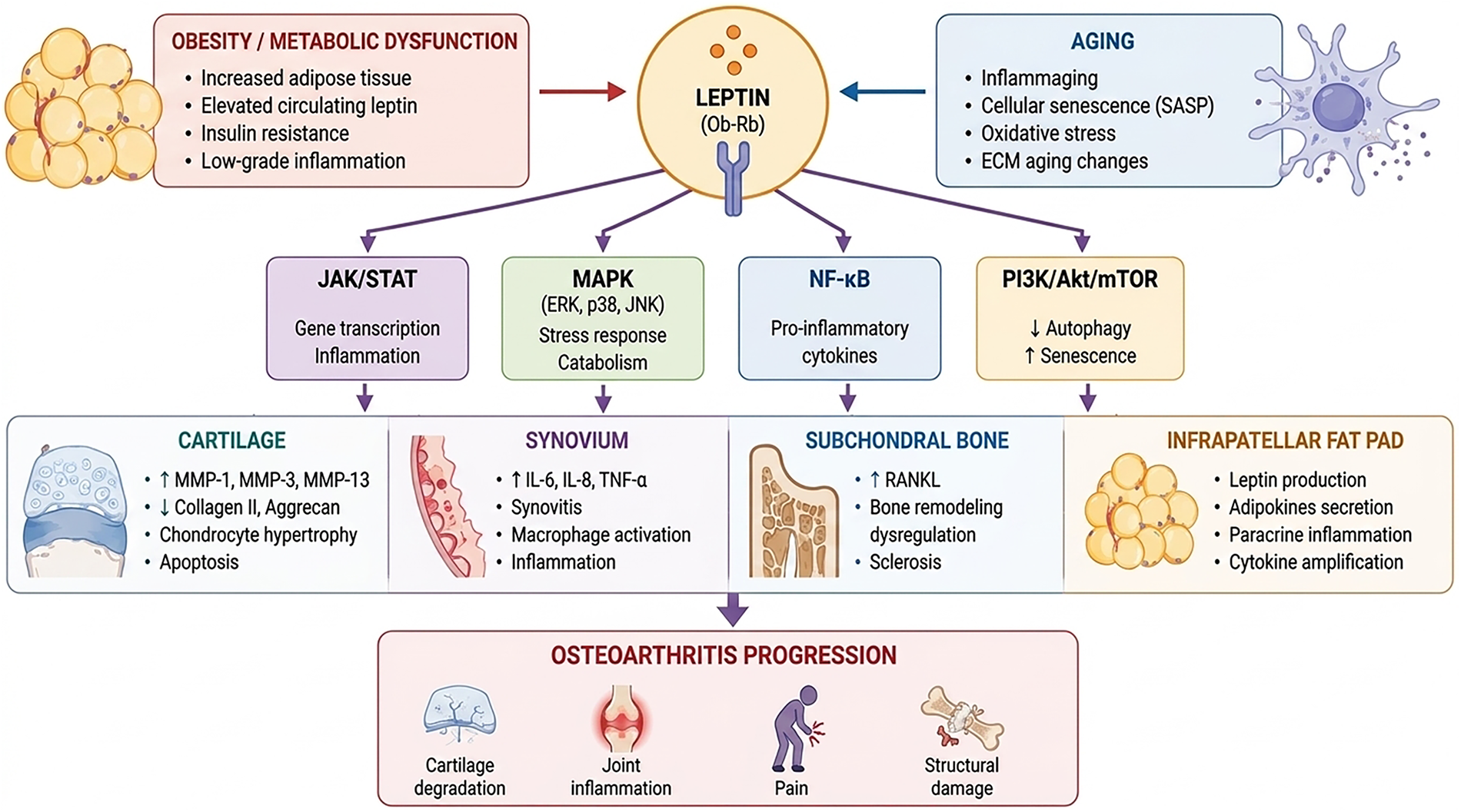
Figure 1: Leptin as a central integrator of metabolic, aging-related, and joint-specific pathways in osteoarthritis. Conditions such as obesity and metabolic dysfunction lead to increased circulating leptin levels, insulin resistance, and chronic low-grade inflammation, while aging contributes through processes collectively defined as inflammaging, including cellular senescence, oxidative stress, and extracellular matrix alterations. Through binding to its functional receptor (Ob-Rb), leptin activates multiple intracellular signaling pathways, including JAK/STAT, MAPK (ERK, p38, JNK), NF-κB, and PI3K/Akt/mTOR. These cascades regulate gene transcription, inflammatory responses, stress signaling, and the balance between autophagy and senescence, ultimately shaping cellular behavior within joint tissues. Within the osteoarthritic joint, leptin exerts tissue-specific effects. In cartilage, it promotes matrix degradation through the induction of matrix metalloproteinases, enhances chondrocyte hypertrophy, and contributes to apoptotic processes. In the synovium, leptin drives the production of pro-inflammatory cytokines and supports macrophage activation, sustaining chronic synovial inflammation. At the level of subchondral bone, it contributes to dysregulated remodeling through alterations in RANKL signaling, favoring sclerosis and structural imbalance. In parallel, the infrapatellar fat pad acts as a local source of leptin and other adipokines, amplifying paracrine inflammatory signaling within the joint microenvironment. Collectively, these interconnected mechanisms lead to the progression of osteoarthritis, characterized by cartilage degradation, persistent inflammation, pain, and structural joint damage.
Leptin exerts a complex and context-dependent role in bone metabolism, acting through both central and peripheral mechanisms that influence bone remodeling. Ob-Rs are widely expressed in bone-related cells, including osteoblasts, osteoclasts, and bone marrow stromal cells, supporting the concept that leptin directly participates in skeletal homeostasis [6,44–46]. At the central level, leptin signaling in the hypothalamus modulates bone metabolism through the sympathetic nervous system. Activation of hypothalamic Ob-Rs has been shown to suppress bone formation via β2-adrenergic signaling, which reduces osteoblastic activity and promotes osteoclastogenesis through increased expression of receptor activator of NF-κB ligand (RANKL). This pathway suggests an indirect inhibitory role of leptin on bone formation [47]. Conversely, peripheral effects of leptin appear more heterogeneous and, in some contexts, even anabolic [48]. Experimental studies have demonstrated that leptin can promote osteoblast differentiation and enhance bone formation by shifting mesenchymal stem cell commitment away from adipogenesis toward osteogenesis [49]. In addition, leptin has been shown to modulate the RANKL/osteoprotegerin axis, thereby influencing osteoclast differentiation and bone resorption [50]. However, the net effect of leptin on bone remains controversial and highly context-dependent. While some studies support its anabolic and protective role, others indicate that elevated leptin levels, such as those observed in obesity, may contribute to dysregulated bone remodeling. Under physiological conditions, leptin maintains skeletal homeostasis by promoting osteoblast differentiation and ensuring balanced bone remodeling [51].
Conversely, chronic hyperleptinemia and obesity-associated leptin resistance derail these regulatory networks, triggering aberrant intracellular signaling, altered osteoblast and osteoclast activity, and impaired bone quality [52], partly reconciling the seemingly conflicting anabolic and catabolic profiles reported in the literature. For instance, aberrant leptin signaling has been associated with increased activation of MAPK pathways (including ERK1/2 and p38), which are linked to inflammatory responses and altered osteoblast function [53–56]. Crucially, the overall impact of leptin on bone appears to depend on the balance between central inhibitory effects and peripheral anabolic/catabolic actions. In pathological conditions such as obesity and aging, both highly relevant in OA, this balance is likely shifted toward impaired bone quality rather than physiological homeostasis. Alterations in leptin signaling have also been implicated in impaired bone healing and remodeling observed in metabolic conditions. High-fat diet models have demonstrated that leptin resistance or dysregulation negatively affects bone formation, partly through JAK2/STAT3-dependent mechanisms in skeletal stem cells. This is particularly relevant in OA, where subchondral bone remodeling is a pathological hallmark and a key driver of disease progression [57–59]. Such structural abnormalities are characterized by an initial increase in bone turnover, followed by aberrant bone formation that results in subchondral sclerosis, increased bone mineral density, and reduced tissue elasticity. The resulting changes impair the ability of subchondral bone to dissipate mechanical loads, leading to abnormal stress transmission to the overlying articular cartilage [60]. Moreover, disruption of the osteochondral interface facilitates pathological biochemical crosstalk between subchondral bone and articular cartilage, establishing a vicious cycle of tissue degeneration that further accelerates OA progression [61]. Overall, in the context of OA, leptin should not be considered purely anabolic or catabolic for bone tissue, but rather a dysregulator of bone remodeling dynamics, contributing to subchondral sclerosis and abnormal mechanical properties of the osteochondral unit.
Articular cartilage represents one of the primary targets of leptin activity in OA, where this adipokine exerts predominantly pro-inflammatory and catabolic effects, although context-dependent anabolic responses have also been described. Chondrocytes, the only resident cells of cartilage, express functional Ob-R, making them directly responsive to leptin signaling [62–64]. In OA conditions, leptin levels are increased both systemically and locally within the joint microenvironment, and chondrocytes display an enhanced sensitivity to leptin stimulation. This altered responsiveness is particularly evident in obesity-associated OA, where leptin resistance at the systemic level paradoxically coexists with heightened local leptin activity in joint tissues [14,37,41,65,66]. This dissociation between systemic and local leptin activity represents a key mechanism potentially explaining its tissue-specific effects in OA. At the molecular level, leptin promotes cartilage degradation through the activation of multiple transduction routes, including JAK2/STAT3, MAPK (ERK, p38, JNK), and NF-κB [17]. These pathways converge on the upregulation of key catabolic mediators, such as matrix metalloproteinases (MMP-1, MMP-3, MMP-9, and MMP-13), which are directly involved in the breakdown of ECM components, particularly type II collagen and aggrecan. In addition, leptin stimulates the expression of iNOS, leading to increased nitric oxide production, which contributes to chondrocyte apoptosis and further matrix degradation [17,67,68]. A critical feature of leptin activity in cartilage is its strong synergistic interaction with classical pro-inflammatory cytokines, particularly IL-1β and TNF-α [68,69]. Co-stimulation with leptin and IL-1β amplifies the production of inflammatory mediators such as prostaglandin E2 (PGE2), cyclooxygenase-2 (COX-2), and additional MMPs, thereby exacerbating cartilage damage [40,70,71]. This synergism positions leptin not merely as an effector molecule, but as an amplifier of cytokine-driven inflammation within the OA joint. Beyond its catabolic role, leptin also influences chondrocyte phenotype and differentiation. It has been shown to promote hypertrophic differentiation, as indicated by increased expression of markers such as type X collagen, which is typically associated with endochondral ossification and cartilage degeneration [72]. Moreover, leptin can induce cytoskeletal remodeling via RhoA/ROCK signaling pathways, affecting chondrocyte morphology and mechanotransduction [73]. Leptin is also implicated in the regulation of cellular senescence and autophagy in chondrocytes, two processes that are increasingly recognized as central in OA pathogenesis [6]. High leptin levels have been associated with reduced autophagic activity, mediated in part through activation of the mTOR pathway, and with increased expression of senescence markers such as p53/p21 [74]. These effects contribute to impaired cellular homeostasis and reduced regenerative capacity of cartilage. Interestingly, some studies have reported anabolic effects of leptin, including stimulation of chondrocyte proliferation and increased synthesis of proteoglycans and collagen. These apparently conflicting findings suggest a dose- and context-dependent effect of leptin on cartilage homeostasis. While baseline physiological levels may support normal chondrocyte metabolism and ECM turnover, supraphysiological and chronic elevation, as observed in obesity and aging, shift its activity toward catabolic, pro-inflammatory, and degenerative signaling pathways [6,66,75,76]. Interestingly, leptin and Ob-R are expressed at low levels in normal or minimally affected cartilage, but their expression progressively increases with OA progression. This suggests that disease stage is a key determinant of chondrocyte responsiveness to leptin, with relatively preserved cartilage potentially retaining anabolic responses, whereas advanced OA cartilage appears to shift toward predominantly catabolic signaling [63]. Finally, discrepancies among studies may partly reflect heterogeneity in the experimental models employed. Deprived of their native ECM and three-dimensional mechanical cues, in vitro 2D monolayers of primary chondrocytes may exhibit altered leptin signaling and cellular responses. In parallel, ex vivo cartilage explants preserve ECM architecture and can modulate leptin diffusion and bioavailability, although they lack systemic metabolic and inflammatory inputs [77]. In vivo models add further complexity, reproducing systemic alterations such as hyperleptinemia or leptin resistance [65]. However, species-specific differences in cartilage biology and leptin signaling may influence the interpretation and translatability of experimental findings [78]. Its ability to integrate inflammatory signaling, matrix remodeling, and cellular dysfunction makes it a key target for translational studies.
The synovium plays a central role in the pathophysiology of OA as a key source of inflammatory mediators contributing to joint degeneration. In this context, leptin has emerged as an important regulator of synovial inflammation, acting at the interface between metabolic dysfunction and immune activation [79]. Leptin is consistently detected in the synovial fluid of OA patients, often at concentrations comparable to or higher than those observed in serum, suggesting a significant contribution from local production within the joint microenvironment. Synovial fibroblasts, as well as infiltrating immune cells, express functional Ob-R, making them direct targets of leptin signaling [80–82]. At the cellular level, leptin promotes a pro-inflammatory phenotype in synovial cells by inducing the production of cytokines and chemokines, including IL-6 and IL-8 [82,83]. This effect is mediated through the activation of key intracellular networks, such as NF-κB and MAPK, which amplify inflammatory responses and sustain cytokine release. Importantly, synovial responses to leptin may differ depending on disease stage, as leptin contributes to synovial inflammation through a staged process involving initiation, maintenance, and amplification of inflammatory responses.
In early OA, leptin-driven signaling appears to contribute primarily to low-grade synovitis and immune cell recruitment, whereas in more advanced disease, it may participate in the persistence of a chronic inflammatory state characterized by synovial hyperplasia and persistent cytokine production [84–86]. In the maintenance phase, leptin contributes to synovial hyperplasia, increased vascular permeability, and recruitment of immune cells, all of which are hallmarks of synovitis in OA [6,87,88]. A key element of this process is the involvement of macrophages, which are increasingly recognized as central effectors of synovial inflammation. Leptin has been shown to promote macrophage polarization toward a pro-inflammatory phenotype, thereby sustaining cytokine production and reinforcing local inflammatory loops [89]. In addition, leptin acts synergistically with classical pro-inflammatory cytokines, particularly IL-1β and TNF-α, reinforcing a chronic low-grade inflammatory state within the joint. During this amplification phase, such cooperative interactions intensify and perpetuate downstream inflammatory signaling, which not only exacerbates synovial dysfunction and tissue remodeling but also drives cartilage degradation through the release of soluble mediators that diffuse into adjacent articular tissues [37,69]. IPFP further contributes to this process by acting as a local source of adipokines and inflammatory factors [6]. Comparative analyses reveal that the IPFP expresses leptin and other adipokines at both the mRNA and protein levels, with leptin expression being significantly higher in OA patients than in healthy controls [90]. Through the continuous release of these mediators into the synovial environment, the IPFP sustains a feed-forward loop that links metabolic alterations to persistent joint inflammation. This IPFP-synovium axis represents a crucial component of the metabolic–inflammatory network driving OA progression, amplifying local inflammatory responses while promoting structural joint degeneration. This concept is further supported by clinical evidence, although findings remain heterogeneous. Some observational studies have reported significantly higher synovial fluid leptin concentrations in patients with more advanced radiographic OA. Specifically, one clinical cohort showed that leptin levels progressively increased across Kellgren–Lawrence grades, reaching the highest concentrations in grade IV disease, suggesting that local leptin accumulation may be associated with structural disease burden [18]. However, other studies have not confirmed significant differences in absolute synovial fluid leptin concentrations across radiographic stages, indicating that the relationship between local leptin levels and structural severity remains inconsistent across different clinical cohorts. In human OA joints, leptin has been consistently detected in synovial fluid and articular cartilage, where its expression is markedly increased compared with healthy tissue. Importantly, leptin expression within cartilage and osteophytes correlates with the histological grades of cartilage destruction, as assessed by the Mankin score [91,92]. Overall, IPFP-derived leptin-mediated synovial activation represents a critical component of OA pathogenesis, highlighting the importance of targeting local inflammatory networks in addition to structural joint damage. Rather than acting as a transient inflammatory trigger, leptin appears to contribute to the persistence and amplification of synovial inflammation, thereby sustaining disease chronicity.
The IPFP, also known as Hoffa’s fat pad, is increasingly recognized as an active player in the pathogenesis of OA rather than a passive structural component [93,94]. Located intracapsularly but extrasynovially, the IPFP is strategically positioned to directly influence the joint microenvironment through the secretion of adipokines, cytokines, and growth factors [6]. Among these mediators, leptin is abundantly produced by the IPFP and released into the synovial fluid, where it acts as a key amplifier of local inflammatory responses in OA by promoting synovial fibroblast activation and enhancing the release of pro-inflammatory mediators, thereby sustaining chronic synovitis [95]. Compared to subcutaneous adipose tissue, the IPFP exhibits a distinct secretory profile characterized by elevated expression of pro-inflammatory cytokines, including IL-6, TNF-α, and other chemokines, which synergize with leptin signaling to amplify joint inflammation [70]. At the cellular level, the IPFP contains adipocytes, fibroblasts, immune cells, and vascular components, all of which contribute to its immunometabolic activity. Notably, the IPFP is not a static adipose depot but a dynamically remodeling tissue, whose phenotype shifts toward a pro-inflammatory and fibrotic state in OA. In OA conditions, structural alterations such as fibrosis, vascular remodeling, and immune cell infiltration are commonly observed, exacerbating its pro-inflammatory phenotype [96]. Leptin produced within the IPFP can act in a paracrine manner on adjacent tissues, including synovium, cartilage, and subchondral bone, thereby promoting a leptin-driven network of inter-tissue crosstalk that sustains OA progression [6,97]. Specifically, leptin binding to synovial fibroblasts stimulates the production of IL-6 and IL-8, reinforcing synovial inflammation [83,98]. These mediators diffuse within the joint microenvironment and contribute to the activation of neighbouring chondrocytes, where leptin further promotes matrix degradation through increased expression of catabolic enzymes [84,99,100]. Concurrently, leptin modulates osteoblast and osteoclast activity within the osteochondral unit, contributing to aberrant subchondral bone remodeling [101]. Through this coordinated exchange of inflammatory and catabolic mediators, IPFP-derived leptin establishes a self-sustaining inflammatory loop that functionally links inter-tissue signaling and joint degeneration. This positions the IPFP as a central hub of OA pathophysiology, orchestrating inflammation, inter-tissue crosstalk, and ECM degradation. Emerging evidence also indicates that the IPFP undergoes structural and functional alterations in OA. Through leptin-driven modulation of nociceptive signaling within the joint, potentially involving the sensitization of peripheral nociceptors and the upregulation of neuroinflammatory mediators such as nerve growth factor (NGF) and substance P within the synovial environment, IPFP-derived leptin may contribute to peripheral sensitization and pain amplification in OA [102–104].
This provides a mechanistic link between metabolic inflammation and clinical symptomatology, particularly pain severity [6,94,105]. Importantly, the IPFP has been identified as a potential source of mediators involved in pain sensitization, linking metabolic inflammation to clinical symptoms. The progressive fibrosis and inflammatory remodeling of the IPFP may further enhance mechanical stress within the joint and sustain nociceptive input, thereby contributing to chronic pain states [106]. In line with this, IPFP-derived leptin concentrations within the synovial fluid have been associated with pain severity in patients with end-stage knee and hip OA, where higher levels correlate with increased Western Ontario and McMaster Universities Osteoarthritis Index (WOMAC) and Visual Analogue Scale (VAS) pain scores [80]. Importantly, the accessibility of the IPFP during surgical procedures, such as joint arthroplasty, makes it a valuable target for translational research. The analysis of leptin expression and signaling pathways in IPFP tissue, combined with parallel evaluation in cartilage and synovium, may provide critical insights into patient-specific disease mechanisms [14,24]. Overall, the IPFP represents a key source of leptin and inflammatory mediators within the OA joint. Its persistent secretory activity supports the concept that IPFP-derived signals actively drive OA progression, rather than merely reflecting systemic metabolic status.
4 Leptin, Obesity and Osteoarthritis
Obesity is one of the most well-established risk factors for OA; however, obesity-associated OA cannot be explained exclusively by increased mechanical loading. The involvement of non-weight-bearing joints, such as the hands, supports the contribution of systemic metabolic alterations and inflammatory mechanisms beyond biomechanical stress [91]. Accordingly, obesity-associated OA is increasingly recognized as a distinct metabolic phenotype characterized by adipose dysfunction, chronic low-grade inflammation, and adipokine dysregulation [15,27].
A key feature of obesity is the transformation of adipose tissue from an energy storage organ into an active immune tissue. Adipocyte hypertrophy and remodeling promote immune cell infiltration and a shift toward a pro-inflammatory adipose tissue phenotype, resulting in increased secretion of leptin and other inflammatory mediators, including TNF-α and IL-6 [107]. This obesity-associated inflammatory state, often referred to as metabolic inflammation or metaflammation, contributes to a systemic environment that favors joint inflammation and tissue degeneration [107].
Within this context, leptin represents a major molecular link between adiposity and OA progression. Increased adipose tissue mass leads to chronic hyperleptinemia, which differs from physiological leptin signaling involved in energy homeostasis [106]. Although obesity is associated with central leptin resistance, peripheral joint tissues may remain responsive to leptin, allowing persistent activation of pro-inflammatory and catabolic pathways within the OA joint microenvironment [15,26,27]. Thus, leptin may contribute to OA progression despite impaired systemic metabolic signaling.
Importantly, obesity-associated hyperleptinemia occurs within the broader framework of metabolic syndrome, including central obesity, insulin resistance, dyslipidemia, and altered inflammatory profiles [107–109]. These metabolic abnormalities contribute to the establishment of a catabolic joint environment by promoting inflammatory signaling, ECM degradation, synovial activation, and abnormal subchondral bone remodeling. In this setting, leptin acts synergistically with other obesity-related mediators to amplify pathways involved in OA progression, including NF-κB and MAPK signaling [6,15,27,93].
During OA, adipose tissue, particularly the IPFP, functions as a local endocrine organ secreting a network of pro-inflammatory adipokines, including resistin, visfatin, chemerin, and, paradoxically, adiponectin [97,110,111]. Rather than acting independently, these mediators converge on common inflammatory signaling pathways, including NF-κB, MAPK, and JAK/STAT, which are also activated by leptin. Resistin has been associated with OA clinical severity and may synergize with leptin through activation of the NF-κB/MMP axis [112], whereas visfatin enhances IL-6 and TNF-α production via MAPK signalling in synovial fibroblasts and promotes chondrocyte apoptosis [113,114]. Chemerin contributes to innate immune cell recruitment through CMKLR1 and reinforces TNF-α-dependent inflammatory loops, thereby amplifying leptin-mediated responses [115]. Although adiponectin is generally regarded as an anti-inflammatory adipokine in systemic metabolism, it may exert catabolic effects in OA cartilage through AdipoR1/AMPK/JNK signaling [116]. Consequently, the inflammatory status of the OA joint is determined not by leptin alone but by the integrated adipokine milieu, in which the reduced adiponectin/leptin ratio observed in obesity-associated OA shifts the balance toward persistent inflammation, cartilage catabolism, and synovitis.
The metabolic OA phenotype also involves local amplification of systemic metabolic alterations. Intra-articular adipose tissues, particularly the IPFP, may function as local sources of leptin and other adipokines, reinforcing the interaction between adipose tissue dysfunction and joint inflammation [106,117,118]. This bidirectional communication between systemic metabolic imbalance and local joint responses provides a mechanistic explanation for the development of obesity-associated OA.
These findings support the concept that obesity-related OA is not merely a consequence of mechanical overload but a metabolically driven disease process in which adipokine dysregulation contributes to disease initiation and progression.
5 Leptin and Aging: The Role of Inflammaging in Osteoarthritis
Aging is the most significant risk factor for OA [119,120], and increasing evidence indicates that leptin is a key mediator linking age-related metabolic dysfunction to chronic joint inflammation and tissue degeneration. Although aging is characterized by progressive loss of cellular homeostasis, chronic low-grade inflammation (inflammaging), and accumulation of molecular damage [6,119,121], these processes are also associated with profound alterations in leptin production, signaling, and tissue responsiveness. Consequently, leptin has emerged as a central component of the aging-associated inflammatory network that promotes OA progression [122]. Aging-related inflammaging is characterized by increased circulating levels of IL-1β, IL-6, TNF-α, and C-reactive protein [123–125]. Rather than representing an isolated systemic phenomenon, this inflammatory milieu enhances leptin production and modifies leptin responsiveness within joint tissues, thereby amplifying inflammatory and catabolic signaling [126]. Likewise, senescent chondrocytes acquire a senescence-associated secretory phenotype (SASP), characterized by increased secretion of pro-inflammatory cytokines, MMPs, and other catabolic mediators, creating a microenvironment in which leptin can further potentiate inflammatory responses and ECM degradation [6].
Emerging evidence also identifies leptin as an important immunometabolic regulator linking aging, obesity, and OA. Through its effects on immune and joint-resident cells, leptin integrates systemic metabolic alterations with local inflammatory responses.
In parallel, age-related impairment of autophagy, mitochondrial quality control, and oxidative stress increases chondrocyte susceptibility to leptin-mediated signaling [127–131]. Alterations in leptin signaling, including the development of central leptin resistance with relative preservation of peripheral leptin responsiveness, may further promote dysregulated inflammatory responses within the joint. Consequently, despite reduced central leptin sensitivity, peripheral tissues may continue to respond to leptin, allowing persistent activation of pro-inflammatory and catabolic pathways in the OA microenvironment [6,14,24].
Moreover, leptin has emerged as a direct regulator of cellular senescence and senescence-associated signaling pathways in aging joints. Cellular senescence in OA chondrocytes is primarily driven by activation of the p16 INK4a/Rb and p53/p21 CIP1 pathways, which induce irreversible cell-cycle arrest in response to oxidative stress, DNA damage, and metabolic dysfunction [8,132]. Emerging evidence suggests that leptin signaling through the Ob-R/JAK2/STAT3 axis may contribute to the activation of senescence-associated molecular responses and promote the acquisition of the SASP phenotype. In parallel, leptin activates NF-κB signaling, a central regulator of SASP maintenance, thereby increasing the production of inflammatory cytokines and catabolic mediators, including IL-6, IL-1β, MMP-3, and MMP-13 [133,134]. Through this mechanism, leptin may amplify the senescence–inflammation feedback loop, in which SASP factors promote local inflammatory propagation, ECM degradation, and progressive joint degeneration [6,42]. Furthermore, leptin-mediated JAK/STAT and MAPK signaling may exacerbate mitochondrial oxidative stress and metabolic dysfunction, further reinforcing the interaction between senescence, inflammation, and tissue degeneration [135].
Beyond cartilage, aging-related alterations in adipose tissue, including increased fat mass and changes in adipokine secretion, further contribute to elevated leptin levels and systemic inflammation. Recent evidence suggests that dysfunctional adipose tissue may also represent a source of senescence-associated inflammatory mediators, including leptin, IL-6, TNF-α, and MCP-1, thereby linking adipose tissue aging to systemic inflammaging and joint pathology [136]. These metabolic disturbances exacerbate the pro-inflammatory milieu and promote inter-tissue crosstalk within the joint, involving cartilage, synovium, subchondral bone, and the IPFP [6,137]. Overall, leptin should be considered not only as a mediator of metabolic dysfunction but also as a component of the aging-associated inflammatory network that drives OA progression. Its role in linking systemic inflammaging to local tissue degeneration highlights its importance as a central integrator of age-related joint pathology.
6 Leptin as a Biomarker in Osteoarthritis
The identification of reliable biomarkers for OA remains a major unmet need, particularly for early diagnosis, disease stratification, and monitoring of disease progression. In this context, leptin has emerged as a promising candidate biomarker due to its role at the intersection between metabolic dysfunction and joint inflammation. Elevated leptin concentrations have been consistently reported in both serum and synovial fluid (SF) of patients with OA, with individual studies and meta-analyses demonstrating significantly increased levels compared with healthy controls, particularly within the joint compartment [18,138]. The enrichment of leptin in SF supports the concept of local accumulation or production and highlights its potential role as a marker of intra-articular pathological processes [91]. Several studies have demonstrated positive associations between leptin concentrations and disease severity, assessed through radiographic grading systems such as the Kellgren–Lawrence scale and histological cartilage damage [18,19,91]. These findings indicate that leptin reflects structural alterations across different anatomical sites, including non–weight-bearing joints, supporting a contribution of metabolic-inflammatory mechanisms beyond mechanical loading alone. Beyond structural associations, leptin may also contribute directly to OA pathophysiology and symptom generation. Experimental models have shown that leptin deficiency protects against cartilage damage and reduces pain sensitivity, whereas restoration of leptin signaling reinstates structural degeneration and mechanical hyperalgesia [14,24]. These findings support a potential causal role for leptin in linking metabolic dysfunction, joint damage, and pain. At the molecular level, leptin exhibits concentration-dependent effects, with elevated levels promoting a catabolic phenotype characterized by increased MMPs expression and induction of hypertrophic chondrocyte pathways [76]. This biological complexity is relevant for biomarker interpretation, as leptin concentrations may reflect both physiological and pathological processes depending on the local and systemic context.
Leptin should therefore be considered within a broader metabolic-inflammatory network rather than as an isolated marker. Multi-biomarker studies indicate that leptin provides complementary information when integrated with other adipokines and systemic inflammatory markers [139]. In a cross-sectional MRI-based study of early knee OA (n = 137), leptin was independently associated with osteophyte size, cartilage integrity, synovitis, and effusion, whereas adiponectin was associated with pain and function and hs-CRP with meniscal pathology, demonstrating distinct biological domains captured by different biomarkers within the same panel [140]. Similarly, serum studies have shown that leptin and resistin provide partially distinct information depending on radiographic severity and metabolic status, supporting their complementary rather than redundant use [141].
The prognostic relevance of leptin-containing biomarker combinations has also been supported by ratio-based and machine-learning approaches. In the Osteoarthritis Initiative progression cohort, leptin/CRP and CRP/MCP-1 ratios combined with age and BMI showed high accuracy in predicting structural progression, with particularly strong prognostic sensitivity of leptin-related measures in men [142]. Longitudinal analyses from the VIDEO cohort demonstrated that leptin clustered within an inflammatory-metabolic principal component together with adipsin and CRP, primarily associated with pain and symptom burden assessed by WOMAC scores, whereas other biomarker clusters reflected structural damage or cytokine-driven inflammation [143]. These findings support leptin as a marker of a specific metabolic-inflammatory axis that partially overlaps but remains distinct from classical inflammatory and structural OA pathways. Emerging evidence further indicates that leptin is associated with specific OA phenotypes and endotypes, particularly the metabolic or obesity-associated phenotype characterized by obesity, metabolic syndrome, and systemic low-grade inflammation. In this setting, leptin acts both as a surrogate marker and as a potential mediator of metabolic dysregulation, linking adipose tissue expansion, IPFP activity, synovial inflammation, cartilage metabolism, and subchondral bone remodeling [144]. Patients with isolated knee OA and higher BMI exhibit increased leptin concentrations compared with other OA phenotypes, supporting its potential value in metabolic subtype discrimination [141]. Furthermore, Mendelian randomization studies have shown that genetically increased leptin levels are associated with higher OA risk, particularly knee OA, supporting a potential causal contribution rather than a purely associative relationship [145].
At the tissue level, the IPFP functions as an active endocrine structure involved in the metabolic OA phenotype representing a potential therapeutic target [146]. From an endotype perspective, leptin is most strongly associated with the metabolic-inflammatory OA axis rather than the classical inflammatory endotype driven predominantly by IL-1, TNF-α, and synovitis-related pathways [147]. However, substantial overlap exists between these biological domains, suggesting that OA phenotypes represent a continuum rather than strictly separated entities. Unsupervised clustering approaches have identified distinct OA subgroups based on metabolic, systemic, and local inflammatory characteristics, including leptin-high metabolic phenotypes associated with worse structural and symptomatic outcomes [148]. Beyond single-marker interpretation, several molecular and precision medicine frameworks have been proposed to formalize OA phenotyping and biomarker-driven stratification. The OA endotype framework proposed by Mobasheri et al. describes overlapping molecular subtypes, including inflammatory, mechanical, metabolic, and low-repair endotypes, with leptin and adiponectin positioned within the metabolic axis as upstream regulators of IL-1β, IL-6, and matrix-degrading enzyme production [149]. Similarly, Roman-Blas et al. proposed that OA phenotypes require tailored biomarker combinations and outcome measures, with adipokines such as leptin being particularly relevant for identifying metabolically driven phenotypes rather than representing universal markers of disease severity [150]. Integrative systems-based approaches further support the concept of multi-biomarker endotyping. A 15-biomarker panel identified distinct OA molecular profiles characterized by differences in tissue turnover and systemic inflammation, reinforcing the biological heterogeneity of OA and the need for phenotype-oriented strategies [147]. Similarly, metabolic OA frameworks define this phenotype through the combination of obesity, metabolic syndrome, and increased biomarkers such as hsCRP and leptin, supporting the role of leptin in patient stratification [151]. Broader integrative models further identify leptin as a systemic mediator linking adipose tissue expansion with joint degeneration, with metabolomic signatures supporting its involvement in metabolic OA biology [152].
From a therapeutic perspective, leptin has been investigated as a potential marker of differential treatment response, although clinical validation remains limited. Experimental studies suggest that leptin-high metabolic phenotypes may represent subgroups more responsive to interventions targeting metabolic pathways. In high-fat diet models, metformin showed stronger chondroprotective effects compared with normal-diet conditions, partly through reduction of adipose-derived leptin signaling, suggesting that leptin-related pathways may contribute to metabolic treatment responsiveness [153]. Clinical evidence, however, remains insufficient to support leptin-based treatment allocation. Long-term vitamin D supplementation did not significantly modify leptin or related inflammatory adipokines, suggesting limited value of leptin stratification for this intervention [154]. Conversely, intra-articular biological therapies such as Wharton’s jelly mesenchymal stem cells have been associated with reductions in synovial leptin concentrations together with changes in other adipokines, indicating that leptin may have potential value as a pharmacodynamic marker of local therapeutic response [155]. Expert consensus statements suggest that inflammatory and metabolic OA endotypes, including leptin-high phenotypes, may eventually contribute to treatment selection strategies; however, no leptin-based stratification approach has yet been validated in randomized clinical trials [156]. Similarly, adipokine-targeting strategies remain at a preclinical or early translational stage, and leptin levels are not currently used for clinical treatment allocation [157].
Despite its strong biological rationale and translational potential, several limitations currently prevent the implementation of leptin as a routine clinical biomarker. The absence of standardized assays, validated reference ranges, and OA-specific thresholds remains a major barrier. Circulating leptin concentrations are influenced by sex, adiposity, age, and circadian variation, requiring careful interpretation and limiting direct comparison between studies [158]. In addition, leptin interpretation remains strongly dependent on metabolic context. The close relationship with BMI and fat mass makes it difficult to separate leptin-specific effects from broader obesity-associated inflammatory and mechanical mechanisms. This limitation is particularly relevant because leptin differences between OA patients and controls are less evident in non-obese individuals, reducing its discriminatory value in lean OA phenotypes [141,159]. Sex-related differences further complicate interpretation, as women consistently exhibit higher leptin concentrations than men and may show different biomarker associations [142]. Another unresolved issue is the relationship between systemic and local leptin compartments. Although serum leptin is the most accessible measurement, synovial fluid and IPFP-derived leptin may better represent intra-articular biology, although they are less feasible for routine longitudinal assessment [144,146]. Furthermore, studies evaluating synovial leptin have generally included relatively small cohorts and require validation in larger independent populations before clinical application can be considered [160]. Methodological variability represents an additional barrier to clinical translation. No standardized analytical protocol currently exists for leptin quantification, and most studies rely on ELISA-based assays that differ in methodology, calibration, and analytical performance, limiting comparability across studies [156]. Likewise, no validated serum or synovial fluid cut-off values have been established for OA diagnosis, disease staging, prognosis, or treatment stratification, and no study has defined clinically useful sensitivity or specificity thresholds for leptin as a standalone biomarker. Furthermore, the absence of regulatory qualification of leptin-containing biomarker panels, the lack of validated biochemical surrogate endpoints for OA clinical trials, and the heterogeneity of OA populations remain important obstacles to translation [156]. Importantly, no study to date has formally established clinically applicable sensitivity and specificity values for leptin as a standalone diagnostic or prognostic biomarker in OA, preventing its evaluation against predefined clinical performance criteria. Although multi-omics studies consistently implicate leptin-associated pathways, these findings have not yet been converted into clinically actionable tools [161,162]. Finally, although longitudinal studies provide stronger evidence than cross-sectional investigations, prospective data remain relatively limited, and available studies suggest that leptin improves prognostic performance mainly when incorporated into multimarker models rather than used as an individual biomarker.
Clinical Studies Evaluating Leptin as a Biomarker of OA Severity and Progression
Clinical studies consistently support an association between leptin concentrations and OA presence, severity, and clinical manifestations; however, its performance as an independent biomarker remains limited by biological variability and the complexity of OA phenotypes.
Several studies have investigated the relationship between leptin levels and structural disease severity. While plasma leptin concentrations have been positively associated with radiographic severity after adjustment for sex and BMI, findings regarding synovial fluid leptin concentrations remain conflicting. Some cohorts have reported progressively increasing synovial fluid leptin levels with advancing Kellgren–Lawrence grade, whereas others have found no significant differences in absolute synovial fluid leptin concentrations across radiographic stages [18,163]. Similarly, serum leptin has been associated with imaging markers of cartilage composition. An inverse correlation between circulating leptin concentrations and the delayed gadolinium-enhanced magnetic resonance imaging of cartilage (dGEMRIC) index has been reported, suggesting a relationship between increased leptin levels and reduced cartilage glycosaminoglycan content. However, this association was significant only in women, highlighting the importance of sex-specific biological differences in leptin interpretation and biomarker performance [164]. Despite these associations, the relationship between systemic leptin levels and local joint pathology remains inconsistent. In a large cohort of patients with end-stage knee OA, serum leptin concentrations were significantly higher than in healthy controls but were not associated with cartilage histopathology and showed only weak correlations with synovial inflammatory markers. These findings suggest that circulating leptin may predominantly reflect systemic metabolic status rather than the extent of local structural damage [92]. Compared with circulating measurements, SF leptin may provide a more direct representation of the intra-articular environment, and its levels have been positively associated with MMPs expression supporting a mechanistic link between local leptin accumulation and cartilage matrix degradation [68]. However, evidence supporting SF leptin as a clinically applicable biomarker remains limited, as available studies have generally involved small cohorts and require confirmation in larger independent populations [160]. Longitudinal studies provide additional evidence that leptin is associated with structural progression, although its predictive performance remains modest when used alone. In the ADVANCE cohort, higher serum leptin concentrations were associated with subsequent joint space narrowing. However, prediction of incident osteophyte formation remained limited when leptin was incorporated into a multimarker model with COMP and IL-1β, achieving an AUROC of 0.586 (95% CI 0.524–0.646). These findings indicate that leptin may provide prognostic value mainly as part of a biomarker combination rather than as an individual predictor [165]. Similarly, weight-loss intervention studies have provided insights into the relationship between leptin dynamics and disease progression. Reductions in circulating leptin during weight-loss interventions were associated with slower cartilage volume loss; however, these associations largely disappeared after adjustment for the degree of weight reduction. This suggests that leptin changes may primarily represent obesity-related metabolic improvements rather than an independent determinant of structural progression [159]. Among clinical manifestations, pain represents one of the most consistently reported associations with leptin. Higher serum leptin concentrations have been independently associated with increased risk of knee pain and painful radiographic OA after adjustment for age, body mass, ethnicity, and socioeconomic factors [166]. Moreover, reductions in serum leptin following combined behavioural weight-loss interventions correlated with improvements in WOMAC pain scores. However, mediation analyses indicated that these effects were largely explained by weight reduction itself, suggesting that leptin acts primarily as a marker of adiposity-related metabolic changes rather than as an independent mediator of pain [167]. The association between leptin and symptoms may extend beyond weight-bearing joints. Studies in hand OA have also reported relationships between leptin concentrations and clinical manifestations, suggesting that leptin-related mechanisms are not exclusively dependent on mechanical loading. Nevertheless, findings remain heterogeneous, as some knee OA cohorts have reported no significant association between baseline leptin concentrations and pain severity or functional disability [168].
Overall, clinical evidence indicates that leptin captures relevant biological information across multiple OA domains, including structural severity, cartilage composition, progression, and symptom burden. However, its clinical value appears to depend strongly on biological context, particularly metabolic status, sex, and the distinction between systemic and local compartments.
Therefore, current evidence supports leptin as a complementary biomarker within multimarker and phenotype-driven approaches rather than as a validated standalone marker for OA diagnosis, prognosis, or treatment stratification. Future prospective studies integrating leptin with imaging, clinical characteristics, and additional molecular biomarkers will be required to determine its precise role in precision medicine approaches for OA.
7 Therapeutic Implications and Targeting Leptin Pathways in Osteoarthritis
Given its central role in linking metabolic dysfunction, inflammation, and joint degeneration, leptin represents an attractive target for therapeutic intervention in OA. However, its pleiotropic nature and widespread physiological functions make direct targeting of leptin signaling particularly challenging [6,37]. Moreover, despite extensive experimental evidence supporting its pathogenic role, most proposed therapeutic approaches remain at the preclinical stage. To date, no leptin-targeted therapy has been approved or clinically validated for OA, and no successful clinical trials have demonstrated the efficacy of directly targeting leptin signaling in patients with OA.
Against this backdrop, one of the most extensively investigated preclinical approaches involves modulation of leptin signaling pathways rather than direct inhibition of leptin itself. Key intracellular cascades activated by leptin, including JAK/STAT, MAPK, PI3K/Akt, and NF-κB, are critically involved in the regulation of inflammation, matrix degradation, and cellular senescence in joint tissues [6,66]. Pharmacological inhibitors targeting these pathways have shown promising results in preclinical research by reducing the expression of MMPs, inflammatory cytokines, and catabolic mediators [169–171]. For instance, inhibition of NF-κB signaling has been associated with decreased cartilage degradation and attenuation of synovial inflammation, highlighting its relevance as a downstream effector of leptin activity [172]. Another promising approach focuses on restoring the balance between anabolic and catabolic processes within cartilage. Given the involvement of leptin in suppressing autophagy through activation of the mTOR pathway, mTOR inhibitors such as rapamycin have been investigated as potential therapeutic agents. These compounds have been shown to enhance autophagic activity, reduce chondrocyte apoptosis, and limit OA progression in in vivo models [173]. In addition to canonical mTOR signaling, leptin has been shown to regulate autophagy and apoptosis through alternative mechanisms, including the LOXL3 route, further contributing to chondrocyte dysfunction. Accordingly, pharmacological inhibition of mTOR using agents such as rapamycin or AZD8055, as well as small-molecule inhibitors of the PI3K/Akt/mTOR axis (e.g., LY294002), has demonstrated the ability to restore autophagic flux, reduce catabolic factor expression, and attenuate structural joint alterations, including subchondral bone changes in experimental studies [174,175]. Moreover, genetic or pharmacological suppression of mTOR activity has been associated with decreased expression of key degradative enzymes such as MMP-13, reinforcing the relevance of this pathway in leptin-mediated cartilage degeneration [74]. In parallel, modulation of leptin-induced pro-apoptotic signaling represents an additional therapeutic opportunity. Activation of JAK2/STAT3 and MAPK-related pathways, including JNK signaling, contributes to chondrocyte apoptosis and stress responses. Experimental evidence suggests that inhibition of JAK/STAT signaling can attenuate leptin-induced apoptosis, while restoration of regulatory molecules such as dual-specificity phosphatases may partially counteract JNK-mediated effects [66]. Similarly, endogenous inhibitors of leptin signaling, including SOCS-3, have been shown to negatively regulate leptin activity in preclinical experiments, highlighting potential strategies to fine-tune leptin responsiveness rather than completely abolish its function [41,176]. Targeting the inflammatory microenvironment represents an additional strategy. Since leptin acts synergistically with pro-inflammatory cytokines such as IL-1β and TNF-α, therapies aimed at reducing systemic and local inflammation may indirectly mitigate leptin-mediated effects [40]. Lifestyle interventions, including weight loss and physical activity, have been shown to decrease circulating leptin levels and improve clinical outcomes in OA patients, supporting the importance of metabolic modulation in disease management [177]. More recently, interest has emerged in targeting local sources of leptin within the joint, particularly the IPFP. Given its role as a reservoir of adipokines and inflammatory mediators, strategies aimed at modulating IPFP inflammation or secretory activity may represent a novel therapeutic avenue [106]. Finally, direct modulation of leptin signaling itself has been proposed as a potential strategy. Targeting the functional leptin receptor isoform using monoclonal antibodies or high-affinity binding molecules may represent a feasible approach to limit leptin activity [6]. In parallel, naturally occurring compounds such as resveratrol and curcumin have shown the ability to reduce leptin levels and inflammatory mediators, suggesting a potential role as adjunctive therapies [178,179]. Emerging evidence also highlights the regulatory role of microRNAs, such as miR-27, which can suppress leptin expression and inhibit NF-κB signaling, leading to reduced expression of catabolic enzymes including MMP-9 and MMP-13 [180]. Furthermore, the pleiotropic nature of leptin, which plays essential roles in energy homeostasis, immune regulation, and neuroendocrine function, raises significant concerns regarding the feasibility of systemic inhibition due to potential metabolic and immunological side effects [181]. Importantly, no leptin pathway inhibitors have been explored off-label and evaluated in clinical settings for OA in the context of therapeutic repurposing. This absence of clinical investigation reflects several key barriers. First, the dual and concentration-dependent nature of leptin biology, whereby physiological levels may exert chondroprotective effects while pathological elevations promote catabolic responses, raises concerns about the safety of systemic blockade [182]. Second, leptin resistance is commonly present in obese OA patients, potentially limiting the added value of further pathway modulation [182]. Third, systemic inhibition is constrained by safety considerations related to the complex biological role of leptin, particularly in energy balance, immune function, and bone metabolism, making long-term targeting in a chronic condition such as OA problematic [183]. Fourth, local delivery could theoretically circumvent systemic risks, but no intra-articular leptin antagonist has been developed for clinical use [184]. In this context, leptin is more consistently interpreted as a disease-modulating factor or bystander amplifying metabolic and inflammatory signals, rather than a primary druggable driver in current clinical practice. These limitations highlight the need for more selective approaches, such as targeting downstream transduction networks or modulating tissue-specific leptin activity within the joint microenvironment. A precision medicine framework, in which leptin-related mechanisms are addressed in metabolically defined OA subgroups, may therefore represent a more realistic therapeutic strategy. In addition, inter-individual variability in leptin levels and signaling responsiveness, influenced by factors such as age, sex, and metabolic status, complicates patient selection for targeted therapies [185]. Overall, modulation of leptin signaling pathways rather than direct inhibition remains the most promising direction for future OA therapies. Nevertheless, translating these encouraging experimental findings into clinically effective and safe interventions remains a major challenge. Future translational and clinical studies are required to validate these approaches, bridge the current bench-to-bedside gap, and determine their safety and efficacy in clinical settings. A schematic overview of current and emerging strategies targeting leptin signaling in OA is provided in figure 2.
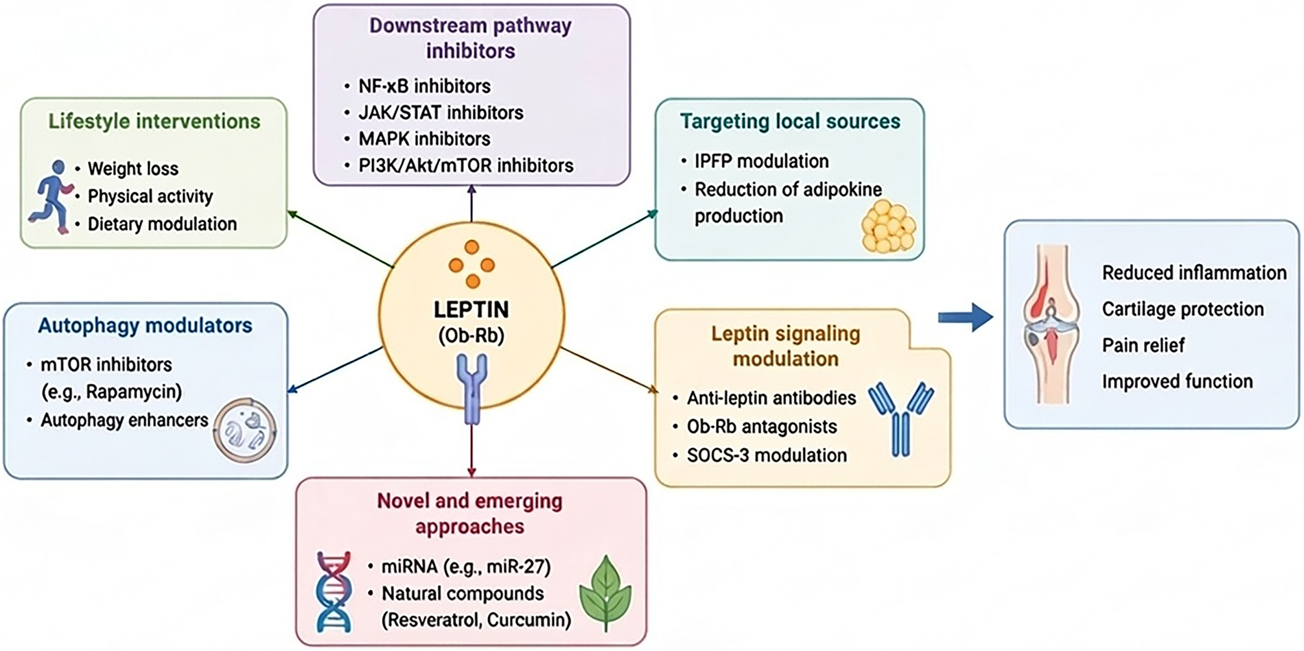
Figure 2: Therapeutic strategies targeting leptin signaling in osteoarthritis. Schematic representation of current and emerging approaches to modulate leptin-driven pathways in osteoarthritis. Strategies include lifestyle interventions (weight loss, physical activity, dietary modulation), pharmacological inhibition of downstream signaling pathways (NF-κB, JAK/STAT, MAPK, PI3K/Akt/mTOR), and modulation of autophagy (e.g., mTOR inhibitors such as rapamycin). Targeting local sources of leptin, particularly the infrapatellar fat pad (IPFP), represents an additional approach to reduce intra-articular adipokine production. Direct modulation of leptin signaling can be achieved through anti-leptin antibodies, Ob-Rb antagonists, and SOCS-3 regulation. Novel and emerging strategies include microRNA-based approaches (e.g., miR-27) and natural compounds such as resveratrol and curcumin. Collectively, these interventions aim to reduce inflammation, limit cartilage degradation, alleviate pain, and improve joint function.
8 Clinical Implications: The Inflammatory–Metabolic Osteoarthritis Phenotype
Although OA has traditionally been considered a non-inflammatory degenerative disease, it is now widely recognized that a chronic low-grade inflammatory component plays a crucial role in its pathophysiology [186]. Importantly, this inflammatory state differs substantially from the acute, systemic, and immune-driven inflammation observed in classical inflammatory arthritis, as it is typically persistent, low-grade, and closely linked to metabolic dysfunction and aging [187]. Within this framework, an “inflammatory–metabolic OA phenotype” has been proposed. This phenotype does not represent an overlap with immune-mediated diseases such as rheumatoid arthritis, but rather identifies a subgroup of OA patients in whom local synovial inflammation and systemic metabolic alterations are particularly relevant drivers of disease expression and progression.
From a clinical standpoint, this phenotype is frequently encountered in daily practice [188]. Patients often present with obesity, metabolic syndrome, or other features of metabolic dysregulation, and commonly exhibit signs of increased synovial activity, including joint effusion and imaging-detected synovitis [188]. A characteristic feature of this subgroup is the presence of pain that appears disproportionate to the degree of structural joint damage observed on radiographic evaluation, suggesting that inflammatory and metabolic factors significantly contribute to symptom generation beyond purely mechanical mechanisms [189]. Recognition of this phenotype has important clinical implications, as improved patient stratification may facilitate the identification of individuals at higher risk of disease progression, persistent symptoms, and suboptimal response to conventional treatments. More importantly, it supports a shift toward therapeutic strategies targeting the biological mechanisms underlying disease progression rather than focusing exclusively on structural joint damage [189].
Although no universally accepted diagnostic criteria for the inflammatory–metabolic OA phenotype have been established [190], several clinical and biological features have been consistently used in research settings to identify this subgroup. Patients typically fulfill standard clinical and/or radiographic criteria for OA and present with obesity (most commonly defined as BMI ≥ 30 kg/m2) and/or metabolic syndrome according to established ATP III or IDF criteria [191,192]. Additional features frequently include evidence of chronic low-grade inflammation, such as mildly elevated hsCRP levels, together with clinical or imaging signs of synovitis. Pain disproportionate to radiographic disease severity further supports the presence of this phenotype [193]. Although circulating and synovial leptin concentrations are consistently elevated in patients with metabolic OA and correlate with disease severity and inflammatory activity, no validated diagnostic threshold has yet been established for clinical use. Therefore, leptin should be regarded as a promising mechanistic and stratification biomarker rather than a routine diagnostic marker [145,194]. Collectively, these readily available clinical and metabolic characteristics may provide a practical framework for patient stratification while awaiting prospective validation of standardized diagnostic criteria.
In this context, adipokines, particularly leptin, may represent a key mechanistic link between metabolic dysfunction and the clinical expression of this phenotype. Beyond its metabolic role, leptin exerts pro-inflammatory effects within the joint microenvironment, promoting synovial activation, cytokine release, and cartilage degradation. At the same time, leptin has been implicated in pain modulation, potentially contributing to peripheral and central sensitization processes that characterize patients with disproportionate pain.
At the central level, leptin may contribute to pain amplification through modulation of neuro-immune signaling within the spinal cord and supraspinal structures [195]. Leptin can cross the blood–brain barrier via a saturable transport system and acts on Ob-R expressed in key nociceptive regulatory regions, including the hypothalamus and spinal dorsal horn. Within the central nervous system, leptin signaling activates JAK2/STAT3 and NF-κB pathways in microglia, promoting the release of pro-inflammatory cytokines such as IL-1β, TNF-α, and IL-6, which are established mediators of central sensitization [41]. In parallel, leptin-associated glial activation enhances astrocytic reactivity and disrupts glutamate homeostasis, further increasing dorsal horn neuronal excitability and reducing endogenous descending pain inhibition [196]. Collectively, these mechanisms provide a biologically plausible link between systemic metabolic inflammation and central amplification of nociceptive signaling in OA pain. This dual role positions leptin as a critical mediator bridging systemic metabolic alterations with local joint inflammation and pain perception.
The recognition of this phenotype also has important translational implications. Identifying patients characterized by metabolic dysfunction, chronic low-grade inflammation, and elevated leptin signaling may improve patient stratification, facilitate the development of personalized therapeutic approaches, and support the identification of individuals most likely to benefit from interventions targeting inflammatory and metabolic pathways. Overall, integrating the concept of an inflammatory–metabolic OA phenotype into OA research and clinical practice provides a valuable framework for linking molecular mechanisms, including leptin-driven signaling, with clinical heterogeneity, ultimately supporting a more comprehensive and clinically meaningful understanding of OA pathogenesis and the development of targeted therapeutic strategies.
9 Future Perspectives and Current Limitations of Leptin Research in Osteoarthritis
Despite growing evidence supporting the involvement of leptin in OA, several limitations still hinder its clinical translation as both a biomarker and a therapeutic target. Most mechanistic evidence derives from in vitro studies and experimental animal models, whereas prospective human studies specifically designed to establish causality remain limited. Although elevated serum and synovial fluid leptin concentrations are consistently associated with OA, current evidence cannot determine whether leptin actively drives disease progression or primarily reflects obesity-related metabolic dysfunction. Furthermore, many experimental studies employ leptin concentrations that exceed physiological intra-articular levels, raising concerns regarding the translational relevance of some reported catabolic effects [37,69].
Another important challenge is the context-dependent nature of leptin signaling. Under physiological conditions, leptin may exert anabolic effects by supporting cartilage homeostasis, whereas chronic hyperleptinemia associated with obesity promotes inflammation, ECM degradation, and cartilage catabolism. Consequently, studies including both lean and obese patients may underestimate the specific contribution of leptin to the metabolic OA phenotype. Likewise, although Mendelian randomization studies support a causal role of obesity in OA development, the independent contribution of leptin has not yet been demonstrated, and its pathogenic role remains difficult to distinguish from the broader metabolic alterations associated with adiposity [141,197,198]. Future studies should therefore clarify whether leptin represents a true disease-driving factor, identify biologically relevant intra-articular thresholds, and determine how peripheral leptin responsiveness is influenced by obesity-associated leptin resistance.
Several emerging research areas may substantially improve our understanding of leptin biology in OA. Sex-related differences are increasingly recognized as an important source of biological variability. Women exhibit higher circulating leptin concentrations than men, and postmenopausal increases in leptin parallel the higher incidence of OA, while transcriptomic studies suggest that female patients are more frequently associated with inflammatory OA molecular subtypes [199–202]. However, whether the estrogen–leptin axis directly contributes to disease progression remains unclear, highlighting the need for sex-stratified mechanistic and clinical investigations.
Technological advances, particularly single-cell RNA sequencing and spatial transcriptomics, are also redefining OA as a highly heterogeneous disease. These approaches have identified distinct immune and stromal cell populations within the joint and have demonstrated tissue-specific adipokine expression, including leptin, providing unprecedented opportunities to characterize leptin-responsive cellular subsets and their spatial organization during disease progression [202–205]. In parallel, increasing evidence supports interactions between gut microbiota, metabolic dysfunction, and adipokine regulation. Although still largely based on observational and experimental studies, microbiome-derived metabolites may influence leptin secretion and contribute to the metabolic-inflammatory environment characteristic of obesity-associated OA, suggesting new opportunities for microbiome-targeted interventions [206–208].
Another emerging concept is the role of leptin as an immunometabolic regulator linking metabolic status with innate and adaptive immune responses. Beyond its endocrine functions, leptin promotes pro-inflammatory immune cell activation and amplifies inflammatory signaling within the OA joint, while recent evidence suggests additional neuroimmune interactions within adipose tissues that may also involve the infrapatellar fat pad [69,209,210]. However, because leptin also plays fundamental roles in systemic metabolism and immune homeostasis, systemic inhibition is unlikely to represent a safe therapeutic strategy [27].
From a translational perspective, future precision medicine approaches should consider leptin as one component of a broader metabolic-inflammatory network rather than as an isolated biomarker. Integration of adipokine profiling with metabolomics, transcriptomics, and molecular phenotyping may improve patient stratification and facilitate the identification of metabolic OA endotypes that are more likely to benefit from targeted interventions [211–213]. Although no leptin-specific therapy has yet entered clinical trials for OA, local modulation of leptin-related signaling pathways and indirect reduction of leptin through metabolically active interventions, including weight-loss strategies and GLP-1 receptor agonists, represent promising translational directions [27,214].
Overall, while current evidence identifies leptin as a key mediator linking obesity, metabolic dysfunction, inflammation, and joint degeneration, significant gaps remain regarding its causal role, biological context, and therapeutic applicability. Addressing these limitations through longitudinal clinical studies, standardized biomarker assessment, sex-specific analyses, and integrated multi-omics approaches will be essential to determine whether leptin can ultimately serve as a clinically useful biomarker and therapeutic target in OA.
The evidence discussed in this review positions leptin as a critical node at the intersection of metabolic dysfunction, aging, and joint degeneration in OA. Rather than acting as a simple adipokine, leptin integrates systemic and local signals, contributing to the dysregulation of cellular homeostasis within the joint. A key emerging concept is that leptin does not function as a uniform disease driver, but rather as a context-dependent mediator whose effects are shaped by tissue type, metabolic status, and inflammatory milieu. This helps explain the apparent paradox of leptin displaying both protective and deleterious effects in different experimental and clinical settings. Across cartilage, synovium, subchondral bone, and the IPFP, leptin participates in a complex network of signaling pathways that collectively promote inflammation, ECM degradation, and structural joint remodeling. However, the literature reveals markedly conflicting anabolic and catabolic profiles for leptin across these tissues. A structured summary of key papers covering major tissues and thematic areas is provided in the Supplementary Tables S1–S7, to facilitate navigation of the literature. These discrepancies likely reflect differences in experimental conditions, including physiological vs. chronic supraphysiological leptin exposure, as well as the stage of disease progression, where relatively preserved tissues may retain anabolic, homeostatic responses that are progressively lost and replaced by catabolic pathways in advanced disease. Furthermore, leptin signaling is strongly influenced by the experimental model, with differences observed between simplified in vitro systems, ex vivo tissue explants, and in vivo models that incorporate systemic metabolic and immune factors. Beyond experimental context, leptin is also embedded within broader processes such as obesity-related inflammation and aging-associated inflammaging. Accordingly, OA should not be viewed as a purely local degenerative disease, but rather as a systemic disorder in which metabolic and immune alterations play a central role. In this framework, leptin represents a key molecular link between systemic metabolic dysfunction and local joint pathology. Despite substantial progress in understanding leptin biology in OA, several key questions remain unresolved. These include whether leptin acts as a primary driver or an amplifier of disease processes. Addressing this question will require strategies to selectively modulate peripheral leptin signaling, as well as a better understanding of how these pathways interact with other metabolic and inflammatory mediators during disease progression. However, major methodological and biological barriers currently prevent the use of leptin as a clinical biomarker. Its interpretation is heavily confounded by sex, age, and BMI, which significantly reduce its discriminatory value, particularly in lean OA phenotypes. Rather than a standalone tool, recent data suggest that leptin can only improve prognostic performance when integrated into wider multimarker panels. A growing body of evidence supports the concept that OA is a heterogeneous disease composed of distinct clinical and molecular phenotypes rather than a single uniform entity. Among these, a metabolic phenotype has been proposed, characterized by obesity, systemic low-grade inflammation, and dysregulated adipokine signaling, particularly involving leptin (figure 3). In this subgroup of patients, elevated levels in both circulation and synovial fluid reflect not only increased adiposity but also an active contribution to joint pathology. This phenotype is associated with enhanced inflammatory signaling, accelerated cartilage degradation, and more severe structural progression compared to non-metabolic forms of OA. Within this framework, leptin may serve as both a potential biomarker and a contributing factor to local pathology. Patients with a metabolic OA phenotype are therefore the most likely to exhibit leptin-associated structural alterations, making them potential candidates for therapies targeting leptin-related pathways or their downstream signaling cascades. However, the identification of this phenotype in clinical practice remains challenging due to overlapping features with other OA subtypes and the influence of confounding metabolic factors.
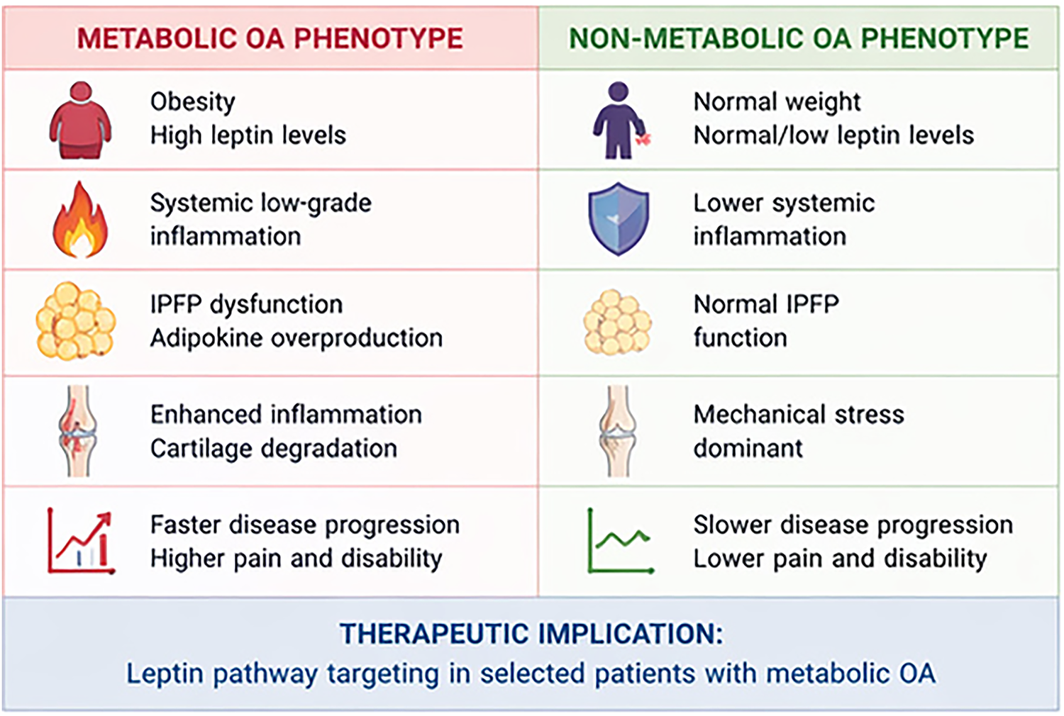
Figure 3: Metabolic vs. non-metabolic osteoarthritis: implications for precision medicine. Comparison between metabolic and non-metabolic osteoarthritis (OA) phenotypes highlighting key differences in systemic and local pathophysiological features. The metabolic OA phenotype is characterized by obesity, elevated leptin levels, systemic low-grade inflammation, and infrapatellar fat pad (IPFP) dysfunction leading to increased adipokine production. These alterations contribute to enhanced joint inflammation, accelerated cartilage degradation, and more rapid disease progression with greater pain and disability. In contrast, the non-metabolic OA phenotype is associated with normal body weight, lower leptin levels, reduced systemic inflammation, preserved IPFP function, and a predominance of mechanical stress as the main driver of joint damage. These differences support a precision medicine approach in OA, where targeting leptin-related pathways may be particularly beneficial in patients with a metabolic phenotype.
As such, integration of clinical data with molecular profiling, including adipokine levels and inflammatory markers, will be essential to reliably define this subgroup. Overall, the recognition of a leptin-associated metabolic phenotype supports a shift toward precision medicine in OA, where therapeutic strategies are tailored according to underlying molecular mechanisms rather than solely clinical presentation. This stratification will be essential for the development of targeted and mechanism-based therapeutic strategies. In this context, translating precision medicine into clinical practice requires robust strategies for patient stratification and therapeutic decision-making. Multimodal molecular profiling represents a promising approach to achieving this goal. Systemic adipokine signatures, including leptin-related indices, may provide an accessible first-level tool for identifying patients driven by metabolic dysregulation. These circulating markers should be integrated with synovial inflammatory profiles and joint-derived mediators, enabling a more accurate characterization of local disease activity and its interaction with systemic metabolic alterations. Furthermore, the integration of multi-omics data, including transcriptomic, proteomic, and metabolomic profiles, may refine risk stratification by identifying patients with faster disease progression or distinct molecular patterns of disease. From a therapeutic perspective, a major challenge remains the effective targeting of leptin-related pathways without disrupting their physiological roles in energy homeostasis and immune regulation. Addressing this issue will require a deeper understanding of tissue-specific signaling dynamics and the development of selective modulatory approaches. In addition, integrative approaches combining circulating biomarkers, synovial fluid analysis, and tissue-level molecular profiling may improve patient stratification and support the implementation of precision medicine strategies in OA. To date, all proposed leptin-targeted strategies remain confined to preclinical models, with no successful clinical translation in OA. This highlights a persistent gap between experimental findings and clinical application and underscores the need for more selective and clinically applicable approaches. Ultimately, clarifying the role of leptin in OA may contribute to a broader conceptual shift in the field, moving from a structural “wear-and-tear” model toward a systemic, metabolically driven disease paradigm.
Acknowledgement: During the preparation of this manuscript, Figures were developed using the author’s original scripts in combination with FigureLabs AI (https://figurelabs.ai/). All AI-assisted outputs were carefully reviewed, edited, and validated by the author, who assumes full responsibility for the accuracy, integrity, and content of this publication.
Funding Statement: The authors received no specific funding for this study.
Author Contributions: Conceptualization: Gabriele Ricciardi, Mariagiovanna Ballato and Maurizio Martini; Investigation: Gabriele Di Carlo, Domenico Donadio and Emanuela Germanà; Supervision: Maurizio Martini, Biagio Zampogna and Marco Ferlazzo; Writing—original draft: Gabriele Ricciardi, Mariagiovanna Ballato, Flavio Corpina and Guido Fadda; Writing—review & editing: Gabriele Ricciardi, Mariagiovanna Ballato, Carmela Lipari and Danilo Leonetti. All authors reviewed and approved the final version of the manuscript.
Availability of Data and Materials: Not Applicable.
Ethics Approval: Not applicable.
Conflicts of Interest: Author Dr. Flavio Corpina is the founder of Foresight Consulting. Author Dr. Carmela Lipari was employed by Foresight Consulting. The remaining authors declare that the research was conducted in the absence of any commercial or financial relationships that could be construed as a potential conflict of interest.
Supplementary Materials: The supplementary material is available online at https://www.techscience.com/doi/10.32604/ecn.2026.084855/s1.
Abbreviations
| OA | Osteoarthritis |
| ECM | Extracellular matrix |
| SASP | Senescence-associated secretory phenotype |
| IL | Interleukin |
| TNF | Tumor necrosis factor |
| IPFP | Infrapatellar Fat Pad |
| JAK/STAT | Janus kinase/signal transducer and activator of transcription |
| SOCS-3 | Suppressor of cytokine signaling-3 |
| ROS | Reactive oxygen species |
References
1. Tang SA, Zhang C, Oo WM, et al. Osteoarthritis. Nat Rev Dis Primers 2025;11:10. doi:10.1038/s41572-025-00594-6. [Google Scholar] [CrossRef]
2. Gu YT, Chen J, Meng ZL, et al. Research progress on osteoarthritis treatment mechanisms. Biomed Pharmacother 2017;93:1246–52. doi:10.1016/j.biopha.2017.07.034. [Google Scholar] [CrossRef]
3. Chen D, Shen J, Zhao W, et al. Osteoarthritis: toward a comprehensive understanding of pathological mechanism. Bone Res 2017;5:16044. doi:10.1038/boneres.2016.44. [Google Scholar] [CrossRef]
4. Chen T, Chen W, Xu T, et al. Osteoarthritis: epidemiology, pathogenesis, and treatment. MedComm 2026;7(4):e70727. doi:10.1002/mco2.70727. [Google Scholar] [CrossRef]
5. Qiu L, Alhaskawi A, Moqbel SAA. Osteoarthritis: multitissue pathology, molecular mechanisms, clinical management, and emerging precision and regenerative therapies. Front Pharmacol 2025;16:1697192. doi:10.3389/fphar.2025.1697192. [Google Scholar] [CrossRef]
6. Liu Z, Xie W, Li H et al. Novel perspectives on leptin in osteoarthritis: focus on aging. Genes Dis 2024;11(6):101159. doi:10.1016/j.gendis.2023.101159. [Google Scholar] [CrossRef]
7. Yagi M, Endo K, Komori K, Sekiya I. Comparison of the effects of oxidative and inflammatory stresses on rat chondrocyte senescence. Sci Rep 2023;13(1):7697. doi:10.1038/s41598-023-34825-1. [Google Scholar] [CrossRef]
8. Huang K, Cai H. The role of chondrocyte senescence in osteoarthritis pathogenesis and therapeutic implications. Exp Gerontol 2025;208:112828. doi:10.1016/j.exger.2025.112828. [Google Scholar] [CrossRef]
9. Loeser RF. Aging and osteoarthritis: the role of chondrocyte senescence and aging changes in the cartilage matrix. Osteoarthr Cartil 2009;17(8):971–79. doi:10.1016/j.joca.2009.03.002. [Google Scholar] [CrossRef]
10. Motta F, Barone E, Sica A, Selmi C. Inflammaging and osteoarthritis. Clin Rev Allergy Immunol 2023;64(2):222–38. doi:10.1007/s12016-022-08941-1. [Google Scholar] [CrossRef]
11. Zhang Q, Zhao YX, Li LF, et al. Metabolism-related adipokines and metabolic diseases: their role in osteoarthritis. J Inflamm Res 2025;18:1207–33. doi:10.2147/JIR.S499835. [Google Scholar] [CrossRef]
12. Huang S, Chen J, Zhang H, et al. Inflammatory mechanisms underlying metabolic syndrome-associated and potential treatments. Osteoarthr Cartil Open 2025;7(2):100614. doi:10.1016/j.ocarto.2025.100614. [Google Scholar] [CrossRef]
13. Francisco V, Pino J, Gonzalez-Gay MA, et al. Adipokines and inflammation: is it a question of weight? Br J Pharmacol 2018;175(10):1569–79. doi:10.1111/bph.14181. [Google Scholar] [CrossRef]
14. Fu Y, Batushansky A, Kinter M, Huebner JL, Kraus VB, Griffin TM. Effects of leptin and body weight on inflammation and knee osteoarthritis phenotypes in female rats. JBMR Plus 2023;7(7):e10754. doi:10.1002/jbm4.10754. [Google Scholar] [CrossRef]
15. La Cava A. Leptin in inflammation and autoimmunity. Cytokine 2017;98:51–8. doi:10.1016/j.cyto.2016.10.011. [Google Scholar] [CrossRef]
16. Hua KF, Li LH, Yu HC, Wong WT, Hsu HT. Leptin induces MMP-1 expression through the RhoA/ERK1/2/NF-κB axis in human intervertebral disc cartilage endplate-derived stem cells. J Inflamm Res 2023;16:5235–48. doi:10.2147/JIR.S431026. [Google Scholar] [CrossRef]
17. Hui W, Litherland GJ, Elias MS, et al. Leptin produced by joint white adipose tissue induces cartilage degradation via upregulation and activation of matrix metalloproteinases. Ann Rheum Dis 2012;71(3):455–62. doi:10.1136/annrheumdis-2011-200372. [Google Scholar] [CrossRef]
18. Ku JH, Lee CK, Joo BS, et al. Correlation of synovial fluid leptin concentrations with the severity of osteoarthritis. Clin Rheumatol 2009;28(12):1431–5. doi:10.1007/s10067-009-1242-8. [Google Scholar] [CrossRef]
19. Morales Abaunza RA, Rojas ÁP, Rojas C, et al. Levels of serum leptin in patients with primary hand osteoarthritis. Rev Colomb De Reumatol Engl Ed 2020;27(1):20–5. doi:10.1016/j.rcreue.2019.12.005. [Google Scholar] [CrossRef]
20. Wei K. Association between serum leptin and accelerated biological aging in US adults: findings from NHANES III. J Nutr Health Aging 2025;29(12):100695. doi:10.1016/j.jnha.2025.100695. [Google Scholar] [CrossRef]
21. Zhao X, Dong Y, Zhang J, et al. Leptin changes differentiation fate and induces senescence in chondrogenic progenitor cells. Cell Death Dis 2016;7(4):e2188. doi:10.1038/cddis.2016.68. [Google Scholar] [CrossRef]
22. Obradovic M, Sudar-Milovanovic E, Soskic S, et al. Leptin and obesity: role and clinical implication. Front Endocrinol 2021;12:585887. doi:10.3389/fendo.2021.585887. [Google Scholar] [CrossRef]
23. Hu W, Zhu H, Gong F. Leptin and leptin resistance in obesity: current evidence, mechanisms and future directions. Endocr Connect 2025;14(9):e250521. doi:10.1530/EC-25-0521. [Google Scholar] [CrossRef]
24. Liu Z, Xiao T, Liu H. Leptin signaling and its central role in energy homeostasis. Front Neurosci 2023;17:1238528. doi:10.3389/fnins.2023.1238528. [Google Scholar] [CrossRef]
25. Dalamaga M, Chou SH, Shields K, Papageorgiou P, Polyzos SA, Mantzoros CS. Leptin at the intersection of neuroendocrinology and metabolism: current evidence and therapeutic perspectives. Cell Metab 2013;18(1):29–42. doi:10.1016/j.cmet.2013.05.010. [Google Scholar] [CrossRef]
26. Martin SS, Qasim A, Reilly MP. Leptin resistance: a possible interface of inflammation and metabolism in obesity-related cardiovascular disease. J Am Coll Cardiol 2008;52(15):1201–10. doi:10.1016/j.jacc.2008.05.060. [Google Scholar] [CrossRef]
27. de Candia P, Prattichizzo F, Garavelli S, Alviggi C, La Cava A, Matarese G. The pleiotropic roles of leptin in metabolism, immunity, and cancer. J Exp Med 2021;218(5):e20191593. doi:10.1084/jem.20191593. [Google Scholar] [CrossRef]
28. Zikopoulos A, Moustakli E, Katopodis P, et al. Leptin receptor b (LEPRb) mutations disrupt hypothalamic control of the reproductive axis. Int J Mol Sci 2026;27(5):2482. doi:10.3390/ijms27052482. [Google Scholar] [CrossRef]
29. Wauman J, Zabeau L, Tavernier J. The leptin receptor complex: heavier than expected? Front Endocrinol 2017;8:30. doi:10.3389/fendo.2017.00030. [Google Scholar] [PubMed] [CrossRef]
30. Jafarpour S, Khosravi S, Janghorbani M, et al. Association of serum and follicular fluid leptin and in vitro Fertilization/ICSI outcome: a systematic review and meta-analysis. J Gynecol Obstet Hum Reprod 2021;50(6):101924. doi:10.1016/j.jogoh.2020.101924. [Google Scholar] [CrossRef]
31. Hu X, Li J, Fu M, Zhao X, Wang W. The JAK/STAT signaling pathway: from bench to clinic. Sig Transduct Target Ther 2021;6(1):402. doi:10.1038/s41392-021-00791-1. [Google Scholar] [CrossRef]
32. Molnar V, Matišić V, Kodvanj I, et al. Cytokines and chemokines involved in osteoarthritis pathogenesis. Int J Mol Sci 2021;22(17):9208. doi:10.3390/ijms22179208. [Google Scholar] [CrossRef]
33. Andrei C, Mihai DP, Nitulescu GM, Nitulescu G, Zanfirescu A. Modulating autophagy in osteoarthritis: exploring emerging therapeutic drug targets. Int J Mol Sci 2024;25(24):13695. doi:10.3390/ijms252413695. [Google Scholar] [CrossRef]
34. Lepetsos P, Papavassiliou KA, Papavassiliou AG. Redox and NF-κB signaling in osteoarthritis. Free Radic Biol Med 2019;132(4):90–100. doi:10.1016/j.freeradbiomed.2018.09.025. [Google Scholar] [CrossRef]
35. Ni S, Li D, Wei H, Miao KS, Zhuang C. PPARγ attenuates interleukin-1β-induced cell apoptosis by inhibiting NOX2/ROS/p38MAPK activation in osteoarthritis chondrocytes. Oxid Med Cell Longev 2021;2021:5551338. doi:10.1155/2021/5551338. [Google Scholar] [CrossRef]
36. Han Z, Wang K, Ding S, Zhang M. Cross-talk of inflammation and cellular senescence: a new insight into the occurrence and progression of osteoarthritis. Bone Res 2024;12(1):69. doi:10.1038/s41413-024-00375-z. [Google Scholar] [CrossRef]
37. Ait Eldjoudi D, Cordero Barreal A, Gonzalez-Rodríguez M, et al. Leptin in osteoarthritis and rheumatoid arthritis: player or bystander? Int J Mol Sci 2022;23(5):2859. doi:10.3390/ijms23052859. [Google Scholar] [CrossRef]
38. Wang T, He C. Pro-inflammatory cytokines: the link between obesity and osteoarthritis. Cytokine Growth Factor Rev 2018;44(9):38–50. doi:10.1016/j.cytogfr.2018.10.002. [Google Scholar] [CrossRef]
39. Otero M, Lago R, Lago F, Reino JJ, Gualillo O. Signalling pathway involved in nitric oxide synthase type II activation in chondrocytes: synergistic effect of leptin with interleukin-1. Arthritis Res Ther 2005;7(3):R581–91. doi:10.1186/ar1708. [Google Scholar] [CrossRef]
40. Vuolteenaho K, Koskinen A, Kukkonen M, et al. Leptin enhances synthesis of proinflammatory mediators in human osteoarthritic cartilage—mediator role of NO in leptin-induced PGE2, IL-6, and IL-8 production. Mediat Inflamm 2009;2009(6):345838. doi:10.1155/2009/345838. [Google Scholar] [CrossRef]
41. Koskinen-Kolasa A, Vuolteenaho K, Korhonen R, Moilanen T, Moilanen E. Catabolic and proinflammatory effects of leptin in chondrocytes are regulated by suppressor of cytokine signaling-3. Arthritis Res Ther 2016;18(1):215. doi:10.1186/s13075-016-1112-0. [Google Scholar] [CrossRef]
42. Dessie G, Ayelign B, Akalu Y, Shibabaw T, Molla MD. Effect of leptin on chronic inflammatory disorders: insights to therapeutic target to prevent further cardiovascular complication. Diabetes Metab Syndr Obes 2021;14:3307–22. doi:10.2147/DMSO.S321311. [Google Scholar] [CrossRef]
43. Forny-Germano L, de Felice FG, Vieira MNDN. The role of leptin and adiponectin in obesity-associated cognitive decline and Alzheimer’s disease. Front Neurosci 2018;12:1027. doi:10.3389/fnins.2018.01027. [Google Scholar] [CrossRef]
44. Cosme D, Gomes AC. Leptin levels and bone mineral density: a friend or a foe for bone loss? A systematic review of the association between leptin levels and low bone mineral density. Int J Mol Sci 2025;26(5):2066. doi:10.3390/ijms26052066. [Google Scholar] [CrossRef]
45. Ducy P, Amling M, Takeda S, et al. Leptin inhibits bone formation through a hypothalamic relay: a central control of bone mass. Cell 2000;100(2):197–207. doi:10.1016/s0092-8674(00)81558-5. [Google Scholar] [CrossRef]
46. Reid IR, Baldock PA, Cornish J. Effects of leptin on the skeleton. Endocr Rev 2018;39(6):938–59. doi:10.1210/er.2017-00226. [Google Scholar] [CrossRef]
47. Shi H, Chen M. The brain-bone axis: unraveling the complex interplay between the central nervous system and skeletal metabolism. Eur J Med Res 2024;29(1):317. doi:10.1186/s40001-024-01918-0. [Google Scholar] [CrossRef]
48. Scheller EL, Song J, Dishowitz MI, Soki FN, Hankenson KD, Krebsbach PH. Leptin functions peripherally to regulate differentiation of mesenchymal progenitor cells. Stem Cells 2010;28(6):1071–80. doi:10.1002/stem.432. [Google Scholar] [CrossRef]
49. Motyl KJ, Rosen CJ. Understanding leptin-dependent regulation of skeletal homeostasis. Biochimie 2012;94(10):2089–96. doi:10.1016/j.biochi.2012.04.015. [Google Scholar] [CrossRef]
50. Martin A, de Vittoris R, David V, et al. Leptin modulates both resorption and formation while preventing disuse-induced bone loss in tail-suspended female rats. Endocrinology 2005;146(8):3652–9. doi:10.1210/en.2004-1509. [Google Scholar] [CrossRef]
51. Zhang B, Yang L, Zeng Z, et al. Leptin potentiates BMP9-induced osteogenic differentiation of mesenchymal stem cells through the activation of JAK/STAT signaling. Stem Cells Dev 2020;29(8):498–510. doi:10.1089/scd.2019.0292. [Google Scholar] [CrossRef]
52. Cai F, Yusufu A, Liu K, et al. High-fat diet causes undesirable bone regeneration by altering the bone marrow environment in rats. Front Endocrinol 2023;14:1088508. doi:10.3389/fendo.2023.1088508. [Google Scholar] [CrossRef]
53. Han YC, Ma B, Guo S, et al. Leptin regulates disc cartilage endplate degeneration and ossification through activation of the MAPK-ERK signalling pathway in vivo and in vitro. J Cell Mol Med 2018;22(4):2098–109. doi:10.1111/jcmm.13398. [Google Scholar] [CrossRef]
54. Thomas T. The complex effects of leptin on bone metabolism through multiple pathways. Curr Opin Pharmacol 2004;4(3):295–300. doi:10.1016/j.coph.2004.01.009. [Google Scholar] [CrossRef]
55. Li Y, Yao L, Huang Y, et al. Leptin enhances M1 macrophage polarization and impairs tendon-bone healing in rotator cuff repair: a rat model. Clin Orthop Relat Res 2025;483(5):939–51. doi:10.1097/corr.0000000000003428. [Google Scholar] [CrossRef]
56. Philbrick KA, Branscum AJ, Wong CP, Turner RT, Iwaniec UT. Leptin increases particle-induced osteolysis in female ob/ob mice. Sci Rep 2018;8(1):14790. doi:10.1038/s41598-018-33173-9. [Google Scholar] [CrossRef]
57. Maisenbacher TC, Ehnert S, Histing T, Nüssler AK, Menger MM. Advantages and limitations of diabetic bone healing in mouse models: a narrative review. Biomedicines 2023;11(12):3302. doi:10.3390/biomedicines11123302. [Google Scholar] [CrossRef]
58. Brown ML, Yukata K, Farnsworth CW, et al. Delayed fracture healing and increased callus adiposity in a C57BL/6J murine model of obesity-associated type 2 diabetes mellitus. PLoS One 2014;9(6):e99656. doi:10.1371/journal.pone.0099656. [Google Scholar] [CrossRef]
59. Mostoufi-Moab S, Magland J, Isaacoff EJ, et al. Adverse fat depots and marrow adiposity are associated with skeletal deficits and insulin resistance in long-term survivors of pediatric hematopoietic stem cell transplantation. J Bone Miner Res 2015;30(9):1657–66. doi:10.1002/jbmr.2512. [Google Scholar] [CrossRef]
60. Luo P, Yuan QL, Yang M, Wan X, Xu P. The role of cells and signal pathways in subchondral bone in osteoarthritis. Bone Joint Res 2023;12(9):536–45. doi:10.1302/2046-3758.129.bjr-2023-0081.r1. [Google Scholar] [CrossRef]
61. Zhou X, Cao H, Yuan Y, Wu W. Biochemical signals mediate the crosstalk between cartilage and bone in osteoarthritis. Biomed Res Int 2020;2020:5720360. doi:10.1155/2020/5720360. [Google Scholar] [CrossRef]
62. Scotece M, Mobasheri A. Leptin in osteoarthritis: focus on articular cartilage and chondrocytes. Life Sci 2015;140:75–8. doi:10.1016/j.lfs.2015.05.025. [Google Scholar] [CrossRef]
63. Simopoulou T, Malizos KN, Iliopoulos D, et al. Differential expression of leptin and leptin’s receptor isoform (Ob-Rb) mRNA between advanced and minimally affected osteoarthritic cartilage; effect on cartilage metabolism. Osteoarthr Cartil 2007;15(8):872–83. doi:10.1016/j.joca.2007.01.018. [Google Scholar] [CrossRef]
64. Azamar-Llamas D, Hernández-Molina G, Ramos-Ávalos B, Furuzawa-Carballeda J. Adipokine contribution to the pathogenesis of osteoarthritis. Mediat Inflamm 2017;2017(6):5468023. doi:10.1155/2017/5468023. [Google Scholar] [CrossRef]
65. Bao JP, Chen WP, Feng J, Hu PF, Shi ZL, Wu LD. Leptin plays a catabolic role on articular cartilage. Mol Biol Rep 2010;37(7):3265–72. doi:10.1007/s11033-009-9911-x. [Google Scholar] [CrossRef]
66. Zhang ZM, Shen C, Li H et al. Leptin induces the apoptosis of chondrocytes in an in vitro model of osteoarthritis via the JAK2-STAT3 signaling pathway. Mol Med Rep 2016;13(4):3684–90. doi:10.3892/mmr.2016.4970. [Google Scholar] [CrossRef]
67. Otero M, Gomez Reino JJ, Gualillo O. Synergistic induction of nitric oxide synthase type II: in vitro effect of leptin and interferon-gamma in human chondrocytes and ATDC5 chondrogenic cells. Arthritis Rheum 2003;48(2):404–9. doi:10.1002/art.10811. [Google Scholar] [CrossRef]
68. Koskinen A, Vuolteenaho K, Nieminen R, Moilanen T, Moilanen E. Leptin enhances MMP-1, MMP-3 and MMP-13 production in human osteoarthritic cartilage and correlates with MMP-1 and MMP-3 in synovial fluid from OA patients. Clin Exp Rheumatol 2011;29(1):57–64. doi:10.1016/s1063-4584(08)60275-7. [Google Scholar] [CrossRef]
69. Cordero-Barreal A, González-Rodríguez M, Ruiz-Fernández C, et al. An update on the role of leptin in the immuno-metabolism of cartilage. Int J Mol Sci 2021;22(5):2411. doi:10.3390/ijms22052411. [Google Scholar] [CrossRef]
70. Clockaerts S, Bastiaansen-Jenniskens YM, Feijt C, et al. Cytokine production by infrapatellar fat pad can be stimulated by interleukin 1β and inhibited by peroxisome proliferator activated receptor α agonist. Ann Rheum Dis 2012;71(6):1012–18. doi:10.1136/annrheumdis-2011-200688. [Google Scholar] [CrossRef]
71. Gómez R, Scotece M, Conde J, Gómez-Reino JJ, Lago F, Gualillo O. Adiponectin and leptin increase IL-8 production in human chondrocytes. Ann Rheum Dis 2011;70(11):2052–54. doi:10.1136/ard.2010.145672. [Google Scholar] [CrossRef]
72. Ben-Eliezer M, Phillip M, Gat-Yablonski G. Leptin regulates chondrogenic differentiation in ATDC5 cell-line through JAK/STAT and MAPK pathways. Endocrine 2007;32(2):235–44. doi:10.1007/s12020-007-9025-y. [Google Scholar] [CrossRef]
73. Li Z, Liang J, Wu WK, et al. Leptin activates RhoA/ROCK pathway to induce cytoskeleton remodeling in nucleus pulposus cells. Int J Mol Sci 2014;15(1):1176–88. doi:10.3390/ijms15011176. [Google Scholar] [CrossRef]
74. Zhao X, Huang P, Li G, Lv Z, Hu G, Xu Q. Activation of the leptin pathway by high expression of the long form of the leptin receptor (Ob-Rb) accelerates chondrocyte senescence in osteoarthritis. Bone Joint Res 2019;8(9):425–36. doi:10.1302/2046-3758.89.BJR-2018-0325.R2. [Google Scholar] [CrossRef]
75. Figenschau Y, Knutsen G, Shahazeydi S, Johansen O, Sveinbjörnsson B. Human articular chondrocytes express functional leptin receptors. Biochem Biophys Res Commun 2001;287(1):190–7. doi:10.1006/bbrc.2001.5543. [Google Scholar] [CrossRef]
76. Primrose JG, Jain L, Bolam SM, et al. Concentration-dependent effects of leptin on osteoarthritis-associated changes in phenotype of human chondrocytes. Connect Tissue Res 2023;64(5):457–68. doi:10.1080/03008207.2023.2214249. [Google Scholar] [CrossRef]
77. Houtman E, van Hoolwerff M, Lakenberg N, et al. Human osteochondral explants: reliable biomimetic models to investigate disease mechanisms and develop personalized treatments for osteoarthritis. Rheumatol Ther 2021;8(1):499–515. doi:10.1007/s40744-021-00287-y. [Google Scholar] [PubMed] [CrossRef]
78. Rios JL, Sapède D, Djouad F, et al. Animal models of osteoarthritis part 1-preclinical small animal models: challenges and opportunities for drug development. Curr Protoc 2022;2(11):e596. doi:10.1002/cpz1.596. [Google Scholar] [CrossRef]
79. Panichi V, Costantini S, Grasso M, Arciola CR, Dolzani P. Innate immunity and synovitis: key players in osteoarthritis progression. Int J Mol Sci 2024;25(22):12082. doi:10.3390/ijms252212082. [Google Scholar] [CrossRef]
80. Lübbeke A, Finckh A, Puskas GJ, et al. Do synovial leptin levels correlate with pain in end stage arthritis? Int Orthop 2013;37(10):2071–9. doi:10.1007/s00264-013-1982-6. [Google Scholar] [CrossRef]
81. Bokarewa M, Bokarew D, Hultgren O, Tarkowski A. Leptin consumption in the inflamed joints of patients with rheumatoid arthritis. Ann Rheum Dis 2003;62(10):952–6. doi:10.1136/ard.62.10.952. [Google Scholar] [CrossRef]
82. Xiong H, Li W, Ke J, Fang W, Li B, Wei L. Leptin levels in the synovial fluid of patients with temporomandibular disorders. J Oral Maxillofac Surg 2019;77(3):493–8. doi:10.1016/j.joms.2018.09.012. [Google Scholar] [CrossRef]
83. Muraoka S, Kusunoki N, Takahashi H, Tsuchiya K, Kawai S. Leptin stimulates interleukin-6 production via Janus kinase 2/signal transducer and activator of transcription 3 in rheumatoid synovial fibroblasts. Clin Exp Rheumatol 2013;31(4):589–5. doi:10.3410/f.718008680.793476266. [Google Scholar] [CrossRef]
84. Sanchez-Lopez E, Coras R, Torres A, Lane NE, Guma M. Synovial inflammation in osteoarthritis progression. Nat Rev Rheumatol 2022;18(5):258–75. doi:10.1038/s41584-022-00749-9. [Google Scholar] [CrossRef]
85. Robinson WH, Lepus CM, Wang Q, et al. Low-grade inflammation as a key mediator of the pathogenesis of osteoarthritis. Nat Rev Rheumatol 2016;12(10):580–92. doi:10.1038/nrrheum.2016.136. [Google Scholar] [CrossRef]
86. Yan M, Zhang J, Yang H, Sun Y. The role of leptin in osteoarthritis. Medicine 2018;97(14):e0257. doi:10.1097/md.0000000000010257. [Google Scholar] [CrossRef]
87. Xiao K, Liu C, Tu Z, et al. Activation of the NF-κB and MAPK signaling pathways contributes to the inflammatory responses, but not cell injury, in IPEC-1 cells challenged with hydrogen peroxide. Oxid Med Cell Longev 2020;2020:5803639. doi:10.1155/2020/5803639. [Google Scholar] [CrossRef]
88. Wayupat A, Kongtawelert P, Pothacharoen P, Shwe TH, Phitak T. Leptin potentiates the inflammatory effect of interleukin-1 beta on synoviocytes. potential preventive role of DHA and EPA in osteoarthritis cell model. Biochemistry 2025;90(8):1153–67. doi:10.1134/S0006297925600863. [Google Scholar] [CrossRef]
89. Pérez-Pérez A, Sánchez-Jiménez F, Vilariño-García T, Sánchez-Margalet V. Role of leptin in inflammation and vice versa. Int J Mol Sci 2020;21(16):5887. doi:10.3390/ijms21165887. [Google Scholar] [CrossRef]
90. Conde J, Scotece M, López V, et al. Differential expression of adipokines in infrapatellar fat pad (IPFP) and synovium of osteoarthritis patients and healthy individuals. Ann Rheum Dis 2014;73(3):631–3. doi:10.1136/annrheumdis-2013-204189. [Google Scholar] [CrossRef]
91. Dumond H, Presle N, Terlain B, et al. Evidence for a key role of leptin in osteoarthritis. Arthritis Rheum 2003;48(11):3118–29. doi:10.1002/art.11303. [Google Scholar] [CrossRef]
92. de Boer TN, van Spil WE, Huisman AM, et al. Serum adipokines in osteoarthritis; comparison with controls and relationship with local parameters of synovial inflammation and cartilage damage. Osteoarthr Cartil 2012;20(8):846–53. doi:10.1016/j.joca.2012.05.002. [Google Scholar] [CrossRef]
93. Zeng N, Yan ZP, Chen XY, Ni GX. Infrapatellar fat pad and knee osteoarthritis. Aging Dis 2020;11(5):1317. doi:10.14336/ad.2019.1116. [Google Scholar] [CrossRef]
94. Mallia I, Fioravanti A, Guiducci S. Infrapatellar fat pad in knee osteoarthritis: a comprehensive review of pathophysiology and targeted therapeutic strategies. Int J Mol Sci 2025;26(21):10408. doi:10.3390/ijms262110408. [Google Scholar] [CrossRef]
95. Crofford L, Mehta H, Roessler B, Mancuso P. Leptin induces production of eicosanoids and proinflammatory cytokines in human synovial fibroblasts. Arthritis Res Ther 2004;6:64. doi:10.1186/ar1400. [Google Scholar] [CrossRef]
96. Portnoy DM, Paiola M, Tymm C, Winchester R, Mor A, Gartshteyn Y. Understanding the inflammatory aspect of osteoarthritis: lessons from immune checkpoint inhibitors. J Clin Med 2026;15(2):658. doi:10.3390/jcm15020658. [Google Scholar] [CrossRef]
97. Ioan-Facsinay A, Kloppenburg M. An emerging player in knee osteoarthritis: the infrapatellar fat pad. Arthritis Res Ther 2013;15(6):225. doi:10.1186/ar4422. [Google Scholar] [CrossRef]
98. Tong KM, Shieh DC, Chen CP, et al. Leptin induces IL-8 expression via leptin receptor, IRS-1, PI3K, Akt cascade and promotion of NF-κB/p300 binding in human synovial fibroblasts. Cell Signal 2008;20(8):1478–88. doi:10.1016/j.cellsig.2008.04.003. [Google Scholar] [CrossRef]
99. Pearson MJ, Herndler-Brandstetter D, Tariq MA, et al. IL-6 secretion in osteoarthritis patients is mediated by chondrocyte-synovial fibroblast cross-talk and is enhanced by obesity. Sci Rep 2017;7(1):3451. doi:10.1038/s41598-017-03759-w. [Google Scholar] [CrossRef]
100. Braun S, Zaucke F, Brenneis M, et al. The corpus adiposum infrapatellare (Hoffa’s fat pad)-the role of the infrapatellar fat pad in osteoarthritis pathogenesis. Biomedicines 2022;10(5):1071. doi:10.3390/biomedicines10051071. [Google Scholar] [CrossRef]
101. Mutabaruka MS, Aoulad Aissa M, Delalandre A, Lavigne M, Lajeunesse D. Local leptin production in osteoarthritis subchondral osteoblasts may be responsible for their abnormal phenotypic expression. Arthritis Res Ther 2010;12(1):R20. doi:10.1186/ar2925. [Google Scholar] [CrossRef]
102. Ohashi Y, Uchida K, Fukushima K, Inoue G, Takaso M. Mechanisms of peripheral and central sensitization in osteoarthritis pain. Cureus 2023;15(2):e35331. doi:10.7759/cureus.35331. [Google Scholar] [CrossRef]
103. Ko KR, Lee H, Han SH, et al. Substance P, a promising therapeutic target in musculoskeletal disorders. Int J Mol Sci 2022;23(5):2583. doi:10.3390/ijms23052583. [Google Scholar] [CrossRef]
104. Kinfe TM, Buchfelder M, Chaudhry SR, et al. Leptin and associated mediators of immunometabolic signaling: novel molecular outcome measures for neurostimulation to treat chronic pain. Int J Mol Sci 2019;20(19):4737. doi:10.3390/ijms20194737. [Google Scholar] [CrossRef]
105. Cen H, Yan Q, Meng T, et al. Quantitative infrapatellar fat pad signal intensity alteration as an imaging biomarker of knee osteoarthritis progression. RMD Open 2023;9(1):e002565. doi:10.1136/rmdopen-2022-002565. [Google Scholar] [CrossRef]
106. Wang Q, Liu D, Hu F, et al. The infrapatellar fat pad in osteoarthritis: from pathophysiology to a novel therapeutic target. Int J Mol Sci 2026;27(5):2369. doi:10.3390/ijms27052369. [Google Scholar] [CrossRef]
107. Zhong J, Yang G. Metabolic syndrome-osteoarthritis axis: beyond mechanical loading toward a cardiometabolic OA phenotype. Biomed Pharmacother 2026;200(5):119583. doi:10.1016/j.biopha.2026.119583. [Google Scholar] [CrossRef]
108. Abu-Amer Y. Cross-regulation of inflammation and metabolic mechanisms in osteoarthritis: recent advances bridging the gap to novel treatments. Connect Tissue Res 2025;66(5):339–44. doi:10.1080/03008207.2025.2500530. [Google Scholar] [CrossRef]
109. Wei G, Lu K, Umar M, et al. Risk of metabolic abnormalities in osteoarthritis: a new perspective to understand its pathological mechanisms. Bone Res 2023;11(1):63. doi:10.1038/s41413-023-00301-9. [Google Scholar] [CrossRef]
110. MaᶜDonald IJ, Liu SC, Huang CC, Kuo SJ, Tsai CH, Tang CH. Associations between adipokines in arthritic disease and implications for obesity. Int J Mol Sci 2019;20(6):1505. doi:10.3390/ijms20061505. [Google Scholar] [CrossRef]
111. Jiang T, Su S, Tian R, et al. Immunoregulatory orchestrations in osteoarthritis and mesenchymal stromal cells for therapy. J Orthop Transl 2025;55(2):38–54. doi:10.1016/j.jot.2025.08.009. [Google Scholar] [CrossRef]
112. Zhang Z, Zhang Z, Kang Y, et al. Resistin stimulates expression of chemokine genes in chondrocytes via combinatorial regulation of C/EBPβ and NF-κB. Int J Mol Sci 2014;15(10):17242–55. doi:10.3390/ijms151017242. [Google Scholar] [CrossRef]
113. Wu MH, Tsai CH, Huang YL, Fong YC, Tang CH. Visfatin promotes IL-6 and TNF-α production in human synovial fibroblasts by repressing miR-199a-5p through ERK, p38 and JNK signaling pathways. Int J Mol Sci 2018;19(1):190. doi:10.3390/ijms19010190. [Google Scholar] [CrossRef]
114. Han DF, Li Y, Xu HY, Li RH, Zhao D. An update on the emerging role of visfatin in the pathogenesis of osteoarthritis and pharmacological intervention. Evid Based Complement Altern Med 2020;2020:8303570. doi:10.1155/2020/8303570. [Google Scholar] [CrossRef]
115. Kennedy AJ, Davenport AP. International union of basic and clinical pharmacology CIII: chemerin receptors CMKLR1 (Chemerin1) and GPR1 (Chemerin2) nomenclature, pharmacology, and function. Pharmacol Rev 2018;70(1):174–96. doi:10.1124/pr.116.013177. [Google Scholar] [CrossRef]
116. Kang EH, Lee YJ, Kim TK, et al. Adiponectin is a potential catabolic mediator in osteoarthritis cartilage. Arthritis Res Ther 2010;12(6):R231. doi:10.1186/ar3218. [Google Scholar] [CrossRef]
117. Mobasheri A, Castro-Domínguez F, Gualillo O, Kluzek S, Ruiz SP, Largo R. Targeting synovial inflammation in knee osteoarthritis: translational insights for diagnosis and therapy. J Orthop Transl 2026;56(5):101012. doi:10.1016/j.jot.2025.10.004. [Google Scholar] [CrossRef]
118. Pereira Herrera B, Emanuel K, Emans PJ, van Griensven M, Cillero-Pastor B. Infrapatellar fat pad as a source of biomarkers and therapeutic target for knee osteoarthritis. Arthritis Res Ther 2025;27(1):81. doi:10.1186/s13075-025-03517-8. [Google Scholar] [CrossRef]
119. Hügle T, Geurts J, Nüesch C, Müller-Gerbl M, Valderrabano V. Aging and osteoarthritis: an inevitable encounter? J Aging Res 2012;2012(1):950192. doi:10.1155/2012/950192. [Google Scholar] [CrossRef]
120. Anderson AS, Loeser RF. Why is osteoarthritis an age-related disease? Best Pract Res Clin Rheumatol 2010;24(1):15–26. doi:10.1016/j.berh.2009.08.006. [Google Scholar] [CrossRef]
121. Baechle JJ, Chen N, Makhijani P, Winer S, Furman D, Winer DA. Chronic inflammation and the hallmarks of aging. Mol Metab 2023;74:101755. doi:10.1016/j.molmet.2023.101755. [Google Scholar] [CrossRef]
122. Koenig S, Luheshi GN, Wenz T, Gerstberger R, Roth J, Rummel C. Leptin is involved in age-dependent changes in response to systemic inflammation in the rat. Brain Behav Immun 2014;36:128–38. doi:10.1016/j.bbi.2013.10.019. [Google Scholar] [CrossRef]
123. Cesari M, Penninx BW, Pahor M, et al. Inflammatory markers and physical performance in older persons: the InCHIANTI study. J Gerontol A Biol Sci Med Sci 2004;59(3):242–8. doi:10.1093/gerona/59.3.m242. [Google Scholar] [CrossRef]
124. Krabbe KS, Pedersen M, Bruunsgaard H. Inflammatory mediators in the elderly. Exp Gerontol 2004;39(5):687–99. doi:10.1016/j.exger.2004.01.009. [Google Scholar] [CrossRef]
125. Morrisette-Thomas V, Cohen AA, Fülöp T, et al. Inflamm-aging does not simply reflect increases in pro-inflammatory markers. Mech Ageing Dev 2014;139:49–57. doi:10.1016/j.mad.2014.06.005. [Google Scholar] [CrossRef]
126. Chung HY, Kim DH, Lee EK, et al. Redefining chronic inflammation in aging and age-related diseases: proposal of the senoinflammation concept. Aging Dis 2019;10(2):367–82. doi:10.14336/AD.2018.0324. [Google Scholar] [CrossRef]
127. Wang ZJ, Zhang HB, Chen C, Huang H, Liang JX. Effect of PPARG on AGEs-induced AKT/MTOR signaling-associated human chondrocytes autophagy. Cell Biol Int 2018;42(7):841–8. doi:10.1002/cbin.10951. [Google Scholar] [CrossRef]
128. Guo P, Alhaskawi A, Adel Abdo Moqbel S, Pan Z. Recent development of mitochondrial metabolism and dysfunction in osteoarthritis. Front Pharmacol 2025;16:1538662. doi:10.3389/fphar.2025.1538662. [Google Scholar] [CrossRef]
129. Lei R, Zhou C. The role of reactive oxygen species-mitophagy regulation in the treatment of osteoarthritis by active Chinese herbal medicine monomers: a review. Front Med 2025;12:1686190. doi:10.3389/fmed.2025.1686190. [Google Scholar] [CrossRef]
130. Tan S, Sun Y, Li S, Wu H, Ding Y. The impact of mitochondrial dysfunction on osteoarthritis cartilage: current insights and emerging mitochondria-targeted therapies. Bone Res 2025;13(1):77. doi:10.1038/s41413-025-00460-x. [Google Scholar] [CrossRef]
131. Zhao S, Xu H, Chen J, et al. Key role of STAT3 in osteoarthritis: from pathogenic mechanisms to targeted therapy. Int J Nanomed 2025;20:14075–96. doi:10.2147/IJN.S558209. [Google Scholar] [CrossRef]
132. Razdan S, Tomar A, Bawari S. Senescence associated osteoarthritis: emerging therapeutics and future directions. Clin Immunol Commun 2026;9:30–41. doi:10.1016/j.clicom.2026.01.003. [Google Scholar] [CrossRef]
133. Wang Y, Jing X, Li Y. Research progress on cellular senescence in the pathogenesis and treatment of osteoarthritis and temporomandibular joint osteoarthritis. J Oral Facial Pain Headache 2026;40(3):14–25. doi:10.22514/jofph.2026.033. [Google Scholar] [CrossRef]
134. Wen J, Liu J, Wan L, Wang F. New insights into the role of cellular senescence and rheumatic diseases. Front Immunol 2025;16:1557402. doi:10.3389/fimmu.2025.1557402. [Google Scholar] [CrossRef]
135. Zheng L, He S, Wang H, Li J, Liu Y, Liu S. Targeting cellular senescence in aging and age-related diseases: challenges, considerations, and the emerging role of senolytic and senomorphic therapies. Aging Dis 2024;15(6):2554–94. doi:10.14336/AD.2024.0206. [Google Scholar] [CrossRef]
136. Lin C, Yang J, Su H, Zhang X, Wang B. The bidirectional role of obesity and aging in the pathogenesis of osteoarthritis: molecular mechanisms, epigenetic insights, and therapeutic implications. J Inflamm Res 2025;18:12637–76. doi:10.2147/JIR.S514521. [Google Scholar] [CrossRef]
137. Rosini S, Saviola G, Rosini S, Baldissarro E, Molfetta L. Adipokines: do they affect the osteochondral unit? Rheumato 2025;5(3):9. doi:10.3390/rheumato5030009. [Google Scholar] [CrossRef]
138. Lee YH, Song GG. Circulating leptin level in osteoarthritis and associations between leptin receptor polymorphisms and disease susceptibility: a meta-analysis. Int J Rheum Dis 2023;26(7):1305–13. doi:10.1111/1756-185X.14730. [Google Scholar] [CrossRef]
139. Collins KH, Lenz KL, Welhaven HD, et al. Adipose-derived leptin and complement factor D mediate osteoarthritis severity and pain. Sci Adv 2025;11(17):eadt5915. doi:10.1126/sciadv.adt5915. [Google Scholar] [CrossRef]
140. Chong TKY, Tan JR, Ma CA, Wong SBS, Leung YY. Association of adipokines with severity of knee osteoarthritis assessed clinically and on magnetic resonance imaging. Osteoarthr Cartil Open 2023;5(4):100405. doi:10.1016/j.ocarto.2023.100405. [Google Scholar] [CrossRef]
141. Lambova SN, Batsalova T, Moten D, et al. Serum leptin and resistin levels in knee osteoarthritis-clinical and radiologic links: towards precise definition of metabolic type knee osteoarthritis. Biomedicines 2021;9(8):1019. doi:10.3390/biomedicines9081019. [Google Scholar] [CrossRef]
142. Bonakdari H, Jamshidi A, Pelletier JP, Abram F, Tardif G, Martel-Pelletier J. A warning machine learning algorithm for early knee osteoarthritis structural progressor patient screening. Ther Adv Musculoskelet Dis 2021;13:1759720X21993254. doi:10.1177/1759720X21993254. [Google Scholar] [CrossRef]
143. Zhu J, Ruan G, Cen H, et al. Association of serum levels of inflammatory markers and adipokines with joint symptoms and structures in participants with knee osteoarthritis. Rheumatology 2022;61(3):1044–52. doi:10.1093/rheumatology/keab479. [Google Scholar] [CrossRef]
144. Gao YH, Zhao CW, Liu B et al. An update on the association between metabolic syndrome and osteoarthritis and on the potential role of leptin in osteoarthritis. Cytokine 2020;129:155043. doi:10.1016/j.cyto.2020.155043. [Google Scholar] [CrossRef]
145. Fan J, Zhu J, Sun L, Li Y, Wang T, Li Y. Causal association of adipokines with osteoarthritis: a mendelian randomization study. Rheumatology 2021;60(6):2808–15. doi:10.1093/rheumatology/keaa719. [Google Scholar] [CrossRef]
146. Chou LS, Zhang J, Jildeh TR. Metabolic functions of the infrapatellar fat pad: implications for knee health and pathology. JBJS Rev 2024;12(10):146. doi:10.2106/jbjs.rvw.24.00110. [Google Scholar] [CrossRef]
147. Thudium CS, Hannani MT, Collins JE, et al. Diagnosing, treating and monitoring the inflammatory endotype in osteoarthritis clinical trials. Expert Rev Mol Diagn 2025;25(10):709–22. doi:10.1080/14737159.2025.2550639. [Google Scholar] [CrossRef]
148. Calvet J, García-Manrique M, Berenguer-Llergo A, et al. Metabolic and inflammatory profiles define phenotypes with clinical relevance in female knee osteoarthritis patients with joint effusion. Rheumatology 2023;62(12):3875–85. doi:10.1093/rheumatology/kead135. [Google Scholar] [CrossRef]
149. Mobasheri A, Saarakkala S, Finnilä M, Karsdal MA, Bay-Jensen AC, van Spil WE. Recent advances in understanding the phenotypes of osteoarthritis. F1000Research 2019;8:2091. doi:10.12688/f1000research.20575.1. [Google Scholar] [CrossRef]
150. Roman-Blas JA, Mendoza-Torres LA, Largo R, Herrero-Beaumont G. Setting up distinctive outcome measures for each osteoarthritis phenotype. Ther Adv Musculoskelet Dis 2020;12:1759720X20937966. doi:10.1177/1759720X20937966. [Google Scholar] [CrossRef]
151. Brom M, Saxer F, Mindeholm L, Schieker M, Conaghan PG. Is it time to revisit the role of interleukin-1 inhibitors in osteoarthritis? Diabetes Metab Syndr Obes 2025;18:1753–64. doi:10.2147/DMSO.S520465. [Google Scholar] [CrossRef]
152. Batushansky A, Zhu S, Komaravolu RK, South S, Mehta-D’souza P, Griffin TM. An initiative of osteoarthritis and cartilage. Obesity and metabolic factors in OA. Osteoarthr Cartil 2022;30(4):501–15. doi:10.1016/j.joca.2021.06.013. [Google Scholar] [CrossRef]
153. Li D, Ruan G, Zhang Y, et al. Metformin attenuates osteoarthritis by targeting chondrocytes, synovial macrophages and adipocytes. Rheumatology 2023;62(4):1652–61. doi:10.1093/rheumatology/keac467. [Google Scholar] [CrossRef]
154. Zheng S, Wang B, Han W, et al. Vitamin D supplementation and inflammatory and metabolic biomarkers in patients with knee osteoarthritis: post hoc analysis of a randomised controlled trial. Br J Nutr 2018;120(1):41–8. doi:10.1017/S0007114518001174. [Google Scholar] [CrossRef]
155. Günay AE, Karaman I, Guney A, et al. Assessment of clinical, biochemical, and radiological outcomes following intra-articular injection of Wharton jelly-derived mesenchymal stromal cells in patients with knee osteoarthritis: a prospective clinical study. Medicine 2022;101(37):e30628. doi:10.1097/MD.0000000000030628. [Google Scholar] [CrossRef]
156. Karsdal MA, Rovati LC, Tambiah J, et al. The inflammatory endotype in osteoarthritis: reflections from the 2024 OARSI clinical trials symposium (CTS) with a special emphasis on feasibility for clinical development. Osteoarthr Cartil Open 2025;7(2):100572. doi:10.1016/j.ocarto.2025.100572. [Google Scholar] [CrossRef]
157. Xie C, Chen Q. Adipokines: New therapeutic target for osteoarthritis? Curr Rheumatol Rep 2019;21(12):71. doi:10.1007/s11926-019-0868-z. [Google Scholar] [CrossRef]
158. Arden N, Richette P, Cooper C, et al. Can we identify patients with high risk of osteoarthritis progression who will respond to treatment? A focus on biomarkers and frailty. Drugs Aging 2015;32(7):525–35. doi:10.1007/s40266-015-0276-7. [Google Scholar] [CrossRef]
159. King LK, Henneicke H, Seibel MJ, March L, Anandacoomarasmy A. Association of adipokines and joint biomarkers with cartilage-modifying effects of weight loss in obese subjects. Osteoarthr Cartil 2015;23(3):397–404. doi:10.1016/j.joca.2014.11.020. [Google Scholar] [CrossRef]
160. Rousseau JC, Garnero P. Biological markers in osteoarthritis. Bone 2012;51(2):265–77. doi:10.1016/j.bone.2012.04.001. [Google Scholar] [CrossRef]
161. Rai MF, Collins KH, Lang A, et al. Three decades of advancements in osteoarthritis research: insights from transcriptomic, proteomic, and metabolomic studies. Osteoarthr Cartil 2024;32(4):385–97. doi:10.1016/j.joca.2023.11.019. [Google Scholar] [CrossRef]
162. Liu Y, Molchanov V, Brass D, Yang T. Recent advances in omics and the integration of multi-omics in osteoarthritis research. Arthritis Res Ther 2025;27(1):100. doi:10.1186/s13075-025-03563-2. [Google Scholar] [CrossRef]
163. Staikos C, Ververidis A, Drosos G, Manolopoulos VG, Verettas DA, Tavridou A. The association of adipokine levels in plasma and synovial fluid with the severity of knee osteoarthritis. Rheumatology 2013;52(6):1077–83. doi:10.1093/rheumatology/kes422. [Google Scholar] [CrossRef]
164. Anandacoomarasamy A, Giuffre BM, Leibman S, et al. Delayed gadolinium-enhanced magnetic resonance imaging of cartilage: Clinical associations in obese adults. J Rheumatol 2009;36(5):1056–62. doi:10.3899/jrheum.080997. [Google Scholar] [CrossRef]
165. O’Sullivan O, Valdes AM, Watson F, Kluzek S, Bull AMJ, Bennett AN. Insights into knee post-traumatic osteoarthritis pathophysiology from the relationship of serum biomarkers to radiographic features in the ADVANCE cohort. Arthritis Res Ther 2025;27(1):207. doi:10.1186/s13075-025-03648-y. [Google Scholar] [CrossRef]
166. O’Sullivan O, Stocks J, Schofield S, et al. Association of serum biomarkers with radiographic knee osteoarthritis, knee pain and function in a young, male, trauma-exposed population—findings from the ADVANCE study. Osteoarthr Cartil 2024;32(12):1636–46. doi:10.1016/j.joca.2024.07.016. [Google Scholar] [CrossRef]
167. Huebner JL, Landerman LR, Somers TJ, et al. Exploratory secondary analyses of a cognitive-behavioral intervention for knee osteoarthritis demonstrate reduction in biomarkers of adipocyte inflammation. Osteoarthr Cartil 2016;24(9):1528–34. doi:10.1016/j.joca.2016.04.002. [Google Scholar] [CrossRef]
168. Durmus D, Alayli G, Aliyazicioglu Y, Buyukakıncak O, Canturk F. Effects of glucosamine sulfate and exercise therapy on serum leptin levels in patients with knee osteoarthritis: preliminary results of randomized controlled clinical trial. Rheumatol Int 2013;33(3):593–9. doi:10.1007/s00296-012-2401-9. [Google Scholar] [CrossRef]
169. Roujeau C, Jockers R, Dam J. New pharmacological perspectives for the leptin receptor in the treatment of obesity. Front Endocrinol 2014;5(6):167. doi:10.3389/fendo.2014.00167. [Google Scholar] [CrossRef]
170. Yang WH, Tsai CH, Fong YC, et al. Leptin induces oncostatin M production in osteoblasts by downregulating miR-93 through the Akt signaling pathway. Int J Mol Sci 2014;15(9):15778–90. doi:10.3390/ijms150915778. [Google Scholar] [CrossRef]
171. Collotta D, Franchina MP, Carlucci V, Collino M. Recent advances in JAK inhibitors for the treatment of metabolic syndrome. Front Pharmacol 2023;14:1245535. doi:10.3389/fphar.2023.1245535. [Google Scholar] [CrossRef]
172. Choi MC, Jo J, Park J, Kang HK, Park Y. NF-κB signaling pathways in osteoarthritic cartilage destruction. Cells 2019;8(7):734. doi:10.3390/cells8070734. [Google Scholar] [CrossRef]
173. Xue JF, Shi ZM, Zou J, Li XL. Inhibition of PI3K/AKT/mTOR signaling pathway promotes autophagy of articular chondrocytes and attenuates inflammatory response in rats with osteoarthritis. Biomed Pharmacother 2017;89:1252–61. doi:10.1016/j.biopha.2017.01.130. [Google Scholar] [CrossRef]
174. Huang ZM, Du SH, Huang LG, Li JH, Xiao L, Tong P. Leptin promotes apoptosis and inhibits autophagy of chondrocytes through upregulating lysyl oxidase-like 3 during osteoarthritis pathogenesis. Osteoarthr Cartil 2016;24(7):1246–53. doi:10.1016/j.joca.2016.02.009. [Google Scholar] [CrossRef]
175. Lin C, Shao Y, Zeng C, et al. Blocking PI3K/AKT signaling inhibits bone sclerosis in subchondral bone and attenuates post-traumatic osteoarthritis. J Cell Physiol 2018;233(8):6135–47. doi:10.1002/jcp.26460. [Google Scholar] [CrossRef]
176. Bao JP, Xu LH, Ran JS, Xiong Y, Wu LD. Vaspin prevents leptin-induced inflammation and catabolism by inhibiting the activation of nuclear factor-κB in rat chondrocytes. Mol Med Rep 2017;16(3):2925–30. doi:10.3892/mmr.2017.6911. [Google Scholar] [CrossRef]
177. Khalafi M, Hossein Sakhaei M, Kheradmand S, Symonds ME, Rosenkranz SK. The impact of exercise and dietary interventions on circulating leptin and adiponectin in individuals who are overweight and those with obesity: a systematic review and meta-analysis. Adv Nutr 2023;14(1):128–46. doi:10.1016/j.advnut.2022.10.001. [Google Scholar] [CrossRef]
178. Eseberri I, Lasa A, Churruca I, Portillo MP. Resveratrol metabolites modify adipokine expression and secretion in 3T3-L1 pre-adipocytes and mature adipocytes. PLoS One 2013;8(5):e63918. doi:10.1371/journal.pone.0063918. [Google Scholar] [CrossRef]
179. Alalaiwe A, Fang JY, Lee HJ, Chiu CH, Hsu CY. The demethoxy derivatives of curcumin exhibit greater differentiation suppression in 3T3-L1 adipocytes than curcumin: a mechanistic study of adipogenesis and molecular docking. Biomolecules 2021;11(7):1025. doi:10.3390/biom11071025. [Google Scholar] [CrossRef]
180. Zhou B, Li H, Shi J. miR-27 inhibits the NF-κB signaling pathway by targeting leptin in osteoarthritic chondrocytes. Int J Mol Med 2017;40(2):523–30. doi:10.3892/ijmm.2017.3021. [Google Scholar] [CrossRef]
181. Park HK, Ahima RS. Physiology of leptin: energy homeostasis, neuroendocrine function and metabolism. Metabolism 2015;64(1):24–34. doi:10.1016/j.metabol.2014.08.004. [Google Scholar] [CrossRef]
182. Pallu S, Francin PJ, Guillaume C, et al. Obesity affects the chondrocyte responsiveness to leptin in patients with osteoarthritis. Arthritis Res Ther 2010;12(3):R112. doi:10.1186/ar3048. [Google Scholar] [CrossRef]
183. Stefanakis K, Upadhyay J, Ramirez-Cisneros A, Patel N, Sahai A, Mantzoros CS. Leptin physiology and pathophysiology in energy homeostasis, immune function, neuroendocrine regulation and bone health. Metabolism 2024;161:156056. doi:10.1016/j.metabol.2024.156056. [Google Scholar] [CrossRef]
184. Shentu CY, Wang HB, Peng X, et al. Progress and challenges of topical delivery technologies meditated drug therapy for osteoarthritis. Int J Nanomed 2024;19:8337–52. doi:10.2147/IJN.S466437. [Google Scholar] [CrossRef]
185. Becerril S, Rodríguez A, Catalán V, et al. Sex- and age-dependent changes in the adiponectin/leptin ratio in experimental diet-induced obesity in mice. Nutrients 2023;15(1):73. doi:10.3390/nu15010073. [Google Scholar] [CrossRef]
186. Sokolove J, Lepus CM. Role of inflammation in the pathogenesis of osteoarthritis: latest findings and interpretations. Ther Adv Musculoskelet Dis 2013;5(2):77–94. doi:10.1177/1759720X12467868. [Google Scholar] [CrossRef]
187. Naumovs V, Groma V, Mednieks J. From low-grade inflammation in osteoarthritis to neuropsychiatric sequelae: a narrative review. Int J Mol Sci 2022;23(24):16031. doi:10.3390/ijms232416031. [Google Scholar] [CrossRef]
188. Deveza LA, Nelson AE, Loeser RF. Phenotypes of osteoarthritis: current state and future implications. Clin Exp Rheumatol 2019;37(5):64–72. doi:10.1016/j.joca.2019.06.011. [Google Scholar] [CrossRef]
189. Coaccioli S, Sarzi-Puttini P, Zis P, Rinonapoli G, Varrassi G. Osteoarthritis: new insight on its pathophysiology. J Clin Med 2022;11(20):6013. doi:10.3390/jcm11206013. [Google Scholar] [CrossRef]
190. Deveza LA, Loeser RF. Is osteoarthritis one disease or a collection of many? Rheumatology 2018;57 (suppl_4):iv34–42. doi:10.1093/rheumatology/kex417. [Google Scholar] [CrossRef]
191. Courties A, Berenbaum F, Sellam J. The phenotypic approach to osteoarthritis: a look at metabolic syndrome-associated osteoarthritis. Joint Bone Spine 2019;86(6):725–30. doi:10.1016/j.jbspin.2018.12.005. [Google Scholar] [CrossRef]
192. Wan M, Gray-Gaillard EF, Elisseeff JH. Cellular senescence in musculoskeletal homeostasis, diseases, and regeneration. Bone Res 2021;9(1):41. doi:10.1038/s41413-021-00164-y. [Google Scholar] [CrossRef]
193. Bedson J, Croft PR. The discordance between clinical and radiographic knee osteoarthritis: a systematic search and summary of the literature. BMC Musculoskelet Disord 2008;9(1):116. doi:10.1186/1471-2474-9-116. [Google Scholar] [CrossRef]
194. Dong N, Gao YH, Liu B, et al. Differential expression of adipokines in knee osteoarthritis patients with and without metabolic syndrome. Int Orthop 2018;42(6):1283–9. doi:10.1007/s00264-018-3761-x. [Google Scholar] [CrossRef]
195. Doyen PJ, Vergouts M, Pochet A, et al. Inflammation-associated regulation of RGS in astrocytes and putative implication in neuropathic pain. J Neuroinflamm 2017;14(1):209. doi:10.1186/s12974-017-0971-x. [Google Scholar] [CrossRef]
196. Yi MH, Liu YU, Umpierre AD et al. Optogenetic activation of spinal microglia triggers chronic pain in mice. PLoS Biol 2021;19(3):e3001154. doi:10.1371/journal.pbio.3001154. [Google Scholar] [CrossRef]
197. Yuan J, Wang D, Zhang Y, Dou Q. Genetically predicted obesity and risk of hip osteoarthritis. Eat Weight Disord 2023;28(1):11. doi:10.1007/s40519-023-01538-3. [Google Scholar] [CrossRef]
198. Meng H, Jiang L, Song Z, Wang F. Causal associations of circulating lipids with osteoarthritis: a bidirectional Mendelian randomization study. Nutrients 2022;14(7):1327. doi:10.3390/nu14071327. [Google Scholar] [CrossRef]
199. Salem AM. Variation of leptin during menstrual cycle and its relation to the hypothalamic-pituitary-gonadal (HPG) axis: a systematic review. Int J Womens Health 2021;13:445–58. doi:10.2147/IJWH.S309299. [Google Scholar] [CrossRef]
200. Murawska-Ciałowicz E, Kaczmarek A, Kałwa M, Oniszczuk A. Influence of training and single exercise on leptin level and metabolism in obese overweight and normal-weight women of different age. Int J Environ Res Public Health 2022;19(19):12168. doi:10.3390/ijerph191912168. [Google Scholar] [CrossRef]
201. Varghese M, Griffin C, Abrishami S, et al. Sex hormones regulate metainflammation in diet-induced obesity in mice. J Biol Chem 2021;297(5):101229. doi:10.1016/j.jbc.2021.101229. [Google Scholar] [CrossRef]
202. Steinberg J, Southam L, Fontalis A, et al. Linking chondrocyte and synovial transcriptional profile to clinical phenotype in osteoarthritis. Ann Rheum Dis 2021;80(8):1070–4. doi:10.1136/annrheumdis-2020-219760. [Google Scholar] [CrossRef]
203. Liu W, Chen Y, Zeng G, et al. Single-cell profiles of age-related osteoarthritis uncover underlying heterogeneity associated with disease progression. Front Mol Biosci 2021;8:748360. doi:10.3389/fmolb.2021.748360. [Google Scholar] [CrossRef]
204. Tong Y, Zhou H, Zhou B, Xu K. Exploration of biomarkers in osteoarthritis based on bioinformatics. Medicine 2021;100(31):e26730. doi:10.1097/md.0000000000026730. [Google Scholar] [CrossRef]
205. Yang R, Xu T, Zhang L, et al. A single-cell atlas depicting the cellular and molecular features in human anterior cruciate ligamental degeneration: a single cell combined spatial transcriptomics study. eLife 2023;12:e85700. doi:10.7554/eLife.85700. [Google Scholar] [CrossRef]
206. Rushing B, McRitchie S, Arbeeva L, et al. Untargeted fecal metabolomics to investigate the role of the microbiome and nutrients in osteoarthritis. Curr Dev Nutr 2021;5:47. doi:10.1093/cdn/nzab033_047. [Google Scholar] [CrossRef]
207. May KS, den Hartigh LJ. Modulation of adipocyte metabolism by microbial short-chain fatty acids. Nutrients 2021;13(10):3666. doi:10.3390/nu13103666. [Google Scholar] [CrossRef]
208. Wei Z, Li F, Pi G. Association between gut microbiota and osteoarthritis: a review of evidence for potential mechanisms and therapeutics. Front Cell Infect Microbiol 2022;12:812596. doi:10.3389/fcimb.2022.812596. [Google Scholar] [PubMed] [CrossRef]
209. Kiernan K, MacIver NJ. The role of the adipokine leptin in immune cell function in health and disease. Front Immunol 2021;11:622468. doi:10.3389/fimmu.2020.622468. [Google Scholar] [CrossRef]
210. Ramos-Ramírez P, Malmhäll C, Tliba O, Rådinger M, Bossios A. Adiponectin/AdipoR1 axis promotes IL-10 release by human regulatory T cells. Front Immunol 2021;12:677550. doi:10.3389/fimmu.2021.677550. [Google Scholar] [CrossRef]
211. Zhai G. The role of metabolomics in precision medicine of osteoarthritis: how far are we? Osteoarthr Cartil Open 2021;3(4):100170. doi:10.1016/j.ocarto.2021.100170. [Google Scholar] [CrossRef]
212. Mantripragada VP, Csorba A, Bova W, et al. Assessment of clinical, tissue, and cell-level metrics identify four biologically distinct knee osteoarthritis patient phenotypes. Cartilage 2022;13(1):19476035221074003. doi:10.1177/19476035221074003. [Google Scholar] [CrossRef]
213. Goode AP, Hu D, George SZ, et al. Biomarker clusters differentiate phenotypes of lumbar spine degeneration and low back pain: the johnston county osteoarthritis project. Osteoarthr Cartil Open 2022;4(3):100270. doi:10.1016/j.ocarto.2022.100270. [Google Scholar] [CrossRef]
214. Kim JE, Kim JS, Jo MJ, et al. The roles and associated mechanisms of adipokines in development of metabolic syndrome. Molecules 2022;27(2):334. doi:10.3390/molecules27020334. [Google Scholar] [CrossRef]
Cite This Article
 Copyright © 2026 The Author(s). Published by Tech Science Press.
This work is licensed under a Creative Commons Attribution 4.0 International License , which permits unrestricted use, distribution, and reproduction in any medium, provided the original work is properly cited.
 Downloads
 Citation Tools
