
 Login
Login Register
 Submit a Paper
 Propose a Special lssue
 Open Access
REVIEW
GM-CSF: from emergency myelopoiesis to central innate immune memory
Instituto de Biotecnología y Biomedicina (BIOTECMED) and Departamento de Microbiología y Ecología, Facultad de Ciencias Biológicas, Universitat de València, Burjassot, Spain
* Corresponding Author: Alberto Yáñez. Email:
European Cytokine Network 2026, 37(3), 193-206. https://doi.org/10.32604/ecn.2026.086544
Received 01 June 2026; Accepted 31 August 2026; Issue published 28 September 2026
 View Full Text
 Download PDF
Abstract
Granulocyte-macrophage colony-stimulating factor (GM-CSF) is a pleiotropic cytokine classically recognized as a central regulator of emergency myelopoiesis, promoting the expansion and differentiation of hematopoietic stem and progenitor cells (HSPCs) towards the myeloid lineage during infection and inflammation. In addition, GM-CSF enhances myeloid cell recruitment and activation, boosting antimicrobial functions and inflammatory responses. This coordinated capacity to enhance myelopoiesis while amplifying effector functions reflects a broader property of GM-CSF to functionally reprogram the hematopoietic system, not only increasing myeloid output but also shaping the inflammatory potential of downstream myeloid cells. However, this enhanced potential must be tightly regulated to avoid tissue damage and the development of chronic inflammatory or autoimmune disorders driven by GM-CSF-activated cells. In this review, we integrate current knowledge of GM-CSF biology with emerging evidence supporting its role in shaping central innate immune memory. We highlight its capacity to induce divergent memory programs, promoting either trained immunity or tolerance depending on GM-CSF receptor (GM-CSFR) expression levels and downstream signaling strength across distinct stages of myelopoiesis. Finally, we discuss the relevance of these properties in different contexts and their current and potential therapeutic implications.Keywords
Cytokines are key intercellular mediators that orchestrate immune homeostasis and host defense via autocrine or paracrine signaling. Their multiple effector functions are shaped by context, driving different outcomes depending on the cells that provide, and/or receive the cytokine-mediated signals. In this network, granulocyte-macrophage colony-stimulating factor (GM-CSF) was first described as a member of the CSF family [1], a group of cytokines that regulate mammalian myelopoiesis. Its three canonical members, M-CSF (CSF-1), G-CSF (CSF-3), and GM-CSF (CSF-2), are involved in the process of myeloid lineage specification from bone marrow (BM) progenitors, and several murine models have demonstrated that M-CSF and G-CSF are essential at steady-state for monocyte and macrophage, and granulocyte differentiation, respectively [2,3]. In contrast, although GM-CSF supports granulocyte and macrophage differentiation in vitro, studies using GM-CSF- or GM-CSF receptor (GM-CSFR)-deficient mice revealed that its role in steady-state myelopoiesis is largely redundant, aside from the absence of alveolar macrophages and reductions in specific subsets of tissue-resident dendritic cells (DCs) [4–6]. Instead, GM-CSF expression is typically induced as part of the host response to infection or tissue injury, where it plays a central role in emergency myelopoiesis by reinforcing hematopoietic stem and progenitor cells (HSPCs) production and release of myeloid cells from the BM. Commitment of HSPCs to the myeloid lineage is triggered by transcriptional programs associated with myeloid differentiation activated by GM-CSF, such as PU.1. Besides, GM-CSF can also drive activation of mature myeloid cells, enhancing their antimicrobial effector functions and priming their inflammatory cytokine production. The flip side of potentiating inflammation is that GM-CSF-activated phagocytes can also generate tissue damage and, ultimately autoimmune or inflammatory disorders, highlighting the dual role of GM-CSF as a mediator of both protective and pathogenic inflammatory responses [7,8].
This concept aligns with the emerging paradigm of innate immune memory, whereby myeloid cells undergo metabolic and epigenetic reprogramming after microbial or inflammatory stimulation, leading to long-term changes in the magnitude and quality of secondary responses. Importantly, this adaptation extends beyond mature innate cells to HSPCs, positioning the bone marrow as a central reservoir of innate immune memory [9,10].
To facilitate the discussion presented throughout this review, it is first necessary to establish a conceptual framework that distinguishes several related, yet non-equivalent, biological processes. Although these concepts are closely related, emergency myelopoiesis, myeloid skewing, trained immunity, tolerance, and central innate immune memory describe distinct biological phenomena and should not be used interchangeably. Emergency myelopoiesis refers to the rapid increase in myeloid cell production in response to infection or tissue injury, whereas myeloid skewing denotes a preferential commitment of hematopoietic progenitors toward the myeloid lineage at the expense of alternative differentiation programs. By contrast, innate immune memory describes the capacity of innate immune cells to undergo long-lasting functional reprogramming following a primary stimulus, resulting in either enhanced responsiveness (trained immunity) or attenuated inflammatory responses (tolerance) upon secondary challenge. Importantly, these functional outcomes are thought to arise from sustained metabolic, epigenetic, and transcriptional remodeling rather than simply increased myeloid output. When such long-term reprogramming occurs at the level of HSPCs, it is referred to as central innate immune memory, allowing functionally reprogrammed myeloid cells to be continuously generated long after the initial stimulus has disappeared. Although emergency myelopoiesis and central innate immune memory coexist during inflammatory responses and may share upstream mediators such as GM-CSF, they represent related but distinct processes. Throughout this review, we distinguish changes in hematopoietic output from persistent functional programming of HSPCs and their progeny, and discuss how GM-CSF may contribute to both processes depending on the cellular context and signaling strength.
Here, we summarize the classical functions of GM-CSF in myelopoiesis and inflammation, with emphasis on how signal strength and receptor conformation shape these cellular outcomes and highlighting its context-dependent effects. We then integrate these functions with the emerging evidence supporting a role for GM-CSF as a regulator of innate immune memory, acting at multiple stages of the hematopoietic compartment to shape long-term functional reprogramming of both mature myeloid cells and HSPCs, with important future implications for infection, chronic inflammation, cancer, and therapeutic intervention.
2 GM-CSF Cell Biology and Signaling
GM-CSF is rarely detectable in the peripheral blood of healthy individuals and is produced basally in nonsterile tissues such as the lung, gut, and skin, but it increases in response to various inflammatory stimuli and is rapidly consumed by surrounding cells [11]. The GM-CSFR is a heterodimeric complex consisting of a cytokine-specific α chain (CD116, GMRα), which is expressed at low levels and tightly regulated on target cells, and a signal-transducing β common chain (CD131, βc chain), which is shared with the IL-3 and IL-5 receptors (figure 1) [12]. GMRα initially binds to GM-CSF by surrounding the cytokine, but with low affinity and rapid dissociation kinetics. Subsequent assembly of the GMRα-βc heterodimeric complex stabilizes the interaction, increasing the affinity while lowering the dissociation kinetics [13,14].
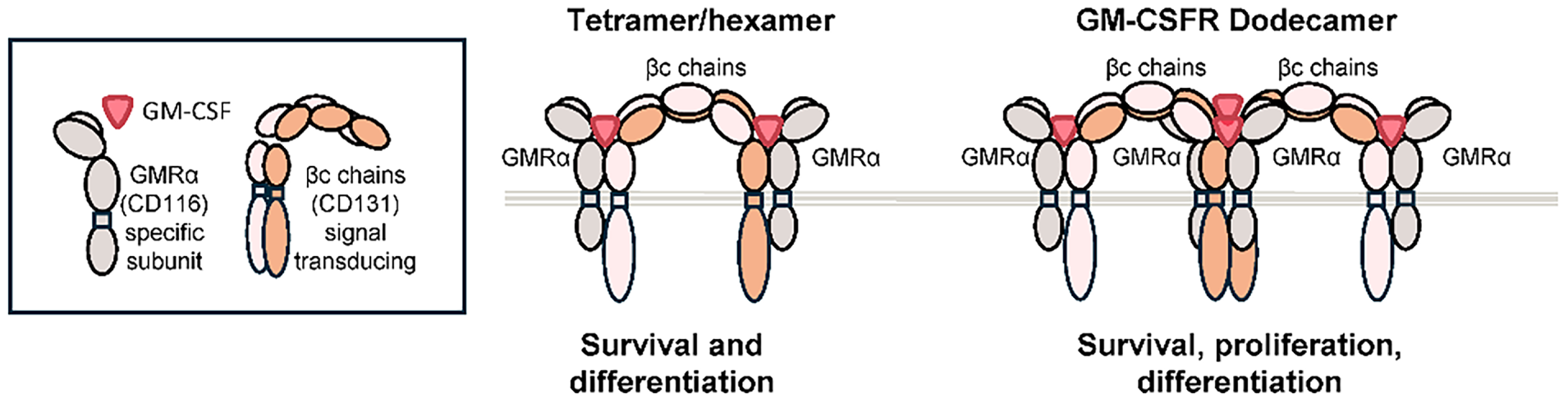
Figure 1: GM-CSFR subunits and receptor conformations. GM-CSFR is a heterodimeric receptor composed of a cytokine-specific α chain (GMRα) and a common β chain (βc chain). Ligand binding promotes assembly of a signaling hexamer, whose dimerization generates a high-affinity dodecamer that brings the βc cytoplasmic domains into close proximity, enabling transphosphorylation and kinase activation. Differential signaling outcomes associated with each receptor conformation are indicated.
The crystal structure of GM-CSF bound to its heterodimeric receptor has been solved, providing important structural insights into the interaction between the GM-CSFR and downstream signaling. The receptor assembles into a tetrameric structure comprising two intercalated βc chains at the core enclosed by two GMRα chains, each binding one GM-CSF molecule, resulting in a 6-protein ligand-receptor signaling complex, or hexamer (figure 1). The βc subunit plays a main role in signal transduction, yet signaling specificity is explained by the unique cytoplasmic domains of the GMRα subunits, whose deletion abolishes ligand-induced signaling [15].
This hexameric complex appears to dimerize forming a dodecamer structure composed of two hexamers, placing the cytoplasmic domains of the βc chains into close proximity, thus facilitating transphosphorylation and kinase activation. This generates a high-affinity conformation which engages multiple downstream pathways robustly, leading to GM-CSF-mediated survival, proliferation, differentiation and activation. By contrast, hexameric intermediates transduce predominantly survival and differentiation signals, as next detailed (figure 1) [14,16,17]. Other studies determined similar differential activation in response to low levels or high doses of GM-CSF, driven by a phosphorylation switch in the βc chains that allows for differential signaling cascades. Signaling occurs either through Ser585 at lower concentrations, leading to cell survival, or through Tyr577 at higher concentrations inducing regulation of broader functions [18,19]. Thus, the pleiotropic effect elicited by GM-CSF relies on the different signaling intensities, that is the net result of the specific cell-type GM-CSFR expression level, but also the GM-CSF quantity, and ultimately, the receptor stoichiometry.
GM-CSFR can trigger activation of the canonical pathways, including JAK2/STAT5 (Janus kinase 2/signal transducer and activator of transcription 5), the MAPK cascade (Ras/Raf/MEK/ERK) and the PI3K/Akt (phosphatidylinositol-3-kinase/protein kinase B) axis, as well as the NF-κB (nuclear factor kappa B) pathway. Downstream crosstalk further regulates mTORC1 (mammalian target of rapamycin complex 1) and β-catenin levels, which, together with the above-mentioned signaling modules, fine-tune the diverse cellular outcomes orchestrated by GM-CSF (figure 2).
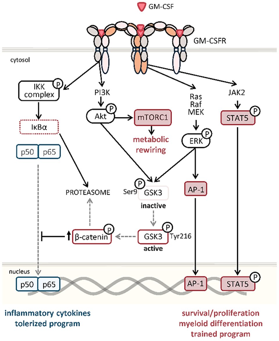
Figure 2: Overview of GM-CSF receptor signaling and its proposed role in regulating innate immune memory programs. GM-CSF binding activates multiple intracellular signaling pathways, including JAK2/STAT5, PI3K/Akt/mTOR, MAPK/ERK, NF-κB, and β-catenin, leading to context-dependent regulation of cell survival, proliferation, differentiation, and inflammatory activation. The canonical signaling pathways downstream of GM-CSFR are well established, whereas the interactions between β-catenin and NF-κB signaling, and their contribution to long-term hematopoietic stem and progenitor cell (HSPC) programming and innate immune memory, represent an emerging model supported by recent experimental evidence. The balance between inflammatory and regulatory programs is likely influenced by multiple factors, including GM-CSFR expression levels, ligand concentration, cell type, developmental stage, stimulus, and inflammatory context. Consequently, the signaling network depicted should be interpreted as a context-dependent conceptual framework rather than a universal GM-CSFR signaling cascade. Arrows indicate activation, blunt-ended lines indicate inhibition and “P” denotes phosphorylation.
The JAK2/STAT5 pathway has been extensively associated with progenitor survival, proliferation, and myeloid differentiation programs, while also contributing to inflammatory activation in mature myeloid cells [20]. Its activation requires the dodecamer high-affinity structure, which results in JAK2 clustering enabling phosphorylation of the βc chains, a signaling event that contributes to cell proliferation in response to GM-CSF, as revealed by mutant receptor analyses [17]. In line with this, only high concentrations of GM-CSF induce signaling via Tyr577, which strongly activates this pathway, driving phosphorylation of either the STAT5A or STAT5B isoforms [12]. Moreover, in human ex vivo differentiation of macrophages, STAT5 activity contributes to macrophage activation, including boosted phagocytic capacity and inflammatory cytokine production, whereas its inhibition reduces IL-6, IL-8 and TNF-α levels and promotes apoptosis in activated macrophages [21].
In parallel, activation of the MAPK pathway promotes progenitor proliferation and regulates lineage commitment during hematopoiesis [22]. Downstream, MAPK signaling induces AP-1 family members (such as c-Fos and c-Jun), and physical PU.1-c-Jun interaction is crucial for activation of PU.1 target genes required for myeloid commitment during macrophage differentiation [23] (figure 2). Specifically, sustained ERK signaling downstream of GM-CSF promotes monocyte survival in vitro and enhanced antigen-presenting features [24].
The PI3K/Akt pathway is engaged at both basal and high GM-CSF signaling strengths, contributing primarily to cell survival by inducing anti-apoptotic programs. This pathway can be activated via βc-chain phosphorylation or via direct interaction with the GMRα in a mechanism independent of JAK2, highlighting its flexibility in response to distinct receptor conformations [18]. In myeloid cells, GM-CSF-derived Akt modulation contributes to macrophage polarization or differentiation into DCs with varying mature and immature phenotypes [25,26].
Activation of NF-κB downstream of GM-CSFR signaling, is classically regarded as a central mediator of the primary inflammatory response. Early studies indicated that GM-CSF-induced Akt activation could regulate the transcriptional activity of NF-κB. However, direct activation of NF-κB through the GMRα subunit has also been described, promoting phosphorylation of the IKK activation complex and subsequent transcription of pro-inflammatory mediators [25] (figure 2). Besides this role, NF-κB signaling has also been linked to GM-CSF-mediated upregulation of anti-apoptotic members of the Bcl-2 family, thereby supporting survival across multiple hematopoietic lineages [17,23].
Further studies have demonstrated that GM-CSF signaling can also modulate the β-catenin pathway, whose central upstream regulator is glycogen synthase kinase 3β (GSK3β) (figure 2). During hematopoiesis, activation of GSK3β by phosphorylation at Tyr216 contributes to the tight regulation of HSPC self-renewal and differentiation by maintaining low intracellular levels of β-catenin through proteasomal degradation. In contrast, inhibition of GSK3β, by phosphorylation at Ser9, promotes β-catenin stabilization and transcriptional activation, a process associated with skewed myeloid differentiation and HSPC exhaustion [27,28].
Interestingly, the signaling pathways engaged downstream of GM-CSFR integrate ligand concentration, receptor stoichiometry, and cellular context to determine distinct biological outputs. Low-intensity signaling, associated with basal GM-CSF concentrations or lower-order receptor assemblies, preferentially activates PI3K/Akt- and NF-κB-dependent survival programs. By contrast, stronger GM-CSF signaling through high-affinity dodecameric receptor complexes promotes robust activation of JAK2/STAT5, MAPK/ERK, and NF-κB pathways, driving inflammatory gene expression, metabolic rewiring, and myeloid differentiation programs [12,17–19]. In this context, coordinated Akt and ERK activation converges on mTORC1 signaling, a central regulator of metabolic rewiring required for inflammatory effector functions, while enhanced NF-κB activity amplifies pro-inflammatory cytokine production. Notably, both Akt and ERK have been shown to induce inhibitory Ser9 phosphorylation of GSK3β [28,29], suggesting that β-catenin activation may represent an additional downstream layer integrating GM-CSF signaling strength with hematopoietic fate decisions and functional reprogramming.
Consistent with these graded signaling responses noted above, dose- and time-dependent effects of GM-CSF on monocyte and macrophage differentiation have been reported, suggesting that GM-CSF does not instruct a single activation state but rather licenses a spectrum of functional programs in different contexts. Sun et al. reported that different concentrations of GM-CSF generated myeloid populations with distinct inflammatory or suppressive properties, in fact, cells differentiated with a high concentration of GM-CSF were more potent in generating cytokines and chemokines [30]. Accordingly, in inflammatory settings, dose-dependent neutralization of GM-CSF differentially regulates monocyte expansion and functional activation, affecting both cell numbers and polarization programs [31].
Lastly, transition from activated monocytes to suppressor monocytes has been described in human and mouse inflammatory monocytes to be mediated by a first “licensing” step that requires the GM-CSF-induced PI3K/AKT/mTOR pathway [32]. In this context, myeloid-derived suppressor cells (MDSCs) are a heterogeneous population of immature cells of monocytic and granulocytic lineages with potent immunosuppressive activity that expand during cancer, chronic inflammation, and infection [33]. Their expansion and functional activation are driven by multiple inflammatory mediators, among which GM-CSF, in combination with other cytokines such as IL-6, represents a major inducer of MDSC development. These signals converge on key intracellular pathways including Ras/MAPK, PI3K/Akt, JAK/STAT, and TGF-β-associated signaling, which collectively regulate the expression of transcription factors and effector molecules involved in MDSC proliferation, survival, and suppressive activity, including iNOS, IL-10, and arginase-1 [34]. Together, these observations support the mechanistic framework to explain the diverse and context-dependent functions of GM-CSF discussed throughout this review.
3.1 Cellular Sources of GM-CSF
Under homeostatic conditions, alveolar epithelial cells constitute the major source of GM-CSF in the lung and are essential for alveolar macrophage differentiation and maintenance [35], while resident DC subsets are maintained by local tissue-derived GM-CSF signals in skin and mucosal tissues, largely provided by stromal compartments [36].
During inflammation, GM-CSF serves as a key communication hub linking local tissue perturbation with systemic myeloid responses. Both hematopoietic and non-hematopoietic cell types can produce GM-CSF, although this typically requires an activating stimulus. Importantly, the relative contribution of each cellular source is context-dependent and remains incompletely defined.
Among immune cells, activated T helper (Th) cells (Th1 and Th17) represent a major source of GM-CSF during inflammation and autoimmunity as observed in both mouse models and patients. Additionally, a novel subset of GM-CSF-producing T cells (Th-GM) has recently been identified and associated with the development of experimental autoimmune encephalomyelitis (EAE), a murine model of multiple sclerosis, [20,37]. In addition to T cells, other hematopoietic populations contribute to GM-CSF production in a highly context-dependent manner. Innate response activator B cells, located in the peritoneal and pleural cavities, produce GM-CSF upon pattern-recognition receptor (PRR) stimulation, thereby enhancing myeloid function and promoting early IgM production via autocrine GM-CSF signaling which reinforces early innate protection during infection [38,39]. By contrast, under chronic inflammatory conditions, the same B cell-derived GM-CSF axis becomes pathogenic, as observed in multiple sclerosis (MS) patients [40]. Similarly, natural killer (NK) cells can produce GM-CSF during tissue inflammation, where it contributes to either protective antimicrobial immunity or, conversely, to pathological amplification of inflammatory circuits in autoimmune arthritis [41,42].
An additional physiological regulator of GM-CSF production is the commensal microbiota. Increasing evidence indicates that microbiota-derived signals contribute to the maintenance of steady-state hematopoiesis and myeloid cell development by promoting tonic cytokine production at barrier tissues. In the intestine, microbial recognition by resident myeloid and stromal cells induces the production of IL-1β and IL-23, which subsequently stimulate type 3 innate lymphoid cells (ILC3s) and activated T cells to secrete GM-CSF. Locally produced GM-CSF promotes the differentiation and functional maturation of intestinal macrophages and dendritic cells, thereby contributing to epithelial integrity, immune homeostasis, and antimicrobial defense [43,44]. Beyond its local effects, the intestinal microbiota also influences steady-state myelopoiesis through circulating microbial products and metabolites that engage PRRs and shape hematopoietic activity in the bone marrow [45], although the direct contribution of microbiota-induced GM-CSF to signal in HSPCs remains to be unequivocally established. Moreover, Pires et al. found in a model of colorectal cancer that the cytokine TL1A stimulates production of GM-CSF by ILC3s, which triggers emergency granulopoiesis, neutrophil recruitment and adaptation in inflamed tissue, thereby promoting colorectal tumorigenesis [46].
In parallel, ILC2s producing GM-CSF have emerged as important regulators of pulmonary immunity during allergic inflammation or infection [47]. Furthermore, recent evidence also indicates that bone marrow-resident ILC2s can produce GM-CSF to support hematopoietic recovery under stress conditions [48,49]. Although HSPCs are not a predominant source of GM-CSF, they can produce this cytokine in response to PRR stimulation [50]. GM-CSF-producing HSPCs have also been identified in tumor-bearing mice and during Candida albicans infection [51,52].
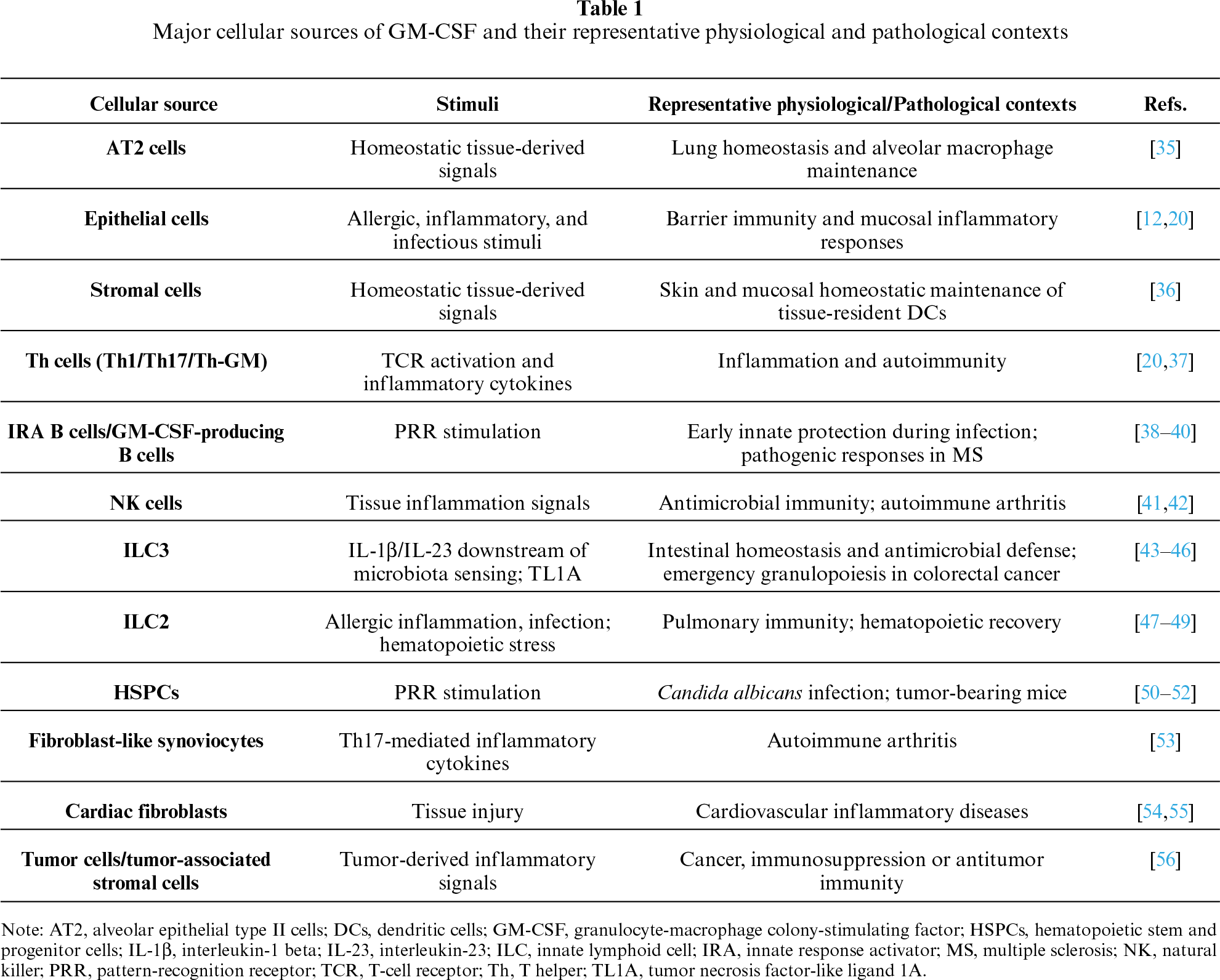
Importantly, non-hematopoietic tissue-resident cells also represent a major source of GM-CSF in inflammatory settings. Epithelial cells can rapidly produce GM-CSF in response to allergic or inflammatory stimuli, acting as an early signal that promotes barrier-associated immune activation and amplifies local inflammatory circuits. In addition, stromal cells contribute substantially to GM-CSF production in pathological contexts. Fibroblast-like synoviocytes produce GM-CSF and play a key role in the initiation and maintenance of autoimmune arthritis [53], while cardiac fibroblasts strongly upregulate GM-CSF following tissue injury and have been implicated in diverse cardiovascular pathologies [54,55]. Lastly, tumor cells and tumor-associated stromal compartments also constitute an important source of GM-CSF with context-dependent outcomes ranging from immunosuppression and tumor progression to reinforced antigen presentation and antitumor immunity, as discussed below [56]. The major cellular sources of GM-CSF under homeostatic and inflammatory conditions are summarized in table 1.
GM-CSF acts on a broad range of cells acting on both hematopoietic progenitors and mature innate immune cells to couple bone marrow output with peripheral inflammatory demands. Among differentiated innate cells, monocytes and macrophages represent central GM-CSF targets. On these cells, GM-CSF potentiates their survival, antimicrobial activity, and inflammatory potential, including increased phagocytosis, reactive oxygen species production, and triggers an inflammatory signature in monocytes [20,57,58]. Regarding dendritic cells, GM-CSF was the first cytokine shown to promote DC differentiation from human and mouse monocytes (moDCs) or hematopoietic progenitors in vitro, typically in combination with IL-4 [59,60]. However, the precise contribution of this cytokine in vivo remains under active debate and has been closely linked to the heterogeneity of GM-CSF-responsive DC populations. Recent evidence indicates that GM-CSF licenses differentiation and/or maturation of a distinct DC3 subset characterized by potent inflammatory and antigen-presenting capacities. Independently of their ontogeny, GM-CSF-responsive DC populations display enhanced MHC-II expression, cytokine production, and T-cell priming capacity [61–63]. In neutrophils, GM-CSF signaling sustains survival and tissue recruitment through upregulation of adhesion molecules, while simultaneously potentiating antimicrobial functions including reactive oxygen species production, phagocytosis and neutrophil extracellular trap formation [64–66]. Moreover, in eosinophils, GM-CSF similarly extends cell survival and potentiates their effector functions, while in the lung, resident basophils have been shown to be regulated by GM-CSF to further imprint macrophages in the pulmonary niche [67,68].
Upstream of differentiated cells, GM-CSFR is also expressed throughout the HSPC compartment, although at lower levels in uncommitted progenitors (HSCs) than in downstream myeloid-committed progenitors such as granulocyte-monocyte progenitors (GMPs). Upon interaction, GM-CSF promotes progenitor proliferation and biases their myeloid lineage commitment by inducing lineage-defining transcription programs [7,69–71]. Beyond regulating progenitor expansion and lineage commitment, the sustained expression of GM-CSFR throughout the myeloid progenitor hierarchy suggests that GM-CSF signaling may also influence the functional properties of downstream progeny. Therefore, GM-CSFR on the surface of HSPCs enables them to regulate myelopoiesis accordingly, but it is also plausible that HSPCs may integrate GM-CSF-derived signals to tune the inflammatory potential of their downstream progeny, as previously demonstrated in mature cells. These emerging concepts are further discussed below.
3.3 GM-CSF during Emergency Myelopoiesis
The hematopoiesis process is highly controlled by several cytokines, growth factors, and cell-cell interactions; however, it shows flexibility and responsiveness to sudden hematopoietic perturbations like blood loss, infection, or other pathologic conditions that disrupt the hematopoietic balance. Thus, a demand-adapted hematopoietic response is developed to boost cellular lineages as required. In the context of infection or injury, myeloid cells are rapidly consumed within tissues and are replenished by mobilized bone marrow monocytes and neutrophils. This acute supply is achieved by emergency myelopoiesis, consisting of transcriptional, epigenetic and metabolic mechanisms that act to prioritize rapid production of myeloid cells at the expense of other lineages [72,73].
Infection-induced activation of HSPCs is mediated by a combination of direct and indirect pathways. On one side, HSPCs can directly recognize pathogen-associated molecular patterns (PAMPs) that potentiate their differentiation into myeloid cells by PRR interaction [74–76]. On the other side, infection triggers the production of cytokines and alarmins by hematopoietic and non-hematopoietic cells, either systemically or within the bone marrow niche, driving indirect activation of HSPCs that potentiate and amplify direct sensing.
CSFs that regulate homeostatic hematopoiesis can play distinct roles during emergency myelopoiesis as exemplified by GM-CSF. In classical studies using GM-CSF-deficient mice, impaired GM-CSF signaling resulted in defective control of Listeria monocytogenes infection due to depletion of myeloid cells in the bone marrow and diminished neutrophil recruitment into the inflamed tissues [77]. Similarly, during chronic infection with Mycobacterium avium GM-CSF-deficient mice showed impaired ability to increase BM myeloid colony forming units (CFUs) in comparison to wild-type controls [78].
In later works, Mitroulis and colleagues showed that β-glucan administration in mice expands myeloid-biased long-term HSCs (LT-HSCs) and multipotent progenitors (MPPs) in association with increased IL-1β and GM-CSF signaling, leading to metabolic rewiring of glucose and cholesterol pathways. Importantly, they further demonstrated that β-glucan administration upregulated GM-CSFR βc chain expression in HSPCs and activated downstream GM-CSFR signaling, as revealed by increased STAT5 phosphorylation. Consistent with this, antibody-mediated blockade of GM-CSF in β-glucan-treated mice diminished the expansion of hematopoietic progenitors in comparison to β-glucan injection alone [79]. Together with this, GM-CSF has been described to enhance extramedullary hematopoiesis as shown by the trafficking of HSPCs to the spleen, and their myeloid progeny into tissues during some inflammatory diseases (such as atherosclerosis or colitis) and cancer [51,80,81]; or to drive leukocyte invasion during myocardial infarction by stimulating a distinct myeloid-biased progenitor subset [54]. Accordingly, two recent studies have demonstrated that GM-CSF production by BM-resident ILC2 drives emergency myelopoiesis in the context of chemotherapy or aging-derived inflammation. Firstly, Sudo et al. showed that GM-CSF knockout mice treated with 5-FU revealed a severe loss of myeloid lineage cells, which was rescued by transferring BM ILC2s from wild-type mice [48]. In line with this, Naef et al. demonstrated that secreted myeloid differentiation factors (particularly GM-CSF and IL-6) resulted in a myeloid-skewed HSC output and reduced self-renewal [49].
In the context of fungal infections where GM-CSF has been shown to be highly relevant, our group has demonstrated that, in vivo C. albicans challenge expands uncommitted progenitors, primes them for enhanced differentiation in the bone marrow early during infection, and mobilizes them to the spleen in a partially GM-CSF-dependent mechanism as demonstrated by antibody-mediated blockade [52]. Additionally, we found upregulation of the Csf2rb gene, the GM-CSFR βc chain, in bone marrow HSPCs very early after infection in line with the aforementioned reports [52].
4 Emerging Roles in Innate Immune Memory
4.1 Innate Immune Memory in Mature Myeloid Cells
Immunological memory was traditionally considered an exclusive trait of adaptive immunity, whereas innate immunity was viewed as a rapid but nonspecific defense system lacking long-term functional adaptation. However, memory-like properties of innate immune cells have now been described across multiple vertebrate and invertebrate species, revealing an evolutionarily conserved mechanism of immune adaptation. In 2011, the term “trained immunity” was first introduced to describe the immunological memory acquired from past insults that generates a protective inflammatory response through innate immune cells [82]. Subsequent studies demonstrated that similar molecular programs have been implicated in generating the opposite outcome, that is a diminished response upon secondary stimulation, as classically described following LPS exposure [83]. Accordingly, the broader concept of “innate immune memory” is now used to encompass both enhanced (trained immunity) and diminished (tolerance) secondary responses [84].
Innate immune memory is not antigen-specific but instead provides broad protection against subsequent challenges through long-term epigenetic remodeling and metabolic rewiring that alter chromatin accessibility and transcriptional responses upon restimulation [85,86]. Such programs can be triggered by diverse microbial, but also nonmicrobial stimuli, including GM-CSF. In this context, this cytokine primes proinflammatory cytokine production upon restimulation, inducing a trained phenotype in murine macrophages and human monocytes [87,88]. Mechanistically, GM-CSF stimulation of human monocytes has been shown to regulate inflammatory responses through coordinated STAT5, Akt, and NF-κB signaling upon microbial restimulation [89]. More recently, an additional layer involving metabolic-epigenetic crosstalk has been described, in which active Liver X Receptor (LXR) signaling contributes to GM-CSF-induced trained immunity in monocytes [90].
4.2 Central Innate Immune Memory
The concepts outlined above establish an important distinction between quantitative adaptations of hematopoiesis and qualitative changes in immune function. While emergency myelopoiesis primarily ensures rapid replenishment of myeloid cells during inflammation, accumulating evidence indicates that inflammatory stimuli can also durably reprogram HSPCs, giving rise to the concept of central innate immune memory.
While initial studies on innate immune memory mainly focused on differentiated myeloid cells, the persistence of trained responses in healthy volunteers receiving the live vaccine bacillus Calmette-Guérin (BCG) against tuberculosis endures from months up to at least one year [91]. These long-lasting effects strongly suggest that reprogramming might be taking place at the level of progenitor cells, thereby sustaining altered functional phenotypes beyond the lifespan of innate immune cells. Central innate immune memory proposes that stimulation of BM progenitors programs their metabolic and epigenetic states, and these changes are sustained through differentiation, ultimately generating progeny with altered phenotype, either trained or tolerized, upon secondary challenge [92–94].
Two conceptual frameworks have emerged to describe central innate immune memory in the current literature. One defines it primarily as a sustained change in lineage output (enhanced myelopoiesis) as previously discussed, while the other emphasizes the functional assessment of the derived mature myeloid cells, focusing on their heightened responsiveness, cytokine production, and antimicrobial capacity. Indeed, several studies have demonstrated that inflammatory signals driving emergency myelopoiesis can also imprint long-term transcriptional and epigenetic programs that shape these functional properties of the downstream myeloid output. Indeed, this functional HSPCs reprogramming can enhance host protection against infection or inflammatory challenges [79,95,96]. In this context, our group revisited the C. albicans PCA2 live vaccine model [97] (with a non-virulent strain) to better delve into the mechanisms by which HSPCs mediate protection against reinfection with a virulent strain of C. albicans [52]. BM HSPCs are expanded and reprogrammed early during infection to produce trained macrophages when ex vivo differentiated, and to produce increased amounts of proinflammatory cytokines themselves upon restimulation. Besides, their adoptive transfer is sufficient to protect mice against infection. Mechanistically, transcriptomic analyses in HSPCs revealed that autocrine GM-CSF signaling contributes to the trained phenotype and is essential for vaccine-induced protection [52].
This emerging evidence together with the well-established role of GM-CSF in emergency myelopoiesis, positions this cytokine as a strong candidate regulator of functional reprogramming in hematopoietic progenitors. However, the precise mechanisms by which GM-CSF steers immune memory across the hierarchical compartments of bone marrow HSPCs to coordinate and sustain their programming were not fully elucidated. Addressing this question, our recent work supports a model in which GM-CSFR expression levels across the myelopoietic hierarchy appear to regulate opposing innate memory programs that are long-lasting and persist beyond primary hematopoietic reconstitution in vivo. Mechanistically, myeloid-committed progenitors express higher levels of the GM-CSFR subunits than uncommitted progenitors, enabling GM-CSF to strongly activate the STAT5, ERK and PI3K/Akt-mTOR pathways and generate trained macrophages characterized by enhanced proinflammatory cytokine responses. In these progenitors, ERK- and PI3K/Akt-mediated inhibition of GSK3 promotes β-catenin accumulation, preventing NF-κB nuclear translocation and favoring STAT5- and mTOR-dependent trained programming (figure 2). In contrast, in upstream uncommitted progenitors, GM-CSF triggers only weak Akt and ERK activation but promotes translocation of NF-κB to the nucleus, allowed by constitutively active GSK3 and thereby low β-catenin levels, initiating a tolerization program (figures 2 and 3). Importantly, we further demonstrated that this stage-specific and opposing immune programming was observed in fungal infection-driven models of trained immunity and was conserved in equivalent human bone marrow progenitor populations [70]. While the role of GM-CSFR expression levels and downstream STAT5, ERK and PI3K/Akt-mTOR signaling is supported by several experimental studies, the integration of these pathways with β-catenin signaling and NF-κB regulation to generate divergent trained and tolerized programs should be considered a recently proposed mechanistic model that remains to be validated across different biological settings. Importantly, the proposed model should not be interpreted as suggesting that GM-CSF alone is sufficient to determine the final outcome of innate immune memory programming. Instead, GM-CSF signaling is likely integrated with additional environmental cues, cytokines, metabolic signals, and epigenetic regulators that collectively shape the functional state ultimately acquired by HSPCs and their progeny.
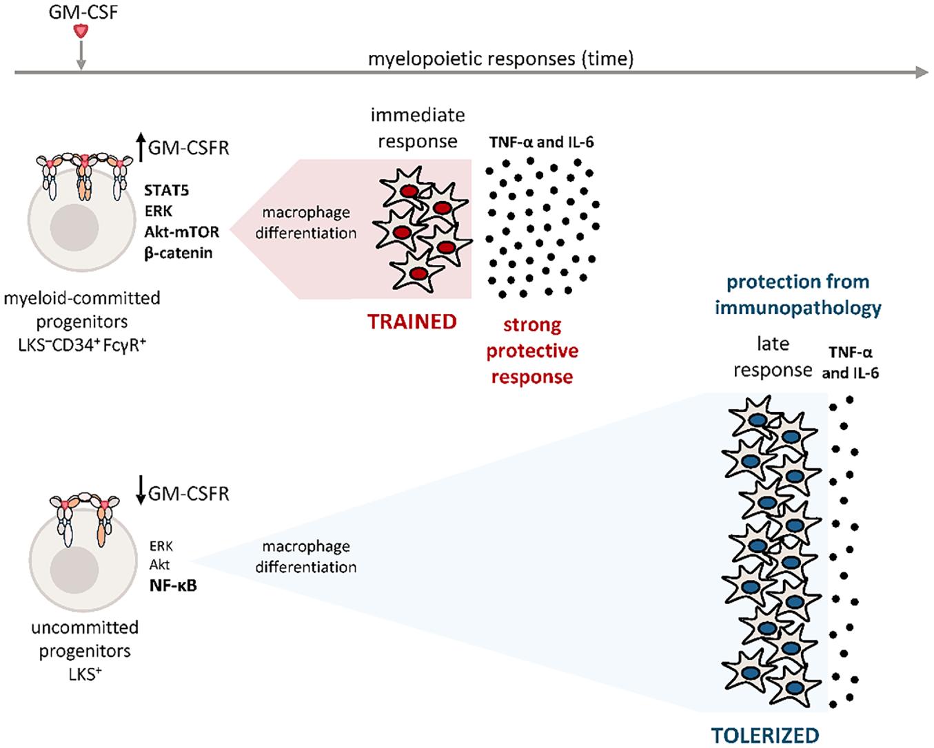
Figure 3: Proposed model whereby GM-CSF differentially programs opposing innate immune memory states across the hematopoietic hierarchy. Transient GM-CSF stimulation differentially programs hematopoietic progenitor subsets according to their stage of myeloid commitment and GM-CSFR expression levels. Myeloid-committed progenitors (defined as LSK–CD34+FcγR+ cells), which express higher levels of GM-CSFR subunits and display strong activation of STAT5, ERK, and Akt–mTOR signaling pathways, generate trained macrophages characterized by enhanced proinflammatory responses upon secondary stimulation. In contrast, upstream uncommitted progenitors identified as LSK+ progenitors (Lin–Sca-1+c-Kit+ cells), exhibit lower GM-CSFR expression and preferential NF-κB nuclear translocation, resulting in the generation of tolerized macrophages with reduced inflammatory activity [70]. These findings support a model in which GM-CSF not only promotes myelopoiesis but also functionally programs distinct and opposing innate immune memory states across the hematopoietic hierarchy, potentially balancing protective inflammation with prevention of excessive immunopathology.
These observations support a model in which GM-CSF signaling represents one mechanism by which functionally divergent innate memory programs can emerge in different hematopoietic compartments. We propose that immediate myeloid-committed progenitors preferentially adopt a trained program to enhance rapid effector responses, whereas upstream progenitors acquire a tolerized state that may limit excessive inflammation and prevent cytokine-driven pathology (figure 3) [70]. GM-CSF activity has long been related to cytokine dose, duration of exposure, inflammatory microenvironment and even combinatorial signaling with other cytokines. This novel mechanism could provide an additional layer of regulation in how opposite phenotypes are generated and the transition between inflammatory and regulatory memory programs. It will therefore be interesting to elucidate how these distinct GM-CSF-derived programs contribute temporally to immune responses in vivo, or across different pathological contexts with potential implications for the development of targeted immunomodulatory therapies.
5 GM-CSF Role in Disease and Clinical Relevance
As described throughout this review, GM-CSF plays a key role at the intersection of hematopoiesis, inflammation and disease. Beyond its well-known role in triggering myeloid-differentiation, GM-CSF functions as a context-dependent inflammatory cytokine that reinforces myeloid cells for key effector functions in tissues. However, the same properties that make this cytokine a promising target in cytopenic, immunotherapy or infectious settings, are the ones that need to be blunted in inflammatory pathologies [20]. Given its broad biological relevance, several therapeutic strategies aiming either to enhance or to block GM-CSF signaling are currently under investigation or already approved in multiple clinical contexts. If GM-CSF indeed contributes to central innate immune memory, therapeutic manipulation of this pathway may have consequences beyond the immediate inflammatory response. In addition to its acute effects on mature myeloid cells, transient enhancement or inhibition of GM-CSF signaling could potentially influence long-term HSPC programming, thereby modifying the quantity and functional properties of myeloid cells generated after treatment. Although this possibility remains largely unexplored in humans, it provides an attractive conceptual framework for interpreting both the sustained therapeutic benefits and the delayed adverse effects reported in different clinical settings.
Recombinant human GM-CSF (rhGM-CSF), also referred to as Sargramostim or Leukine®, is clinically used to accelerate myeloid reconstitution after bone-marrow transplantation, improve survival in patients exposed to myeloablative radiation, and mobilize HSPCs to peripheral blood for leukapheresis and autologous transplantation [98–100]. Besides in hematology, GM-CSF has also been explored to reverse sepsis-associated immunosuppression. Randomized clinical trials have shown that rhGM-CSF administration restores monocyte function and improves leukocyte responsiveness, contributing to sustained infection control and improved clinical parameters [101]. Likewise, in severe invasive fungal infections, particularly in neutropenic or otherwise immunocompromised hosts, adjunctive rhGM-CSF treatment with antifungal therapy, has been associated with improved neutrophil recovery, enhanced fungicidal activity, and increased macrophage activation and phagocytosis [102]. While the clinical benefits of rhGM-CSF are attributed to accelerated myeloid recovery and enhanced function of mature myeloid cells, it remains possible that some of these effects also involve functional reprogramming of HSPCs. If GM-CSF induces persistent changes in hematopoietic progenitors, transient therapeutic administration could generate myeloid cells with enhanced antimicrobial or immunoregulatory properties that persist beyond the period of cytokine exposure. Whether such long-term functional adaptation contributes to the efficacy of rhGM-CSF in transplantation, sepsis, or invasive fungal infections remains unknown and deserves further investigation.
In the context of Coronavirus disease 2019 (COVID-19), GM-CSF plays an essential role in mediating alveolar macrophage homeostasis and lung inflammation, in fact, inhaled GM-CSF was assessed in patients with COVID-19-related acute hypoxemic respiratory failure [103]. Conversely, in the later stages of COVID-19, increased GM-CSF levels lead to the activation of monocytes and macrophages and may indirectly contribute to acute respiratory distress syndrome contributing to lung damage. Here, GM-CSF neutralization is associated with faster clinical improvements and slower progression to severe disease or death in some high-risk COVID-19 patients [104]. The timing-dependent effects observed during COVID-19 further illustrate that the biological consequences of GM-CSF signaling depend not only on cytokine availability but also on the inflammatory context in which signaling occurs. Whether therapeutic modulation during different phases of disease also influences subsequent HSPC programming or innate immune adaptation remains an open question.
In the context of cancer, many tumors secrete GM-CSF either directly from malignant cells or indirectly through the immune and nonimmune compartments of the tumor microenvironment, thereby reshaping tumor-associated macrophage responses and systemic myelopoiesis [12,105]. While in some settings this favors the accumulation of immunosuppressive myeloid populations (MDSCs) and tumor progression, GM-CSF can also potentiate antitumor immunity. GM-CSF has classically been used in cancer therapy as an immune adjuvant, particularly in patients receiving chemotherapy for solid tumors, enhancing myeloid recovery and promoting humoral and cellular immune responses. In addition, GM-CSF combined with immune checkpoint blockade or cancer vaccine strategies has shown promising antitumor activity in several settings. Clinically, GM-CSF has been incorporated into approved therapeutic vaccines for hormone refractory metastatic prostate cancer, based on patient immune cells activated ex vivo with a GM-CSF fusion protein; and for the treatment of inoperable metastatic melanoma, using an oncolytic herpesvirus engineered to express GM-CSF [106,107].
By contrast, multiple studies have underscored GM-CSF as a driver of tissue inflammation and pain in diverse pathological settings including arthritis and joint disease, neuroinflammation, lung inflammation, cardiovascular injury, multiple sclerosis, and autoimmunity [20]. Notably, pathological GM-CSF signaling amplifies inflammatory cytokine production, leukocyte recruitment, and tissue infiltration, thereby contributing to chronic inflammation and tissue damage. Consequently, genetic deletion and neutralization studies in experimental models, together with early- and mid-phase clinical trials, indicate that blocking GM-CSF or its receptor may provide therapeutic benefit in inflammatory disorders. Likewise, GM-CSF blockade has shown protective effects in experimental models of allergic airway inflammation and cardiovascular inflammatory disease, further supporting its broad contribution to tissue-specific inflammatory pathology. In fact, administration of monoclonal antibodies against GM-CSF or to GMRα in rheumatoid arthritis has shown encouraging efficacy signals and it is generally well tolerated. Clinical trials have reported overall improvements in inflammatory biomarkers, with manageable adverse effects. Thus far, neither blocking strategy has been associated with major pulmonary toxicity [108,109]. However, these findings should be interpreted cautiously. Prolonged or profound systemic neutralization remains a theoretical concern due to the essential role of GM-CSF in alveolar macrophage maintenance and surfactant clearance, as exemplified by pulmonary alveolar proteinosis, a disease caused by defective GM-CSF signaling [12].
6 Concluding Remarks and Future Perspectives
Overall, the evidence discussed throughout this review highlights the remarkable functional versatility of GM-CSF and emphasizes the importance of considering its biological effects within the appropriate cellular and inflammatory context. Accordingly, blockade of the GM-CSF axis could theoretically compromise protective mechanisms (emergency myelopoiesis, antibacterial and antifungal host defenses, and tissue repair following inflammatory injury) particularly in individuals with chronic infections or impaired immune function. Although such long-term consequences have not yet been clearly demonstrated in clinical practice, they warrant careful evaluation as the therapeutic use of GM-CSF-targeting agents continues to expand. Future therapeutic strategies may benefit from more selective approaches, including bispecific antibodies combining GM-CSF blockade with cell type-specific targeting, potentially limiting the systemic effects associated with broad inhibition of the GM-CSF axis.
Taken together, GM-CSF emerges as a pleiotropic yet therapeutically targetable cytokine whose context-dependent effects can either enhance host defense and tissue repair or amplify pathogenic inflammation. This conceptual framework suggests that therapeutic manipulation of GM-CSF may not only influence the immediate activation of mature myeloid cells but could also modify the functional state of hematopoietic progenitors, thereby shaping future innate immune responses. Although direct evidence for such long-term effects in humans is still lacking, integrating HSPC programming into the interpretation of GM-CSF-targeted therapies may improve our understanding of both therapeutic efficacy and delayed adverse effects. Future clinical studies should therefore evaluate not only conventional inflammatory endpoints but also biomarkers of long-term hematopoietic and innate immune reprogramming.
Acknowledgement: The authors thank members of the laboratory for insightful discussions and critical reading of the manuscript.
Funding Statement: Research of the authors is supported by the Spanish Ministry of Science, Innovation & Universities (MICIU)/Agencia Estatal de Investigación (AEI)/FEDER, EU (PID2024-160294NB-I00/AEI/10.13039/501100011033) and CIAICO/2023/028 (Generalitat Valenciana; Programa per a la promoció de la investigació cientifica, el desenvolupament tecnològic i la innovació en la Comunitat Valenciana. Subvencions a grups d’investigació consolidats 2023) to AY and MLG. PG is a recipient of the fellowship “Atracció de talent” from the University of Valencia.
Author Contributions: The authors confirm contribution to the paper as follows: Conceptualization: Alberto Yáñez, M. Luisa Gil; writing—original draft preparation: Paula Guerrero; writing—review and editing: Alberto Yáñez, M. Luisa Gil, Paula Guerrero; funding acquisition: Alberto Yáñez, M. Luisa Gil. All authors reviewed and approved the final version of the manuscript.
Availability of Data and Materials: Not applicable.
Ethics Approval: Not applicable.
Conflicts of Interest: The authors declare no conflicts of interest.
References
1. Burgess AW, Metcalf D. The nature and action of granulocyte-macrophage colony stimulating factors. Blood 1980;56(6):947–58. doi:10.1182/blood.V56.6.947.947. [Google Scholar] [CrossRef]
2. Lieschke GJ, Grail D, Hodgson G, et al. Mice lacking granulocyte colony-stimulating factor have chronic neutropenia, granulocyte and macrophage progenitor cell deficiency, and impaired neutrophil mobilization. Blood 1994;84(6):1737–46. doi:10.1182/blood.V84.6.1737.1737. [Google Scholar] [CrossRef]
3. Wiktor-Jedrzejczak W, Bartocci A, Ferrante AW Jr, et al. Total absence of colony-stimulating factor 1 in the macrophage-deficient osteopetrotic (op/op) mouse. Proc Natl Acad Sci U S A 1990;87(12):4828–32. doi:10.1073/pnas.87.12.4828. [Google Scholar] [CrossRef]
4. Stanley E, Lieschke GJ, Grail D, et al. Granulocyte/macrophage colony-stimulating factor-deficient mice show no major perturbation of hematopoiesis but develop a characteristic pulmonary pathology. Proc Natl Acad Sci U S A 1994;91(12):5592–6. doi:10.1073/pnas.91.12.5592. [Google Scholar] [CrossRef]
5. Guilliams M, De Kleer I, Henri S, et al. Alveolar macrophages develop from fetal monocytes that differentiate into long-lived cells in the first week of life via GM-CSF. J Exp Med 2013;210(10):1977–92. doi:10.1084/jem.20131199. [Google Scholar] [CrossRef]
6. van de Laar L, Coffer PJ, Woltman AM. Regulation of dendritic cell development by GM-CSF: molecular control and implications for immune homeostasis and therapy. Blood 2012;119(15):3383–93. doi:10.1182/blood-2011-11-370130. [Google Scholar] [CrossRef]
7. Becher B, Tugues S, Greter M. GM-CSF: from growth factor to central mediator of tissue inflammation. Immunity 2016;45(5):963–73. doi:10.1016/j.immuni.2016.10.026. [Google Scholar] [CrossRef]
8. Chavakis T, Wielockx B, Hajishengallis G. Inflammatory modulation of hematopoiesis: linking trained immunity and clonal hematopoiesis with chronic disorders. Annu Rev Physiol 2022;84(1):183–207. doi:10.1146/annurev-physiol-052521-013627. [Google Scholar] [CrossRef]
9. Vuscan P, Kischkel B, Joosten LAB, Netea MG. Trained immunity: general and emerging concepts. Immunol Rev 2024;323(1):164–85. doi:10.1111/imr.13326. [Google Scholar] [CrossRef]
10. Ruffinatto L, Groult Y, Iacono J, Sarrazin S, de Laval B. Hematopoietic stem cell a reservoir of innate immune memory. Front Immunol 2024;15:1491729. doi:10.3389/fimmu.2024.1491729. [Google Scholar] [CrossRef]
11. Hamilton JA, Achuthan A. Colony stimulating factors and myeloid cell biology in health and disease. Trends Immunol 2013;34(2):81–9. doi:10.1016/j.it.2012.08.006. [Google Scholar] [CrossRef]
12. Dougan M, Dranoff G, Dougan SK. GM-CSF, IL-3, and IL-5 family of cytokines: regulators of inflammation. Immunity 2019;50(4):796–811. doi:10.1016/j.immuni.2019.03.022. [Google Scholar] [CrossRef]
13. Hamilton JA, Anderson GP. Mini ReviewGM-CSF biology. Growth Factors 2004;22(4):225–31. doi:10.1080/08977190412331279881. [Google Scholar] [CrossRef]
14. Broughton SE, Hercus TR, Nero TL, et al. Conformational changes in the GM-CSF receptor suggest a molecular mechanism for affinity conversion and receptor signaling. Structure 2016;24(8):1271–81. doi:10.1016/j.str.2016.05.017. [Google Scholar] [CrossRef]
15. Polotskaya A, Zhao Y, Lilly ML, Kraft AS. A critical role for the cytoplasmic domain of the granulocyte-macrophage colony-stimulating factor alpha receptor in mediating cell growth. Cell Growth Differ 1993;4(6):523–1. doi:10.32388/ldmrqi. [Google Scholar] [CrossRef]
16. Hansen G, Hercus TR, McClure BJ, et al. The structure of the GM-CSF receptor complex reveals a distinct mode of cytokine receptor activation. Cell 2008;134(3):496–507. doi:10.1016/j.cell.2008.05.053. [Google Scholar] [CrossRef]
17. Perugini M, Brown AL, Salerno DG, et al. Alternative modes of GM-CSF receptor activation revealed using activated mutants of the common β-subunit. Blood 2010;115(16):3346–3. doi:10.1182/blood-2009-08-235846. [Google Scholar] [CrossRef]
18. Guthridge MA, Barry EF, Felquer FA, et al. The phosphoserine-585–dependent pathway of the GM-CSF/IL-3/IL-5 receptors mediates hematopoietic cell survival through activation of NF-κB and induction of bcl-2. Blood 2004;103(3):820–7. doi:10.1182/blood-2003-06-1999. [Google Scholar] [CrossRef]
19. Guthridge MA, Powell JA, Barry EF, et al. Growth factor pleiotropy is controlled by a receptor Tyr/Ser motif that acts as a binary switch. EMBO J 2006;25(3):479–89. doi:10.1038/sj.emboj.7600948. [Google Scholar] [CrossRef]
20. Hamilton JA. GM-CSF in inflammation. J Exp Med 2020;217(1):e20190945. doi:10.1084/jem.20190945. [Google Scholar] [CrossRef]
21. Nagenborg J, Jin H, Ruder AV, et al. GM-CSF-activated STAT5A regulates macrophage functions and inflammation in atherosclerosis. Front Immunol 2023;14:1165306. doi:10.3389/fimmu.2023.1165306. [Google Scholar] [CrossRef]
22. Zarrabi M, Afzal E, Asghari MH, Mohammad M, Es HA, Ebrahimi M. Inhibition of MEK/ERK signalling pathway promotes erythroid differentiation and reduces HSCs engraftment in ex vivo expanded haematopoietic stem cells. J Cell Mol Med 2018;22(3):1464–74. doi:10.1111/jcmm.13379. [Google Scholar] [CrossRef]
23. Zhao X, Bartholdy B, Yamamoto Y, et al. PU.1-c-Jun interaction is crucial for PU.1 function in myeloid development. Commun Biol 2022;5(1):961. doi:10.1038/s42003-022-03888-7. [Google Scholar] [PubMed] [CrossRef]
24. Achuthan A, Aslam ASM, Nguyen Q, et al. Glucocorticoids promote apoptosis of proinflammatory monocytes by inhibiting ERK activity. Cell Death Dis 2018;9(3):267. doi:10.1038/s41419-018-0332-4. [Google Scholar] [PubMed] [CrossRef]
25. Vergadi E, Ieronymaki E, Lyroni K, Vaporidi K, Tsatsanis C. Akt signaling pathway in macrophage activation and M1/M2 polarization. J Immunol 2017;198(3):1006–14. doi:10.4049/jimmunol.1601515. [Google Scholar] [CrossRef]
26. Zhan Y, Lew AM, Chopin M. The pleiotropic effects of the GM-CSF rheostat on myeloid cell differentiation and function: more than a numbers game. Front Immunol 2019;10:2679. doi:10.3389/fimmu.2019.02679. [Google Scholar] [PubMed] [CrossRef]
27. Brown AL, Salerno DG, Sadras T, et al. The GM-CSF receptor utilizes β-catenin and Tcf4 to specify macrophage lineage differentiation. Differentiation 2012;83(1):47–59. doi:10.1016/j.diff.2011.08.003. [Google Scholar] [CrossRef]
28. Cross DA, Alessi DR, Cohen P, Andjelkovich M, Hemmings BA. Inhibition of glycogen synthase kinase-3 by insulin mediated by protein kinase B. Nature 1995;378(6559):785–9. doi:10.1038/378785a0. [Google Scholar] [CrossRef]
29. Ding Q, Xia W, Liu JC, et al. Erk associates with and primes GSK-3β for its inactivation resulting in upregulation of β-catenin. Mol Cell 2005;19(2):159–70. doi:10.1016/j.molcel.2005.06.009. [Google Scholar] [CrossRef]
30. Sun L, Rautela J, Delconte RB, et al. GM-CSF quantity has a selective effect on granulocytic vs. monocytic myeloid development and function. Front Immunol 2018;9:1922. doi:10.3389/fimmu.2018.01922. [Google Scholar] [CrossRef]
31. Louis C, Cook AD, Lacey D, et al. Specific contributions of CSF-1 and GM-CSF to the dynamics of the mononuclear phagocyte system. J Immunol 2015;195(1):134–44. doi:10.4049/jimmunol.1500369. [Google Scholar] [CrossRef]
32. Ribechini E, Hutchinson JA, Hergovits S, et al. Novel GM-CSF signals via IFN-γR/IRF-1 and AKT/mTOR license monocytes for suppressor function. Blood Adv 2017;1(14):947–60. doi:10.1182/bloodadvances.2017006858. [Google Scholar] [PubMed] [CrossRef]
33. Gabrilovich DI, Nagaraj S. Myeloid-derived suppressor cells as regulators of the immune system. Nat Rev Immunol 2009;9(3):162–74. doi:10.1038/nri2506. [Google Scholar] [CrossRef]
34. Wang Y, Jia A, Bi Y, Wang Y, Liu G. Metabolic regulation of myeloid-derived suppressor cell function in cancer. Cells 2020;9(4):1011. doi:10.3390/cells9041011. [Google Scholar] [CrossRef]
35. Gschwend J, Sherman SPM, Ridder F, et al. Alveolar macrophages rely on GM-CSF from alveolar epithelial type 2 cells before and after birth. J Exp Med 2021;218(10):e20210745. doi:10.1084/jem.20210745. [Google Scholar] [CrossRef]
36. Greter M, Helft J, Chow A, et al. GM-CSF controls nonlymphoid tissue dendritic cell homeostasis but is dispensable for the differentiation of inflammatory dendritic cells. Immunity 2012;36(6):1031–46. doi:10.1016/j.immuni.2012.03.027. [Google Scholar] [CrossRef]
37. Sheng W, Yang F, Zhou Y, et al. STAT5 programs a distinct subset of GM-CSF-producing T helper cells that is essential for autoimmune neuroinflammation. Cell Res 2014;24(12):1387–402. doi:10.1038/cr.2014.154. [Google Scholar] [CrossRef]
38. Weber GF, Chousterman BG, Hilgendorf I, et al. Pleural innate response activator B cells protect against pneumonia via a GM-CSF-IgM axis. J Exp Med 2014;211(6):1243–56. doi:10.1084/jem.20131471. [Google Scholar] [CrossRef]
39. Rauch PJ, Chudnovskiy A, Robbins CS, et al. Innate response activator B cells protect against microbial sepsis. Science 2012;335(6068):597–601. doi:10.1126/science.1215173. [Google Scholar] [CrossRef]
40. Li R, Rezk A, Miyazaki Y, et al. Proinflammatory GM-CSF–producing B cells in multiple sclerosis and B cell depletion therapy. Sci Transl Med 2015;7(310):eaab4176. doi:10.1126/scitranslmed.aab4176. [Google Scholar] [CrossRef]
41. Louis C, Souza-Fonseca-Guimaraes F, Yang Y, et al. NK cell–derived GM-CSF potentiates inflammatory arthritis and is negatively regulated by CIS. J Exp Med 2020;217(5):e2019142103192020c. doi:10.1084/jem.20191421. [Google Scholar] [CrossRef]
42. Domínguez-Andrés J, Feo-Lucas L, Minguito de la Escalera M, González L, López-Bravo M, Ardavín C. Inflammatory Ly6Chigh monocytes protect against candidiasis through IL-15-driven NK cell/neutrophil activation. Immunity 2017;46(6):1059–72.e4. doi:10.1016/j.immuni.2017.05.009. [Google Scholar] [CrossRef]
43. Mortha A, Chudnovskiy A, Hashimoto D, et al. Microbiota-dependent crosstalk between macrophages and ILC3 promotes intestinal homeostasis. Science 2014;343(6178):1249288. doi:10.1126/science.1249288. [Google Scholar] [CrossRef]
44. Castro-Dopico T, Fleming A, Dennison TW, et al. GM-CSF calibrates macrophage defense and wound healing programs during intestinal infection and inflammation. Cell Rep 2020;32(1):107857. doi:10.1016/j.celrep.2020.107857. [Google Scholar] [CrossRef]
45. Khosravi A, Yáñez A, Price JG, et al. Gut microbiota promote hematopoiesis to control bacterial infection. Cell Host Microbe 2014;15(3):374–81. doi:10.1016/j.chom.2014.02.006. [Google Scholar] [CrossRef]
46. Pires S, Yang W, Frigerio S, et al. Innate lymphoid cells activated by the cytokine TL1A link colitis to emergency granulopoiesis and the recruitment of tumor-promoting neutrophils. Immunity 2026;59(2):372–87.e7. doi:10.1016/j.immuni.2025.12.008. [Google Scholar] [CrossRef]
47. Brooksby JJ, Kobayashi T, Iijima K, et al. Group 2 innate lymphoid cells program pulmonary adaptive immunity via granulocyte-macrophage colony-stimulating factor. J Allergy Clin Immunol 2026;158(1):236–52. doi:10.1016/j.jaci.2026.02.028. [Google Scholar] [CrossRef]
48. Sudo T, Motomura Y, Okuzaki D, et al. Group 2 innate lymphoid cells support hematopoietic recovery under stress conditions. J Exp Med 2021;218(5):e20200817. doi:10.1084/jem.20200817. [Google Scholar] [CrossRef]
49. Naef P, Jaeger-Ruckstuhl CA, Schnüriger N, et al. IL-33/ST2 signaling in ILC2s drives exhaustion and myeloid skewing of HSCs in response to hematopoietic stress and aging. iScience 2025;28(5):112378. doi:10.1016/j.isci.2025.112378. [Google Scholar] [CrossRef]
50. Zhao JL, Ma C, O’Connell RM, et al. Conversion of danger signals into cytokine signals by hematopoietic stem and progenitor cells for regulation of stress-induced hematopoiesis. Cell Stem Cell 2014;14(4):445–59. doi:10.1016/j.stem.2014.01.007. [Google Scholar] [CrossRef]
51. Wu C, Ning H, Liu M, et al. Spleen mediates a distinct hematopoietic progenitor response supporting tumor-promoting myelopoiesis. J Clin Invest 2018;128(8):3425–38. doi:10.1172/JCI97973. [Google Scholar] [CrossRef]
52. Bono C, Guerrero P, Jordán-Pla A, et al. GM-CSF programs hematopoietic stem and progenitor cells during Candida albicans vaccination for protection against reinfection. Front Immunol 2021;12:790309. doi:10.3389/fimmu.2021.790309. [Google Scholar] [CrossRef]
53. Hirota K, Hashimoto M, Ito Y, et al. Autoimmune Th17 cells induced synovial stromal and innate lymphoid cell secretion of the cytokine GM-CSF to initiate and augment autoimmune arthritis. Immunity 2018;48(6):1220–32.e5. doi:10.1016/j.immuni.2018.04.009. [Google Scholar] [CrossRef]
54. Anzai A, Choi JL, He S, et al. The infarcted myocardium solicits GM-CSF for the detrimental oversupply of inflammatory leukocytes. J Exp Med 2017;214(11):3293–310. doi:10.1084/jem.20170689. [Google Scholar] [CrossRef]
55. Lee KMC, Achuthan AA, Hamilton JA. GM-CSF: a promising target in inflammation and autoimmunity. Immunotargets Ther 2020;9:225–40. doi:10.2147/ITT.S262566. [Google Scholar] [PubMed] [CrossRef]
56. Kumar A, Taghi Khani A, Sanchez Ortiz A, Swaminathan S. GM-CSF: a double-edged sword in cancer immunotherapy. Front Immunol 2022;13:901277. doi:10.3389/fimmu.2022.901277. [Google Scholar] [CrossRef]
57. Croxford AL, Lanzinger M, Hartmann FJ, et al. The cytokine GM-CSF drives the inflammatory signature of CCR2+ monocytes and licenses autoimmunity. Immunity 2015;43(3):502–14. doi:10.1016/j.immuni.2015.08.010. [Google Scholar] [CrossRef]
58. Lazarus HM, Pitts K, Wang T, et al. Recombinant GM-CSF for diseases of GM-CSF insufficiency: correcting dysfunctional mononuclear phagocyte disorders. Front Immunol 2023;13:1069444. doi:10.3389/fimmu.2022.1069444. [Google Scholar] [CrossRef]
59. Sallusto F, Lanzavecchia A. Efficient presentation of soluble antigen by cultured human dendritic cells is maintained by granulocyte/macrophage colony-stimulating factor plus interleukin 4 and downregulated by tumor necrosis factor alpha. J Exp Med 1994;179(4):1109–18. doi:10.1084/jem.179.4.1109. [Google Scholar] [CrossRef]
60. Inaba K, Inaba M, Romani N, et al. Generation of large numbers of dendritic cells from mouse bone marrow cultures supplemented with granulocyte/macrophage colony-stimulating factor. J Exp Med 1992;176(6):1693–702. doi:10.1084/jem.176.6.1693. [Google Scholar] [PubMed] [CrossRef]
61. Segura E. Monocyte-derived dendritic cells: an updated view on an old concept. Immunol Rev 2025;336(1):e70075. doi:10.1111/imr.70075. [Google Scholar] [CrossRef]
62. Helft J, Böttcher J, Chakravarty P, et al. GM-CSF mouse bone marrow cultures comprise a heterogeneous population of CD11c+MHCII+ macrophages and dendritic cells. Immunity 2015;42(6):1197–211. doi:10.1016/j.immuni.2015.05.018. [Google Scholar] [CrossRef]
63. Sonderegger I, Iezzi G, Maier R, Schmitz N, Kurrer M, Kopf M. GM-CSF mediates autoimmunity by enhancing IL-6-dependent Th17 cell development and survival. J Exp Med 2008;205(10):2281–94. doi:10.1084/jem.20071119. [Google Scholar] [PubMed] [CrossRef]
64. Kobayashi SD, Voyich JM, Whitney AR, DeLeo FR. Spontaneous neutrophil apoptosis and regulation of cell survival by granulocyte macrophage-colony stimulating factor. J Leukoc Biol 2005;78(6):1408–18. doi:10.1189/jlb.0605289. [Google Scholar] [CrossRef]
65. Wright HL, Thomas HB, Moots RJ, Edwards SW. RNA-seq reveals activation of both common and cytokine-specific pathways following neutrophil priming. PLoS One 2013;8(3):e58598. doi:10.1371/journal.pone.0058598. [Google Scholar] [CrossRef]
66. Yousefi S, Mihalache C, Kozlowski E, Schmid I, Simon HU. Viable neutrophils release mitochondrial DNA to form neutrophil extracellular traps. Cell Death Differ 2009;16(11):1438–44. doi:10.1038/cdd.2009.96. [Google Scholar] [CrossRef]
67. Cohen M, Giladi A, Gorki AD, et al. Lung single-cell signaling interaction map reveals basophil role in macrophage imprinting. Cell 2018;175(4):1031–44.e18. doi:10.1016/j.cell.2018.09.009. [Google Scholar] [CrossRef]
68. Griseri T, Arnold IC, Pearson C, et al. Granulocyte macrophage colony-stimulating factor-activated eosinophils promote interleukin-23 driven chronic colitis. Immunity 2015;43(1):187–99. doi:10.1016/j.immuni.2015.07.008. [Google Scholar] [CrossRef]
69. Chavakis T, Mitroulis I, Hajishengallis G. Hematopoietic progenitor cells as integrative hubs for adaptation to and fine-tuning of inflammation. Nat Immunol 2019;20(7):802–11. doi:10.1038/s41590-019-0402-5. [Google Scholar] [CrossRef]
70. Guerrero P, Bono C, Sobén M, et al. GM-CSF receptor expression determines opposing innate memory phenotypes at different stages of myelopoiesis. Blood 2024;143(26):2763–77. doi:10.1182/blood.2024024330. [Google Scholar] [CrossRef]
71. Regan-Komito D, Swann JW, Demetriou P, et al. GM-CSF drives dysregulated hematopoietic stem cell activity and pathogenic extramedullary myelopoiesis in experimental spondyloarthritis. Nat Commun 2020;11(1):155. doi:10.1038/s41467-019-13853-4. [Google Scholar] [CrossRef]
72. Boettcher S, Manz MG. Regulation of inflammation- and infection-driven hematopoiesis. Trends Immunol 2017;38(5):345–57. doi:10.1016/j.it.2017.01.004. [Google Scholar] [CrossRef]
73. Swann JW, Olson OC, Passegué E. Made to order: emergency myelopoiesis and demand-adapted innate immune cell production. Nat Rev Immunol 2024;24(8):596–613. doi:10.1038/s41577-024-00998-7. [Google Scholar] [CrossRef]
74. Nagai Y, Garrett KP, Ohta S, et al. Toll-like receptors on hematopoietic progenitor cells stimulate innate immune system replenishment. Immunity 2006;24(6):801–12. doi:10.1016/j.immuni.2006.04.008. [Google Scholar] [PubMed] [CrossRef]
75. Yáñez A, Megías J, O’Connor JE, Gozalbo D, Gil ML. Candida albicans induces selective development of macrophages and monocyte derived dendritic cells by a TLR2 dependent signalling. PLoS One 2011;6(9):e24761. doi:10.1371/journal.pone.0024761. [Google Scholar] [CrossRef]
76. Megías J, Yáñez A, Moriano S, O’Connor JE, Gozalbo D, Gil ML. Direct toll-like receptor-mediated stimulation of hematopoietic stem and progenitor cells occurs in vivo and promotes differentiation toward macrophages. Stem Cells 2012;30(7):1486–95. doi:10.1002/stem.1110. [Google Scholar] [CrossRef]
77. Zhan Y, Lieschke GJ, Grail D, Dunn AR, Cheers C. Essential roles for granulocyte-macrophage colony-stimulating factor (GM-CSF) and G-CSF in the sustained hematopoietic response of Listeria monocytogenes-infected mice. Blood 1998;91(3):863–9. doi:10.1182/blood.v91.3.863.863_863_869. [Google Scholar] [CrossRef]
78. Zhan Y, Cheers C. Haemopoiesis in mice genetically lacking granulocyte-macrophage colony stimulating factor during chronic infection with Mycobacterium avium. Immunol Cell Biol 2000;78(2):118–23. doi:10.1046/j.1440-1711.2000.00891.x. [Google Scholar] [CrossRef]
79. Mitroulis I, Ruppova K, Wang B, et al. Modulation of myelopoiesis progenitors is an integral component of trained immunity. Cell 2018;172(1–2):147–61.e12. doi:10.1016/j.cell.2017.11.034. [Google Scholar] [CrossRef]
80. Griseri T, McKenzie BS, Schiering C, Powrie F. Dysregulated hematopoietic stem and progenitor cell activity promotes interleukin-23-driven chronic intestinal inflammation. Immunity 2012;37(6):1116–29. doi:10.1016/j.immuni.2012.08.025. [Google Scholar] [CrossRef]
81. Robbins CS, Chudnovskiy A, Rauch PJ, et al. Extramedullary hematopoiesis generates ly-6ChighMonocytes that infiltrate atherosclerotic lesions. Circulation 2012;125(2):364–74. doi:10.1161/circulationaha.111.061986. [Google Scholar] [CrossRef]
82. Netea MG, Quintin J, van der Meer JWM. Trained immunity: a memory for innate host defense. Cell Host Microbe 2011;9(5):355–61. doi:10.1016/j.chom.2011.04.006. [Google Scholar] [CrossRef]
83. Bekkering S, Domínguez-Andrés J, Joosten LAB, Riksen NP, Netea MG. Trained immunity: reprogramming innate immunity in health and disease. Annu Rev Immunol 2021;39(1):667–93. doi:10.1146/annurev-immunol-102119-073855. [Google Scholar] [CrossRef]
84. López-Collazo E, Del Fresno C. Endotoxin tolerance and trained immunity: breaking down immunological memory barriers. Front Immunol 2024;15:1393283. doi:10.3389/fimmu.2024.1393283. [Google Scholar] [CrossRef]
85. Caldwell BA, Li L. Epigenetic regulation of innate immune dynamics during inflammation. J Leukoc Biol 2024;115(4):589–606. doi:10.1093/jleuko/qiae026. [Google Scholar] [CrossRef]
86. Cheng SC, Quintin J, Cramer RA, et al. mTOR- and HIF-1α-mediated aerobic glycolysis as metabolic basis for trained immunity. Science 2014;345(6204):1250684. doi:10.1126/science.1250684. [Google Scholar] [CrossRef]
87. Fleetwood AJ, Lawrence T, Hamilton JA, Cook AD. Granulocyte-macrophage colony-stimulating factor (CSF) and macrophage CSF-dependent macrophage phenotypes display differences in cytokine profiles and transcription factor activities: implications for CSF blockade in inflammation. J Immunol 2007;178(8):5245–52. doi:10.4049/jimmunol.178.8.5245. [Google Scholar] [CrossRef]
88. Borriello F, Iannone R, Di Somma S, et al. GM-CSF and IL-3 modulate human monocyte TNF-α production and renewal in in vitro models of trained immunity. Front Immunol 2017;7:680. doi:10.3389/fimmu.2016.00680. [Google Scholar] [CrossRef]
89. Vázquez Marrero VR, Dresler M, Haggadone MD, Lu A, Shin S. GM-CSF engages multiple signaling pathways to enhance pro-inflammatory cytokine responses in human monocytes during Legionella infection. Infect Immun 2025;93(7):e00565–24. doi:10.1128/iai.00565-24. [Google Scholar] [CrossRef]
90. Liu Y, Hamid A, Hardege H, et al. Active LXR signaling, coupled with elevated mitochondrial and glycolytic metabolism contributes to GM-CSF-induced trained immunity. Front Immunol 2026;16:1685796. doi:10.3389/fimmu.2025.1685796. [Google Scholar] [CrossRef]
91. Kleinnijenhuis J, Quintin J, Preijers F, et al. Long-lasting effects of BCG vaccination on both heterologous Th1/Th17 responses and innate trained immunity. J Innate Immun 2014;6(2):152–8. doi:10.1159/000355628. [Google Scholar] [CrossRef]
92. Yáñez A, Hassanzadeh-Kiabi N, Ng MY, et al. Detection of a TLR2 agonist by hematopoietic stem and progenitor cells impacts the function of the macrophages they produce. Eur J Immunol 2013;43(8):2114–25. doi:10.1002/eji.201343403. [Google Scholar] [CrossRef]
93. Megías J, Martínez A, Yáñez A, Goodridge HS, Gozalbo D, Gil ML. TLR2, TLR4 and Dectin-1 signalling in hematopoietic stem and progenitor cells determines the antifungal phenotype of the macrophages they produce. Microbes Infect 2016;18(5):354–63. doi:10.1016/j.micinf.2016.01.005. [Google Scholar] [PubMed] [CrossRef]
94. Bono C, Martínez A, Megías J, Gozalbo D, Yáñez A, Gil ML. Dectin-1 stimulation of hematopoietic stem and progenitor cells occurs in vivo and promotes differentiation toward trained macrophages via an indirect cell-autonomous mechanism. mBio 2020;11(3):e00781–20. doi:10.1128/mBio.00781-20. [Google Scholar] [CrossRef]
95. Kaufmann E, Sanz J, Dunn JL, et al. BCG educates hematopoietic stem cells to generate protective innate immunity against tuberculosis. Cell 2018;172(1–2):176–90.e19. doi:10.1016/j.cell.2017.12.031. [Google Scholar] [CrossRef]
96. Kain BN, Tran BT, Luna PN, et al. Hematopoietic stem and progenitor cells confer cross-protective trained immunity in mouse models. iScience 2023;26(9):107596. doi:10.1016/j.isci.2023.107596. [Google Scholar] [CrossRef]
97. Bistoni F, Verducci G, Perito S, et al. Immunomodulation by a low-virulence, agerminative variant of Candida albicans. Further evidence for macrophage activation as one of the effector mechanisms of nonspecific anti-infectious protection. J Med Vet Mycol 1988;26(5):285–99. doi:10.1080/02681218880000401. [Google Scholar] [CrossRef]
98. Brandt SJ, Peters WP, Atwater SK, et al. Effect of recombinant human granulocyte-macrophage colony-stimulating factor on hematopoietic reconstitution after high-dose chemotherapy and autologous bone marrow transplantation. N Engl J Med 1988;318(14):869–76. doi:10.1056/NEJM198804073181401. [Google Scholar] [CrossRef]
99. Nemunaitis J, Rabinowe SN, Singer JW, et al. Recombinant granulocyte-macrophage colony-stimulating factor after autologous bone marrow transplantation for lymphoid cancer. N Engl J Med 1991;324(25):1773–8. doi:10.1056/nejm199106203242504. [Google Scholar] [CrossRef]
100. Weaver C, Schulman K, Buckner C. Mobilization of peripheral blood stem cells following myelosuppressive chemotherapy: a randomized comparison of filgrastim, sargramostim, or sequential sargramostim and filgrastim. Bone Marrow Transplant 2001;27(2):S23–9. doi:10.1038/sj.bmt.1702865. [Google Scholar] [CrossRef]
101. Schefold JC. Immunostimulation using granulocyte- and granulocyte-macrophage colony stimulating factor in patients with severe sepsis and septic shock. Crit Care 2011;15(2):136. doi:10.1186/cc10092. [Google Scholar] [CrossRef]
102. Chen TK, Batra JS, Michalik DE, et al. Recombinant human granulocyte-macrophage colony-stimulating factor (rhu GM-CSF) as adjuvant therapy for invasive fungal diseases. Open Forum Infect Dis 2022;9(11):ofac535. doi:10.1093/ofid/ofac535. [Google Scholar] [CrossRef]
103. Bosteels C, Van Damme KFA, De Leeuw E, et al. Loss of GM-CSF-dependent instruction of alveolar macrophages in COVID-19 provides a rationale for inhaled GM-CSF treatment. Cell Rep Med 2022;3(12):100833. doi:10.1016/j.xcrm.2022.100833. [Google Scholar] [CrossRef]
104. Mu X, Liu K, Li H, Wang FS, Xu R. Granulocyte-macrophage colony-stimulating factor: an immunotarget for sepsis and COVID-19. Cell Mol Immunol 2021;18(8):2057–8. doi:10.1038/s41423-021-00719-3. [Google Scholar] [CrossRef]
105. Bayne LJ, Beatty GL, Jhala N, et al. Tumor-derived granulocyte-macrophage colony-stimulating factor regulates myeloid inflammation and T cell immunity in pancreatic cancer. Cancer Cell 2012;21(6):822–35. doi:10.1016/j.ccr.2012.04.025. [Google Scholar] [CrossRef]
106. Kantoff PW, Higano CS, Shore ND, et al. Sipuleucel-T immunotherapy for castration-resistant prostate cancer. N Engl J Med 2010;363(5):411–22. doi:10.1056/nejmoa1001294. [Google Scholar] [CrossRef]
107. Bommareddy PK, Patel A, Hossain S, Kaufman HL. Talimogene laherparepvec (T-VEC) and other oncolytic viruses for the treatment of melanoma. Am J Clin Dermatol 2017;18(1):1–15. doi:10.1007/s40257-016-0238-9. [Google Scholar] [CrossRef]
108. Burmester GR, McInnes IB, Kremer JM, et al. Mavrilimumab, a fully human granulocyte–macrophage colony-stimulating factor receptor α monoclonal antibody. Arthritis Rheumatol 2018;70(5):679–89. doi:10.1002/art.40420. [Google Scholar] [CrossRef]
109. Constantinescu CS, Asher A, Fryze W, et al. Randomized phase 1b trial of MOR103, a human antibody to GM-CSF, in multiple sclerosis. Neurol Neuroimmunol Neuroinflamm 2015;2(4):e117. doi:10.1212/NXI.0000000000000117. [Google Scholar] [CrossRef]
Cite This Article
 Copyright © 2026 The Author(s). Published by Tech Science Press.
This work is licensed under a Creative Commons Attribution 4.0 International License , which permits unrestricted use, distribution, and reproduction in any medium, provided the original work is properly cited.
 Downloads
 Citation Tools
