
 Submit a Paper
 Propose a Special lssue
 Open Access
ARTICLE

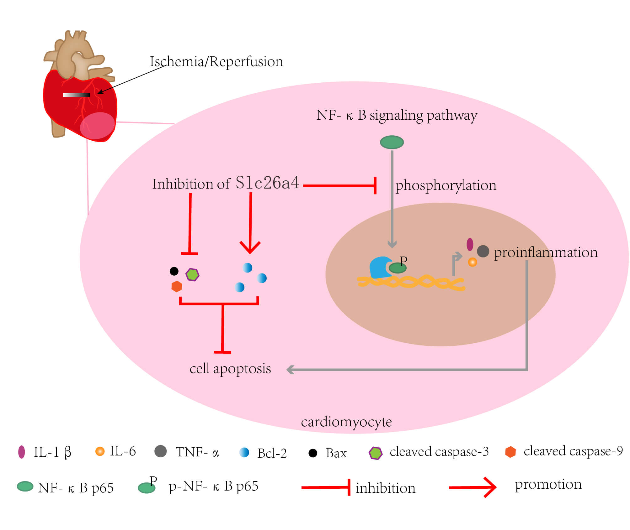
Inhibition of SLC26A4 regulated by electroacupuncture suppresses the progression of myocardial ischemia-reperfusion injury
1 Department of Endocrinology, Heilongjiang University of Chinese Medicine, Harbin, 150040, China
2 Department of Cardiology, Heilongjiang University of Chinese Medicine, Harbin, 150040, China
3 College of Clinical Medicine, Heilongjiang University of Chinese Medicine, Harbin, 150040, China
4 Department of Nephrology, Beijing Hospital of Integrated Traditional Chinese and Western Medicine, Beijing, 100039, China
5 College of Acupuncture and Massage, Heilongjiang University of Chinese Medicine, Harbin, 150040, China
6 Department of Acupuncture, Heilongjiang University of Chinese Medicine, Harbin, 150040, China
* Corresponding Author: YING KONG. Email:
(This article belongs to the Special Issue: Advances in Biomarker Research: Unveiling the Pathways to Precision Medicine)
BIOCELL 2024, 48(4), 665-675. https://doi.org/10.32604/biocell.2024.046342
Received 27 September 2023; Accepted 05 December 2023; Issue published 09 April 2024
 View Full Text
 Download PDF
Abstract
Introduction: Myocardial ischemia-reperfusion (IR) injury has received widespread attention due to its damaging effects. Electroacupuncture (EA) pretreatment has preventive effects on myocardial IR injury. SLC26A4 is a Na+ independent anion reverse transporter and has not been reported in myocardial IR injury. Objectives: To find potential genes that may be regulated by EA and explore the role of this gene in myocardial IR injury. Methods: RNA sequencing and bioinformatics analysis were performed to obtain the differentially expressed genes in the myocardial tissue of IR rats with EA pretreatment. Myocardial infarction size was detected by TTC staining. Serum CK, creatinine kinase-myocardial band, Cardiac troponin I, and lactate dehydrogenase levels were determined by ELISA. The effect of SLC26A4 on cardiomyocyte apoptosis was explored by TUNEL staining and western blotting. The effects of SLC26A4 on inflammation were determined by HE staining, ELISA, and real-time PCR. The effect of SLC26A4 on the NF-κB pathway was determined by western blotting. Results: SLC26A4 was up-regulated in IR rats but downregulated in IR rats with EA pretreatment. Compared with IR rats, those with SLC26A4 knockdown exhibited improved cardiac function according to decreased myocardial infarction size, reduced serum LDH/CK/CK-MB/cTnI levels, and elevated left ventricular ejection fraction and fractional shortening. SLC26A4 silencing inhibited myocardial inflammation, cell apoptosis, phosphorylation, and nuclear translocation of NF-κB p65. Conclusion: SLC26A4 exhibited promoting effects on myocardial IR injury, while the SLC26A4 knockdown had an inhibitory effect on the NF-κB pathway. These results further unveil the role of SLC26A4 in IR injury.Graphic Abstract
Keywords
Supplementary Material
Supplementary Material FileCite This Article
 Copyright © 2024 The Author(s). Published by Tech Science Press.
This work is licensed under a Creative Commons Attribution 4.0 International License , which permits unrestricted use, distribution, and reproduction in any medium, provided the original work is properly cited.
 Downloads
 Citation Tools