
 Login
Login Register
 Submit a Paper
 Propose a Special lssue
 Open Access
ARTICLE
Isorhynchophylline Suppresses Eicosanoid Production via miR-200a-Mediated Inhibition of the FOXC1/NF-κB Axis in Inflammatory Macrophages
1 Department of Respiratory Medicine, Hubei Provincial Hospital of Traditional Chinese Medicine, Wuhan, China
2 Department of Endocrinology, Hubei Provincial Hospital of Traditional Chinese Medicine, Wuhan, China
* Corresponding Author: Jinyue Zhu. Email:
# These authors contributed equally to this work
BIOCELL 2026, 50(10), 11 https://doi.org/10.32604/biocell.2026.082951
Received 26 March 2026; Accepted 04 June 2026; Issue published 22 September 2026
 View Full Text
 Download PDF
Abstract
Background: Macrophage-derived eicosanoids are involved in airway inflammation associated with asthma, but the mechanisms regulating their production remain incompletely understood. The study aimed to evaluate the effects of isorhynchophylline (IRN) on eicosanoid production and inflammatory activation in LPS/IFN-γ-stimulated THP-1-derived macrophages and to further investigate whether these effects are mediated through the miR-200a/FOXC1/NF-κB axis. Methods: THP-1 cells were differentiated into M0 (PMA) and polarized into M1 (LPS+IFN-γ) phenotypes. IRN (5–20 μM) was applied. Eicosanoids (PGE2, LTB4) were measured via ELISA; COX-2, 5-LOX, mPGES-1, FOXC1, p-IκBα/IκBα, and p-p65/p65 were measured via Western blotting; and miR-200a and target genes were measured via RT-qPCR. Polarization markers (CD86/CD206, TNF-α/IL-10, and iNOS/Arg1) were assessed. Results: IRN dose-dependently reduced PGE2 and LTB4 release and downregulated COX-2, 5-LOX, and mPGES-1 expression in LPS/IFN-γ-stimulated macrophages (p < 0.05). IRN increased miR-200a expression, suppressed FOXC1 expression, and attenuated NF-κB activation, as indicated by reduced p-IκBα/IκBα and p-p65/p65 levels and decreased nuclear p65 accumulation (p < 0.01). IRN also reduced M1-associated markers, including CD86, iNOS, and TNF-α, while increasing M2-associated markers, including CD206, Arg1, and IL-10 (p < 0.01). miR-200a knockdown weakened the inhibitory effects of IRN on FOXC1/NF-κB signaling and eicosanoid-producing enzymes, whereas miR-200a overexpression reproduced these effects (p < 0.001). Dual-luciferase assays confirmed that miR-200a directly bound to the FOXC1 3′UTR. FOXC1 overexpression or NF-κB activation partially reversed the suppressive effects of IRN, whereas NF-κB inhibition mimicked the IRN-induced phenotype (p < 0.01). Conclusions: IRN suppresses eicosanoid production and inflammatory activation in LPS/IFN-γ-stimulated THP-1-derived macrophages, partly through the miR-200a/FOXC1/NF-κB axis. These findings provide cell-based mechanistic evidence that requires validation in primary airway macrophages and disease-specific models.Keywords
Bronchial asthma is a heterogeneous chronic airway disease characterized by persistent inflammation, airway hyperresponsiveness, and remodeling. It affects more than 300 million people worldwide [1,2]. Standard therapies such as inhaled corticosteroids (ICSs), β2-agonists, and leukotriene receptor antagonists can control symptoms in many patients. However, a subset (~10%) of patients with asthma exhibit poor responsiveness to high-dose steroids [2], leading to uncontrolled severe asthma with frequent exacerbations. Moreover, long-term steroid use and other current treatments can cause significant side effects, and relapses remain common. These clinical challenges highlight the need for new therapeutic strategies that go beyond symptomatic bronchodilation or broad immunosuppression.
Dysregulated arachidonic acid (AA) metabolism is an important component of airway inflammation and has been implicated in asthma pathophysiology [3]. AA is enzymatically converted by the cyclooxygenase (COX) and lipoxygenase (5-LOX) pathways into proinflammatory eicosanoids. Notably, prostaglandin E2 (PGE2) and leukotrienes (e.g., LTB4, LTC4, LTD4) are strongly implicated in asthma pathogenesis [4,5]. These AA-derived eicosanoids participate in airway inflammation: LTB4 is a potent chemoattractant for neutrophils and eosinophils [6,7,8], whereas cysteinyl leukotrienes (LTC4/D4/E4) induce bronchoconstriction, vascular leakage (airway edema), and mucus hypersecretion [9]. Although some antileukotriene drugs (e.g., montelukast, a CysLT1 receptor antagonist) and a 5-LOX inhibitor (zileuton) have been developed to target this pathway [10,11], these agents block downstream effects rather than correct upstream enzymatic overactivity. Therefore, investigating how these enzymes and eicosanoid mediators are regulated may help clarify macrophage-related inflammatory mechanisms relevant to airway inflammation.
Macrophages are important contributors to airway inflammation and can serves as sources of eicosanoid mediators in asthma. In the asthmatic airway microenvironment, macrophages may acquire M1-like features, including increased expression of inflammatory enzymes such as COX-2 and 5-LOX and enhanced production of proinflammatory AA-derived mediators [12,13]. For example, exposure to stimuli such as LPS (a bacterial endotoxin) triggers nuclear factor kappa-B (NF-κB)-dependent induction of COX-2 in macrophages, leading to excessive PGE2 release [14]. Similarly, 5-LOX activation in M1 macrophages drives LTB4 synthesis, propagating a vicious cycle of inflammation [15]. In contrast, alternatively activated M2 macrophages produce anti-inflammatory lipid mediators (e.g., lipoxins) that help resolve inflammation [16]. The molecular mechanisms that govern this imbalance in macrophage eicosanoid-related pathways during airway inflammation remain incompletely understood. In particular, the potential epigenetic regulation of COX-2/5-LOX expression by microRNAs, and its crosstalk with key inflammatory signaling pathways (such as NF-κB) have not been fully elucidated.
Recent evidence suggests that microRNAs can serve as critical modulators of inflammatory networks in asthma [17]. Among these, microRNA-200a (miR-200a) has emerged as a regulatory miRNA of interest. Our team’s previous work in a mouse asthma model identified miR-200a as a mediator of the anti-asthmatic effects of isorhynchophylline (IRN) [18]. IRN is a natural indole alkaloid derived from Uncaria rhynchophylla, a medicinal herb. In OVA-sensitized asthmatic mice, IRN treatment significantly attenuated airway inflammation, reducing eosinophil infiltration in bronchoalveolar fluid, collagen deposition in lung tissue, and the levels of IgE and Th2 cytokines [18]. IRN also inhibited the pathological proliferation of airway smooth muscle cells (ASMCs) in vitro. Mechanistic exploration revealed that IRN upregulated miR-200a in airway tissues, which in turn suppressed the transcription factor forkhead box C1 (FOXC1) and downstream NF-κB signaling [18]. Notably, blocking miR-200a abrogated the beneficial effects of IRN, indicating that this miRNA is a key upstream effector of IRN action [18].
FOXC1 is a transcription factor not classically studied in asthma; however, emerging data suggest that it may modulate inflammatory pathways. For example, in osteoarthritic synoviocytes, IL-1β exposure was shown to downregulate miR-200a-3p, leading to increased FOXC1 expression and increased production of inflammatory cytokines and enzymes [19]. This finding suggests a general model in which inflammatory stimuli can relieve the repression of FOXC1 by miR-200a, unleashing the proinflammatory effects of FOXC1, potentially via NF-κB or other signaling pathways. We hypothesize that a similar axis may operate in inflammatory macrophages, and contribute to the regulation of NF-κB activation and eicosanoid-producing enzymes.
In the present study, we used LPS/IFN-γ-stimulated THP-1-derived macrophages as an inflammatory macrophage model to investigate whether IRN regulates eicosanoid production through the miR-200a-/FOXC1/NF-κB pathway in macrophages, thereby rebalancing the level of eicosanoid-producing enzymes and mediators. We addressed two key questions: (1) Does IRN suppress M1 macrophage activation and eicosanoid production through the upregulation of miR-200a? (2) Does miR-200a directly target FOXC1 to modulate NF-κB signaling and downstream inflammatory enzymes? To answer these questions, we examined the effects of IRN on COX-2/5-LOX-dependent eicosanoid production, macrophage inflammatory marker expression, miR-200a expression, FOXC1/NF-κB signaling, and p65 nuclear translocation. Gain- and loss-of-function experiments involving miR-200a, FOXC1 overexpression, and pharmacological modulation of NF-κB were further performed to clarify the mechanistic relationship among these factors. Although this study does not directly model asthma and relies on a THP-1-derived macrophage system, it provides mechanistic evidence for further validation in primary airway macrophages and asthma-specific experimental models.
Fetal bovine serum (Cat. No. FSD500) was purchased from Excel Bio (Shanghai, China). High-glucose DMEM (Cat. No. C11995500BT) and RPMI-1640 medium (Cat. No. 11875093) were obtained from Gibco (Waltham, MA, USA). RIPA lysis buffer (Cat. No. P0013B), PMSF (Cat. No. ST728), APS (Cat. No. ST005), TEMED (Cat. No. ST728), penicillin-streptomycin solution (100×, Cat. No. C0222), and PBS (Cat. No. C0221A) were purchased from Beyotime Biotechnology (Shanghai, China). Acrylamide/bis solution (30%, Cat. No. BL513A), Tris-Base (Cat. No. BS083), TBS powder (Cat. No. BL602A), BSA protein standard (Cat. No. BL673A), and Tween-20 (Cat. No. BS100) were obtained from Biosharp (Hefei, China). SDS (Cat. No. 3250), glycine (Cat. No. 1275), and skim milk (Cat. No. 1172) were purchased from BioFroxx (Einhausen, Germany). A BCA protein assay kit (Cat. No. WB6501), 5× SDS sample buffer (Cat. No. WB2001), and ECL reagents (Cat. No. P2100) were purchased from NCM Biotech (Suzhou, China). Methanol (Cat. No. HB05) was obtained from Guangzhou Chemical (Guangzhou, China). Antibodies against COX-2 (Cat. No. 27308-1-AP), 5-LOX (Cat. No. 10021-1-Ig), FOXC1 (Cat. No. 55365-1-AP), IκBα (Cat. No. 10268-1-AP), p-IκB (Cat. No. 68999-1-Ig), p65 (Cat. No. 10745-1-AP), p-p65 (Cat. No. 82335-1-RR), GAPDH (Cat. No. 60004-1-Ig), Lamin B1 (Cat. No. 12987-1-AP), CD86 (Cat. No. APC-FcA98043), CD206 (Cat. No. APC-FcA98031), HRP-conjugated goat anti-rabbit IgG (Cat. No. SA00001-2), and goat anti-mouse IgG (Cat. No. SA00001-1) were purchased from Proteintech (Wuhan, China). An anti-mPGES-1 antibody (Cat. No. ab180589) was obtained from Abcam (Cambridge, UK). ELISA kits for PGE2 (Cat. No. SEKSM-0034), IL-10 (Cat. No. SEKH-0018), and TNF-α (Cat. No. SEKH-0047) were purchased from Solarbio (Beijing, China), and the LTB4 ELISA kit (Cat. No. ml027372) was obtained from mlbio (Shanghai, China). Isorhynchophylline (IRN, Cat. No. HY-N0766), BAY 11-7082 (Cat. No. HY-13453), and NF-κB activator 1 (Cat. No. HY-134476) were purchased from MedChemExpress (Monmouth Junction, NJ, USA). TRIzol (Cat. No. 15596026CN) and Lipofectamine 3000 Transfection Reagent (Cat. No. L3000015) were purchased from Invitrogen (Thermo Fisher Scientific, Waltham, MA, USA). Nuclear and Cytoplasmic Protein Extraction Kit (Cat. No. P0028) was purchased from Beyotime (Shanghai, China).
2.2 Cell Culture and Experimental Design
THP-1 human monocytic cells (CL-0233) and HEK293 cells (CL-0001) were obtained from Procell (Wuhan, China). The cell lines were authenticated by short tandem repeat (STR) profiling and confirmed to be free from mycoplasma contamination. THP-1 cells were cultured in RPMI-1640 supplemented with 10% FBS and 100 U/mL penicillin + 100 μg/mL streptomycin at 37°C and 5% CO2. At ~90% confluence, the cells were collected (1000 rpm, 5 min; 4-20R refrigerated high/low-speed universal centrifuge, Hunan Hengnuo Instrument Equipment Co., Ltd., Changsha, China), resuspended in complete medium, and passaged at a 1:3 ratio. HEK293 cells were maintained in DMEM (high glucose) with 10% FBS and antibiotics under the same conditions; when ~90% confluent, the cells were rinsed twice with 0.01 M PBS (pH 7.4), digested with 0.25% trypsin-EDTA (~30 s), neutralized with complete medium, collected (1000 rpm, 5 min), and passaged at a 1:5 ratio.
For M0/M1 modeling, THP-1 monocytes were differentiated into M0 macrophages using PMA (100 nM, 48 h; HY-18739, MedChemExpress, Monmouth Junction, NJ, USA) followed by culture for 24 h in PMA-free medium. M1 polarization was induced by stimulation with LPS (100 ng/mL, HY-D1056, MedChemExpress, Monmouth Junction, NJ, USA) and IFN-γ (20 ng/mL, HY-P7025, MedChemExpress, Monmouth Junction, NJ, USA) for 24 h. The experimental design consisted of four parts: Part I (IRN dose-response): M0 (untreated), M1 (model), M1+IRN-L (5 μM), M1+IRN-M (10 μM), and M1+IRN-H (20 μM) (IRN added after M1 induction). Part II (role of miR-200a): M1, M1+IRN-H (20 μM), M1+IRN-H+KD-miR-200a, M1+IRN-H+KD-NC, M1+OE-miR-200a, and M1+OE-NC. Part III (miR-200a-FOXC1 binding): dual-luciferase assay in HEK293 cells (see Section 2.7). Part IV (FOXC1/NF-κB axis): M1, M1+IRN-H, M1+IRN-H+OE-FOXC1, M1+OE-FOXC1, M1+IRN-H+NF-κB-A (NF-κB activator 1, 1 μM), M1+NF-κB-A (1 μM), and M1+NF-κB-I (BAY 11-7082, 1 μM; HY-13453, MedChemExpress, Monmouth Junction, NJ, USA).
For miR-200a gain- and loss-of-function experiments, THP-1-derived M1 macrophages were transfected with miR-200a mimic or miR-200a inhibitor, respectively, with the corresponding mimic negative control or inhibitor negative control used as controls. For FOXC1 overexpression, cells were transfected with a FOXC1 overexpression plasmid or the corresponding empty vector control. Briefly, THP-1-derived M1 macrophages were seeded in 6-well plates at approximately 5 × 10⁵ cells/well in 2 mL complete medium. miR-200a mimic, miR-200a inhibitor, or the corresponding negative controls were transfected at a final concentration of 50 nM, whereas FOXC1 overexpression plasmid or empty vector was transfected using the indicated plasmid amount. Transfections were performed using Lipofectamine 3000 Transfection Reagent (5 μL/well) according to the manufacturer’s instructions. For each well, oligonucleotides or plasmid DNA and Lipofectamine 3000 were separately diluted in Opti-MEM reduced-serum medium, gently mixed, and incubated for 15 min at room temperature to allow complex formation. The transfection complexes were then added dropwise to the cells. After 6 h, the medium was replaced with fresh complete medium. For the IRN-combination groups, cells were treated with IRN-H (20 μM) after transfection. Cells were harvested 24 h after transfection for subsequent analyses. To modulate NF-κB signaling, M1 macrophages were treated with NF-κB activator 1 (1 μM) or the NF-κB inhibitor BAY 11-7082 (1 μM). Cells were collected after treatment for further analysis.
2.3 Enzyme-Linked Immunosorbent Assay (ELISA)
The cell-free supernatants were collected and clarified (1000× g, 10 min). PGE2, LTB4, IL-10, and TNF-α were quantified with commercial ELISA kits strictly following the manufacturers’ instructions. The absorbance at 450 nm was read on a Multiskan MK3 microplate reader (Thermo Fisher Scientific, Waltham, MA, USA). Standard curves were generated using serially diluted standards provided in each kit, and the concentrations of target analytes were calculated from the corresponding standard curves. Each sample was measured in duplicate.
The cells were lysed in ice-cold RIPA buffer (containing protease/phosphatase inhibitors). After clarification and BCA quantification, equal amounts of protein (25 μg) were resolved via SDS-PAGE and transferred to PVDF membranes. The membranes were blocked (5% skim milk/TBST), incubated with primary antibodies (4°C, overnight) and HRP-conjugated secondary antibodies (RT, 1 h), developed via enhanced chemiluminescence (ECL), and analyzed via ImageJ (version 1.5; National Institutes of Health, Bethesda, MD, USA). The targets included COX-2 (1:1000), 5-LOX (1:1000), mPGES-1 (1:1000), FOXC1 (1:1000), IκBα (1:5000), p-IκB (1:2000), p65 (1:1000), and p-p65 (1:2000); GAPDH (1: 50,000) and Lamin B1 (1:5000) served as loading controls for the total and nuclear fractions, respectively.
2.5 Reverse Transcription Quantitative Polymerase Chain Reaction (RT-qPCR)
Total RNA was isolated with TRIzol. RNA concentration and purity were assessed by measuring the absorbance at 260 and 280 nm, and samples with an A260/A280 ratio of 1.8–2.1 were used for subsequent reverse transcription. For mRNA detection, cDNA was synthesized using HiScript III RT SuperMix (Cat. No. R323-01; Vazyme Biotech Co., Ltd., Nanjing, China) with random primers. The reverse transcription program was as follows: 37°C for 15 min and 85°C for 5 s. For miRNA detection, reverse transcription was performed using stem-loop primers specific for miR-200a and U6. The RT-qPCR was performed using SYBR Green on a CFX96 Touch Real-Time PCR Detection system (Bio-Rad Laboratories, Hercules, CA, USA). The RT-qPCR cycling conditions were as follows: initial denaturation at 95°C for 30 s, followed by 40 cycles of 95°C for 10 s and 60°C for 30 s. Melt curve analysis was performed at the end of each run to confirm amplification specificity. No-template controls were included in each experiment to monitor potential contamination. GAPDH (mRNA) and U6 (miRNA) were used as internal controls. Relative expression was calculated via the 2−ΔΔCt method. The primer sequences for FOXC1, COX-2, 5-LOX, iNOS, Arg-1, GAPDH, miR-200a and U6 are provided in Table A1.
2.6 Flow Cytometric Analysis of Macrophage Polarization
After the indicated treatments described in Section 2.2, THP-1-derived macrophages were harvested, washed twice with 0.01 M PBS (pH 7.4), and adjusted to approximately 1 × 10⁶ cells/mL. To reduce nonspecific antibody binding, cells were incubated with a human Fc receptor blocking reagent for 10 min at room temperature before antibody staining. Cell viability was assessed using 7-AAD viability staining, and dead cells were excluded from the analysis. Aliquots (≈50 μL) were stained with fluorophore-conjugated anti-CD86 and anti-CD206 antibodies (10 μL each) for 60 min at room temperature in the dark, washed, and resuspended in 0.01 M PBS (pH 7.4) for acquisition on a BD FACSCalibur™ flow cytometer (BD Biosciences, San Jose, CA, USA). Single-stained controls were used for compensation, and fluorescence-minus-one controls and isotype controls were used to establish gating thresholds. The percentages of CD86+ (M1) and CD206+ (M2) cells were quantified.
2.7 Dual-Luciferase Reporter Assay
The human FOXC1 3′UTR containing the predicted miR-200a binding site (~±500 bp) was cloned and inserted into psiCHECK2.0 (WT); a seed-mutant construct (MUT) was generated via site-directed mutagenesis. HEK293 cells were seeded in 96-well plates (1.5 × 10⁴ cells/well) and cultured in 100 μL complete medium per well. Cells were cotransfected with 300 ng of the WT or MUT FOXC1 3′UTR reporter plasmid and 50 nM of miR-200a mimic or negative control via 0.3 μL Lipofectamine 3000 Transfection Reagent per well, according to the manufacturer’s instructions. At 48 h after transfection, firefly and Renilla luciferase activities were measured sequentially. Relative luciferase activity was calculated by normalizing Renilla luciferase activity to firefly luciferase activity.
2.8 Nuclear/Cytoplasmic Fractionation
Cytosolic and nuclear proteins were extracted via a stepwise fractionation kit following the manufacturer’s protocol. Briefly, the cells were washed with cold 0.01 M PBS (pH 7.4). Approximately 20 μL of cell pellet was resuspended in 200 μL cytoplasmic protein extraction reagent A containing 1 mM PMSF, vortexed for 5 s, and incubated on ice for 10 min. Cytoplasmic protein extraction reagent B (10 μL) was then added, followed by vortexing for 5 s, incubation on ice for 1 min, vortexing again for 5 s, and centrifugation at 12,000× g for 5 min at 4°C. The supernatant was collected as the cytoplasmic fraction. The nuclear pellet was resuspended in 50 μL nuclear protein extraction reagent containing 1 mM PMSF, vortexed for 15 s every 2 min during a 30-min incubation on ice, and centrifuged at 12,000× g for 10 min at 4°C. The supernatant was collected as the nuclear fraction. The cytoplasmic and nuclear fractions were mixed with 1× SDS loading buffer and heated at 95°C for 5 min before Western blot analysis of p65 distribution. GAPDH was used as the loading control for the cytoplasmic fraction, and Lamin B1 was used as the loading control for the nuclear fraction.
The data are expressed as the means ± SDs from ≥3 independent experiments. Before parametric testing, data normality was assessed using the Shapiro-Wilk test, and homogeneity of variance was assessed using Levene’s test. For two-group comparisons, unpaired Student’s t tests were used. For comparisons among multiple predefined treatment groups, one-way ANOVA followed by Tukey’s post hoc test was used. For the dual-luciferase reporter assay, which had a complete 2 × 2 factorial structure with reporter construct and miRNA treatment as the two factors, two-way ANOVA followed by Tukey’s multiple-comparison test was used. p < 0.05 was considered statistically significant. Analyses and plots were generated with GraphPad Prism 9.5.1 (GraphPad Software, Boston, MA, USA); densitometry was performed in ImageJ (version 1.5; National Institutes of Health, Bethesda, MD, USA); figures were arranged in Adobe Illustrator 2023 (Adobe Inc., San Jose, CA, USA).
3.1 IRN Suppresses Eicosanoid Production and M1-Like Inflammatory Polarization Activation in THP-1-Derived Macrophages
To investigate whether IRN regulates eicosanoid production in inflammatory macrophages, THP-1-derived M1-like macrophages were treated with increasing concentrations of IRN. Compared with M0 cells, M1 polarization markedly elevated the secretion of PGE2 and LTB4, whereas IRN reduced the secretion of both metabolites in a dose-dependent manner, with the most pronounced suppression occurring at 20 μM IRN (p < 0.001) (Fig. 1A). Consistent with these findings, Western blot analysis revealed that COX-2, 5-LOX, and mPGES-1 protein levels were strongly induced in THP-1-derived M1 macrophages, and IRN treatment led to the progressive downregulation of all three enzymes in a dose-dependent manner (p < 0.05) (Fig. 1B).
Given that miR-200a is predicted to regulate FOXC1 and COX-2/mPGES-1- and 5-LOX-related responses, we next examined its expression. RT-qPCR demonstrated that miR-200a was markedly downregulated in THP-1-derived M1 macrophages but restored by IRN treatment in a dose-dependent manner (p < 0.01) (Fig. 1C). In contrast, FOXC1, COX-2, and 5-LOX mRNA levels were significantly elevated in THP-1-derived M1 macrophages (p < 0.001) but reduced by IRN treatment in a dose-dependent manner (p < 0.01) (Fig. 1C).
Moreover, IRN reduced M1-associated markers and increased M2-associated markers, as evidenced by decreased CD86 expression, TNF-α secretion, and iNOS mRNA levels, along with increased CD206 expression, IL-10 secretion, and Arg-1 mRNA levels (p < 0.01), suggesting a partial shift toward a less inflammatory macrophage phenotype (Fig. 1D–F). These results suggest that IRN suppresses eicosanoid-producing enzyme expression and M1-associated inflammatory markers in LPS/IFN-γ-stimulated THP-1-derived macrophages, potentially through miR-200a upregulation and FOXC1 inhibition.
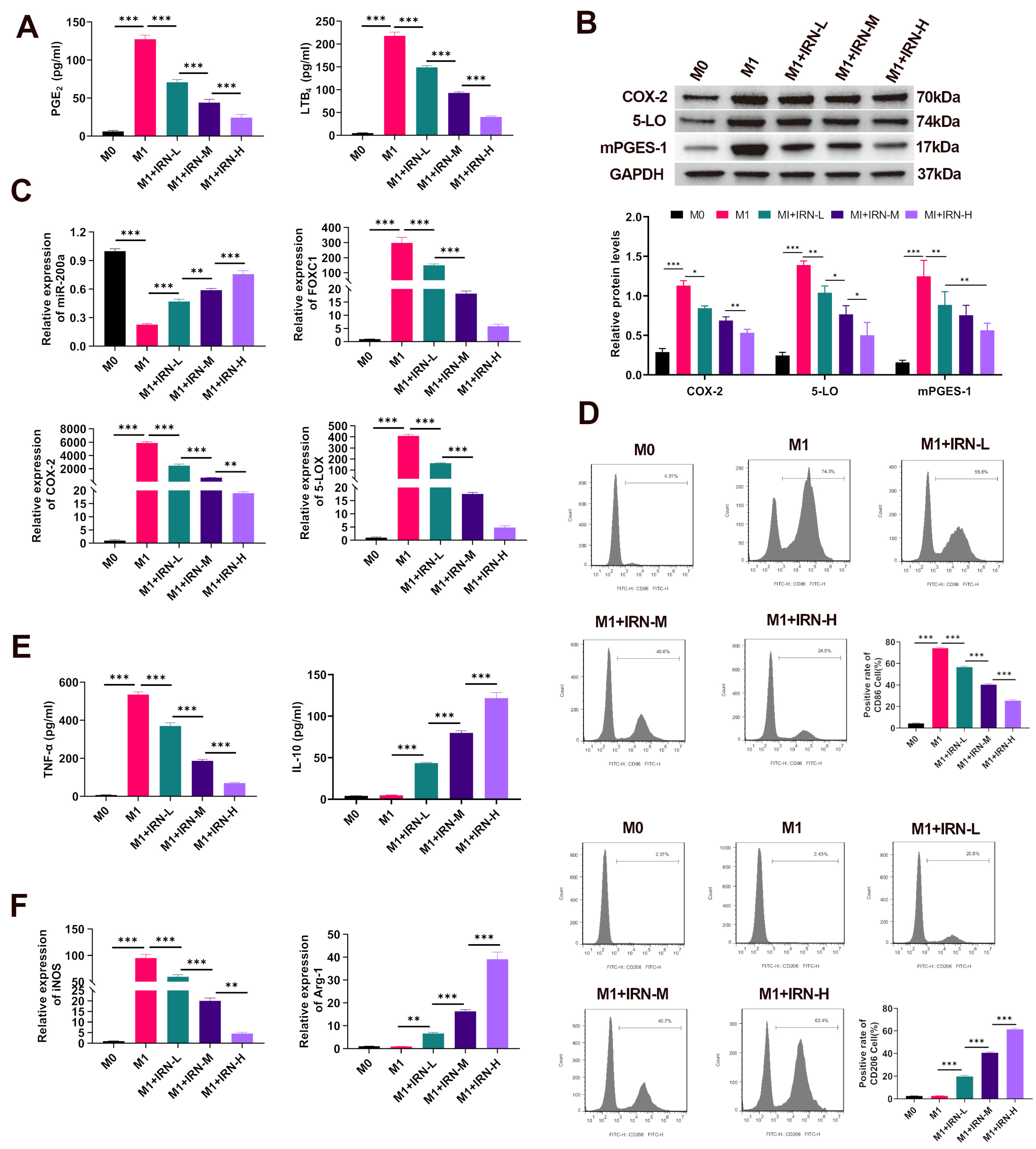
Figure 1: Effects of IRN on eicosanoid production and inflammatory marker expression in LPS/IFN-γ-stimulated THP-1-derived macrophages. (A) ELISA detection of PGE2 and LTB4 levels in cell culture supernatants. (B) Western blot analysis of COX-2, 5-LOX, and mPGES-1 protein expression in THP-1-derived macrophages. (C) RT-qPCR measurement of the miR-200a, FOXC1, COX-2, and 5-LOX mRNA levels. (D) Flow cytometry analysis of the M1 marker CD86 and the M2 marker CD206. (E) ELISA quantification of TNF-α (M1 marker) and IL-10 (M2 marker) in the cell culture supernatants. (F) RT-qPCR analysis of iNOS (M1 marker) and Arg-1 (M2 marker) mRNA expression. *p < 0.05, **p < 0.01, ***p < 0.001. n = 3.
3.2 miR-200a Contributes to the Inhibitory Effects of IRN on Eicosanoid-Producing Enzymes and FOXC1/NF-κB Signaling
To confirm the role of miR-200a in IRN-mediated anti-inflammatory activity, miR-200a was silenced or overexpressed in THP-1-derived M1 macrophages with or without IRN treatment. RT-qPCR confirmed effective overexpression and knockdown, with miR-200a levels highest in the overexpression group and lowest in the knockdown group (p < 0.01) (Fig. 2A).
The ELISA results revealed a similar trend, with IRN and miR-200a overexpression reducing PGE2 and LTB4 secretion, whereas miR-200a knockdown reversed this suppression (p < 0.001) (Fig. 2B). These findings indicate that miR-200a contributes to the IRN-induced suppression of eicosanoid-producing enzymes and NF-κB activation via FOXC1 downregulation.
Western blot analysis revealed that IRN markedly decreased the COX-2, 5-LOX, mPGES-1, FOXC1, p-IκBα/IκBα, and p-p65/p65 ratios compared with those in untreated THP-1-derived M1 macrophages, but these effects were abolished by miR-200a knockdown. In contrast, miR-200a overexpression mimicked the effects of IRN and further reduced the levels of these proteins when miR-200a was combined with IRN (p < 0.001) (Fig. 2C).
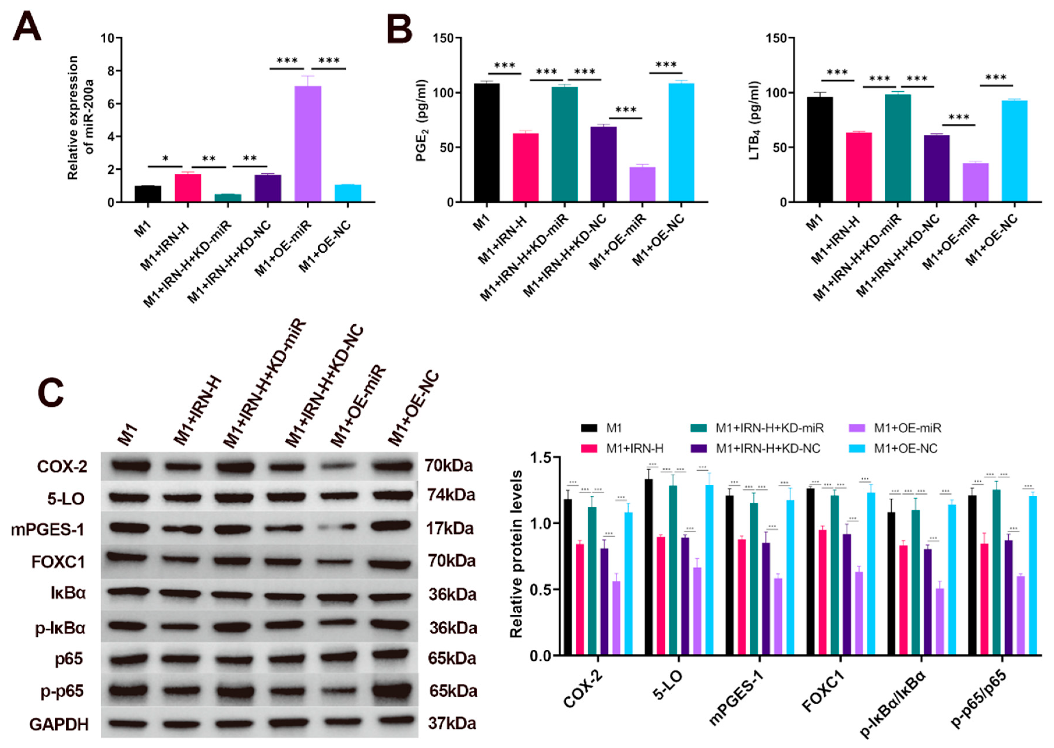
Figure 2: Role of miR-200a in the IRN-mediated suppression of eicosanoid-producing enzymes and FOXC1/NF-κB signaling. (A) RT-qPCR detection of miR-200a expression levels in THP-1-derived M1 macrophages following overexpression or knockdown with or without IRN treatment. (B) ELISA measurement of PGE2 and LTB4 levels in culture supernatants. (C) Western blot analysis of COX-2, 5-LOX, mPGES-1, FOXC1, p-IκBα/IκBα, and p-p65/p65 protein expression levels. *p < 0.05, **p < 0.01, ***p < 0.001. n = 3.
3.3 miR-200a Directly Targets FOXC1 by Binding to Its 3′UTR
To verify whether FOXC1 is a direct downstream target of miR-200a, a dual-luciferase reporter assay was performed in HEK293 cells. Cotransfection of miR-200a mimics with the wild-type FOXC1 3′UTR significantly reduced luciferase activity by approximately 50% compared with that of the negative control (p < 0.001), whereas mutation of the predicted binding site abolished this suppression (Fig. 3). These data support FOXC1 as a direct target of miR-200a in this reporter system.
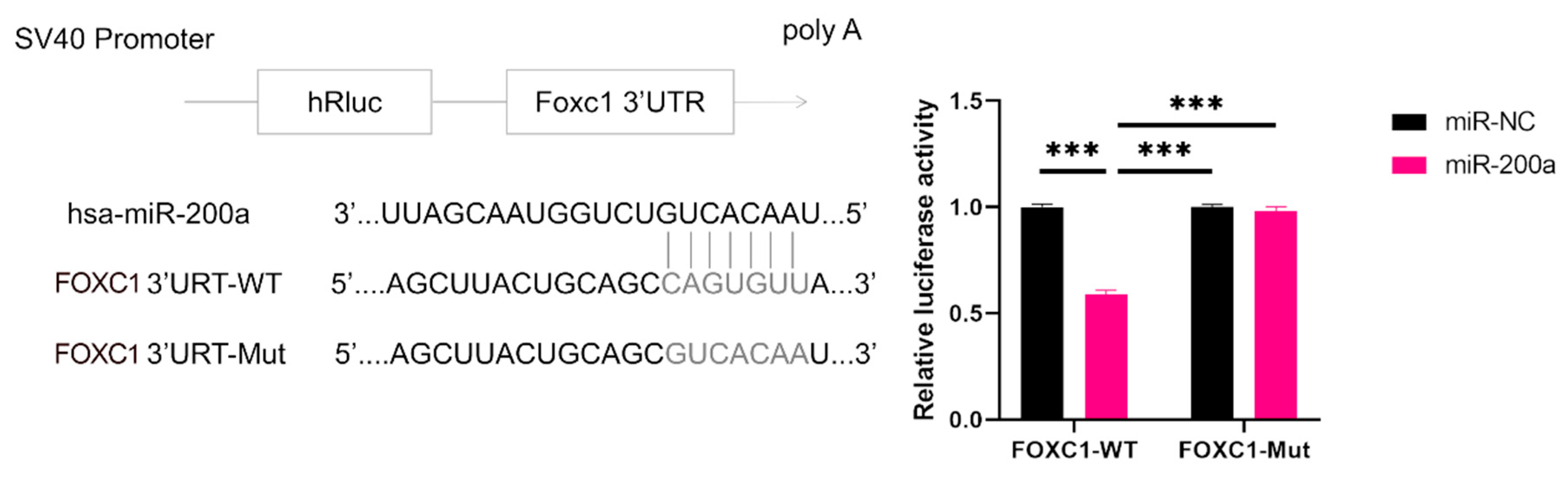
Figure 3: Validation of FOXC1 as a direct target of miR-200a via a dual-luciferase reporter assay. Luciferase reporter assay in HEK293 cells cotransfected with wild-type FOXC1 3′UTR (FOXC1-WT) or mutant FOXC1 3′UTR (FOXC1-Mut) constructs together with miR-200a mimics or a negative control (miR-NC). Relative luciferase activity was measured to evaluate the direct binding of miR-200a to the FOXC1 3′UTR. ***p < 0.001. n = 3.
3.4 FOXC1/NF-κB Signaling Contributes to the Suppressive Effects of IRN in THP-1-Derived Inflammatory Macrophage
To further confirm the involvement of FOXC1/NF-κB signaling in the effects of IRN, FOXC1 was overexpressed, or NF-κB signaling was modulated in THP-1-derived M1 macrophages. Compared with IRN treatment, FOXC1 overexpression or NF-κB activation significantly increased the COX-2, 5-LOX, mPGES-1, p-IκBα/IκBα, and p-p65/p65 ratios, whereas NF-κB inhibition produced effects similar to those of IRN treatment (p < 0.05) (Fig. 4A).
ELISA revealed corresponding increases in PGE2 and LTB4 levels upon FOXC1 overexpression or NF-κB activation and decreases upon NF-κB inhibition (p < 0.01) (Fig. 4B). Furthermore, nuclear fractionation assays revealed that FOXC1 overexpression and NF-κB activation promoted p65 nuclear translocation, which was suppressed by IRN and NF-κB inhibition (p < 0.01) (Fig. 4C). Collectively, these results indicate that miR-200a/FOXC1/NF-κB axis contributes to the suppression of eicosanoid production and inflammatory activation by IRN in LPS/IFN-γ-stimulated THP-1-derived macrophages.
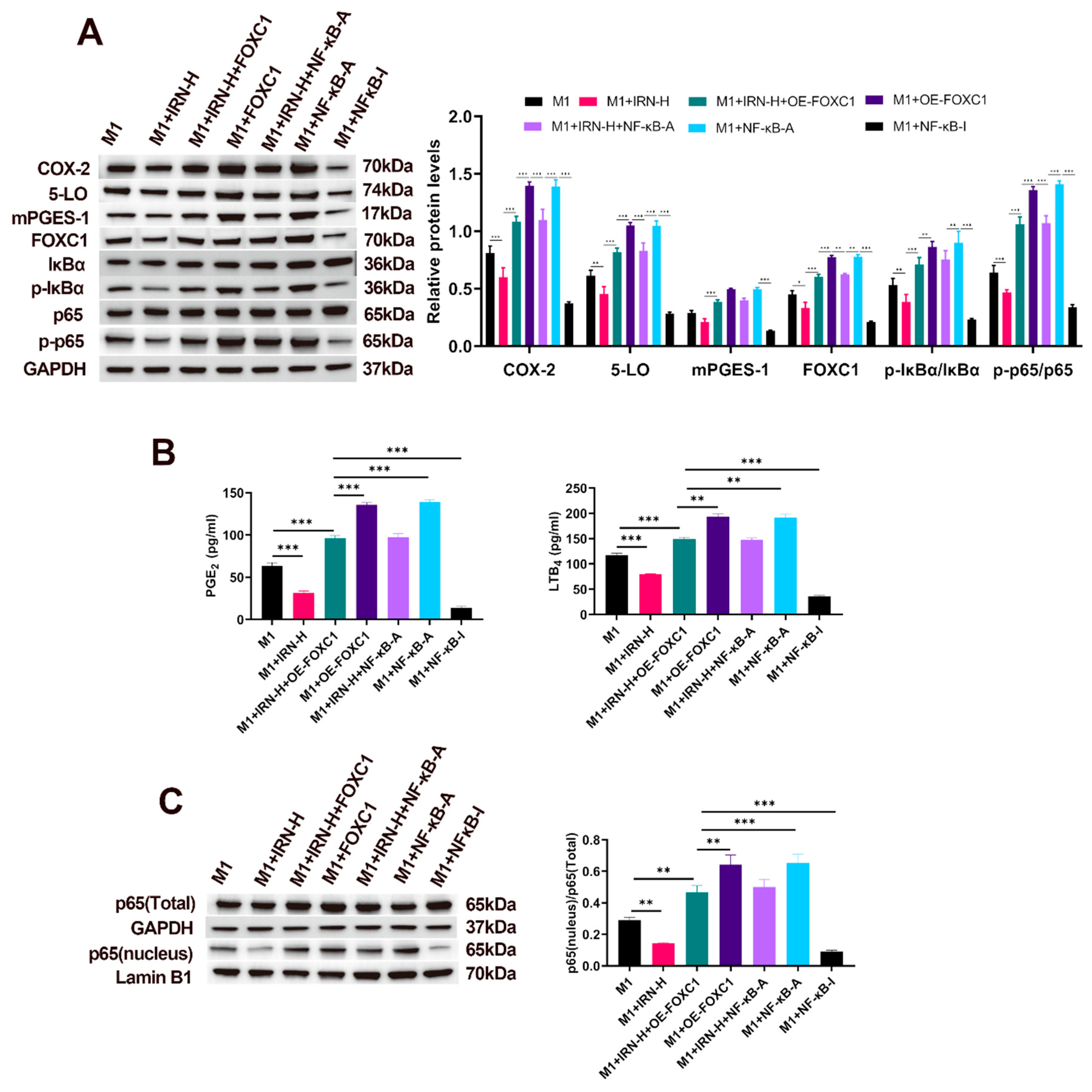
Figure 4: FOXC1/NF-κB signaling contributes to IRN-mediated suppression of eicosanoid production in THP-1-derived macrophages. (A) Western blot detection of AA metabolism-related enzymes (COX-2, 5-LOX, mPGES-1) and FOXC1/NF-κB pathway proteins (FOXC1, p-IκBα/IκBα, p-p65/p65) in THP-1-derived M1 macrophages following FOXC1 overexpression, NF-κB activation, or NF-κB inhibition, with or without IRN treatment. (B) ELISA measurement of the AA metabolites PGE2 and LTB4 in the culture supernatants of the above groups. (C) Nuclear fractionation Western blot analysis of p65 localization to assess NF-κB nuclear translocation in the indicated groups. *p < 0.05, **p < 0.01, ***p < 0.001. n = 3.
In this study, we identified a miR-200a/FOXC1/NF-κB signaling axis that regulates eicosanoid production and inflammatory activation in LPS/IFN-γ-stimulated THP-1-derived macrophages. IRN increased miR-200a expression, suppressed FOXC1 expression, inhibited NF-κB activation, and reduced the expression of eicosanoid-producing enzymes, including COX-2, 5-LOX, and mPGES-1. These changes were accompanied by reduced PGE2 and LTB4 release and attenuation of M1-associated inflammatory markers. These findings are consistent with our previous in vivo study in asthmatic mice, in which IRN increased miR-200a expression and inhibited NF-κB activity in airway tissues, with a particular focus on ASMC proliferation and miR-200a/FOXC1-related signaling [18]. The present study extends these observations to THP-1-derived inflammatory macrophages and links this pathway to eicosanoid-producing enzyme expression and lipid mediator release. The relationship between ASMCs and macrophages is biologically relevant in airway inflammation. Activated macrophages can release cytokines, chemokines, and lipid mediators that promote ASMC proliferation, migration, and contractile dysfunction, thereby contributing to airway remodeling and hyperresponsiveness. Conversely, ASMCs can secrete inflammatory mediators that recruit and activate macrophages, forming a reciprocal inflammatory crosstalk within the airway wall. Importantly, however, the present study was conducted in an in vitro inflammatory macrophage model and does not directly establish an anti-asthmatic effect. This study might provide mechanistic evidence relevant to macrophage-driven inflammatory processes implicated in airway diseases such as asthma, which requires further validation in asthma-specific systems.
Here, we found that IRN suppressed the inflammatory program of LPS/IFN-γ-stimulated THP-1-derived macrophages. IRN-treated cells produced lower levels of PGE2 and LTB4, indicating reduced activity of COX-2/mPGES-1 and 5-LOX-related eicosanoid pathways. This finding is consistent with a previous study showing that IRN attenuated LPS-induced inflammatory responses in RAW264.7 macrophages and human umbilical vein endothelial cells through inhibition of the NF-κB/NLRP3 signaling pathway, supporting the anti-inflammatory activity of IRN in macrophage-related inflammation [20]. Our study further linked IRN to the suppression of eicosanoid-producing enzymes through the miR-200a/FOXC1/NF-κB axis. Because macrophage-derived lipid mediators are involved in airway inflammation, these findings may have potential relevance to asthma-related inflammatory mechanisms, although they should not be interpreted as direct evidence of asthma alleviation [21,22]. From a pathophysiological perspective, reduced LTB4 production would be expected to limit neutrophil and eosinophil recruitment to the airways [23,24,25]. In addition, the reduction in 5-LOX expression may indicate a broader dampening of leukotriene biosynthesis, although cysteinyl leukotrienes were not directly measured in the present study [26]. IRN also reduced PGE2 production. Although the role of PGE2 in airway inflammation is context-dependent, in our model, this change most likely reflects suppression of the macrophage COX-2/mPGES-1 inflammatory axis. Whether these changes ultimately translate into reduced airway inflammation or improved airway function requires further validation in primary airway macrophages and asthma-specific in vivo models [18]. Also, it should be noted that LTB4 and cysteinyl leukotrienes are generated through related but distinct branches of the 5-LOX pathway and exert different biological effects in airway inflammation. Although reduced 5-LOX expression may suggest that IRN affects leukotriene-associated pathways, the present study measured only LTB4 and did not directly quantify LTC4, LTD4, or LTE4. Therefore, our findings should be interpreted as evidence that IRN regulates selected eicosanoid mediators and enzymes, rather than comprehensive proof of broad AA metabolism regulation.
At the molecular level, IRN reduced the expression of COX-2, 5-LOX, and mPGES-1 in THP-1-derived macrophages, suggesting coordinated suppression of the prostaglandin- and leukotriene-generating branches of AA metabolism. This upstream regulation differs conceptually from receptor-level blockade of individual lipid mediators, such as leukotriene receptor antagonism by montelukast, which blocks leukotriene signaling but does not directly reduce eicosanoid production at the source [10]. Current anti-inflammatory therapies, including glucocorticoids, can broadly suppress inflammation, but steroid insensitivity remains an important challenge in a subset of patients with severe asthma [27]. The AA-derived eicosanoids generated through the COX and 5-LOX pathways, particularly prostaglandins and leukotrienes, are involved in airway inflammation. These mediators contribute to inflammatory cell recruitment, airway hyperresponsiveness, bronchoconstriction, mucus secretion, and vascular leakage, all of which are closely related to the pathogenesis of asthma [28]. Therefore, although the present study does not directly model asthma, the ability of IRN to suppress macrophage-derived eicosanoid-producing enzymes may represent a disease-relevant anti-inflammatory mechanism that warrants further validation in asthma models. In addition to reducing eicosanoid-producing enzymes and inflammatory mediator release, IRN decreased M1-associated markers, including CD86, iNOS, and TNF-α, while increasing M2-associated markers, including CD206, Arg1, and IL-10. These findings suggest that IRN attenuates the M1-like inflammatory state and promotes a partial shift toward a less inflammatory macrophage phenotype. Rather than establishing a complete macrophage phenotype switch or a direct disease-modifying effect, these results indicate that IRN may influence inflammatory activation and selected eicosanoid-related pathways in activated THP-1-derived macrophages.
Mechanistically, our findings suggest that miR-200a regulates the FOXC1/NF-κB axis and thereby influences inflammatory enzyme expression. The dual-luciferase reporter assay confirmed that miR-200a directly binds to the 3′UTR of FOXC1 mRNA, indicating that FOXC1 is a direct target of miR-200a. Under LPS/IFN-γ stimulation, miR-200a expression was reduced, whereas FOXC1 expression and NF-κB activation were increased. This pattern is consistent with previous evidence showing that inflammatory stimuli, including IL-1β, can downregulate miR-200a, and with reports suggesting that FOXC1 can participate in inflammatory signaling pathways [29,30]. Functionally, FOXC1 overexpression enhanced p65 nuclear translocation and increased the expression of COX-2, 5-LOX, and mPGES-1, whereas IRN treatment restored miR-200a expression and suppressed FOXC1/NF-κB activation. Moreover, miR-200a knockdown weakened the inhibitory effects of IRN on FOXC1, NF-κB signaling, eicosanoid-producing enzymes, and inflammatory mediator release, while miR-200a overexpression reproduced the anti-inflammatory pattern induced by IRN. This interpretation is consistent with previous evidence that miR-200a-3p directly targets FOXC1 in osteoarthritic synovial fibroblasts, where the miR-200a/FOXC1 axis was implicated in inflammatory and pathological signaling [19]. Although FOXC1 overexpression was associated with increased COX-2 and 5-LOX expression, further studies using promoter-reporter assays, chromatin immunoprecipitation, or protein-interaction analyses are needed to determine whether FOXC1 directly regulates these enzyme-encoding genes or acts indirectly through NF-κB-related pathways.
These findings suggest that IRN may regulate macrophage inflammatory responses at an upstream signaling level. In this study, IRN and miR-200a overexpression reduced multiple eicosanoid-producing enzymes and inflammatory mediators rather than affecting a single downstream target, suggesting that this pathway may coordinate both metabolic and inflammatory regulation in THP-1-derived macrophages. Unlike receptor-level blockade of individual lipid mediators, such as leukotriene receptor antagonism, IRN may act upstream of selected eicosanoid-producing enzymes by restoring miR-200a expression and suppressing FOXC1/NF-κB signaling in this THP-1-derived macrophage model. In parallel, IRN reduced M1-associated markers and increased M2-associated markers, indicating a partial shift toward a less inflammatory macrophage state. These findings support IRN as a candidate compound for further investigation of macrophage inflammatory regulation. However, its potential relevance to asthma or other airway inflammatory diseases should be evaluated in appropriate in vivo and translational models.
However, there are limitations to our study. Firstly, the present study was conducted using LPS/IFN-γ-stimulated THP-1-derived macrophages, which represent a simplified inflammatory macrophage model rather than an asthma-specific experimental system. THP-1 cells are a leukemia-derived human monocytic cell line, and although PMA-differentiated THP-1-derived macrophages are widely used for mechanistic studies of macrophage activation, they cannot fully recapitulate the phenotype, tissue adaptation, metabolic state, or stimulus responsiveness of primary human airway macrophages or alveolar macrophages. Moreover, this model does not reproduce the complex immune microenvironment of asthma, which involves airway epithelial cells, type 2 cytokines, eosinophils, mast cells, structural cells, allergen-driven immune responses, and multicellular interactions within the airway wall. Therefore, the present findings should be interpreted as cell-based evidence that IRN suppresses macrophage inflammatory activation and selected eicosanoid-related responses in vitro, rather than as direct evidence that IRN alleviates asthma or regulates airway macrophage behavior in vivo. Future studies using primary human airway macrophages, bronchoalveolar lavage-derived macrophages, allergen-induced asthma models, and in vivo miR-200a or FOXC1 manipulation are needed to determine whether this mechanism contributes to asthma pathogenesis or treatment responses. Secondly, although our data support the involvement of the miR-200a/FOXC1/NF-κB axis in THP-1-derived inflammatory macrophages, all experiments were performed in cell-based systems. In vivo validation using allergen-induced asthma models, macrophage-specific miR-200a or FOXC1 manipulation, and primary human airway macrophages will be needed to determine whether this mechanism contributes to asthma pathogenesis or treatment responses. Thirdly, luciferase assay confirmed the direct targeting of FOXC1 by miR-200a in a generic cell line, but in primary human airway macrophages, the regulation could be more complex. Future studies should validate this interaction in primary human airway macrophages and determine whether endogenous FOXC1 expression is regulated by miR-200a under asthma-relevant inflammatory conditions. Fourthly, although PGE2 and LTB4 were measured as representative COX- and 5-LOX-related products, we did not quantify cysteinyl leukotrienes, including LTC4, LTD4, and LTE4, which are clinically important 5-LOX-derived mediators in asthma pathogenesis. Therefore, the current data do not support a conclusion that IRN broadly regulates the entire AA metabolic network. Future studies using targeted lipidomic profiling or specific assays for cysteinyl leukotrienes are needed to determine whether IRN affects these clinically relevant leukotriene products. Finally, we did not investigate in detail how FOXC1 mechanistically activates NF-κB, which is an area for future molecular studies (e.g., does FOXC1 upregulate specific cytokines or interact with NF-κB subunits?). It will also be valuable to determine whether the miR-200a/FOXC1 axis influences other genes as well or if IRN has effects on other cell types.
In conclusion, this study provides evidence that IRN suppresses eicosanoid production and inflammatory activation in LPS/IFN-γ-stimulated THP-1-derived macrophages through a miR-200a-dependent mechanism. IRN upregulated miR-200a, inhibited FOXC1 expression, attenuated NF-κB activation, and reduced COX-2, 5-LOX, and mPGES-1 expression, thereby decreasing PGE2 and LTB4 release. These findings suggested that the miR-200a/FOXC1/NF-κB axis is involved in the regulation of inflammatory responses in THP-1-derived macrophages. Although the present study does not directly model asthma, these findings may be relevant to macrophage-driven airway inflammation and warrant further validation in asthma-specific experimental systems.
Acknowledgement:
Funding Statement: The authors received no specific funding for this study.
Author Contributions: The authors confirm contribution to the paper as follows: study conception and design: Lu Yang, Man Tian and Jinyue Zhu. Investigation, data collection, analysis, and interpretation of results: Lu Yang and Man Tian. Draft manuscript preparation: Lu Yang. Resource, supervision, reviewed and revised the manuscript: Jinyue Zhu. All authors reviewed and approved the final version of the manuscript.
Availability of Data and Materials: The data that support the findings of this study are available from the Corresponding Author, Jinyue Zhu, upon reasonable request.
Ethics Approval: Not applicable.
Conflicts of Interest: The authors declare no conflicts of interest.
Abbreviations
| AA | arachidonic acid |
| Arg1 | arginase 1 |
| ASMC | airway smooth muscle cell |
| COX-2 | cyclooxygenase-2 |
| FOXC1 | forkhead box C1 |
| HEK293 | human embryonic kidney 293 cells |
| IFN-γ | interferon-γ |
| IL-10 | interleukin-10 |
| IRN | isorhynchophylline |
| LPS | lipopolysaccharide |
| LTB4 | leukotriene B4 |
| 5-LOX | 5-lipoxygenase |
| mPGES-1 | microsomal prostaglandin E synthase-1 |
| NF-κB | nuclear factor kappa B |
| PGE2 | prostaglandin E2 |
| PMA | phorbol 12-myristate 13-acetate |
| TNF-α | tumor necrosis factor-α |
Table A1: The primer sequences.
| Primer | Sequence (5′-3′) |
|---|---|
| FOXC1-F | TGTTCGAGTCACAGAGGATCG |
| FOXC1-R | ACAGTCGTAGACGAAAGCTCC |
| COX-2-F | CTGGCGCTCAGCCATACAG |
| COX-2-R | CGCACTTATACTGGTCAAATCCC |
| 5-LOX-F | CTCAAGCAACACCGACGTAAA |
| 5-LOX-R | CCTTGTGGCATTTGGCATCG |
| iNOS-F | AGGGACAAGCCTACCCCTC |
| iNOS-R | CTCATCTCCCGTCAGTTGGT |
| Arg-1-F | TGGACAGACTAGGAATTGGCA |
| Arg-1-R | CCAGTCCGTCAACATCAAAACT |
| GAPDH-F | AGATCCCTCCAAAATCAAGTGG |
| GAPDH-R | GGCAGAGATGATGACCCTTTT |
| miR-200a-3p | GCTAACACTGTCTGGTAACGATGT |
| UR-PloyA | GCTGTCAACGATACGCTACGTAAC |
| U6-F | CTCGCTTCGGCAGCACA |
| U6-R | AACGCTTCACGAATTTGCGT |
References
1. Marshall CL , Hasani K , Mookherjee N . Immunobiology of steroid-unresponsive severe asthma. Front Allergy. 2021; 2: 718267. doi:10.3389/falgy.2021.718267. [Google Scholar] [CrossRef]
2. Jayasooriya SM , Devereux G , Soriano JB , Singh N , Masekela R , Mortimer K , et al. Asthma: Epidemiology, risk factors, and opportunities for prevention and treatment. Lancet Respir Med. 2025; 13( 8): 725– 38. doi:10.1016/S2213-2600(24)00383-7. [Google Scholar] [CrossRef]
3. Luo Y , Jin M , Lou L , Yang S , Li C , Li X , et al. Role of arachidonic acid lipoxygenase pathway in Asthma. Prostaglandins Other Lipid Mediat. 2022; 158: 106609. doi:10.1016/j.prostaglandins.2021.106609. [Google Scholar] [CrossRef]
4. Bok SH , Jang HE , Kim KH , Park DH . Puriton attenuates the asthma severity in ovalbumin-induced murine model via balancing Th1/Th2 and inhibiting inflammation. PLoS One. 2025; 20( 5): e0322792. doi:10.1371/journal.pone.0322792. [Google Scholar] [CrossRef]
5. Sabu Kattuman EE , Teegala LR , Darzi S , Thodeti CK , Paruchuri S . Leukotrienes: Bridging the inflammatory gap in asthma and inflammatory bowel diseases (IBD). Compr Physiol. 2025; 15( 3): e70022. doi:10.1002/cph4.70022. [Google Scholar] [CrossRef]
6. Golenkina EA , Viryasova GM , Galkina SI , Kondratenko ND , Gaponova TV , Romanova YM , et al. Redox processes are major regulators of leukotriene synthesis in neutrophils exposed to bacteria Salmonella typhimurium; the way to manipulate neutrophil swarming. Front Immunol. 2024; 15: 1295150. doi:10.3389/fimmu.2024.1295150. [Google Scholar] [CrossRef]
7. Golenkina EA , Viryasova GM , Galkina SI , Iakushkina IV , Gaponova TV , Romanova YM , et al. ATP and formyl peptides facilitate chemoattractant leukotriene-B4 synthesis and drive calcium fluxes, which may contribute to neutrophil swarming at sites of cell damage and pathogens invasion. Biomedicines. 2024; 12( 6): 1184. doi:10.3390/biomedicines12061184. [Google Scholar] [CrossRef]
8. Shioda R , Jo-Watanabe A , Okuno T , Saeki K , Nakayama M , Suzuki Y , et al. The leukotriene B4/BLT1-dependent neutrophil accumulation exacerbates immune complex-mediated glomerulonephritis. FASEB J. 2023; 37( 2): e22789. doi:10.1096/fj.202201936R. [Google Scholar] [CrossRef]
9. Lee M , Boyce JA , Barrett NA . Cysteinyl leukotrienes in allergic inflammation. Annu Rev Pathol Mech Dis. 2025; 20: 115– 41. doi:10.1146/annurev-pathmechdis-111523-023509. [Google Scholar] [CrossRef]
10. Goel K , Singh D , Goyal R , Bansal S , Singh S , Sharma N , et al. New insights on pharmacological potential of montelukast: A comprehensive review. Inflammopharmacology. 2025; 33( 8): 4205– 35. doi:10.1007/s10787-025-01850-7. [Google Scholar] [CrossRef]
11. Bouchette D , Patel P , Preuss CV . Zileuton. In: StatPearls. Treasure Island, FL, USA: StatPearls Publishing; 2025. [Google Scholar]
12. Saradna A , Do DC , Kumar S , Fu QL , Gao P . Macrophage polarization and allergic asthma. Transl Res. 2018; 191: 1– 14. doi:10.1016/j.trsl.2017.09.002. [Google Scholar] [CrossRef]
13. Sheppe AEF , Edelmann MJ . Roles of eicosanoids in regulating inflammation and neutrophil migration as an innate host response to bacterial infections. Infect Immun. 2021; 89( 8): e00095– 21. doi:10.1128/IAI.00095-21. [Google Scholar] [CrossRef]
14. Yubolphan R , Phongsiri K , Mongkhammee N , Pidech J , Roytrakul S , Suwattanasophon C , et al. Anti-inflammatory effects of hen egg white hydrolysate and its specific peptides IS8, PA11, and PK8 on LPS-induced macrophage inflammation. Food Sci Nutr. 2025; 13( 8): e70713. doi:10.1002/fsn3.70713. [Google Scholar] [CrossRef]
15. Bruggink V , Gutjahr C , Decker A , Engelbrecht H , Beekmann U , Kralisch D , et al. Ginsenosides from Panax ginseng modulate lipid mediator profiles in human leukocytes by interference with cellular 5-lipoxygenase activity. Biochem Pharmacol. 2025; 236: 116882. doi:10.1016/j.bcp.2025.116882. [Google Scholar] [CrossRef]
16. Lee TE , Shin KC , Park JB , Oh DK . Specialized pro-resolving mediators: Biosynthetic pathways, biocatalytic synthesis, and applications. Biotechnol Adv. 2025; 84: 108682. doi:10.1016/j.biotechadv.2025.108682. [Google Scholar] [CrossRef]
17. Zimbru RI , Zimbru EL , Bojin FM , Haidar L , Andor M , Harich OO , et al. Connecting the dots: How microRNAs link asthma and atherosclerosis. Int J Mol Sci. 2025; 26( 8): 3570. doi:10.3390/ijms26083570. [Google Scholar] [CrossRef]
18. Zhu J , Wang W , Wu X . Isorhynchophylline exerts anti-asthma effects in mice by inhibiting the proliferation of airway smooth muscle cells: The involvement of miR-200a-mediated FOXC1/NF-κB pathway. Biochem Biophys Res Commun. 2020; 521( 4): 1055– 60. doi:10.1016/j.bbrc.2019.10.178. [Google Scholar] [CrossRef]
19. Wang J , Wang Y , Zhang H , Gao W , Lu M , Liu W , et al. Forkhead box C1 promotes the pathology of osteoarthritis by upregulating β-catenin in synovial fibroblasts. FEBS J. 2020; 287( 14): 3065– 87. doi:10.1111/febs.15178. [Google Scholar] [CrossRef]
20. Wang LH , Gu ZW , Li J , Yang WQ , Li YL , Qi DM , et al. Isorhynchophylline inhibits inflammatory responses in endothelial cells and macrophages through the NF-κB/NLRP3 signaling pathway. BMC Complement Med Ther. 2023; 23( 1): 80. doi:10.1186/s12906-023-03902-3. [Google Scholar] [CrossRef]
21. Vieira BM , Paiva MB , Gaspar-Elsas MIC , Xavier-Elsas PP . Coordinated regulation of eosinophil production and migration by glucocorticoids, prostaglandins, and cysteinyl-leukotrienes. Int Immunopharmacol. 2025; 148: 114067. doi:10.1016/j.intimp.2025.114067. [Google Scholar] [CrossRef]
22. Yokomizo T , Shimizu T . The leukotriene B4 receptors BLT1 and BLT2 as potential therapeutic targets. Immunol Rev. 2023; 317( 1): 30– 41. doi:10.1111/imr.13196. [Google Scholar] [CrossRef]
23. Archambault AS , Poirier S , Lefebvre JS , Robichaud PP , Larose MC , Turcotte C , et al. 20-Hydroxy- and 20-carboxy-leukotriene (LT) B4 downregulate LTB4-mediated responses of human neutrophils and eosinophils. J Leukoc Biol. 2019; 105( 6): 1131– 42. doi:10.1002/JLB.MA0718-306R. [Google Scholar] [CrossRef]
24. Hackenbroch C , Cimen T , Gross C , Rubenzucker S , Burkard P , Groß N , et al. Neutrophil recruitment depends on platelet-derived leukotriene B4. Blood Adv. 2025; 9( 9): 2226– 30. doi:10.1182/bloodadvances.2024014947. [Google Scholar] [CrossRef]
25. Park H , Harmalkar DS , Wei JD , Sun S , Kwon J , Lee CH , et al. Discovery of a novel BLT2 antagonist for the treatment of inflammatory airway diseases. Eur J Med Chem. 2023; 261: 115864. doi:10.1016/j.ejmech.2023.115864. [Google Scholar] [CrossRef]
26. Bradding P , Porsbjerg C , Côté A , Dahlén SE , Hallstrand TS , Brightling CE . Airway hyperresponsiveness in asthma: The role of the epithelium. J Allergy Clin Immunol. 2024; 153( 5): 1181– 93. doi:10.1016/j.jaci.2024.02.011. [Google Scholar] [CrossRef]
27. Kawano T . New therapeutic targets and drugs for the severe asthma. Yakugaku Zasshi. 2024; 144( 12): 1045– 50. doi:10.1248/yakushi.24-00145. [Google Scholar] [CrossRef]
28. Sanak M . Eicosanoid mediators in the airway inflammation of asthmatic patients: What is new? Allergy Asthma Immunol Res. 2016; 8( 6): 481. doi:10.4168/aair.2016.8.6.481. [Google Scholar] [CrossRef]
29. Qiu D , Zhao N , Chen Q , Wang M . FOXC1 aggravates the ischemia-reperfusion induced injury in renal tubular epithelial cells by activating NF-κB/NLRP3 signaling. J Biochem Mol Toxicol. 2025; 39( 5): e70301. doi:10.1002/jbt.70301. [Google Scholar] [CrossRef]
30. Peng S , Shen L , Yu X , Wu J , Zha L , Xia Y , et al. miR-200a attenuated oxidative stress, inflammation, and apoptosis in dextran sulfate sodium-induced colitis through activation of Nrf2. Front Immunol. 2023; 14: 1196065. doi:10.3389/fimmu.2023.1196065. [Google Scholar] [CrossRef]
Cite This Article
 Copyright © 2026 The Author(s). Published by Tech Science Press.
This work is licensed under a Creative Commons Attribution 4.0 International License , which permits unrestricted use, distribution, and reproduction in any medium, provided the original work is properly cited.
 Downloads
 Citation Tools
