
 Login
Login Register
 Submit a Paper
 Propose a Special lssue
 Open Access
REVIEW
Cancer and fibrosis: two different fates for a single player. Role of cytokines
1 Department of Medical and Surgical Sciences, University of Foggia, Foggia, Italy
2 Department of Clinical and Experimental Medicine, University of Foggia, Foggia, Italy
* Corresponding Authors: Roberto Zefferino. Email: ; Massimo Conese. Email:
European Cytokine Network 2026, 37(3), 225-248. https://doi.org/10.32604/ecn.2026.079385
Received 20 January 2026; Accepted 02 July 2026; Issue published 28 September 2026
 View Full Text
 Download PDF
Abstract
Cancer and fibrosis are two pathological conditions that although of different clinical appearance have several shared characteristics, most likely because of a common etiology (for example in case of Silica). Here, we present a comprehensive review of the literature, with respect to Crystalline Silica, a known carcinogenic and fibrogenic agent, aiming to highlight common denominators between both diseases, as well as their differences. The results from various studies point to the involvement of the immune response, cancer-associated fibroblasts (CAFs), cellular communication and various signaling pathways, including Notch, Wnt/β-catenin, and YAP/TAZ in their respective pathogenesis. In particular, pro-inflammatory cytokines are reported to trigger a stromal reaction mediated by adaptive immunity, leading to fibrosis. Conversely, an imbalance between the Treg and Teff cells alongside the activity of CAFs promotes cancer development. The Wnt/β-catenin pathway appears to play a pivotal role in these divergent responses. We propose a working hypothesis based on the mechanisms of action of silica and its effect with the perspective of developing targeted therapies for silica-induced disease.Keywords
In 1966, Allison et al. showed that the cytotoxic effect of silica on macrophages occurs through the contact of silica with secondary lysosomes, triggering enzyme release in the tissue environment, which in turn would recall fibroblasts [1]. Sixty years ago, the pathogenesis of fibrogenesis seemed clear. Nowadays, it is known that Crystalline Silica (CS) is a certain carcinogen [2] and it is also capable of inducing a particular pneumoconiosis named silicosis [3,4]. However, some questions are still unresolved. We do not know why, in some subjects exposed to dust containing CS, silicosis occurs, while in others, lung cancer occurs. Consequently, what are the mechanisms that cells use either to escape cancer or to escape fibrosis and determine cancer? Besides this, the role of various signaling systems, cells, and processes should be considered. It can therefore be argued that silica either acts as a standard cytotoxic agent or induces a distinct biological response in certain individuals, potentially explaining the observed variations in cellular and tissue behavior.
The aim of this article is to answer the above questions but also to verify the current state of scientific literature. Building upon these inquiries, this review seeks to integrate several traditionally disparate elements: the toxicological agent CS, its pathological outcomes (specifically carcinogenesis and fibrosis), and the mechanistic pathways through which these effects are determined.
We conducted a narrative review with a structured search of PubMed/Medical Literature Analysis and Retrieval System Online (MEDLINE, https://pubmed.ncbi.nlm.nih.gov), and Scopus (https://www.scopus.com) using controlled vocabulary and free-text terms for “silica”, “silicosis”, “lung cancer”, “alveolar epithelial cells”, “macrophages”, “neutrophils”, “immunity”, and “microenvironment”. No artificial intelligence (AI)-driven analysis, data generation, or study design support was employed.
3 Pathogenic Mechanisms and Microenvironment Alterations
“There is no requirement to classify respirable CS (RCS) as a carcinogen if silicosis is used as the pivotal endpoint for classification”: this is a conclusion of an evaluation arising from Classification Labelling Packaging (CLP), Globally Harmonized System of Classification and Labelling of Chemicals (GHS) [5] that fixed according to this classification some terms: “Dust is defined as solid particles of a substance or a mixture suspended in a gas (usually air)” [5] and “The respirable fraction is the mass fraction of inhaled particles penetrating to the unciliated airways” [5]. Silica is harmful only if its dimensions are <5 μm, because only in this case it reaches unciliated airways [6]. Then only RCS would be capable of inducing damage, and indeed, there is no evidence that specific health effects due to CS dust exposure are caused by other dust fractions, by other occurrences of CS, or by other routes of exposure to CS [7]. Thus, as stated, “…the substance of relevance to classification is RCS. This is the case whether it is present alone or when present in a mixture.” [5,8]. Another conclusion derived by epidemiological studies concerns the circumstance that lung cancer is more frequent in the subjects affected by silicosis and much less in workers only exposed to RCS [9], then actually one might conclude that silicosis should be considered a biomarker of susceptibility to lung cancer.
In this review, we will discuss the two main outcomes of exposure to RCS, i.e., fibrosis vs. cancer. To build up a pathogenetic continuum, we will present first the initial damage by RCS and DAMP release, subsequent innate immune activation (inflammasome, NF-κB), its evolution into chronic inflammation, which brings divergent adaptive immune and stromal responses (Treg/effector imbalance, CAF activation, Notch signaling), with final outcome decision (fibrosis vs. cancer).
Two mechanisms regarding silica and cell damage/death and subsequent inflammation have been proposed: (1) silicon-based free radicals directly react with cellular components like DNA, carbohydrates, proteins and lipids and cause macrophage activation and death through interaction with membrane constituents [10]; (2) macrophages and/or alveolar cell activation which follows silica phagocytosis results in the release of cytotoxic enzymes, reactive oxygen species (ROS), reactive nitrogen species (RNS), inflammatory cytokines and chemokines [10–12]. Eventually, cell death occurs with the release of the silica particle, and the recruitment and activation of polymorphonuclear neutrophils/leukocytes (PMNs) and additional alveolar macrophages [10]. The result would be oxidative stress and lung injury that would be capable of stimulating alveolar macrophages and/or alveolar epithelial cells to produce growth factors and fibrogenic mediators, along with fibroblast activation and pulmonary fibrosis [13]. Thus, a continuous ingestion-reingestion cycle, with cell activation and death, occurs. The persistent inflammation due to prolonged recruitment of macrophages and PMNs represents the primary step in silicosis [10] (figure 1A). An important role would be played by the generation of ROS present on silica or generated by stimulated cells; they would damage the lung epithelia and might induce DNA damage or cell proliferation contributing to carcinogenesis [14]. Figure 1B represents the pathogenesis of silica-associated cancer. It is possible to observe that the repeated injury to alveolar epithelial cells/macrophages and the inflammatory process ensuing from cell damage are common to the pathogenesis of fibrosis. Eventually, the genotoxic effects prevail, and activation of proto-oncogenes to oncogenes, as well as the loss of onco-suppressor genes, pave the way to cancer. The pre-neoplastic niche exerts a pivotal role in tumorigenesis, driven by an altered Treg/Teff ratio and the activity of cancer-associated fibroblasts (CAFs).
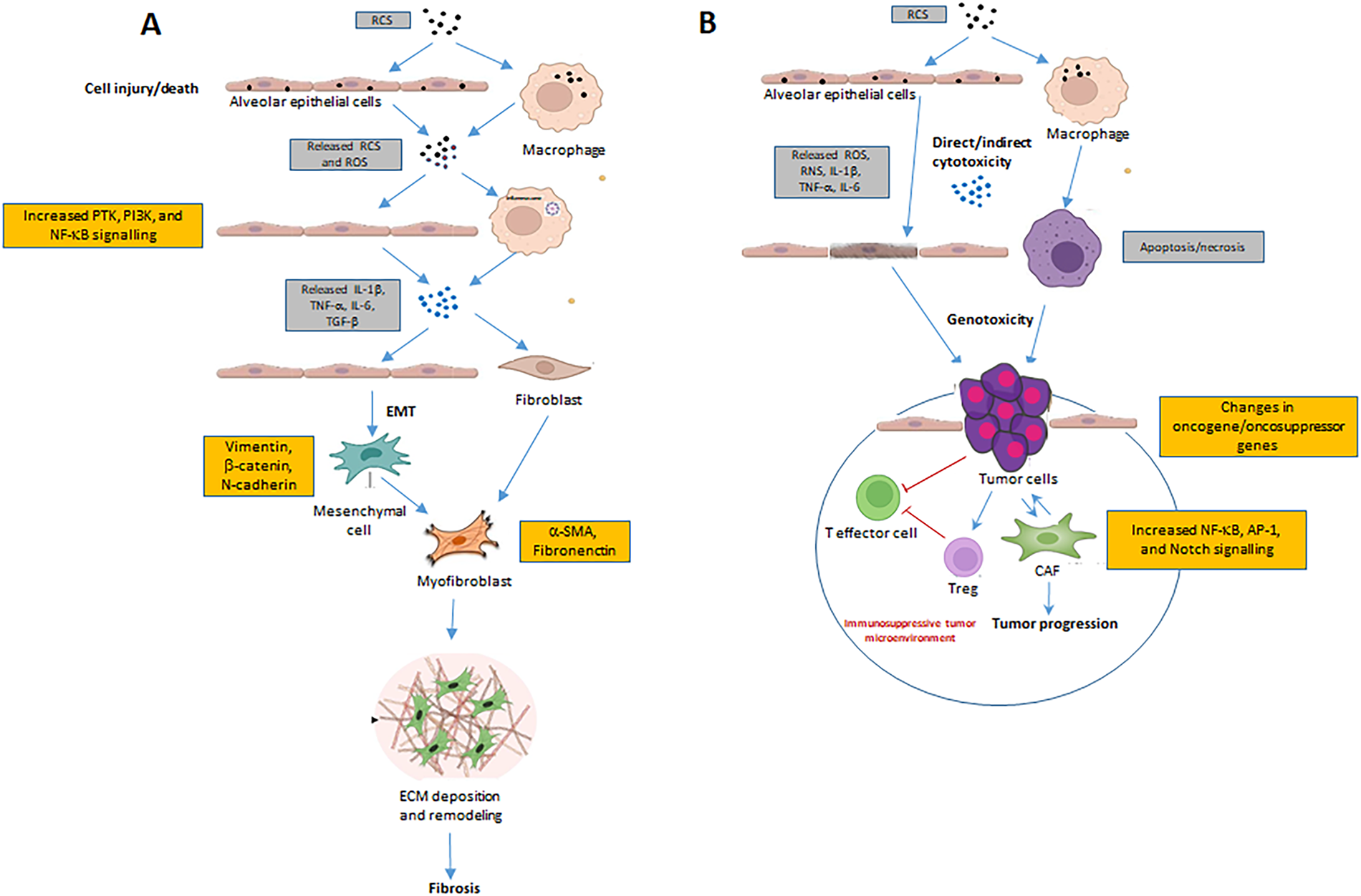
Figure 1: Silica toxic, fibrotic, and carcinogenic effects. (A) Silica dust exposure as respirable crystalline silica (RCS) can activate macrophages, damage alveolar epithelial cells, and then initiate inflammation reactions through the activation of PTK, PI3K, and NF-κB pathways. In turn, these inflammatory factors induce multiple signaling pathways, such as TGF-β and TNF-α signaling pathways. Repeated inflammation and cell death promote the formation of fibroblasts and myofibroblasts, increase extracellular matrix (ECM) deposition and remodeling, accelerate the release of fibroblasts and epithelial-mesenchymal transition (EMT) process, thus resulting in the destruction of alveolar structures and fibrosis. (B) Silica can influence the process of carcinogenesis via several pathways. Persistence of RCS in the lungs is toxic to alveolar epithelial cells and resident macrophages either directly or indirectly via reactive oxygen species (ROS). Activated macrophages produce cytokines/chemokines/oxidants known to facilitate cancer development, while oxidants induce epithelial cell genotoxicity/injury/proliferation. In response to the chronic inflammation induced by prolonged RCS exposure (upper portion of figure), physiological host processes designed to minimize ongoing immune stimulation are triggered. This results in the creation of an immunosuppressive microenvironment characterized by the presence of regulatory T cells (Treg). Cancer-associated fibroblasts (CAFs) and tumor cells are linked by a vicious crosstalk that ultimately leads to tumor progression. Abbreviations: IL: interleukin; NF-κB: nuclear factor-κB; PI3K: phosphoinositide 3-kinase; PTK: protein tyrosine kinase; TGF-β: transforming growth factor-β; RNS: reactive nitrogen species; TNF-α: tumor necrosis factor-α.
Vallyathan et al. [15] demonstrated that silicon-based free radicals can be generated and that freshly ground silica is more biologically reactive than aged silica, because the former activates a greater respiratory burst in alveolar macrophages than the latter. In addition, freshly ground silica exhibits a greater cytotoxic effect on cellular membrane integrity, i.e., a 1.5-fold increase in LDH release from macrophages, a 36-fold increase in hemolytic activity, and a three-fold increase in the ability to induce lipid peroxidation. Because acute silicosis is frequently associated with occupations in which freshly fractured CS of respirable size is generated, the present study suggests that fracture-generated silicon-based radicals may play a significant role in the pathogenesis of this disease.
A recently validated critical mechanism in silicosis is ferroptosis [16]. Indeed, Sun et al. [17] demonstrated that Silica was capable of inducing ferroptosis via acyl-CoA synthetase long-chain family member 4 (ACSL4), in particular by reducing the activity of glutathione peroxidase 4 (GPX4) through the ACSL4/p38 MAPK signaling pathway, thereby provoking ferroptosis of hepatocytes. Notably, Ferrostatin-1 (Fer-1, known ferroptosis inhibitor) abrogated the effect induced by silica [17]. Ferroptosis was also observed in in vivo and in vitro studies in macrophages, which, upon exposure to SiO2, showed iron overload, oxidative stress, lipid peroxidation, and gene expression alterations, including a decrease in GPX4 expression [18]. Ferroptosis may cause cell membrane damage [19] and release pro-inflammatory damage-associated molecular patterns (DAMPs) that trigger the innate immune response, including the alarmin ATP and pro-inflammatory cytokines [20]. Moreover, the ferroptotic macrophages potentially foster the activation of fibroblasts into myofibroblasts and promote collagen deposition by secreting pro-fibrotic and pro-inflammatory factors, such as IL-1β, TNF-α, and TGF-β. Fer-1 attenuated ferroptosis in SiO2-treated RAW264.7 macrophages by inhibiting lipid peroxidation and cell death [18]. Moreover, considering that in dendritic cells (DCs), retinoic acid (RA) interferes with lymphocyte homing and immune cell trafficking, and it was demonstrated that overactivation of RA signaling promotes a type I interferon pathway that enhances Th2-associated airway inflammation [21,22], Li et al. [23] showed that silica-induced ferroptosis in DCs provokes lipid peroxidation and subsequent activation of RA signaling. This interaction enhances cGAS-STING and IL-1β signaling, driving both inflammation and fibrosis. They also verified that ferrostatin-1 was capable of attenuating fibrosis in a mouse model of silicosis [23].
In silicosis, the role of mitochondrial damage should not be neglected. Zhou et al. [24] demonstrated that silica-induced mitochondrial DNA (mtDNA) leakage activates innate immunity through the inflammatory cGAS-STING-type I interferon and IL-1β pathways, while mitochondrial ROS (mtROS) drives TGF-β1 maturation and myofibroblast activation. Mitochondrial fusion, fission, and mitophagy constitute an integrated quality control mechanism designed to maintain cellular health by segregating and eliminating dysfunctional components. This cycle, often described as a “kiss and run” process, ensures that damaged mitochondria are removed (“mitophagy”) before they can propagate oxidative damage throughout the mitochondrial network [25]. Silica was demonstrated to induce a leakage of this mechanism, inducing altered mitophagy, and this is involved in inflammation and fibrosis [26].
Cell death by ferroptosis and mitochondrial damage are acquiring more and more importance in the development of inflammation/innate immune responses and should be further studied to find novel therapeutic targets.
3.3 RCS and Acute/Chronic Inflammation
Ingestion of silica crystals by resident macrophages of the small airways elicits an inflammatory response characterized by the release of cytokines, such as interleukin (IL)-1β and tumor necrosis factor (TNF)-α, that becomes chronic following continuous exposure to RCS.
In this context of acute inflammation, nuclear factor κB (NF-κB) plays an important role. NF-κB is a transcription factor that controls the gene expression of various molecular signals, such as growth factors, chemokines, cytokines, and adhesion molecules involved in the inflammatory response. The canonical NF-κB pathway is activated mostly by the stimulation of proinflammatory receptors, such as the TNF receptor superfamily, the Toll-Like receptor family (TLRs), and by cytokine receptors for the interleukins [27]. NF-κB is present in the cytoplasm as an inactive form bound to an inhibitory protein (IκB), and it can be activated when IκB is phosphorylated by IκB kinase (IKK). Once activated, NF-κB can translocate to the nucleus, where it binds to gene promoters on DNA. This binding activates transcription of mRNA, which induces translational production of chemokines, cytokines, and growth factors. ROS are proinflammatory since they cause the activation of NF-κB by modifying IKK, leading to IκB degradation, freeing NF-κB to enter the nucleus and activate pro-inflammatory genes [28].
Silica is capable of inducing the phosphorylation of IκB and thus the activation of NF-κB [29]. Because both the phosphorylation and activation of IκB are inhibited by antioxidants, it might be that silica-induced oxidants activate phosphorylation of IκB, which results in the activation of NF-κB [30,31]. It is known that IκB is phosphorylated by protein tyrosine kinase (PTK); in fact, PTK inhibitors have been shown to block the phosphorylation of IκB and the activation of NF-κB [32]. Studies on RAW macrophages have proposed that silica-induced oxidants activate tyrosine phosphorylation of IκB by PTK, which results in NF-κB activation [29,33]. Silica was shown to induce the activity of phosphoinositide 3-kinase (PI3K) in RAW macrophages, and this activation was inhibited by antioxidants. Also, the inhibition of PI3K partially inhibited the silica-induced activation of NF-κB, suggesting that PI3K is involved [34].
Summarizing silica-induced signaling pathway for NF-κB, silica-induced ROS activate PTK and PI3K, which induce steps critical to inactivation of IκB and activation of NF-κB [35].
To understand how silica induces macrophage-driven inflammation, Hornung and colleagues [36] reported that silica crystals activated the NLRP3 inflammasome, requiring phagocytosis and subsequent lysosomal swelling and damage.
Regarding ROS, several Authors gave evidence to the inflammasome, which represents an intracellular multiprotein complex activated by ROS. In particular, the sensor protein is NLRP3 (nucleotide-binding oligomerization domain-like receptor) that is activated by ROS and other stimuli. It has been hypothesized that a loop exists between ROS and NLRP3 [37]: ROS would activate the NLRP3 inflammasome that produces IL-1β, which induces inflammatory responses and recruits neutrophils and macrophages; in turn, these cells produce ROS and lead to further inflammatory responses. Furthermore, IL-1β promotes intracellular accumulation of ROS by disturbing antioxidant enzymes.
Lam and colleagues [38] verified the role of NLRP3 in a study on mice that were administered silica; in particular, mice with a deficit in acute NLRP3-mediated inflammation showed significantly reduced pulmonary transforming growth factor-β (TGF-β) and α-smooth muscle actin (α-SMA) expression, tissue damage, and fibrosis in the chronic phase of disease progression. Importantly, this included reduced silicotic nodule size and cellularity.
NLRP3, apart from the classic activation through the classical unfolded protein response (UPR), can be activated via ER stress via an alternative pathway [39]. In this context, a very relevant role is played by thioredoxin interacting protein (TXNIP), a cellular protein present normally in the nucleus that is activated and shaped in cytoplasm when intracellular ROS increment. It disables the control of ROS production usually played by thioredoxin (TRX), incrementing the ER stress, and this relationship could explain the persistence of chronic inflammation [40].
Downstream of the activation of NF-κB and the inflammasome, the cytokines TNF and IL-1 appear to play a particularly important role in silica-induced inflammation [41]. TNF-α is a cytokine that is thought to be a key mediator in the pathogenesis of silicosis, in particular in silica-induced inflammation and fibrosis. Nitric oxide (NO), synthesized by macrophages upon stimulation by silica, may also play a role in the activation of NF-κB, and thus TNF-α production [42]. In silica-exposed mice lacking the gene for inducible NO synthase (iNOS), there was indeed a decrease in the amount of TNF-α produced, along with a decrease in histological evidence of silica-induced lung damage and inflammation [43]. TNF is not directly chemotactic but can stimulate production of chemotactic cytokines, including chemokines. The same authors showed that the main molecule capable of inducing neutrophils, macrophages, and lymphocytes attraction was macrophage inflammatory protein (MIP-2). This chemokine acts along with the other chemokine MIP-1α [44,45]. Mice immunized with antibody to TNF-α show an attenuated increase in lung chemokine mRNA expression, this circumstance determining an evident reduced inflammatory response to silica exposure [46]. Moreover, TNF-α induces the expression of adhesion molecules on endothelial cells and the production of IL-8, MCP-1, prostaglandin E2 (PGE2), and prostacyclin derived by arachidonic acid metabolism, as well as the production of ROS by phagocytic cells [47].
In addition to TNF, IL-1 plays a critical role in the lung response to silica [41]. Bronchoalveolar (BAL) cells of rats exposed to silica inhalation produced IL-1 following the activation of NF-κB [48,49]. A prominent feature of silicosis is the development of granulomas. It was demonstrated in rats lacking functional IL-1 (IL-1 knock out) exposed to silica that the occurrence of granulomas was very much limited [50]. Porter et al. [51] examined the progression of lung inflammation and damage in rats after the cessation of silica exposure. Rats were exposed to silica by inhalation for 20, 40, or 60 days, and a portion of each group was examined for 36 days post-exposure. Inflammation, as indicated by BAL PMNs, was increased in the 36-day post-exposure period in rats exposed for 40 or 60 but not 20 days. This indicated that in these two groups, recruitment of PMNs into the lung continued without further silica exposure. Fibrosis, on the other hand, was increased in all groups in the 36-day post-exposure period, while it was not present consistently after exposure ended in the 20-day exposure group only. The fact that the PMN influx did not progress in the rats exposed for 20 days, while fibrosis did, suggests that lavage PMNs do not predict fibrogenicity. The authors also pointed to the consistency of this finding with the lack of PMN influx seen in human chronic silicosis despite the progressive nature of the disease [51].
In particular, when the CS particle arrives in the lung, the outcome of this deposition might depend on the ability to produce proinflammatory cytokines [52]. In particular, if the amount of particles is high, the outcome will be acute inflammation, and IL-1 will elicit the release of histamine from mast cells at the site of inflammation [53]. Histamine triggers early vasodilatation and an increase in vascular permeability. Chronic inflammation may develop following acute inflammation or derive from chronic exposure. Some cytokines, such as IL-1 and TNF, are able to maintain both types of inflammation (acute and chronic) because the target cells of their actions are also monocytes and macrophages [54]. During the chronic phase of inflammation, cytokine interactions result in monocyte chemotaxis to the site of inflammation where macrophage activating factors (MAF), such as interferon (IFN)-γ, monocyte chemotactic protein 1 (MCP-1; also referred to chemokine (C-C motif) ligand 2 (CCL2)), and other molecules then activate the macrophages while migration inhibition factors (MIF), such as granulocyte macrophage-colony stimulating factor (GM-CSF) and IFN-γ itself, retain them at the inflammatory site [55].
Another process that could lead to scarring and fibrosis, for example, in periodontitis [56], is the inadequate resolution of inflammation. The class switch of eicosanoid pathways in neutrophils is an important way to end acute inflammation [57,58]. This class switch is mediated by the up-regulation of 15-lipoxygenase by neutrophils late in inflammation. In fact, it is known that in the early phase of inflammation, neutrophils use only 5-lipoxygenase for the production of leukotrienes [59]. The series of enzymatic reactions leads to lipoxins (LXs), such as LXA4 and LXB4 [60,61]. These are receptor agonists that stimulate the resolution of inflammation and promote homeostasis through a number of mechanisms that include limiting the migration of PMN into sites of inflammation and modulating the phenotype of macrophages to stimulate the uptake of apoptotic PMN without secreting proinflammatory cytokines [62–64]. The resolution of inflammation occurs if the cause of the last is removed, but with continued exposure to silica, resolution may not occur.
3.4 Silica and Epigenetics: Divergence between Fibrosis and Cancer
An epigenetic perspective may serve to discuss how the fate determination between fibrosis and cancer. DNA methylation, histone modifications, and non-coding RNAs can determine cell fate toward fibrosis or carcinogenesis (table 1).
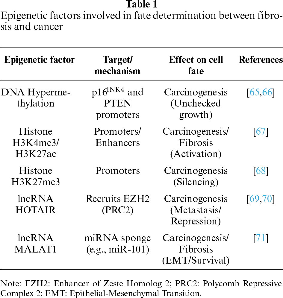
DNA methylation (addition of methyl groups to CpG sites) typically acts as a gene silencing mechanism. Aberrant hypermethylation in p16INK4 and PTEN is considered a hallmark of cancer initiation and progression [65,66]. High levels of H3K4me3 (Histone H3 Lysine 4 Trimethylation) and H3K27ac (Histone H3 Lysine 27 Acetylation) are often associated with enhanced transcription of oncogenes [67], while increased H3K27me3 (Histone H3 Lysine 27 Trimethylation) is frequently linked to the silencing of tumor suppressor genes [68]. In the context of fibrosis, H3K4me3 and H3K27ac are often elevated at the promoter and enhancer regions of pro-fibrotic genes, leading to their upregulation, the accumulation of extracellular matrix (ECM), and the activation of fibroblasts into myofibroblasts [72]. Long non-coding RNAs (lncRNAs) regulate gene expression by acting as scaffolds, guides, or decoys for epigenetic machinery. lncRNA HOTAIR (Hox Transcript Antisense RNA) recruits the catalytic subunit EZH2 of the histone methyltransferase PRC2 (Polycomb Repressive Complex 2), causing profound epigenetic silencing of tumor suppressors and promoting cancer metastasis [69,70]. On the other hand, lncRNA MALAT1 (Metastasis-Associated Lung Adenocarcinoma Transcript 1) acts as a sponge for tumor-suppressive miRNAs. It is highly upregulated in metastatic cancers and is also heavily involved in promoting epithelial-to-mesenchymal transition (EMT) and renal fibrosis by activating the STAT3/NF-κB axis [71].
While considering that engineered Silica nanoparticles (SiNPs) (often amorphous) have distinctly different toxicokinetics compared to occupational RCS, it could be useful to understand that their use in nanomedicine is not free from risks, also in order to stimulate new research about the effect of RCS; in fact, in 2024, Zheng et al. reviewed for the first time epigenetic changes induced by SiNPs [73]. SiNPs induce epigenetic modification in GC-2 spd cells (an immortalized mouse testicular cell line), i.e., numerous extensive DNA methylation changes, including 51.0% hyperdifferentially methylated regions and 49.0% hypodifferentially methylated regions [74]. SiNPs also provoke an abnormal expression of miRNAs. The abnormal expression of these miRNAs leads to reproductive dysfunction, endothelial dysfunction, and even tumorigenesis [75]. Evidence demonstrates a strong association between epigenetics and the development of immune-mediated pulmonary diseases, including silicosis [76]. Wang et al. [77] showed an evident demethylation of cultured fibroblasts of SD rats induced by SiO2, and that SiO2 decreased the genomic DNA methylation levels of fibroblasts co-cultured with macrophages. This might explain the activation mechanism of fibroblasts during silicosis [76]. Umemura et al. [78] gave evidence of methylation of five suppressor genes in serum DNA, this methylation is more frequently present in silicosis patients who have silicosis and cancer than in silicosis patients without cancer.
Relevant observations were done by Blanco et al. [79], who, by using a model of silica-induced multistep lung carcinogenesis driven by chronic inflammation to study the evolution of molecular markers and genetic alterations, verified that p53 nuclear levels increase in epithelial cells during the progression from normal to advanced preneoplastic tissues, remaining high in most tumors. Instead, p16 resulted in overexpression in hyperplastic tissues, but its expression is decreased or lost in advanced bronchiolar dysplasias and in 40% of the tumors. If the first result was expected, the second has to be explained. The Authors supposed that the p16INK4a gene was hypermethylated, and this represents an important step because this gene is linked to the transition from hyperplasia to dysplasia and tumor through the existence of tumorigenesis barriers that slow or inhibit the progression of preneoplastic lesions to neoplasia [80,81]. One such barrier involves DNA replication stress, which leads to activation of the DNA damage checkpoint and, thereby, to apoptosis or cell cycle arrest mediated by either p53 or p16 [82,83]. Thus, while these events avoid cancer, the lack of them allows the cell to progress toward cancer. This observation could explain why silicosis patients develop cancer or fibrosis.
Multiple mechanisms are involved in DNA methylation induced by SiNPs, including the regulation of DNA methyltransferases (DNMTs), DNA demethylases, such as those belonging to the ten-eleven translocation (TET) protein family, and DNA methylation substrates [73]. Since no studies have reported the effects of SiNPs on the expression of TET2 and TET3, future research on the mechanism of SiNP-induced DNA methylation is needed.
Overall, all these epigenetic mechanisms often form a positive feedback loop: inflammation leads to epigenetic alterations, which, in turn, facilitate further inflammatory or oncogenic pathways, solidifying a pro-fibrotic or pro-cancerous environment.
In this context, both the dose of Silica and specific genetic or epigenetic events can determine the divergent fate.
3.5 Silica, Cancer, and Fibrosis: Divergence on the Immune Response
The process that leads to either fibrosis or cancer could depend on the features of the tissue microenvironment. At first glance, summarizing, we might affirm that if immunosuppression predominates, cancer would ensue; otherwise, an increment of immune reaction would lead to fibrosis. However, the interrelationship among different cell types is gaining complexity. Hereafter, we give evidence to the possible roles of molecular signaling that permit the orchestration of the numerous and varied cell types, considering known fibrogenic and carcinogenic agents.
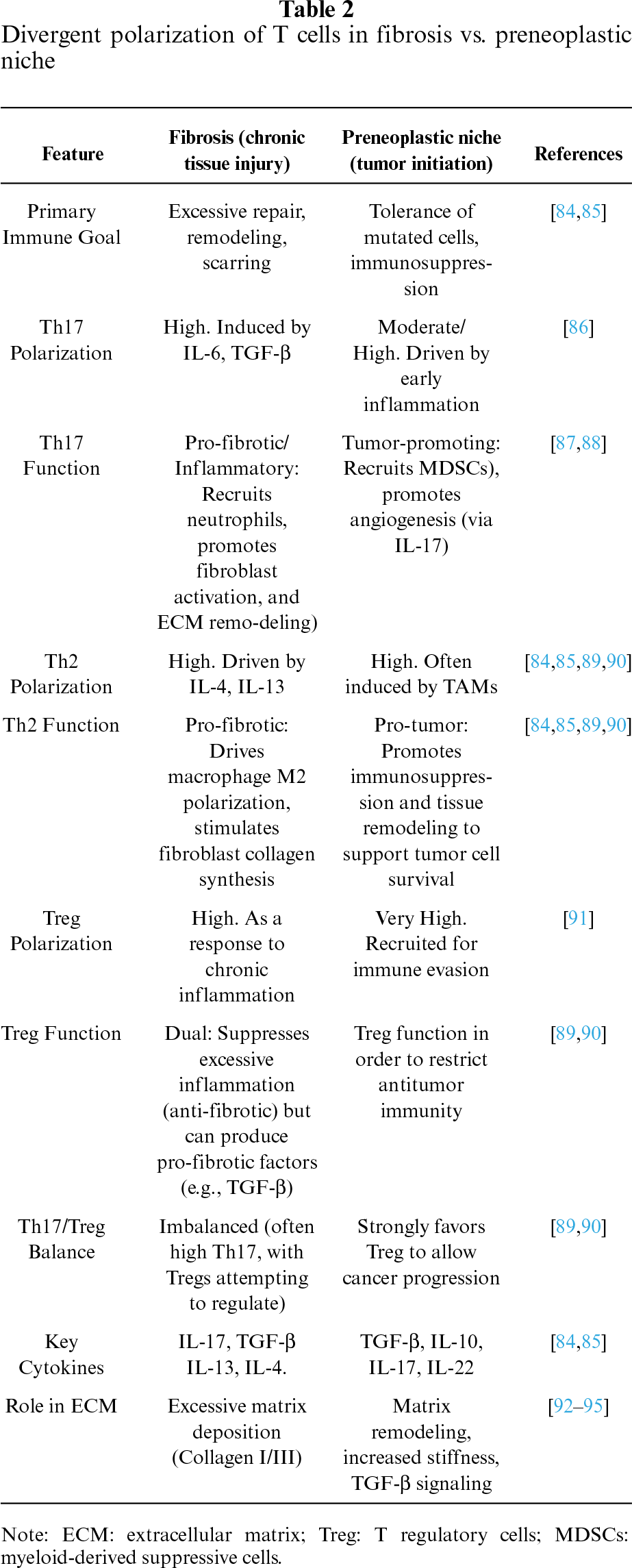
Table 2 compares the immune polarization and function of T helper 17 (Th17), T helper 2 (Th2), and Regulatory T cells (Treg) in chronic tissue fibrosis and the preneoplastic (pre-cancerous) niche. While both environments are characterized by chronic inflammation and tissue remodeling, their immune landscapes differ in their ultimate role in tissue destruction vs. tumor promotion [84,85].
Silica exposure induces molecular and cellular biological alterations of immune cells [91]. It is well known that the association between silica inhalational exposure and autoimmune disease is particularly in the context of intense exposure [96]. The CD95/Fas molecule is well known as a cell death receptor that causes apoptosis, which is particularly important in lymphocytes [97,98]. One quarter of silicosis patients have anti-CD95/Fas autoantibody [99]. Under this condition, if Fas does not function properly, responder T lymphocytes show a longer survival, inciting autoimmune diseases or clinical symptoms of inflammatory reaction to self/non-self-antigens. Furthermore, in silicosis patients, anti-caspase 8-autoantibodies were found [100], and caspase 8 is the main player of apoptosis downstream to Fas. In addition, soluble Fas secreted from the cells into the surrounding area is capable of blocking Fas ligand, inhibiting apoptosis [101,102], and also higher levels of soluble Fas were revealed in these patients [103]. Higher serum soluble IL-2 receptor (sIL-2R) levels were found in autoimmune diseases and in silicosis patients. sIL-2R is an activation marker for T cells and might explain the cause of the chronic activation of responder T cells in silicosis patients [91]. The same authors show a chronic activation of T regulatory cells (Treg) and earlier loss of Treg by Fas mediated apoptosis [91]. Overall, these findings indicate that chronic activation of responder T cells and Treg occurred in the peripheral blood of silicosis patients by chronic and recurrent exposure to silica particles, and responder T cells may then survive for a longer period, whereas Treg may be lost by apoptosis. It has also been shown that PBMC from silicosis patients showed decreased expression of physiological inhibitory molecules for Fas-mediated apoptosis, such as i-flice, sentrin, survivin, and inhibitor of caspase activated DNase (ICAD) [104]. These findings suggest that there exist two subpopulations of T cells in silicosis patients, one comprises T cells resistant against Fas-mediated apoptosis and is now recognized as chronically activated responder T cells. The other subpopulation manifested accelerated Fas-mediated apoptosis (and was also sensitive to functional anti-Fas autoantibody, as described above) and is now recognized as chronically activated Treg [91]. Then silicosis would determine an imbalance of Treg and responder T cells, and this would result in altered regulation of the immune response, explaining the higher appearance of autoimmune disease and fibrosis in silicosis patients.
Another important effect on immune regulation is played by Silica on IL-17/Th17 cells. In particular, Tang et al. [86] gave evidence that in a co-culture system (macrophages and lymphocytes), silica stimulation increased the level of IL-17A and ROR-γt, which indicated that IL-17A and Th17 response took part in Silica-induced immune response in vitro. As an antibody directed towards IL-17 determined a shift of immune response in the direction of Th2 through Treg cells, it can be inferred that Silica unbalanced the immune response by downregulating the Treg arm and thus progression to chronic inflammation and fibrosis.
In the tumor microenvironment (TME), an important mechanism that tumors use to overcome host responses is immune escaping. This function is allowed by, among others, the T cell landscape. In particular, there exists a crosstalk between T cell subsets, TME non-tumoral suppressive cells, and tumor cells, by which T cells may change their features, from tumor suppressor becoming pro-tumoral [87]. In this context, it appears relevant the role of Th17 cells, capable of inhibiting effector T cell proliferation and cytokine production [88]. On the contrary, Th17 cells may also promote anti-tumor immunity. They determine the activation of cytotoxic T cells and provoke a shift to IFN-γ-producing Th1 cells [105], favoring anti-proliferative, pro-apoptotic, and anti-angiogenetic function [106,107]. Th17 cells may also transform into Th1 or Treg, determining opposite effects, respectively, as anti- or pro-tumoral [89,90].
Other cells, immune and non-immune cells, play a role in immune escape, such as macrophages. Tumor-associated macrophages (TAMs) are attracted by MCP-1 and MIP-1α in hypoxic areas of TME [108,109]. TAMs show a M2-like phenotype and are capable of easing the tumor progression through secreting suppressive cytokines, chemokines, proteolytic enzymes, growth factors, and upregulating inhibitory checkpoint receptors on T cells [110]. Furthermore, TAMs secrete ARG1, iNOS, TGF-β, IL-10, and ROS, which provoke CD8+ T cell exhaustion and dysfunction [111], and TGF-β that upregulates the expression of TIM-3, PD-1, and CTLA-4 (Cytotoxic T-Lymphocyte-Associated Protein 4) on T cells, and inhibits IFN-γ and granzyme B [112]. Moreover, they promote PD-L1 expression on monocytes, which favors PD-1+T cell exhaustion [111–113]. Finally, the role of indoleamine 2,3-dioxygenase appears relevant as it attenuates effector T cell function by depleting TME of essential amino acids, such as L-arginine and tryptophan [113]. In fact, effector T cells starved of tryptophan are unable to proliferate and go into G1 cell cycle arrest [114] and, in addition, are more sensitive to apoptosis [115]. In this context, Notch signaling, IL-12, and IL-27 play a relevant role, since their interaction can provoke exhaustion of IL-10-producing regulatory T cells (“Tr1”). In fact, IL-12 and IL-27 plus signals produced by the Notch1L/Notch1 act on the IL-10 gene, making it suitable for activation in the majority of polarized Th subsets [116], leading to exhaustion of IL-10-producing T cells as frequently observed in cancer [117–120].
Macrophage polarization (M1 vs. M2) differs significantly between the fibrotic and pre-neoplastic niche based on the primary requirement for either strict repair or adaptive, chronic remodeling. In the fibrotic niche, macrophages typically show a transient M1 to persistent M2 switch, where the M2 phenotype is directly responsible for excessive tissue repair and scar formation [121]. In the pre-neoplastic niche, the M2 phenotype (often M2d/TAMs) is similarly dominant but specialized towards immunosuppression, angiogenesis, and tumor-cell-assisted invasion rather than solely structural tissue repair [122,123].
More recently, single-cell transcriptomic evidence has revealed significant heterogeneity and dynamic shifts in macrophage populations within the silicotic microenvironment, moving beyond the simple M1/M2 polarization model. Studies identify a core transition from fatty acid-binding protein 4 (FABP4)+ resident alveolar macrophages [124] to a pro-fibrotic secreted phosphoprotein (SPP1)+ macrophage subset that drives fibrosis, particularly within silicotic nodules [125].
As evidence of the above facts, we would like to highlight the different roles played by cytokines (see IL-17), which may at first glance seem contradictory. Although the cells involved in these processes appear more structured, nevertheless they also modify themselves in response to the conditions of the TME. Chronic inflammation normally occurs following exposure to silica, but it cannot be ruled out that a clear proliferation of regulatory T cells (Treg) capable of facilitating the onset of cancer may also occur.
3.6 Silica and Mechanisms of Fibrosis
After an injury occurs to epithelial tissues, the repair ensues by the formation of granulation tissues rich in smooth muscle actin (SMA)-positive myofibroblasts (a hallmark of activated fibroblasts), platelets, newly formed blood vessels, macrophages, and other inflammatory cells and ECM. TGF-β signal pathway plays a pivotal role in the emergence of myofibroblasts, which contribute to the production of matrix metalloproteinase (MMP) and ECM proteins, such as collagen I, fibronectin, and hyaluronic acid [92,126–128]. The injured tissues are then eliminated, and ECM proteins are generated de novo [126–129]. If a sustained activation of myofibroblasts occurs, a dysfunctional repair mechanism appears, leading to the accumulation of fibrotic ECM, which is rich in collagen fibers and resistant to MMP-mediated degradation [92–94]. The fibrotic ECM is capable of inhibiting epithelial cell polarity and stimulates epithelial cell proliferation, conditions allowing tumor formation and development [130,131]. Different authors show that the presence of fibrotic lesions significantly increases the risk of cancer in numerous tissues, including the lungs, liver, and breast [130–133]. Although silicosis has a distinct etiology and histopathology, also in idiopathic pulmonary fibrosis (IPF), there is an association with a higher incidence of lung cancers as compared with the general population [134], showing that the alterations of TME could be the cause of this higher incidence because IPF is characterized by scar tissue accumulation in the lung interstitium. It was demonstrated that the start of this disease is the injury to alveolar type II (ATII) cells that triggers production of TGF-β, which leads in turn to proliferation of macrophages, platelets, and myofibroblasts in the damaged areas, leading to the formation of fibroblastic foci. Fibroblastic foci containing myofibroblasts seem to be an indicator of poor prognosis and decreased survival [135].
At the level of the lung, it is known that the triggers implicated in IPF pathogenesis are initial microinjuries to the alveolar epithelium that provoke alveolar epithelial cell damage with abnormal re-epithelization and repair [136]. In this context, epithelial and endothelial cells produce high levels of fibrogenic mediators, such as TGF-β, PDGF, Wnt, or Wnt-1–inducible signaling protein-1, which, in concert, promote the differentiation of resident quiescent fibroblasts into myofibroblasts. The over-expression or persistent activation of the Wnt/β-catenin pathway can lead to aberrant fibrosis progression and related tissue dysfunction, in collaboration with RAS and/or TGF-β1 activity [137]. Epithelial and endothelial cells then acquire mesenchymal phenotypes, which, along with the recruitment of circulating fibrocytes and bone marrow–derived progenitor cells to the lung, contribute to fibrosis. In turn, these myofibroblasts secrete TGF-β, which induces ATII cell apoptosis, perpetuating the aberrant wound healing process. The myofibroblast also secretes high levels of tissue inhibitors of matrix metalloproteases (TIMPs) and ECM proteins, promoting collagen deposition and contributing to fibrosis [138].
In the field of molecular signaling, TGF-β1 represents the isoform most closely related to the development of IPF [139]. It is secreted in an inactivated form via its attachment to a latency-associated peptide and to a latent TGF-β binding protein. Several papers have given evidence that its activation can be derived by different upstream agents, (a) such as MMP9 and MMP2 [140]; (b) changes in pH (acidic conditions); (c) ROS [141]; (d) thrombospondin-1 [142]; (e) tissue stiffness [143]; (f) integrins αVβ3, αVβ5, αVβ8, and αVβ6. These different mechanisms play an important pivotal role in TGF-β activation in fibrotic disorders [144].
Moreover, TGF-β is produced by alveolar macrophages, neutrophils, activated alveolar epithelial cells, endothelial cells, fibroblasts, and myofibroblasts [145]. The latter, once activated, in addition, produce pleiotropic growth factors, showing chemotactic and proliferative activity, inducing macrophage and fibroblast recruitment as well as fibroblast proliferation via platelet-derived growth factor (PDGF) expression [146]. TGF-β also stimulates in fibroblasts the expression of several proinflammatory and fibrogenic cytokines, such as TNF-α, PDGF, IL-1β, or IL-13, thus promoting and perpetuating the fibrotic response [147–149]. In fact, the repetitive injury of the alveolar epithelium is a certain cause of the aberrant wound healing process in IPF [150].
The first signal for platelet activation, the formation of the fibrin-rich clot formation, and the generation of a strong antifibrinolytic cascade is the disruption of epithelial cells and the upregulation of epithelial-derived plasminogen activated inhibitor (PAI)-1 by a process driven by TGF-β1/Smad3 [151]. Several authors support the association between alveolar epithelial cell apoptosis and lung fibrosis. The mechanisms that cause alveolar epithelial cell death are not fully disclosed, however it is known that TGF-β plays a pivotal role. TGF-β is a potent inducer of apoptosis in alveolar epithelial cells [152], via Fas-mediated caspase-8 activation and down-regulation of p21. In particular, TGF-β promotes the imbalance of Bcl family members by stimulating Bax and Bid in the murine lung [138].
During injury, ROS promote a particular microenvironment capable of altering the balance between MMPs and their inhibitors (tissue inhibitors of metalloproteinases (TIMPs)) and by directly or indirectly activating TGF-β. In turn, TGF-β produces ROS as part of its signaling pathway [153]. Guo and colleagues [154] have shown that nicotinamide adenine dinucleotide phosphate (NADPH) oxidase 4 is involved in lung fibroblast-to-myofibroblast differentiation. Under normal conditions, ATII cells can transdifferentiate into ATI cells. In the course of injury and stress conditions, both cell types undergo cell death, with subsequent hyperplasia to re-epithelialize damaged and denuded areas or incrementing epithelial mesenchymal transition (EMT), which represents a possible source of activated myofibroblasts in the lung subepithelium. Li and coworkers [155] gave evidence about the main role of ATII cells in fibrosis development. In particular, they revealed that a loss of TGF-β receptor signaling in ATII cells determined an evident protection against bleomycin-induced fibrosis by preventing epithelial apoptosis along with a decreasing of myofibroblast activation, and by impeding protease imbalance (TIMP/MMP) [155].
Considering all these studies, TGF-β can be considered a key initiator of fibrotic processes. In IPF, highly active myofibroblasts are present in subepithelial regions that constitute fibroblastic foci and are localized mostly in close proximity to injured or hyperplastic ATII cells. The origin of these contractile α-SMA-positive myofibroblasts is controversial, nevertheless they are considered to be the main cause of increased ECM deposition in IPF [156]. There are several theories regarding their origin; indeed, they would derive from proliferating and activated resident lung fibroblasts, ATI or ATII cells via the process of EMT; bone marrow–derived fibroblast; pericytes of the lung interstitium [157].
Along with this role of TGF-β, the microenvironment favors ECM deposition by inducing an imbalance between matrix-degrading enzymes and their inhibitors. Some authors have hypothesized that MMP-7 may release preformed TGF-β from the ECM, playing an important effect on epithelial repair process [158]. The role of IL-17 was highlighted by Wilson et al. [159], by showing a novel synergistic role of IL-17A and TGF-β in the development of bleomycin–induced fibrosis.
It is known the role of YAP signaling as well as mechanosensor and mechanotransducer at cell-ECM/biomaterial interfaces [160]. A positive feedback loop between fibroblast activation and ECM deposition mediated by CD44-RhoA-YAP signaling pathway in experimental silicosis was demonstrated [161]. Activated fibroblasts through the CD44-RhoA-YAP signaling pathway drive ECM production and accumulation, further stimulating fibroblast activation. Upstream blockade by anti-CD44 antibody or downstream blockade by dihydrotanshinone I (DHI, a lipophilic component of traditional Chinese medicine Salvia Miltiorrhiza Bunge) [162], as well as verteporfin (VP, a small molecule YAP inhibitor) [163], delays CS-induced lung fibrosis.
YAP and UPR are functionally intertwined adaptive pathways, as cells depend on YAP to resolve ER stress and expand the endoplasmic reticulum (ER)‘s folding capacity [164]. As a physicochemical stressor, silica dust chronically activates the PERK-eIF2α, IRE1α-XBP1, and ATF6 pathways [165]. Recent evidence indicates that while the adaptive response of the UPR has cytoprotective effects in early stages, sustained activation induces apoptosis via CHOP and promotes collagen synthesis through XBP1s in hepatic stellate cells as induced by TGF-β [166,167]. Although silica is known to activate the XBP1 stress response generally, whether this specific activation directly drives fibrogenesis in the lung remains to be verified.
Also, YAP and the senescence-associated secretory phenotype (SASP) are intimately connected. Research indicates that the YAP-TEAD complex enables senescent cells to survive by easing ER stress, allowing the cells to continuously secrete their profibrotic SASP [168]. Senescence is induced in fibroblast and alveolar epithelial cells by silica via oxidative stress. Particularly interesting appears the observation of Lian et al. [169] who verified an evident increment of growth differentiation factor 15 (GDF15) using alveolar epithelial cells (MLE-12) and mouse embryonic fibroblast cells (NIH/3T3) treated with SiO2. Senescent ATII cells could facilitate epithelial cell EMT and fibroblast activation in a GDF15-dependent manner. GDF15, as well as IL-6 and TGF-β, belongs to SASP factors, and it is produced by fibroblast and ATII cells via p53 during oxidative stress and plays a pro-fibrotic role. In the silicosis context, SASP could perpetuate the inflammation, leading to cellular senescence present in the nodule and thereby establishing a “senescence-inflammation-fibrosis” vicious cycle [170].
Another mechanism that has been recently studied in silicosis, and in particular in the macrophages, is the signalling functions of exosomes and microvesicles. Interestingly, SASP correlates to hyperactive secretion not only of cytokines, pro-fibrotic growth factors, but also of exosomes, which drive fibrosis [171]. The literature shows that the signaling mediated by exosomal miRNAs between fibroblasts, macrophages, and epithelial cells participates in the development of lung fibrosis [172]. Exosome-originating miRNAs regulate key processes, such as TGF-β signalling, critical for fibroblast activation and deposition of ECM components. Therefore, exosomal miRNAs that shift cell-to-cell communication and cellular mechanisms can regulate the progression of pulmonary fibrosis. In particular, when alveolar macrophages phagocytose silica, they release exosomes with altered, pro-fibrotic cargo—including specific microRNAs (such as miR-155, miR-21, and miR-107) and potentially mitochondrial components—that are taken up by fibroblasts [173]. MiR-107 is critical to exosomal-mediated lung fibrosis because it modulates significant molecular pathways of fibroblast activation and ECM deposition. Therefore, miR-107 negatively regulates the cell cycle signaling pathway through exosomal transfer, promoting fibrosis progression [174]. MiR-125a-5p directly targets Smurf1 to potentiate TGF-β/Smad signaling, upregulating SMAD1, ID1, α-SMA, and collagen and driving fibroblast to-myofibroblast trans-differentiation in vitro [175]. Also, circular RNAs (circRNAs) carried by exosomes act as critical regulators of EMT in fibrotic diseases, facilitating intercellular communication between damaged cells (such as renal tubular epithelial cells, macrophages, or cancer-associated fibroblasts) and target cells. These exosomal circRNAs, often highly stable due to their closed-loop structure, are selectively packaged into exosomes to modulate the expression of fibrotic markers, promote myofibroblast differentiation, and drive the deposition of ECM. Mechanistically, exosomal circRNAs often act as “sponges” for miRNAs, preventing them from silencing their target mRNAs. In fibrosis, this mechanism leads to the upregulation of profibrotic transcription factors or EMT-related signaling proteins, such as TGF-β or Wnt/β-catenin [176].
Freire and colleagues [177] verified the role of silica induced inflammation in the occurrence of cancer in a particular experimental model where N-nitrosodimethylamine (NDMA) was used to cause lung cancer in Balb/c mice. The combination of silica and NDMA was capable to increment three-fold the occurrence of adenocarcinomas in respect to mice treated only with NDMA. These results demonstrated that local chronic inflammation was able to reduce significantly the latency period for the appearance of preneoplastic lung lesions and promote lung carcinogenesis [177]. Adenomas from NDMA-silica–treated mice showed an increment of activation of caspase-3 when compared to those from NDMA-only–treated mice, suggesting that silica promotes apoptosis. Malignant transformation is usually associated with an immunosuppressive microenvironment. Microarray expression data showed that genes associated with immunosuppression, such as programmed death (PD)-1, transforming growth factor-β1 (TGF-β1), lymphocyte-activation gene 3 (LAG3), MCP-1, and FOXP3, presented higher expression levels in adenomas from NDMA-silica–treated mice compared to mice treated with NDMA only [177]. Furthermore, a marked increase in Tregs in adenomas in NDMA-silica—treated mice was found. These lymphocytes were also present in areas of the lung distant from adenomas in a more represented way in the NDMA-silica in comparison with NDMA-only treated mice. Altogether, these studies revealed that silica-mediated chronic inflammation could promote the development of an immunosuppressive microenvironment, which is favorable for the generation of preneoplastic lesions that potentially contribute to the progression of adenomas to adenocarcinomas [177]. Other authors [178] found that silicon dioxide (SiO2) nanoparticles (NPs) resulted in immunotoxicity because they reduced B- and T-cell proliferation in the spleen of mice fed with NPs. Moreover, they showed a reduction of Th1 response and a slight increase of IFN-γ. Also, the ionic characteristics of NPs played a role, indeed negatively charged SiO2 NPs were revealed to have the most potent in vivo immunotoxicity. These experimental data refer to NPs and could not correlate directly with RCS but it seems to link to Silica as element and appears useful to mention it, also in light of the observations of Shi et al. [179] that verified the role of ROS in the carcinogenesis through different mechanisms, such as genotoxic effect (DNA damage); stimulation of oncogenes; increment of cytokines, such as IL-1 and TNF; activation of NF-κB and AP-1; mutation of the p53 gene, a known tumor suppressor gene; lipid peroxidation products and a rise in intracellular calcium.
With regard to the first mechanism, Silica induces DNA strand breaks in vitro, and this mechanism is blocked by ROS scavenger agents. It is known that Silica binds to DNA through the formation of hydrogen bonds with its silanol groups to the DNA phosphate backbone, thus bringing the DNA strand close to the sites of ROS production on the silica surface. It has also been demonstrated that silica (via interaction with hydroxyl radical) produces 8-hydroxydeoxyguanine—a modified DNA base important in mutagenesis and carcinogenesis, showing the link between ROS formation and DNA damage [180].
An interesting study was published by Driscoll and colleagues [181], which found that BAL cells, derived from rats exposed to silica via intratracheal instillation, showed more DNA mutations than control rats. The authors deepened the previous observation, adding BAL taken by rats exposed to silica to rat alveolar RLE-6TN cells in culture. Surprisingly, BAL cells enriched for neutrophils were significantly more mutagenic to RLE-6TN cells than BAL cells enriched for macrophages. This effect was prevented if the antioxidant catalase was added, indicating the known role of neutrophils in producing ROS [181].
Johnston et al. [182] showed that rats exposed to crystalline or amorphous silica by inhalation for 13 weeks revealed an increment of BAL neutrophils. However, the cytotoxicity was more evident in the amorphous silica-exposed than the CS-exposed animals. After a post-exposure recovery of eight months, the previous inflammatory alterations were found only in the rats exposed to CS, while these parameters had regressed in the amorphous silica-exposed rats. Only CS exposure resulted in a significant increase in hypoxanthine phosphoribosyltransferase 1 mutation frequency in rat alveolar epithelial cells, but this was not observed with amorphous silica. Altogether, these results may indicate that the increment of cytotoxicity occurring through apoptosis represents a mechanism that preserves tissues from malignant transformation. Therefore, the evasion of (or lack of) acute cytotoxicity is an important factor that allows target cells to survive and accumulate secondary genotoxic effects.
Although the previously cited work [182] suggested that inflammation alone is not responsible for the development of mutagenic effects in rat lungs after exposure to silica particles, Shi et al. [179] hypothesized that TNF and IL-1 could play a role in the carcinogenesis, particularly TNF-α, which would act through NF-κB and activator Protein-1 (AP-1). AP-1 is composed of homo- or heterodimers of the protein products of individual members within the Jun (c-Jun, c-Jun-B, and c-Jun D) and Fos (c-Fos, FOS B, Fra-1, and Fra-2) immediate-early response gene families [35]. AP-1, activated by oxidants and silica, would act as a promoter, stimulating oncogenes and being involved in neoplastic transformation, tumor progression, and metastasis [35]. Another effect of silica would be to inhibit the p53 suppressor gene [179], its inhibition making it easier for the passage of genetic lesions in cells with DNA damage to the new generation of cells. Liu et al. [183] confirmed the important role of p53 by examining the p53 gene mutations in lung cancer in workers with silicosis.
Linked to inflammation and fibrosis, as we have recalled in Section 3.6, SASP is also involved in cancer. Actually, SASP has been recognized in the TME as a double-edged sword. While cellular senescence serves as an intrinsic defence mechanism to protect cells from malignant transformation, senescent cells can promote tumor progression to overt malignancy, primarily through a set of factors known as SASPs, including chemokines, growth factors, cytokines, and stromal MMPs [184].
3.8 Role of Notch, YAP/TAZ, and Wnt/β-Catenin and Their Interplay
While the involvement of both immune and non-immune cells has been previously noted, it is essential to elucidate the distinct signaling pathways operating within the microenvironment.
In order to discover a link between inflammation and fibrosis and the latter and cancer, some studies have focused on the role of Notch-Jagged signalling [185]. In mammals, there are four Notch receptors (Notch1–4) and five Notch ligands (Delta-like (DLL) 1, 3, 4 and Jagged (JAG) 1–2). When a Notch receptor interacts with a Notch ligand in an adjacent signaling cell, it undergoes proteolytic cleavage by A Disintegrin And Metalloprotease domain 10 (ADAM10) and the γ-secretase complex, which leads to the release of the Notch intracellular domain (NICD). Eventually, the NICD enters the nucleus and activates the transcription of Notch target genes [186].
Aberrant Notch-Jagged signaling through JAG1 was found in different tumours and was observed to be associated with the expression of IL-1, IL-4, and TGF-β, all known cytokines as well as pro-oncogenic factors [185].
We could cite the observations in kidney fibrosis, where some authors [187] demonstrated an increase of Notch Jagged signaling through a reduction in the expression of Hoxa5 protein. In colorectal cancer, evidence has demonstrated the importance of Notch signaling, which is normally regulated by zinc finger protein 671 [188].
It could also be useful to cite the role of Notch signaling in glioblastoma [189] and in gastric cancer [190] to remark the important role of this signaling because many studies point to the oncogenic role of the Notch pathway in tumor cells, as well as a central regulator in non-malignant inflammatory disorders [191]. Consequently, various therapeutic strategies are now being pursued to antagonize Notch signals in cancer and inflammatory disorders, including fibrosis [191,192]. However, it must be emphasized that the involvement of the Notch pathway in the pathogenesis of silica-related malignancies has not yet been well characterized.
While studies of Notch signaling across various malignancies reveal divergent and potentially paradoxical behaviors, several authors suggest that this pathway operates via a threshold-dependent mechanism rather than a binary on/off schematic. Specifically, Notch signaling is characterized by three distinct levels of control that govern its activity: (a) cis inhibition; (b) lateral inhibition and activation of the same signal; and (c) trans activation [193–195]. The Notch juxtacrine signaling system engages in complex crosstalk with other cellular and tissue regulatory mechanisms, such as YAP/TAZ and Wnt. For example, evidence obtained in the liver suggests that the Wnt pathway acts as a negative regulator of YAP/TAZ, thereby functioning as a tumor suppressor in this context [196–198]. Indeed, inactivation of Wnt/B-catenin determines activation of YAP/TAZ that increments Notch, then acting as a tumor inducer [197].
Regarding the fibrotic process, Yuan et al. [199] demonstrated that interstitial macrophages (IMs) exhibit significantly upregulated Notch3 expression during silicosis progression. Utilizing myeloid-specific Notch3-knockout mice, the authors established that Notch3 signaling not only promotes IM recruitment—specifically by regulating CCR2 expression—but also modulates their pro-inflammatory phenotype. Notably, conditional Notch3-knockout mice showed a marked attenuation of CS-induced pulmonary inflammation and fibrosis. Moreover, the Wnt/β-catenin pathway is activated in the lung epithelium of IPF, characterized by progressive loss of lung function distinguished by severe pulmonary epithelial injury, fibroblast activation, ECM deposition, and distorted lung development [200].
The fibrotic ECM, characterized by altered stiffness and composition, drives both fibrotic progression and epithelial carcinogenesis through mechanotransduction pathways involving integrins and YAP/TAZ. As the primary downstream effectors of the Hippo signaling pathway, YAP/TAZ play a pivotal role in regulating organ size, cell proliferation, and lineage fate determination [201]. Acting as bona fide mechanosensors, these proteins mediate cellular responses to physical cues at both cell-ECM and biomaterial interfaces [160]. While an exhaustive review of this pathway is beyond the scope of the present article, in this regard we refer readers to Heng et al. [202], it is noteworthy that TNF-α can also activate YAP/TAZ, although the precise underlying mechanisms remain to be fully elucidated [203,204]. Starting from these important observations, it could be hypothesized that CS, by inducing TNF-α, triggers the Hippo-YAP/TAZ axis. Furthermore, extensive crosstalk exists between YAP/TAZ and Notch signaling [197], both of which are implicated in the pathogenesis of fibrosis and cancer. The factors determining these divergent pathological outcomes remain unknown; however, they may depend on tissue- or cell-type-specific contexts. Such pleiotropic effects could be mediated by the expression of specific TEA domain-containing transcription factor isoforms that interact with YAP/TAZ within the cell nucleus [202], or by the nuanced, non-binary regulatory mechanisms of the Notch pathway previously discussed. Indeed, a review of the literature indicates that during fibrosis pathogenesis, activation of the Wnt/β-catenin pathway induces the YAP/TAZ pathway, which in turn triggers the expression of Notch receptors and/or ligands [205,206].
In summary, CS induces epithelial injury primarily via ROS production, subsequently triggering a robust immune response. While high-dose exposure results in acute inflammation, chronic exposure—typical of occupational settings—leads to persistent inflammation and subsequent maladaptive tissue repair. Within this context, the microenvironment plays a strategic role, governed by the complex interplay between immune and non-immune cells, including fibroblasts, CAFs, and alveolar epithelial cells. Furthermore, the influence of diverse signaling modalities—comprising intercellular communication, juxtacrine, and paracrine signaling—cannot be overlooked, as they may subvert expected pathological outcomes. We hypothesize that silica-induced inflammation leads to fibrosis when the pro-inflammatory response predominates; conversely, a T-cell landscape characterized by an enrichment of Treg may facilitate a transition toward carcinogenesis. Ultimately, we propose that cellular dynamics exert a more decisive influence than individual signaling pathways, given that the latter appear strictly tissue- and cell-type-specific, suggesting their role is subordinate to the cellular context; the clearest demonstration of this affirmation seems to be Notch signaling because activation or reduction of this signaling appears always paradoxical because it is tissue and cell-type specific: in this way the cellular dynamics would appear more important than signaling pathway.
3.9 Silica, TME, and Cancer-Associated Fibroblasts
Coming to the question of why cancer is promoted and not fibrosis, it could be that some mechanism does not occur, or some other mechanisms may have a role. TME is the site where this choice occurs (figure 2). The trigger could be at the level of intracellular signaling pathways activated in various cell types, including immune cells and fibroblasts, or it could be that particular cytokines are pivotal for guiding a different fate. Nevertheless, TME can be studied in another way, considering the communication between cells, since fibroblasts and immune cells can regulate each other via gap junctions [207].
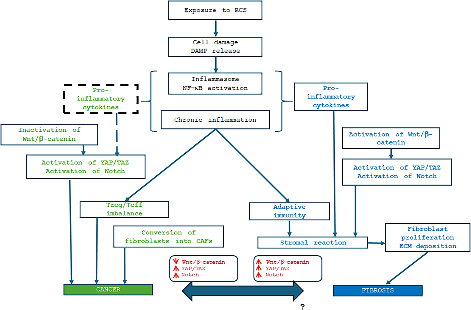
Figure 2: Sequential biological events following exposure to respirable crystalline silica (RCS). This process, driven by inflammation, results in two distinct outcomes: fibrosis or carcinogenesis. Specifically, as shown in the blue section (right side), pro-inflammatory cytokines trigger a stromal reaction mediated by adaptive immunity, leading to fibrosis. Conversely, as depicted in the green section (left side), an imbalance between Treg and Teff cells, alongside the activity of cancer-associated fibroblasts (CAFs), promotes cancer development. A pivotal role is played by the Wnt/β-catenin pathway: its activation leads to the activation of both Notch and YAP/TAZ signaling, driving the fibrotic fate (blue section). On the contrary, the inactivation of Wnt/β-catenin results in the activation of YAP/TAZ signaling and the activation of Notch, fostering a pro-tumorigenic environment (green section). At the bottom of the figure, we represent the link between these two pathological fates. The bidirectional arrow indicates: (a) the well-documented potential for fibrosis to progress into cancer; and (b) a theoretical working hypothesis represented by the opposite direction. This suggests the possibility of ‘reprogramming’ or transforming the cancer phenotype into a fibrotic one (denoted by a question mark). In this context, the modulation of Wnt/β-catenin, Notch, and YAP/TAZ signaling (highlighted in the red boxes) may represent a promising therapeutic strategy. DAMP, damage-associated molecular pattern.
Forty years have passed since Dvorak [208] described cancer as a non-healing wound. In fact, in normal tissue activated fibroblasts lead to healing, impeding the spread of cancer cells. On the other hand, if an atypical wound healing process, like the keloid process occurs in the tumor stroma, the result is an excess of fibers accumulated by fibroblasts. This immature fibrosis can promote tumor infiltrating growth, in fact, for example, colon cancer patients with this phenotype may have an undesirable prognosis [209,210].
In order to examine and “dissect” the complex links between fibrosis and carcinogenesis and to try to provide a theory capable to explain the different direction due to exposure to silica, we cannot neglect cancer associated fibroblasts (CAFs), a very controversial topic [211]. The first cause of this dispute concerns their heterogeneity, in fact, they can derive from resident fibroblasts, mesenchymal stem cells (MSCs), epithelial, pericytes, adipocytes, and endothelial cells [212]. The identification of CAFs in the tumor stroma needs the evaluation of their morphology and the expression of specific markers. Morphologically, they are large spindle-shaped cells similar to smooth muscle cells, showing myofilaments and electron dense patches [213]. They express α-SMA [214]. A marker that permits the identification of activated CAF is the fibroblast activation protein α (FAPα), a cytomembrane protein selectively expressed in various types of human epithelial cancer [215], along with podoplanin-a, S100A4, vimentin, fibroblast specific protein-1 (FSP-1), and platelet-derived growth factor (PDGF) receptors α and β [216].
Thus, CAFs can reasonably be described as a “cell state” rather than a “cell type” [217]. Different growth factors and cytokines have been identified as inducing CAF differentiation in progenitors, some of which derive from the feedback loop between cancer cells and CAFs in the TME [218]. CAFs also stably maintain their transcriptome and metabolic profiling through an autocrine method. Once propagated in vitro, CAFs are capably to retain activated and tumor-promoting traits despite a lack of ongoing interactions with carcinoma cells, it is likely that this trait is acquired by epigenetic alterations induced by cancer cells that stabilize in offspring [218].
The crosstalk between cancer cells and fibroblasts has been identified as the leading cause of the formation of the malignant phenotype of cancer. In this perspective, the role of cytokines and different regulatory factors present in the ECM appears strategic. For example, Cullen et al. [219] reported a pathway that mainly involves cancer cells producing PDGF, which induces fibroblast proliferation and the expression of insulin-like growth factor (IGF)-1 and IGF-2. In turn, IGFs secreted by fibroblasts through biunivocal crosstalk induce cancer cell proliferation and the synthesis of PDGF [219]. Another role of cancer cells is the induction of MMP production by fibroblasts, which results in ECM degradation and enhancement of the invasiveness of cancer cells [95]. In this loop, fibroblasts secrete growth factors, including HGF [220], keratinocyte growth factor (KGF) [221], and IGF-1 and -2 [222], which in turn stimulate the proliferation of cancer cells. The effect shown by Hawinkels et al. [223], following local cell-cell interactions between breast cancer cells and fibroblasts, occurs on numerous genes, also involving the regulation of the expression of TGF-β-altered genes.
These signaling pathways would act as positive feedback loops, which result in an increment of tumoral mass and/or amplification of signaling molecules, leading to tumor therapy resistance. The involvement of TGF-β signaling is known, leading to stimulating myofibroblast differentiation. On the contrary, the inhibition of TGF-β signaling in stromal fibroblasts results in a significant regression in tumor growth. However, these antitumor effects may vary depending on individual tumor models [224]. The pivotal role of the TGF-β signaling pathway is underlined by the circumstance that it has become a therapeutic target due to its capacity to instruct a pro-tumorigenic program in tumor stromal cells by using cancer cells crosstalk [225].
It is known that there are subpopulations of CAFs according to distinct functional states [226]. This then raises the question of what determines the CAFs’ heterogeneity. It is possible that the CAFs’ heterogeneity characterizes different functions [227,228]. Furthermore, alterations in CAFs show a remarkable spectrum of organ/tissue specificity. For example, caveolin (CAV)-1 was found to induce glycol-metabolic reprogramming in breast CAFs [229], while CAV-1-induced aerobic glycolysis was not completely verifiable in oral CAFs [230]. Therefore, these alterations in CAFs seem to exist only in some types of cancers. It is the opinion of some authors that the heterogeneity of CAFs in the same organ or tissue would depend on their precursor fibroblasts [231]. Most of the time, CAFs are derived from the activated local tissue-resident fibroblasts, fibrocytes recruited from bone marrow, MSCs, and stellate cells, or are the products of the mesenchymal transition of epithelial and endothelial cells, and the transdifferentiation of pericytes, smooth muscle cells, and adipocytes [232,233]. Based on their origin, the functions and markers of CAF subtypes will be different and unique.
Whether CAFs can switch in distinct functional states or subtypes, has been suggested by studies on the cytokine network. IL-1 signaling induces the generation of inflammatory CAFs, whereas TGF-β antagonizes CAF switching from an inflammatory phenotype to a myofibroblast phenotype [234,235]. That would demonstrate the important role of TME and molecular signaling as well as the specific cell types producing signaling molecules. Wu et al. found that gastric cancer-derived HtrA Serine Peptidase 1 (encoded by the gene HTRA1) promoted CAFs generation from normal fibroblasts through the activation of the NF-κB/bFGF/FGF2 signaling pathway [236]. It was also shown that the CAF acquisition of secretory phenotype by activating NF-κB induced by CXCR2 signaling [237], noting that the NF-κB signaling pathway was involved in regulating CAF factor secretion. Also, in oral squamous carcinoma cells, CAFs exhibited increased CXCL1 secretion in an NF-κB-dependent manner after being stimulated with IL-1β [238]. An inflammatory environment in pancreatic cancer was demonstrated as cancer cell-derived IL-1α determined an increment of expression of COX-2, CXCL8, CCL20, and IL-6 in CAFs [239]. In this perspective, NF-κB would play a role in the crosstalk between cancer cells and CAFs. In fact, CAF-driven NF-κB was verified as a pro-inflammatory gene signature critical for tumor progression. In human breast cancer, CAF-derived CXCL1, IL-6, and COX-2, known as targets of the NF-κB transcription factor, were shown to be associated with tumor-promoting inflammation and tumor invasiveness [240]. ROS triggered NF-κB activation and STAT3 in CAFs to upregulate CCL2, while the inhibition of CCL2 could reduce tumor growth of oral cancer in a mouse model [241]. Also, in squamous cell carcinoma, the pro-inflammatory signaling driven by CAFs appeared to be NF-κB-dependent [242]. Other signaling pathways in CAFs, including Notch, and crosstalk of CAFs with cancer cells, as well as targeted therapy of CAF-induced signaling pathways, can be found in detail in Wu et al. [226]. In our opinion, silica could also act in this way because, as shown above, silica is capable of activating NF-κB directly and through the increment of ROS.
4 Silica, Fibrosis, and Cancer: A Working Hypothesis
Table 3 represents the decisive nodes that distinguish a fibrosis-type microenvironment from a tumor-promoting microenvironment.
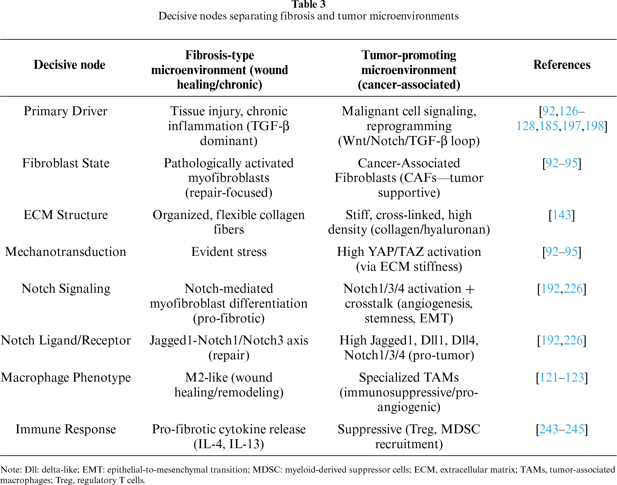
A positive loop exists that underlies the crosstalk between cancer cells and CAFs. The last produced PDGF that determines proliferation of fibroblasts and expression of IGF-1 and IGF-2, which in turn induce proliferation of cancer cells [219,222]. At the end, this activation results in MMP generation, mediating degradation of the ECM that increases the invasiveness of the tumour [95]. Notch signalling seems to play a strategic role. Indeed, its block is associated with Tregs increase, whereas its overexpression reduces Tregs [246–248], and this reduction would explain the increment of autoimmune diseases found in silicosis patients [100]. Moreover, when the Notch signaling is halted, an increment of IL-4 and IL-13 and polarization of TAMs toward a M2 phenotype is observed, and this promotes angiogenesis and “not-eat me” immune editing. Vice versa, if Notch signaling is activated, TAMs polarize toward M1, increasing IL-12, antigen presentation capacity, phagocytosis, and thus toward “eat me” response. Activated Notch signaling provokes fibrosis and the increment of the M1 antitumour activity. On the other hand, the block of Notch signaling avoids fibrosis and induces tumour [243–245]. Therefore, it is important to highlight that M1 and M2 macrophages succumb to transcriptomic mechanisms that transform them. Such a transcriptomic mechanism was demonstrated in silicosis by Liu et al. [124], who revealed an important feature of some fibroblasts capable of inducing permanent inflammation through FABP4, which regulates the activation of the PI3K/AKT/mTOR pathway, participating in SiO2-induced M1 polarization of macrophages and the accumulation of intracellular cholesterol components. More recently, Begka and colleagues [249] were able to establish in a silicotic mouse model that, via transcriptomic profiling, uncovered an interplay between macrophages, neutrophils, fibroblasts, and AT2 epithelial cells, potentially perpetuating silicotic lesions through their dynamic talk. Similarly, in a silicosis mouse model, Cao et al. [250] demonstrated the presence of two types of endothelial cells, one capable of driving inflammation, the other one involved in repair mechanisms, showing a pathological shift toward inflammation-amplifying endothelial cells and impaired reparative capacity during silicosis progression.
In this complex interplay, silica could increment Notch signaling via ROS and NF-κB signaling, inducing fibrosis or reduce this signaling and thus determine cancer. If in this context, this signaling is decisive has yet to be defined: it is a hypothesis. CAF subtype specification is determined by graded or threshold-dependent Notch1 signaling activity, rather than a simple ON/OFF mechanism, functioning as a “molecular switch” that governs CAF heterogeneity [251,252], unveiling Notch1 signaling as a potential therapeutic target for tumors or fibrosis.
Today we can affirm that the study of this Notch signaling could result strategic and be the next phase in the study of silica toxic mechanisms. This study could help to answer the question, considering Silica, a known fibrogenic and carcinogenic agent, which are the conditions that either transform fibrosis into cancer or that avoid cancer and induce fibrosis. Further studies would verify if cancer is a fibrosis that failed or if the fibrosis is an unsuccessful cancer.
Table 4 provides an overall summary of the main topics and targets that make a difference in the pathogenesis of lung fibrosis vs. lung cancer as induced by Silica.
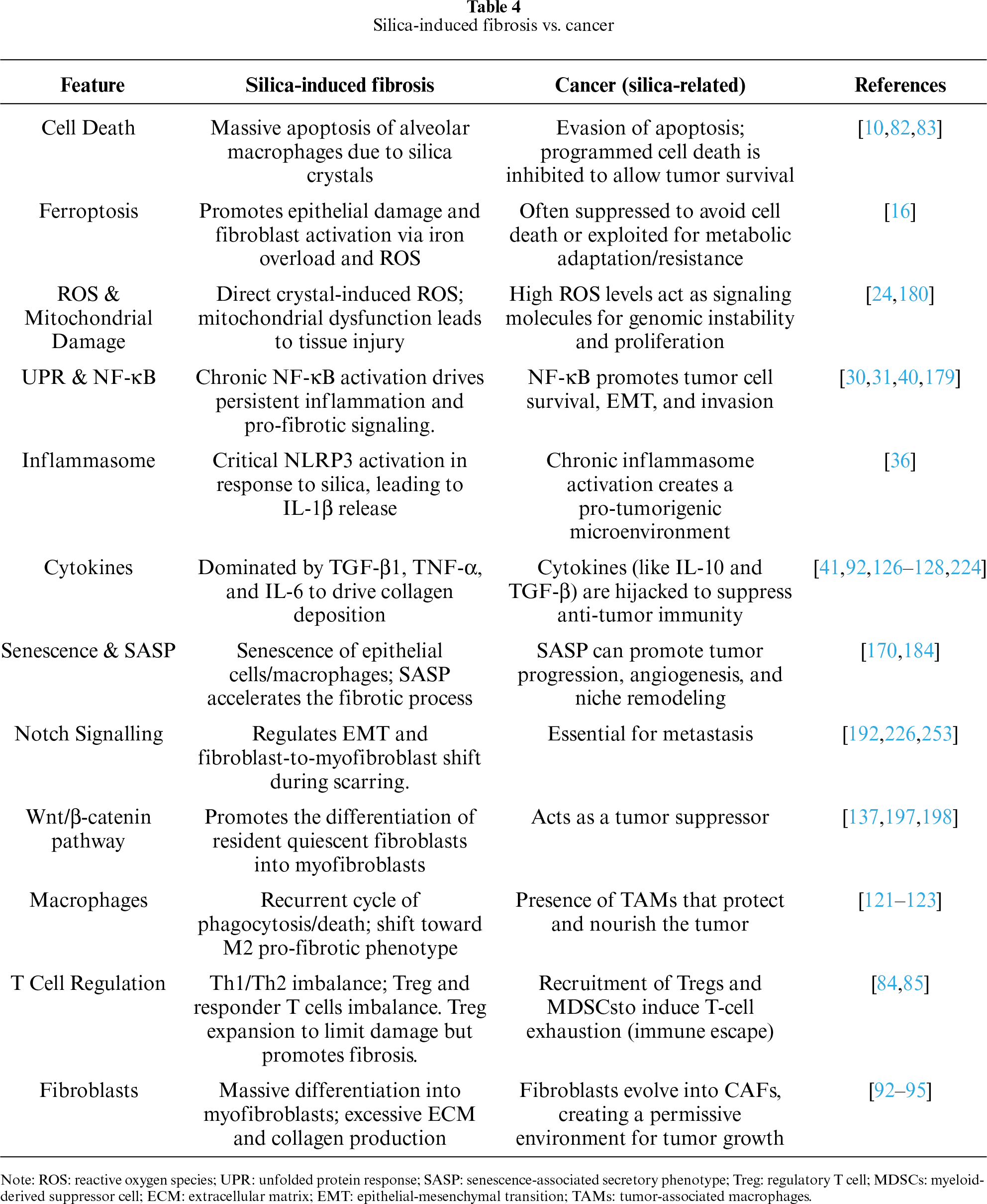
A hypothesis can be derived from our review, as illustrated in figure 2. As discussed previously, proinflammatory cytokines determine a context of chronic inflammation, however the immune system plays a pivotal role in the divergent pathogenesis of cancer vs. fibrosis, In cancer, the imbalance between Treg/Teff (ratio > 1) modulates an immune escape that, together with CAFs and the inactivation of Wnt/β-catenin, leads to cancer; instead if the Treg/Teff ratio is <1, a stromal reaction occurs that turns out to fibrosis. Cancer and fibrosis show different mechanisms only in the last pathogenic phases where the role the immune system overlaps with that of microenvironment: here different cells communicate with each other through intercellular communication, juxtacrine, and paracrine signaling and these messages seem to be tissue and cellular specific, because the same signal can play opposite role if it is activated or is inactivated. This leads to our working hypothesis because an important role emerges regarding to Wnt/β-catenin that controls, through other molecular signaling such as Notch and YAP/TAZ, the different fates, however these latter signaling pathways act in a tissue and cellular specific fashion. We could hypothesize that, despite its canonical oncogenic role, in this specific microenvironment if Wnt/β-catenin is inactivated the cancer is enabled, vice versa if it is activated the fibrosis occurs. If we could modulate this interplay, we would transform cancer in fibrosis: it would be an important success in the fight against cancer but for now it is just a working hypothesis. Nowadays it is known that fibrosis can transform in cancer: will it be possible a day to do otherwise?
5 Therapeutic Targets and Early Molecular Detection
Targeting Notch, TGF-β, or CAF-associated pathways offers promising therapeutic avenues to shift disease trajectories from progressive fibrosis or cancer toward resolution and prevention. These pathways are deeply interconnected, frequently co-activated in pathological stroma, and drive both fibrotic deposition and cancer progression. TGF-β inhibition directly reduces myofibroblast differentiation and ECM deposition. For example, relaxin-2, TGF-β receptor (TβRI) inhibitors, and integrin αvβ6 antagonists (e.g., BG00011) have demonstrated success in reducing fibrosis in lung, liver, and kidney models [254]. Besides its inhibitory action on the TGF-β pathway, the pleiotropic hormone relaxin attenuated fibrosis via a Wnt-dependent mechanism [137]. On the other hand, TGF-β inhibition (e.g., bintrafusp alfa or galunisertib) reduces CAF activation, reverses EMT, and lowers resistance to chemotherapy by breaking down the physical fibrotic barrier [254]. Given the Notch activation in both fibrosis and cancer, it is not surprising that inhibition of Notch would be therapeutic in these two conditions. Notch inhibition (e.g., via γ-secretase inhibitors—GSIs) reduces myofibroblast activation and triggers apoptosis of existing myofibroblasts, promoting the regression of established fibrosis [192]. Inhibiting Notch (specifically Notch3 or JAG1) reduces cancer cell migration, invasion, and EMT. Currently, clinical trials for Notch signaling, encompassing GSIs, ADAM inhibitors, antibodies targeting Notch receptors or ligands, Notch transcription complex inhibitors, and γ-secretase modulators (GSMs) are undergoing [253]. The challenge will lie in identifying methods to finely modulate Notch inhibition under both conditions. As to CAFs, several strategies have been proposed to either eliminate tumor-promoting CAFs or reprogram them. However, the plasticity and heterogeneity of CAFs complicate the understanding of their properties and present difficulties for clinical application [255]. At the moment, because of some therapeutic options with the same operational effect (i.e., inhibition), it is difficult to therapeutically steer the response towards a less harmful outcome. Moreover, while targeting the pathways of fibrosis/cancer shows efficacy in preclinical models, their critical role in normal tissue homeostasis requires precise, context-dependent, or selective therapeutic strategies to avoid adverse effects.
In order to detect disease before irreversible fibrosis occurs, effective immune phenotyping and longitudinal monitoring of high-risk silicosis patients—particularly those exposed to engineered stone or sandblasting—require transitioning from reliance on radiology to molecular-level surveillance. Key strategies involve monitoring for persistent macrophage activation, accelerated oxidative stress, and T-cell-mediated immune imbalance (Th1/Th2/Th17) using minimally invasive techniques [256]. High-risk monitoring should include measuring neopterin in serum/urine (marker of cellular immune response) and increased TNF-α or IL-1 release [256]. Elevated malondialdehyde (MDA), 8-isoprostanes, and reduced superoxide dismutase (SOD) could be considered as markers of oxidative stress and lipid peroxidation, as well as of disease progression, especially when used in combination with exhaled breath condensate (EBC) to measure volatile organic compounds (VOCs) and oxidative markers, offering a non-invasive method for longitudinal surveillance [257]. The investigation of leukocyte/lymphocyte subsets in silicosis patients led to the discovery that the proportions of memory B cells, naïve helper T cells, and the CD4+/CD8+ T cells’ ratio in the peripheral blood of patients with silicosis were significantly decreased, while the percentages of plasma cells, memory helper T cells, and regulatory T cells were significantly increased, as compared with healthy controls [258]. Profiling exosomes released by macrophages, containing miRNAs (e.g., miR-125a-5p, miR-107, miR-23a-3p) that regulate TGF-β and NF-κB pathways, in serum or BALF offers high-resolution detection of early fibrotic activity [172] could represent another chance, but perhaps it would need to better understand their practical limitations.
Given its nature as a narrative review, this work does not purport to be exhaustive. Rather, our primary objective is to delineate potential trajectories for future research and to stimulate further intellectual discourse on these topics. Whether our hypotheses are ultimately validated or refuted, we contend that this conceptual framework should contribute to the field. The limitations of this study are represented by the inherent constraints of a narrative review, which may result in the unintentional omission of certain relevant studies; the non-exhaustive characterization of the diverse signaling pathways involved in fibrosis and carcinogenesis; the potentially provocative nature of our conclusions, which, while not yet fully substantiated by experimental data, are intended to serve as conceptual catalysts for future investigations.
Starting from the analysis of silica exposure, the mechanisms underlying two distinct pathological outcomes were explored. The objective was to determine how different signals and cells that constitute microenvironment may determine what we consider individual susceptibility factors or the intrinsic physicochemical properties of the toxic substance that can exert a more decisive influence on the development of either silicosis or malignancy. This review examined the progressive cascade—characterized by DAMPs, ROS, chronic inflammation, and immune modulation—that culminates in either fibrosis or cancer. Perhaps, we should modify our approach by seeking to connect these complex interactions considering them as a whole, examining the cellular dialogue that can alter the microenvironment, since this appears to be the place where everything is crucial. In particular, it seems important to answer some questions regarding the microenvironment, where immune and non-immune cells share a physical space, producing mediators that allow them to communicate with each other.
In fact, the diverse signaling pathways governing intercellular communication are critical, as they appear to dictate these divergent cellular fates. As a provocative conceptual proposal, and considering that fibrosis can precede oncogenesis, the possibility of modulating the oncogenic trajectory toward a fibrotic phenotype through the Wnt/β-catenin pathway should be suggested. Its activation should lead to the subsequent activation of Notch and YAP/TAZ signaling, and this crosstalk requires more attention and further investigations.
In conclusion, an intricate network of cells, mediators (including cytokines) and signaling pathways are involved in the modification of the microenvironment to build either fibrosis or cancer. Advancing mechanistic resolution and translational validation of Notch, YAP/TAZ and Wnt/β-catenin pathways should be essential for transforming silicosis or pre-neoplastic microenvironment from descriptive frameworks into clinically actionable platforms. These efforts may ultimately enable the development of targeted strategies that mitigate either silicosis or cancer or halt the progression of silica-derived diseases.
Acknowledgement: Not applicable.
Funding Statement: The authors received no specific funding for this study.
Author Contributions: The authors confirm contribution to the paper as follows: Conceptualisation, Roberto Zefferino; writing—original draft preparation, review and editing, Roberto Zefferino, Massimo Conese, Aglaja Zefferino, Annalucia Carbone, Sante Di Gioia; visualisation, Massimo Conese; supervision, Roberto Zefferino. All authors reviewed and approved the final version of the manuscript.
Availability of Data and Materials: Not applicable.
Ethics Approval: Not applicable.
Conflicts of Interest: The authors declare no conflicts of interest.
References
1. Allison AC, Harington JS, Birbeck M. An examination of the cytotoxic effects of silica on macrophages. J Exp Med 1966;124(2):141–54. doi:10.1084/jem.124.2.141. [Google Scholar] [CrossRef]
2. Guha N, Straif K, Benbrahim-Tallaa L. The IARC monographs on the carcinogenicity of crystalline silica. Med Lav 2011;102(4):310–20; [Google Scholar]
3. The National Institute for Occupational Safety and Health. Health effects of occupational exposure to respirable crystalline silica. Washington, DC, USA: US Department of Health and Human Services, CDC, National Institute for Occupational Safety and Health; 2002. [Google Scholar]
4. Mazurek JM, Attfield MD. Silicosis mortality among young adults in the United States, 1968–2004. Am J Ind Med 2008;51(8):568–78. doi:10.1002/ajim.20597. [Google Scholar] [CrossRef]
5. Morfeld P. Respirable crystalline silica: rationale for classification according to the CLP* regulation and within the framework of the globally harmonised system (GHS) of classification and labelling of chemicals. 2010 [cited 2026 Jan 1]. Available from: https://safesilica.eu/wp-content/uploads/2021/07/Summary-Morfeld-Classification-CLP_GHS_RCS.pdf. [Google Scholar]
6. CEN EN 481. Workplace atmospheres: size fraction definitions for measurement of airborne particles in the workplace. Brussels, Belgium: Comité Européen de Normalisation; 1993. [Google Scholar]
7. Greim H. Silica, crystalline: quartz dust, cristobalite dust and tridymite dust (respirable fraction). The MAK collection for occupational health and safety. Weinheim, Germany: Verlag Wiley-VCH; 2000. p. 206–71. [Google Scholar]
8. Cocco P. Occupational exposure to silica and autoimmune diseases: a systematic review. Work Health 2025;1(1):2. doi:10.53941/wah.2025.100002. [Google Scholar] [CrossRef]
9. Erren TC, Glende CB, Morfeld P, Piekarski C. Is exposure to silica associated with lung cancer in the absence of silicosis? A meta-analytical approach to an important public health question. Int Arch Occup Environ Health 2009;82(8):997–1004. doi:10.1007/s00420-008-0387-0. [Google Scholar] [CrossRef]
10. Yang B, Liu X, Peng C, Meng X, Jia Q. Silicosis: from pathogenesis to therapeutics. Front Pharmacol 2025;16:1516200. doi:10.3389/fphar.2025.1516200. [Google Scholar] [CrossRef]
11. Hamilton RF Jr, Thakur SA, Holian A. Silica binding and toxicity in alveolar macrophages. Free Radic Biol Med 2008;44(7):1246–58. doi:10.1016/j.freeradbiomed.2007.12.027. [Google Scholar] [CrossRef]
12. Fubini B, Hubbard A. Reactive oxygen species (ROS) and reactive nitrogen species (RNS) generation by silica in inflammation and fibrosis. Free Radic Biol Med 2003;34(12):1507–16. doi:10.1016/s0891-5849(03)00149-7. [Google Scholar] [CrossRef]
13. Du SL, Zhou YT, Hu HJ, Lin L, Zhang ZQ. Silica-induced ROS in alveolar macrophages and its role on the formation of pulmonary fibrosis via polarizing macrophages into M2 phenotype: a review. Toxicol Mech Methods 2025;35(2):89–100. doi:10.1080/15376516.2024.2400323. [Google Scholar] [CrossRef]
14. Sato T, Shimosato T, Klinman DM. Silicosis and lung cancer: current perspectives. Lung Cancer 2018;9:91–101. doi:10.2147/lctt.s156376. [Google Scholar] [CrossRef]
15. Vallyathan V, Shi XL, Dalal NS, Irr W, Castranova V. Generation of free radicals from freshly fractured silica dust. Potential role in acute silica-induced lung injury. Am Rev Respir Dis 1988;138(5):1213–9. doi:10.1164/ajrccm/138.5.1213. [Google Scholar] [CrossRef]
16. Yan M, Zhang J, Ji G, Fang X, Mo C. Ferroptosis in pneumoconiosis: from molecular and cellular mechanisms to therapeutic strategies. Med Res 2025;1(2):239–56. doi:10.1002/mdr2.70020. [Google Scholar] [CrossRef]
17. Sun M, Sun Q, Li T et al. Silica nanoparticles induce liver lipid metabolism disorder via ACSL4-mediated ferroptosis. Environ Pollut 2024;359(10159):124590. doi:10.1016/j.envpol.2024.124590. [Google Scholar] [CrossRef]
18. Liu T, Bao R, Wang Q et al. SiO2-induced ferroptosis in macrophages promotes the development of pulmonary fibrosis in silicosis models. Toxicol Res 2022;11(1):42–51. doi:10.1093/toxres/tfab105. [Google Scholar] [CrossRef]
19. Yan B, Ai Y, Sun Q et al. Membrane damage during ferroptosis is caused by oxidation of phospholipids catalyzed by the oxidoreductases POR and CYB5R1. Mol Cell 2021;81(2):355–69.e10. doi:10.1016/j.molcel.2020.11.024. [Google Scholar] [CrossRef]
20. Proneth B, Conrad M. Ferroptosis and necroinflammation, a yet poorly explored link. Cell Death Differ 2019;26(1):14–24. doi:10.1038/s41418-018-0173-9. [Google Scholar] [CrossRef]
21. Wu R, Li X, Ma N et al. Bacterial quorum sensing molecules promote allergic airway inflammation by activating the retinoic acid response. iScience 2020;23(7):101288. doi:10.1016/j.isci.2020.101288. [Google Scholar] [CrossRef]
22. Zhang Z, Li J, Zheng W et al. Peripheral lymphoid volume expansion and maintenance are controlled by gut microbiota via RALDH+ dendritic cells. Immunity 2016;44(2):330–42. doi:10.1016/j.immuni.2016.01.004. [Google Scholar] [CrossRef]
23. Li X, Tan J, Xiong W, Feng Y, Zhang Z. Silica-induced ferroptosis activates retinoic acid signaling in dendritic cells to drive inflammation and fibrosis in silicosis. Int Immunopharmacol 2025;149(409–422):114244. doi:10.1016/j.intimp.2025.114244. [Google Scholar] [CrossRef]
24. Zhou Q, Yi G, Chang M et al. Activation of Sirtuin3 by honokiol ameliorates alveolar epithelial cell senescence in experimental silicosis via the cGAS-STING pathway. Redox Biol 2024;74(6):103224. doi:10.1016/j.redox.2024.103224. [Google Scholar] [CrossRef]
25. Twig G, Hyde B, Shirihai OS. Mitochondrial fusion, fission and autophagy as a quality control axis: the bioenergetic view. Biochim Biophys Acta 2008;1777(9):1092–7. doi:10.1016/j.bbabio.2008.05.001. [Google Scholar] [CrossRef]
26. Zhou YT, Li S, Du SL, Zhao JH, Cai YQ, Zhang ZQ. The multifaceted role of macrophage mitophagy in SiO2-induced pulmonary fibrosis: a brief review. J Appl Toxicol 2024;44(12):1854–67. doi:10.1002/jat.4612. [Google Scholar] [CrossRef]
27. Morgan MJ, Liu ZG. Crosstalk of reactive oxygen species and NF-κB signaling. Cell Res 2011;21(1):103–15. doi:10.1038/cr.2010.178. [Google Scholar] [CrossRef]
28. Lingappan K. NF-κB in oxidative stress. Curr Opin Toxicol 2018;7:81–6. doi:10.1016/j.cotox.2017.11.002. [Google Scholar] [CrossRef]
29. Kang JL, Pack IS, Hong SM, Lee HS, Castranova V. Silica induces nuclear factor-κB activation through tyrosine phosphorylation of IκB-α in RAW264.7 macrophages. Toxicol Appl Pharmacol 2000;169(1):59–65. doi:10.1006/taap.2000.9039. [Google Scholar] [PubMed] [CrossRef]
30. Chen F, Lu Y, Demers LM et al. Role of hydroxyl radical in silica-induced NF-κB activation in macrophages. Ann Clin Lab Sci 1998;28(1):1–13; [Google Scholar]
31. Ding M, Chen F, Shi X, Yucesoy B, Mossman B, Vallyathan V. Diseases caused by silica: mechanisms of injury and disease development. Int Immunopharmacol 2002;2(2–3):173–82. doi:10.1016/s1567-5769(01)00170-9. [Google Scholar] [CrossRef]
32. Kang JL, Jung HJ, Lee K, Kim HR. Src tyrosine kinases mediate crystalline silica-induced NF-κB activation through tyrosine phosphorylation of IκB-α and p65 NF-κB in RAW 264.7 macrophages. Toxicol Sci 2006;90(2):470–7. doi:10.1093/toxsci/kfj096. [Google Scholar] [CrossRef]
33. Kang JL, Go YH, Hur KC, Castranova V. Silica-induced nuclear factor-κB activation: involvement of reactive oxygen species and protein tyrosine kinase activation. J Toxicol Environ Health A 2000;60(1):27–46; [Google Scholar]
34. Kang JL, Lee HS, Pack IS, Hur KC, Castranova V. Phosphoinositide 3-kinase activity leads to silica-induced NF-κB activation through interacting with tyrosine-phosphorylated IκB-α and contributing to tyrosine phosphorylation of p65 NF-κB. Mol Cell Biochem 2003;248(1–2):17–24. doi:10.1023/a:1024163630166. [Google Scholar] [CrossRef]
35. Castranova V. Signaling pathways controlling the production of inflammatory mediators in response to crystalline silica exposure: role of reactive oxygen/nitrogen species. Free Radic Biol Med 2004;37(7):916–25. doi:10.1016/j.freeradbiomed.2004.05.032. [Google Scholar] [CrossRef]
36. Hornung V, Bauernfeind F, Halle A et al. Silica crystals and aluminum salts activate the NALP3 inflammasome through phagosomal destabilization. Nat Immunol 2008;9(8):847–56. doi:10.1038/ni.1631. [Google Scholar] [CrossRef]
37. Dominic A, Le N-T, Takahashi M. Loop between NLRP3 inflammasome and reactive oxygen species. Antioxid Redox Signal 2022;36(10–12):784–96. doi:10.1089/ars.2020.8257. [Google Scholar] [CrossRef]
38. Lam M, Barry KT, Hodges CJ et al. NLRP3 deficiency abrogates silica-induced neutrophil infiltration, pulmonary damage and fibrosis. Respir Res 2025;26(1):109. doi:10.1186/s12931-025-03192-y. [Google Scholar] [CrossRef]
39. Menu P, Mayor A, Zhou R et al. ER stress activates the NLRP3 inflammasome via an UPR-independent pathway. Cell Death Dis 2012;3(1):e261. doi:10.1038/cddis.2011.132. [Google Scholar] [CrossRef]
40. Choi EH, Park SJ. TXNIP: a key protein in the cellular stress response pathway and a potential therapeutic target. Exp Mol Med 2023;55(7):1348–56. doi:10.1038/s12276-023-01019-8. [Google Scholar] [CrossRef]
41. Driscoll KE, Guthrie GD. Crystalline silica and silicosis. In: McQueen CA, editor. Comprehensive toxicology. 2nd edition. Amsterdam, The Netherlands: Elsevier; 2010. p. 331–50. [Google Scholar]
42. Kang JL, Lee K, Castranova V. Nitric oxide up-regulates DNA-binding activity of nuclear factor-κB in macrophages stimulated with silica and inflammatory stimulants. Mol Cell Biochem 2000;215(1–2):1–9. doi:10.1023/a:1026581301366. [Google Scholar] [CrossRef]
43. Zeidler P, Hubbs A, Battelli L, Castranova V. Role of inducible nitric oxide synthase-derived nitric oxide in silica-induced pulmonary inflammation and fibrosis. J Toxicol Environ Health A 2004;67(13):1001–26. doi:10.1080/15287390490447296. [Google Scholar] [CrossRef]
44. Driscoll KE. Macrophage inflammatory proteins: biology and role in pulmonary inflammation. Exp Lung Res 1994;20(6):473–90. doi:10.3109/01902149409031733. [Google Scholar] [CrossRef]
45. Driscoll KE, Hassenbein DG, Carter J et al. Macrophage inflammatory proteins 1 and 2: expression by rat alveolar macrophages, fibroblasts, and epithelial cells and in rat lung after mineral dust exposure. Am J Respir Cell Mol Biol 1993;8(3):311–8. doi:10.1165/ajrcmb/8.3.311. [Google Scholar] [CrossRef]
46. Driscoll KE, Hassenbein DG, Carter JM, Kunkel SL, Quinlan TR, Mossman BT. TNF α and increased chemokine expression in rat lung after particle exposure. Toxicol Lett 1995;82–83:483–9. doi:10.1016/0378-4274(95)03578-8. [Google Scholar] [CrossRef]
47. Driscoll K, Guthrie G. Crystalline silica and silicosis. In: Roth R, editor. Comprehensive toxicology Vol 8 toxicology of the respiratory system. New York, NY, USA: Elsevier Science, 1997. p. 373–92. [Google Scholar]
48. Oghiso Y, Kubota Y. Enhanced interleukin 1 production by alveolar macrophages and increase in Ia-positive lung cells in silica-exposed rats. Microbiol Immunol 1986;30(11):1189–98. doi:10.1111/j.1348-0421.1986.tb03047.x. [Google Scholar] [CrossRef]
49. Zhang J, Yang X, Yang Y et al. NF-κB mediates silica-induced pulmonary inflammation by promoting the release of IL-1β in macrophages. Environ Toxicol 2022;37(9):2235–43. doi:10.1002/tox.23590. [Google Scholar] [CrossRef]
50. Srivastava KD, Rom WN, Jagirdar J, Yie TA, Gordon T, Tchou-Wong KM. Crucial role of interleukin-1β and nitric oxide synthase in silica-induced inflammation and apoptosis in mice. Am J Respir Crit Care Med 2002;165(4):527–33. doi:10.1164/ajrccm.165.4.2106009. [Google Scholar] [CrossRef]
51. Porter DW, Hubbs AF, Mercer R et al. Progression of lung inflammation and damage in rats after cessation of silica inhalation. Toxicol Sci 2004;79(2):370–80. doi:10.1093/toxsci/kfh110. [Google Scholar] [CrossRef]
52. Dostert C, Petrilli V, Van Bruggen R, Steele C, Mossman BT, Tschopp J. Innate immune activation through Nalp3 inflammasome sensing of asbestos and silica. Science 2008;320(5876):674–7. doi:10.1126/science.1156995. [Google Scholar] [CrossRef]
53. Subramanian N, Bray MA. Interleukin 1 releases histamine from human basophils and mast cells in vitro. J Immunol 1987;138(1):271–5. doi:10.4049/jimmunol.138.1.271. [Google Scholar] [CrossRef]
54. Rimal B, Greenberg AK, Rom WN. Basic pathogenetic mechanisms in silicosis: current understanding. Curr Opin Pulm Med 2005;11(2):169–73. doi:10.1097/01.mcp.0000152998.11335.24. [Google Scholar] [CrossRef]
55. Feghali CA, Wright TM. Cytokines in acute and chronic inflammation. Front Biosci 1997;2(4):d12–26. doi:10.2741/a171. [Google Scholar] [CrossRef]
56. Cekici A, Kantarci A, Hasturk H, Van Dyke TE. Inflammatory and immune pathways in the pathogenesis of periodontal disease. Periodontol 2000 2014;64(1):57–80. doi:10.1111/prd.12002. [Google Scholar] [CrossRef]
57. Levy BD, Clish CB, Schmidt B, Gronert K, Serhan CN. Lipid mediator class switching during acute inflammation: signals in resolution. Nat Immunol 2001;2(7):612–9. doi:10.1038/89759. [Google Scholar] [CrossRef]
58. Van Dyke TE. Control of inflammation and periodontitis. Periodontol 2000 2007;45(1):158–66. doi:10.1111/j.1600-0757.2007.00229.x. [Google Scholar] [CrossRef]
59. Serhan CN. A search for endogenous mechanisms of anti-inflammation uncovers novel chemical mediators: missing links to resolution. Histochem Cell Biol 2004;122(4):305–21. doi:10.1007/s00418-004-0695-8. [Google Scholar] [CrossRef]
60. Kantarci A, Van Dyke TE. Lipoxins in chronic inflammation. Crit Rev Oral Biol Med 2003;14(1):4–12; [Google Scholar]
61. Van Dyke TE, Serhan CN. Resolution of inflammation: a new paradigm for the pathogenesis of periodontal diseases. J Dent Res 2003;82(2):82–90. doi:10.1177/154405910308200202. [Google Scholar] [CrossRef]
62. Maddox JF, Hachicha M, Takano T, Petasis NA, Fokin VV, Serhan CN. Lipoxin A4 stable analogs are potent mimetics that stimulate human monocytes and THP-1 cells via a G-protein-linked lipoxin A4 receptor. J Biol Chem 1997;272(11):6972–8. doi:10.1074/jbc.272.11.6972. [Google Scholar] [CrossRef]
63. Maddox JF, Serhan CN. Lipoxin A4 and B4 are potent stimuli for human monocyte migration and adhesion: selective inactivation by dehydrogenation and reduction. J Exp Med 1996;183(1):137–46. doi:10.1084/jem.183.1.137. [Google Scholar] [CrossRef]
64. Serhan CN, Fiore S, Brezinski DA, Lynch S. Lipoxin A4 metabolism by differentiated HL-60 cells and human monocytes: conversion to novel 15-oxo and dihydro products. Biochemistry 1993;32(25):6313–9. doi:10.1021/bi00076a002. [Google Scholar] [CrossRef]
65. Kaneto H, Sasaki S, Yamamoto H et al. Detection of hypermethylation of the p16INK4A gene promoter in chronic hepatitis and cirrhosis associated with hepatitis B or C virus. Gut 2001;48(3):372–7. doi:10.1136/gut.48.3.372. [Google Scholar] [CrossRef]
66. Ramadan A, Hashim M, Abouzid A, Swellam M. Clinical impact of PTEN methylation status as a prognostic marker for breast cancer. J Genet Eng Biotechnol 2021;19(1):66. doi:10.1186/s43141-021-00169-4. [Google Scholar] [CrossRef]
67. Beacon TH, Delcuve GP, Lopez C et al. The dynamic broad epigenetic (H3K4me3, H3K27ac) domain as a mark of essential genes. Clin Epigenetics 2021;13(1):138. doi:10.1186/s13148-021-01126-1. [Google Scholar] [CrossRef]
68. Borkiewicz L. Histone 3 lysine 27 trimethylation signature in breast cancer. Int J Mol Sci 2021;22(23):12853. doi:10.3390/ijms222312853. [Google Scholar] [CrossRef]
69. Hamad H, Othman G, Kardagh R. Investigating the role of HOTAIR and MALAT1 long noncoding RNAs and their relations with bone marrow environment in acute myeloid leukemia subtypes: biomarkers and treatment response. Adv Hematol 2025;2025(1):3459924. doi:10.1155/ah/3459924. [Google Scholar] [CrossRef]
70. Lee SH, Li Y, Kim H, Eum S, Park K, Lee CH. The role of EZH1 and EZH2 in development and cancer. BMB Rep 2022;55(12):595–601. doi:10.5483/bmbrep.2022-0174. [Google Scholar] [CrossRef]
71. Hao L, Wu W, Xu Y et al. LncRNA-MALAT1: a key participant in the occurrence and development of cancer. Molecules 2023;28(5):2126. doi:10.3390/molecules28052126. [Google Scholar] [CrossRef]
72. Basta MD, Petruk S, Mazo A, Walker JL. Fibrosis—the tale of H3K27 histone methyltransferases and demethylases. Front Cell Dev Biol 2023;11:1193344. doi:10.3389/fcell.2023.1193344. [Google Scholar] [CrossRef]
73. Zheng M, Chen Z, Xie J et al. The genetic and epigenetic toxicity of silica nanoparticles: an updated review. Int J Nanomed 2024;19:13901–23. doi:10.2147/ijn.s486858. [Google Scholar] [CrossRef]
74. Sang Y, Liu J, Li X et al. The effect of SiNPs on DNA methylation of genome in mouse spermatocytes. Environ Sci Pollut Res Int 2021;28(32):43684–97. doi:10.1007/s11356-021-13459-8. [Google Scholar] [CrossRef]
75. Zhou G, Ren L, Yin H et al. The alterations of miRNA and mRNA expression profile and their integration analysis induced by silica nanoparticles in spermatocyte cells. NanoImpact 2021;23(3):100348. doi:10.1016/j.impact.2021.100348. [Google Scholar] [CrossRef]
76. Liu Y, Li H, Xiao T, Lu Q. Epigenetics in immune-mediated pulmonary diseases. Clin Rev Allergy Immunol 2013;45(3):314–30. doi:10.1007/s12016-013-8398-3. [Google Scholar] [CrossRef]
77. Wang YX, Yao W, Gao B, Yao YC, Wu YM, Hao CF. DNA methylation levels of genome in cFb transdifferentiation induced by SiO2 in rats. Chin J Ind Hyg Occup Dis 2012;30(5):326–30. [Google Scholar]
78. Umemura S, Fujimoto N, Hiraki A et al. Aberrant promoter hypermethylation in serum DNA from patients with silicosis. Carcinogenesis 2008;29(9):1845–9. doi:10.1093/carcin/bgn169. [Google Scholar] [CrossRef]
79. Blanco D, Vicent S, Fraga MF et al. Molecular analysis of a multistep lung cancer model induced by chronic inflammation reveals epigenetic regulation of p16 and activation of the DNA damage response pathway. Neoplasia 2007;9(10):840–52. doi:10.1593/neo.07517. [Google Scholar] [CrossRef]
80. Bartkova J, Rezaei N, Liontos M et al. Oncogene-induced senescence is part of the tumorigenesis barrier imposed by DNA damage checkpoints. Nature 2006;444(7119):633–7. doi:10.1038/nature05268. [Google Scholar] [CrossRef]
81. Gorgoulis VG, Vassiliou LV, Karakaidos P et al. Activation of the DNA damage checkpoint and genomic instability in human precancerous lesions. Nature 2005;434(7035):907–13. doi:10.1038/nature03485. [Google Scholar] [CrossRef]
82. Helton ES, Chen X. p53 modulation of the DNA damage response. J Cell Biochem 2007;100(4):883–96. doi:10.1002/jcb.21091. [Google Scholar] [CrossRef]
83. Jacobs JJ, de Lange T. p16INK4a as a second effector of the telomere damage pathway. Cell Cycle 2005;4(10):1364–8. doi:10.4161/cc.4.10.2104. [Google Scholar] [PubMed] [CrossRef]
84. Wang Y, Li J, Nakahata S, Iha H. Complex role of regulatory T cells (Tregs) in the tumor microenvironment: their molecular mechanisms and bidirectional effects on cancer progression. Int J Mol Sci 2024;25(13):7346. doi:10.3390/ijms25137346. [Google Scholar] [CrossRef]
85. Kang HX, Fu YF, Wu H et al. The immune dysregulation of fibrosis: insights into immune-fibrotic crosstalk and potential therapeutic targets. Front Immunol 2026;17:1770516. doi:10.3389/fimmu.2026.1770516. [Google Scholar] [CrossRef]
86. Tang W, Liu F, Chen Y et al. Reduction of IL-17A might suppress the Th1 response and promote the Th2 response by boosting the function of Treg cells during silica-induced inflammatory response in vitro. Mediat Inflamm 2014;2014(2):570894. doi:10.1155/2014/570894. [Google Scholar] [CrossRef]
87. Maggi E, Munari E, Landolina N, Mariotti FR, Azzarone B, Moretta L. T cell landscape in the microenvironment of human solid tumors. Immunol Lett 2024;270(1):106942. doi:10.1016/j.imlet.2024.106942. [Google Scholar] [CrossRef]
88. Downs-Canner S, Berkey S, Delgoffe GM et al. Suppressive IL-17A+Foxp3+ and ex-Th17 IL-17AnegFoxp3+ Treg cells are a source of tumour-associated Treg cells. Nat Commun 2017;8(1):14649. doi:10.1038/ncomms14649. [Google Scholar] [CrossRef]
89. Najafi S, Mirshafiey A. The role of T helper 17 and regulatory T cells in tumor microenvironment. Immunopharmacol Immunotoxicol 2019;41(1):16–24. doi:10.1080/08923973.2019.1566925. [Google Scholar] [CrossRef]
90. Pan Y, Yang W, Tang B et al. The protective and pathogenic role of Th17 cell plasticity and function in the tumor microenvironment. Front Immunol 2023;14:1192303. doi:10.3389/fimmu.2023.1192303. [Google Scholar] [CrossRef]
91. Lee S, Matsuzaki H, Kumagai-Takei N et al. Silica exposure and altered regulation of autoimmunity. Environ Health Prev Med 2014;19(5):322–9. doi:10.1007/s12199-014-0403-9. [Google Scholar] [CrossRef]
92. Hinz B. Formation and function of the myofibroblast during tissue repair. J Investig Dermatol 2007;127(3):526–37. doi:10.1038/sj.jid.5700613. [Google Scholar] [CrossRef]
93. Wipff PJ, Rifkin DB, Meister JJ, Hinz B. Myofibroblast contraction activates latent TGF-β1 from the extracellular matrix. J Cell Biol 2007;179(6):1311–23. doi:10.1083/jcb.200704042. [Google Scholar] [CrossRef]
94. Wynn TA, Ramalingam TR. Mechanisms of fibrosis: therapeutic translation for fibrotic disease. Nat Med 2012;18(7):1028–40. doi:10.1038/nm.2807. [Google Scholar] [CrossRef]
95. Shay G, Lynch CC, Fingleton B. Moving targets: emerging roles for MMPs in cancer progression and metastasis. Matrix Biol 2015;44–46:200–6. doi:10.1016/j.matbio.2015.01.019. [Google Scholar] [CrossRef]
96. Fireman EM, Fireman Klein E. Association between silicosis and autoimmune disease. Curr Opin Allergy Clin Immunol 2024;24(2):45–50. doi:10.1097/aci.0000000000000966. [Google Scholar] [CrossRef]
97. Nagata S. Fas and Fas ligand: a death factor and its receptor. Adv Immunol 1994;57:129–44. doi:10.1016/s0065-2776(08)60672-0. [Google Scholar] [CrossRef]
98. Nagata S, Golstein P. The Fas death factor. Science 1995;267(5203):1449–56. doi:10.1126/science.7533326. [Google Scholar] [CrossRef]
99. Takata-Tomokuni A, Ueki A, Shiwa M et al. Detection, epitope-mapping and function of anti-Fas autoantibody in patients with silicosis. Immunology 2005;116(1):21–9. doi:10.1111/j.1365-2567.2005.02192.x. [Google Scholar] [CrossRef]
100. Ueki A, Isozaki Y, Tomokuni A et al. Intramolecular epitope spreading among anti-caspase-8 autoantibodies in patients with silicosis, systemic sclerosis and systemic lupus erythematosus, as well as in healthy individuals. Clin Exp Immunol 2002;129(3):556–61. doi:10.1046/j.1365-2249.2002.01939.x. [Google Scholar] [CrossRef]
101. Cascino I, Papoff G, Eramo A, Ruberti G. Soluble Fas/Apo-1 splicing variants and apoptosis. Front Biosci 1996;1(4):d12–8. doi:10.2741/a112. [Google Scholar] [CrossRef]
102. Cheng J, Zhou T, Liu C et al. Protection from Fas-mediated apoptosis by a soluble form of the Fas molecule. Science 1994;263(5154):1759–62. doi:10.1126/science.7510905. [Google Scholar] [CrossRef]
103. Tomokuni A, Aikoh T, Matsuki T et al. Elevated soluble Fas/APO-1 (CD95) levels in silicosis patients without clinical symptoms of autoimmune diseases or malignant tumours. Clin Exp Immunol 1997;110(2):303–9. doi:10.1111/j.1365-2249.1997.tb08332.x. [Google Scholar] [CrossRef]
104. Otsuki T, Tomokuni A, Sakaguchi H, Hyodoh F, Kusaka M, Ueki A. Reduced expression of the inhibitory genes for fas-mediated apoptosis in silicosis patients; 2000, 42(4):163–8. [Google Scholar]
105. Guery L, Hugues S. Th17 cell plasticity and functions in cancer immunity. Biomed Res Int 2015;2015:314620. doi:10.1155/2015/314620. [Google Scholar] [CrossRef]
106. Cope A, Le Friec G, Cardone J, Kemper C. The Th1 life cycle: molecular control of IFN-gamma to IL-10 switching. Trends Immunol 2011;32(6):278–86. doi:10.1016/j.it.2011.03.010. [Google Scholar] [CrossRef]
107. Hu X, Liu X, Moisan J et al. Synthetic RORgamma agonists regulate multiple pathways to enhance antitumor immunity. Oncoimmunology 2016;5(12):e1254854. doi:10.1080/2162402x.2016.1254854. [Google Scholar] [CrossRef]
108. Li C, Xu X, Wei S et al. Tumor-associated macrophages: potential therapeutic strategies and future prospects in cancer. J Immunother Cancer 2021;9(1):e001341. doi:10.1136/jitc-2020-001341. [Google Scholar] [CrossRef]
109. Gazzaniga S, Bravo AI, Guglielmotti A et al. Targeting tumor-associated macrophages and inhibition of MCP-1 reduce angiogenesis and tumor growth in a human melanoma xenograft. J Investig Dermatol 2007;127(8):2031–41. doi:10.1038/sj.jid.5700827. [Google Scholar] [CrossRef]
110. Lin Y, Xu J, Lan H. Tumor-associated macrophages in tumor metastasis: biological roles and clinical therapeutic applications. J Hematol Oncol 2019;12(1):76. doi:10.1186/s13045-019-0760-3. [Google Scholar] [CrossRef]
111. Jiang Y, Li Y, Zhu B. T-cell exhaustion in the tumor microenvironment. Cell Death Dis 2015;6(6):e1792. doi:10.1038/cddis.2015.162. [Google Scholar] [CrossRef]
112. Wang D, Yang L, Yue D et al. Macrophage-derived CCL22 promotes an immunosuppressive tumor microenvironment via IL-8 in malignant pleural effusion. Cancer Lett 2019;452:244–53. doi:10.1016/j.canlet.2019.03.040. [Google Scholar] [CrossRef]
113. Zhang Z, Liu S, Zhang B, Qiao L, Zhang Y, Zhang Y. T cell dysfunction and exhaustion in cancer. Front Cell Dev Biol 2020;8:17. doi:10.3389/fcell.2020.00017. [Google Scholar] [CrossRef]
114. Munn DH, Sharma MD, Baban B et al. GCN2 kinase in T cells mediates proliferative arrest and anergy induction in response to indoleamine 2,3-dioxygenase. Immunity 2005;22(5):633–42. doi:10.1016/j.immuni.2005.03.013. [Google Scholar] [CrossRef]
115. Lee GK, Park HJ, Macleod M, Chandler P, Munn DH, Mellor AL. Tryptophan deprivation sensitizes activated T cells to apoptosis prior to cell division. Immunology 2002;107(4):452–60. doi:10.1046/j.1365-2567.2002.01526.x. [Google Scholar] [CrossRef]
116. Gruarin P, Maglie S, De Simone M et al. Eomesodermin controls a unique differentiation program in human IL-10 and IFN-gamma coproducing regulatory T cells. Eur J Immunol 2019;49(1):96–111. doi:10.1002/eji.201847722. [Google Scholar] [CrossRef]
117. Mascanfroni ID, Takenaka MC, Yeste A et al. Metabolic control of type 1 regulatory T cell differentiation by AHR and HIF1-α. Nat Med 2015;21(6):638–46. doi:10.1038/nm.3868. [Google Scholar] [CrossRef]
118. Roncarolo MG, Gregori S, Bacchetta R, Battaglia M, Gagliani N. The biology of t regulatory type 1 cells and their therapeutic application in immune-mediated diseases. Immunity 2018;49(6):1004–19. doi:10.1016/j.immuni.2018.12.001. [Google Scholar] [CrossRef]
119. Alfen JS, Larghi P, Facciotti F et al. Intestinal IFN-gamma-producing type 1 regulatory T cells coexpress CCR5 and programmed cell death protein 1 and downregulate IL-10 in the inflamed guts of patients with inflammatory bowel disease. J Allergy Clin Immunol 2018;142(5):1537–47.e8. doi:10.1016/j.jaci.2017.12.984. [Google Scholar] [CrossRef]
120. Bonnal RJP, Rossetti G, Lugli E et al. Clonally expanded EOMES(+) Tr1-like cells in primary and metastatic tumors are associated with disease progression. Nat Immunol 2021;22(6):735–45. doi:10.1038/s41590-021-00930-4. [Google Scholar] [CrossRef]
121. Yang H, Cheng H, Dai R, Shang L, Zhang X, Wen H. Macrophage polarization in tissue fibrosis. PeerJ 2023;11:e16092; [Google Scholar]
122. Zhang W, Wang M, Ji C, Liu X, Gu B, Dong T. Macrophage polarization in the tumor microenvironment: emerging roles and therapeutic potentials. Biomed Pharmacother 2024;177(3):116930. doi:10.1016/j.biopha.2024.116930. [Google Scholar] [CrossRef]
123. Chen S, Li S, Wang H. Remodeling tumor-associated macrophages in the tumor microenvironment. Oncol Transl Med 2024;10(6):281–5. doi:10.1097/ot9.0000000000000063. [Google Scholar] [CrossRef]
124. Liu S, Zhang Y, Wan F et al. The regulatory role of FABP4 in macrophage polarization and lipid accumulation in silicosis. Cell Signal 2025;135(2):112041. doi:10.1016/j.cellsig.2025.112041. [Google Scholar] [CrossRef]
125. Sun D, Wang Y, Ma R, Dilixiati N, Song Y, Ye Q. SPP1+ macrophages contribute to silica-induced pulmonary inflammation and fibrosis via the ERK/JNK signaling pathway. Am J Respir Cell Mol Biol 2026;74(7):936–48. doi:10.1093/ajrcmb/aanag027. [Google Scholar] [CrossRef]
126. Thannickal VJ, Lee DY, White ES et al. Myofibroblast differentiation by transforming growth factor-β1 is dependent on cell adhesion and integrin signaling via focal adhesion kinase. J Biol Chem 2003;278(14):12384–9. doi:10.1074/jbc.m208544200. [Google Scholar] [CrossRef]
127. Scharenberg MA, Pippenger BE, Sack R et al. TGF-β-induced differentiation into myofibroblasts involves specific regulation of two MKL1 isoforms. J Cell Sci 2014;127(Pt 5):1079–91. doi:10.1242/jcs.142075. [Google Scholar] [CrossRef]
128. Foster DS, Jones RE, Ransom RC, Longaker MT, Norton JA. The evolving relationship of wound healing and tumor stroma. JCI Insight 2018;3(18):e99911. doi:10.1172/jci.insight.99911. [Google Scholar] [CrossRef]
129. Carthy JM. TGFβ signaling and the control of myofibroblast differentiation: implications for chronic inflammatory disorders. J Cell Physiol 2018;233(1):98–106. doi:10.1002/jcp.25879. [Google Scholar] [CrossRef]
130. Uehara T, Ainslie GR, Kutanzi K et al. Molecular mechanisms of fibrosis-associated promotion of liver carcinogenesis. Toxicol Sci 2013;132(1):53–63. doi:10.1093/toxsci/kfs342. [Google Scholar] [CrossRef]
131. Lederer DJ, Martinez FJ. Idiopathic pulmonary fibrosis. N Engl J Med 2018;378(19):1811–23. doi:10.1056/nejmra1705751. [Google Scholar] [CrossRef]
132. Zhao M, Dumur CI, Holt SE, Beckman MJ, Elmore LW. Multipotent adipose stromal cells and breast cancer development: think globally, act locally. Mol Carcinog 2010;49(11):923–7. doi:10.1002/mc.20675. [Google Scholar] [PubMed] [CrossRef]
133. Cox TR, Erler JT. Molecular pathways: connecting fibrosis and solid tumor metastasis. Clin Cancer Res 2014;20(14):3637–43. doi:10.1158/1078-0432.ccr-13-1059. [Google Scholar] [CrossRef]
134. Yoon JH, Nouraie M, Chen X et al. Characteristics of lung cancer among patients with idiopathic pulmonary fibrosis and interstitial lung disease—analysis of institutional and population data. Respir Res 2018;19(1):195. doi:10.1186/s12931-018-0899-4. [Google Scholar] [CrossRef]
135. Tomassetti S, Gurioli C, Ryu JH et al. The impact of lung cancer on survival of idiopathic pulmonary fibrosis. Chest 2015;147(1):157–64. doi:10.1378/chest.14-0359. [Google Scholar] [CrossRef]
136. Selman M, King TE, Pardo A, American Thoracic S, European Respiratory S, American College of Chest P. Idiopathic pulmonary fibrosis: prevailing and evolving hypotheses about its pathogenesis and implications for therapy. Ann Intern Med 2001;134(2):136–51. doi:10.7326/0003-4819-134-2-200101160-00015. [Google Scholar] [CrossRef]
137. Somanader DVN, Zhao P, Widdop RE, Samuel CS. The involvement of the Wnt/β-catenin signaling cascade in fibrosis progression and its therapeutic targeting by relaxin. Biochem Pharmacol 2024;223(12):116130. doi:10.1016/j.bcp.2024.116130. [Google Scholar] [CrossRef]
138. Fernandez IE, Eickelberg O. The impact of TGF-β on lung fibrosis: from targeting to biomarkers. Proc Am Thorac Soc 2012;9(3):111–6; [Google Scholar]
139. Xaubet A, Marin-Arguedas A, Lario S et al. Transforming growth factor-β1 gene polymorphisms are associated with disease progression in idiopathic pulmonary fibrosis. Am J Respir Crit Care Med 2003;168(4):431–5. doi:10.1164/rccm.200210-1165oc. [Google Scholar] [CrossRef]
140. Dancer RC, Wood AM, Thickett DR. Metalloproteinases in idiopathic pulmonary fibrosis. Eur Respir J 2011;38(6):1461–7. doi:10.1183/09031936.00024711. [Google Scholar] [CrossRef]
141. Bargagli E, Olivieri C, Bennett D, Prasse A, Muller-Quernheim J, Rottoli P. Oxidative stress in the pathogenesis of diffuse lung diseases: a review. Respir Med 2009;103(9):1245–56. doi:10.1016/j.rmed.2009.04.014. [Google Scholar] [CrossRef]
142. Crawford SE, Stellmach V, Murphy-Ullrich JE et al. Thrombospondin-1 is a major activator of TGF-β1 in vivo. Cell 1998;93(7):1159–70. doi:10.1016/s0092-8674(00)81460-9. [Google Scholar] [CrossRef]
143. Hinz B. Tissue stiffness, latent TGF-β1 activation, and mechanical signal transduction: implications for the pathogenesis and treatment of fibrosis. Curr Rheumatol Rep 2009;11(2):120–6. doi:10.1007/s11926-009-0017-1. [Google Scholar] [CrossRef]
144. Margadant C, Sonnenberg A. Integrin-TGF-β crosstalk in fibrosis, cancer and wound healing. EMBO Rep 2010;11(2):97–105. doi:10.1038/embor.2009.276. [Google Scholar] [CrossRef]
145. Aschner Y, Downey GP. Transforming growth factor-β: master regulator of the respiratory system in health and disease. Am J Respir Cell Mol Biol 2016;54(5):647–55. doi:10.1165/rcmb.2015-0391tr. [Google Scholar] [CrossRef]
146. Buechler MB, Fu W, Turley SJ. Fibroblast-macrophage reciprocal interactions in health, fibrosis, and cancer. Immunity 2021;54(5):903–15. doi:10.1016/j.immuni.2021.04.021. [Google Scholar] [CrossRef]
147. Jiao J, Li L, Yao W, Qin W, Hao C, Lu L. Influence of silica exposure for lung silicosis rat. Dis Markers 2021;2021(4):6268091. doi:10.1155/2021/6268091. [Google Scholar] [CrossRef]
148. Baraut J, Farge D, Jean-Louis F et al. Transforming growth factor-β increases interleukin-13 synthesis via GATA-3 transcription factor in T-lymphocytes from patients with systemic sclerosis. Arthritis Res Ther 2015;17(1):196. doi:10.1186/s13075-015-0708-0. [Google Scholar] [CrossRef]
149. Budi EH, Schaub JR, Decaris M, Turner S, Derynck R. TGFβ as a driver of fibrosis: physiological roles and therapeutic opportunities. J Pathol 2021;254(4):358–73. doi:10.1002/path.5680. [Google Scholar] [CrossRef]
150. Wilson MS, Wynn TA. Pulmonary fibrosis: pathogenesis, etiology and regulation. Mucosal Immunol 2009;2(2):103–21. doi:10.1038/mi.2008.85. [Google Scholar] [CrossRef]
151. Dennler S, Itoh S, Vivien D, ten Dijke P, Huet S, Gauthier JM. Direct binding of smad3 and smad4 to critical TGF β-inducible elements in the promoter of human plasminogen activator inhibitor-type 1 gene. EMBO J 1998;17(11):3091–100. doi:10.1093/emboj/17.11.3091. [Google Scholar] [CrossRef]
152. Siegel PM, Massague J. Cytostatic and apoptotic actions of TGF-β in homeostasis and cancer. Nat Rev Cancer 2003;3(11):807–21. doi:10.1038/nrc1208. [Google Scholar] [CrossRef]
153. Kinnula VL, Fattman CL, Tan RJ, Oury TD. Oxidative stress in pulmonary fibrosis: a possible role for redox modulatory therapy. Am J Respir Crit Care Med 2005;172(4):417–22; [Google Scholar]
154. Guo W, Saito S, Sanchez CG et al. TGF-β1 stimulates HDAC4 nucleus-to-cytoplasm translocation and NADPH oxidase 4-derived reactive oxygen species in normal human lung fibroblasts. Am J Physiol Lung Cell Mol Physiol 2017;312(6):L936–44. doi:10.1152/ajplung.00256.2016. [Google Scholar] [CrossRef]
155. Li M, Krishnaveni MS, Li C et al. Epithelium-specific deletion of TGF-β receptor type II protects mice from bleomycin-induced pulmonary fibrosis. J Clin Investig 2011;121(1):277–87. doi:10.1172/jci42090. [Google Scholar] [CrossRef]
156. Eickelberg O, Laurent GJ. The quest for the initial lesion in idiopathic pulmonary fibrosis: gene expression differences in IPF fibroblasts. Am J Respir Cell Mol Biol 2010;42(1):1–2. doi:10.1165/rcmb.2009-0245ed. [Google Scholar] [CrossRef]
157. Scotton CJ, Chambers RC. Molecular targets in pulmonary fibrosis: the myofibroblast in focus. Chest 2007;132(4):1311–21. doi:10.1378/chest.06-2568. [Google Scholar] [CrossRef]
158. Hall MC, Young DA, Waters JG et al. The comparative role of activator protein 1 and Smad factors in the regulation of Timp-1 and MMP-1 gene expression by transforming growth factor-β1. J Biol Chem 2003;278(12):10304–13. doi:10.1074/jbc.m212334200. [Google Scholar] [CrossRef]
159. Wilson MS, Madala SK, Ramalingam TR et al. Bleomycin and IL-1β-mediated pulmonary fibrosis is IL-17A dependent. J Exp Med 2010;207(3):535–52. doi:10.1084/jem.20092121. [Google Scholar] [CrossRef]
160. Dupont S. Role of YAP/TAZ in cell-matrix adhesion-mediated signalling and mechanotransduction. Exp Cell Res 2016;343(1):42–53. doi:10.1016/j.yexcr.2015.10.034. [Google Scholar] [CrossRef]
161. Li S, Li C, Zhang Y et al. Targeting mechanics-induced fibroblast activation through CD44-RhoA-YAP pathway ameliorates crystalline silica-induced silicosis. Theranostics 2019;9(17):4993–5008. doi:10.7150/thno.35665. [Google Scholar] [CrossRef]
162. Zhao W, Li C, Gao H, Wu Q, Shi J, Chen X. Dihydrotanshinone I attenuates atherosclerosis in ApoE-deficient mice: role of NOX4/NF-κB mediated lectin-like oxidized LDL receptor-1 (LOX-1) of the endothelium. Front Pharmacol 2016;7(117):418. doi:10.3389/fphar.2016.00418. [Google Scholar] [CrossRef]
163. Corvaisier M, Bauzone M, Corfiotti F et al. Regulation of cellular quiescence by YAP/TAZ and cyclin E1 in colon cancer cells: implication in chemoresistance and cancer relapse. Oncotarget 2016;7(35):56699–712. doi:10.18632/oncotarget.11057. [Google Scholar] [CrossRef]
164. Wu H, Wei L, Fan F et al. Integration of hippo signalling and the unfolded protein response to restrain liver overgrowth and tumorigenesis. Nat Commun 2015;6(1):6239. doi:10.1038/ncomms7239. [Google Scholar] [CrossRef]
165. Li S, Zhao J, Han G, Zhang X, Li N, Zhang Z. Silicon dioxide-induced endoplasmic reticulum stress of alveolar macrophages and its role on the formation of silicosis fibrosis: a review article. Toxicol Res 2023;12(6):1024–33. doi:10.1093/toxres/tfad099. [Google Scholar] [CrossRef]
166. Martinotti S, Ranzato E. Targeting the unfolded protein response with natural products: therapeutic potential in ER stress-related diseases. Int J Mol Sci 2025;26(18):8814. doi:10.3390/ijms26188814. [Google Scholar] [CrossRef]
167. Maiers JL, Kostallari E, Mushref M et al. The unfolded protein response mediates fibrogenesis and collagen I secretion through regulating TANGO1 in mice. Hepatology 2017;65(3):983–98. doi:10.1002/hep.28921. [Google Scholar] [CrossRef]
168. Anerillas C, Mazan-Mamczarz K, Herman AB et al. The YAP-TEAD complex promotes senescent cell survival by lowering endoplasmic reticulum stress. Nat Aging 2023;3(10):1237–50. doi:10.21203/rs.3.rs-2451188/v1. [Google Scholar] [CrossRef]
169. Lian W, Cheng D, Sun W et al. Senescent alveolar type II epithelial cells-secreted GDF15 promotes silicosis progression via interfering intercellular communication. Ecotoxicol Environ Saf 2025;292(12):117917. doi:10.1016/j.ecoenv.2025.117917. [Google Scholar] [CrossRef]
170. Wan R, Wang L, Zhu M, Li W, Duan Y, Yu G. Cellular senescence: a troy horse in pulmonary fibrosis. Int J Mol Sci 2023;24(22):16410. doi:10.3390/ijms242216410. [Google Scholar] [CrossRef]
171. Lee AH, Ghosh D, Koh IL, Dawson MR. Senescence-associated exosomes transfer miRNA-induced fibrosis to neighboring cells. Aging 2023;15(5):1237–56. doi:10.18632/aging.204539. [Google Scholar] [CrossRef]
172. Gupta G, Goyal A, Ilma B et al. Exosomal miRNAs as biomarkers and therapeutic targets in silicosis-related lung fibrosis. Mol Biol Rep 2025;52(1):585. doi:10.1007/s11033-025-10687-w. [Google Scholar] [CrossRef]
173. Brigstock DR. Extracellular vesicles in organ fibrosis: mechanisms, therapies, and diagnostics. Cells 2021;10(7):1596. doi:10.3390/cells10071596. [Google Scholar] [CrossRef]
174. Xia J, Wang D, Guo W et al. Exposure to micron-grade silica particles triggers pulmonary fibrosis through cell-to-cell delivery of exosomal miR-107. Int J Biol Macromol 2024;266(Pt 1):131058. doi:10.1016/j.ijbiomac.2024.131058. [Google Scholar] [CrossRef]
175. Wang D, Hao C, Zhang L et al. Exosomal miR-125a-5p derived from silica-exposed macrophages induces fibroblast transdifferentiation. Ecotoxicol Environ Saf 2020;192(111):110253. doi:10.1016/j.ecoenv.2020.110253. [Google Scholar] [CrossRef]
176. Wei L, Liu L, Bai M, Ning X, Sun S. CircRNAs: versatile players and new targets in organ fibrosis. Cell Commun Signal 2023;21(1):90. doi:10.1186/s12964-023-01051-1. [Google Scholar] [CrossRef]
177. Freire J, Ajona D, de Biurrun G et al. Silica-induced chronic inflammation promotes lung carcinogenesis in the context of an immunosuppressive microenvironment. Neoplasia 2013;15(8):913–24. doi:10.1593/neo.13310. [Google Scholar] [CrossRef]
178. Kim JH, Kim CS, Ignacio RM et al. Immunotoxicity of silicon dioxide nanoparticles with different sizes and electrostatic charge. Int J Nanomed 2014;9(Suppl 2):183–93. doi:10.2147/ijn.s57934. [Google Scholar] [CrossRef]
179. Shi X, Castranova V, Halliwell B, Vallyathan V. Reactive oxygen species and silica-induced carcinogenesis. J Toxicol Environ Health B Crit Rev 1998;1(3):181–97. doi:10.1080/10937409809524551. [Google Scholar] [CrossRef]
180. Shi X, Mao Y, Daniel LN, Saffiotti U, Dalal NS, Vallyathan V. Silica radical-induced DNA damage and lipid peroxidation. Environ Health Perspect 1994;102(Suppl 10):149–54. doi:10.2307/3432231. [Google Scholar] [CrossRef]
181. Driscoll KE, Deyo LC, Carter JM, Howard BW, Hassenbein DG, Bertram TA. Effects of particle exposure and particle-elicited inflammatory cells on mutation in rat alveolar epithelial cells. Carcinogenesis 1997;18(2):423–30. doi:10.1093/carcin/18.2.423. [Google Scholar] [CrossRef]
182. Johnston CJ, Driscoll KE, Finkelstein JN et al. Pulmonary chemokine and mutagenic responses in rats after subchronic inhalation of amorphous and crystalline silica. Toxicol Sci 2000;56(2):405–13. doi:10.1093/toxsci/56.2.405. [Google Scholar] [CrossRef]
183. Liu B, Guan R, Zhou P et al. A distinct mutational spectrum of p53 and K-ras genes in lung cancer of workers with silicosis. J Environ Pathol Toxicol Oncol 2000;19(1–2):1–7; [Google Scholar]
184. Dong Z, Luo Y, Yuan Z, Tian Y, Jin T, Xu F. Cellular senescence and SASP in tumor progression and therapeutic opportunities. Mol Cancer 2024;23(1):181. doi:10.1186/s12943-024-02096-7. [Google Scholar] [CrossRef]
185. Xiu MX, Liu YM, Kuang BH. The oncogenic role of Jagged1/Notch signaling in cancer. Biomed Pharmacother 2020;129:110416. doi:10.1016/j.biopha.2020.110416. [Google Scholar] [CrossRef]
186. Kopan R, Ilagan MX. The canonical notch signaling pathway: unfolding the activation mechanism. Cell 2009;137(2):216–33. doi:10.1016/j.cell.2009.03.045. [Google Scholar] [CrossRef]
187. Xiao X, Wang W, Guo C et al. Hypermethylation leads to the loss of HOXA5, resulting in JAG1 expression and NOTCH signaling contributing to kidney fibrosis. Kidney Int 2024;106(1):98–114. doi:10.1016/j.kint.2024.02.023. [Google Scholar] [CrossRef]
188. Wang Y, Chen FR, Wei CC et al. Zinc finger protein 671 has a cancer-inhibiting function in colorectal carcinoma via the deactivation of Notch signaling. Toxicol Appl Pharmacol 2023;458(2021):116326. doi:10.1016/j.taap.2022.116326. [Google Scholar] [CrossRef]
189. Tsung AJ, Guda MR, Asuthkar S et al. Methylation regulates HEY1 expression in glioblastoma. Oncotarget 2017;8(27):44398–409. doi:10.18632/oncotarget.17897. [Google Scholar] [CrossRef]
190. Patrad E, Khalighfard S, Amiriani T, Khori V, Alizadeh AM. Molecular mechanisms underlying the action of carcinogens in gastric cancer with a glimpse into targeted therapy. Cell Oncol 2022;45(6):1073–117. doi:10.1007/s13402-022-00715-3. [Google Scholar] [CrossRef]
191. Allen F, Maillard I. Therapeutic targeting of notch signaling: from cancer to inflammatory disorders. Front Cell Dev Biol 2021;9:649205. doi:10.3389/fcell.2021.649205. [Google Scholar] [CrossRef]
192. Hu B, Phan SH. Notch in fibrosis and as a target of anti-fibrotic therapy. Pharmacol Res 2016;108(2):57–64. doi:10.1016/j.phrs.2016.04.010. [Google Scholar] [CrossRef]
193. Kovall RA, Gebelein B, Sprinzak D, Kopan R. The canonical notch signaling pathway: structural and biochemical insights into shape, sugar, and force. Dev Cell 2017;41(3):228–41. doi:10.1016/j.devcel.2017.04.001. [Google Scholar] [CrossRef]
194. Guruharsha KG, Kankel MW, Artavanis-Tsakonas S. The Notch signalling system: recent insights into the complexity of a conserved pathway. Nat Rev Genet 2012;13(9):654–66. doi:10.1038/nrg3272. [Google Scholar] [CrossRef]
195. Yaron A, Sprinzak D. The cis side of juxtacrine signaling: a new role in the development of the nervous system. Trends Neurosci 2012;35(4):230–9. doi:10.1016/j.tins.2011.12.003. [Google Scholar] [CrossRef]
196. Patel SH, Camargo FD, Yimlamai D. Hippo signaling in the liver regulates organ size, cell fate, and carcinogenesis. Gastroenterology 2017;152(3):533–45. doi:10.1053/j.gastro.2016.10.047. [Google Scholar] [CrossRef]
197. Totaro A, Castellan M, Di Biagio D, Piccolo S. Crosstalk between YAP/TAZ and notch signaling. Trends Cell Biol 2018;28(7):560–73. doi:10.1016/j.tcb.2018.03.001. [Google Scholar] [CrossRef]
198. Kim W, Khan SK, Gvozdenovic-Jeremic J et al. Hippo signaling interactions with Wnt/β-catenin and Notch signaling repress liver tumorigenesis. J Clin Investig 2017;127(1):137–52. doi:10.1172/jci88486. [Google Scholar] [CrossRef]
199. Yuan H, You Y, He Y et al. Crystalline silica-induced proinflammatory interstitial macrophage recruitment through Notch3 signaling promotes the pathogenesis of silicosis. Environ Sci Technol 2023;57(39):14502–14. doi:10.1021/acs.est.3c03980. [Google Scholar] [CrossRef]
200. Richeldi L, Collard HR, Jones MG. Idiopathic pulmonary fibrosis. Lancet 2017;389(10082):1941–52. doi:10.1016/s0140-6736(17)30866-8. [Google Scholar] [CrossRef]
201. Piccolo S, Cordenonsi M, Dupont S. Molecular pathways: YAP and TAZ take center stage in organ growth and tumorigenesis. Clin Cancer Res 2013;19(18):4925–30. doi:10.1158/1078-0432.ccr-12-3172. [Google Scholar] [CrossRef]
202. Heng BC, Zhang X, Aubel D et al. An overview of signaling pathways regulating YAP/TAZ activity. Cell Mol Life Sci 2021;78(2):497–512. doi:10.1007/s00018-020-03579-8. [Google Scholar] [CrossRef]
203. Kim HB, Kim M, Park YS et al. Prostaglandin E2 activates YAP and a positive-signaling loop to promote colon regeneration after colitis but also carcinogenesis in mice. Gastroenterology 2017;152(3):616–30. doi:10.1053/j.gastro.2016.11.005. [Google Scholar] [CrossRef]
204. Choi HJ, Kim NE, Kim BM, Seo M, Heo JH. TNF-α-induced YAP/TAZ activity mediates leukocyte-endothelial adhesion by regulating VCAM1 expression in endothelial cells. Int J Mol Sci 2018;19(11):3428. doi:10.3390/ijms19113428. [Google Scholar] [CrossRef]
205. Papavassiliou KA, Sofianidi AA, Spiliopoulos FG, Gogou VA, Gargalionis AN, Papavassiliou AG. YAP/TAZ signaling in the pathobiology of pulmonary fibrosis. Cells 2024;13(18):1519. doi:10.3390/cells13181519. [Google Scholar] [CrossRef]
206. Zhu T, Ma Z, Wang H et al. YAP/TAZ affects the development of pulmonary fibrosis by regulating multiple signaling pathways. Mol Cell Biochem 2020;475(1–2):137–49. doi:10.1007/s11010-020-03866-9. [Google Scholar] [CrossRef]
207. Zefferino R, Piccoli C, Di Gioia S, Capitanio N, Conese M. How cells communicate with each other in the tumor microenvironment: suggestions to design novel therapeutic strategies in cancer disease. Int J Mol Sci 2021;22(5):2550. doi:10.3390/ijms22052550. [Google Scholar] [CrossRef]
208. Dvorak HF. Tumors: wounds that do not heal. Similarities between tumor stroma generation and wound healing. N Engl J Med 1986;315(26):1650–9; [Google Scholar]
209. Ueno H, Jones AM, Wilkinson KH, Jass JR, Talbot IC. Histological categorisation of fibrotic cancer stroma in advanced rectal cancer. Gut 2004;53(4):581–6. doi:10.1136/gut.2003.028365. [Google Scholar] [CrossRef]
210. Shin N, Son GM, Shin DH et al. Cancer-associated fibroblasts and desmoplastic reactions related to cancer invasiveness in patients with colorectal cancer. Ann Coloproctol 2019;35(1):36–46. doi:10.3393/ac.2018.09.10. [Google Scholar] [CrossRef]
211. Tao L, Huang G, Song H, Chen Y, Chen L. Cancer associated fibroblasts: an essential role in the tumor microenvironment. Oncol Lett 2017;14(3):2611–20. doi:10.3892/ol.2017.6497. [Google Scholar] [CrossRef]
212. Anderberg C, Pietras K. On the origin of cancer-associated fibroblasts. Cell Cycle 2009;8(10):1461–2. doi:10.4161/cc.8.10.8557. [Google Scholar] [CrossRef]
213. Liu Y, Hu T, Shen J et al. Separation, cultivation and biological characteristics of oral carcinoma-associated fibroblasts. Oral Dis 2006;12(4):375–80. doi:10.1111/j.1601-0825.2005.01207.x. [Google Scholar] [CrossRef]
214. Sugimoto H, Mundel TM, Kieran MW, Kalluri R. Identification of fibroblast heterogeneity in the tumor microenvironment. Cancer Biol Ther 2006;5(12):1640–6. doi:10.4161/cbt.5.12.3354. [Google Scholar] [CrossRef]
215. Park JE, Lenter MC, Zimmermann RN, Garin-Chesa P, Old LJ, Rettig WJ. Fibroblast activation protein, a dual specificity serine protease expressed in reactive human tumor stromal fibroblasts. J Biol Chem 1999;274(51):36505–12. doi:10.1074/jbc.274.51.36505. [Google Scholar] [CrossRef]
216. Kim HM, Jung WH, Koo JS. Expression of cancer-associated fibroblast related proteins in metastatic breast cancer: an immunohistochemical analysis. J Transl Med 2015;13(1):222. doi:10.1186/s12967-015-0587-9. [Google Scholar] [CrossRef]
217. Madar S, Goldstein I, Rotter V. ‘Cancer associated fibroblasts’—more than meets the eye. Trends Mol Med 2013;19(8):447–53; [Google Scholar]
218. Yoshida GJ, Azuma A, Miura Y, Orimo A. Activated fibroblast program orchestrates tumor initiation and progression; molecular mechanisms and the associated therapeutic strategies. Int J Mol Sci 2019;20(9):2256. doi:10.37247/pamb.1.2020.2. [Google Scholar] [CrossRef]
219. Cullen KJ, Smith HS, Hill S, Rosen N, Lippman ME. Growth factor messenger RNA expression by human breast fibroblasts from benign and malignant lesions. Cancer Res 1991;51(18):4978–85; [Google Scholar]
220. Jia CC, Wang TT, Liu W et al. Cancer-associated fibroblasts from hepatocellular carcinoma promote malignant cell proliferation by HGF secretion. PLoS One 2013;8(5):e63243. doi:10.1371/journal.pone.0063243. [Google Scholar] [CrossRef]
221. Lin J, Liu C, Ge L et al. Carcinoma-associated fibroblasts promotes the proliferation of a lingual carcinoma cell line by secreting keratinocyte growth factor. Tumour Biol 2011;32(3):597–602. doi:10.1007/s13277-011-0158-5. [Google Scholar] [CrossRef]
222. Weroha SJ, Haluska P. The insulin-like growth factor system in cancer. Endocrinol Metab Clin N Am 2012;41(2):335–50. doi:10.1016/j.ecl.2012.04.014. [Google Scholar] [CrossRef]
223. Hawinkels LJ, Paauwe M, Verspaget HW et al. Interaction with colon cancer cells hyperactivates TGF-β signaling in cancer-associated fibroblasts. Oncogene 2014;33(1):97–107. doi:10.1038/onc.2012.536. [Google Scholar] [PubMed] [CrossRef]
224. Bierie B, Moses HL. TGF-β and cancer. Cytokine Growth Factor Rev 2006;17(1–2):29–40. doi:10.1016/j.cytogfr.2005.09.006. [Google Scholar] [CrossRef]
225. Lonning S, Mannick J, McPherson JM. Antibody targeting of TGF-β in cancer patients. Curr Pharm Biotechnol 2011;12(12):2176–89. doi:10.2174/138920111798808392. [Google Scholar] [CrossRef]
226. Wu F, Yang J, Liu J et al. Signaling pathways in cancer-associated fibroblasts and targeted therapy for cancer. Signal Transduct Target Ther 2021;6(1):218. doi:10.1038/s41392-021-00641-0. [Google Scholar] [CrossRef]
227. LeBleu VS, Neilson EG. Origin and functional heterogeneity of fibroblasts. FASEB J 2020;34(3):3519–36. doi:10.1096/fj.201903188r. [Google Scholar] [CrossRef]
228. Ishii G, Ochiai A, Neri S. Phenotypic and functional heterogeneity of cancer-associated fibroblast within the tumor microenvironment. Adv Drug Deliv Rev 2016;99(Pt B):186–96. doi:10.1016/j.addr.2015.07.007. [Google Scholar] [CrossRef]
229. Pavlides S, Whitaker-Menezes D, Castello-Cros R et al. The reverse Warburg effect: aerobic glycolysis in cancer associated fibroblasts and the tumor stroma. Cell Cycle 2009;8(23):3984–4001. doi:10.4161/cc.8.23.10238. [Google Scholar] [CrossRef]
230. Yang J, Shi X, Yang M et al. Glycolysis reprogramming in cancer-associated fibroblasts promotes the growth of oral cancer through the lncRNA H19/miR-675-5p/PFKFB3 signaling pathway. Int J Oral Sci 2021;13(1):12. doi:10.1038/s41368-021-00115-7. [Google Scholar] [CrossRef]
231. Chen X, Song E. Turning foes to friends: targeting cancer-associated fibroblasts. Nat Rev Drug Discov 2019;18(2):99–115. doi:10.1038/s41573-018-0004-1. [Google Scholar] [CrossRef]
232. Xia Y, Zhen L, Li H et al. MIRLET7BHG promotes hepatocellular carcinoma progression by activating hepatic stellate cells through exosomal SMO to trigger Hedgehog pathway. Cell Death Dis 2021;12(4):326. doi:10.1038/s41419-021-03494-1. [Google Scholar] [PubMed] [CrossRef]
233. Liu T, Han C, Wang S et al. Cancer-associated fibroblasts: an emerging target of anti-cancer immunotherapy. J Hematol Oncol 2019;12(1):86. doi:10.1186/s13045-019-0770-1. [Google Scholar] [CrossRef]
234. Biffi G, Oni TE, Spielman B et al. IL1-induced JAK/STAT signaling is antagonized by TGFβ to shape CAF heterogeneity in pancreatic ductal adenocarcinoma. Cancer Discov 2019;9(2):282–301. doi:10.1158/2159-8290.CD-18-0710. [Google Scholar] [CrossRef]
235. Ding N, Yu RT, Subramaniam N et al. A vitamin D receptor/SMAD genomic circuit gates hepatic fibrotic response. Cell 2013;153(3):601–13. doi:10.1016/j.cell.2013.03.028. [Google Scholar] [CrossRef]
236. Wu H, Ma S, Xiang M, Tong S. HTRA1 promotes transdifferentiation of normal fibroblasts to cancer-associated fibroblasts through activation of the NF-κB/bFGF signaling pathway in gastric cancer. Biochem Biophys Res Commun 2019;514(3):933–9. doi:10.1016/j.bbrc.2019.05.076. [Google Scholar] [CrossRef]
237. Awaji M, Saxena S, Wu L et al. CXCR2 signaling promotes secretory cancer-associated fibroblasts in pancreatic ductal adenocarcinoma. FASEB J 2020;34(7):9405–18. doi:10.1096/fj.201902990r. [Google Scholar] [CrossRef]
238. Wei LY, Lee JJ, Yeh CY et al. Reciprocal activation of cancer-associated fibroblasts and oral squamous carcinoma cells through CXCL1. Oral Oncol 2019;88:115–123. doi:10.1016/j.oraloncology.2018.11.002. [Google Scholar] [CrossRef]
239. Tjomsland V, Spangeus A, Valila J et al. Interleukin 1α sustains the expression of inflammatory factors in human pancreatic cancer microenvironment by targeting cancer-associated fibroblasts. Neoplasia 2011;13(8):664–75. doi:10.1593/neo.11332. [Google Scholar] [CrossRef]
240. Erez N, Glanz S, Raz Y, Avivi C, Barshack I. Cancer associated fibroblasts express pro-inflammatory factors in human breast and ovarian tumors. Biochem Biophys Res Commun 2013;437(3):397–402. doi:10.1016/j.bbrc.2013.06.089. [Google Scholar] [CrossRef]
241. Li X, Xu Q, Wu Y et al. A CCL2/ROS autoregulation loop is critical for cancer-associated fibroblasts-enhanced tumor growth of oral squamous cell carcinoma. Carcinogenesis 2014;35(6):1362–70. doi:10.1093/carcin/bgu046. [Google Scholar] [CrossRef]
242. Erez N, Truitt M, Olson P, Arron ST, Hanahan D. Cancer-associated fibroblasts are activated in incipient neoplasia to orchestrate tumor-promoting inflammation in an NF-κB-dependent manner. Cancer Cell 2010;17(2):135–47. doi:10.3410/f.2137001.2311072. [Google Scholar] [CrossRef]
243. Liu T, Hu B, Choi YY et al. Notch1 signaling in FIZZ1 induction of myofibroblast differentiation. Am J Pathol 2009;174(5):1745–55. doi:10.2353/ajpath.2009.080618. [Google Scholar] [CrossRef]
244. Keewan E, Naser SA. Notch-1 signaling modulates macrophage polarization and immune defense against mycobacterium avium paratuberculosis infection in inflammatory diseases. Microorganisms 2020;8(7):1006. doi:10.3390/microorganisms8071006. [Google Scholar] [CrossRef]
245. Lin Y, Zhao JL, Zheng QJ et al. Notch signaling modulates macrophage polarization and phagocytosis through direct suppression of signal regulatory protein α expression. Front Immunol 2018;9:1744. doi:10.3389/fimmu.2018.01744. [Google Scholar] [CrossRef]
246. Mao L, Zhao ZL, Yu GT et al. Gamma-secretase inhibitor reduces immunosuppressive cells and enhances tumour immunity in head and neck squamous cell carcinoma. Int J Cancer 2018;142(5):999–1009. doi:10.1002/ijc.31115. [Google Scholar] [CrossRef]
247. Ikemoto T, Sugimoto K, Shimada M et al. Clinical role of Notch signaling pathway in intraductal papillary mucinous neoplasm of the pancreas. J Gastroenterol Hepatol 2015;30(1):217–22. doi:10.17116/hirurgia201911181. [Google Scholar] [CrossRef]
248. Ortiz-Martinez F, Gutierrez-Avino FJ, Sanmartin E et al. Association of notch pathway down-regulation with triple negative/basal-like breast carcinomas and high tumor-infiltrating FOXP3+ Tregs. Exp Mol Pathol 2016;100(3):460–8. doi:10.1016/j.yexmp.2016.04.006. [Google Scholar] [CrossRef]
249. Begka C, Macowan M, Cardwell B et al. Integrated human and mouse single-cell profiling reveals immune-stromal niche driving silicosis. Mucosal Immunol 2026;19(2):1822–34. doi:10.1016/j.mucimm.2026.01.003. [Google Scholar] [CrossRef]
250. Cao D, Cui H, Chen Z, Li H, Li B, Wang J. Single-cell transcriptomics reveals predominantly inflammatory endothelial cell responses and suppressed vascular repair in silicosis. Front Immunol 2025;16:1629226. doi:10.3389/fimmu.2025.1629226. [Google Scholar] [CrossRef]
251. Shao H, Kong R, Ferrari ML, Radtke F, Capobianco AJ, Liu ZJ. Notch1 pathway activity determines the regulatory role of cancer-associated fibroblasts in melanoma growth and invasion. PLoS One 2015;10(11):e0142815. doi:10.1371/journal.pone.0142815. [Google Scholar] [CrossRef]
252. Du Y, Shao H, Moller M, Prokupets R, Tse YT, Liu ZJ. Intracellular Notch1 signaling in cancer-associated fibroblasts dictates the plasticity and stemness of melanoma stem/initiating cells. Stem Cells 2019;37(7):865–75. doi:10.1002/stem.3013. [Google Scholar] [CrossRef]
253. Shi Q, Xue C, Zeng Y et al. Notch signaling pathway in cancer: from mechanistic insights to targeted therapies. Signal Transduct Target Ther 2024;9(1):128. doi:10.1038/s41392-024-01828-x. [Google Scholar] [CrossRef]
254. Shi X, Young CD, Zhou H, Wang X. Transforming growth factor-β signaling in fibrotic diseases and cancer-associated fibroblasts. Biomolecules 2020;10(12):1666. doi:10.3390/biom10121666. [Google Scholar] [CrossRef]
255. Yamazaki M, Ishimoto T. Targeting cancer-associated fibroblasts: eliminate or reprogram? Cancer Sci 2025;116(3):613–21. doi:10.1111/cas.16443. [Google Scholar] [CrossRef]
256. Pandey JK, Agarwal D. Biomarkers: A potential prognostic tool for silicosis. Indian J Occup Environ Med 2012;16(3):101–7. doi:10.4103/0019-5278.111746. [Google Scholar] [CrossRef]
257. Pau MC, Zinellu E, Piras B et al. Oxidative stress biomarkers in silicosis: A systematic review. Diseases 2026;14(3):98. doi:10.3390/diseases14030098. [Google Scholar] [CrossRef]
258. Jimenez-Gomez G, Campos-Caro A, Garcia-Nunez A, Gallardo-Garcia A, Molina-Hidalgo A, Leon-Jimenez A. Analysis of immune cell subsets in peripheral blood from patients with engineered stone silica-induced lung inflammation. Int J Mol Sci 2024;25(11):5722. doi:10.3390/ijms25115722. [Google Scholar] [CrossRef]
Cite This Article
 Copyright © 2026 The Author(s). Published by Tech Science Press.
This work is licensed under a Creative Commons Attribution 4.0 International License , which permits unrestricted use, distribution, and reproduction in any medium, provided the original work is properly cited.
 Downloads
 Citation Tools
