
 Login
Login Register
 Submit a Paper
 Propose a Special lssue
 Open Access
REVIEW
Gout as a systemic cardio-inflammatory disease: molecular and clinical links between hyperuricaemia and cardiovascular remodelling
Department of Physiology, Faculty of Medicine, Universiti Malaya, Lembah Pantai, Kuala Lumpur, Malaysia
* Corresponding Authors: Nelli Giribabu. Email: ; Naguib Salleh. Email:
European Cytokine Network 2026, 37(3), 293-318. https://doi.org/10.32604/ecn.2026.080872
Received 17 February 2026; Accepted 25 August 2026; Issue published 28 September 2026
 View Full Text
 Download PDF
Abstract
This narrative review summarises epidemiological associations and evaluates proposed mechanisms linking gout and hyperuricaemia with cardiovascular dysfunction and remodelling. Gout is a crystal arthropathy that is increasingly recognised as also carrying systemic cardio-inflammatory features. Hyperuricaemia and monosodium urate (MSU) crystals trigger Toll-like receptor 4/nuclear factor-kappa B (TLR4/NF-κB) and NOD-, LRR- and pyrin domain-containing protein 3 (NLRP3) inflammasome signalling, driving IL-1β, IL-6, and TNF-α release together with xanthine oxidase (XO)–derived oxidative stress. Experimental studies in hyperuricaemia and non-gout cardiovascular models suggest that these cascades contribute to endothelial dysfunction and promote cardiac fibroblasts through TGF-β/Smad, JAK2/STAT3/HMGCS2, and Wnt/β-catenin pathways, promoting extracellular matrix (ECM) deposition and progressive cardiac remodelling. Population studies consistently associate gout with atrial fibrillation, ischaemic heart disease, stroke, and heart failure after adjustment for multiple cardiovascular risk factors. Multi-omics analyses of gout have identified the candidate hub genes CXCL8, PTGS2 and IL10 in gout. Separately, and by extrapolation rather than from gout-derived myocardial tissue, single-cell analysis of end-stage heart failure has implicated macrophage programmed cell-death pathways including ferroptosis and anoikis in cardiac remodelling, while experimental hyperuricaemia has implicated polyamine depletion and lumican suppression in mitochondrial dysfunction and ECM dysregulation. These latter findings are hypothesis-generating and have not been validated in patients with gout. Clinically, a single prospective observational study reported that guideline-concordant treatment combining a xanthine oxidase inhibitor (XOI) with colchicine improved flow-mediated dilation and reduced serum IL-1β, IL-6 and high-sensitivity C-reactive protein (hsCRP); no randomised cardiovascular outcome trial of this combination has been undertaken. In summary, gout is best regarded at present as a clinically important marker of increased cardiovascular risk rather than as an established independent causal exposure.Keywords
Redefining Gout as a Systemic Cardio-Inflammatory Disease
Gout has long been characterised as a localized arthropathy dictated by the intra-articular deposition of monosodium urate (MSU) crystals. Nevertheless, growing observational evidence indicates that gout is associated with adverse cardiovascular outcomes that persist after adjustment for traditional metabolic risk factors such as obesity, hypertension and diabetes mellitus, supporting the view that it carries systemic cardio-inflammatory features [1–3]. Evidence from population studies indicates that cardiovascular disease is more frequent among patients with gout than among matched controls; outcome-specific prevalence estimates are presented in Section 2.1. Longitudinal analyses further reveal that patients with gout have a substantially elevated risk of ischaemic heart disease, stroke, and heart failure [4]. In addition, atrial fibrillation risk is reported to be increased approximately 1.38-fold after adjustment for conventional cardiovascular risk factors [5,6]. A US-based retrospective study involving a predominantly Black cohort (self-reported race per institutional electronic health records) similarly demonstrated a strong association between gout and adverse cardiovascular outcomes, including angina, coronary artery disease, myocardial infarction, peripheral artery disease, and heart failure, compared to age- and sex-matched control groups [7].
The mechanistic basis for this clinical association involves interconnected biochemical pathways driven by hyperuricaemia-induced oxidative stress, wherein MSU crystals operate as systemic danger signals that trigger innate immune reactions and sustained low-grade vascular inflammation. XO functions not only as the terminal enzyme in purine catabolism but also as a primary source of intracellular reactive oxygen species (ROS). This enzymatic ROS production accelerates endothelial dysfunction by rapidly degrading nitric oxide (NO) bioavailability. Simultaneously, soluble urate and precipitated MSU crystals act as damage-associated molecular patterns (DAMPs) that trigger TLR4 and prime the NLRP3 inflammasome. This molecular cascade drives the proteolytic cleavage and subsequent secretion of interleukin-1β (IL-1β) and interleukin-18 (IL-18), sustaining a systemic inflammatory state that promotes adverse cardiac remodelling and interstitial fibrosis [8–12].
Recent advances in high-throughput transcriptomics including bulk RNA sequencing, and single-cell RNA sequencing (scRNA-seq), and spatial multi-omics have provided highly resolved insights into these cardiovascular pathologies associated with gout. Bulk transcriptomic analyses have identified several gout-linked gene networks enriched in immunoregulatory and inflammatory pathways. These networks include hub genes involved in leukocyte activation and NF-κB signalling, such as CXCL8, PTGS2, and IL10 [13,14]. Further, scRNA-seq mapping of failing human hearts has identified distinct macrophage subpopulations exhibiting highly coordinated programmed cell death profiles, including active ferroptosis and anoikis-related pathways, which modulate structural remodelling phenotypes in end-stage heart failure [15]. Complementing these cellular maps, single-cell and spatial transcriptomic profiling of a hyperuricaemia murine model has identified ribosomal and metabolic adaptations linking localised urate stress to tissue injury; that atlas was generated in kidney rather than myocardium [16]. Spatially resolved profiling of human cardiac tissue has been reported separately and does not concern hyperuricaemia [17].
These scientific insights may have principal translational implications for clinical gout therapeutic strategies. In a single prospective observational study of patients initiating guideline-concordant gout therapy, combined XOI and colchicine treatment was associated with improved endothelial function and with reductions in inflammatory biomarkers. Because that study was uncontrolled, these changes should be interpreted as within-cohort change rather than as a treatment effect [18]. However, the magnitude of benefits may vary according to baseline cardiovascular risk and endothelial function. Concurrently, newly identified pathways involving polyamine insufficiency which impairs mitochondrial energetics, DNA replication, and protein synthesis alongside lumican suppression, which alters transforming growth factor-β (TGF-β) bioavailability and collagen fibrillogenesis, have emerged as promising mechanistic targets that may influence ECM remodelling in gout-associated CVD [19,20].
While several recent reviews have compiled evidence on the epidemiological associations and broader metabolic links between hyperuricaemia and cardiovascular disease (CVD) [21–23], the current review extends this literature by integrating state-of-the-art multi-omics datasets. Through the synthesis of bulk transcriptomic profiles, single-cell RNA sequencing landscapes and spatial tissue architecture, this review proposes a unified framework linking gout to systemic cardio-inflammatory disease. Throughout the review, a deliberate distinction is maintained among serum urate concentration, asymptomatic hyperuricaemia, clinically diagnosed gout, acute gout flares and direct MSU crystal exposure, as these represent related but biologically and clinically distinct exposures. Accordingly, the specific exposure investigated in each study is explicitly identified when the corresponding evidence is discussed. Furthermore, this review summarizes the molecular and cellular biomarkers involved in the principal pathophysiological pathways linking gout to cardiovascular remodelling (figure 1), briefly examines emerging peptide-based therapeutic candidates at an early stage of development, and contextualizes relevant clinical care models in relation to the most recent European expert consensus statement on hyperuricaemia and cardiovascular risk.
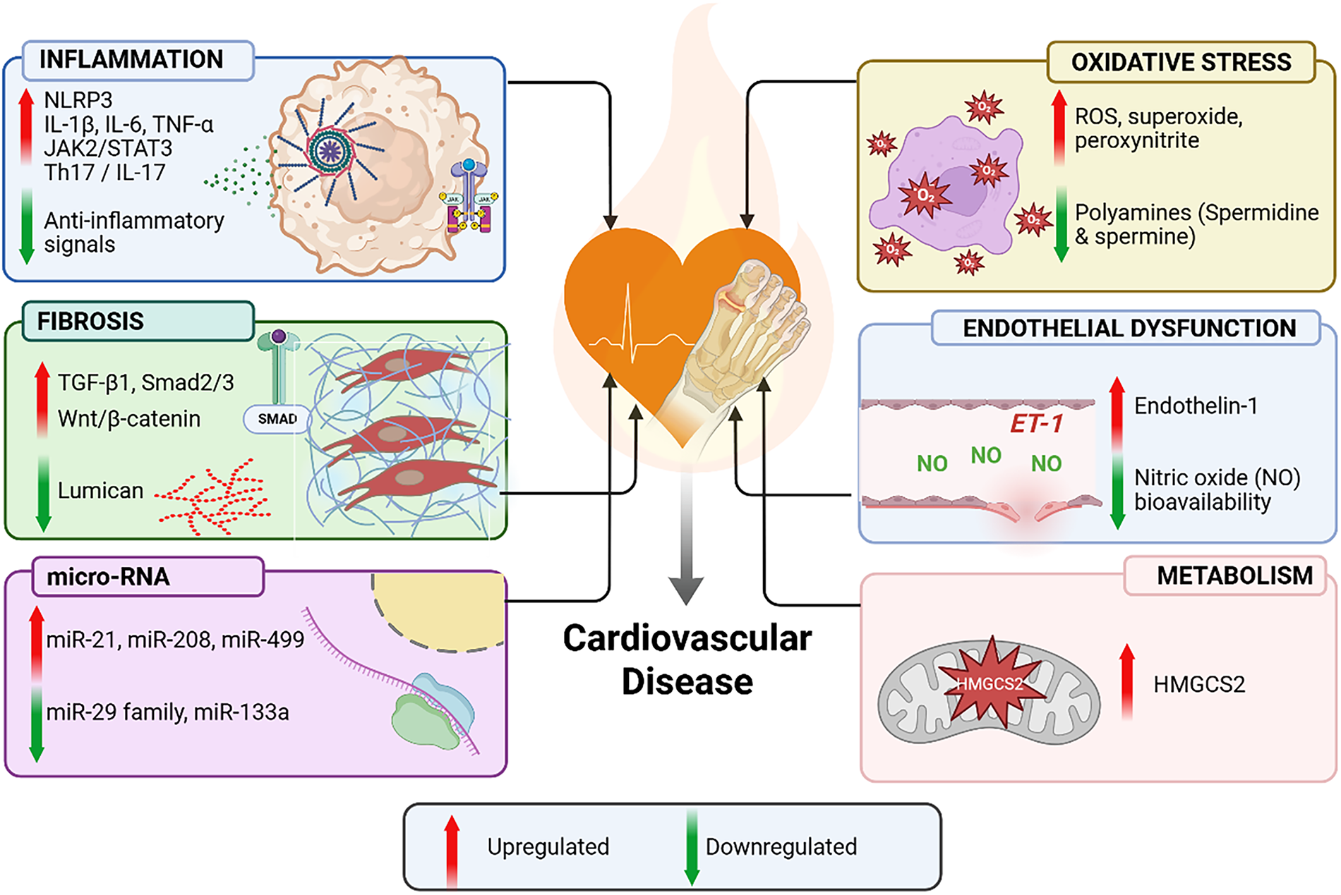
Figure 1: Molecular pathways implicated in urate-associated cardiac remodelling. Schematic summary of upregulated (↑) and downregulated (↓) molecular and cellular changes reported in gout and experimental hyperuricaemia, organised by pathophysiological domain. Arrow direction is the primary encoding. Inflammation: activation of the NLRP3 inflammasome together with increased pro-inflammatory cytokines (IL-1β, IL-6, TNF-α), JAK2/STAT3 activation, and Th17/IL-17 signalling, with concurrent suppression of anti-inflammatory signals. Oxidative stress: elevated reactive oxygen species (ROS), superoxide, and peroxynitrite production paralleled by depletion of cardioprotective polyamines (spermidine, spermine). Fibrosis: upregulation of TGF-β1/Smad2/3 and Wnt/β-catenin signalling with downregulation of lumican. Endothelial dysfunction: increased endothelin-1 (ET-1) with reduced nitric oxide (NO) bioavailability. Metabolism: increased HMGCS2 expression contributing to mitochondrial dysfunction. miRNA: upregulation of pro-fibrotic miR-21, miR-208 and miR-499 with downregulation of anti-fibrotic miR-29 family and miR-133a. Most nodes shown derive from hyperuricaemia or from non-gout cardiac disease models, and none has been validated in myocardial tissue from patients with gout; connectors should therefore be read as proposed rather than established. Created in BioRender. https://BioRender.com/8t4aqs5.
2 Clinical and Epidemiological Links between Gout and Cardiovascular Disease
2.1 Major Observational and Genetic Epidemiological Studies and Risk Quantification
The prevalence of CVD is notably high among patients with gout, a finding consistently reported by studies conducted across diverse countries and healthcare systems (table 1) [1,4,6]. A matched cohort study within the UK Clinical Practice Research Datalink (CPRD), comprising 152,663 individuals with incident gout and 709,981 age-, sex-, and practice-matched controls, reported a significantly elevated risk of CVD (hazard ratio [HR] of 1.58, 95% confidence interval [CI] 1.52–1.63) in patients with gout over a median follow-up period of 6.5 years. The risk increased across all twelve CVD categories examined. Even after adjusting for confounding variables including age, sex, body mass index (BMI), smoking status, diabetes, hypertension and chronic kidney disease, gout remained independently associated with increased cardiovascular risk, yielding an adjusted Hazard Ratio of 1.31 (95% CI 1.27–1.36) which represents a 31% higher relative risk [1].
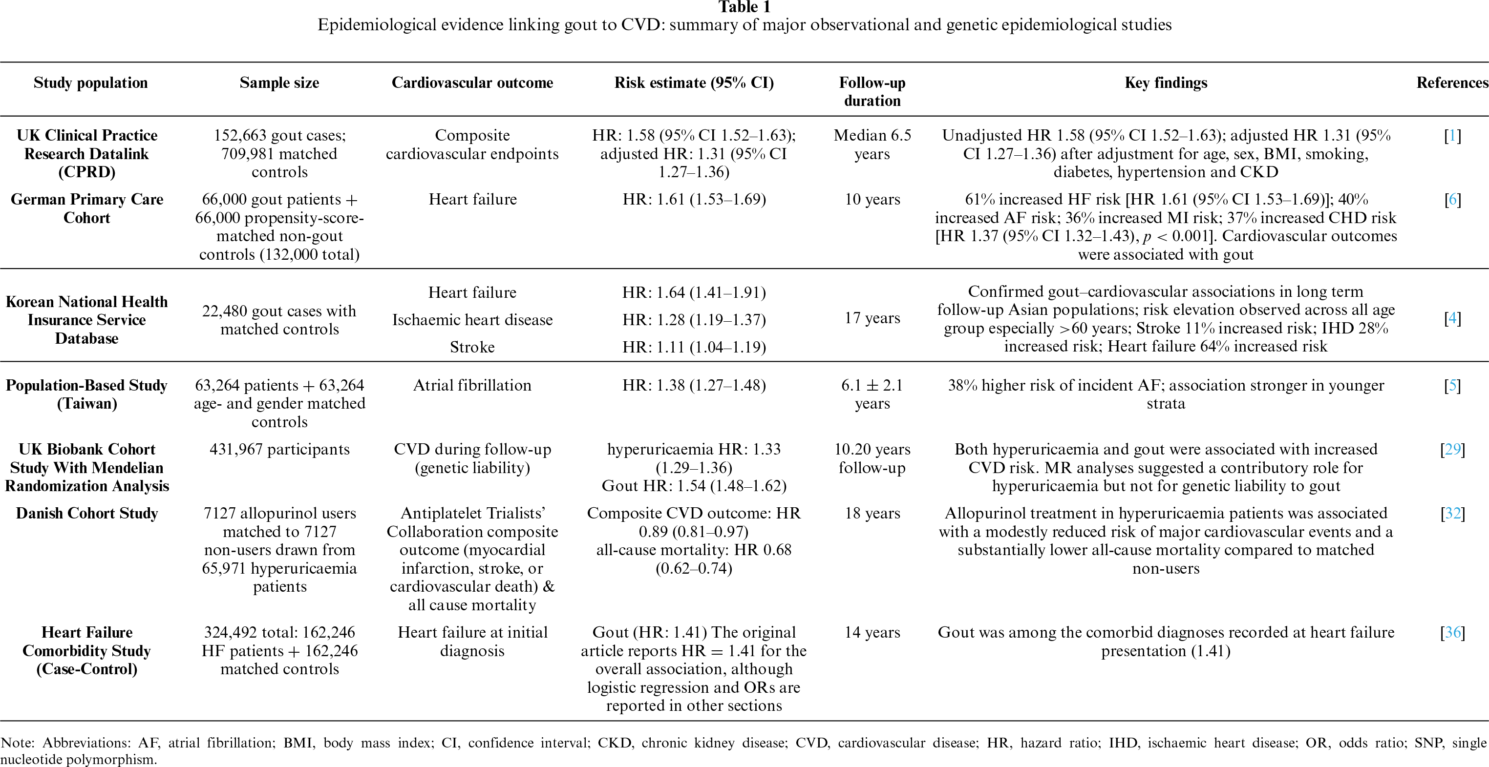
Similar findings have been reported in other population cohorts. Results from a primary care study involving 132,000 participants in Germany demonstrated an elevated incidence of major CVDs in gout patients over a 10-year period, including heart failure (14.7% vs. 8.5%), atrial fibrillation (12.6% vs. 8.4%), angina pectoris (5.2% vs. 2.9%), myocardial infarction (3.1% vs. 2.2%), and coronary heart disease (16.5% vs. 11.8%) [6]. Additionally, a matched cohort study within the Korean National Health Insurance Service database, comprising 22,480 gout patients, reported a 64% increased risk of heart failure (hazard ratio (HR): 1.64, 95% confidence interval (CI): 1.41–1.91), a 28% increased risk of ischaemic heart disease (HR: 1.28, 95% CI: 1.19–1.37), and an 11% increased risk of stroke (HR: 1.11, 95% CI: 1.04–1.19) [4]. The consistency of these findings across large population-based cohorts from the United Kingdom, Germany, and Korea supports the robustness of the association between gout and cardiovascular risk across diverse healthcare systems and demographic settings.
A robust association between serum uric acid concentrations and CVD further supports these observations [1,22–24]. A significantly greater cardiovascular risk has been reported within the first 120 days following gout diagnosis, with the highest HR observed within the first 60 days. This timeline suggests that acute inflammatory gout flares may contribute to triggering cardiovascular events, likely via mechanisms such as atherosclerotic plaque rupture and destabilization [25,26]. Concurrently, the chronic inflammatory state inherent to gouty arthritis may promote long-term cardiovascular remodelling risk [27]. Collectively, these large population-based studies consistently demonstrate that gout is associated with an increased risk of multiple cardiovascular outcomes across diverse populations. However, because these findings are derived from observational studies, they establish robust associations rather than causality.
Beyond the overall epidemiological association, cardiovascular risk varies across patient subgroups and should also be interpreted in light of genetic evidence examining causality. Epidemiological evidence highlights that CVD risk in patients with gout varies across demographic strata. A Swedish population study reported that women with gout were diagnosed at an older age and had a greater overall burden of comorbidities than men. Although, after age standardization, men exhibited a higher prevalence of established cardiovascular conditions, including coronary heart disease, atrial fibrillation, congestive heart failure, and stroke [28]. Analyses from the UK CPRD, which evaluated incident cardiovascular outcomes during follow-up, demonstrated that women with gout experienced a greater relative increase in cardiovascular risk than men (women: HR: 1.88, 95% CI: 1.75–2.02; men: HR: 1.49, 95% CI: 1.43–1.56) [1]. These findings are not contradictory, as the Swedish study assessed cardiovascular comorbidities present at the time of gout diagnosis, whereas the UK study examined the subsequent development of CVD. The greater relative risk observed among women may reflect sex-specific biological factors, such as hormonal alterations that could affect urate metabolism, vascular function, and systemic inflammatory responses.
Notably, age-stratified analyses revealed a clear pattern where younger individuals with gout have a disproportionately higher relative cardiovascular risk compared with age-matched individuals without gout [1]. These findings are consistent with a possible link between chronic inflammation in gout and premature development of CVD [27]. Nevertheless, the absolute burden of cardiovascular complications remains substantial among older adults; in individuals aged 60 years and older, the risk of developing heart failure was increased by 69% (HR 1.69, 95% CI: 1.43–2.01) in gout [4]. Consequently, CVD risk assessment and aggressive management of modifiable risk factors should be considered across all age groups, with particular attention to younger patients and women who may experience a disproportionately greater relative increase in cardiovascular risk.
Mendelian randomisation (MR) studies have provided crucial insights into the potential causal links between hyperuricaemia, gout and CVD [29]. For instance, a UK Biobank study utilizing 81 single nucleotide polymorphisms (SNPs) associated with hyperuricaemia and 6 SNPs associated with gout demonstrated that genetic liability to hyperuricaemia was significantly linked to an increased risk of CVD (odds ratio: 1.09, 95% CI: 1.03–1.16). In contrast, a genetic predisposition for gout itself showed no significant causal relationship with CVD [29]. These observations indicate that baseline hyperuricaemia may contribute more directly to cardiovascular pathogenesis, whereas the observed association between clinical gout and CVD may also be influenced by inflammatory, metabolic, and comorbidity-related factors beyond genetic susceptibility to gout alone.
This discrepancy may reflect both biological differences between hyperuricaemia and clinical gout, as well as limitations inherent to MR analyses. MR isolates the lifelong impact of genetically elevated urate. Clinical gout, by contrast, is a complex downstream disease state characterised by MSU crystal deposition, fluctuating inflammatory peaks and chronic macrophage activation [29]. In addition, clinical gout frequently co-clusters with profound cardiometabolic risk factors such as obesity, hypertension, chronic kidney disease and metabolic syndrome [30,31]. Because these cardiometabolic comorbidities are not fully captured by the narrow genetic instruments optimised solely for gout, the genetic association between gout and CVD may be attenuated in MR models. These inferences are further constrained by assumptions intrinsic to MR itself: horizontal pleiotropy, in which variants influence the outcome through pathways other than the exposure; the requirement for strong, valid instruments; and the assumption of no unmeasured confounding of the instrument–outcome relationship. Genetic liability to an exposure is also not equivalent to the effect of a pharmacological intervention on that exposure in later life.
Although these epidemiological and genetic studies consistently demonstrate an association between gout and increased cardiovascular risk, a pivotal question remains whether gout itself is causally responsible or whether it primarily reflects the cumulative burden of cardiometabolic disease. This distinction is essential for interpreting observational findings and guiding therapeutic strategies.
2.3 Confounding, Causal Inference and Therapeutic Implications in Gout
Gout does not occur in isolation. Chronic kidney disease, obesity, hypertension, type 2 diabetes, alcohol intake, diuretic and other urate-raising medications, obstructive sleep apnoea and diet are each independently associated with both hyperuricaemia and cardiovascular disease, and several may lie on the causal pathway between them. Statistical adjustment cannot fully separate confounding from mediation and adjusting for a mediator such as chronic kidney disease may attenuate a genuine effect while leaving residual confounding by unmeasured factors. The observational estimates summarised above should therefore be read as adjusted associations rather than as causal effect estimates, and the discordance between those estimates and the null Mendelian randomisation finding for genetic liability to gout is consistent with gout acting substantially as a marker of accumulated cardiometabolic and inflammatory burden.
While observational data from a Danish population suggested that allopurinol treatment was associated with a lower risk of the Antiplatelet Trialists’ Collaboration composite outcome (myocardial infarction, stroke or cardiovascular death) and with lower all-cause mortality in patients with hyperuricaemia [32], ALL-HEART found no reduction in cardiovascular events with allopurinol versus usual care in ischaemic heart disease [33], while CARES—a non-inferiority safety trial comparing two urate-lowering agents rather than testing urate lowering against no treatment—reported higher cardiovascular mortality with febuxostat [34]. The difference between observational and RCT data may stem from variations in patient characteristics, disease stage, and underlying mechanisms [35]. Evidence suggests that cardiovascular benefits of urate-lowering therapy are variable, and treatment initiated during later stages of CVD may be insufficient to reverse established vascular damage [35].
These conflicting findings may be partly explained by viewing gout as an indicator of severe or prolonged hyperuricaemia. Clinical gout generally reflects a high cumulative burden of urate exposure and frequently coexists with cardiometabolic comorbidities [3]. It also serves as a marker of a severe chronic inflammatory burden, in which soluble urate contributes to endothelial and vascular smooth muscle cell dysfunction through alterations in cellular metabolism and innate immune responses. Supporting this concept, a prospective observational study reported that combining colchicine with a XOI was associated with improved endothelial function and reduced systemic inflammatory markers, including hsCRP and IL-1β [18]; the interpretation of vascular dysfunction as an intrinsic feature of gout is developed elsewhere [3]. Therefore, the relationship between hyperuricaemia, gout and CVD should be regarded as a complex, interconnected systemic association, rather than a simple, direct consequence of elevated serum urate levels alone [3].
3 Molecular Mechanisms: From Hyperuricaemia to Cardiac Fibrosis
3.1 Activation of Innate Immune Reactions and Inflammasomes
The pathogenesis of CVD in gout involves substantial innate immune activation via several pattern recognition receptor pathways [37,38]. Table 2 provides an overview of the molecular mechanisms linking hyperuricaemia and gout to cardiac fibrosis and heart failure. MSU crystals can act as DAMPs that initiate TLR4 dependent MyD88-NF-κB signalling pathways, resulting in the upregulation of inflammatory gene expression in immune and vascular cells [39]. MSU crystals promote assembly and activation of the NLRP3 inflammasome which further activates caspase-1 enzyme that cleaves pro-IL-1β and pro-IL-18 proteins into their mature bioactive forms [40]. Concurrently, in HepG2 hepatocytes and in associated human observational analyses, soluble uric acid has been shown to engage the IκB kinase/IκBα/NF-κB signalling cascade, with an increase in expression of acute-phase inflammatory mediators such as CRP, fibrinogen, ferritin and complement C3 [41]. Activation of NLRP3 inflammasomes requires potassium (K+) efflux, which is also necessary for caspase-1 activation [42]. Inflammasome assembly is further regulated by metabolic and organelle-associated signalling pathways, including mitochondrial stress responses [43]. Besides cytokine secretion, inflammasome activation can also trigger gasdermin D–mediated pyroptosis, a lytic inflammatory cell death pathway increasingly implicated in inflammation-related cardiovascular injury [43,44].
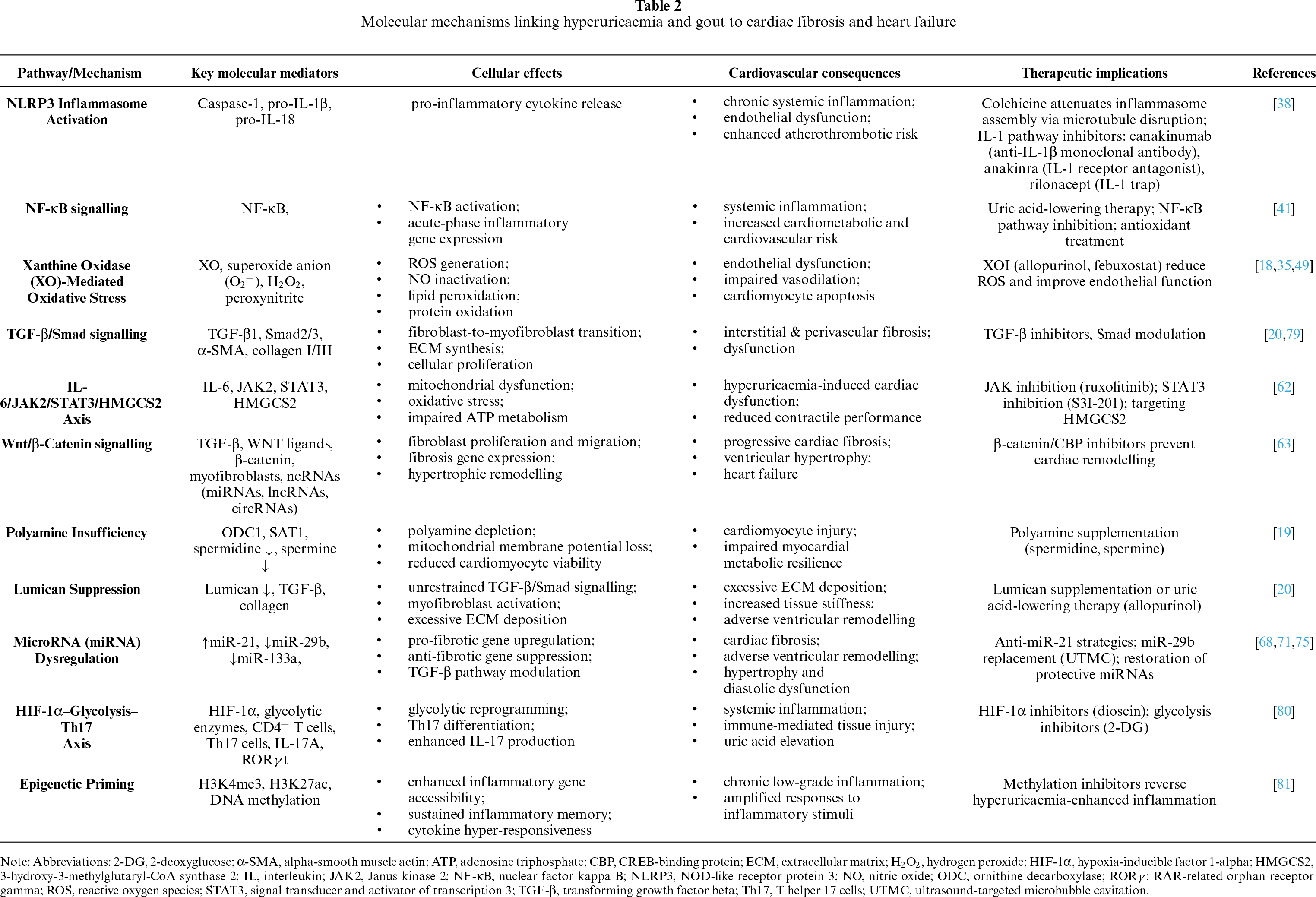
The downstream effects of localized inflammasome activation initiate systemic cascades that can adversely affect cardiovascular structure and function. For example, IL-1 family cytokines and secondary inflammatory mediators can contribute to vascular dysfunction, and maladaptive cardiovascular remodelling [45,46]. Furthermore, consistent low-grade inflammation, driven by prolonged exposure to innate immune cytokines in gout may lead to the development of cardiovascular fibrosis, a process preceded by repeated microvascular injury that promotes structural tissue remodelling [45,46]. Clinical studies showing that low-dose colchicine reduces cardiovascular events after myocardial infarction provide indirect translational support for targeting innate immune mechanisms; those trials were conducted in coronary disease populations and did not evaluate the gout-to-remodelling pathway. This benefit is related to this drug’s ability to disrupt microtubule function, reduce neutrophil activation and lower cytokine release in cardiovascular tissue compartments [47,48].
3.2 Oxidative Stress, Endothelial Dysfunction and Vascular Injury
XO is a critical enzyme pathway that connects purine catabolism to the systemic cardiovascular dysfunction observed in gout [35,49]. In addition to its role in uric acid production, XO generates substantial ROS, specifically superoxide anion (O2−) and hydrogen peroxide (H2O2). This ROS burden fuels vascular oxidative stress and stimulates downstream signalling pathways such as NF-κB, which in turn promote inflammation and hypertrophic remodelling; the supporting evidence here derives from diabetic nephropathy and diabetic cardiac models and is cited as general oxidative-stress background rather than urate-specific cardiovascular evidence [50,51]. While this fundamental metabolic stress paradigm has been characterised in hepatic models where antioxidant treatment in the human HepG2 cell line reduces uric acid-induced NF-κB activation [41], similar oxidative and inflammatory mechanisms have also been reported in cardiovascular endothelial cells and cardiac fibroblasts [52,53]. Within endothelial cells and cardiac fibroblasts, urate-induced oxidative stress promotes inflammatory signalling, impairs NO bioavailability, and contributes to pathways involved in cardiovascular remodelling [52,53]. Collectively, these findings identify XO-derived oxidative stress as an upstream driver linking hyperuricaemia to endothelial injury and cardiovascular remodelling.
Beyond initiating oxidative stress, persistent hyperuricaemia disrupts endothelial homeostasis and promotes vascular dysfunction. Chronic hyperuricaemia has been associated with endothelial dysfunction and microvascular inflammation at serum urate concentrations below the saturation threshold, which is commonly cited as approximately 6.8 mg/dL (≈405 µmol/L) under physiological temperature and pH, although the precise value is temperature- and pH-dependent [21]. Within human umbilical vein endothelial cells (HUVECs), intracellular urate accumulation upregulates XO activity. This oxidative pathway has been shown to impair HUVEC function, impair endothelium-derived NO bioavailability, and may drive cardiovascular structural remodelling [53]. Furthermore, XO-derived superoxide rapidly reacts with NO to form peroxynitrite (ONOO-), reducing NO bioavailability, increasing oxidative stress, impairing endothelium-dependent vasodilation, and promoting a prothrombotic vascular environment [54].
Persistent endothelial dysfunction subsequently amplifies vascular inflammation by promoting leukocyte recruitment and inflammatory signalling. In gout, the concurrent production of urate and ROS creates a pro-inflammatory vascular environment. For instance, hyperuricaemia drives vascular injury, while concurrent oxidative stress impairs endothelial integrity, resulting in inflammatory activation characterised by heightened cytokine release, programmed cell death, and localized microvascular activation [50]. Specifically, this local endothelial activation via IL-1β and TNF-α upregulates key adhesion molecules including vascular cell adhesion molecule-1 (VCAM-1), intercellular adhesion molecule-1 (ICAM-1) and E-selectin on vascular endothelium, promoting leukocyte adhesion and recruitment [53].
In parallel with endothelial injury, oxidative stress also affects cardiac fibroblasts, providing a direct link to structural cardiac remodelling. Uric acid may also affect cardiac fibroblasts in which exposure to urate enhances the extracellular signal-regulated kinase (ERK) and activator protein-1 (AP-1) signalling pathways, resulting in increased endothelin-1 (ET-1) expression and enhanced proliferative and contractile cellular activities [52]. Antioxidant treatments have been shown to reduce ERK phosphorylation and ET-1 expression, confirming that ROS specifically from NADPH oxidase and XO are necessary for fibroblast activation [52]. In rat cardiac fibroblasts, these findings indicate that uric acid increases ET-1 expression, which may promote vasoconstriction and vascular remodelling [52].
Given the central role of XO in the pathogenesis of CVD, pharmacological inhibition of XO represents a promising strategy to interrupt this oxidative–inflammatory cascade. XOI may provide vascular benefits beyond simple urate reduction by protecting the vascular endothelium from ROS-mediated damage [55]. This is supported by clinical observations demonstrating improved vascular function following treatment with XOI [18].
Importantly, the consequences of chronic endothelial dysfunction extend beyond impaired vasodilation and inflammatory activation. Continuous exposure to elevated soluble urate, together with increased concentrations of cytokines such as IL-1β and TGF-β, has been proposed to promote endothelial-to-mesenchymal transition (EndMT), a process in which endothelial cells lose their endothelial characteristics and acquire a migratory, pro-fibrotic myofibroblast-like phenotype [56]. Although EndMT has been implicated in cardiac fibrosis, urate-induced cardiac EndMT has not been demonstrated in gout and therefore remains a hypothetical mechanism requiring direct validation. If confirmed, this phenotypic transition could promote fibroblast activation, ECM deposition, and adverse cardiac remodelling [57].
Taken together, these findings support a stepwise model in which XO-derived oxidative stress initiates endothelial dysfunction, amplifies vascular inflammation, promotes fibroblast activation, and ultimately contributes to pathological cardiac remodelling. This coordinated interplay between metabolic dysfunction, chronic inflammatory activation and progressive cardiac remodelling may characterise gout-associated cardiovascular pathology.
3.3 Fibroblast Activation & ECM Remodelling
Fibroblast activation plays a key role in structural remodelling and functional decline of the heart in chronic inflammatory and metabolic conditions associated with gout. Multiple signalling pathways regulate the transition of fibroblasts into myofibroblasts, increased cell proliferation, and excessive deposition of ECM, which are typical features of cardiac fibrosis [57,58]. Among these pathways, transforming growth factor-β (TGF-β)/Smad signalling is a principal regulator of cardiac fibrosis [58]. The activation of Smad2 and Smad3 by TGF-β1 is a critical step in cardiac remodelling; by increasing α-smooth muscle actin (α-SMA) expression, this pathway induces myofibroblast differentiation, promoting collagen production and enhancing myofibroblast contractile force that collectively drive ECM accumulation and progressive cardiac fibrosis [59,60].
Beyond TGF-β signalling, chronic hyperuricemia may also contribute to cardiac fibroblast activation and ECM remodelling. In a mouse model of myocardial infarction, hyperuricemia aggravated adverse cardiac fibrotic remodelling and promoted aberrant myofibroblast activation and ECM deposition, which were associated with reduced fibroblast derived llumican [20]. In parallel, inflammatory signalling involving interleukin-6 (IL-6) may contribute to cardiovascular injury through inflammatory and oxidative stress mechanisms [61]. In the context of hyperuricemia, Peng et al. [62] demonstrated activation of the IL6/JAK2/STAT3 pathway, with STAT3 promoting promoting 3-hydroxy-3-methylglutaryl-CoA synthase 2 (HMGCS2) expression, a mitochondrial enzyme that normally regulates ketogenesis; and contributing to mitochondrial dysfunction, oxidative stress, and impaired cardiomyocyte energy metabolism [62]. Thus, hyperuricemia may contribute to adverse cardiac remodelling through both fibroblast-mediated ECM deposition and cardiomyocyte metabolic dysfunction.
Multiple pro-fibrotic signalling pathways converge on fibroblast activation. In addition to TGF-β signalling, the Wnt/β-catenin pathway also contributes to cardiac fibrogenesis via coordinated regulation of fibroblast activation and ECM accumulation [63]. While canonical signalling promotes fibroblast proliferation and migration, non-canonical signalling regulates cytoskeletal remodelling and enhances contractile activity and matrix turnover. Together, these complementary signalling pathways exacerbate cardiac fibrotic remodelling of the heart [63]. HOX transcript antisense RNA (HOTAIR) and other long non-coding RNAs have been reported to influence fibroblast behaviour by activating Wnt signalling through upregulation of URI1 in models of myocardial fibrosis unrelated to gout; the relevance of this axis to gout-associated fibrosis is uncertain [28]. Together, the evidence supports the concept that Wnt/β-catenin signalling represents a candidate therapeutic target for limiting fibrotic remodelling, although it has not been evaluated in gout [28].
3.4 Changes in MicroRNA (miRNA) Regulatory Networks in Gout-Associated Cardiac Fibrosis
In addition to protein-based signalling pathways, chronic inflammation in gout also influences post-transcriptional gene regulation through microRNAs (miRNAs) [64]. These non-coding RNA molecules function as key regulators of cardiac remodelling by regulating gene networks that control fibroblast activation, ECM synthesis, and cardiomyocyte hypertrophy [64,65]. The progression of cardiac fibrosis has been linked to over 60 miRNAs, including a subset termed “inflamma-miRs”, in various inflammatory cardiovascular disease models. Whether these signatures are present in gout has not been established [65].
Experimental studies further support the role of miRNAs as multicellular regulators of cardiac remodelling. A functional screen of 194 differentially expressed miRNAs in heart failure models indicated that miRNAs act as multicellular regulators of cardiac remodelling [66]. 31 miRNA mimics, including miR-145-3p, miR-486-3p, and miR-891a-3p, significantly reduced cardiomyocyte size under hypertrophic conditions induced by phenylephrine with several of these mimics exhibiting anti-hypertrophic effects [66]. In summary, the dysregulated miRNA networks in chronic inflammatory diseases such as gout may drive adverse cardiac remodelling via coordinated effects on hypertrophy, fibrosis, and inflammation which could involve modulation of cardiac fibroblast collagen production and macrophage polarisation by distinct miRNAs.
3.4.1 Pro-Fibrotic miRNAs Driving Cardiac Fibrosis
The miRNA signature in cardiac fibrosis contains a number of key pro-fibrotic regulators that are relevant in gout-associated inflammation [67,68]. miR-21 is one of the key pro-fibrotic regulators of the TGF-βsignalling pathway, particularly involved in post-transcriptional suppression of TGF-β receptor III (TGF-β RIII) expression that mediates myofibroblast differentiation and collagen production in cardiac fibroblasts [68]. An in vivo rodent study showed that miR-21 upregulation occurs within seven days of myocardial infarction while deposition of collagen and elevation of TGF-β1 protein is pronounced in border zones of infarcted myocardium [68,69].
Together, these observations indicate miR-21 as one of the central post-transcriptional mediators of cardiac fibrogenesis, although its specific contribution to gout-associated cardiac remodelling remains to be determined.
The miR-208 family and miR-499-5p are closely tied to pathological cardiac hypertrophy. Clinical studies in hypertensive patients have shown that these circulating miRNA levels are associated with left ventricular mass index [70]. Whether these miRNAs serve as early indicators of hypertrophic injury in gout has not been examined.
3.4.2 Suppression of Anti-Fibrotic miRNAs and Therapeutic Implications
Counterbalancing these pro-fibrotic miRNAs are several anti-fibrotic miRNAs that suppress ECM deposition and pathological remodelling. Experimental studies showed that miR-29 family, including miR-29a, miR-29b, miR-29c serve as a pivotal regulator of ECM via their anti-fibrotic effect [71,72]. The miR-29 family targets genes encoding ECM components such as collagens, fibrillin and elastin in cardiac fibroblasts and damaged myocardium [69,71]; supporting cross-organ evidence from liver fibrosis is consistent with this role but is not cardiac-specific [69,73]. These effects inhibit profibrotic TGF-β-dependent signalling pathways that regulate ECM synthesis during ventricular remodelling [74] and may have a role in gout-related cardiac remodelling.
These findings have stimulated interest in miRNA replacement therapy as a potential strategy for limiting cardiac fibrosis. Delivery of miR-29b-loaded microbubbles using ultrasound-targeted microbubble cavitation (UTMC), a novel delivery system for miR-29b [71] significantly elevated intracellular miR-29b expression and reduced fibrotic genes in-vitro. In vivo studies using angiotensin II-infused mice models revealed that UTMC-mediated administration of miR-29b mitigated cardiac fibrotic remodelling, underscoring its potential in models of pressure and neurohormonal cardiac fibrosis; applicability to urate-driven remodelling remains untested [71].
Similarly, miRNA-133a is another important anti-fibrotic regulator that targets genes involved in cardiac hypertrophy and fibrosis as evidence from clinical studies involving individuals with arterial hypertension and hypertensive heart disease which reveal markedly reduced circulating miR-133a levels in comparison to healthy controls [75]. These findings support a role for this miRNA in restraining adverse cardiac remodelling in hypertensive heart disease; it has not been studied in gout.
3.4.3 Circulating miRNA Biomarkers
Circulating miRNAs have emerged as promising biomarkers for detecting cardiac involvement in inflammatory and CVD. Recent profiling reveals that panels of circulating miRNAs, including miR-21, miR-155, and miR-221 serve as potential indicators of non-ischaemic myocardial inflammation, particularly in conditions such as viral myocarditis [76]. In contrast, ischaemic conditions such as acute myocardial infarction (AMI) and coronary artery disease (CAD) exhibit distinct plasma exosomal miRNA profiles [77]. Although direct evidence of myocardial injury in gout is currently lacking, these findings suggest that circulating and exosomal miRNAs may have potential as disease-specific biomarkers capable of differentiating inflammatory and ischaemic forms of myocardial injury. Further studies are required to validate this hypothesis in patients with gout. To date, no classifier of gout-related myocardial injury has been developed or validated.
In addition to tracking acute inflammatory profiles, circulating miRNAs associated with chronic structural remodelling are highly pertinent in identifying progressive gout related cardiac diseases. For instance, reduced circulating miR-19b levels have been linked to increase myocardial collagen remodelling in patients experiencing aortic stenosis and heart failure [67]. Furthermore, a controlled clinical study indicates that a specific panel of circulating exosomal miRNAs, particularly upregulated miR-27a-5p and downregulated miR-139-3p, serves as a stable, high-quality biomarker for tracking chronic heart failure in patients with hyperuricaemia [78]. This panel achieved an area under the curve (AUC) of 0.899 (95% CI 0.812–0.987). An AUC is a measure of discrimination across all classification thresholds and is not equivalent to the proportion of patients correctly classified; sensitivity and specificity at a defined threshold should be reported separately. Functional enrichment analyses indicated that these differentially expressed miRNAs were enriched for predicted AMPK–mTOR-related targets, which may be associated with autophagic responses during pathological cardiac remodelling [78]. In summary, current evidence supports that longitudinal changes in circulating miRNA signatures may help identify the transition from acute inflammation to chronic cardiac remodelling in gout. Despite this, prospective validation in gout cohorts remains necessary before clinical implementation.
4 Transcriptomic insights into Gout-Associated Cardiovascular Remodelling: Current Evidence and Translational Gaps
To our knowledge, transcriptomic studies directly profiling myocardial tissue from patients with gout remain limited. The evidence below therefore combines gout data from joint and blood compartments with cardiac data from non-gout disease models. Although direct transcriptomic evidence from human gout myocardium remains limited, advanced transcriptomic technologies nonetheless provide valuable insight into molecular pathways that may facilitate cardiovascular remodelling associated with hyperuricaemia and gout. Bulk RNA sequencing facilitates the identification of population-level inflammatory mRNA signatures, scRNA-seq elucidates cellular heterogeneity and state-specific responses, while spatial transcriptomics combines gene expression with tissue architecture across affected organs.
4.1 Bulk RNA Analysis: Determination of Inflammatory Signatures at the Population Level
Comprehensive transcriptome analysis has revealed 329 genes associated with gout-related tissue inflammation, among which CXCL8, PTGS2, and IL10 emerged as key regulatory genes involved in cell-cell adhesion, leukocyte activation, and NF-κB signalling [14]. Functional enrichment analysis has further shown that many of these identified genes converged on immunological and cardiometabolic pathways, including interleukin-17 (IL-17) signalling, C-type lectin receptor signalling, Advanced Glycation End-products–Receptor for Advanced Glycation End-products (AGE-RAGE) signalling, and pathways associated with atherosclerosis, highlighting systemic inflammatory and metabolic disturbances in gout [14]. Weighted gene co-expression network analysis (WGCNA) highlighted the central role of hub genes, which displayed strong network connection and potential diagnostic importance [14]. Consistent with these findings, protein-protein interaction (PPI) network analysis identified several inflammatory nodes such as CCL3, CCL18, IL1B, CXCL1, TNF, CCR7, IL7R, CCR2, CCR5, and FCGR3A, thereby emphasising the importance of coordinated immunological signalling in gout pathogenesis [14].
Integrated analysis of the bulk transcriptomic dataset GSE160170 and the single-cell transcriptomic dataset GSE211783 strengthened the evidence supporting these molecular signatures and inflammatory pathways in gout [14]. In addition, comparative transcriptomic analysis of gout and atherosclerosis revealed 41 shared differentially expressed genes (DEGs), highlighting the molecular similarities between these disorders with key inflammatory mediators included CCR2, CCR5, CCL3, and TNF. These were further prioritised by Least Absolute Shrinkage and Selection Operator (LASSO) regression as candidate shared features of the two conditions [14]. Shared differential expression and model selection do not by themselves establish diagnostic performance, and independent validation has not been reported.
Regulatory network reconstruction also revealed miRNA-mRNA interactions (e.g., hsa-miR-203a-3p targeting TNF and CCR5) and transcription factor–mRNA motifs (RELA, NFKB1 control CCR2/CCR5/CCL3/TNF), highlighting coordinated inflammatory regulatory networks in gout [14]. Molecular docking pipelines nominated candidate compounds such as pergolide with favourable predicted binding energies (<−6.0 kcal/mol) [14]. This is an in-silico nomination only; biochemical, selectivity, safety and in vivo validation would be required before any repurposing claim could be made. Overall, bulk transcriptomic analyses consistently identify inflammatory and cardiometabolic gene networks that provide a molecular basis for the systemic manifestations of gout and their potential cardiovascular implications.
4.2 Single-Cell RNA Sequencing: Heterogeneity and Functional Diversity of Cells
4.2.1 Cardiac Macrophage Heterogeneity in Gout
Single-cell RNA sequencing investigation of human cardiac tissue has identified significant immunological heterogeneity in heart failure, with macrophages constituting the most transcriptionally variable immune cell type [15]. Four macrophage subtypes were identified, each occupying disease-associated trajectories representing dynamic activation states. Whether these states also occur in gout-related heart failure is unknown: no gout-specific cardiac single-cell dataset is currently available. Meanwhile, functional pathway analysis revealed subtype-specific activation of programmed cell death pathways related to macrophage, notably ferroptosis and anoikis [15]. Macrophage markers including CD163, FPR1 and VSIG4 were associated with these subtypes and may represent candidate markers for future validation. Although derived from non-gout cardiac tissue, these findings identify macrophage heterogeneity as a potential mechanism warranting dedicated studies in gout-associated cardiac remodelling.
4.2.2 Monocyte Subpopulations in Gout
Single-cell transcriptomics analyses have revealed both convergent and disease-specific activation of monocyte subpopulations in gout which have also been observed in severe coronavirus illness (COVID-19) [82]. In both diseases, the fraction of classical monocytes was elevated relative to healthy controls, underscoring a common transition towards innate immune activation. Despite this overlap, differential gene expression and integrative Mendelian randomisation studies indicated unique molecular drivers including NLRP3, which was positively correlated with susceptibility to severe COVID-19, whereas IER3 exhibited a disease-specific connection with gout. These findings are presented as a methodological comparison rather than as cardiac evidence: although gout flare and severe COVID-19 show partly overlapping monocyte activation at the cellular level, the governing transcriptional programmes are disease-specific, and the samples analysed were non-cardiac [82].
Analysis of intercellular communication by ligand–receptor inference methods uncovered intricate cytokine–cytokine receptor signalling networks among monocyte subsets, notably highlighting the enrichment of IL-17-associated signalling pathways [82]. These findings underscore the pivotal role of classical monocytes in orchestrating systemic inflammatory responses in both severe COVID-19 and gout. These findings imply that gout shares common innate immune activation pathways with other inflammatory diseases while maintaining disease-specific transcriptional programmes.
4.2.3 Diversity of Immune Cells in Acute Gout
A study of single-cell profiling using scRNA-seq performed on peripheral blood mononuclear cells and synovial fluid from patients with acute gout has uncovered significant immune cell heterogeneity across tissue compartments, identifying 31 immune cell populations in blood and 28 in synovial fluid. Acute gout showed increased numbers of naïve CD4+ T-cells and classical monocytes, while plasmacytoid dendritic cells and intermediate monocytes were more prevalent in healthy individuals. This alteration in cellular composition signifies a high inflammatory immunological microenvironment characteristic of acute gout flares [83]. Functional scoring analyses revealed compartment-specific immune states, with inflammation scores highest in synovial fluid, followed by peripheral blood from patients with acute gout, whereas energy scores showed the inverse trend, indicating dynamic immune-metabolic reprogramming during acute inflammatory gout episodes. Together, these findings demonstrate that acute gout is characterised by extensive immune-cell heterogeneity and dynamic immune-metabolic reprogramming, supporting the concept that gout involves systemic inflammatory mechanisms extending beyond the affected joints [83].
4.3 Tissue Architecture and Molecular Geography Using Spatial Transcriptomics
Spatial transcriptomics has emerged as a powerful technique for mapping tissue architecture together with localized molecular features, allowing the spatial localisation of pathophysiological processes relevant to disease development [17,84]. No spatial transcriptomic map of the hyperuricaemic myocardium has been published. Consequently, current evidence relies on hyperuricaemic renal models together with spatial transcriptomic studies of cardiovascular disease to infer mechanisms potentially relevant to gout-associated cardiac remodelling. Hyperuricaemia nephropathy models demonstrate the capacity of this technology to resolve localized tissue architecture, although they are renal models and therefore cannot directly substantiate myocardial spatial mechanisms. Accordingly, single-cell and spatial transcriptomic profiling of hyperuricaemia nephropathy using a urate oxidase knockout mouse model has generated a high-resolution spatiotemporal atlas of hyperuricaemia-induced nephropathy [16].
The spatial transcriptomic analysis showed a number of emerging targets and pathways, particularly those associated with ribosome function and metabolism involved in uric acid excretion [16]. Beyond renal pathology, spatially resolved transcriptomic and multi-omic approaches have been successfully applied to human cardiovascular tissues, revealing spatially organized cellular niches, signalling pathways, and disease-specific molecular programs relevant to cardiovascular pathophysiology and therapeutic targeting. The combined use of single-cell RNA sequencing and spatial transcriptomics enables the identification of molecular processes that may not be apparent from single-modality analyses, thereby offering a more comprehensive view of tissue pathophysiology [17,84]. In summary, spatial transcriptomics complements bulk and single-cell transcriptomic approaches by providing spatial context, thereby facilitating a more comprehensive understanding of tissue-specific molecular mechanisms that may underlie cardiovascular remodelling in gout.
5 New Mechanistic Knowledge: Combining Multi-Omics Data
Multi-omics integration identifies convergent pathogenic circuits linking innate immunity, oxidative stress, fibroblast activation, metabolic rewiring, and ECM dysregulation.
5.1 RNA Modifications, Editing and Alternative Splicing in Hyperuricaemia and Gout
At the post-transcriptional level, recent investigations into RNA modifications, editing, and alternative splicing have uncovered previously underappreciated regulatory mechanisms involved in the pathogenesis of hyperuricaemia and gout [85]. In vitro modelling using THP-1, HEK293, and HUVEC cell lines showed that 48-h exposure to soluble uric acid or MSU crystals altered RNA modification patterns in a cell type-specific manner [85]. In these models, exposure to uric acid or MSU specifically influenced the expression of enzymes responsible for 5-methylcytosine (m5C), pseudouridine (Ψ), and N6-methyladenosine (m6A) RNA modifications, indicating that hyperuricaemia with or without gout reshapes multiple layers of epitranscriptomic regulation [85].
Alternative splicing analyses further showed that exposure to uric acid or MSU crystals affected the splicing patterns of transcripts including Bcl-x, Smac and Hipk3 in multiple cell lines [85]. Consistent with these in vitro findings, clinical profiling of patients across the acute, inter-critical and chronic tophaceous stages of gout revealed aberrant expression of the adenosine-to-inosine (A-to-I) RNA editing enzymes ADAR1 and ADAR2, alongside significant differential alternative splicing of BCL-x, SMAC, HIPK3, TP53 and NFKB1, compared with healthy controls [85].
Upregulation of ADAR2 expression was observed in HEK293 cells following uric acid administration. These findings raise the possibility that hyperuricaemia influences RNA editing activity, although changes in ADAR2 expression alone do not establish altered global RNA editing; direct editome quantification would be required. Collectively, these findings identify RNA modification, RNA editing, and alternative splicing as interconnected post-transcriptional regulatory mechanisms with potential relevance to inflammatory signalling, apoptosis, and cellular stress responses in hyperuricaemia and gout [85].
5.2 Chromatin and DNA Methylation as a Result of Epigenetic Priming
Apart from post-transcriptional regulation, hyperuricaemia also induces epigenetic changes at the chromatin level that may sustain inflammatory memory. These epigenetic modifications have been linked to urate-induced priming in human monocytes, a phenomenon characterised using chromatin immunoprecipitation sequencing (ChIP-seq) [81]. Analysis of the transcriptionally permissive histone marks H3K4me3 and H3K27ac showed that elevated urate exposure was associated with increased inflammatory responsiveness in vitro, and broad-spectrum methylation inhibitors attenuated this effect. Because such inhibitors are non-specific, this constitutes pharmacological evidence that is consistent with, rather than proof of, an epigenetic contribution [81].
Further evidence supports the role of epigenetic dysregulation beyond histone modifications. The evaluation of H3K4me3 and H3K27ac demonstrated unique chromatin profiles in urate-primed monocytes relative to control cells, with gene-specific variability noted at loci such as MED24, CSF3, TAF1C, DNAAF1, HCAR2, IDO1, SNRPC, and APOE, suggesting differential histone modification patterns subsequent to urate exposure [81]. A recent genome-wide DNA methylation study identified specific methylation sites that differentiate hyperuricaemic from normouricaemic individuals [81,86]. Altered expression of DNMT3A, DNMT3B and HDAC3/7 was associated with subclinical atherosclerosis in analyses adjusted for traditional cardiovascular risk factors. This is an association between transcript expression and an imaging phenotype; neither methylation status nor mediation was assessed, so a causal interpretation is not supported [86].
5.3 Metabolic Rewiring: The HIF-1 α-Glycolysis-Th17 Axis in Gout
Metabolic reprogramming represents another important layer of multi-omics regulation. Hypoxia-inducible factor-1α (HIF-1α), a pivotal regulator of immune-metabolic reprogramming, has emerged as a significant element in gout pathogenesis via its influence on CD4+ T-cell metabolism and differentiation [80]. Increased HIF-1α expression in CD4+ T-cells has been observed in gout patients and urate oxidase (Uox)-knockout mice, with both CD4+ T-cell-specific genetic deletion and pharmacological suppression of HIF-1α reducing disease symptoms in experimental models. Transcriptomic and pathway analyses indicated that HIF-1α signalling facilitates Th17 differentiation and IL-17 related inflammatory pathways, while metabolic profiling validated that glycolysis is a crucial downstream pathway connecting HIF-1α activity to Th17 proliferation and IL-17 synthesis [80].
In addition to joint inflammation, IL-17, the hallmark cytokine of Th17 cells, has been associated with chronic vascular inflammation, leukocyte migration to the arterial wall, and atherosclerosis in both murine models and humans [87]. In psoriasis, IL-17A inhibition improves cutaneous inflammation, and some studies report favourable changes in vascular imaging surrogates. This is indirect evidence from a different immune-mediated disease: whether it reflects a shared Th17/IL-17 pathway operating in gout is untested, and no trial has demonstrated a reduction in cardiovascular events with IL-17A blockade [88].
5.4 Polyamine Insufficiency and Lumican-Mediated ECM Organization in Gout
In experimental hyperuricaemia, uric acid exposure disrupts the equilibrium of polyamines—positively charged cellular stabilisers that regulate gene expression and mitochondrial survival in cardiomyocytes—leading to metabolic susceptibility and cellular damage [19]. Experimental investigations indicate that increased uric acid disrupts local cardiomyocyte ornithine metabolism, a cross-compartment metabolic pathway bridging the mitochondria and cytoplasm. This alteration shifts metabolic flux to disturb polyamine equilibrium, resulting in diminished concentrations of downstream polyamines spermidine and spermine in cardiomyocytes. This deficiency of polyamines correlates with mitochondrial dysfunction, characterised by diminished mitochondrial membrane potential and decreased cellular viability. Conversely, exogenous supplementation with either spermine or spermidine partially restores mitochondrial integrity and cardiomyocyte survival in hyperuricaemia stress [19]. These findings indicate that disrupted polyamine metabolism serves as a metabolic mechanism by which hyperuricaemia induces cardiomyocyte damage.
In parallel with metabolic dysfunction, hyperuricaemia also alters ECM organisation through suppression of lumican. Under hyperuricaemic conditions, a marked reduction in fibroblast-derived lumican production amplifies the activation of the TGF-β/Smad signalling axis. This hyperactivation of the TGF-β/Smad pathway contributes to the pathological transition of quiescent resident fibroblast into myofibroblasts and excessive matrix deposition. In experimental hyperuricaemia superimposed on myocardial infarction, this mechanism was associated with exacerbated fibrotic remodelling, impaired cardiac function and increased mortality; the model does not establish spontaneous remodelling in gout, whereas lumican supplementation or uric acid reduction therapy mitigates these pathological alterations [20]. These findings collectively reveal polyamine deficiency-induced metabolic damage and lumican-dependent deregulation of ECM remodelling as parallel mechanisms through which hyperuricaemia contributes to cardiac injury and fibrosis.
5.5 An Integrated Model of Gout-Related CVD and Clinical Implications
Cross-platform synthesis supports a cohesive model (figure 2) wherein elevated serum uric acid acts as a potential metabolic stressor and risk marker, leading to innate immune activation via TLR-mediated NLRP3 inflammasome and NF-κB signalling, in conjunction with oxidative stress induced by XO-dependent ROS generation [89]. Simultaneously, hyperuricaemia stimulates fibroblast-related profibrotic pathways, encompassing TGF-β/Smad signalling, JAK2/STAT3/HMGCS2-dependent metabolic and infla mmatory mechanisms, and Wnt/β-catenin activation, all of which jointly facilitate pathological cardiac remodelling [62,89].
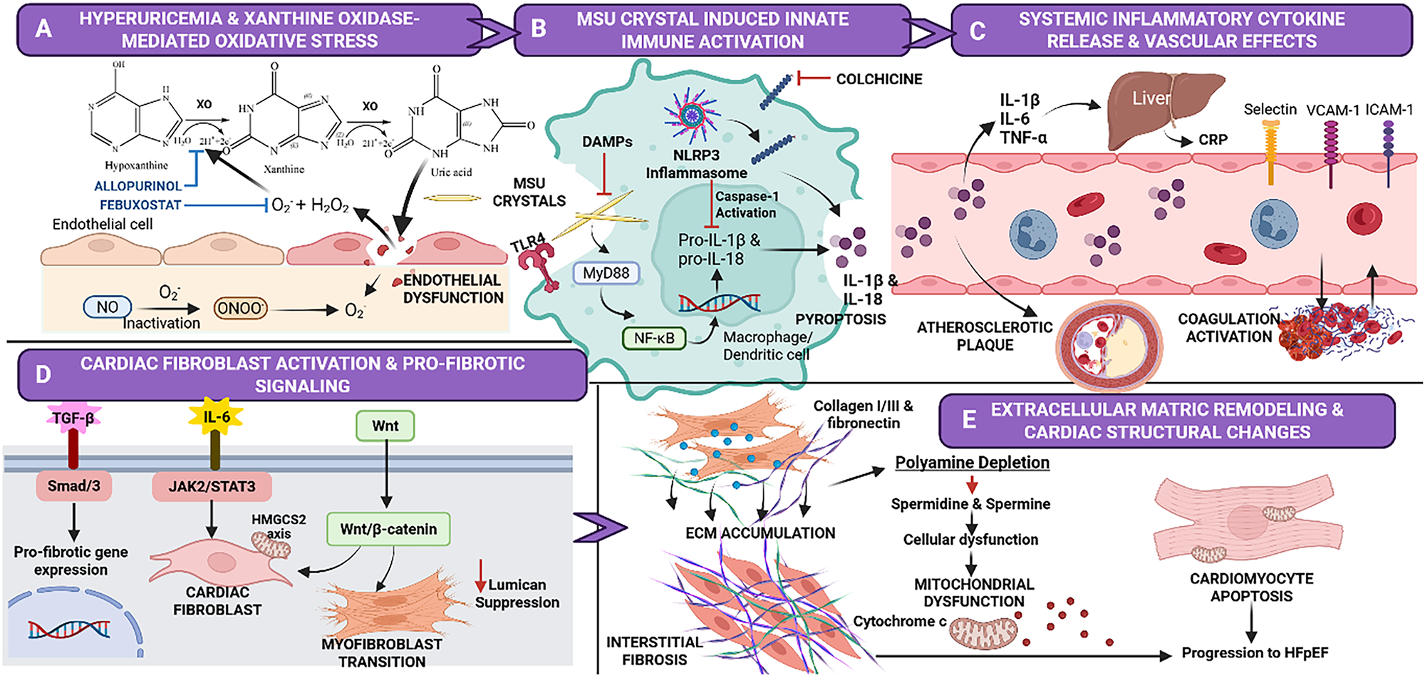
Figure 2: Pathophysiological mechanisms linking hyperuricaemia and gout to CVD. (A) Hyperuricaemia and xanthine oxidase-mediated oxidative stress. Xanthine oxidase (XO) converts hypoxanthine to xanthine and ultimately to uric acid, releasing superoxide (O2−) and hydrogen peroxide (H2O2). The resulting ROS inactivate endothelial nitric oxide (NO) and generate peroxynitrite (ONOO−), causing endothelial dysfunction. Allopurinol and febuxostat block this cascade by inhibiting XO. (B) Monosodium urate (MSU) crystal-induced innate immune activation. MSU crystals and damage-associated molecular patterns (DAMPs) engage toll-like receptor 4 (TLR4) on macrophages and dendritic cells, priming the NLRP3 inflammasome via MyD88 and NF-κB. Caspase-1 then cleaves pro-IL-1β and pro-IL-18 into their active forms, releasing cytokines and triggering pyroptosis. Colchicine attenuates this pathway by disrupting microtubule-dependent inflammasome assembly. (C) Systemic inflammatory cytokine release and vascular effects. IL-1β, IL-6, and TNF-α released into the circulation drive hepatic C-reactive protein (CRP) synthesis and induce endothelial expression of E-selectin, VCAM-1 and ICAM-1. The resulting leukocyte recruitment, atherosclerotic plaque formation, and coagulation activation establish a pro-thrombotic vascular phenotype. (D) Cardiac fibroblast activation and pro-fibrotic signalling. Three pathways converge to drive fibrogenesis: TGF-β/Smad2/3 activates pro-fibrotic gene transcription; IL-6/JAK2/STAT3 induces the HMGCS2 axis; and Wnt/β-catenin promotes myofibroblast transition. Concurrent lumican suppression amplifies fibroblast-to-myofibroblast conversion and pathological matrix remodelling. (E) Extracellular matrix (ECM) remodelling and cardiac structural changes. Activated myofibroblasts deposit excess collagen I/III and fibronectin, producing interstitial fibrosis. Polyamine depletion (spermidine, spermine) impairs mitochondrial function, releases cytochrome c, and induces cardiomyocyte apoptosis. The cumulative effect is adverse cardiac remodelling and heart failure phenotypes; the specific culmination in heart failure with preserved ejection fraction (HFpEF) is a hypothesis rather than a demonstrated endpoint. Nodes derived from discrete preclinical studies, including lumican suppression and polyamine depletion, should not be read as equivalent in evidential weight to replicated epidemiology. Created in BioRender. https://BioRender.com/vn4ffny.
Integrating these molecular layers, dysregulation of miRNAs at the post-transcriptional level (e.g., upregulation of pro-fibrotic miR-21 and miR-208 and downregulation of anti-fibrotic miR-29 and miR-133a) acts as a key regulatory hub that integrates inflammatory and fibrotic signalling to perpetuate epigenetic priming and maladaptive cardiac remodelling. Together with alterations in RNA editing and alternative splicing, this regulatory network reconfigures inflammatory and fibrotic gene expression programs, thereby maintaining epigenetic priming and inflammatory memory [81,85]. Immuno-metabolic reprogramming exacerbates disease progression, as HIF-1α-dependent glycolysis accelerates Th17 proliferation and IL-17-mediated inflammation [80]. Simultaneously, hyperuricaemia causes polyamine depletion and lumican suppression, exacerbating mitochondrial dysfunction and ECM disorganisation, thereby contributing to both metabolic and structural cardiac injury [19,20].
Therapeutically, combining urate-lowering therapy through XO inhibition with established anti-inflammatory strategies, including colchicine or IL1β blocking agents, together with emerging approaches such as polyamine restoration, lumican preservation and anti-fibrotic miRNA modulation, provide a mechanistic rationale for comprehensive cardioprotection in gout. Cumulatively, these findings provide a systems-level rationale for combining urate-lowering, anti-inflammatory and anti-fibrotic strategies to mitigate cardiovascular remodelling in gout. Although several targets remain experimental, multi-omics integration provides a framework for identifying future precision therapies. Overall, these integrated multi-omics findings provide a systems-level framework linking molecular mechanisms with clinical cardiovascular manifestations, thereby reinforcing the concept of gout as a systemic cardio-inflammatory disease rather than an isolated crystal arthropathy.
6 Cardiac Arrhythmia: Gout-Associated Atrial Fibrillation (AF)
6.1 Epidemiological Associations and Risk Quantification
Atrial fibrillation is a frequently documented cardiovascular complication of gout, with numerous large-scale observational studies revealing significant associations even after adjusting for key cardiovascular and metabolic variables [5,90–92]. Using data from Taiwan’s National Health Insurance Research Database (NHIRD), a nationwide population-based cohort study of 63,264 patients with newly diagnosed gout and 63,264 age- and gender matched controls reported a 1.38-fold increased risk of incident AF, with the association being stronger among younger patients [5]. In accordance with these findings, registry-based data from Sweden revealed a greater prevalence of AF in persons with gout compared to the general population, reinforcing the strength of this link [91]. Recent meta-analyses have consistently reported that hyperuricaemia and gout are associated with an elevated risk of AF, with dose–response relationships described between serum uric acid concentration and AF incidence [90,92].
The dose-dependent association between serum uric acid concentration and AF risk is consistent with a contributory pathogenic role for hyperuricaemia in atrial arrhythmogenesis [92]. Although a dose–response gradient strengthens biological plausibility, it does not by itself establish causality because residual confounding and reverse causation cannot be excluded. The persistence of this association in individuals with asymptomatic hyperuricaemia further suggests that elevated uric acid levels, rather than acute inflammatory flares alone, may enhance AF development through chronic pathophysiological mechanisms [5,90].
Temporal analyses and observational evidence indicate that the incidence of AF is heightened during both acute gout flares and inter-critical intervals, suggesting the engagement of many pathophysiological processes throughout distinct phases of the disease [93]. Acute gout flares may transiently precipitate AF through rapid systemic inflammation and autonomic activation, while chronic hyperuricaemia has been hypothesised to mediate structural and electrical remodelling of the atria, potentially establishing a substrate for persistent arrhythmia. In addition, chronic inflammation may induce atrial fibrosis and electrophysiological heterogeneity via central and peripheral inflammatory signalling pathways. The reciprocal association between AF and heart failure complicates the clinical progression in individuals with gout, as each illness may predispose the other through common haemodynamic and neurohormonal pathways [91]. These epidemiological observations suggest that both acute inflammatory flares and chronic hyperuricaemia may drive atrial remodelling through distinct but complementary mechanisms. The epidemiological association between gout and AF is supported by mechanistic studies demonstrating progressive structural and electrical remodelling of the atria.
6.2 Structural Atrial Remodelling and Inflammatory Mechanisms in Gout
The pathophysiology of AF linked to gout involves progressive structural and electrical remodelling of the atria, driven by persistent inflammation and oxidative stress. Prolonged inflammatory activation induces atrial fibroblast activation, excessive ECM accumulation, and atrial fibrosis, resulting in the breakdown of normal myocardial structure. The structural modifications induce areas of conduction delay and electrical irregularity, hence heightening vulnerability to re-entrant arrhythmias and promoting the onset and persistence of AF [93,94]. Pro-inflammatory cytokines, such as IL-1β and IL-18 are pivotal in this process. In macrophages, MSU crystals actively trigger the NLRP3 inflammasome and caspase-1 activation, driving the proteolytic processing and secretion of these local cytokines to stimulate myofibroblast differentiation and collagen synthesis [79,95].
Beyond fibrosis, soluble urate has been reported to accumulate intracellularly in atrial myocytes through urate transporters, based on experimental evidence [95]. Intracellular urate generates ROS through NADPH oxidase and activates ERK/Akt and heat shock factor 1 (HSF1) signalling, thereby increasing heat shock protein 70 (Hsp70) expression and stabilizing Kv1.5 potassium channels. Enhanced Kv1.5 activity shortens atrial action potential duration in experimental atrial myocytes, providing a potential electrophysiological mechanism contributing to AF susceptibility [95]. Consistent with these mechanistic observations, a meta-analysis of observational studies reported that gout was associated with an increased risk of AF (adjusted HR 1.31; 95% CI 1.00–1.70) [96]. However, the proposed urate-mediated electrophysiological mechanism has not been directly confirmed in human atrial tissue [95].
Current evidence suggests that these systemic inflammatory and oxidative changes act predominantly indirectly on myocardial tissue. Direct MSU deposition within atrial myocardium has not been systematically evaluated, although urate deposition has been reported in cardiac and valvular tissue by dual-energy CT [5,95]. Population-based studies and meta-analyses are consistent with a contributory role for chronic hyperuricaemia in atrial arrhythmogenesis, although residual confounding by obesity, alcohol intake, hypertension and renal impairment cannot be excluded [90,92,97]. In addition, excessive adiposity and alcohol intake, which are common risk factors for gout, are independently linked to an elevated risk of atrial fibrillation, thereby underscoring the notion that chronic metabolic-inflammatory conditions associated with gout lead to structural changes in the atria and increased vulnerability to arrhythmias [5]. Furthermore, oxidative stress resulting from heightened XO activity has been shown to injure cardiomyocytes, induce cardiomyocyte death and disturb redox homeostasis, supporting a mechanistic link between urate excess, structural cardiac remodelling and heart failure [54,98,99].
The resultant structural disarray creates an arrhythmogenic state marked by conduction delay, localised conduction block, and increased vulnerability to triggered activity stemming from delayed after-depolarizations (DADs). Concurrently, the upregulation of ET-1 due to oxidative stress facilitates atrial remodelling by directly altering atrial myocyte electrophysiology and indirectly augmenting sympathetic nervous system activity [100]. ET-1 extends action potential length, increases intracellular calcium accumulation, and encourages calcium leaking from the sarcoplasmic reticulum, thus enabling prolonged after-depolarizations and ectopic activity [101]. Ultimately, this establishes a conducive environment for the onset of AF and prolonging its duration, contributing to the disease’s resistance to standard antiarrhythmic treatments [93]. Viewed collectively, these findings suggest that chronic inflammation, oxidative stress and atrial structural remodelling act synergistically to create an arrhythmogenic substrate that may explain the increased incidence of AF observed in patients with gout. Beyond myocardial remodelling, endothelial dysfunction and coronary microvascular injury may further mediate the development of an arrhythmogenic substrate in gout.
6.3 Gout-Related Microvascular Dysfunction and Endothelial Injury
Gout-associated microvascular dysfunction and endothelial injury link metabolic urate stress with atrial remodelling and AF. At the cellular level, soluble uric acid accumulation generates ROS, driving electrical and structural atrial remodelling [95]. Hyperuricaemia and chronic inflammation impair coronary microvascular function. Patients with gout exhibit reduced coronary flow reserve (CFR) and flow-mediated dilation (FMD), consistent with subclinical endothelial and coronary microvascular dysfunction [102]. This compromised vascular state may foster a localized pro-inflammatory atrial environment that alters calcium homeostasis and activates fibrotic pathways, increasing vulnerability to AF [103].
Persistent endothelial injury further amplifies inflammatory signalling through activation of AGE-RAGE axis. Elevated urate concentrations directly drive endothelial injury via the HMGB1-RAGE signalling pathway [104]. In endothelial cells, elevated urate upregulates HMGB1 secretion and RAGE expression, downregulating endothelial nitric oxide synthase (eNOS) expression and consequently reducing nitric oxide (NO) production [104]. Experimental studies indicate that this axis activates NF-κB to amplify pro-inflammatory cytokines, reducing microvascular responsiveness and increasing endothelial permeability [104].
In patients with concurrent gout-related heart failure, the interplay of metabolic vascular dysfunction and haemodynamic stress alters atrial electrophysiology. Increased atrial pressures lead to heightened stretch of atrial myocytes. This mechanical strain triggers spontaneous sarcoplasmic reticulum Ca2+ leakage during diastole via oxidative stress-induced dysfunction of ryanodine receptor channels (RyR2) [105]. Although endothelial dysfunction and microvascular injury may promote an arrhythmogenic atrial substrate, the RyR2 mechanism described above derives from pressure-overloaded hearts and has not yet been reported under hyperuricaemic conditions [105].
7 Ventricular Remodelling and Heart Failure Pathogenesis in Gout
7.1 Diastolic Dysfunction & Structural Changes of Left Ventricles
Gout is associated with adverse changes in left ventricular (LV) structure and function in cross-sectional echocardiographic studies (figure 3). Several echocardiographic studies have showed evidence of subclinical myocardial dysfunction and early cardiac remodelling in patients with gout, even in the absence of overt heart failure [106–108]. In a large echocardiographic study of asymptomatic individuals, both hyperuricaemia and gout were associated with greater LV wall thickness, increased LV mass index (LVMI), larger left atrial volume, reduced tissue Doppler-derived LV e’, indicating impaired diastolic function [108]. Concurrently, gout-specific echocardiographic investigations have identified associations between elevated metabolic risk factors, LV hypertrophy, increased left atrial volume index (LAVI), and subclinical myocardial dysfunction [106].
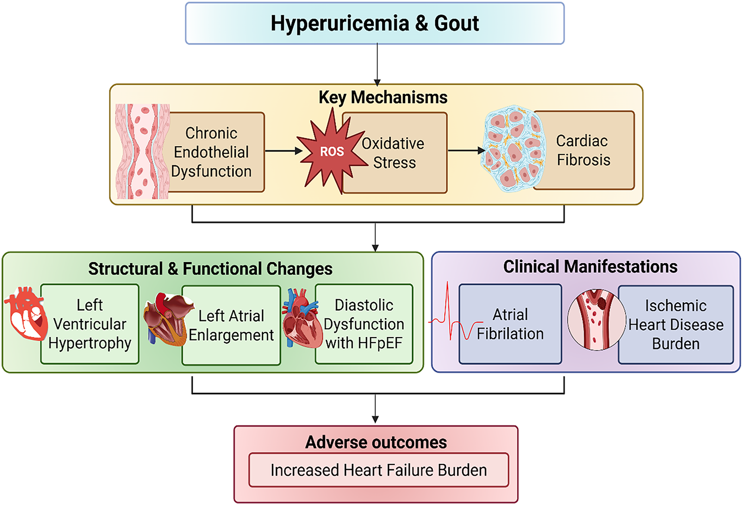
Figure 3: Cardiovascular clinical manifestations of hyperuricaemia and gout. Chronic endothelial dysfunction, oxidative stress and cardiac fibrosis are associated with left ventricular hypertrophy, diastolic dysfunction, left atrial enlargement and atrial fibrillation, shown here as associated manifestations rather than as a deterministic progression. These changes accompany an increased burden of heart failure and ischaemic heart disease in patients with gout and persistent hyperuricaemia. Chronic kidney disease, hypertension, obesity, diabetes and concurrent treatment act as confounders and effect modifiers of these associations. Created in BioRender. https://BioRender.com/zcd2up5.
In a large cross-sectional echocardiographic cohort of asymptomatic Asian adults, gout was independently associated with LV hypertrophy (LVH), left atrial enlargement (LAE) and impaired diastolic performance [108]. These observations suggest that the persistent inflammatory and metabolic burden characteristics of gout may contribute to adverse cardiac remodelling and increase long-term heart failure risk. The diastolic dysfunction observed in gout resembles early features commonly associated with heart failure with preserved ejection fraction (HFpEF), including impaired ventricular relaxation despite systolic function. Although much of the mechanistic evidence derives from broader cardiovascular fibrosis literature, persistent inflammatory signalling and oxidative stress may activate cardiac fibroblasts and promote ECM deposition, leading to progressive interstitial fibrosis [57,58]. Given the role of chronic inflammation in the pathogenesis of HFpEF, gout may identify patients at higher risk of progression from subclinical diastolic dysfunction to symptomatic heart failure. Whether gout acceleration of HFpEF progression is modifiable has not been established, and prospective gout-specific HFpEF studies are required. This point of view underscores the need for heightened clinical vigilance and early cardiovascular risk assessment in patients with gout.
Left atrial enlargement (LAE) is a recurrent observation in patients with gout, serving as a reliable surrogate marker for a persistent elevated left ventricular filling pressures [106,108]. An elevated left atrial volume index is associated with the severity of diastolic dysfunction and offers prognostic value for future heart failure risk. Progressive left atrial enlargement in patients with gout may reflect ongoing subclinical haemodynamic stress driven by poor ventricular compliance and chronically elevated filling pressures. Ultimately, this cumulative structural remodelling may provide a substrate for the development of both symptomatic heart failure and AF.
Collectively, these echocardiographic findings suggest that structural and functional myocardial abnormalities may develop before clinically overt heart failure becomes apparent. These structural abnormalities may ultimately progress to clinically overt heart failure, prompting consideration of the inflammatory mechanisms that underlie myocardial dysfunction in gout.
7.2 Association of Inflammatory Cardiomyopathy and Heart Failure in Gout
The concept of inflammatory cardiomyopathy provides a compelling clinical framework for understanding the development of heart failure in patients with gout. Chronic systemic inflammation and persistent immune activation drive chronic myocardial dysfunction, triggering adverse cardiac remodelling. Epidemiological data consistently reinforce this association. For instance, a large case-control study assessing clinical comorbidities at the time of initial heart failure presentation identified gout as a highly prevalent coexisting condition [36].
Supporting this observation, a population-based longitudinal cohort study has documented a higher cumulative incidence of heart failure in patients with gout than in propensity-score-matched controls over a 10-year follow-up period [6]. These findings indicate that gout is associated with earlier onset of heart failure after propensity-score matching for measured cardiovascular covariates; the mediating mechanism was not assessed. This association further supported by a nationwide registry data, which revealed that patients with gout experience markedly elevated rates of incident heart failure even after stringent multivariable correction for conventional metabolic and cardiovascular covariates [4].
At the molecular level, chronic systemic immune activation compromises myocardial performance via prolonged inflammatory signalling and intensify oxidative stress [109,110]. Pro-inflammatory cytokines, specifically IL-1β, IL-6 and TNF-α exert direct cardiotoxic effects including negative inotropic effects, impaired sarcoplasmic reticulum calcium handling, and promotion of cardiomyocyte apoptosis [111]. Moreover, this sustained cytokine exposure disrupts mitochondrial respiration and cardiac energetics with the resulting collapse in intracellular ATP which compromises cardiomyocyte contractile performance and accelerates adverse ventricular remodelling [109,110]. A key consequence of persistent myocardial inflammation is activation of profibrotic pathways that drive ECM remodelling.
7.3 Mechanisms of Gout-Related Cardiac Fibrosis: Remodelling of the ECM
Cardiac fibrosis represents a hallmark mechanism in heart failure progression, characterised by pathological remodelling of the cardiac ECM which increases myocardial rigidity and compromises both systolic and diastolic performance of the heart [57,112]. This structural disarray fundamentally stems from the activation and phenotypic trans-differentiation of resident cardiac fibroblasts into pro-fibrotic myofibroblasts [57,58,112].
Gout may drive a profibrotic milieu, particularly through hyperuricaemia and comorbid inflammation, with experimental evidence implicating urate excess in local tissue injury, immune activation and cellular apoptosis [113]. At the molecular level, this structural shift is fuelled by the direct disruption of endogenous negative regulators of fibrosis. In experimental models, elevated uric acid reduces fibroblast-derived lumican, a proteoglycan that binds and restrains profibrotic signalling complexes [20].
Reduced lumican disinhibits the canonical TGF-β/Smad signalling axis, increasing downstream Smad2/3 phosphorylation and promoting myofibroblast activation and proliferation [20,113,114]. Consequently, TGF-β1 facilitates fibrosis by enhancing cardiac fibroblast proliferation and activating the transcription of ECM genes, such as collagen types I and III, which expedites excessive matrix deposition in myocardium [60,114]. Concurrently, dysregulation of Wnt/β-catenin signalling acts in concert with TGF-β cascade to augment the expression of fibrosis-related genes, hence strengthening myofibroblast activation and ECM build-up during cardiac remodelling [63,115]. These findings suggest that hyperuricaemia may amplify several converging profibrotic pathways that promote ECM accumulation and ventricular remodelling.
7.4 Gout-Related Metabolic Dysfunction and Cardiac Energetics
Recent data suggests that hyperuricaemia and gout are linked to maladaptive changes in cardiac metabolism, which exacerbate the onset and advancement of heart failure by disrupting cellular energetics. Heart failure is characterised by mitochondrial dysfunction, reduced ATP production and increased oxidative stress. This cascade induces an energy deficit that restricts the capacity of cardiomyocytes to fulfil metabolic requirements, especially during elevated hemodynamic workload [116]. In the context of hyperuricaemia, these metabolic abnormalities are further compounded, thereby aggravating mitochondrial inefficiency and undermining cardiac energy homeostasis.
Increased serum uric acid levels have been showed to activate inflammatory signalling pathways in cardiomyocytes, notably the IL-6/JAK2/STAT3 axis, which is crucial for controlling mitochondrial metabolism and cellular energy homeostasis [62]. High uric acid levels directly stimulate this cytokine cascade to transcriptionally upregulate the expression of 3-hydroxy-3-methyglutaryl-CoA synthase 2 (HMGCS2) in myocardial tissue. Dysregulation of JAK2/STAT3/HMGCS2 signalling pathway has been linked to compromised mitochondrial activity, heightened oxidative stress, and irregular ATP generation, leading to severe energy deficiency in cardiomyocytes under hyperuricaemia condition. Notably, targeted pharmacological suppression of this axis using the JAK inhibitor ruxolitinib or the STAT3 inhibitor S3I-201 downregulates HMGCS2 expression, ameliorates mitochondrial dysfunction, suppresses oxidative stress and restores cellular ATP levels, thereby attenuating uric acid-induced cardiac functional abnormalities in these experimental models [62].
Beyond inflammatory cytokine networks, hyperuricaemia damages cardiomyocytes by disrupting polyamine metabolism. High uric acid alters upstream ornithine metabolism [19]. Uric acid increased expression of both ornithine decarboxylase 1 (ODC1), the rate-limiting synthetic enzyme, and spermidine/spermine N1-acetyltran-sferase 1 (SAT1), which drives acetylation and export. The net effect was depletion of the cardioprotective polyamines spermidine and spermine, consistent with SAT1-mediated catabolism and export exceeding ODC1-driven synthesis despite compensatory upregulation of the synthetic arm. This deficiency directly induces mitochondrial membrane depolarisation, oxidative stress, and diminished cardiomyocyte viability. Conversely, exogenous supplementation with spermidine or spermine significantly restored mitochondrial membrane potential and rescues cell survival, underscoring polyamine replenishment as a potential therapeutic target for alleviating hyperuricaemia-related cardiac dysfunction [19]. Together, these findings highlight metabolic reprogramming as an additional mechanism linking hyperuricaemia with impaired cardiac energetics and ventricular remodelling.
8 Therapeutic Approach and Clinical Implications in Gout
8.1 Prevention of Oxidative Stress and Cardiovascular Protection by XO Inhibition
XOIs are the cornerstone of urate-lowering therapy (ULT) in gout and may provide benefits beyond systemic urate reduction. Clinical evidence indicates that guideline-based gout treatment combining ULT with colchicine improves endothelial function as revealed by increased flow-mediated dilation (FMD), while reducing key systemic inflammatory markers such as IL-1β, IL-6, and hsCRP [18].
However, vascular deposition of MSU crystals contributes to localized chronic inflammation, it likely represents only a small part of the mechanism linking gout to CVD. Cardiovascular risk is also driven by the pro-oxidant effects of uric acid itself and increased XO activity. Excessive XO activity promotes urate production and vascular oxidative stress, contributing to cardiovascular injury [35].
Despite this, whether these biological improvements translate into long-term cardiovascular benefits remains uncertain. It is suggested that the mixed cardiovascular outcomes observed with XO inhibition may reflect the complex physiological role of XO. While excessive XO activity contributes to oxidative stress and vascular injury, basal XO activity is involved in normal redox signalling and host defense. Consequently, the cardiovascular effects of intensive XO inhibition may vary depending on the patient’s underlying cardiovascular risk, disease phenotype and baseline XO activity [35].
These differences between ULTs may help explain why patients with gout and existing CVD respond differently to treatment. In the CARES trial, febuxostat, a potent XOI, was associated with a higher risk of cardiovascular mortality in certain high-risk patients [34]. This safety signal was subsequently contextualised by the Febuxostat versus Allopurinol Streamlined Trial (FAST), a prospective, randomised, open-label, non-inferiority trial enrolling 6128 patients with gout aged ≥60 years over a median follow-up of 4.0 years, which found that febuxostat was non-inferior to allopurinol for the primary composite cardiovascular endpoint (adjusted HR 0.85, 95% CI 0.70–1.03) [33]. The contrasting findings suggest that while febuxostat requires careful patient selection in severe pre-existing disease, its risk profile is conditional rather than universal. Therefore, treatment decisions should be individualised, taking into account both effectiveness of urate lowering and the patient’s cardiovascular risk profile, particularly in those with heart failure or other cardiovascular comorbidities.
Reflecting this, the current European expert consensus statement recognises hyperuricaemia as a relevant cardiovascular and renal risk factor that may aid risk stratification in selected high-risk populations. Despite this, because randomised trials have not identified clear cardiovascular or renal benefits from routine ULT, the consensus statement does not recommend treatment of asymptomatic hyperuricaemia solely for cardiovascular risk reduction. Instead, a risk-based and individualized approach is advocated particularly for patients with symptomatic disease, very high serum urate levels or substantial cardiovascular risk [117].
8.2 The Use of Colchicine and Inflammasome Inhibitors
Colchicine is a well-established anti-inflammatory agent whose therapeutic benefits extend beyond gout to CVD through modulation of the innate immune response and inflammasome-mediated inflammation. Its cardiovascular protective effects have been reported in several large RCTs.
The COLCOT trial demonstrated that low-dose colchicine (0.5 mg daily) administered after a recent myocardial infarction reduced the relative risk of major adverse cardiovascular events by approximately 23%, providing evidence that targeting inflammation can improve cardiovascular outcomes independently of lipid lowering. This trial enrolled patients after myocardial infarction and was not gout-specific [48]. At the same time, the LoDoCo2 trial revealed that long-term colchicine therapy in patients with chronic coronary disease reduced the relative risk of cardiovascular events by approximately 31%, supporting a role in secondary prevention of atherosclerotic CVD; this population likewise was not selected for gout [47].
Colchicine exerts its anti-inflammatory actions by disrupting microtubule polymerisation, which inhibits neutrophil chemotaxis, attenuates inflammasome formation, and reduces the generation of pro-inflammatory cytokines. Experimental and translational studies indicate that colchicine attenuates NLRP3 inflammasome assembly through microtubule disruption, reducing subsequent maturation of interleukin-1β (IL-1β) and interleukin-18 (IL-18), cytokines central to vascular inflammation and plaque destabilisation [96,118,119]. These pathways are especially pertinent in gout, where the activation of the NLRP3 inflammasome by MSU crystals precipitates both acute exacerbations and persistent low-grade inflammation [37].
The role of inflammasome in CVD is further supported by trials targeting downstream inflammatory pathways. The CANTOS trial showed that IL-1β inhibition reduced recurrent cardiovascular events without lipid lowering, supporting a causal role for IL-1β-mediated inflammation in recurrent atherosclerotic events; all-cause mortality was unchanged and fatal infection was increased [120]. Colchicine targets overlapping elements of the IL-1β/IL-6/CRP axis, highlighting shared inflammatory mechanisms between gout and CVD. The clinical importance of targeting these inflammatory pathways is further supported by randomized trials evaluating selective cytokine inhibition.
Although dedicated cardiovascular outcome trials in patients with gout are limited, observational studies suggest that colchicine may reduce myocardial infarction risk, improve endothelial function and suppress systemic inflammation, supporting its potential role in cardiovascular risk reduction beyond urate lowering [121]. Chronic usage of low-dose colchicine is typically well tolerated, with gastrointestinal discomfort being the most prevalent side effect. Long-term clinical data indicate that serious toxicity is uncommon at low doses provided that dose modification is applied in renal or hepatic impairment. Myotoxicity, cytopenia and interactions with CYP3A4 and P-glycoprotein inhibitors remain clinically important, and colchicine is contraindicated in severe renal or hepatic impairment when such interacting agents are co-prescribed [122,123]. These findings establish colchicine as a clinically significant anti-inflammatory drug that targets inflammasome-driven pathways related to gout and atherosclerotic CVD.
Beyond colchicine, targeted inhibition of the IL-1 pathway further highlights the role of inflammasome mediated inflammation in gout and CVD. Canakinumab, a monoclonal antibody against IL-1β, reduced recurrent cardiovascular events in the CANTOS trial, providing evidence that inflammation contributes directly to atherosclerotic disease progression [120]. In gout, IL-1 inhibitors including canakinumab, anakinra and rilonacept have reported efficacy in reducing gout flares and controlling inflammation especially in patients who are unsuitable for conventional therapies [124,125]. These findings establish the NLRP3/IL-1β axis as a shared therapeutic target linking gout-associated inflammation with cardiovascular disease.
8.3 Combined Therapeutic Strategies and Risk Factor Intervention in Gout
Emerging evidence supports combination therapeutic strategies that target both hyperuricaemia and inflammation to reduce cardiovascular risk in patients with gout. Hyperuricaemia and inflammation represent distinct but interconnected contributors in cardiovascular pathophysiology, facilitated by oxidative stress, endothelial dysfunction, and cytokine activation caused by inflammasomes. Thus, the integration of urate-lowering medication with anti-inflammatory treatment provides a rational therapeutic approach by addressing both metabolic and inflammatory contributors to CVD [31,37].
XOI diminish uric acid-related oxidative stress and enhance endothelial function, whereas colchicine mitigates crystal-induced and inflammasome-mediated inflammation. Although dedicated cardiovascular outcome trials evaluating this combination are lacking, clinical and molecular investigations suggest additional vascular benefits when urate-lowering medication is integrated with anti-inflammatory approaches. Improvements in inflammatory biomarkers and endothelial function reported with colchicine therapy may complement the vascular effects of urate reduction. These are surrogate outcomes; no trial has shown that this dual-target approach reduces clinical cardiovascular events [118,121].
In addition to gout-specific treatments, effective cardiovascular risk mitigation necessitates rigorous control of associated cardiometabolic risk factors commonly found with gout, such as metabolic syndrome, hypertension, insulin resistance, and dyslipidaemia. Inhibitors of the renin–angiotensin system confer recognised cardiovascular protection. Among these, losartan has a modest uricosuric effect mediated by URAT1 inhibition; this is not a class effect of angiotensin receptor blockers and is not shared by angiotensin-converting enzyme inhibitors, some of which may raise serum urate [31]. Statin therapy is fundamental in managing cardiovascular risk in patients with gout, especially those with established atherosclerotic disease or multiple risk factors and may provide supplementary anti-inflammatory effects relevant to residual inflammatory risk not fully addressed by lipid reduction alone [120].
Alongside proven pharmaceutical approaches, recent preclinical research has shown novel molecular pathways that may play a role in gout-related cardiovascular and myocardial remodelling. Changes in polyamine metabolism and ECM regulators, such as lumican, have been associated with metabolic inefficiency, mitochondrial impairment, and aberrant fibrotic remodelling in experimental models of cardiac injury and remodelling. Although therapies targeting polyamine homeostasis and lumican-mediated ECM regulation remain experimental, these pathways may represent potential targets for future cardiovascular interventions in gout. Additional translational and clinical investigations are necessary prior to the consideration of such strategies for clinical use.
8.4 Clinical Practice Integration and Stratification of Gout-Related Cardiovascular Risk
Accurate cardiovascular risk assessment in patients with gout requires consideration of disease specific factors that typical cardiovascular risk prediction models do not fully capture. Widely utilised instruments like the Framingham Risk Score and pooled cohort-based Atherosclerotic Cardiovascular Disease (ASCVD) risk calculators predominantly emphasise traditional risk factors and may undervalue cardiovascular risk in patients with gout, where chronic inflammation, hyperuricaemia, renal impairment, and metabolic comorbidities exacerbate cardiovascular burden. Extensive epidemiological studies and meta-analyses have repeatedly shown elevated risks of coronary heart disease, heart failure, atrial fibrillation, and cardiovascular mortality in patients with gout, even after controlling for conventional risk variables [126,127].
The cardiovascular risk linked to gout and hyperuricaemia is notably heightened in younger adults and women, groups typically seen as having a low absolute cardiovascular risk. This discrepancy between relative and absolute risk highlights a limitation of conventional risk calculators and supports the proposal that gout be treated as a risk-enhancing condition, as has been suggested for other chronic inflammatory diseases. This proposal requires formal validation and is not yet endorsed by current risk-calculator guidance. Integrating gout status into clinical risk stratification may enhance the identification of patients who could benefit from earlier or more extensive preventive measures, especially those classified as borderline or intermediate risk by traditional models [127,128].
A comprehensive cardiovascular assessment in gout patients should integrate traditional cardiovascular risk factors with gout-specific factors, including hyperuricaemia, inflammatory burden, renal dysfunction and metabolic comorbidities. The initial clinical examination must encompass a thorough evaluation of the components of metabolic syndrome, renal function, and systemic inflammation markers, which often coexist with gout and independently mediate CVD. Increasing evidence suggests that hyperuricaemia is associated with oxidative stress and chronic low-grade inflammation, which eventually contributes to endothelial dysfunction. Since these factors contribute to subclinical CVD, a thorough multisystem approach is a necessity for risk evaluation [126].
Evaluating cardiac rhythm is crucial in gout patients, since increasing evidence associates hyperuricaemia and gout with atrial fibrillation. A recent systematic review and meta-analysis revealed that hyperuricaemia and gout are strongly correlated with an elevated incidence of atrial fibrillation, endorsing the necessity of rhythm monitoring in the cardiovascular assessment of this demographic [90]. Electrocardiography may be informative where clinically indicated, detecting atrial fibrillation, conduction abnormalities or electrocardiographic indicators of left ventricular hypertrophy that warrant further assessment. There is no evidence of benefit, cost-effectiveness or guideline endorsement for universal electrocardiographic screening in gout.
Echocardiography serves as a significant supplementary instrument for evaluating cardiovascular risk in patients with gout, especially for identifying subclinical myocardial impairment. Hyperuricaemia has been independently linked to detrimental echocardiographic indicators of myocardial architecture and function, encompassing anomalies indicative of early diastolic dysfunction, even in the absence of overt heart failure [129]. A thorough echocardiographic evaluation, incorporating tissue Doppler imaging, left atrial volume assessment, and estimation of left ventricular filling pressures, can detect early myocardial dysfunction that occurs prior to clinical manifestations. Recognising these subclinical abnormalities should lead to enhanced care of inflammation, hyperuricaemia, and related cardiometabolic risk factors to prevent the advancement to symptomatic heart failure. Although prospective validation is required, integrating gout-specific factors into cardiovascular assessment may facilitate earlier identification of patients at increased cardiovascular risk.
9.1 Synthesis of Evidence: Gout as a Systemic Cardio-Inflammatory Disease
The accumulated evidence supports the view that gout is an independent cardiovascular risk marker in adjusted observational studies and should be regarded as a crystal arthropathy with systemic cardio-inflammatory features. This framing must be reconciled with Mendelian randomisation data showing no significant causal effect of genetic liability to gout on cardiovascular disease [29], which suggests that gout may index cumulative metabolic and inflammatory burden rather than act as an independent causal exposure. Large-scale cohort studies have consistently shown that gout is associated with a significant risk of CVDs independent of the traditional risk factors. Hazard ratios for incident heart failure are approximately 1.61 in the German cohort [6] and 1.64 in the Korean cohort [4]; the corresponding estimate is 1.28 for ischaemic heart disease [4] and 1.38 for atrial fibrillation [5]. The value of 1.41 reported in table 1 derives from a case-control study of comorbidity at heart failure presentation [36] and is an odds-type measure that should not be pooled with these hazard ratios. The consistency of epidemiological associations across multiple large-scale studies, diverse patient populations, and distinct healthcare systems provides strong evidence for an elevated cardiovascular risk that remains significant even after rigorous confounder adjustment.
The mechanistic links between hyperuricaemia, inflammasome activation, and chronic inflammation to specific cardiovascular pathophysiology, including endothelial dysfunction, cardiac fibrosis, and arrhythmogenesis, underpin the biological plausibility for observed clinical associations. The distinction between hyperuricaemia and clinical gout, supported by Mendelian randomization studies, suggests that a diagnosis of gout may represent a marker of cumulative metabolic and inflammatory burden rather than elevated serum urate levels alone. Current therapeutic interventions, including XOIs and colchicine, demonstrate the clinical relevance of these mechanistic insights and support the potential for cardiovascular event prevention in patients with gout. This translation from mechanism to therapy provides a basis for optimising existing treatments and developing novel strategies based on emerging pathogenic insights.
Taken as a whole, the evidence summarized in this review highlights how multiple interconnected pathways converge on cardiac remodelling in gout. Oxidative stress, inflammasome activation, endothelial dysfunction, and chronic cytokine-mediated inflammation are supported by both experimental and clinical studies [18,30,31]. Conversely, emerging pathways including polyamine insufficiency, lumican-mediated ECM regulation, and miRNA dysregulation require further validation in human studies as the data remain largely experimental and preclinical in nature [19,20,81,85]. Overall, inflammatory and endothelial biomarkers appear most relevant for translation into clinical practice, whereas multi-omics-derived mechanisms remain promising but require further investigation.
9.2 Clinical Practice Recommendations
Integrating cardiovascular risk assessment into routine gout management requires a systematic approach that balances acute flare management with long-term cardioprotection. Cardiovascular risk assessment should be routine in gout care, including traditional risk factor assessment, evaluation for metabolic syndrome and identification of gout-specific risk enhancers. Electrocardiography and echocardiography may be considered in selected patients with symptoms, long-standing disease or multiple risk factors, together with natriuretic peptides and inflammatory markers where clinically indicated. No trial has evaluated whether routine cardiac imaging in gout improves outcomes.
Therapeutic strategies should target a serum urate goal of <6.0 mg/dL (<360 µmol/L), with a lower target of <5.0 mg/dL (<300 µmol/L) in tophaceous or severe disease, alongside control of systemic inflammation. Combination XOI and low-dose colchicine therapy may be used for established gout indications; dedicated cardiovascular outcome evaluation of this combination has not been performed, and it cannot presently be prioritised on cardioprotective grounds. Management of blood pressure, lipids and glycaemia should follow current hypertension, lipid, diabetes and cardiovascular guidelines, applied with clinical judgement. Risk-factor management should not rely solely on traditional risk calculators, which have not been formally calibrated in gout populations and may under-classify risk in younger patients and in women.
Establishing joint cardio-rheumatology pathways represents a vital evolution in treating this systemic disease. Developing integrated clinical pathways and formalized referral networks for high-risk patients is a critical milestone for modern healthcare delivery. Finally, targeted educational initiatives for healthcare providers regarding the intersecting cardiovascular risks of gout are essential to translate these clinical insights into optimized patient outcomes.
9.3 Research Priorities for the Future and Targets for Gout Therapy
9.3.1 Precision Medicine and Diagnostics
Future research must prioritize the development and validation of precision medicine frameworks capable of personalizing therapy based on genetic, biomarker, and clinical risk profiles. Point-of-care testing for inflammatory biomarkers, metabolomic signatures and genetic variants could facilitate real-time, patient-specific optimisation of therapy, if analytically and clinically validated.
9.3.2 Next-Generation Pre-Clinical Targets
Translating novel mechanistic insights into clinical breakthroughs requires robust, long-term investigation into targets that move beyond conventional anti-inflammatory and urate-lowering paradigms. Among these, polyamine supplementation warrants investigation given the polyamine insufficiency documented in experimental hyperuricaemia; conversely, moving exogenous polyamines from experimental models where they show clear cardioprotective effects to clinical translation will require developing stable formulations, determining ideal dosing, and establishing long-term safety profiles. In parallel, strategies aimed at replacing or preserving lumican expression offer an innovative pathway specifically to halt gout-associated cardiac fibrosis. Furthermore, miRNA-based options are uniquely positioned to combat cardiac complications due to their capacity to modulate multiple overlapping pathogenic pathways simultaneously. In this arena, developing cardiac-targeted delivery systems for anti-fibrotic miRNAs (such as the miR-29 family and miR-133a) could effectively shield against progressive cardiac remodelling. Finally, bioactive peptides derived from food sources have been investigated in preclinical models for dual xanthine oxidase–inhibitory and anti-inflammatory activity; even so, no candidate has advanced to dose-finding or safety characterisation in cardiovascular models, and this avenue remains hypothesis-generating [130].
9.3.3 Innovative Clinical Trial Design
Future clinical trials evaluating gout-associated CVD will require innovative designs that accommodate the chronic, multi-systemic nature of both pathologies. Relying on composite primary endpoints that combine traditional Major Adverse Cardiovascular Events (MACE) with outcomes specific to gout, such as joint disease activity, quality of life, and inflammatory biomarker kinetics, will provide a more holistic evaluation of therapeutics efficacy. Ultimately, the objective is to prevent catastrophic cardiovascular events while preserving quality of life by managing the complementary articular and systemic branches of this complex immune inflammatory disease.
9.4 Limitations of This Review
As a narrative review, this article synthesizes a broad spectrum of previously published clinical and pre-clinical literature rather than introducing original experimental data. Consequently, the strength of our conclusions remains contingent upon the underlying methodological quality, inherent heterogeneity, and reporting standards of the included primary literature (e.g., residual observational confounding, variable follow-up durations, and disparate covariate adjustments). Furthermore, potential publication bias must be acknowledged, as studies with statistically significant outcomes are disproportionately represented in the literature. Finally, because the compiled evidence spans diverse patient populations, animal models, and clinical endpoints, direct comparisons across datasets warrant caution, and causal inference should be interpreted conservatively. A substantial translational gap exists in preclinical gout research. Most mammals, including rodents, express hepatic urate oxidase (uricase), which humans and higher primates lack; rodent hyperuricaemia therefore requires pharmacological or genetic uricase disruption, often at supraphysiological urate exposures, and baseline urate handling differs fundamentally from human physiology. Sex differences and CKD confounding are likewise incompletely modelled.
Several further limitations bear directly on how this synthesis should be read. First, no systematic search strategy, PRISMA flow or formal risk-of-bias assessment was applied; studies were selected narratively, and selection bias cannot be excluded. Second, and most consequentially, much of the mechanistic evidence discussed derives from hyperuricaemia models rather than from gout, and urate-exposure models do not reproduce MSU crystal-driven inflammation, so mechanistic inferences about gout remain indirect. Third, several mechanistic conclusions rest on single primary studies without independent replication. Fourth, the transcriptomic and epigenetic findings summarised here derive from blood and synovial compartments rather than myocardium, and no transcriptomic study has yet profiled cardiac tissue from patients with gout.
The scope of this review is also bounded in other respects. It concentrates on molecular and cellular mechanisms rather than on clinical trial design, health economics or cost-effectiveness. It does not encompass all omics modalities: proteomic and metabolomic evidence is not covered in depth, and a more comprehensive integration of these layers would be a valuable subject for future work. The epidemiological evidence is drawn disproportionately from UK, German, Korean, Swedish, Taiwanese and Danish cohorts, so generalisability to other populations is uncertain, and unexamined genetic, dietary and environmental factors may modify the gout–cardiovascular relationship elsewhere. Finally, the multi-omics techniques highlighted here carry practical barriers to adoption, including substantial cost, high computational resource demands and a requirement for specialised bioinformatics expertise, which currently limit their use in routine clinical or smaller research settings.
9.5 Future Research Directions
Several priorities follow from these limitations. Randomised trials of urate-lowering therapy should stratify patients by baseline inflammatory burden, disease stage and comorbidity, and incorporate longer follow-up, since heterogeneity in these factors is a plausible explanation for the inconsistent cardiovascular results observed to date. Circulating miRNA panels require prospective, longitudinal validation in larger and more diverse gout cohorts, with demonstration that they distinguish gout-related myocardial injury from other inflammatory and ischaemic causes, and with standardised protocols for extraction, quantification and bioinformatic analysis to ensure reproducibility across centres. Epigenetic signatures warrant longitudinal study to determine whether they precede and predict cardiovascular events rather than merely accompanying them. Mendelian randomisation analyses stratified by sex and age, where genetic data permit, would help clarify the sex- and age-specific patterns observed in observational data. Mechanistic candidates including polyamine restoration and lumican preservation require direct testing in gout-relevant models before clinical evaluation, and future work should prioritise more accessible and cost-effective multi-omics methodologies to broaden their applicability.
Acknowledgement: Not applicable.
Funding Statement: This work was supported by the NCS Natural Products Sdn. Bhd. (PV054-2025), which provided financial support for the Graduate Research Assistant (GRA) involved in the study.
Author Contributions: Rajallectchumy Subramaniam: conceptualisation, investigation, visualisation, writing—original draft. Nelli Giribabu: conceptualisation, methodology, writing—review and editing, supervision. Naguib Salleh: conceptualisation, writing—review and editing, supervision, funding acquisition. All authors reviewed and approved the final version of the manuscript.
Availability of Data and Materials: Not applicable. This article is a narrative review and does not generate new datasets. All data discussed in this article are derived from previously published studies, which are cited within the text and listed in the References.
Ethics Approval: Not applicable.
Conflicts of Interest: The authors declare no competing financial or non-financial interests. The funder had no role in the design of the review, the interpretation of the literature, or the decision to submit for publication.
References
1. Ferguson LD, Molenberghs G, Verbeke G, et al. Gout and incidence of 12 cardiovascular diseases: a case-control study including 152 663 individuals with gout and 709 981 matched controls. Lancet Rheumatol 2024;6(3):e156–67. doi:10.1016/S2665-9913(23)00338-7. [Google Scholar] [CrossRef]
2. Hansildaar R, Vedder D, Baniaamam M, Tausche AK, Gerritsen M, Nurmohamed MT. Cardiovascular risk in inflammatory arthritis: rheumatoid arthritis and gout. Lancet Rheumatol 2021;3(1):e58–70. doi:10.1016/S2665-9913(20)30221-6. [Google Scholar] [CrossRef]
3. Pillinger MH, Toprover M. The fifth element: Is vascular dysfunction an intrinsic feature of gout? Semin Arthritis Rheum 2025;72S(7):152679. doi:10.1016/j.semarthrit.2025.152679. [Google Scholar] [CrossRef]
4. Kang HS, Lee NE, Yoo DM, et al. An elevated likelihood of stroke, ischemic heart disease, or heart failure in individuals with gout: a longitudinal follow-up study utilizing the National Health Information database in Korea. Front Endocrinol 2023;14:1195888. doi:10.3389/fendo.2023.1195888. [Google Scholar] [CrossRef]
5. Kuo YJ, Tsai TH, Chang HP, et al. The risk of atrial fibrillation in patients with gout: a nationwide population-based study. Sci Rep 2016;6(1):32220. doi:10.1038/srep32220. [Google Scholar] [CrossRef]
6. Sedighi J, Luedde M, Gaensbacher-Kunzendorf J, Sossalla S, Kostev K. The association between gout and subsequent cardiovascular events: a retrospective cohort study with 132,000 using propensity score matching in primary care outpatients in Germany. Clin Res Cardiol 2025;114(9):1185–90. doi:10.1007/s00392-024-02537-9. [Google Scholar] [CrossRef]
7. Chandrakumar HP, Puskoor AV, Chillumuntala S, et al. Assessment of cardiovascular disease among predominantly black gout patients. J Clin Rheumatol 2023;29(4):202–6. doi:10.1097/RHU.0000000000001948. [Google Scholar] [CrossRef]
8. He B, Nie Q, Wang F, et al. Hyperuricemia promotes the progression of atherosclerosis by activating endothelial cell pyroptosis via the ROS/NLRP3 pathway. J Cell Physiol 2023;238(8):1808–22. doi:10.1002/jcp.31038. [Google Scholar] [CrossRef]
9. Li P, Kurata Y, Taufiq F, et al. Kv1.5 channel mediates monosodium urate-induced activation of NLRP3 inflammasome in macrophages and arrhythmogenic effects of urate on cardiomyocytes. Mol Biol Rep 2022;49(7):5939–52. doi:10.1007/s11033-022-07378-1. [Google Scholar] [CrossRef]
10. Polito L, Bortolotti M, Battelli MG, Bolognesi A. Xanthine oxidoreductase: a leading actor in cardiovascular disease drama. Redox Biol 2021;48:102195. doi:10.1016/j.redox.2021.102195. [Google Scholar] [CrossRef]
11. Toldo S, Mezzaroma E, Buckley LF, et al. Targeting the NLRP3 inflammasome in cardiovascular diseases. Pharmacol Ther 2022;236(113):108053. doi:10.1016/j.pharmthera.2021.108053. [Google Scholar] [CrossRef]
12. Yu W, Cheng JD. Uric acid and cardiovascular disease: an update from molecular mechanism to clinical perspective. Front Pharmacol 2020;11:582680. doi:10.3389/fphar.2020.582680. [Google Scholar] [CrossRef]
13. Gu H, Yu H, Qin L, et al. MSU crystal deposition contributes to inflammation and immune responses in gout remission. Cell Rep 2023;42(10):113139. doi:10.1016/j.celrep.2023.113139. [Google Scholar] [CrossRef]
14. Yuan Y, Gao Z, Chen J, Liu Y, Zhou J. Integrative bioinformatics analysis and experimental validation reveals key genes and regulatory mechanisms in the development of gout. Front Genet 2025;16:1598835. doi:10.3389/fgene.2025.1598835. [Google Scholar] [CrossRef]
15. Wei J, Sun Y, Qu BX, et al. Deciphering macrophage differentiation and cell death dynamics in heart failure: a single-cell sequencing odyssey. Front Immunol 2025;16:1604226. doi:10.3389/fimmu.2025.1604226. [Google Scholar] [CrossRef]
16. Chang H, Tao Q, Wei L, Wang Y, Tu C. Spatiotemporal landscape of kidney in a mouse model of hyperuricemia at single-cell level. FASEB J 2025;39(2):e70292. doi:10.1096/fj.202401801RR. [Google Scholar] [CrossRef]
17. Nguyen Q, Tung LW, Lin B, et al. Spatial transcriptomics in human cardiac tissue. Int J Mol Sci 2025;26(3):995. doi:10.3390/ijms26030995. [Google Scholar] [CrossRef]
18. Toprover M, Shah B, Oh C, et al. Initiating guideline-concordant gout treatment improves arterial endothelial function and reduces intercritical inflammation: a prospective observational study. Arthritis Res Ther 2020;22(1):169. doi:10.1186/s13075-020-02260-6. [Google Scholar] [CrossRef]
19. Lin C, Zheng Q, Yu H, et al. Uric acid-induced cardiomyocytic polyamines’ insufficience: a potential mechanism mediates cardiomyocytic injury. Front Endocrinol 2025;16:1504614. doi:10.3389/fendo.2025.1504614. [Google Scholar] [CrossRef]
20. Zhuang Z, Liu A, Zhang J, et al. Hyperuricemia suppresses lumican, exacerbating adverse remodeling after myocardial infarction by promoting fibroblast phenotype transition. J Transl Med 2024;22(1):983. doi:10.1186/s12967-024-05778-4. [Google Scholar] [CrossRef]
21. Du L, Zong Y, Li H, et al. Hyperuricemia and its related diseases: mechanisms and advances in therapy. Sig Transduct Target Ther 2024;9(1):212. doi:10.1038/s41392-024-01916-y. [Google Scholar] [CrossRef]
22. Kuwabara M, Hisatome I, Ae R, et al. Hyperuricemia, a new cardiovascular risk. Nutr Metab Cardiovasc Dis 2025;35(3):103796. doi:10.1016/j.numecd.2024.103796. [Google Scholar] [CrossRef]
23. Wang JJ, Yi JK, Zhou LR, et al. A systematic review of the connection between serum uric acid levels and the risk of cardiovascular disease. Front Cardiovasc Med 2025;12:1577952. doi:10.3389/fcvm.2025.1577952. [Google Scholar] [CrossRef]
24. Kuwabara M, Kodama T, Ae R, et al. Update in uric acid, hypertension, and cardiovascular diseases. Hypertens Res 2023;46(7):1714–26. doi:10.1038/s41440-023-01273-3. [Google Scholar] [CrossRef]
25. Cipolletta E, Tata LJ, Nakafero G, Avery AJ, Mamas MA, Abhishek A. Association between gout flare and subsequent cardiovascular events among patients with gout. JAMA 2022;328(5):440–50. doi:10.1001/jama.2022.11390. [Google Scholar] [CrossRef]
26. Grebe A, Hoss F, Latz E. NLRP3 inflammasome and the IL-1 pathway in atherosclerosis. Circ Res 2018;122(12):1722–40. doi:10.1161/circresaha.118.311362. [Google Scholar] [CrossRef]
27. Andrés M. Gout and cardiovascular disease: mechanisms, risk estimations, and the impact of therapies. Gout Urate Cryst Depos Dis 2023;1(3):152–66. doi:10.3390/gucdd1030014. [Google Scholar] [CrossRef]
28. Drivelegka P, Sigurdardottir V, Svärd A, Jacobsson LTH, Dehlin M. Comorbidity in gout at the time of first diagnosis: sex differences that may have implications for dosing of urate lowering therapy. Arthritis Res Ther 2018;20(1):108. doi:10.1186/s13075-018-1596-x. [Google Scholar] [CrossRef]
29. Zhu J, Zeng Y, Zhang H, et al. The association of hyperuricemia and gout with the risk of cardiovascular diseases: a cohort and Mendelian randomization study in UK biobank. Front Med 2022;8:817150. doi:10.3389/fmed.2021.817150. [Google Scholar] [CrossRef]
30. Borghi C, Agabiti-Rosei E, Johnson RJ, et al. Hyperuricaemia and gout in cardiovascular, metabolic and kidney disease. Eur J Intern Med 2020;80:1–11. doi:10.1016/j.ejim.2020.07.006. [Google Scholar] [CrossRef]
31. Feig DI, Kang DH, Johnson RJ. Uric acid and cardiovascular risk. N Engl J Med 2008;359(17):1811–21. doi:10.1056/NEJMra0800885. [Google Scholar] [CrossRef]
32. Larsen KS, Pottegård A, Lindegaard HM, Hallas J. Effect of allopurinol on cardiovascular outcomes in hyperuricemic patients: a cohort study. Am J Med 2016;129(3):299–306.e2. doi:10.1016/j.amjmed.2015.11.003. [Google Scholar] [CrossRef]
33. MacKenzie IS, Hawkey CJ, Ford I, et al. Allopurinol versus usual care in UK patients with ischaemic heart disease (ALL-HEARTa multicentre, prospective, randomised, open-label, blinded-endpoint trial. Lancet 2022;400(10359):1195–205. doi:10.1016/S0140-6736(22)01657-9. [Google Scholar] [CrossRef]
34. White WB, Saag KG, Becker MA, et al. Cardiovascular safety of febuxostat or allopurinol in patients with gout. N Engl J Med 2018;378(13):1200–10. doi:10.1056/nejmoa1710895. [Google Scholar] [CrossRef]
35. Desideri G, Borghi C. Xanthine oxidase inhibition and cardiovascular protection: don’t shoot in the dark. Eur J Intern Med 2023;113(6):10–2. doi:10.1016/j.ejim.2023.04.006. [Google Scholar] [CrossRef]
36. Loosen SH, Roderburg C, Curth O, et al. The spectrum of comorbidities at the initial diagnosis of heart failure a case control study. Sci Rep 2022;12(1):2670. doi:10.1038/s41598-022-06618-5. [Google Scholar] [CrossRef]
37. Dalbeth N, Merriman TR, Stamp LK. Gout. Lancet 2016;388(10055):2039–52. doi:10.1016/S0140-6736(16)00346-9. [Google Scholar] [CrossRef]
38. Poulsen R, Dalbeth N. Gout and NLRP3 inflammasome biology. Arthritis Rheumatol 2025;77(10):1317–26. doi:10.1002/art.43215. [Google Scholar] [CrossRef]
39. Huang Q, Gao W, Mu H, et al. HSP60 regulates monosodium urate crystal-induced inflammation by activating the TLR4-NF-κB-MyD88 signaling pathway and disrupting mitochondrial function. Oxid Med Cell Longev 2020;2020(1):8706898. doi:10.1155/2020/8706898. [Google Scholar] [CrossRef]
40. Kim SK. The mechanism of the NLRP3 inflammasome activation and pathogenic implication in the pathogenesis of gout. J Rheum Dis 2022;29(3):140–53. doi:10.4078/jrd.2022.29.3.140. [Google Scholar] [CrossRef]
41. Spiga R, Marini MA, Mancuso E, et al. Uric acid is associated with inflammatory biomarkers and induces inflammation via activating the NF-κB signaling pathway in HepG2 cells. ATVB 2017;37(6):1241–9. doi:10.1161/atvbaha.117.309128. [Google Scholar] [CrossRef]
42. Muñoz-Planillo R, Kuffa P, Martínez-Colón G, Smith BL, Rajendiran TM, Núñez G. K+ efflux is the common trigger of NLRP3 inflammasome activation by bacterial toxins and particulate matter. Immunity 2013;38(6):1142–53. doi:10.1016/j.immuni.2013.05.016. [Google Scholar] [CrossRef]
43. Swanson KV, Deng M, Ting JP. The NLRP3 inflammasome: molecular activation and regulation to therapeutics. Nat Rev Immunol 2019;19(8):477–89. doi:10.1038/s41577-019-0165-0. [Google Scholar] [CrossRef]
44. Zeng Z, Li G, Wu S, Wang Z. Role of pyroptosis in cardiovascular disease. Cell Prolif 2019;52(2):e12563. doi:10.1111/cpr.12563. [Google Scholar] [CrossRef]
45. Boulet J, Sridhar VS, Bouabdallaoui N, Tardif JC, White M. Inflammation in heart failure: pathophysiology and therapeutic strategies. Inflamm Res 2024;73(5):709–23. doi:10.1007/s00011-023-01845-6. [Google Scholar] [CrossRef]
46. Mann DL. Innate immunity and the failing heart: the cytokine hypothesis revisited. Circ Res 2015;116(7):1254–68. doi:10.1161/CIRCRESAHA.116.302317. [Google Scholar] [CrossRef]
47. Nidorf SM, Fiolet ATL, Mosterd A, et al. Colchicine in patients with chronic coronary disease. N Engl J Med 2020;383(19):1838–47. doi:10.1056/NEJMoa2021372. [Google Scholar] [CrossRef]
48. Tardif JC, Kouz S, Waters DD, et al. Efficacy and safety of low-dose colchicine after myocardial infarction. N Engl J Med 2019;381(26):2497–505. doi:10.1056/NEJMoa1912388. [Google Scholar] [CrossRef]
49. Corry DB, Tuck ML. Uric acid and the vasculature. Curr Hypertens Rep 2006;8(2):116–9. doi:10.1007/s11906-006-0006-y. [Google Scholar] [CrossRef]
50. Al Hroob AM, Abukhalil MH, Alghonmeen RD, Mahmoud AM. Ginger alleviates hyperglycemia-induced oxidative stress, inflammation and apoptosis and protects rats against diabetic nephropathy. Biomed Pharmacother 2018;106(2):381–9. doi:10.1016/j.biopha.2018.06.148. [Google Scholar] [CrossRef]
51. Faria A, Persaud SJ. Cardiac oxidative stress in diabetes: mechanisms and therapeutic potential. Pharmacol Ther 2017;172(3):50–62. doi:10.1016/j.pharmthera.2016.11.013. [Google Scholar] [CrossRef]
52. Cheng TH, Lin JW, Chao HH, et al. Uric acid activates extracellular signal-regulated kinases and thereafter endothelin-1 expression in rat cardiac fibroblasts. Int J Cardiol 2010;139(1):42–9. doi:10.1016/j.ijcard.2008.09.004. [Google Scholar] [CrossRef]
53. Liang WY, Zhu XY, Zhang JW, Feng XR, Wang YC, Liu ML. Uric acid promotes chemokine and adhesion molecule production in vascular endothelium via nuclear factor-kappa B signaling. Nutr Metab Cardiovasc Dis 2015;25(2):187–94. doi:10.1016/j.numecd.2014.08.006. [Google Scholar] [CrossRef]
54. Berry CE, Hare JM. Xanthine oxidoreductase and cardiovascular disease: molecular mechanisms and pathophysiological implications. J Physiol 2004;555(Pt 3):589–606. doi:10.1113/jphysiol.2003.055913. [Google Scholar] [CrossRef]
55. Bove M, Cicero AF, Veronesi M, Borghi C. an evidence-based review on urate-lowering treatments: implications for optimal treatment of chronic hyperuricemia. Vasc Health Risk Manag 2017;13:23–8. doi:10.2147/VHRM.S115080. [Google Scholar] [CrossRef]
56. Cheng W, Li X, Liu D, Cui C, Wang X. Endothelial-to-mesenchymal transition: role in cardiac fibrosis. J Cardiovasc Pharmacol Ther 2021;26(1):3–11. doi:10.1177/1074248420952233. [Google Scholar] [CrossRef]
57. Coeyman SJ, Richardson WJ, Bradshaw AD. Mechanics and matrix: positive feedback loops between fibroblasts and ECM drive interstitial cardiac fibrosis. Curr Opin Physiol 2022;28(4509):100560. doi:10.1016/j.cophys.2022.100560. [Google Scholar] [CrossRef]
58. Cavalera M, Wang J, Frangogiannis NG. Obesity, metabolic dysfunction, and cardiac fibrosis: pathophysiological pathways, molecular mechanisms, and therapeutic opportunities. Transl Res 2014;164(4):323–35. doi:10.1016/j.trsl.2014.05.001. [Google Scholar] [CrossRef]
59. Russo I, Frangogiannis NG. Diabetes-associated cardiac fibrosis: cellular effectors, molecular mechanisms and therapeutic opportunities. J Mol Cell Cardiol 2016;90:84–93. doi:10.1016/j.yjmcc.2015.12.011. [Google Scholar] [CrossRef]
60. Saadat S, Noureddini M, Mahjoubin-Tehran M, et al. Pivotal role of TGF-β/smad signaling in cardiac fibrosis: non-coding RNAs as effectual players. Front Cardiovasc Med 2020;7:588347. doi:10.3389/fcvm.2020.588347. [Google Scholar] [CrossRef]
61. Su JH, Luo MY, Liang N, et al. Interleukin-6: a novel target for cardio-cerebrovascular diseases. Front Pharmacol 2021;12:745061. doi:10.3389/fphar.2021.745061. [Google Scholar] [CrossRef]
62. Peng D, He X, Ren B, et al. JAK2/STAT3/HMGCS2 signaling aggravates mitochondrial dysfunction and oxidative stress in hyperuricemia-induced cardiac dysfunction. Mol Med 2025;31(1):184. doi:10.1186/s10020-025-01246-x. [Google Scholar] [CrossRef]
63. Yousefi F, Shabaninejad Z, Vakili S, et al. TGF-β and WNT signaling pathways in cardiac fibrosis: non-coding RNAs come into focus. Cell Commun Signal 2020;18(1):87. doi:10.1186/s12964-020-00555-4. [Google Scholar] [CrossRef]
64. Zhao Y, Du D, Chen S, Chen Z, Zhao J. New insights into the functions of microRNAs in cardiac fibrosis: from mechanisms to therapeutic strategies. Genes 2022;13(8):1390. doi:10.3390/genes13081390. [Google Scholar] [CrossRef]
65. Zapata-Martínez L, Águila S, de los Reyes-García AM, et al. Inflammatory microRNAs in cardiovascular pathology: another brick in the wall. Front Immunol 2023;14:1196104. doi:10.3389/fimmu.2023.1196104. [Google Scholar] [CrossRef]
66. Verjans R, Derks WJA, Korn K, et al. Functional screening identifies microRNAs as multi-cellular regulators of heart failure. Sci Rep 2019;9(1):6055. doi:10.1038/s41598-019-41491-9. [Google Scholar] [CrossRef]
67. Beaumont J, López B, Ravassa S, et al. microRNA-19b is a potential biomarker of increased myocardial collagen cross-linking in patients with aortic stenosis and heart failure. Sci Rep 2017;7(1):40696. doi:10.1038/srep40696. [Google Scholar] [CrossRef]
68. Liang H, Zhang C, Ban T, et al. A novel reciprocal loop between microRNA-21 and TGFβRIII is involved in cardiac fibrosis. Int J Biochem Cell Biol 2012;44(12):2152–60. doi:10.1016/j.biocel.2012.08.019. [Google Scholar] [CrossRef]
69. van Rooij E, Sutherland LB, Thatcher JE, et al. Dysregulation of microRNAs after myocardial infarction reveals a role of miR-29 in cardiac fibrosis. Proc Natl Acad Sci U S A 2008;105(35):13027–32. doi:10.1073/pnas.0805038105. [Google Scholar] [CrossRef]
70. Kontaraki JE, Marketou ME, Parthenakis FI, et al. Hypertrophic and antihypertrophic microRNA levels in peripheral blood mononuclear cells and their relationship to left ventricular hypertrophy in patients with essential hypertension. J Am Soc Hypertens 2015;9(10):802–10. doi:10.1016/j.jash.2015.07.013. [Google Scholar] [CrossRef]
71. Feroze RA, Kopechek J, Zhu J, Chen X, Villanueva FS. Ultrasound-induced microbubble cavitation for targeted delivery of miR-29b mimic to treat cardiac fibrosis. Ultrasound Med Biol 2023;49(12):2573–80. doi:10.1016/j.ultrasmedbio.2023.08.025. [Google Scholar] [CrossRef]
72. Zhou H, Liu P, Guo X, et al. Fibroblast-derived miR-425-5p alleviates cardiac remodelling in heart failure via inhibiting the TGF-β1/Smad signalling. J Cell Mol Med 2024;28(21):e70199. doi:10.1111/jcmm.70199. [Google Scholar] [CrossRef]
73. Roderburg C, Urban GW, Bettermann K, et al. Micro-RNA profiling reveals a role for miR-29 in human and murine liver fibrosis. Hepatology 2011;53(1):209–18. doi:10.1002/hep.23922. [Google Scholar] [CrossRef]
74. Ma Y, Zou H, Zhu XX, et al. Transforming growth factor β: a potential biomarker and therapeutic target of ventricular remodeling. Oncotarget 2017;8(32):53780–90. doi:10.18632/oncotarget.17255. [Google Scholar] [CrossRef]
75. Koval SM, Snihurska IO, Yushko KO, et al. Circulating microRNA-133a in patients with arterial hypertension, hypertensive heart disease, and left ventricular diastolic dysfunction. Front Cardiovasc Med 2020;7:104. doi:10.3389/fcvm.2020.00104. [Google Scholar] [CrossRef]
76. Młynarska E, Badura K, Kurciński S, et al. The role of microRNA in the pathophysiology and diagnosis of viral myocarditis. Int J Mol Sci 2024;25(20):10933. doi:10.3390/ijms252010933. [Google Scholar] [CrossRef]
77. Guo M, Li R, Yang L, et al. Evaluation of exosomal miRNAs as potential diagnostic biomarkers for acute myocardial infarction using next-generation sequencing. Ann Transl Med 2021;9(3):219. doi:10.21037/atm-20-2337. [Google Scholar] [CrossRef]
78. Chen Z, Shi J, Huang X, et al. Exosomal miRNAs in patients with chronic heart failure and hyperuricemia and the underlying mechanisms. Gene 2025;933:148920. doi:10.1016/j.gene.2024.148920. [Google Scholar] [CrossRef]
79. Frangogiannis NG. Cardiac fibrosis: cell biological mechanisms, molecular pathways and therapeutic opportunities. Mol Aspects Med 2019;65(Pt 7):70–99. doi:10.1016/j.mam.2018.07.001. [Google Scholar] [CrossRef]
80. Song S, Li J, Chen F, et al. HIF-1α in CD4+ T cells drives gout pathogenesis via metabolic reprogramming and Th17 differentiation. J Pharm Anal 2026;16(6):101494. doi:10.1016/j.jpha.2025.101494. [Google Scholar] [CrossRef]
81. Badii M, Gaal OI, Cleophas MC, et al. Urate-induced epigenetic modifications in myeloid cells. Arthritis Res Ther 2021;23(1):202. doi:10.1186/s13075-021-02580-1. [Google Scholar] [CrossRef]
82. Li J, Yang G, Liu J, et al. Integrating transcriptomics, eQTL, and Mendelian randomization to dissect monocyte roles in severe COVID-19 and gout flare. Front Genet 2024;15:1385316. doi:10.3389/fgene.2024.1385316. [Google Scholar] [CrossRef]
83. Chang JG, Tu SJ, Huang CM, et al. Single-cell RNA sequencing of immune cells in patients with acute gout. Sci Rep 2022;12(1):22130. doi:10.1038/s41598-022-25871-2. [Google Scholar] [CrossRef]
84. Carstens JL, Krishnan SN, Rao A, et al. Spatial multiplexing and omics. Nat Rev Meth Primers 2024;4(1):54. doi:10.1038/s43586-024-00330-6. [Google Scholar] [CrossRef]
85. Huang CM, Chen YC, Lai IL, et al. Exploring RNA modifications, editing, and splicing changes in hyperuricemia and gout. Front Med 2022;9:889464. doi:10.3389/fmed.2022.889464. [Google Scholar] [CrossRef]
86. Beik-Khormizi M, Zare-Khormizi MR, Firoozabadi AD, Vakili M, Hekmatimoghaddam S, Pourrajab F. Alteration in epigenetic profile in subclinical atherosclerosis and in high uric acid. Sci Rep 2025;15(1):21079. doi:10.1038/s41598-025-06842-9. [Google Scholar] [CrossRef]
87. von Vietinghoff S, Ley K. Interleukin 17 in vascular inflammation. Cytokine Growth Factor Rev 2010;21(6):463–9. doi:10.1016/j.cytogfr.2010.10.003. [Google Scholar] [CrossRef]
88. Wang Y, Zang J, Liu C, Yan Z, Shi D. Interleukin-17 links inflammatory cross-talks between comorbid psoriasis and atherosclerosis. Front Immunol 2022;13:835671. doi:10.3389/fimmu.2022.835671. [Google Scholar] [CrossRef]
89. Li W, Liu J, Jiao R, et al. Baricitinib alleviates cardiac fibrosis and inflammation induced by chronic sympathetic activation. Int Immunopharmacol 2024;140:112894. doi:10.1016/j.intimp.2024.112894. [Google Scholar] [CrossRef]
90. Deng Y, Li Q, Zhou F, et al. Hyperuricemia and gout are associated with the risk of atrial fibrillation: an updated meta-analysis. Rev Cardiovasc Med 2022;23(5):178. doi:10.31083/j.rcm2305178. [Google Scholar] [CrossRef]
91. Wändell P, Carlsson AC, Sundquist J, Sundquist K. The association between gout and cardiovascular disease in patients with atrial fibrillation. SN Compr Clin Med 2019;1(4):304–10. doi:10.1007/s42399-019-0043-x. [Google Scholar] [CrossRef]
92. Xiong J, Shao W, Yu P, et al. Hyperuricemia is associated with the risk of atrial fibrillation independent of sex: a dose-response meta-analysis. Front Cardiovasc Med 2022;9:865036. doi:10.3389/fcvm.2022.865036. [Google Scholar] [CrossRef]
93. Nattel S, Heijman J, Zhou L, Dobrev D. Molecular basis of atrial fibrillation pathophysiology and therapy: a translational perspective. Circ Res 2020;127(1):51–72. doi:10.1161/CIRCRESAHA.120.316363. [Google Scholar] [CrossRef]
94. Harada M, Nattel S. Implications of inflammation and fibrosis in atrial fibrillation pathophysiology. Card Electrophysiol Clin 2021;13(1):25–35. doi:10.1016/j.ccep.2020.11.002. [Google Scholar] [CrossRef]
95. Taufiq F, Li P, Miake J, Hisatome I. Hyperuricemia as a risk factor for atrial fibrillation due to soluble and crystalized uric acid. Circ Rep 2019;1(11):469–73. doi:10.1253/circrep.CR-19-0088. [Google Scholar] [CrossRef]
96. Leung KSK, Gong M, Liu Y, et al. Association between gout and atrial fibrillation: a meta-analysis of observational studies. F1000Res 2018;7:1924. doi:10.12688/f1000research.17104.1. [Google Scholar] [CrossRef]
97. Zhang CH, Huang DS, Shen D, et al. Association between serum uric acid levels and atrial fibrillation risk. Cell Physiol Biochem 2016;38(4):1589–95. doi:10.1159/000443099. [Google Scholar] [CrossRef]
98. Siwik DA, Colucci WS. Regulation of matrix metalloproteinases by cytokines and reactive oxygen/nitrogen species in the myocardium. Heart Fail Rev 2004;9(1):43–51. doi:10.1023/B:HREV.0000011393.40674.13. [Google Scholar] [CrossRef]
99. Tsutsui H, Kinugawa S, Matsushima S. Oxidative stress and heart failure. Am J Physiol Heart Circ Physiol 2011;301(6):H2181–90. doi:10.1152/ajpheart.00554.2011. [Google Scholar] [CrossRef]
100. Gutierrez A, van Wagoner DR. Oxidant and inflammatory mechanisms and targeted therapy in atrial fibrillation: an update. J Cardiovasc Pharmacol 2015;66(6):523–9. doi:10.1097/FJC.0000000000000313. [Google Scholar] [CrossRef]
101. Duru F. Endothelin and cardiac arrhythmias: do endothelin antagonists have a therapeutic potential as antiarrhythmic drugs? Cardiovasc Res 2001;49(2):272–80. doi:10.1016/s0008-6363(00)00263-7. [Google Scholar] [CrossRef]
102. Konal O, Bölen F, Güvenç TS, et al. Gout and rheumatoid arthritis are associated with subclinical vascular damage, reduced brachial vasoreactivity and coronary microvascular dysfunction: a case-control study. Rheumatol Int 2025;45(5):117. doi:10.1007/s00296-025-05868-6. [Google Scholar] [CrossRef]
103. Hu YF, Chen YJ, Lin YJ, Chen SA. Inflammation and the pathogenesis of atrial fibrillation. Nat Rev Cardiol 2015;12(4):230–43. doi:10.1038/nrcardio.2015.2. [Google Scholar] [CrossRef]
104. Cai W, Duan XM, Liu Y, et al. Uric acid induces endothelial dysfunction by activating the HMGB1/RAGE signaling pathway. BioMed Res Int 2017;2017(2):4391920. doi:10.1155/2017/4391920. [Google Scholar] [CrossRef]
105. Zhang Y, Qi Y, Li JJ, et al. Stretch-induced sarcoplasmic reticulum calcium leak is causatively associated with atrial fibrillation in pressure-overloaded hearts. Cardiovasc Res 2021;117(4):1091–102. doi:10.1093/cvr/cvaa163. [Google Scholar] [CrossRef]
106. Dang W, Luo D, Hu J, Luo H, Xu X, Liu J. Analysis of risk factors for changes of left ventricular function indexes in Chinese patients with gout by echocardiography. Front Physiol 2023;14:1280178. doi:10.3389/fphys.2023.1280178. Erratum in: Front Physiol. 2024;15:1373812. [Google Scholar] [CrossRef]
107. Kim IY, Ye BM, Kim MJ, et al. Association between serum uric acid and left ventricular hypertrophy/left ventricular diastolic dysfunction in patients with chronic kidney disease. PLoS One 2021;16(5):e0251333. doi:10.1371/journal.pone.0251333. [Google Scholar] [CrossRef]
108. Sung KT, Lo CI, Lai YH, et al. Associations of serum uric acid level and gout with cardiac structure, function and sex differences from large scale asymptomatic Asians. PLoS One 2020;15(7):e0236173. doi:10.1371/journal.pone.0236173. [Google Scholar] [CrossRef]
109. Adamo L, Rocha-Resende C, Prabhu SD, Mann DL. Reappraising the role of inflammation in heart failure. Nat Rev Cardiol 2020;17(5):269–85. doi:10.1038/s41569-019-0315-x. [Google Scholar] [CrossRef]
110. Van Linthout S, Tschöpe C. Inflammation-cause or consequence of heart failure or both? Curr Heart Fail Rep 2017;14(4):251–65. doi:10.1007/s11897-017-0337-9. [Google Scholar] [CrossRef]
111. Reina-Couto M, Pereira-Terra P, Quelhas-Santos J, Silva-Pereira C, Albino-Teixeira A, Sousa T. Inflammation in human heart failure: major mediators and therapeutic targets. Front Physiol 2021;12:746494. doi:10.3389/fphys.2021.746494. [Google Scholar] [CrossRef]
112. Travers JG, Kamal FA, Robbins J, Yutzey KE, Blaxall BC. Cardiac fibrosis: the fibroblast awakens. Circ Res 2016;118(6):1021–40. doi:10.1161/CIRCRESAHA.115.306565. [Google Scholar] [CrossRef]
113. Deng Y, Liu F, Yang X, Xia Y. The key role of uric acid in oxidative stress, inflammation, fibrosis, apoptosis, and immunity in the pathogenesis of atrial fibrillation. Front Cardiovasc Med 2021;8:641136. doi:10.3389/fcvm.2021.641136. [Google Scholar] [CrossRef]
114. Khalil H, Kanisicak O, Prasad V, et al. Fibroblast-specific TGF-β-Smad2/3 signaling underlies cardiac fibrosis. J Clin Investig 2017;127(10):3770–83. doi:10.1172/JCI94753. [Google Scholar] [CrossRef]
115. Xu L, Cui WH, Zhou WC, et al. Activation of Wnt/β-catenin signalling is required for TGF-β/Smad2/3 signalling during myofibroblast proliferation. J Cell Mol Med 2017;21(8):1545–54. doi:10.1111/jcmm.13085. [Google Scholar] [CrossRef]
116. Sabbah HN. Targeting mitochondrial dysfunction in the treatment of heart failure. Expert Rev Cardiovasc Ther 2016;14(12):1305–13. doi:10.1080/14779072.2016.1249466. [Google Scholar] [CrossRef]
117. Johnson R, Gruev I, Yotov Y, et al. Expert consensus for the diagnosis and treatment of patients with hyperuricemia and high cardiovascular risk: 2025 update. Eur J Intern Med 2026;146(3):106727. doi:10.1016/j.ejim.2026.106727. [Google Scholar] [CrossRef]
118. Leung YY, Yao Hui LL, Kraus VB. Colchicine—update on mechanisms of action and therapeutic uses. Semin Arthritis Rheum 2015;45(3):341–50. doi:10.1016/j.semarthrit.2015.06.013. [Google Scholar] [CrossRef]
119. Martínez GJ, Robertson S, Barraclough J, et al. Colchicine acutely suppresses local cardiac production of inflammatory cytokines in patients with an acute coronary syndrome. J Am Heart Assoc 2015;4(8):e002128. doi:10.1161/jaha.115.002128. [Google Scholar] [CrossRef]
120. Ridker PM, Everett BM, Thuren T, et al. Antiinflammatory therapy with canakinumab for atherosclerotic disease. N Engl J Med 2017;377(12):1119–31. doi:10.1056/NEJMoa1707914. [Google Scholar] [CrossRef]
121. Crittenden DB, Lehmann RA, Schneck L, et al. Colchicine use is associated with decreased prevalence of myocardial infarction in patients with gout. J Rheumatol 2012;39(7):1458–64. doi:10.3899/jrheum.111533. [Google Scholar] [CrossRef]
122. Slobodnick A, Shah B, Pillinger MH, Krasnokutsky S. Colchicine: old and new. Am J Med 2015;128(5):461–70. doi:10.1016/j.amjmed.2014.12.010. [Google Scholar] [CrossRef]
123. Azani F, Khan MA, Yasin AN, Huma ZE, Aldujeli A, Khan ZU. A systematic review on the effect of colchicine in cardiovascular disease management: from risk reduction to comprehensive care. Cureus 2025;17(11):e97903. doi:10.7759/cureus.97903. [Google Scholar] [CrossRef]
124. Schlesinger N, Pillinger MH, Simon LS, Lipsky PE. Interleukin-1β inhibitors for the management of acute gout flares: a systematic literature review. Arthritis Res Ther 2023;25(1):128. doi:10.1186/s13075-023-03098-4. [Google Scholar] [CrossRef]
125. Jeria-Navarro S, Gomez-Gomez A, Park HS, et al. Effectiveness and safety of anakinra in gouty arthritis: a case series and review of the literature. Front Med 2023;9:1089993. doi:10.3389/fmed.2022.1089993. [Google Scholar] [CrossRef]
126. Choi HK, Curhan G. Independent impact of gout on mortality and risk for coronary heart disease. Circulation 2007;116(8):894–900. doi:10.1161/circulationaha.107.703389. [Google Scholar] [CrossRef]
127. Clarson LE, Chandratre P, Hider SL, et al. Increased cardiovascular mortality associated with gout: a systematic review and meta-analysis. Eur J Prev Cardiolog 2015;22(3):335–43. doi:10.1177/2047487313514895. [Google Scholar] [CrossRef]
128. Zhu Y, Pandya BJ, Choi HK. Prevalence of gout and hyperuricemia in the US general population: the national health and nutrition examination survey 2007–2008: prevalence of gout and hyperuricemia in the US. Arthritis Rheum 2011;63(10):3136–41. doi:10.1002/art.30520. [Google Scholar] [CrossRef]
129. Krishnan E, Hariri A, Dabbous O, Pandya BJ. Hyperuricemia and the echocardiographic measures of myocardial dysfunction. Congest Heart Fail 2012;18(3):138–43. doi:10.1111/j.1751-7133.2011.00259.x. [Google Scholar] [CrossRef]
130. Hao L, Tian Q, Li S, Yang T, Hou H. Dual anti-hyperuricemic and anti-gout effects of novel peptides: xanthine oxidase inhibition, digestive properties, and TLRs-NF-κB pathway suppression in cellular models. Food Biosci 2025;72(4):107488. doi:10.1016/j.fbio.2025.107488. [Google Scholar] [CrossRef]
Cite This Article
 Copyright © 2026 The Author(s). Published by Tech Science Press.
This work is licensed under a Creative Commons Attribution 4.0 International License , which permits unrestricted use, distribution, and reproduction in any medium, provided the original work is properly cited.
 Downloads
 Citation Tools
