
 Login
Login Register
 Submit a Paper
 Propose a Special lssue
 Open Access
ARTICLE
Distinct serum chemokine signatures differentiate rheumatoid arthritis from non-rheumatoid arthritides
1 Central Research Laboratories, Sysmex Corporation, Kobe, Japan
2 Department of Rheumatology and Clinical Immunology, Kobe University Graduate School of Medicine, Kobe, Japan
3 Department of Rheumatology and Clinical Immunology, Graduate School of Medicine, Kyoto University, Kyoto, Japan
4 The Center for Rheumatic Diseases, Shinko Hospital, Kobe, Japan
5 Department of Internal Medicine, Higashi-Kobe Hospital, Kobe, Japan
* Corresponding Author: Hitoshi Uga. Email:
European Cytokine Network 2026, 37(3), 319-329. https://doi.org/10.32604/ecn.2026.083162
Received 30 March 2026; Accepted 05 June 2026; Issue published 28 September 2026
 View Full Text
 Download PDF
Abstract
Background: Early and accurate differential diagnosis of rheumatoid arthritis (RA) remains challenging, particularly in patients with seronegative or non-specific inflammatory arthritides. Because cytokines and chemokines play central roles in immune activation and leukocyte trafficking, we investigated whether serum cytokine and chemokine profiles could discriminate RA from non-rheumatoid arthritides (NRA) and healthy controls (HCs). Methods: One hundred and fifty-seven RA patients (104 anti-cyclic citrullinated peptide antibody (ACPA)-positive and 53 ACPA-negative patients), 33 NRA patients and 86 HCs were included. Fourteen cytokines and twelve chemokines were measured in the sera of the above three cohorts, using high-sensitivity chemi-luminescence immunoassay. Results were analyzed by non-parametric Mann-Whitney U-test and multiple logistic regression analysis. Results: Among the parameters, eight chemokines (CCL3, 4, 11, 20, 27, CXCL9, 10 and 13) were significantly upregulated in RA patients as compared with NRA patients and HCs (Mann-Whitney U-test: p < 0.01). Multiple logistic regression analysis revealed that the combination of four chemokines (CCL3, 11, 27 and CXCL13) can effectively discriminate RA from NRA patients and HCs (Area Under the Curve (AUC) = 0.92), as well as ACPA-negative RA from NRA patients (AUC = 0.73). Conclusions: A serum four-chemokine signature consisting of CCL3, CCL11, CCL27, and CXCL13 discriminated RA, including ACPA-negative RA, from non-rheumatoid arthritides and healthy controls. These findings suggest that serum chemokine profiling may provide adjunctive diagnostic information for the differential diagnosis of RA.Keywords
Supplementary Material
Supplementary Material FileRheumatoid arthritis (RA) is a chronic systemic autoimmune disease characterized by persistent synovial inflammation that leads to progressive cartilage destruction and bone erosion [1–3]. Although the precise etiopathogenesis of RA has not been fully elucidated, substantial evidence indicates that disease initiation and progression are driven by complex interactions among immune cells, synovial stromal cells, and soluble mediators [4], particularly cytokines and chemokines [5–8].
Cytokines play pivotal roles in immune activation and inflammatory signaling, and dysregulation of cytokine networks is a hallmark of RA pathophysiology. These cytokines are produced by various immune and inflammatory cells, including T cells, B cells, macrophages, dendritic cells, neutrophils, and synovial stromal cells. Therefore, circulating cytokine profiles may reflect not only soluble inflammatory mediators but also the activation status and cellular composition of the immune response in RA. The clinical efficacy of biologic agents targeting TNF-α and IL-6 has clearly established the pathogenic relevance of cytokines in established disease [8,9]. Moreover, elevations in cytokines and chemokines have been reported before the clinical onset of RA in individuals at risk or in preclinical RA cohorts [10,11]. These findings suggest that cytokine dysregulation is an early event in RA development.
Chemokines, a specialized subset of cytokines that primarily regulate leukocyte trafficking and tissue organization, are also critically involved in RA pathogenesis. By directing the recruitment, positioning, and retention of immune cells within inflamed synovial tissue, chemokines shape both the cellular composition and chronicity of synovitis [5]. Chemokines are broadly categorized into homeostatic chemokines, which are constitutively expressed and contribute to lymphoid tissue architecture, and inflammatory chemokines, which are induced during inflammation and mediate leukocyte trafficking to sites of inflammation [5]. In RA synovium, both categories contribute to synovial hyperplasia, neovascularization, and deconstruction [12].
Despite extensive research on cytokine and chemokine expression in synovial tissue, the diagnostic utility of circulating cytokines and chemokines remains limited. Serum cytokine concentrations are often affected by their short half-lives, systemic regulatory mechanisms, and ongoing immunosuppressive therapy, which may obscure disease-specific signals [8,13,14]. In contrast, chemokines may more stably reflect ongoing immune cell recruitment and low-grade inflammation, even under treatment, raising the possibility that serum chemokine profiles could provide clinically meaningful diagnostic information [15–18].
Early and accurate diagnosis of RA is essential for timely initiation of disease-modifying anti-rheumatic drugs (DMARDs) and biologic therapies, which have dramatically improved long-term outcomes. Anti-cyclic citrullinated peptide antibodies (ACPA) are highly specific for RA and are incorporated into current classification criteria; however, approximately 20 to 30% of RA patients are ACPA-negative [19]. In this subgroup, differentiation from other inflammatory arthritides remains particularly challenging, underscoring the unmet need for additional diagnostic biomarkers.
Despite accumulating evidence implicating cytokines and chemokines in RA pathogenesis, the clinical utility of circulating cytokine and chemokine profiles for the differential diagnosis of inflammatory arthritides remains insufficiently defined, particularly in patients with ACPA-negative disease. We therefore investigated whether serum cytokine and chemokine profiles could serve as discriminative biomarkers for rheumatoid arthritis among patients with inflammatory arthritides. To address this objective, we comprehensively quantified 26 cytokines and chemokines in patients with RA, patients with non-rheumatoid arthritides (NRA), and healthy controls. We hypothesized that specific cytokine/chemokine signatures would distinguish RA, including ACPA-negative RA, from NRA and may reflect distinct inflammatory characteristics not fully captured by conventional clinical disease activity measures.
This was a retrospective, cross-sectional observational study using stored serum samples and corresponding clinical data obtained from patients with RA, patients with NRA, and healthy controls (HCs). Because the cohort was established and serum samples were collected under ethical approvals dating back to 2007 and 2011, RA patients were classified according to the 1987 revised classification criteria of the American College of Rheumatology, which were the applicable criteria at the time of cohort establishment and sample collection. The use of the 1987 criteria, rather than the 2010 ACR/EULAR criteria, should therefore be considered a characteristic of this long-term retrospective cohort.
Serum samples were obtained from 157 patients with RA (131 women and 26 men; mean age 60 years, range 24–83 years) and 33 patients with NRA (23 women and 10 men; mean age 53 years, range 23–79 years) who attended Kobe University Hospital or Shinko Hospital. Serum samples were collected and analyzed between November 2007 and March 2013.
All RA patients fulfilled the 1987 revised classification criteria of the American College of Rheumatology. At the time of blood sampling, tender joint counts (TJC) and swollen joint counts (SJC), erythrocyte sedimentation rate (ESR), and Disease Activity Score in 28 joints based on C-reactive protein (DAS28-CRP) were assessed. Serum levels of C-reactive protein (CRP), rheumatoid factor (RF), matrix metalloproteinase-3 (MMP-3), anti-cyclic citrullinated peptide antibody (ACPA), and current medications were recorded (table 1).
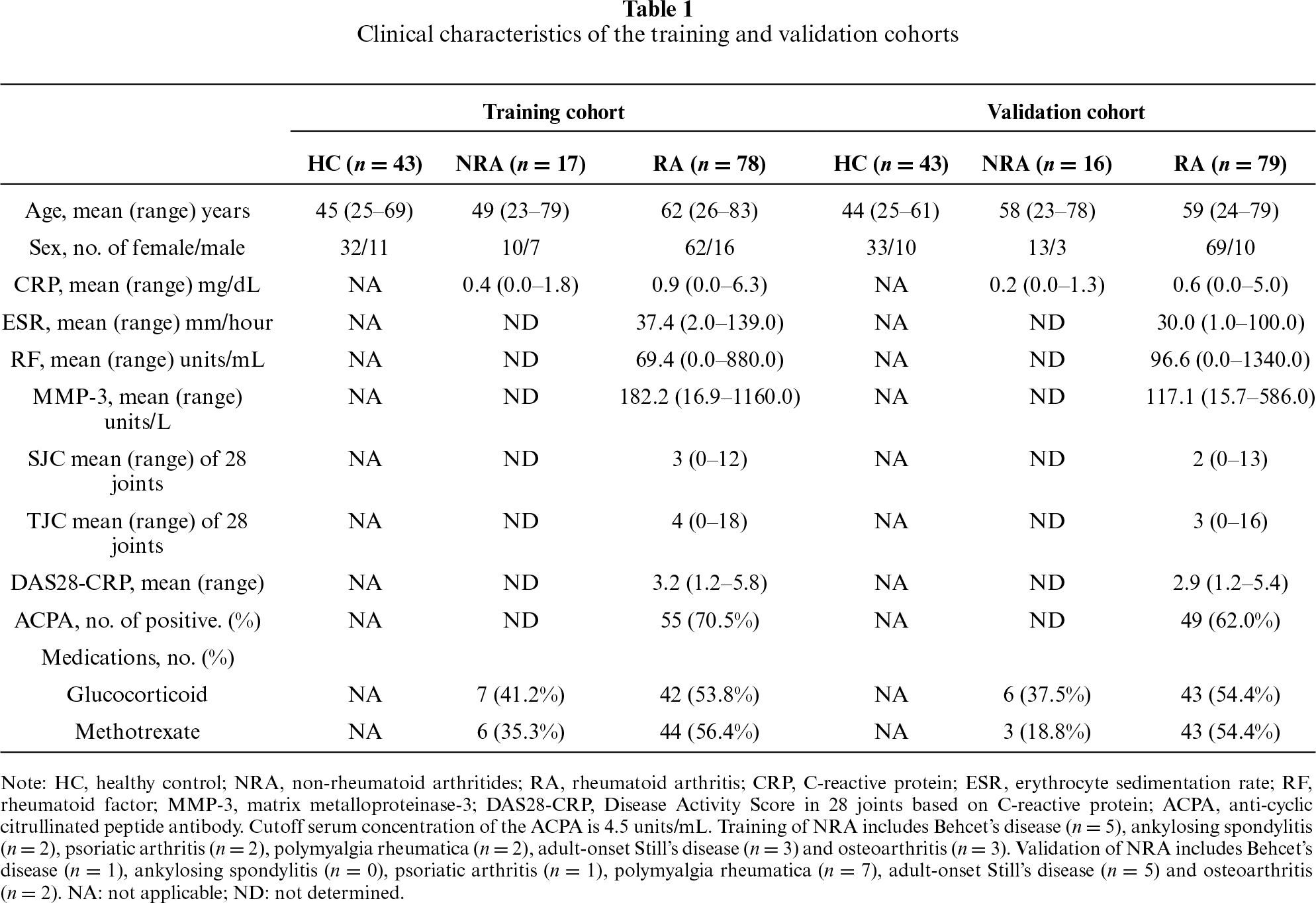
The NRA group consisted of seronegative spondyloarthritis (ankylosing spondylitis, n = 2; psoriatic arthritis, n = 3) and autoantibody-negative inflammatory or degenerative arthritis, including Behçet’s disease (n = 6), polymyalgia rheumatica (n = 9), adult-onset Still’s disease (n = 8), and osteoarthritis (n = 5).
Serum samples from 86 healthy controls (HC; 65 women and 21 men; mean age 44 years, range 25–69 years) were also included.
Patients who were receiving biological DMARDs or targeted synthetic DMARDs at the time of serum sampling were not included in the primary analysis, because these agents directly interfere with cytokine- or cytokine-associated pathways and could markedly modify circulating mediator profiles. The present analysis was therefore restricted to patients who were untreated or treated with conventional therapies, including glucocorticoid and/or methotrexate (MTX), at the time of sampling. In the NRA group, the diagnosis was established by rheumatologists based on clinical features, laboratory findings, and available imaging data. However, TJC, SJC, and composite disease activity indices were not uniformly available for all NRA patients.
This study was conducted in accordance with protocols approved by the ethics committees of Kobe University Hospital (ethics approval number No. 600; approved on 08 November 2007), Shinko Hospital (No. 0947; approved on 11 March 2011), and Sysmex Corporation (No. 2007-08; approved on 20 November 2007). Written informed consent was obtained from all participants.
Whole blood was collected into serum collection tubes without a pro-coagulant. After blood collection, samples were allowed to clot for 20–30 min at room temperature and were then centrifuged at 1500× g for 10 min at room temperature. The separated serum supernatant was aliquoted and immediately frozen at −80°C until analysis. All samples were analyzed within four years of storage. During the analytical procedures, samples were thawed and refrozen no more than three times.
2.3 Cytokine and Chemokine Measurements by ELISA
Serum concentrations of 26 analytes, comprising 14 cytokines and 12 chemokines, were measured. The cytokines analyzed were IL-1β, IL-4, IL-6, IL-10, IL-16, IL-17A, interferon-γ (IFN-γ), TNF-α, TGF-β1, GM-CSF, hepatocyte growth factor (HGF), angiopoietin-2 (Ang-2), stem cell factor (SCF), and vascular endothelial growth factor (VEGF). The chemokines analyzed were IL-8, macrophage migration inhibitory factor (MIF), CCL2/MCP-1, CCL3/MIP-1α, CCL4/MIP-1β, CCL11/eotaxin, CCL20/MIP-3α, CCL27/CTACK, CXCL1/GRO-α, CXCL9/MIG, CXCL10/IP-10, and CXCL13/BLC.
Serum levels of IL-4, IL-10, IL-17A and IFN-γ were determined using in-house high-sensitivity chemiluminescent sandwich ELISA systems. These assays were established to improve discrimination in the low-concentration range of serum cytokines by combining optimized capture/detection antibody pairs with chemiluminescent signal detection using CDP-Star substrate with Sapphire-II enhancer, and chemiluminescence quantified using an AutoLumat LB953 luminometer. This chemiluminescent ELISA-based approach was performed based on previously reported cytokine measurement methods [20,21]. Briefly, 96-well high-binding plates were coated with capture antibodies diluted in PBS and incubated overnight at 4°C. After washing with PBS containing 0.05% Tween-20, the plates were blocked with 1% BSA in PBS for 1–2 h at room temperature. Serum samples and standards were diluted 1:10 with blocking buffer, added at 100 µL per well, and incubated for 2 h at room temperature. After washing, biotinylated detection antibodies were added and incubated for 1–2 h at room temperature. Streptavidin–alkaline phosphatase was then added at a 1:1000 dilution in 1% BSA-containing buffer and incubated for 30–60 min at room temperature. After extensive washing, CDP-Star substrate with Sapphire-II enhancer was added at 100 µL per well. After incubation for 5–10 min, chemiluminescence was measured using an AutoLumat LB953 luminometer at 5-min intervals until light emission reached its peak, usually 20–30 min after substrate addition at room temperature. The limits of quantitation (LOQs) for the in-house high-sensitivity assays were 0.5 pg/mL for IL-4, 1.0 pg/mL for IL-10, 1.5 pg/mL for IL-17A, and 1.5 pg/mL for IFN-γ.
The remaining analytes were measured using commercially available sandwich ELISA kits according to the manufacturer’s instructions, with the modification of signal detection performed using streptavidin-alkaline phosphatase and CDP-Star substrate with Sapphire-II enhancer as described above.
Detailed antibody clones, catalogue numbers, reagent concentrations, kit catalogue numbers, and manufacturers are provided in Supplementary Table S1.
Statistical analyses were performed using StatFlex software (version 6, Artech Co. Ltd., Osaka, Japan). All variables were treated as continuous. A total of 276 subjects were randomly assigned in a 1:1 ratio to either a training cohort (HC n = 43, NRA n = 17, RA n = 78) or a validation cohort (HC n = 43, NRA n = 16, RA n = 79). Group differences were assessed using the non-parametric Mann–Whitney U test. A two-sided p-value < 0.01 was considered statistically significant.
Variables showing significant differences in the training cohort were considered as candidate variables for construction of a diagnostic model to discriminate RA from HC and NRA. Because the primary objective of the diagnostic model was to identify a chemokine-based signature, multivariate candidate variables were restricted to canonical CCL/CXCL chemokines among the factors that showed significant differences between RA and NRA. Spearman’s rank correlation coefficients were calculated to evaluate multicollinearity, and variables with high correlations (r > 0.75) were excluded. When two variables exceeded this threshold, the variable with the lower individual diagnostic performance, as assessed by receiver operating characteristic (ROC) curve analysis, was excluded from subsequent multivariate logistic regression analysis.
Model selection was performed using multivariable logistic regression analysis and guided by Akaike’s Information Criterion (AIC) and ROC curve analysis. All possible combinations of the remaining candidate variables were evaluated in the training cohort, and the optimal model was defined as the model with the lowest AIC and the highest area under the curve (AUC). ROC curves were generated by plotting sensitivity against [1−specificity] at various thresholds. The optimal cutoff value was determined using the Youden index. The corresponding AUC, sensitivity, specificity, positive predictive value (PPV), and negative predictive value (NPV) were calculated. The model derived from the training cohort was subsequently evaluated in the independent validation cohort.
Multi-group comparisons of discriminant values, cytokines and chemokine levels, and RA disease activity categories were performed using the Kruskal–Wallis test followed by Dunn’s post hoc test.
Treatment-related variables, including current glucocorticoid use, current MTX use, drug dose, treatment duration, and cumulative glucocorticoid exposure, were recorded when available but were not included as covariates in the multivariate model-selection procedure. This decision was made because the study was cross-sectional, treatment regimens and doses were heterogeneous, and complete standardized data on treatment duration and cumulative exposure were not available for all patients. The diagnostic models were therefore constructed using serum cytokine and chemokine concentrations alone.
A total of 157 RA patients, 33 NRA patients, and 86 HCs were included in the analysis. Demographic and clinical characteristics are summarized in table 1.
In RA patients, mean (range) laboratory values were as follows: CRP 0.76 (0–6.33) mg/dL, ESR 33.7 (1–139) mm/h, RF 83.4 (0–1340) U/mL, and MMP-3 148.4 (15.7–1160) U/L. ACPA positivity was observed in 104 patients (66.2%).
The mean DAS28-CRP score was 3.0 (range 1.2–5.8). Twenty-six, sixty-nine, and twenty-five patients were classified as having high, moderate, and low disease activity, respectively, while thirty-seven patients were in remission (DAS28-CRP < 2.3).
Regarding treatment, 44 patients received glucocorticoid alone (RA n = 37, NRA n = 7), 54 received glucocorticoid plus MTX (RA n = 48, NRA n = 6), and 42 received MTX alone (RA n = 39, NRA n = 3). The remaining patients (RA n = 33, NRA n = 17) were not receiving glucocorticoid or MTX at the time of serum sampling; these patients were not necessarily all treatment-naïve, and some may have been untreated or off these agents at sampling. Drug dosages varied widely and were not analyzed quantitatively.
3.2 Pro-Inflammatory Cytokines and Chemokines Are Up-Regulated in RA Serum
Serum cytokine and chemokine concentrations were measured using high-sensitivity ELISA. As shown in table 2, 18 of the 26 analytes (69.2%) were significantly elevated in RA patients compared with HCs (p < 0.01): IFN-γ, TNF-α, IL-6, IL-17A, IL-8, CCL20, CXCL13, CCL11, CXCL9, MIF, CCL27, CXCL10, CCL4, HGF, VEGF, IL-16, CCL3 and GM-CSF.
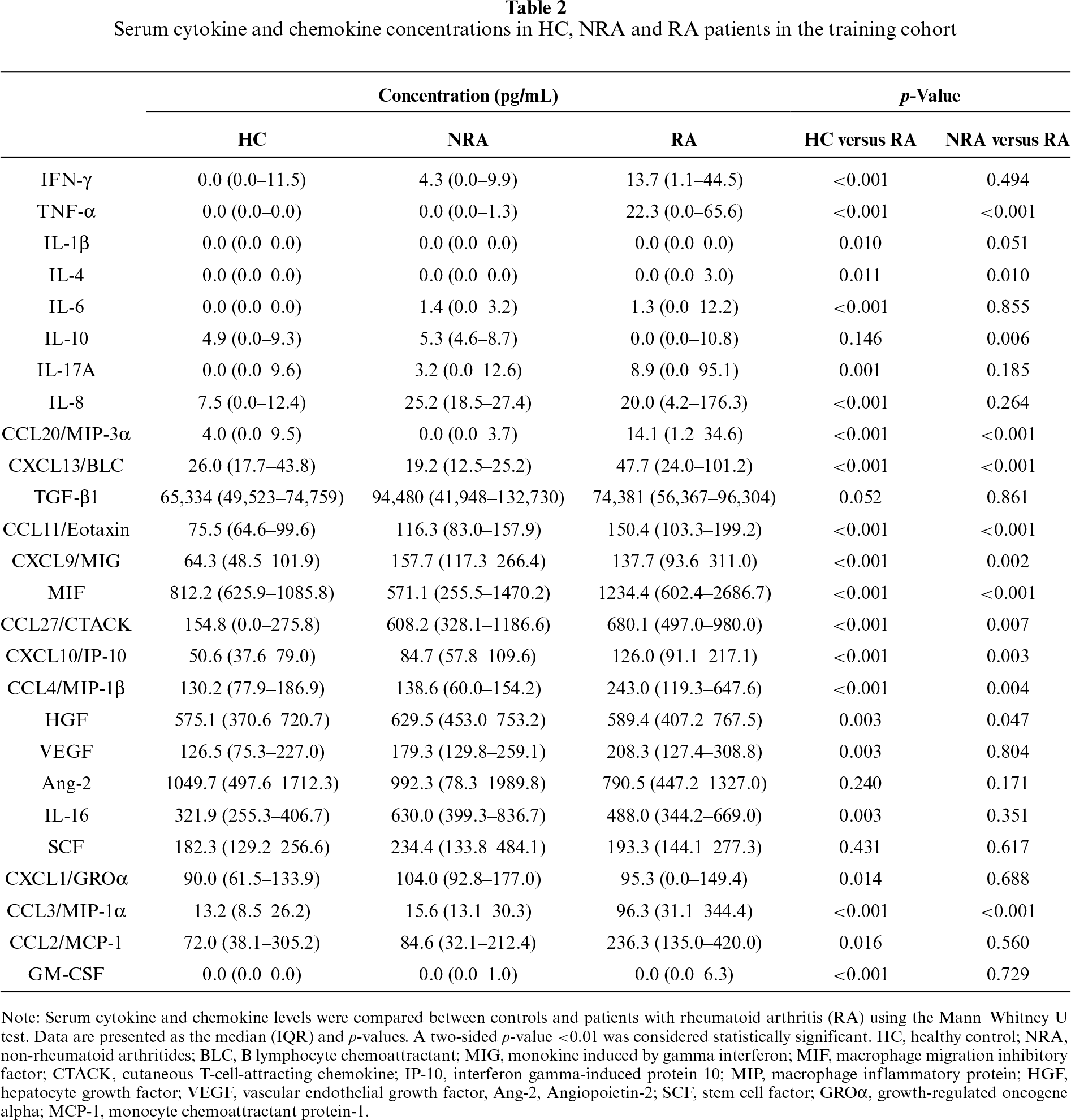
Notably, 10 of the above 18 factors (55.6%) were significantly higher in RA than in NRA patients: TNF-α, CCL20, CXCL13, CCL11, CXCL9, MIF, CCL27, CXCL10, CCL4, and CCL3.
3.3 Diagnostic Accuracy of RA Is Enhanced by a Four-Chemokine Combination
To construct and validate a discriminant model, all subjects were randomly divided into training and validation cohorts. No significant differences in clinical or laboratory variables were observed between the two cohorts (p > 0.05; table 1).
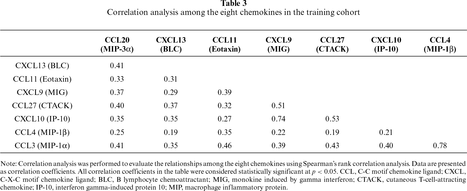
Among the 10 analytes significantly elevated in RA compared with NRA, TNF-α and MIF were not included as candidate variables for construction of the diagnostic model. The primary objective of the present study was to identify a serum chemokine signature capable of discriminating RA from NRA and healthy controls. Therefore, multivariate candidate variables were restricted to canonical CCL/CXCL chemokines. In addition, serum TNF-α levels may be influenced by therapeutic interventions targeting TNF-α or related inflammatory pathways, potentially limiting their robustness as a broadly applicable diagnostic component. Exploratory analyses also suggested that TNF-α and MIF showed less consistent discriminatory performance across patient-group comparisons than the selected chemokines. Accordingly, the multivariate analysis was restricted to the eight canonical chemokines that were significantly higher in RA than in NRA: CCL20, CXCL13, CCL11, CXCL9, CCL27, CXCL10, CCL4, and CCL3. Spearman’s correlation analysis showed that the correlation between CCL3 and CCL4 was 0.78, exceeding the predefined multicollinearity threshold of r > 0.75. Therefore, CCL4 was excluded because its individual AUC was lower than that of CCL3 (tables 3 and 4). Although the correlation between CXCL10 and CCL27 was 0.74, this value did not exceed the predefined threshold; therefore, both CXCL10 and CCL27 were retained for subsequent analysis. The remaining seven chemokines were then subjected to multivariate logistic regression analysis. ROC curve analysis was performed to evaluate the diagnostic performance of each chemokines for distinguishing RA patients from controls, including HC and NRA, in the training cohort (table 4).
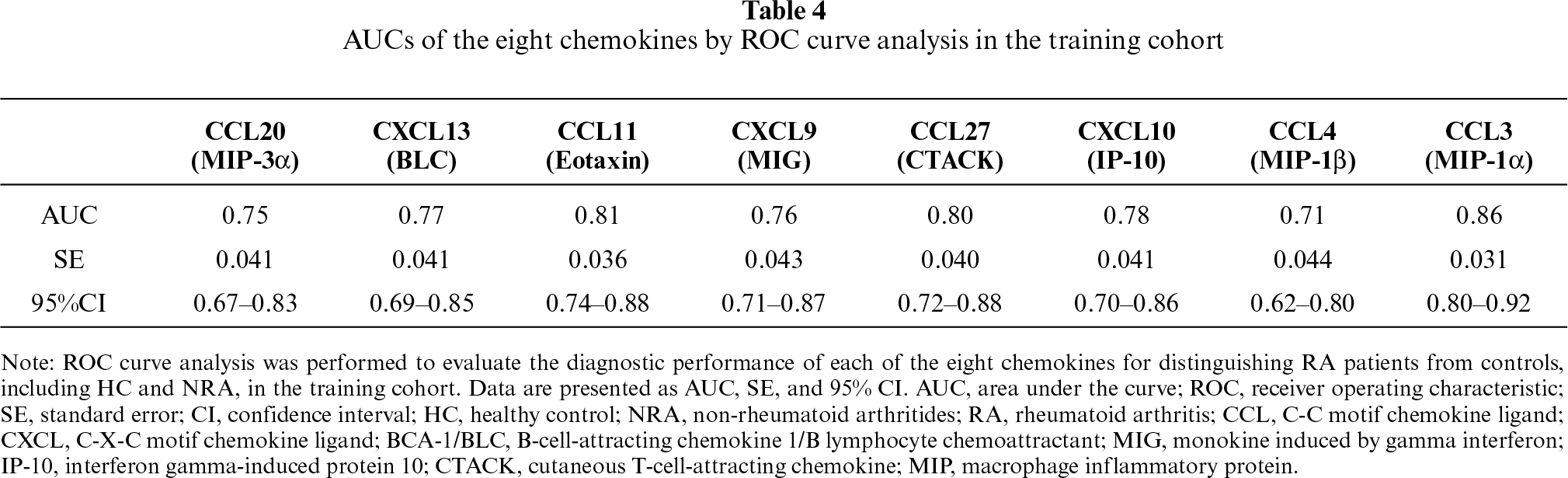
The remaining seven chemokines were subjected to multivariate logistic regression in the training cohort. Among all 127 possible combinations, the model with the lowest AIC and highest AUC was selected (figure 1A). CXCL13, CCL11, CCL27, and CCL3 emerged as independent discriminators (table 5). Individually, these chemokines yielded AUCs of 0.77, 0.81, 0.80, and 0.86, respectively (table 4 and figure 1B).
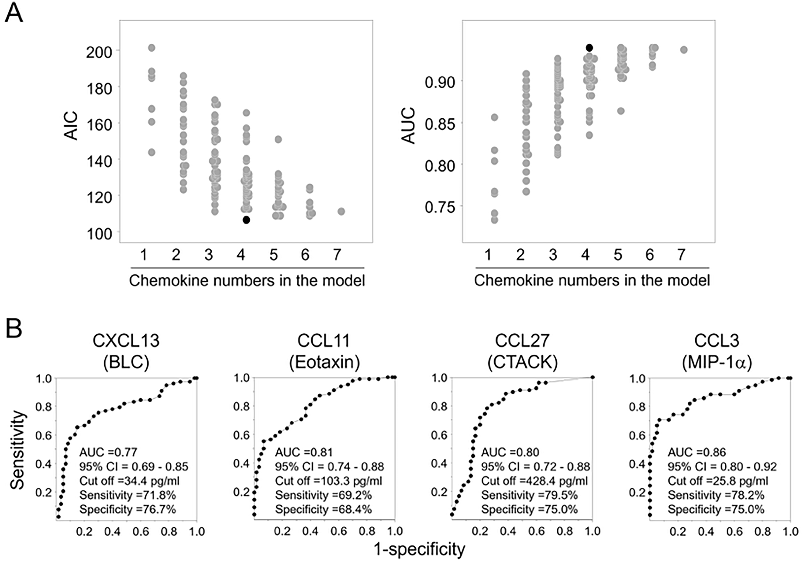
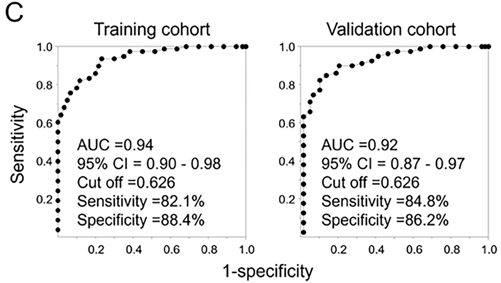
Figure 1: Evaluation of model performance using multiple logistic regression and ROC curve analyses. (A) Multiple logistic regression analysis based on seven chemokines in the training cohort. The seven chemokines were subjected to multivariate logistic regression, and all 127 possible combinations were evaluated. The model with the lowest AIC and highest AUC was selected. (B) ROC curve analysis of the four individual chemokines in the training cohort. (C) ROC curve analysis of the combined four-chemokine model in the training and validation cohorts.
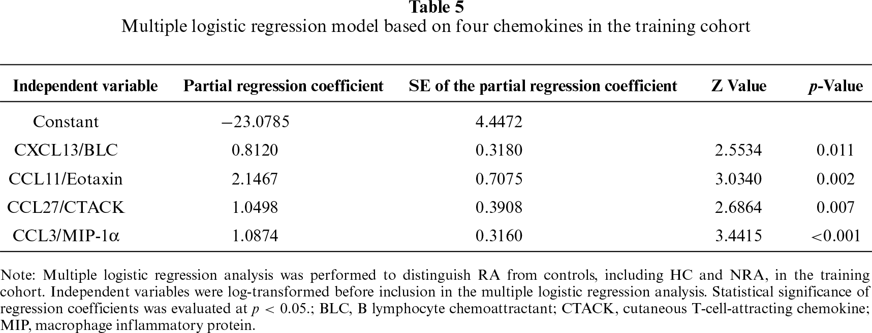
Accordingly, discriminant values were calculated using the following logistic function: Discriminant value = 1/[1 + exp(−(−23.0785 + 0.8120·CXCL13 + 2.1467·CCL11 + 1.0498·CCL27 + 1.0874·CCL3))] (table 5).
The combined four-chemokine model achieved an AUC of 0.94, with a sensitivity of 82.1% and a specificity of 88.4% at a cutoff value of 0.626 in the training cohort. In the independent validation cohort, the model maintained robust performance, yielding an AUC of 0.92, a sensitivity of 84.8%, and a specificity of 86.2% (figure 1C).
An additional analysis to assess potential confounding by age and sex was performed. All four chemokines remained statistically significant independent discriminators, whereas age and sex were not statistically significant. Notably, although both cytokines and chemokines were measured, the final diagnostic model consisted exclusively of chemokines. These findings indicate that the diagnostic utility of the serum profile was driven by the combined chemokine signature rather than by elevation of any single marker.
3.4 Discrimination of ACPA-Negative RA from Non-RA Arthritis
Accurate diagnosis of ACPA-negative RA remains challenging, particularly in differentiating it from other seronegative inflammatory arthritides. Application of the four-chemokine model to this subgroup demonstrated an AUC of 0.73, with sensitivity of 62.5% and specificity of 76.0% at the same cutoff value (0.626) in the validation cohort (figure 2A,B). These findings indicate that the chemokine-based model is useful for distinguishing ACPA-negative RA from NRA.
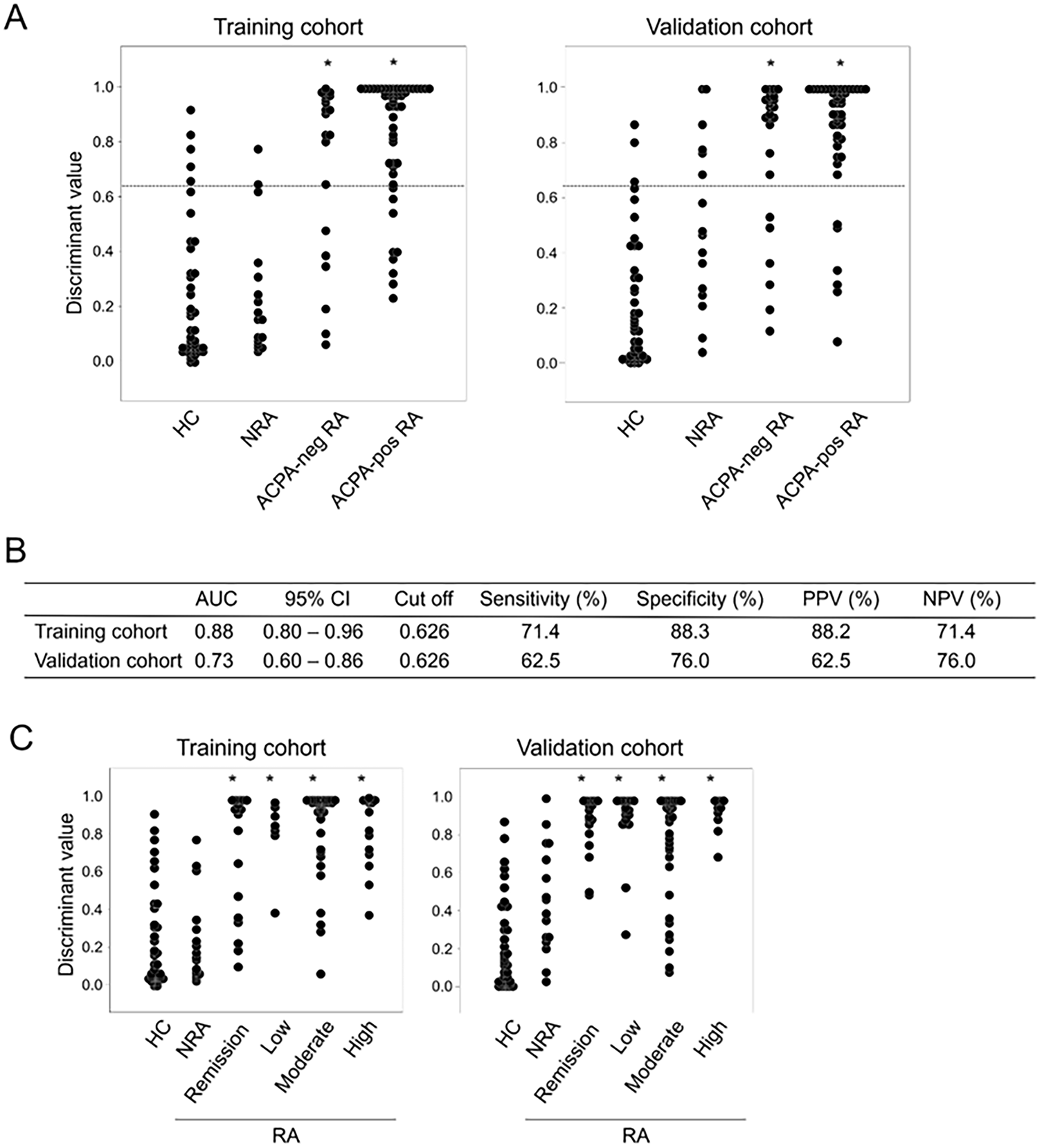
Figure 2: Diagnostic performance and clinical associations of the four-chemokine model. (A) Discriminant values of the chemokine profile among the HC, NRA, ACPA-negative RA, and ACPA-positive RA groups in the training and validation cohorts. (B) Diagnostic performance of the chemokine profile for differentiating ACPA-negative RA from NRA. In the validation cohort, the model achieved an AUC of 0.73 at a cutoff value of 0.626, with a sensitivity of 62.5% and a specificity of 76.0%. (C) Association between the chemokine profile and RA disease activity. Discriminant values of the chemokine profile are shown for the HC, NRA, and RA groups stratified according to DAS28-CRP disease activity categories (remission, low, moderate, and high disease activity) in the training and validation cohorts. *p < 0.01 indicates a statistically significant difference between each RA disease activity group and the combined HC/NRA group, as determined by Dunn’s post hoc test following the Kruskal-Wallis test.
3.5 Association of Chemokine Profile with RA Disease Activity
Finally, we examined the relationship between discriminant scores and RA disease activity. Chemokine concentrations were compared across remission, low, moderate, and high DAS28-CRP subgroups in the RA group. Discriminant values were significantly higher in all RA activity subgroups compared with in HC and NRA groups (figure 2C). These results suggest that the four-chemokine signature may aid in identifying RA patients with low disease activity or early-stage disease, irrespective of ACPA status.
In this study, we comprehensively analyzed serum cytokines and chemokines in patients with RA, NRA, and HC, and demonstrated that a distinct serum chemokine profile can discriminate RA from HC/NRA. Among the analytes examined, chemokines rather than classical cytokines emerged as the most informative diagnostic markers. The combination of four chemokines, CCL3, CCL11, CCL27, and CXCL13 showed high diagnostic accuracy for distinguishing RA from HC/NRA and retained discriminatory power in ACPA-negative RA. These findings indicate that circulating chemokines may reflect RA-associated immune and stromal activation that is not fully captured by conventional laboratory markers.
A notable finding of the present study was that cytokines were not retained in the final model despite their established pathogenic roles in RA. The aim of model construction was to identify a serum chemokine signature capable of discriminating RA from non-rheumatoid arthritides. In addition, serum TNF-α concentrations may be substantially influenced by therapeutic interventions targeting TNF-α or related inflammatory pathways, potentially limiting their utility as broadly applicable diagnostic markers [22,23]. Exploratory analyses also suggested that TNF-α and MIF showed less consistent discriminatory performance across patient-group comparisons than the selected chemokines. These considerations supported the development of a diagnostic model based exclusively on canonical chemokines. In contrast, chemokines regulate leukocyte trafficking, tissue organization, and stromal–immune cell interactions—processes that may persist even under partial clinical remission. Therefore, circulating chemokines may capture residual or subclinical inflammation more consistently than classical pro-inflammatory cytokines. In the present study, the combination of CCL3, CCL11, CCL27, and CXCL13 provided strong discrimination, achieving AUCs of 0.94 in the training cohort and 0.92 in the validation cohort for distinguishing RA from HC/NRA, and retaining modest but potentially clinically useful discriminatory performance in ACPA-negative RA (AUC 0.73) [19]. These findings suggest that serum chemokine profiling may provide diagnostic information complementary to conventional autoantibodies and acute-phase reactants [24]. The additional age- and sex-adjusted analysis further suggests that the discriminatory performance of the four-chemokine signature was not solely attributable to demographic differences among the groups.
Because no single chemokine is specific to RA, the clinical value of the present approach should be interpreted as a disease-associated multivariable signature rather than as the elevation of any individual molecule. This concept is consistent with previous biomarker studies demonstrating that combinations of inflammatory mediators provide more robust diagnostic information than single analytes in heterogeneous RA populations [10,24].
The four chemokines identified in the present model have previously been implicated in leukocyte recruitment, lymphoid organization, and chronic synovial inflammation in RA [15–17,19,20]. CCL3 contributes to recruitment of inflammatory leukocytes into synovial tissue [25,26], whereas CXCL13 promotes ectopic lymphoid organization and local B-cell responses [27–30]. CCL11 [31,32] and CCL27 [33,34] have also been reported to be associated with inflammatory cell trafficking and immune regulation in RA. Collectively, these observations support the biological plausibility of the four-chemokine signature identified in this study.
Concomitant treatment may also have influenced the composition of the final model. Previous studies have shown that MTX [35,36] and glucocorticoids [37,38] can modulate circulating cytokine and chemokine concentrations. Such treatment effects may attenuate cytokine signals and alter chemokine profiles, and therefore should be considered when interpreting serum mediator-based diagnostic models. Nevertheless, the identification of a robust chemokine signature in a real-world cohort receiving conventional therapies may also support its potential clinical relevance.
Elevated discriminant values were observed across RA disease activity categories, including patients with low or moderate DAS28-CRP scores [24]. This finding suggests that chemokine dysregulation may persist despite clinically controlled systemic inflammation and may reflect aspects of disease biology not fully captured by DAS28-CRP. Accordingly, the chemokine profile should be viewed as an adjunctive diagnostic marker rather than a substitute for clinical assessment [19].
Several limitations should be acknowledged. First, this study was based on a long-term retrospective cohort, and RA patients were classified according to the 1987 ACR criteria, which were the applicable criteria at the time of cohort establishment and sample collection. Because the 2010 ACR/EULAR criteria better capture early-stage RA, the use of the 1987 criteria may have limited the inclusion of patients with very early RA and should be considered when interpreting the generalizability of the findings. Second, the HC group was younger than the RA group, and circulating chemokine levels may be influenced by age-related inflammatory changes and sex. Although the age- and sex-adjusted multivariable analysis indicated that the four-chemokine signature remained independently associated with RA, residual confounding due to the retrospective design and demographic imbalance among groups cannot be fully excluded. Third, drug dosages, treatment durations, and cumulative glucocorticoid exposure were heterogeneous and were not quantitatively analyzed. This represents a major methodological limitation, because treatment exposure may have substantially influenced baseline cytokine and chemokine concentrations and may therefore affect interpretation of the serum chemokine signature. Finally, external validation in larger contemporary multicenter cohorts, including patients receiving biologic and targeted synthetic DMARDs, will be required before clinical implementation.
Future studies should include treatment-naïve early RA, age- and sex-matched controls, standardized assessment of disease activity in all disease-control groups, detailed quantitative treatment information, and longitudinal sampling.
In conclusion, our findings demonstrate that a serum chemokine profile consisting of CCL3, CCL11, CCL27, and CXCL13 can effectively discriminate RA from non-RA inflammatory arthritis and healthy controls, including ACPA-negative RA. These results highlight the clinical potential of chemokine profiling as a diagnostic and disease-characterization tool and provide further insight into the immunopathological mechanisms underlying RA. Future validation in independent cohorts and longitudinal studies may establish serum chemokine profiling as a valuable adjunct to current diagnostic strategies in rheumatoid arthritis.
Acknowledgement: The authors thank Dr. Shigetaka Asano and Dr. Alisa E Koch for their valuable advice; Dr. Goh Tsuji, Dr. Sho Sendo, Dr. Masafumi Ikeda and Ms. Mina Yabusaki for helpful discussions; and Ms. Mizuho Kawate, Ms. Yuko Uemura and Ms. Keiko Otsuji for their technical assistance. ChatGPT (OpenAI) was used solely to improve the English language, and the authors take full responsibility for the content of this manuscript.
Funding Statement: This work was fully supported by internal research and development funding from Sysmex Corporation under proposal No. D201002.
Author Contributions: The authors confirm contribution to the paper as follows: study conception and design: Hitoshi Uga and Hirokazu Kurata; data collection: Takahiro Okazawa; analysis and interpretation of results: Takahiro Okazawa, Yoshiaki Miyamoto and Takehiro Hasegawa; analysis of clinical data: Jun Saegusa, Akio Morinobu, and Shunichi Kumagai; draft manuscript preparation: Hitoshi Uga and Hirokazu Kurata. All authors reviewed and approved the final version of the manuscript.
Availability of Data and Materials: The data underlying this article will be shared upon reasonable request to the corresponding author.
Ethics Approval: This study was conducted in accordance with protocols approved by the ethics committees of Kobe University Hospital (ethics approval number No. 600), Shinko Hospital (No. 0947), and Sysmex Corporation (No. 2007-08). Written informed consent was obtained from all participants. The study was carried out in compliance with the Declaration of Helsinki.
Conflicts of Interest: Hitoshi Uga, Yoshiaki Miyamoto and Takehiro Hasegawa are employees of Sysmex Corporation; Takahiro Okazawa and Hirokazu Kurata were formerly employed by Sysmex Corporation. Akio Morinobu has received speaker fee and/or research grant from Astellas, Bristol Myers Squibb, Chugai, Eisai, Eli Lilly, Asahi-Kasei Pharma, Taisho, and Tanabe Pharma. Jun Saegusa has received speaker fees from Eli Lilly Japan K.K., Chugai Pharmaceutical Co., Ltd., and AbbVie GK. Shunichi Kumagai has been serving as an advisor to Sysmex Corporation.
Supplementary Materials: The supplementary material is available online at https://www.techscience.com/doi/10.32604/ecn.2026.083162/s1.
References
1. Ye X, Ren D, Chen Q, et al. Resolution of inflammation during rheumatoid arthritis. Front Cell Dev Biol 2025;13:1556359. doi:10.3389/fcell.2025.1556359. [Google Scholar] [PubMed] [CrossRef]
2. Di Matteo A, Bathon JM, Emery P. Rheumatoid arthritis. Lancet 2023;402(10416):2019–33. doi:10.1016/S0140-6736(23)01525-8. [Google Scholar] [PubMed] [CrossRef]
3. Gao Y, Zhang Y, Liu X. Rheumatoid arthritis: pathogenesis and therapeutic advances. Med Comm 2024;5(3):e509. doi:10.1002/mco2.509. [Google Scholar] [PubMed] [CrossRef]
4. McInnes IB, Schett G. Pathogenetic insights from the treatment of rheumatoid arthritis. Lancet 2017;389(10086):2328–37. doi:10.1016/S0140-6736(17)31472-1. [Google Scholar] [PubMed] [CrossRef]
5. Szekanecz Z. Chemokines and chemokine receptors in arthritis. Front Biosci 2010;S2(1):153–67. doi:10.2741/s53. [Google Scholar] [PubMed] [CrossRef]
6. Zlotnik A, Yoshie O. The chemokine superfamily revisited. Immunity 2012;36(5):705–16. doi:10.1016/j.immuni.2012.05.008. [Google Scholar] [PubMed] [CrossRef]
7. Fujimoto S, Niiro H. Pathogenic role of cytokines in rheumatoid arthritis. JCM 2025;14(18):6409. doi:10.3390/jcm14186409. [Google Scholar] [PubMed] [CrossRef]
8. Avouac J, Kay J, Choy E. Personalised treatment of rheumatoid arthritis based on cytokine profiles and synovial tissue signatures: potentials and challenges. Semin Arthritis Rheum 2025;73:152740. doi:10.1016/j.semarthrit.2025.152740. [Google Scholar] [PubMed] [CrossRef]
9. Brzustewicz E, Bryl E. The role of cytokines in the pathogenesis of rheumatoid arthritis—practical and potential application of cytokines as biomarkers and targets of personalized therapy. Cytokine 2015;76(2):527–36. doi:10.1016/j.cyto.2015.08.260. [Google Scholar] [CrossRef]
10. Kokkonen H, Söderström I, Rocklöv J, Hallmans G, Lejon K, Rantapää Dahlqvist S. Up-regulation of cytokines and chemokines predates the onset of rheumatoid arthritis. Arthritis Rheum 2010;62(2):383–91. doi:10.1002/art.27186. [Google Scholar] [PubMed] [CrossRef]
11. Deane KD, O’Donnell CI, Hueber W, et al. The number of elevated cytokines and chemokines in preclinical seropositive rheumatoid arthritis predicts time to diagnosis in an age-dependent manner. Arthritis Rheum 2010;62(11):3161–72. doi:10.1002/art.27638. [Google Scholar] [PubMed] [CrossRef]
12. Zhang F, Jonsson AH, Nathan A, et al. Deconstruction of rheumatoid arthritis synovium defines inflammatory subtypes. Nature 2023;623(7987):616–24. doi:10.1038/s41586-023-06708-y. [Google Scholar] [PubMed] [CrossRef]
13. Han P, Liu X, He J, Han L, Li J. Overview of mechanisms and novel therapies on rheumatoid arthritis from a cellular perspective. Front Immunol 2024;15:1461756. doi:10.3389/fimmu.2024.1461756. [Google Scholar] [PubMed] [CrossRef]
14. Ridgley LA, Anderson AE, Pratt AG. What are the dominant cytokines in early rheumatoid arthritis? Curr Opin Rheumatol 2018;30(2):207–14. doi:10.1097/bor.0000000000000470. [Google Scholar] [PubMed] [CrossRef]
15. Miyabe Y. Targeting the chemokine system in rheumatoid arthritis and vasculitis. JMA J 2020;3(3):182–92. doi:10.31662/jmaj.2020-0019. [Google Scholar] [PubMed] [CrossRef]
16. Murayama MA, Shimizu J, Miyabe C, Yudo K, Miyabe Y. Chemokines and chemokine receptors as promising targets in rheumatoid arthritis. Front Immunol 2023;14:1100869. doi:10.3389/fimmu.2023.1100869. [Google Scholar] [PubMed] [CrossRef]
17. Iwamoto T, Okamoto H, Toyama Y, Momohara S. Molecular aspects of rheumatoid arthritis: chemokines in the joints of patients. FEBS J 2008;275(18):4448–55. doi:10.1111/j.1742-4658.2008.06580.x. [Google Scholar] [PubMed] [CrossRef]
18. Loetscher P, Moser B. Homing chemokines in rheumatoid arthritis. Arthritis Res 2002;4(4):233–6. doi:10.1186/ar412. [Google Scholar] [PubMed] [CrossRef]
19. Li K, Wang M, Zhao L, Liu Y, Zhang X. ACPA-negative rheumatoid arthritis: from immune mechanisms to clinical translation. EBioMedicine 2022;83(2):104233. doi:10.1016/j.ebiom.2022.104233. [Google Scholar] [PubMed] [CrossRef]
20. Kameda M, Otsuka M, Chiba H, et al. CXCL9, CXCL10, and CXCL11; biomarkers of pulmonary inflammation associated with autoimmunity in patients with collagen vascular diseases–associated interstitial lung disease and interstitial pneumonia with autoimmune features. PLoS One 2020;15(11):e0241719. doi:10.1371/journal.pone.0241719. [Google Scholar] [CrossRef]
21. Jurisic V. Multiomic analysis of cytokines in immuno-oncology. Expert Rev Proteom 2020;17(9):663–74. doi:10.1080/14789450.2020.1845654. [Google Scholar] [PubMed] [CrossRef]
22. Wei ST, Sun YH, Zong SH, Xiang YB. Serum levels of IL-6 and TNF-α may correlate with activity and severity of rheumatoid arthritis. Med Sci Monit 2015;21:4030–8. doi:10.12659/msm.895116. [Google Scholar] [CrossRef]
23. Jurisic V, Terzic T, Colic S, Jurisic M. The concentration of TNF-alpha correlate with number of inflammatory cells and degree of vascularization in radicular cysts. Oral Dis 2008;14(7):600–5. doi:10.1111/j.1601-0825.2007.01426.x. [Google Scholar] [PubMed] [CrossRef]
24. Skrzypkowska M, Stasiak M, Sakowska J, et al. Cytokines and chemokines multiplex analysis in patients with low disease activity rheumatoid arthritis. Rheumatol Int 2022;42(4):609–19. doi:10.1007/s00296-022-05103-6. [Google Scholar] [PubMed] [CrossRef]
25. Zhang R, Sun P, Jiang Y, et al. Genome-wide haplotype association analysis and gene prioritization identify CCL3 as a risk locus for rheumatoid arthritis. Int J Immunogenet 2010;37(4):273–8. doi:10.1111/j.1744-313X.2010.00920.x. [Google Scholar] [PubMed] [CrossRef]
26. Yang YL, Li XF, Song B, et al. The role of CCL3 in the pathogenesis of rheumatoid arthritis. Rheumatol Ther 2023;10(4):793–808. doi:10.1007/s40744-023-00554-0. [Google Scholar] [PubMed] [CrossRef]
27. Bechman K, Dalrymple A, Southey-Bassols C, Cope AP, Galloway JB. A systematic review of CXCL13 as a biomarker of disease and treatment response in rheumatoid arthritis. BMC Rheumatol 2020;4(1):70. doi:10.1186/s41927-020-00154-3. [Google Scholar] [PubMed] [CrossRef]
28. Shi K, Hayashida K, Kaneko M, et al. Lymphoid chemokine B cell-attracting chemokine-1 (CXCL13) is expressed in germinal center of ectopic lymphoid follicles within the synovium of chronic arthritis patients. J Immunol 2001;166(1):650–5. doi:10.4049/jimmunol.166.1.650. [Google Scholar] [PubMed] [CrossRef]
29. Greisen SR, Schelde KK, Rasmussen TK, et al. CXCL13 predicts disease activity in early rheumatoid arthritis and could be an indicator of the therapeutic ‘window of opportunity’. Arthritis Res Ther 2014;16(5):434. doi:10.1186/s13075-014-0434-z. [Google Scholar] [CrossRef]
30. de Stefano L, Bozzalla Cassione E, Sammali Y, et al. High serum levels of CXCL13 predict lower response to csDMARDs in both ACPA-positive and ACPA-negative early rheumatoid arthritis. Rheumatology 2025;64(5):2461–8. doi:10.1093/rheumatology/keae596. [Google Scholar] [PubMed] [CrossRef]
31. Wakabayashi K, Isozaki T, Tsubokura Y, Fukuse S, Kasama T. Eotaxin-1/CCL11 is involved in cell migration in rheumatoid arthritis. Sci Rep 2021;11(1):7937. doi:10.1038/s41598-021-87199-7. [Google Scholar] [PubMed] [CrossRef]
32. Syversen SW, Goll GL, Haavardsholm EA, Bøyesen P, Lea T, Kvien TK. A high serum level of eotaxin (CCL 11) is associated with less radiographic progression in early rheumatoid arthritis patients. Arthritis Res Ther 2008;10(2):R28. doi:10.1186/ar2381. [Google Scholar] [PubMed] [CrossRef]
33. Davila ML, Xu M, Huang C, et al. CCL27 is a crucial regulator of immune homeostasis of the skin and mucosal tissues. iScience 2022;25(6):104426. doi:10.1016/j.isci.2022.104426. [Google Scholar] [PubMed] [CrossRef]
34. Hudak S, Hagen M, Liu Y, et al. Immune surveillance and effector functions of CCR10+ skin homing T cells. J Immunol 2002;169(3):1189–96. doi:10.4049/jimmunol.169.3.1189. [Google Scholar] [CrossRef]
35. Kremer JM, Lawrence DA, Hamilton R, McInnes IB. Long-term study of the impact of methotrexate on serum cytokines and lymphocyte subsets in patients with active rheumatoid arthritis: correlation with pharmacokinetic measures. RMD 2016;2(1):e000287. doi:10.1136/rmdopen-2016-000287. [Google Scholar] [PubMed] [CrossRef]
36. Boiardi L, Macchioni P, Meliconi R, Pulsatelli L, Facchini A, Salvarani C. Relationship between serum RANTES levels and radiological progression in rheumatoid arthritis patients treated with methotrexate. Clin Exp Rheumatol 1999;17:419–25. doi:10.1002/1529-0131(199907)42:71562::AID-ANR45%3E3.0.CO;2-A. [Google Scholar] [CrossRef]
37. Clarke LL, Jessop DS, Hunt LP, Straub RH, Perry MG, Kirwan JR. Alleviation of morning joint stiffness by low-dose prednisone in rheumatoid arthritis is associated with circadian changes in IL-6 and Cortisol. Int J Clin Rheumatol 2011;6(2):241–9. doi:10.2217/ijr.11.12. [Google Scholar] [CrossRef]
38. Petrackova A, Horak P, Savara J, Skacelova M, Kriegova E. High cumulative glucocorticoid dose is associated with increased levels of inflammation-related mediators in active rheumatoid arthritis. Front Immunol 2024;15:1505615. doi:10.3389/fimmu.2024.1505615. [Google Scholar] [PubMed] [CrossRef]
Cite This Article
 Copyright © 2026 The Author(s). Published by Tech Science Press.
This work is licensed under a Creative Commons Attribution 4.0 International License , which permits unrestricted use, distribution, and reproduction in any medium, provided the original work is properly cited.
 Downloads
 Citation Tools
