
 Login
Login Register
 Submit a Paper
 Propose a Special lssue
 Open Access
REVIEW
Short-chain fatty acids regulate cytokine programs in tumor immunity: mechanisms and translational opportunities
1 Department of Gastroenterology, Renmin Hospital of Wuhan University, Wuhan, China
2 Key Laboratory of Hubei Province for Digestive System Diseases, Renmin Hospital of Wuhan University, Wuhan, China
3 Central Laboratory, Renmin Hospital of Wuhan University, Wuhan, China
4 Department of Oncology, The Second Clinical Medical College, Yangtze University, Jingzhou, China
* Corresponding Authors: Haisen Yin. Email: ; Shiyun Tan. Email:
; Baoping Yu. Email:
(This article belongs to the Special Issue: Inflammation in Disease: When Cytokine Conversations Tell the Whole Story)
European Cytokine Network 2026, 37(3), 259-280. https://doi.org/10.32604/ecn.2026.084574
Received 25 April 2026; Accepted 05 June 2026; Issue published 28 September 2026
 View Full Text
 Download PDF
Abstract
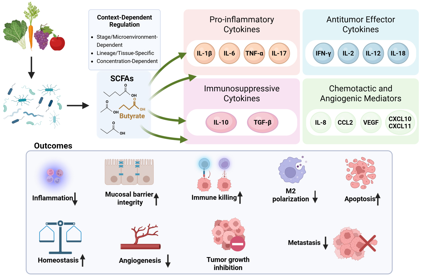
Short-chain fatty acids (SCFAs) are increasingly recognized as active modulators of tumor immunity. However, organizing evidence only by tumor type, metabolite species, or individual pathway may not fully explain their context-dependent effects. This Review clarifies how SCFAs regulate tumor immunity by remodeling cytokine and chemokine networks within the tumor microenvironment and defines the contextual factors that determine whether these effects support or restrain antitumor responses. We organize SCFA-driven effects into four functional cytokine domains: pro-inflammatory, antitumor effector, immunosuppressive, and chemotactic/angiogenic mediators. Across these domains, SCFAs act through inflammasome modulation, receptor-mediated signaling, epigenetic regulation, and metabolic reprogramming. We propose that the translational potential of SCFAs depends on context-aware application, given that their immunological effects are shaped by SCFA subtype, exposure compartment, dose, tumor stage, cellular lineage, tissue environment, and therapeutic context. Importantly, current mechanistic insights remain largely butyrate-based, with limited evidence for other SCFAs or clinically relevant exposure settings. We further discuss the implications of these patterns for immune checkpoint blockade, cell- and virus-based therapies, radiotherapy, and chemotherapy. Overall, SCFA-mediated modulation of cytokine networks provides a useful framework for understanding metabolic regulation of tumor immunity and its translational potential.Graphic Abstract
Keywords
Short-chain fatty acids (SCFAs) are major microbial metabolites generated through the anaerobic fermentation of dietary fiber and constitute a critical interface linking the gut microbiota, host immunity, and colorectal cancer biology, with broader relevance to selected tumor-immune contexts [1]. SCFAs are mainly generated from gut microbial fermentation of microbiota-accessible dietary fibers and other non-digestible carbohydrates [2]. Representative substrates include resistant starch, inulin-type fructans, pectin, β-glucans, arabinoxylans, and galacto-oligosaccharides from vegetables, fruits, legumes, whole grains, and selected prebiotic formulations [3]. Fermentable fiber-rich diets and selected substrates, including pectin and inulin, have been associated with improved antitumor immunity and, in some settings, enhanced immune checkpoint blockade efficacy in melanoma and preclinical colorectal cancer models [4–6]. Nevertheless, high-dose or refined inulin-type soluble fiber may exert context-dependent adverse effects, including dysbiosis-associated hepatocellular carcinoma and inflammation-associated colon tumorigenesis, indicating that fermentable fiber or prebiotic substrates are not uniformly beneficial [7,8]. In the intestinal tract, acetate, propionate, and butyrate are the main SCFAs [9]. Recent studies have shown that these metabolites are not merely end products of microbial metabolism, but also active regulators of transcriptional programs, cellular metabolism, and immune-cell function [10]. In cancer-related settings, experimental and translational studies support the role of SCFAs in intestinal homeostasis and inflammation-related tumorigenesis, and potential associations between SCFAs and treatment response have also been reported in certain tumors [11–13]. Mechanistically, SCFAs act mainly through pathways such as histone deacetylase (HDAC) inhibition, G protein-coupled receptor (GPCR) signaling, and metabolic reprogramming. Through these routes, they also modulate inflammatory programs, including inflammasome-associated signaling [9,14]. These upstream mechanisms do not generate uniform immune outcomes directly. Instead, they are interpreted by distinct responding cell populations, including tumor cells, epithelial cells, macrophages, dendritic cells, CD8+ T cells, natural killer (NK) cells, and stromal cells [9,15]. These cells then translate SCFA-responsive signals into specific cytokine and chemokine outputs that shape downstream immune consequences [14,16]. This is consistent with recent findings in tumor immunometabolism research, which indicate that metabolic reprogramming within the TME can reshape inflammatory signaling, TAM function, CD8+ T cell activity, and therapeutic responsiveness [17–19]. From this perspective, interpreting SCFA activity through their context-dependent remodeling of cytokine and chemokine networks within the TME may provide a useful framework for understanding how these metabolites regulate the tumor microenvironment (TME).
Accumulating evidence indicates that the effects of SCFAs on tumors are not uniform. Rather than exerting simple anti-inflammatory or anti-tumor activities, they exhibit pleiotropic effects. Their modes of action and outcomes may even produce opposite results depending on the disease stage, tissue type, and therapeutic context [1,20,21]. Meanwhile, the direction and magnitude of the immunoregulatory effects exerted by various SCFAs vary with their concentrations, tissue environments, cellular contexts, and therapeutic backgrounds [1,20,22]. Therefore, evaluating the preventive and potential therapeutic relevance of modulating the utilization or levels of SCFAs in vivo is more appropriately conducted within the context-defined framework of cytokine network regulation.
Cytokine and chemokine networks form one of the most immediate functional levels through which SCFA-mediated effects become biologically meaningful in the tumor microenvironment (TME) [14,23]. These networks do not merely reflect inflammatory status; they also shape immune-cell recruitment, activation, exhaustion, vascular remodeling, stromal support, and treatment responsiveness [24,25]. However, most existing summaries of this field still organize the literature by tumor type, signaling mechanism, or individual SCFA species [12–14,20]. These approaches are useful, but they can also fragment the field. As a result, these approaches may obscure a more informative organizing principle: the recurring connection between shared upstream SCFA-responsive mechanisms, downstream cytokine output, and the resulting immunological consequences across different tumor contexts. For this reason, rather than following conventional organization by cancer type, mechanistic pathway, or metabolite class, we employ a cytokine module–based framework. This framework integrates recurrent upstream mechanisms across tumor and SCFA contexts with their downstream cytokine outputs and immune consequences. It also highlights how these relationships are shaped by tissue environment, dominant cellular responders, and therapeutic context.
Hence, this Review focuses on four cytokine modules with direct relevance to tumor immunity. These include pro-inflammatory cytokines, including interleukin (IL)-1β, IL-6, tumor necrosis factor-α (TNF-α), and IL-17; antitumor effector cytokines, including interferon-γ (IFN-γ), IL-2, IL-12, and IL-18; immunosuppressive cytokines, including interleukin-10 and transforming growth factor-β (TGF-β); and chemotactic/angiogenic mediators, including IL-8, C-C motif chemokine ligand 2 (CCL2), C-X-C motif chemokine ligands (CXCL) 10 and 11, and vascular endothelial growth factor (VEGF). The main novelty of this Review lies in using these cytokine modules not merely as descriptive categories, but as an integrative framework for connecting recurrent SCFA-responsive mechanisms with downstream immune outputs. Because current evidence remains unevenly distributed across individual SCFAs, butyrate-centered mechanisms should be interpreted cautiously and should not be automatically generalized to acetate, propionate, valerate, or other SCFAs without direct supporting evidence. In contrast to previous reviews that have mainly summarized SCFAs or microbiota-derived metabolites by tumor type, metabolite class, microbiota composition, or individual signaling pathway, this Review places cytokine- and chemokine-network remodeling at the center of analysis. More specifically, we treat context dependence not as a peripheral caveat, but as the main principle for interpreting apparently conflicting findings. In the sections that follow, we argue that the overall direction of SCFA-associated cytokine remodeling is first constrained by exposure compartment and disease stage, then modified by dominant responding cell lineage and tissue setting, and only then refined by metabolite identity, concentration, and treatment background. Rather than cataloging every reported function of SCFAs in cancer, we examine how SCFAs regulate these four cytokine modules, identify the major determinants of their effects, and discuss the potential therapeutic relevance, translational implications, and mechanistic boundaries of SCFA-mediated immune regulation in cancer.
2 SCFA-Mediated Regulation of Pro-Inflammatory Cytokine Circuits
Within this module, the key determinant is not the cytokine name itself, but whether SCFA exposure occurs in inflammation-dominant or effector-dominant settings. During tumor initiation and progression, IL-1β, IL-6, TNF-α, and IL-17 can act as major drivers of inflammatory and tumor-supportive programs [26–28]. In chronic inflammation-dominant settings, SCFAs more often attenuate pro-inflammatory cytokine amplification and suppress tumor-promoting inflammation [29,30]. By contrast, in effector-dominant or therapy-related immune-activation settings, selected SCFAs may support immune activation or increase tumor-cell susceptibility to immune-mediated injury [31,32]. SCFAs should therefore be viewed here as setting-dependent modulators of pro-inflammatory cytokine circuits rather than as uniformly anti-inflammatory factors (figure 1). Because much of the mechanistic evidence in this section is derived from butyrate-based studies, findings are attributed to individual SCFAs where possible and should not be assumed to apply equally to acetate, propionate, or valerate.
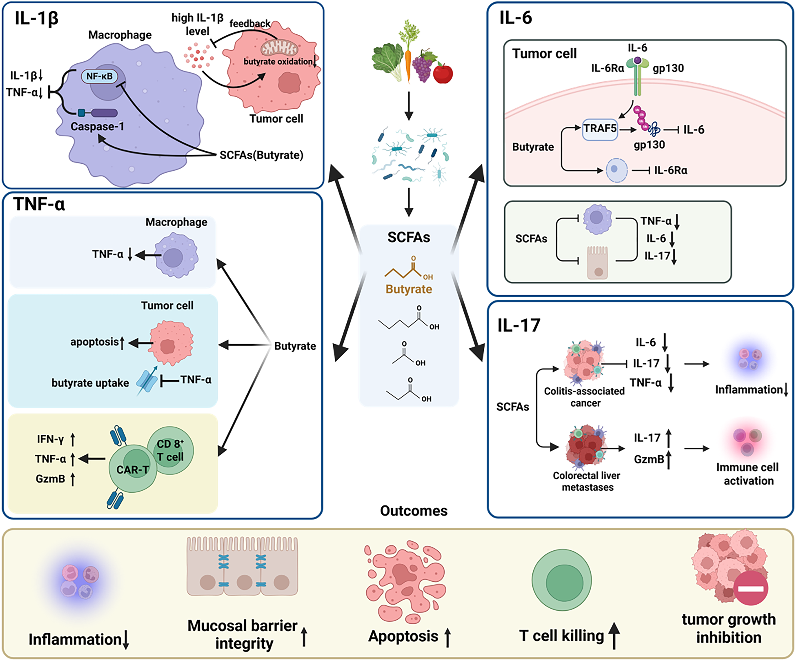
Figure 1: Short-chain fatty acid (SCFA)-mediated regulation of pro-inflammatory cytokine circuits in the tumor microenvironment (TME). SCFAs, particularly butyrate, regulate pro-inflammatory cytokine signaling in the TME in a setting-dependent manner. Most mechanistic evidence in this module is derived from butyrate-based studies, particularly for the IL-1β/NLRP3 and TNF-α axes, whereas acetate, propionate, valerate, or mixed SCFA exposure has been examined in more restricted contexts. In the available evidence, butyrate suppresses the IL-1β axis primarily through inhibition of NLRP3 inflammasome signaling and regulates TNF-α in a lineage-specific manner by limiting inflammatory TNF-α production while enhancing selected cytotoxic programs. For the IL-6/JAK/STAT3 axis, SCFA exposure has been reported to modulate signaling through receptor-proximal or inflammatory pathways. SCFAs may also modulate IL-17 in a stage- and microenvironment-dependent fashion. In chronic inflammation-dominant settings, these effects are associated with reduced inflammatory amplification, improved barrier integrity, and suppression of tumor-promoting inflammation. Arrows indicate activation or enhancement; blunt-ended lines indicate inhibition or attenuation. Upward and downward arrows (↑/↓) denote increased or decreased levels or activity.
2.1 Butyrate-Dominant Regulation of the IL-1β/NLRP3 Inflammasome Axis
IL-1β is a critical node linking innate immune sensing to the amplification of inflammatory circuits, and its production and maturation represent a plausible point of intervention through which SCFAs may modulate the inflammation–tumor axis. In multiple solid tumors, particularly in inflammation-associated tumor models, tumor-associated macrophages (TAMs) are a major source of IL-1β. IL-1β production generally proceeds through two steps: nuclear factor kappa B (NF-κB)-dependent transcriptional priming, followed by NLR family pyrin domain containing 3 (NLRP3)/Caspase-1-mediated proteolytic maturation [33,34]. Accordingly, the NLRP3/IL-1β axis is widely regarded as a therapeutic target for the regulation of a pro-inflammatory TME [34,35].
Among SCFAs, butyrate has shown the most consistent association with suppression of IL-1β-related inflammatory signaling, primarily through inhibition of the NLRP3 inflammasome and modulation of the peroxisome proliferator-activated receptor alpha signaling axis. In colitis-associated cancer and high-fat diet-driven tumor models, butyrate downregulates NLRP3 and Caspase-1, thereby reducing the maturation and secretion of IL-1β and other pro-inflammatory mediators [36–38]. It also upregulates barrier-associated proteins, including zonula occludens-1, occludin, and claudin-1, thereby promoting mucosal barrier repair and reducing chronic inflammatory stimulation [36,39,40]. A similar inhibitory effect on IL-1β has been reported in immune therapy-related inflammatory toxicity, where butyrate increased the PPARα–cytochrome P450 family 4 subfamily X member 1 axis and suppressed NF-κB signaling, leading to reduced IL-1β and TNF-α production in macrophages [41].
Notably, this pathway is constrained by a metabolic–inflammatory negative feedback loop, whereby elevated IL-1β impairs oxidative butyrate metabolism in colorectal cancer (CRC) cells through a p38-dependent mechanism, thereby limiting butyrate activity [42]. Taken together, these findings indicate that butyrate may mitigate chronic inflammation-driven or inflammation-associated tumor initiation and progression by suppressing the NLRP3/IL-1β axis; however, under conditions of high inflammatory burden, cytokine-driven metabolic tolerance may restrict the efficacy of butyrate-mediated regulation, representing an important boundary for therapeutic translation.
2.2 Regulation of the IL-6/JAK/STAT3 Axis by SCFAs
Within the TME, IL-6 serves as a key driver of tumor-supportive inflammatory signaling. It contributes to the maintenance of these pathogenic circuits. Much of its oncogenic activity arises from sustained IL-6/JAK/STAT3 signaling [43,44]. IL-6 derived from tumor, stromal, and immune cells can promote STAT3-dependent immunosuppressive programs through both autocrine and paracrine signaling, thereby facilitating immune evasion [44,45]. The functional consequences of IL-6 signaling also depend on its cellular source, as tumor-derived IL-6 may sustain autocrine STAT3 activation, whereas stromal- or myeloid-derived IL-6 may reinforce paracrine inflammatory and immunosuppressive circuits [44,46].
Current evidence suggests that SCFAs may suppress the IL-6 axis through two principal routes: first, by downregulating IL-6 receptor components and interfering with receptor-proximal signaling; and second, by reducing IL-6 production through inhibition of upstream inflammatory transcriptional programs and inflammasome-related signaling. Regarding the first route, in CRC, butyrate promotes TNF receptor-associated factor 5-dependent ubiquitination and degradation of glycoprotein 130 (gp130) while simultaneously downregulating IL-6 receptor alpha expression, thereby attenuating IL-6–induced STAT signaling and anti-apoptotic programs [47,48]. In multiple myeloma cells, butyrate similarly downregulates gp80/gp130 and is accompanied by G1-phase arrest and apoptosis [49]. Regarding the second route, SCFAs can suppress inflammatory signaling pathways such as NF-κB p65 and NLRP3, thereby reducing IL-6 production in macrophages and colonic epithelial cells [29,30,39]. These effects have been associated with reduced tumor burden and increased apoptosis in models of colitis-associated cancer, together with suppression of inflammatory mediators such as IL-6 [29].
On the other hand, regulation of IL-6 by SCFAs is clearly tissue-specific. In esophageal squamous cell carcinoma, sodium butyrate has been reported to induce IL-6 expression and secretion in cancer cells [50]. Therefore, tissue context and microenvironmental heterogeneity must be considered when interpreting SCFA-mediated regulation of the IL-6 axis; however, because current evidence in this setting is weighted toward butyrate, the direction and extent of these effects should not be generalized across either SCFA species or tumor types.
2.3 Context-Dependent Regulation of TNF-α: Predominantly Butyrate-Based Evidence
In the TME, the function of TNF-α is determined by its cellular source, signaling intensity, and microenvironmental context, rather than reflecting a uniformly protumor or antitumor role [51]. Chronic and sustained TNF-α-driven inflammation tends to promote tumor progression, whereas in selected therapeutic contexts, appropriately localized or controlled TNF-α signaling may enhance antitumor immune responses and facilitate tumor clearance [52,53]. Consistent with this context dependence, regulation of the TNF-α axis by SCFAs is lineage-specific: SCFAs suppress pathological inflammatory signaling in myeloid and epithelial compartments, while enhancing tumor-cell sensitivity to TNF-α-mediated cytotoxicity and, in some lymphoid settings, augmenting TNF-α-associated effector functions.
In inflammatory contexts involving myeloid and epithelial cells, the anti-inflammatory evidence is largely butyrate-weighted, although some studies have used mixed SCFA exposure. SCFAs, particularly butyrate, can suppress inflammatory signaling and reduce TNF-α production in epithelial and myeloid cells through cell-type and stimulus-dependent mechanisms involving NF-κB inhibition as well as broader transcriptional and proteasome/IκB-α regulation; in macrophages, peroxisome proliferator-activated receptor gamma-related effects have also been implicated [30,54–56]. By contrast, in tumor cells, butyrate can sensitize cancer cells to TNF-α-induced apoptosis by downregulating cellular FLICE-like inhibitory protein, upregulating tumor necrosis factor receptor 1, or acting through a p21-dependent mechanism, thereby producing synergistic cytotoxic effects [57–59]. In another tumor-cell context, butyrate has been reported to promote endogenous TNF-α secretion in SW480 cells through the TLR4/MAPK axis, which may contribute to early innate immune activation [60]. However, higher levels of TNF-α may also suppress monocarboxylate transporter 1 through post-transcriptional mechanisms, thereby reducing butyrate uptake and establishing a metabolic–inflammatory negative feedback loop [61,62].
In lymphoid effector cells, SCFAs may instead exert immunostimulatory effects. Butyrate and valerate can enhance mechanistic target of rapamycin (mTOR) activity and inhibit class I HDACs in CD8+ T cells and chimeric antigen receptor T (CAR-T) cells, thereby increasing TNF-α and IFN-γ production and strengthening antitumor effector function [32]. In addition, butyrate has been shown to act directly on Toll-like receptor 5 in CD8+ T cells, activate NF-κB signaling, and upregulate TNF-α, IFN-γ, and granzyme B expression [63]. Therefore, tissue context and microenvironmental heterogeneity must be considered when interpreting SCFA-mediated regulation of the TNF-α axis. However, because current evidence in this setting is weighted toward butyrate, the direction and extent of these effects should not be generalized across either SCFA species or tumor types.
2.4 Stage-Specific Regulation of IL-17 by SCFAs
IL-17 is a key pro-inflammatory cytokine that bridges inflammatory signaling and adaptive immunity, and its regulatory functions vary across different biological contexts. It can either promote tumor progression by sustaining a pro-inflammatory microenvironment, or exert antitumor immune effects by enhancing the recruitment of effector cells [28,64]. The function of IL-17 also depends on its cellular source. In addition to conventional Th17 cells, γδ T cells and group 3 innate lymphoid cells can produce IL-17 in barrier tissues and tumor-associated inflammatory niches [65], which may partly explain the divergent immunological outcomes observed across tumor models.
In models of colitis-associated colorectal cancer, SCFA mixtures can reduce the levels of IL-17, as well as other pro-inflammatory cytokines such as IL-6 and TNF-α, thereby alleviating intestinal inflammation and restraining tumor initiation and progression [29]. However, in lung cancer models, acetate and propionate can upregulate the expression of IL-17, accompanied by elevated levels of granzyme B and perforin, thereby enhancing the activity of cytotoxic cells [66]. Similar phenomena can also be observed in a mouse model of colorectal cancer liver metastasis. Butyrate can increase the proportion of T helper 17 (Th17) cells and promote the secretion of IL-17, thereby enhancing antitumor immunity and reducing metastatic burden [67].
Therefore, the overall regulation of IL-17 by SCFAs varies with tumor type, disease stage, and biological context. When the tissue environment is dominated by chronic inflammation, SCFAs may inhibit IL-17 secretion, thereby attenuating the pro-tumor effects induced by inflammation. Accordingly, the effects of SCFA-based interventions on the IL-17 axis should be interpreted in a stage- and tumor-setting-specific manner, and one should not assume a uniformly stimulatory or inhibitory effect across different SCFA species or tumor settings.
3 SCFA-Mediated Enhancement of Antitumor Effector Cytokine Signaling
Beyond suppressing several tumor-promoting inflammatory cytokines, selected SCFAs, particularly butyrate and valerate in the currently available evidence, may also reinforce antitumor immunity during specific effector phases. These effects are mediated primarily through epigenetic and metabolic mechanisms, including HDAC inhibition and mTOR-related metabolic reprogramming. Through these pathways, SCFAs can potentiate the function of CD8+ T cells, NK cells, and CAR-T cells, thereby increasing the expression and secretion of effector cytokines such as IFN-γ, IL-2, IL-12, and IL-18. In specific preclinical settings, these changes may support effector function in constrained or exhaustion-prone T cells and may reinforce cytotoxic immune responses by promoting antigen presentation and cytotoxic granule release (figure 2).
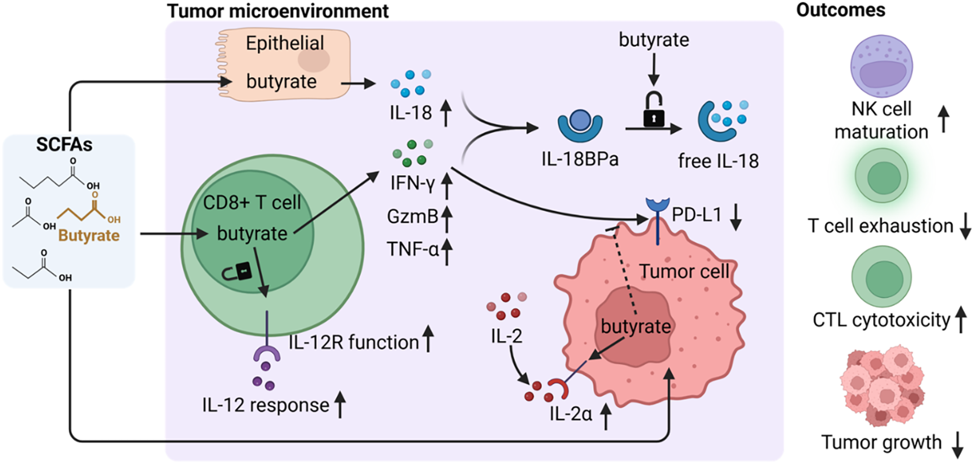
Figure 2: Short-chain fatty acid (SCFA)-mediated enhancement of antitumor effector cytokine signaling in the tumor microenvironment (TME). SCFAs, particularly butyrate, may enhance antitumor effector programs in the TME. In CD8+ T cells and NK cells, they promote the production of IFN-γ, TNF-α, and granzyme B, enhance responsiveness to IL-2 and IL-12, and increase IL-18 bioactivity, thereby potentially strengthening cytotoxic immunity and partially relieving functional exhaustion. In selected contexts, SCFAs may also suppress tumor cell-intrinsic immune escape mechanisms, including IFN-γ-induced PD-L1 upregulation. Together, these effects can promote effector-cell activation, intratumoral cytotoxicity, and tumor growth control. Arrows indicate activation or enhancement; blunt-ended lines indicate inhibition or attenuation. Upward and downward arrows (↑/↓) denote increased or decreased levels or activity.
3.1 SCFAs and IFN-γ-Driven Cytotoxic Immunity
During the antitumor effector phase, IFN-γ is a central cytokine coordinating immune-mediated tumor killing. SCFA-mediated regulation of the IFN-γ axis can be understood at three levels: immune-cell IFN-γ production, tumor- or stromal-cell responsiveness to IFN-γ, and IFN-γ-induced adaptive resistance, including PD-L1 upregulation. Through the combined actions of epigenetic and metabolic mechanisms, SCFAs can enhance the transcriptional accessibility and expression potential of IFN-γ in functionally constrained or exhausted T cells [32].
At the T cell level, acetate can enhance histone acetylation and chromatin accessibility through an acetyl-CoA synthetase–dependent acetyl-CoA supply pathway, thereby increasing the capacity of functionally impaired CD8+ T cells to produce IFN-γ [31]. In addition, short-chain fatty acids such as butyrate can strengthen the effector functions of CD8+ T cells and some CAR-T cells by inhibiting class I histone deacetylases, activating mTOR signaling, and signaling through the GPR109A/HOPX axis, resulting in elevated expression of IFN-γ, granzyme B, and TNF-α [32,68,69]. In other models, acetate can also amplify local IFN-γ and IFN-γ–induced chemokine signaling via GPR43, promoting CD8+ T cell infiltration and suppressing tumor progression [70].
At the tumor-cell level, sodium butyrate can upregulate the expression of STAT1 and enhance IFN-α–induced STAT1 phosphorylation and activation, thereby improving the responsiveness of certain tumor cells to IFN-α signaling [71]. In several tumor models, butyrate also inhibits IFN-γ–induced upregulation of PD-L1, thereby increasing the susceptibility of tumor cells to CD8+ T cell–mediated killing [72,73]. However, this regulatory pattern is strongly influenced by the microenvironment: in colonic mucosal dendritic cell–naive T cell systems, butyrate suppresses the differentiation of IFN-γ+ T cells and skews responses toward a tolerogenic program; whereas in ex vivo settings of peripheral immune cells, butyrate reduces IFN-γ–associated effector responses in CD8+ T cells and NK cells [74,75].
In summary, the effects of short-chain fatty acids on the IFN-γ axis are highly context-dependent. SCFAs can enhance IFN-γ–mediated antitumor effector functions in metabolically restricted or activated T cells, suppress IFN-γ responses in specific tolerogenic microenvironments, or reprogram IFN-γ downstream signaling in tumor cells. The direction and immunological outcomes of these effects depend on SCFA species, target cell type, metabolic state, and microenvironmental conditions, and may involve distinct regulatory layers acting on either IFN-γ production or downstream signaling responses.
3.2 Effects of SCFAs on IL-2 Responsiveness in T Cell Immunity
Beyond the IFN-γ axis, SCFAs can also independently modulate receptor sensitivity and signaling efficiency within the IL-2 axis, a key pathway for T cell growth and function. In some immunotherapeutic settings, however, this regulation may also interfere with treatment efficacy. IL-2 is produced primarily by activated T cells and has intrinsically dual therapeutic effects: it promotes the expansion and cytotoxic activity of effector T cells and NK cells, while also supporting the homeostasis and immunoregulatory function of regulatory T cells (Tregs) [76,77].
Regulation of the IL-2 axis by SCFAs is highly microenvironment-dependent. The direction of this effect appears to depend mainly on the target compartment and treatment phase. On the one hand, butyrate may increase IL-2 receptor alpha (IL-2Rα) expression on certain tumor cells, and butyrate-induced tumor cell-derived apoptotic bodies can synergize with IL-2 immunotherapy to enhance antitumor immune responses [78,79]. Consistent with this, combined treatment with sodium butyrate and IL-2 has shown synergistic antitumor activity in multiple rat models, likely by inducing immunogenic apoptotic bodies and activating tumor-specific immune responses; in some rat tumor models, marked antitumor responses, including complete regression in selected cases, have been reported [79–81]. These findings therefore reflect a tumor-cell or IL-2-combination context rather than a general enhancement of endogenous IL-2 signaling.
On the other hand, in the setting of cytotoxic T lymphocyte-associated protein 4 (CTLA-4) blockade, butyrate may limit therapeutic efficacy by impairing dendritic-cell maturation and costimulatory signaling, thereby weakening T cell priming and reducing memory T cell accumulation; decreased IL-2-associated immune activation has also been observed in treated patients [82]. In this setting, reduced IL-2-associated activation is better interpreted as an indirect consequence of impaired dendritic-cell priming and costimulation, rather than as a direct effect of CTLA-4 blockade on IL-2 production. Overall, the effects of SCFAs on the IL-2 axis are therapy-phase dependent: butyrate may either enhance IL-2 responsiveness and antitumor immunity or dampen IL-2-related immune priming and memory formation, depending on the immune microenvironment and therapeutic context.
3.3 Effects of Butyrate on the IL-12 Response Axis
Butyrate may relieve functional constraints on the IL-12 axis and thereby support type 1 antitumor immunity in selected preclinical settings. IL-12 is one of the key mediators underlying inflammatory tumor states in T cells and improved responsiveness to immune checkpoint blockade, providing a mechanistic basis for its antitumor effects [83,84].
Mechanistically, as an HDAC inhibitor, butyrate upregulates the transcriptional repressor inhibitor of DNA binding 2 (ID2), which in turn antagonizes E2A transcription factor binding to the Il12rb2 promoter, thereby relieving repression of IL-12R β2 expression and restoring CD8+ T cell responsiveness to IL-12. Enhanced IL-12 sensitivity further promotes effector functions, including increased production of IFN-γ and granzyme B, and this effect is associated with tumor growth inhibition in colorectal cancer and lymphoma models [85]. Overall, butyrate appears to strengthen the IL-12 axis by restoring CD8+ T cell responsiveness to IL-12, thereby potentially amplifying type 1 effector functions and supporting antitumor immunity in selected models.
3.4 Context-Dependent Regulation of IL-18 by SCFAs
Regulation of IL-18 by SCFAs is characterized by marked stage specificity and functional duality. In the TME, IL-18 exerts antitumor immune effects by enhancing T helper 1-skewed immunity, activating NK cells, and promoting the infiltration of effector CD8+ T cells into tumor tissues [86,87]. However, sustained or excessive immune enhancement by IL-18 can aggravate inflammation, worsen mucosal damage, and disrupt the integrity of the mucosal barrier, thereby leading to detrimental effects [88].
During hepatic homeostasis and the early phase of immune surveillance, butyrate may instead exert protective antitumor effects through the GPR109A–IL-18 axis. Previous studies have shown that GPR109A is essential for butyrate-induced IL-18 production and tumor-protective mucosal immunity [89]. Subsequent work has further demonstrated that in the liver, butyrate activates GPR109A on Kupffer cells and hepatocytes to promote IL-18 production, thereby supporting the maturation and functional adaptation of liver-resident NK cells [90]. In intestinal epithelial cells, butyrate has likewise been shown to enhance IL-18 transcription and increase intracellular IL-18 accumulation [91]. Because IL-18BP neutralizes IL-18, local IL-18 bioactivity depends on the balance between IL-18 production and IL-18BP-mediated inhibition. In this context, in intestinal epithelial cells and CRC cells, sodium butyrate has been shown to suppress IFN-γ-induced IL-18BP expression, potentially relieving local inhibition of IL-18 bioactivity [92]. Taken together, these studies suggest that butyrate may favor IL-18 activity by enhancing hepatic IL-18 production and limiting IL-18BP induction in epithelial contexts, thereby supporting liver-resident NK-cell maturation and hepatic immune homeostasis [90,92]. In these relatively homeostatic or early immune-surveillance settings, butyrate appears to act mainly through epithelial- and liver-associated GPR109A signaling pathways that support physiological IL-18 production.
By contrast, in chronic inflammation-driven colitis-associated cancer, the role of butyrate is more consistent with that of an “inflammatory brake.” In this setting, the dominant responding cells and signaling pathways differ from those in homeostatic tissues, with butyrate acting mainly on macrophage- and inflammasome-associated inflammatory programs. Butyrate suppresses NLRP3-dependent macrophage activation and reduces IL-18 and IL-1β secretion, thereby alleviating intestinal epithelial injury and limiting colitis-associated cancer progression. In chemotherapy-induced mucosal injury, SCFAs, particularly butyrate, also limit reactive oxygen species generation, dampen NLRP3 inflammasome signaling, and reduce excessive IL-1β and IL-18 production. As a result, they alleviate inflammation and help maintain mucosal barrier integrity [36,39]. Overall, SCFAs regulate the IL-18 axis in a state-dependent manner, promoting immune support under homeostatic conditions but constraining excessive inflammation in chronic inflammatory or injury-associated settings.
Taken together, these findings suggest that SCFAs can strengthen immune surveillance when baseline antitumor immunity is weak. However, in inflammation-dominant settings, they can curb excessive inflammatory responses. This pattern underscores the stage- and microenvironment-dependent regulatory role of SCFAs across different disease stages and microenvironmental conditions.
4 SCFAs and Immunosuppressive Cytokine Circuits
Immune tolerance in the TME is largely sustained by immunosuppressive cytokines, particularly IL-10 and TGF-β [93]. Still, the influence of SCFAs on this network cannot be framed as a simple opposition between pro-inflammatory and anti-inflammatory effects. In preventive or premalignant settings, these metabolites more often support physiological immune tolerance and help maintain tissue homeostasis. Once malignancy is established, or when therapeutic intervention begins, their role may shift. Under these conditions, SCFA-mediated regulation can attenuate M2-like macrophage- and Treg-driven immunosuppressive programs by limiting TAM-derived IL-10 and stromal TGF-β. In this setting, such regulation may contribute to a more immunologically active and antitumor immune phenotype, thereby creating conditions that support effector T cell infiltration and cytotoxic function (figure 3). In this module, the available mechanistic evidence is largely butyrate-centered, although selected findings derive from mixed SCFA exposure or broader SCFA-based experimental settings. Evidence specifically addressing acetate, propionate, or valerate remains comparatively limited.
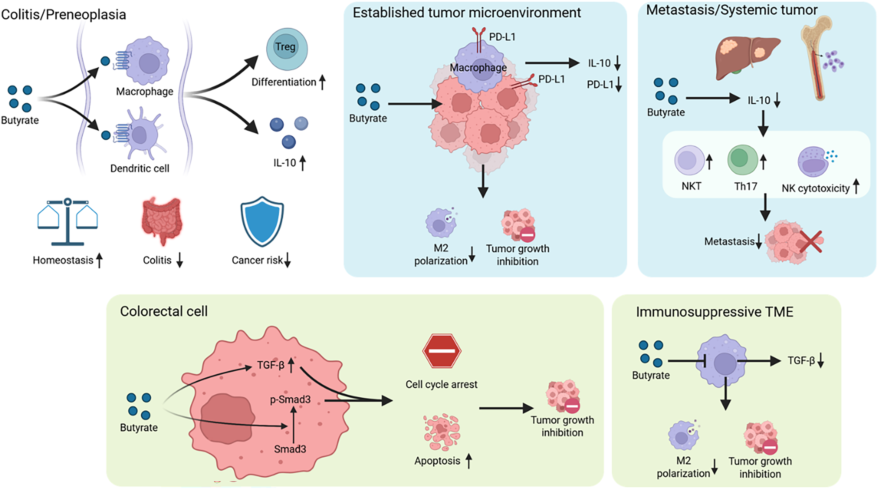
Figure 3: Context-dependent regulation of immunosuppressive cytokine circuits by short-chain fatty acids (SCFAs) in the tumor microenvironment (TME). SCFAs, particularly butyrate, regulate immunosuppressive cytokine networks in the TME in a stage-, tissue-, and microenvironment-dependent manner. In inflammatory or premalignant settings, SCFAs may support IL-10-associated immune tolerance, whereas in established tumors they can weaken IL-10-dominant immunosuppression. SCFAs may also enhance tumor-restraining TGF-β signaling in epithelial or early tumor contexts while reducing tumor-supportive effects associated with stromal- or macrophage-derived TGF-β in advanced disease. These findings indicate that SCFAs dynamically regulate IL-10- and TGF-β-centered suppressive circuits rather than acting simply as anti-inflammatory mediators. Arrows indicate activation or enhancement; blunt-ended lines indicate inhibition or attenuation. Upward and downward arrows (↑/↓) denote increased or decreased levels or activity.
4.1 Stage-Dependent Regulation of the IL-10 Axis by SCFAs
Current evidence, predominantly from butyrate-based studies, suggests that IL-10-dependent immunosuppression is regulated by SCFAs in a stage-dependent manner: during premalignant or inflammatory phases, they more frequently promote IL-10, thereby limiting excessive mucosal inflammation and supporting tissue protection; however, in certain contexts involving established tumors and treatment-related conditions, they may counteract the inhibitory effects of IL-10 on antigen-presenting cell function, the enhancement of the M2-like macrophage program, and its role in promoting immune evasion by suppressing IL-10. IL-10 is produced primarily by innate immune cells, particularly macrophages and dendritic cells [94]. Under physiological conditions, IL-10 exerts anti-inflammatory effects by limiting excessive immune activation [95]. In tumors, however, IL-10 can suppress antigen-presenting cell function and inhibit the production of pro-inflammatory cytokines such as IL-12 and TNF-α, thereby promoting immunosuppression and immune escape [96–98].
Regulation of IL-10 by SCFAs is shaped by disease stage and tissue-specific factors and may therefore proceed in opposite directions across tissues and disease models. In colitis and during the early stages of inflammation-associated colorectal tumorigenesis, butyrate acts as a GPR109A agonist to promote anti-inflammatory programs in colonic macrophages and dendritic cells, support the differentiation of Tregs and IL-10-producing T cells, maintain intestinal homeostasis, and restrain colitis-associated carcinogenesis [89]. These findings mainly support direct effects of butyrate on immune cells involved in mucosal tolerance. By contrast, in established gastrointestinal tumor models, butyrate suppresses PD-L1 and IL-10 within gastric cancer-associated immune compartments and reduces M2 macrophage polarization as well as PD-L1+ TAM infiltration in colorectal cancer, thereby alleviating the immunosuppressive features of the TME and contributing to reduced tumor growth or burden [99,100]. In an in vivo metastatic CRC setting, sodium butyrate reduced IL-10 while increasing the proportions of NK T cells and Th17 cells, accompanied by elevated IL-17 and reduced metastatic burden [67]. In an in vitro NK–myeloma system, SCFAs promoted extracellular vesicle release from NK cells, reduced NK-derived IL-10, and enhanced NK cytotoxicity, collectively suggesting a shift toward a more effector-dominant immune state [101]. This suppressive effect also appears to be concentration-dependent, as high concentrations of butyrate inhibit IL-10 production through signaling pathways involving GPCRs, cluster of differentiation 36, and SRC kinases [56].
Importantly, the effects of SCFAs on IL-10 are constrained by tissue specificity. In lung cancer, intratumoral microbiota-derived butyrate has been associated with early recurrence and metastatic progression. Mechanistically, it promotes tumor-cell H19 expression through HDAC 2 inhibition and enhances M2 macrophage polarization, while butyrate-treated M2 macrophages exhibit increased IL-10 production [102]. This example suggests that SCFAs may also regulate IL-10 indirectly through tumor-cell programs that shape macrophage polarization, rather than solely through direct immune-cell effects. This also suggests that butyrate may exert tumor-promoting effects in specific tissue niches or metastatic ecosystems, which should be considered in translational applications.
4.2 Bidirectional Regulation of TGF-β Signaling by SCFAs
As a central mediator of T cell exclusion and fibrosis, TGF-β is subject to bidirectional regulation by SCFAs, although current mechanistic evidence is weighted predominantly toward butyrate. SCFAs exert stage-dependent effects on the TGF-β axis, enhancing its tumor-suppressive and pro-apoptotic functions in early tumor cells while, in advanced disease, reducing stromal-derived immunosuppressive TGF-β and attenuating its downstream signaling. The cellular sources of TGF-β within the TME are highly heterogeneous and include tumor cells, stromal cells, and infiltrating immune cells [103,104]. In early tumors, TGF-β acts predominantly through Smad-dependent pathways to induce cell-cycle arrest and apoptosis, thereby exerting tumor-suppressive effects [105]. As tumor progression proceeds, cancer cells gradually evade these suppressive constraints, after which TGF-β cooperates with non-canonical pathways to drive epithelial–mesenchymal transition/plasticity, invasion, and distant metastasis, while also promoting stromal remodeling and angiogenesis to establish a tumor-supportive microenvironment [104,106].
SCFAs, particularly butyrate, exert pronounced tissue-specific bidirectional effects on the TGF-β axis. In colonic epithelium and early colorectal cancer models, butyrate enhances canonical TGF-β/Smad3 signaling by upregulating Smad3 expression and phosphorylation, coordinately regulates downstream targets such as ID2, ID3, and plasminogen activator inhibitor-1, promotes G0/G1 arrest and apoptosis, and suppresses anchorage-independent growth of tumor cells [107,108]. Consistent with this, host Smad3 deficiency has been associated with reduced abundance of butyrate-producing bacteria and increased susceptibility to lipopolysaccharide (LPS)-driven inflammation-associated carcinogenesis, suggesting the existence of an interdependent microbiota–host signaling axis [109]. By contrast, in stroma-rich microenvironments such as B16 melanoma, butyrate appears to act predominantly on stromal secretory phenotypes. Specifically, it suppresses polarization of TAMs toward an M2-like state and lowers stromal levels of TGF-β, VEGF, and IL-10, thereby weakening immunosuppression and angiogenesis and restricting tumor progression [110,111].
Taken together, these findings suggest that butyrate–TGF-β interactions provide a mechanistic rationale for further study in context-defined settings, but current evidence remains too limited to support a broadly generalizable translational strategy. Such work may help clarify whether butyrate can preserve tumor-cell sensitivity to TGF-β-mediated growth restraint while limiting stromal TGF-β-associated tumor support.
5 SCFAs and Chemotactic and Angiogenic Signaling in the TME
The efficiency of effector immune-cell infiltration and the structural integrity of the tumor vascular network are central determinants of the immune phenotype of the TME. In this respect, SCFAs display dual regulatory properties. On the one hand, they can reduce myeloid suppressor-cell recruitment and inhibit neovascularization by downregulating IL-8, CCL2, and VEGF. On the other hand, they can promote intratumoral trafficking of CD8+ T cells and NK cells by inducing CXCL10 and CXCL11 expression. This coordinated regulation of immune-cell recruitment and angiogenic signaling may represent one mechanism through which SCFAs contribute to a more inflamed and effector-permissive immune phenotype in selected TME contexts (figure 4). Here again, most mechanistic evidence is derived from butyrate-based studies, whereas propionate-, acetate-, or valerate-specific evidence is available only in selected chemokine or tumor-context settings.
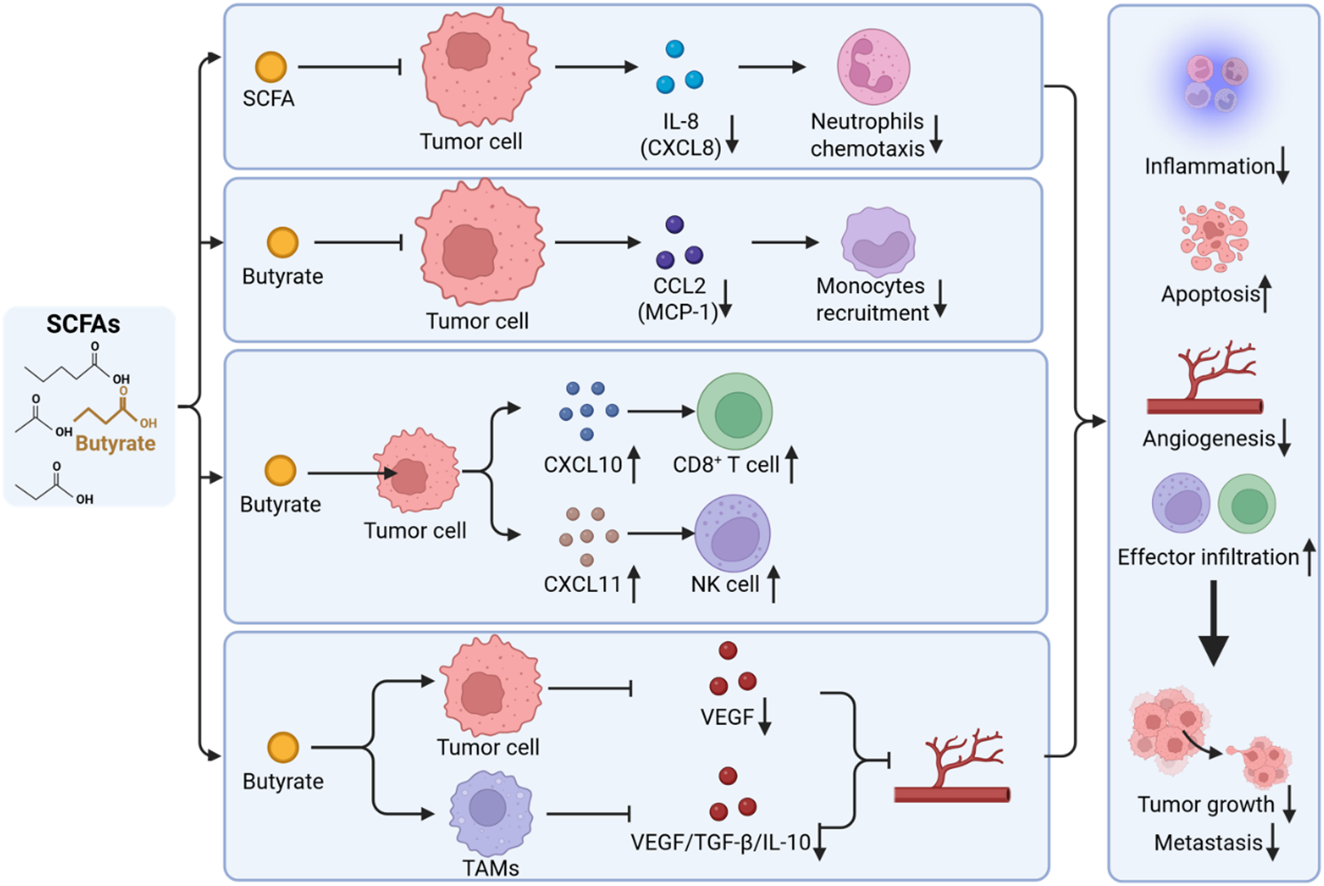
Figure 4: Short-chain fatty acid (SCFA)-mediated regulation of chemokine-driven immune trafficking and angiogenic signaling in the tumor microenvironment (TME). SCFAs, particularly butyrate, regulate chemokine-driven immune trafficking and VEGF-dependent angiogenic signaling in the TME. In many contexts, SCFAs suppress myeloid-cell recruitment and pro-angiogenic signaling associated with IL-8/CXCL8, CCL2, and VEGF, while enhancing CXCL10/CXCL11-dependent recruitment of CD8+ T cells and NK cells. Through these coordinated effects, SCFAs may contribute to a shift in the TME from a myeloid-rich, angiogenic, and immune-excluding state toward a less angiogenic and more immune-permissive phenotype. Arrows indicate activation or enhancement; blunt-ended lines indicate inhibition or attenuation. Upward and downward arrows (↑/↓) denote increased or decreased levels or activity.
5.1 Effects of SCFAs on the IL-8/CXCL8 Axis
Within the cytokine networks governing immune-cell trafficking, IL-8 is a key mediator driving the accumulation of neutrophils and myeloid-derived suppressor cells (MDSCs). SCFAs regulate IL-8 primarily through epigenetic mechanisms and transcriptional interference with inflammatory signaling pathways. IL-8, also known as CXCL8, is a pleiotropic pro-inflammatory chemokine that signals through C-X-C motif chemokine receptors 1 and 2 (CXCR1 and CXCR2) and promotes the recruitment of neutrophils and MDSCs into the TME, thereby helping establish a suppressive microenvironment that limits antitumor immunity [112,113].
Regulation of IL-8 by SCFAs, particularly butyrate, is strongly tissue-dependent. In tumor-associated inflammatory models, butyrate and propionate, acting as HDAC inhibitors, induce histone hyperacetylation and thereby suppress IL-8 transcription [114]. Butyrate also promotes differentiation of CRC cells and is associated with downregulation of TLR4 expression, which may indirectly attenuate LPS responsiveness and downstream NF-κB activation and IL-8 secretion [115]. In vitro studies further show that SCFAs can downregulate inflammatory mediators such as IL-8 and IL-6 in selected intestinal epithelial or macrophage inflammatory models [30].
However, in malignant ascites from ovarian cancer, acetate levels have been positively correlated with IL-8, and acetate has also shown positive associations with lipid-related metabolites, suggesting a distinct mode of metabolic–inflammatory coupling within this fluid microenvironment. The causal basis underlying this association still needs further clarification [116]. Overall, the regulation of the IL-8/CXCL8 axis by short-chain fatty acids exhibits clear species-specificity and microenvironment-dependence. Its directional effect is not fixed, but varies depending on the type of short-chain fatty acid, tissue compartment, and differences in inflammatory-metabolic coupling status under distinct tumor microenvironmental conditions.
5.2 Effects of SCFAs on the CCL2/CCR2 Axis
CCL2 serves as a central node in the recruitment of immunosuppressive cells to the TME and may represent a key axis through which SCFAs modulate tumor immunity. Also known as monocyte chemoattractant protein-1, CCL2 signals via CCR2 to recruit monocytes, MDSCs, and Tregs into tumors and contributes to broader cytokine-mediated TAM recruitment, thereby driving immunosuppression, angiogenesis, and tumor progression [117–119].
In human acute leukemia cell models (HL-60 and U937), high concentrations of butyrate suppress phosphorylation of AKT and MAPK pathway components, including p-ERK1/2 and p-AKT, leading to reductions in CCL2 and C-C motif chemokine ligand 5 at both the transcriptional and protein levels. In this leukemia-cell context, reduced CCL2/CCL5 expression provides a plausible explanation for impaired monocyte migration, whereas Caspase-3 activation more directly reflects butyrate-associated leukemic-cell apoptosis rather than a downstream consequence of CCL2 suppression alone [120]. At the clinical level, however, this axis appears more heterogeneous. In melanoma patients receiving anti-CTLA-4 therapy, higher serum butyrate levels were associated with increased circulating CCL2 and an increased frequency of Tregs, suggesting that systemic SCFA exposure may exert unfavorable immunomodulatory effects in some immunotherapy contexts [82]. This finding should not be interpreted as directly contradicting the leukemia-cell data, because it reflects a different tumor type, exposure compartment, cellular source of CCL2, and treatment setting. In leukemia cell lines, high-concentration butyrate directly affects tumor-cell chemokine production and apoptosis; in melanoma patients receiving CTLA-4 blockade, serum butyrate more likely reflects a systemic immune-metabolic state associated with altered Treg abundance, CCL2 levels, and impaired immune priming. Accordingly, the effects of SCFAs on the CCL2 axis are best interpreted within the specific context of tumor type, exposure compartment, and treatment setting.
5.3 Effects of SCFAs on CXCL10/CXCL11-Mediated Effector-Cell Trafficking
Unlike CCL2, CXCL10 and CXCL11 are generally associated with antitumor immunity. As interferon-inducible CXCR3 ligands produced by tumor, myeloid, and stromal cells, they recruit CXCR3+ effector lymphocytes—particularly CD8+ T cells and, in some contexts, NK cells—and are therefore linked to an inflamed TME and improved responsiveness to immunotherapy [121,122].
In contrast to their suppressive effects on CCL2, SCFAs more often induce CXCL10 and CXCL11 in solid tumors. In CRC cells, butyrate induces DNA damage through its HDAC-inhibitory activity and activates the cyclic GMP-AMP synthase–stimulator of interferon genes–interferon regulatory factor 3 axis (cGAS–STING–IRF3, a cytosolic DNA-sensing pathway), thereby markedly upregulating CXCL10 secretion and major histocompatibility complex class I expression (MHC-I, which supports antigen presentation to CD8+ T cells). These changes are associated with increased CD8+ T cell infiltration and enhanced granzyme B-positive cytotoxic activity and, particularly when butyrate is combined with oncolytic viruses, suggest synergistic potential in selected preclinical settings [123,124]. In hepatocellular carcinoma models, butyrate has also been shown to selectively enhance CXCL11 expression through STAT-mediated chromatin and enhancer remodeling, thereby promoting NK-cell recruitment and augmenting NK cytotoxicity [125]. Taken together, these findings suggest that, in specific preclinical tumor models, butyrate may exert its antitumor effects primarily by inducing CXCL10/CXCL11 to promote the migration of CD8+ T cells or NK cells.
5.4 SCFAs and VEGF-Dependent Angiogenic Signaling
Within angiogenic networks that support tumor survival and metastasis, SCFAs may exert suppressive effects through coordinated metabolic and epigenetic regulation at both the tumor-cell and myeloid-cell levels. In the TME, VEGF is not only a central regulator of angiogenesis but also an important modulator of antitumor immunity. By acting on immune cells expressing VEGF receptors, VEGF can impair dendritic-cell maturation and expand immunosuppressive populations such as Tregs and MDSCs, thereby promoting T cell dysfunction and immune escape [126,127].
At the tumor-cell and endothelial-cell levels, butyrate suppresses VEGF signaling through multiple mechanisms. As an HDAC inhibitor, it inhibits hypoxia-inducible factor 1α (HIF-1α) nuclear translocation and DNA binding, thereby attenuating hypoxia-driven transcriptional activation of VEGF [128,129]. It can also reduce the binding affinity of specificity protein 1 to the neuropilin-1 promoter, thereby downregulating VEGF and its coreceptor neuropilin-1 [130]. In addition, in human lung microvascular endothelial cells, sodium butyrate has been reported to shift VEGF-A isoform balance toward anti-angiogenic variants generated by alternative splicing, thereby increasing the level of anti-angiogenic VEGF [131]. In oral cancer cells, butyrate downregulates vascular endothelial growth factor C and vascular endothelial growth factor D, a finding consistent with its broader anti-angiogenic effects in CRC models, where it suppresses VEGF expression and HIF-1α-related angiogenic signaling [128,132,133].
At the immune-cell level, butyrate may further weaken TAM-mediated VEGF-driven tumor support. In melanoma models, butyrate suppresses polarization of TAMs toward an M2-like phenotype and inhibits TAM proliferation, thereby reducing levels of tumor-supportive factors including VEGF, TGF-β, and IL-10. These changes are associated with relief of immunosuppression and inhibition of tumor growth [110,111]. In summary, the regulation of the VEGF axis by butyrate is generally anti-angiogenic, characterized by simultaneous actions on VEGF transcription and splicing programs within tumor cells as well as the pro-angiogenic microenvironment mediated by TAMs, thereby coordinately restricting angiogenesis and attenuating immunosuppression.
Taken together, predominantly preclinical evidence—much of it centered on butyrate—suggests that SCFAs can, in selected settings, favor a microenvironment characterized by reduced pro-angiogenic and myeloid-recruiting signals such as VEGF, IL-8, and CCL2, together with enhanced effector-cell-attracting signals such as CXCL10 and CXCL11. This coordinated remodeling may contribute to a less angiogenic and more immune-permissive state and, in selected settings, may be associated with stronger antitumor immune responses and improved responsiveness to immunotherapy.
6 Context-Dependent Determinants of SCFA-Mediated Cytokine Regulation
SCFA effects in the TME are not determined by metabolite identity alone. To integrate the heterogeneous findings discussed above, we propose a hierarchical model for interpreting SCFA-mediated cytokine remodeling. In this model, the overall direction of cytokine remodeling is first constrained by exposure compartment and disease stage, then modified by the dominant responding cell lineage and tissue context, and only then refined by SCFA species, concentration, and treatment background. Therefore, whether a given cytokine shift is ultimately interpreted as anti-inflammatory or pro-inflammatory, immunostimulatory or immunosuppressive, depends on this hierarchy rather than on a fixed class effect of SCFAs.
6.1 SCFA Species-Specific Differences
Although SCFAs are often discussed collectively as a metabolic class, individual SCFAs show distinct immune-regulatory profiles once exposure compartment, disease stage, and responding cell populations are considered. Among individual SCFAs, butyrate is currently the best characterized in cancer-related immune regulation. Therefore, mechanisms demonstrated mainly with butyrate should not be automatically interpreted as class effects shared by acetate, propionate, or valerate. Representative evidence links butyrate to inflammatory cytokine control, macrophage-associated immunosuppression, effector-cell activation, and chemokine-mediated immune-cell trafficking [14,32,99,124].
Although butyrate remains the most extensively characterized SCFA in cancer-related cytokine regulation, acetate, propionate, and valerate should not be interpreted simply as weaker or interchangeable analogues of butyrate. Species-specific regulation may reflect differences in metabolic utilization, receptor preference, HDAC-inhibitory activity, effective concentration range, and dominant responding cell type. Acetate is closely linked to acetyl-CoA-dependent epigenetic remodeling and GPR43-associated immune signaling, which may support effector T-cell function in selected contexts, but it may also contribute to tumor-cell metabolic adaptation and checkpoint-related immune resistance under certain conditions [31,70,134]. Propionate may partly overlap with butyrate in HDAC-dependent inflammatory regulation, including suppression of selected inflammatory mediators, yet high-exposure or tumor-cell-intrinsic settings may favor PD-L1-associated immune-evasion programs [114,135]. Valerate has emerging relevance in effector T-cell and CAR-T-cell cytokine programs, particularly in enhancing antitumor effector functions under defined adoptive immunotherapy-related conditions, although its effects remain less systematically characterized than those of butyrate [32,136]. These observations indicate that individual SCFAs differ not only in potency but also in biological direction, and therefore should be interpreted according to exposure compartment, concentration, responding cell lineage, and therapeutic setting rather than treated as a uniform metabolic class.
6.2 Stage- and Microenvironment-Dependent Shifts in SCFA Function
Among the many factors contributing to context specificity, disease stage and tissue differences are the most prominent determinants of short-chain fatty acid function. The response of the same cytokine to butyrate may even shift markedly with disease progression, as exemplified by IL-10. In early inflammation or precancerous conditions, butyrate increases IL-10 production, thereby maintaining intestinal homeostasis and reducing the risk of intestinal inflammation and tissue carcinogenesis. As discussed above, this effect is mainly linked to GPR109A-dependent anti-inflammatory programs in colonic macrophages and dendritic cells. In contrast, within established TME, butyrate may instead attenuate IL-10 signaling, which helps alleviate immune tolerance [67,89,99]. A similar pattern is observed in the regulation of IL-17. During colitis-associated carcinogenesis, a mixture of short-chain fatty acids inhibits IL-17 to limit pro-tumor inflammation; however, in metastatic tumor models, butyrate promotes IL-17 production, thereby enhancing antitumor immunity [29,67]. The regulation of IL-18 shows tumor tissue-specific microenvironmental dependence: under physiological conditions, it is upregulated by butyrate to facilitate barrier-related immune maintenance; whereas under chronic inflammatory injury, IL-18 is downregulated to restrict excessive inflammasome-driven inflammation [36,90].
Similar trends can also be observed in different therapeutic contexts. Butyrate can increase the expression of IL-2Rα/CD25 in nasopharyngeal carcinoma cells, and in rat colorectal tumor models, it can synergize with IL-2 to enhance antitumor efficacy. This may be achieved by butyrate-mediated enhancement of tumor-cell immunogenicity and promotion of antigen presentation [78,80,137]. However, in the setting of CTLA-4 therapy, elevated circulating levels of short-chain fatty acids—particularly butyrate—result in impaired dendritic-cell maturation, reduced costimulatory signaling, and diminished soluble CD25-associated IL-2 signaling, thereby decreasing sensitivity to CTLA-4 blockade [82]. Taken together, these studies collectively suggest that disease stage, tissue differences, and therapeutic context can influence cytokine regulation mediated by the same short-chain fatty acid represented by butyrate, thereby leading to distinct immune and therapeutic effects.
6.3 Lineage- and Tissue-Specific Determinants of SCFA Responses
Once the broader biological context is established, cell lineage and tissue environment emerge as the next key factors shaping SCFA activity. Different cell populations and organ-specific microenvironments do not respond to SCFAs in the same way. In some settings, these differences can even drive opposite patterns of cytokine production and distinct downstream immune effects. In colorectal cancer and multiple myeloma, butyrate reduces gp130/IL-6 receptor signaling and is associated with growth-inhibitory effects. This pattern mainly reflects receptor-component or receptor-proximal regulation. In esophageal squamous cell carcinoma, however, butyrate has been reported to enhance IL-6 transcription in tumor cells, suggesting a tumor-cell-intrinsic transcriptional response rather than a reversal of the same receptor-level mechanism [48–50].
The TNF-α axis likewise shows pronounced lineage- and microenvironment-dependent behavior. SCFAs can suppress expression of inflammatory cytokines, including TNF-α, in macrophage-like myeloid cells. In lymphoid effector cells, by contrast, their effects are more context-dependent: under some conditions, SCFAs enhance TNF-α production in CD8+ T cells or CAR-T cells and strengthen effector function; under other conditions—determined jointly by metabolite species, exposure dose, and the TME—they may instead reduce TNF-α output and induce hyporesponsive or exhaustion-associated phenotypes [30,32,63,136]. IFN-γ shows a similar compartment- and model-dependent regulatory pattern: in local TME, particularly under metabolically constrained and exhaustion-prone T cell states, its production may be maintained or restored; in peripheral immune-cell environments or in some engineered tumor-microenvironment models, however, SCFAs may also dampen IFN-γ-related effector activity, consistent with adaptive shifts across tissue compartments, metabolic settings, and activation states [31,75]. In colonic epithelial contexts, TGF-β signaling may be enhanced to support its tumor-suppressive activity; in melanoma models, by contrast, sodium butyrate has been reported to reduce TGF-β expression together with TAM abundance, a pattern consistent with weakening tumor-supportive stromal or myeloid support [107,110]. Similarly, IL-8 expression is commonly reduced in solid tumor contexts, whereas in malignant fluid microenvironments such as ovarian cancer ascites, IL-8 correlates positively with metabolite levels, suggesting that organ-specific microenvironments may substantially alter the direction of metabolic regulation [30,116].
6.4 Concentration-Dependent Effects and Differential Regulation of SCFA Transport
After the overall direction and principal responding cell populations have been defined, SCFA concentration becomes another key determinant. Transport capacity also helps define the effective window within which cytokine remodeling occurs.
Physiologically achievable SCFA concentrations are highly compartmentalized: luminal and fecal concentrations can reach the millimolar range, whereas concentrations in the portal vein, hepatic vein, and peripheral circulation decline rapidly to the micromolar range [138]. Reliable measurements of SCFA concentrations within tumor tissues remain limited [139]. Therefore, low micromolar or 0.1 mM exposures are more relevant to tracer, transport-related, or low-dose immune-regulatory settings, whereas 10 mM butyrate more closely reflects local intestinal exposure or pharmacological in vitro treatment and should not be directly equated with circulating or intratumoral concentrations.
Concentration is one of the key variables determining the direction of SCFA-mediated immunoregulation. Along the TNF-α-, IL-1β-, and IL-10-related axes, butyrate may exhibit dose-dependent directional switching. In LPS-stimulated human macrophages, low-concentration butyrate at approximately 0.1 mM suppresses TNF-α production in vitro. By contrast, high-concentration butyrate at approximately 10 mM fails to inhibit TNF-α, increases IL-1β production, reduces IL-10 production, and promotes macrophage cell death in vitro [56]. Thus, the 0.1 mM condition may be more informative for low-dose immune-cell modulation, whereas the 10 mM condition is better interpreted as a high local or pharmacological exposure that defines the upper boundary and potential reversal of butyrate activity.
At the same time, inflammatory mediators can also regulate SCFA transporter expression and epithelial SCFA handling in a concentration-dependent manner. Under tracer or low micromolar exposure conditions, such as 10 μM radiolabeled butyrate, uptake appears to be more readily suppressed by inflammation. By contrast, under higher-concentration exposure conditions, such as 10 mM butyrate, uptake may instead be enhanced in some tumor-cell settings [61,140–142]. These examples further indicate that low micromolar tracer concentrations are most useful for defining transporter sensitivity and inflammatory regulation of uptake, whereas millimolar exposure may reveal tumor-cell handling of SCFAs under local intestinal or pharmacological conditions. Accordingly, any discussion of whether SCFAs increase or decrease a given cytokine needs to be grounded in clearly defined experimental boundaries. These include the effective concentration range tested in the relevant model, for example low micromolar tracer concentrations, approximately 0.1 mM low-dose butyrate, or pharmacological exposure around 10 mM butyrate, exposure duration, and the transporter or receptor background of the target cells. Without such parameters, findings that apply only to specific biological settings can be generalized too far. In turn, these variables influence not only how cytokine regulation occurs, but also how far such mechanisms can be translated into practical therapeutic use. However, direct evidence on how different SCFA concentrations regulate cytokine programs within tumor tissues or immune-cell compartments remains limited and requires further study.
7 Translational Considerations for SCFA-Mediated Cytokine Remodeling in Cancer Therapy
From a translational standpoint, current evidence does not justify treating SCFAs as broadly applicable sensitizers with a single and predictable effect on anticancer therapy. This is consistent with recent evidence suggesting that metabolic-immune remodeling can both promote immune evasion and treatment resistance, as well as restore antitumor immunity, with the specific outcome depending on the dominant metabolic pathways, inflammatory mediators, and cell subsets involved [19,143,144]. Data from human association studies, animal models, and ex vivo systems consistently show that their therapeutic influence is heterogeneous and shaped by treatment context. When considering SCFAs in combination strategies, SCFA species or abundance alone cannot fully explain their translational potential. What matters just as much is the direction in which they reprogram cytokine networks within the microenvironment under a specific therapeutic condition. Representative studies on SCFA-mediated cytokine and immune-mediator regulation in treatment-related cancer settings are summarized in Table 1.
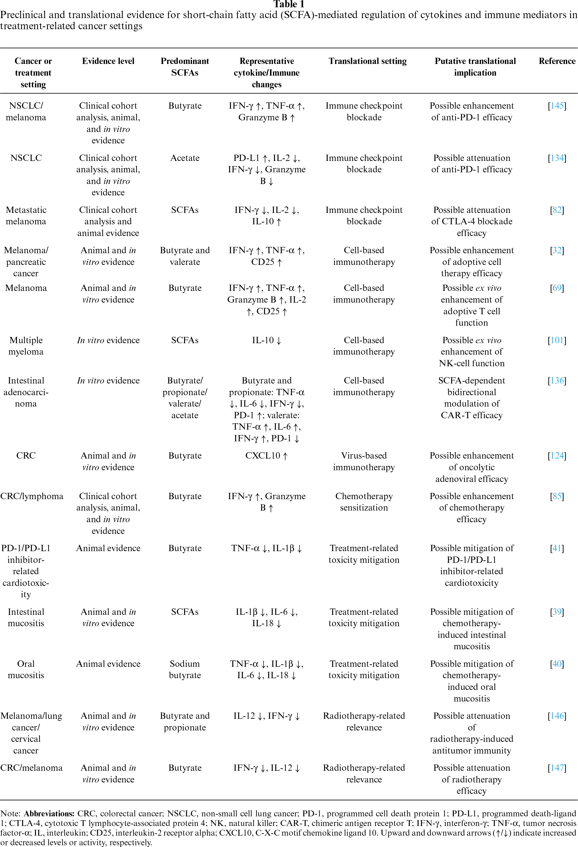
Therefore, the translational significance of SCFAs should be judged within defined therapeutic contexts. The key issue is not whether they can be broadly classified as immune-enhancing or immune-suppressive, but whether the cytokine-network changes they induce match the core immune programs required for a given treatment modality. Accordingly, the following sections examine immune checkpoint blockade, cell- and virus-based therapies, and radiotherapy/chemotherapy, with attention to both potential therapeutic value and practical limits.
7.1 Context-Dependent Modulation of Immune Checkpoint Blockade by SCFAs
In the context of immune checkpoint blockade (ICB) therapy, existing clinical and preclinical evidence is insufficient to support a universal and consistent synergistic effect between SCFAs and ICB efficacy. A more clinically relevant question is whether SCFA-mediated alterations in cytokine expression align with the core immune regulatory programs required for a therapeutic response to ICB. This process may involve multiple aspects, including effective early immune initiation, remodeling of immune function during the effector phase, and enhanced sensitivity of tumor cells to treatment.
In anti-PD-1/PD-L1 therapy, some retrospective human studies and selected preclinical models suggest that butyrate-associated exposure may coincide with improved response when it helps sustain a local pro-effector immune state. Retrospective analyses have suggested that higher serum butyrate levels and related gut microbial features correlate with better therapeutic outcomes in some cohorts, but these associations should be interpreted cautiously because their direction and strength may depend on whether models are adjusted for clinical covariates and potential confounders [63,145]. Animal studies provide mechanistic support for this association. Butyrate, whether derived from Roseburia intestinalis or administered exogenously, can downregulate suppressive factors in the tumor immune microenvironment, including PD-L1 and IL-10. It can also enhance CD8+ T cell-dominant programs involving IFN-γ, TNF-α, and granzyme B production. These changes may create conditions favorable for restoring exhausted T cell function during anti-PD-1 treatment [99,100]. At present, however, this evidence remains stronger for butyrate-centered settings than for SCFAs as a general intervention class, and clinical biomarker associations require validation in adequately adjusted, treatment-specific cohorts.
However, preclinical evidence also indicates that this apparent synergy may be lost when SCFA effects are dominated by tumor cell-intrinsic metabolic reprogramming. In a recent murine colorectal cancer model, butyrate enhanced carnitine palmitoyltransferase 1A-mediated fatty acid oxidation. This increased the metabolic adaptability of tumor cells. It was associated with reduced local CD8+ T cell infiltration and impaired cytotoxicity, ultimately weakening anti-PD-1 efficacy [148]. This finding suggests that when the metabolite is preferentially utilized by tumor metabolic pathways, the microenvironment may shift away from an immune state conducive to checkpoint blockade responsiveness.
Other SCFAs, such as acetate and propionate, may also display distinct tendencies toward immune tolerance. In non-small cell lung cancer models, acetate upregulated tumor PD-L1 through an acetyl-CoA–c-Myc axis [134]. This was accompanied by marked reductions in T cell-derived IL-2 and IFN-γ, as well as increased expression of pro-angiogenic factors such as VEGF. In colorectal cancer in vitro models, high-concentration sodium propionate similarly stabilized PD-L1 mRNA through insulin-like growth factor 2 mRNA-binding protein 3 [135]. The finding for high-dose propionate currently remains limited to in vitro systems. Even so, these data suggest an important caution. When SCFA exposure mainly upregulates checkpoint molecules without inducing compensatory effector cytokines, the result may reflect tumor-intrinsic adaptive resistance rather than true therapeutic sensitization.
By contrast, in the setting of anti-CTLA-4 therapy, current evidence points to inhibitory effects. Clinical and preclinical analyses indicate that high circulating butyrate levels may limit the efficacy of ipilimumab [82]. Mechanistically, excessive butyrate suppresses dendritic-cell maturation and IL-2 production. The efficacy of CTLA-4 blockade depends strongly on early antigen presentation in draining lymph nodes and IL-2-driven immune priming. Impairment of this signaling axis directly undermines the core immune programs required for therapeutic efficacy. This phenomenon also reflects the spatial and temporal specificity of SCFA action. For example, butyrate may enhance local anti-PD-1 responses during the immune effector phase by upregulating IFN-γ. By contrast, during the immune-priming phase, it may attenuate CTLA-4 blockade in lymph nodes by suppressing IL-2-associated activation.
In summary, the role of SCFAs in ICB therapy must be evaluated in the context of both the treatment phase and the site of action, rather than being simply labeled as uniformly beneficial or detrimental. Moreover, as research progresses toward clinical translation, measuring SCFA levels solely in the gut or circulation is often insufficient to inform clinical decision-making. More refined translational research should match cytokine regulatory patterns with the core mechanisms of different ICB regimens. These mechanisms may involve early immune priming or effector-phase immune remodeling.
7.2 Translational Considerations for SCFAs as Ex Vivo Modulators in Cell- and Virus-Based Therapies
In adoptive cell therapy and oncolytic virotherapy, current evidence for SCFA-mediated benefit derives mainly from ex vivo studies and preclinical models. These studies suggest that SCFA action can be more accurately understood as condition-defined and context-specific rather than as broad, systemically applicable immune activation. Based on the available literature, this specificity is reflected in the differential capacity of SCFAs to regulate cytokine networks across distinct cell types and microenvironments.
In the context of enhancing the persistence and effector function of infused cells, ex vivo evidence suggests that SCFAs may reprogram cytokine programs in immune cells under defined preconditioning and activation conditions. For example, T cells pretreated with butyrate or valerate exhibit enhanced secretion of IFN-γ and TNF-α [32,69]. Similarly, butyrate exposure has been associated with downregulation of the inhibitory cytokine IL-10 in NK cells, together with enhanced NK cytotoxicity [101]. However, butyrate and propionate have also been reported to suppress dendritic cell/antigen-presenting cell-dependent antigen-specific CD8+ T cell activation by reducing IL-12 production from antigen-presenting cells. These findings further indicate that SCFA effects may differ by target cell lineage, even when the measured outcome is broadly interpreted as enhanced cytotoxic function.
Therefore, these gains achieved under specific ex vivo activation conditions cannot be directly extrapolated to in vivo settings or to the complex TME. A recent microfluidic on-chip model of human anti–receptor tyrosine kinase-like orphan receptor 1 CAR-T cells in intestinal adenocarcinoma revealed that SCFA-mediated regulation is highly dependent on both metabolite species and microenvironmental context: whereas acetate and valerate tended to preserve a pro-inflammatory state, butyrate and propionate markedly suppressed CAR-T cell release of TNF-α, IL-6, and IFN-γ and simultaneously induced upregulation of exhaustion- and tolerance-associated markers, including PD-1, T cell immunoglobulin and mucin-domain containing-3, and forkhead box P3 [136].
Taken together, these heterogeneous findings suggest that the role of SCFAs in cell therapy is better conceptualized as context-specific cytokine reprogramming rather than as a broadly applicable immune-enhancing strategy. Their translational value depends on cell type, metabolite species, and timing of exposure—in other words, on whether these variables ultimately direct cytokine networks toward sustained effector-cytokine release and immune activation, or instead toward attenuated cytokine output and a hyporesponsive or tolerance-like state. Because most available studies do not systematically compare concentration, exposure duration, and activation route, future work should define these parameters explicitly before SCFAs are considered as adjunctive modulators for cell therapy.
In oncolytic virotherapy, the potential value of SCFAs also appears to be linked to local cytokine remodeling. Mouse studies have shown that butyrate, while enhancing adenoviral infectivity through upregulation of relevant receptors, can also promote local tumor CXCL10 secretion, thereby increasing chemotactic infiltration of CD8+ T cells and producing synergistic antitumor effects in vivo [124].
Overall, current evidence supports positioning SCFAs primarily as candidate ex vivo preconditioning factors or local sensitizers. At present, however, support for this view remains largely confined to ex vivo systems and preclinical models. The central translational challenge is to establish whether treatment-beneficial cytokine patterns seen in preclinical studies—such as increased IFN-γ, TNF-α, or CXCL10—can be reproduced consistently under conditions that meet the requirements for standardized manufacturing, delivery, and clinical safety.
7.3 Compartment-Specific Effects of SCFAs in Radiotherapy and Chemotherapy
In chemotherapy settings, preclinical studies indicate that SCFAs may offer two distinct translational advantages: they can improve treatment sensitivity and lessen tissue toxicity. These effects should be interpreted according to exposure compartment, cancer type, timing of SCFA exposure, and chemotherapy regimen. At the intratumoral immune level, butyrate enhances the cytotoxic function of CD8+ T cells by activating the ID2–IL-12 cytokine axis, and it shows synergistic antitumor activity when combined with oxaliplatin [85]. Outside the tumor, SCFAs exert anti-inflammatory effects in normal tissues. They inhibit NLRP3 inflammasome activation and lower the levels of pro-inflammatory cytokines, including IL-1β, TNF-α, and IL-6. These actions have been linked to reduced chemotherapy-associated intestinal mucositis and cardiotoxicity in preclinical settings [39–41].
In radiotherapy models, SCFA-mediated cytokine regulation is similarly context-dependent. One study found that depletion of butyrate-associated microbial populations with vancomycin unexpectedly enhanced radiotherapy-induced antitumor immunity [146]. Mechanistically, local butyrate suppressed activation of the stimulator of interferon genes pathway in dendritic cells, weakened amplification of key cytokine signals including interferon-β, IL-12, and IFN-γ, and ultimately impaired radiotherapy-induced tumor-specific T cell responses [147]. At the same time, other studies have reported that butyrate or butyrate-producing bacteria can enhance radiosensitivity in colorectal cancer [149,150]. These findings appear to reflect increased tumor cell-intrinsic radiosensitivity and are therefore not necessarily contradictory to the above observations showing that butyrate may reduce radiotherapy benefit by suppressing cytokine-driven immune programs.
Taken together, the impact of SCFAs—particularly butyrate—on therapeutic outcome cannot be reduced to a uniform sensitizing or inhibitory effect. Instead, it must be interpreted within the specific treatment context and in relation to the cytokine-mediated immune state that SCFAs help establish. A systematic evaluation of the associated cytokine changes may therefore provide a more accurate basis for assessing the clinical translational value of SCFAs.
7.4 Contextual Interpretation of SCFA Modulation in Cancer Therapy
Based on current evidence, the therapeutic significance of SCFA-mediated cytokine network remodeling should be interpreted in the context of the specific treatment scenario, rather than simply categorized as a universal sensitizing or inhibitory effect. When SCFA modulation enhances the immune programs required for therapeutic response, its role may offer potential benefits. Examples include maintaining local effector cytokine activation during PD-1/PD-L1 blockade therapy, enhancing effector function during the in vitro preparation of adoptive immune cells, promoting CXCL10-mediated immune cell recruitment in oncolytic virus therapy, or enhancing IL-12-dependent CD8+ T-cell cytotoxicity during chemotherapy. SCFA modulation may also have protective implications when the suppression of excessive pro-inflammatory cytokines helps mitigate treatment-related toxicity in normal tissues.
Conversely, when SCFA modulation interferes with immune programs essential for a particular treatment to be effective, its effects may have potential adverse consequences. This includes impairing dendritic cell-mediated immune priming during CTLA-4 blockade therapy, promoting immune checkpoint upregulation or tumor metabolic adaptation during PD-1/PD-L1 blockade therapy, inducing CAR-T cell exhaustion-like changes under complex microenvironmental exposure, and attenuating STING/type I interferon, IL-12, or IFN-γ-dependent antitumor immunity during radiotherapy. Therefore, SCFA-related interventions should be evaluated on a case-by-case basis according to the treatment modality, site of exposure, predominant SCFA species, cytokine profile, and dosage, rather than being employed as a non-selective adjunctive strategy.
8 Current Limitations and Translational Challenges
The role of SCFAs in remodeling cytokine networks within the TME has drawn growing interest. Even so, their therapy-related significance remains difficult to define because the current evidence base is limited both in depth and in methodology. Most available findings come from in vitro experiments, preclinical tumor models, and retrospective clinical association studies. In contrast, prospective validation studies in well-defined therapeutic settings remain scarce. Available evidence is unevenly distributed across cytokine profiles, SCFA species, and therapeutic contexts. Mechanistic patterns centered on butyrate have been repeatedly confirmed, whereas findings regarding other SCFAs or specific tumor types are mostly based on limited data, with some derived from only a single study. Currently, relatively robust evidence points to the regulatory effects of butyrate on inflammatory and immune effector programs; however, sufficient evidence is still lacking regarding the functions of other SCFAs, compartment-specific exposure patterns, and clinically relevant dose ranges. Besides, strategies designed to increase SCFA exposure are not interchangeable. High-fiber diets, prebiotics, probiotics, fecal microbiota transplantation, SCFA-producing bacterial consortia, and direct SCFA supplementation may differentially affect luminal, circulating, and intratumoral SCFA levels and may differ in kinetics, safety, and off-target effects on the broader microbiota.
Furthermore, significant methodological heterogeneity across studies hinders direct comparisons and the formulation of definitive conclusions. Variations in SCFA species, dosage, intervention duration, administration route, tumor models, and therapeutic regimens all contribute to inconsistent outcomes. For many seemingly contradictory results, it remains unclear whether they reflect genuine biological context dependence or merely arise from differences in experimental design and exposure conditions. The issue of tissue compartment specificity is also unresolved: SCFA levels in feces, circulation, and tumor tissues represent biologically distinct compartments, whose levels and functional relevance may not be consistent. Associations identified in one compartment cannot be directly extrapolated to others.
In addition, most clinical studies provide primarily correlative rather than causal evidence, making it difficult to distinguish whether SCFAs directly exert immunoregulatory effects or merely serve as surrogate markers reflecting gut microbiota and overall metabolic status. Although models such as ex vivo pretreatment, microenvironment chips, and local radiotherapy offer mechanistic insights, their reproducibility and translational value in the complex human exposure system still require prospective validation [136,146,147]. Therefore, future research should establish standardized quantitative methods, enable compartment-specific exposure assessment, and conduct prospective studies integrating metabolomics, single-cell sequencing, and spatial immune profiling. These represent critical translational bottlenecks that must be urgently addressed before SCFA-mediated cytokine regulatory mechanisms can be reliably applied to clinical therapeutic stratification and intervention design.
SCFAs are key metabolic intermediates linking diet, the gut microbiota, and host immunity, and their functions within the TME cannot be adequately captured by a simple binary paradigm of “anti-inflammatory” versus “pro-inflammatory.” Rather, current evidence suggests that viewing SCFA activity through the lens of context-dependent cytokine and chemokine-network modulation may provide a useful perspective for understanding how these metabolites regulate the immune microenvironment. Across pro-inflammatory, effector, immunosuppressive, and chemotactic/angiogenic cytokine modules, SCFAs influence immune activation, immune-cell trafficking, stromal support, angiogenesis, and therapeutic responsiveness through recurring mechanisms that include inflammasome regulation, receptor-associated signaling, epigenetic remodeling, and metabolic reprogramming.
The regulatory effects of SCFAs on cytokine function are not determined by a single factor, but are jointly influenced by multiple variables, including SCFA species, dosage, compartment of action, tumor stage, cellular lineage, tissue microenvironment, and therapeutic context. This complexity also indicates that the same metabolite can either enhance immune surveillance or promote immune tolerance, chronic inflammation, or treatment resistance under different conditions. Therefore, SCFAs should not be regarded as intervention molecules with uniform biological effects, and a balanced interpretation of SCFA biology also requires species-specific analysis because butyrate-centered mechanisms should not be generalized to acetate, propionate, valerate, or other SCFAs without direct evidence.
From a translational perspective, the clinical application of SCFAs relies more on mechanism-guided, precise use matched to disease settings rather than generalized supplementation. Accordingly, future research should move in several specific directions to improve clinical translatability. Future studies should define clinically achievable SCFA concentrations across intestinal, circulating, tumor-tissue, and ex vivo compartments. SCFA species, dose, exposure duration, and delivery route should be standardized, and cytokine-network changes observed in cell or animal models should be validated in prospective patient cohorts with treatment-specific endpoints, including ICB response, cell-therapy persistence, radiotherapy efficacy, chemotherapy sensitivity, and treatment-related toxicity. Safety and feasibility should also be assessed in relation to microbiota variability, tumor type, disease stage, and compatibility with existing therapies.
Overall, SCFAs should not be viewed simply as broad immune enhancers or generally applicable anticancer supplements. Rather, they are context-defined immune-metabolic modulators whose effects depend on where they act, which cells respond, which cytokine program is remodeled, and which therapeutic phase is being targeted.
Acknowledgement: The authors acknowledge BioRender for its support in the preparation of schematic illustrations included in this review.
Funding Statement: This work was funded by the Natural Science Foundation of Hubei Province (Zhiliang Jin), grant number JCZRLH202500649 (https://kjt.hubei.gov.cn/).
Author Contributions: The authors confirm contribution to the paper as follows: Conceptualization, Qi Song, Haisen Yin, Shiyun Tan and Baoping Yu; methodology, Qi Song and Han Zhang; investigation, Qi Song, Ming Li, Zhiliang Jin and Han Zhang; writing—original draft preparation, Qi Song; writing—review and editing, Qi Song, Ming Li, Zhiliang Jin, Haisen Yin, Shiyun Tan and Baoping Yu; visualization, Qi Song; supervision, Haisen Yin, Shiyun Tan and Baoping Yu; project administration, Haisen Yin and Baoping Yu. All authors reviewed and approved the final version of the manuscript.
Availability of Data and Materials: No new data were created or analyzed in this study. Data sharing is not applicable.
Ethics Approval: Not applicable.
Conflicts of Interest: The authors declare no conflicts of interest.
References
1. Mann ER, Lam YK, Uhlig HH. Short-chain fatty acids: linking diet, the microbiome and immunity. Nat Rev Immunol 2024;24(8):577–95. doi:10.1038/s41577-024-01014-8. [Google Scholar] [PubMed] [CrossRef]
2. Vinelli V, Biscotti P, Martini D, et al. Effects of dietary fibers on short-chain fatty acids and gut microbiota composition in healthy adults: a systematic review. Nutrients 2022;14(13):2559. doi:10.3390/nu14132559. [Google Scholar] [PubMed] [CrossRef]
3. Xu T, Wu X, Liu J, et al. The regulatory roles of dietary fibers on host health via gut microbiota-derived short chain fatty acids. Curr Opin Pharmacol 2022;62:36–42. doi:10.1016/j.coph.2021.11.001. [Google Scholar] [PubMed] [CrossRef]
4. Spencer CN, McQuade JL, Gopalakrishnan V, et al. Dietary fiber and probiotics influence the gut microbiome and melanoma immunotherapy response. Science 2021;374(6575):1632–40. doi:10.1126/science.aaz7015. [Google Scholar] [PubMed] [CrossRef]
5. Zhang SL, Mao YQ, Zhang ZY, et al. Pectin supplement significantly enhanced the anti-PD-1 efficacy in tumor-bearing mice humanized with gut microbiota from patients with colorectal cancer. Theranostics 2021;11(9):4155–70. doi:10.7150/thno.54476. [Google Scholar] [PubMed] [CrossRef]
6. Boucher E, Plazy C, Richard ML, et al. Inulin prebiotic reinforces host cancer immunosurveillance via γδ T cell activation. Front Immunol 2023;14:1104224. doi:10.3389/fimmu.2023.1104224. [Google Scholar] [CrossRef]
7. Singh V, Yeoh BS, Chassaing B, et al. Dysregulated microbial fermentation of soluble fiber induces cholestatic liver cancer. Cell 2018;175(3):679–94.e22. doi:10.1016/j.cell.2018.09.004. [Google Scholar] [PubMed] [CrossRef]
8. Tian S, Paudel D, Hao F, et al. Refined fiber inulin promotes inflammation-associated colon tumorigenesis by modulating microbial succinate production. Cancer Rep 2023;6(11):e1863. doi:10.1002/cnr2.1863. [Google Scholar] [PubMed] [CrossRef]
9. Liu XF, Shao JH, Liao YT, et al. Regulation of short-chain fatty acids in the immune system. Front Immunol 2023;14:1186892. doi:10.3389/fimmu.2023.1186892. [Google Scholar] [PubMed] [CrossRef]
10. Mukhopadhya I, Louis P. Gut microbiota-derived short-chain fatty acids and their role in human health and disease. Nat Rev Microbiol 2025;23(10):635–51. doi:10.1038/s41579-025-01183-w. [Google Scholar] [PubMed] [CrossRef]
11. Nomura M, Nagatomo R, Doi K, et al. Association of short-chain fatty acids in the gut microbiome with clinical response to treatment with nivolumab or pembrolizumab in patients with solid cancer tumors. JAMA Netw Open 2020;3(4):e202895. doi:10.1001/jamanetworkopen.2020.2895. [Google Scholar] [PubMed] [CrossRef]
12. Al-Qadami GH, Secombe KR, Subramaniam CB, Wardill HR, Bowen JM. Gut microbiota-derived short-chain fatty acids: impact on cancer treatment response and toxicities. Microorganisms 2022;10(10):2048. doi:10.3390/microorganisms10102048. [Google Scholar] [PubMed] [CrossRef]
13. Feitelson MA, Arzumanyan A, Medhat A, Spector I. Short-chain fatty acids in cancer pathogenesis. Cancer Metastasis Rev 2023;42(3):677–98. doi:10.1007/s10555-023-10117-y. [Google Scholar] [PubMed] [CrossRef]
14. Li S, Duan Y, Luo S, Zhou F, Wu Q, Lu Z. Short-chain fatty acids and cancer. Trends Cancer 2025;11(2):154–68. doi:10.1016/j.trecan.2024.11.003. [Google Scholar] [PubMed] [CrossRef]
15. Robles-Vera I, Toral M, de la Visitación N, Aguilera-Sánchez N, Redondo JM, Duarte J. Protective effects of short-chain fatty acids on endothelial dysfunction induced by Angiotensin II. Front Physiol 2020;11:277. doi:10.3389/fphys.2020.00277. [Google Scholar] [PubMed] [CrossRef]
16. Yi M, Li T, Niu M, et al. Targeting cytokine and chemokine signaling pathways for cancer therapy. Signal Transduct Target Ther 2024;9(1):176. doi:10.1038/s41392-024-01868-3. [Google Scholar] [PubMed] [CrossRef]
17. Zhang H, Li S, Wang D, et al. Metabolic reprogramming and immune evasion: the interplay in the tumor microenvironment. Biomark Res 2024;12(1):96. doi:10.1186/s40364-024-00646-1. [Google Scholar] [PubMed] [CrossRef]
18. Tharp KM, Kersten K, Maller O, et al. Tumor-associated macrophages restrict CD8+ T cell function through collagen deposition and metabolic reprogramming of the breast cancer microenvironment. Nat Cancer 2024;5(7):1045–62. doi:10.1038/s43018-024-00775-4. [Google Scholar] [CrossRef]
19. Praharaj M, Shen F, Lee AJ, et al. Metabolic reprogramming of tumor-associated macrophages using glutamine antagonist jhu083 drives tumor immunity in myeloid-rich prostate and bladder cancers. Cancer Immunol Res 2024;12(7):854–75. doi:10.1158/2326-6066.CIR-23-1105. [Google Scholar] [PubMed] [CrossRef]
20. Kalyanaraman B, Cheng G, Hardy M. The role of short-chain fatty acids in cancer prevention and cancer treatment. Arch Biochem Biophys 2024;761(9):110172. doi:10.1016/j.abb.2024.110172. [Google Scholar] [PubMed] [CrossRef]
21. Saadh MJ, Allela OQB, Ballal S, et al. The effects of microbiota-derived short-chain fatty acids on T lymphocytes: from autoimmune diseases to cancer. Semin Oncol 2025;52(5):152398. doi:10.1016/j.seminoncol.2025.152398. [Google Scholar] [PubMed] [CrossRef]
22. Liu H, Xiong X, Zhu W, et al. Gut microbial metabolites in cancer immunomodulation. Mol Cancer 2025;25(1):8. doi:10.1186/s12943-025-02521-5. [Google Scholar] [PubMed] [CrossRef]
23. Gomes S, Rodrigues AC, Pazienza V, Preto A. Modulation of the tumor microenvironment by microbiota-derived short-chain fatty acids: impact in colorectal cancer therapy. Int J Mol Sci 2023;24(6):5069. doi:10.3390/ijms24065069. [Google Scholar] [PubMed] [CrossRef]
24. Ozga AJ, Chow MT, Luster AD. Chemokines and the immune response to cancer. Immunity 2021;54(5):859–74. doi:10.1016/j.immuni.2021.01.012. [Google Scholar] [PubMed] [CrossRef]
25. Pradhan R, Kundu A, Kundu CN. The cytokines in tumor microenvironment: from cancer initiation-elongation-progression to metastatic outgrowth. Crit Rev Oncol Hematol 2024;196(2):104311. doi:10.1016/j.critrevonc.2024.104311. [Google Scholar] [PubMed] [CrossRef]
26. Grivennikov SI, Greten FR, Karin M. Immunity, inflammation, and cancer. Cell 2010;140(6):883–99. doi:10.1016/j.cell.2010.01.025. [Google Scholar] [PubMed] [CrossRef]
27. Greten FR, Grivennikov SI. Inflammation and cancer: triggers, mechanisms, and consequences. Immunity 2019;51(1):27–41. doi:10.1016/j.immuni.2019.06.025. [Google Scholar] [PubMed] [CrossRef]
28. Zhao J, Chen X, Herjan T, Li X. The role of interleukin-17 in tumor development and progression. J Exp Med 2020;217(1):e20190297. doi:10.1084/jem.20190297. [Google Scholar] [PubMed] [CrossRef]
29. Tian Y, Xu Q, Sun L, Ye Y, Ji G. Short-chain fatty acids administration is protective in colitis-associated colorectal cancer development. J Nutr Biochem 2018;57:103–9. doi:10.1016/j.jnutbio.2018.03.007. [Google Scholar] [PubMed] [CrossRef]
30. Nakkarach A, Foo HL, Song AAL, Mutalib NEA, Nitisinprasert S, Withayagiat U. Anti-cancer and anti-inflammatory effects elicited by short chain fatty acids produced by Escherichia coli isolated from healthy human gut microbiota. Microb Cell Fact 2021;20(1):36. doi:10.1186/s12934-020-01477-z. [Google Scholar] [CrossRef]
31. Qiu J, Villa M, Sanin DE, et al. Acetate promotes T Cell effector function during glucose restriction. Cell Rep 2019;27(7):2063–74.e5. doi:10.1016/j.celrep.2019.04.022. [Google Scholar] [PubMed] [CrossRef]
32. Luu M, Riester Z, Baldrich A, et al. Microbial short-chain fatty acids modulate CD8+ T cell responses and improve adoptive immunotherapy for cancer. Nat Commun 2021;12(1):4077. doi:10.1038/s41467-021-24331-1. [Google Scholar] [CrossRef]
33. Rébé C, Ghiringhelli F. Interleukin-1β and cancer. Cancers 2020;12(7):1791. doi:10.3390/cancers12071791. [Google Scholar] [CrossRef]
34. Sharma BR, Kanneganti TD. NLRP3 inflammasome in cancer and metabolic diseases. Nat Immunol 2021;22(5):550–9. doi:10.1038/s41590-021-00886-5. [Google Scholar] [PubMed] [CrossRef]
35. Yang D, Sun X, Wang H, et al. TREM2 depletion in pancreatic cancer elicits pathogenic inflammation and accelerates tumor progression via enriching IL-1β+ macrophages. Gastroenterology 2025;168(6):1153–69. doi:10.1053/j.gastro.2025.01.244. [Google Scholar] [CrossRef]
36. Shao X, Sun S, Zhou Y, et al. Bacteroides fragilis restricts colitis-associated cancer via negative regulation of the NLRP3 axis. Cancer Lett 2021;523:170–81. doi:10.1016/j.canlet.2021.10.002. [Google Scholar] [PubMed] [CrossRef]
37. Yao Y, Sun S, Gu J, et al. Roux-en-Y reconstruction alleviates radical gastrectomy-induced colitis via down-regulation of the butyrate/NLRP3 signaling pathway. EBioMedicine 2022;86(1):104347. doi:10.1016/j.ebiom.2022.104347. [Google Scholar] [PubMed] [CrossRef]
38. Shao X, Liu L, Zhou Y, et al. High-fat diet promotes colitis-associated tumorigenesis by altering gut microbial butyrate metabolism. Int J Biol Sci 2023;19(15):5004–19. doi:10.7150/ijbs.86717. [Google Scholar] [PubMed] [CrossRef]
39. Yue X, Wen S, Long-Kun D, et al. Three important short-chain fatty acids (SCFAs) attenuate the inflammatory response induced by 5-FU and maintain the integrity of intestinal mucosal tight junction. BMC Immunol 2022;23(1):19. doi:10.1186/s12865-022-00495-3. [Google Scholar] [PubMed] [CrossRef]
40. Zeng J, Tan Z, Zhang K, et al. Butyrate enhances recovery of chemotherapy-induced oral mucositis by enhancing tight junction protein expression and inhibiting inflammatory responses. Med Oral Patol Oral Cir Bucal 2025;30(5):e657–63. doi:10.4317/medoral.27080. [Google Scholar] [PubMed] [CrossRef]
41. Chen Y, Liu Y, Wang Y, et al. Prevotellaceae produces butyrate to alleviate PD-1/PD-L1 inhibitor-related cardiotoxicity via PPARα-CYP4X1 axis in colonic macrophages. J Exp Clin Cancer Res 2022;41(1):1. doi:10.1186/s13046-021-02201-4. [Google Scholar] [CrossRef]
42. Johnstone M, Bennett N, Standifer C, et al. Characterization of the pro-inflammatory cytokine IL-1β on butyrate oxidation in colorectal cancer cells. J Cell Biochem 2017;118(6):1614–21. doi:10.1002/jcb.25824. [Google Scholar] [CrossRef]
43. Fisher DT, Appenheimer MM, Evans SS. The two faces of IL-6 in the tumor microenvironment. Semin Immunol 2014;26(1):38–47. doi:10.1016/j.smim.2014.01.008. [Google Scholar] [PubMed] [CrossRef]
44. Johnson DE, O’Keefe RA, Grandis JR. Targeting the IL-6/JAK/STAT3 signalling axis in cancer. Nat Rev Clin Oncol 2018;15(4):234–48. doi:10.1038/nrclinonc.2018.8. [Google Scholar] [PubMed] [CrossRef]
45. Kuo IY, Yang YE, Yang PS, et al. Converged Rab37/IL-6 trafficking and STAT3/PD-1 transcription axes elicit an immunosuppressive lung tumor microenvironment. Theranostics 2021;11(14):7029–44. doi:10.7150/thno.60040. [Google Scholar] [PubMed] [CrossRef]
46. Morgan EL, Macdonald A. Autocrine STAT3 activation in HPV positive cervical cancer through a virus-driven Rac1-NFκB-IL-6 signalling axis. PLoS Pathog 2019;15(6):e1007835. doi:10.1371/journal.ppat.1007835. [Google Scholar] [CrossRef]
47. Yuan H, Liddle FJ, Mahajan S, Frank DA. IL-6-induced survival of colorectal carcinoma cells is inhibited by butyrate through down-regulation of the IL-6 receptor. Carcinogenesis 2004;25(11):2247–55. doi:10.1093/carcin/bgh246. [Google Scholar] [PubMed] [CrossRef]
48. Yuan Y, Li B, Kuang Y, et al. The fiber metabolite butyrate reduces gp130 by targeting TRAF5 in colorectal cancer cells. Cancer Cell Int 2020;20(1):212. doi:10.1186/s12935-020-01305-9. [Google Scholar] [PubMed] [CrossRef]
49. Lavelle D, Chen YH, Hankewych M, DeSimone J. Histone deacetylase inhibitors increase p21(WAF1) and induce apoptosis of human myeloma cell lines independent of decreased IL-6 receptor expression. Am J Hematol 2001;68(3):170–8. doi:10.1002/ajh.1174. [Google Scholar] [PubMed] [CrossRef]
50. Wang LS, Chow KC, Wu CW. Expression and up-regulation of interleukin-6 in oesophageal carcinoma cells by n-sodium butyrate. Br J Cancer 1999;80(10):1617–22. doi:10.1038/sj.bjc.6690571. [Google Scholar] [PubMed] [CrossRef]
51. Mercogliano MF, Bruni S, Mauro F, Elizalde PV, Schillaci R. Harnessing tumor necrosis factor alpha to achieve effective cancer immunotherapy. Cancers 2021;13(3):564. doi:10.3390/cancers13030564. [Google Scholar] [PubMed] [CrossRef]
52. Johansson A, Hamzah J, Payne CJ, Ganss R. Tumor-targeted TNFα stabilizes tumor vessels and enhances active immunotherapy. Proc Natl Acad Sci U S A 2012;109(20):7841–6. doi:10.1073/pnas.1118296109. [Google Scholar] [CrossRef]
53. Landskron G, De la Fuente M, Thuwajit P, Thuwajit C, Hermoso MA. Chronic inflammation and cytokines in the tumor microenvironment. J Immunol Res 2014;2014(2):149185. doi:10.1155/2014/149185. [Google Scholar] [PubMed] [CrossRef]
54. Yin L, Laevsky G, Giardina C. Butyrate suppression of colonocyte NF-kappa B activation and cellular proteasome activity. J Biol Chem 2001;276(48):44641–6. doi:10.1074/jbc.M105170200. [Google Scholar] [PubMed] [CrossRef]
55. Wenzel TJ, Gates EJ, Ranger AL, Klegeris A. Short-chain fatty acids (SCFAs) alone or in combination regulate select immune functions of microglia-like cells. Mol Cell Neurosci 2020;105:103493. doi:10.1016/j.mcn.2020.103493. [Google Scholar] [PubMed] [CrossRef]
56. Jiang M, Incarnato D, Modderman R, et al. Low butyrate concentrations exert anti-inflammatory and high concentrations exert pro-inflammatory effects on macrophages. J Nutr Biochem 2025;144:109962. doi:10.1016/j.jnutbio.2025.109962. [Google Scholar] [PubMed] [CrossRef]
57. Chopin V, Slomianny C, Hondermarck H, Le Bourhis X. Synergistic induction of apoptosis in breast cancer cells by cotreatment with butyrate and TNF-alpha, TRAIL, or anti-Fas agonist antibody involves enhancement of death receptors’ signaling and requires P21(waf1). Exp Cell Res 2004;298(2):560–73. doi:10.1016/j.yexcr.2004.04.038. [Google Scholar] [CrossRef]
58. Pajak B, Orzechowski A. Sodium butyrate-dependent sensitization of human colon adenocarcinoma COLO 205 cells to TNF-alpha-induced apoptosis. J Physiol Pharmacol 2007;58(Suppl 3):163–76. [Google Scholar] [PubMed]
59. Pajak B, Gajkowska B, Orzechowski A. Sodium butyrate sensitizes human colon adenocarcinoma COLO 205 cells to both intrinsic and TNF-alpha-dependent extrinsic apoptosis. Apoptosis 2009;14(2):203–17. doi:10.1007/s10495-008-0291-9. [Google Scholar] [PubMed] [CrossRef]
60. Xiao T, Wu S, Yan C, et al. Butyrate upregulates the TLR4 expression and the phosphorylation of MAPKs and NK-κB in colon cancer cell in vitro. Oncol Lett 2018;16(4):4439–47. doi:10.3892/ol.2018.9201. [Google Scholar] [CrossRef]
61. Couto MR, Gonçalves P, Catarino TA, Martel F. The effect of inflammatory status on butyrate and folate uptake by tumoral (CaCo-2) and non-tumoral (IEC-6) intestinal epithelial cells. Cell J 2017;19(Suppl 1):96–105. doi:10.22074/cellj.2017.4859. [Google Scholar] [PubMed] [CrossRef]
62. Couto MR, Andrade N, Magro F, Martel F. Bile salts and proinflammatory cytokines inhibit MCT1-mediated cellular uptake of butyrate and interfere with its antiproliferative properties. Exp Cell Res 2023;429(2):113670. doi:10.1016/j.yexcr.2023.113670. [Google Scholar] [PubMed] [CrossRef]
63. Kang X, Liu C, Ding Y, et al. Roseburia intestinalis generated butyrate boosts anti-PD-1 efficacy in colorectal cancer by activating cytotoxic CD8+ T cells. Gut 2023;72(11):2112–22. doi:10.1136/gutjnl-2023-330291. [Google Scholar] [CrossRef]
64. Vitiello GA, Miller G. Targeting the interleukin-17 immune axis for cancer immunotherapy. J Exp Med 2020;217(1):e20190456. doi:10.1084/jem.20190456. [Google Scholar] [PubMed] [CrossRef]
65. Sutton CE, Mielke LA, Mills KHG. IL-17-producing γδ T cells and innate lymphoid cells. Eur J Immunol 2012;42(9):2221–31. doi:10.1002/eji.201242569. [Google Scholar] [CrossRef]
66. Qian X, Chen Z, Ji XM, et al. Qingfei mixture modulates the immune responses in lung cancer through modulating mTOR signaling and gut microbiota-derived short-chain fatty acids. Heliyon 2024;10(8):e29404. doi:10.1016/j.heliyon.2024.e29404. [Google Scholar] [PubMed] [CrossRef]
67. Ma X, Zhou Z, Zhang X, et al. Sodium butyrate modulates gut microbiota and immune response in colorectal cancer liver metastatic mice. Cell Biol Toxicol 2020;36(5):509–15. doi:10.1007/s10565-020-09518-4. [Google Scholar] [PubMed] [CrossRef]
68. Yu X, Ou J, Wang L, et al. Gut microbiota modulate CD8+ T cell immunity in gastric cancer through Butyrate/GPR109A/HOPX. Gut Microbes 2024;16(1):2307542. doi:10.1080/19490976.2024.2307542. [Google Scholar] [CrossRef]
69. Gaskarth DA, Fan S, Highton AJ, Kemp RA. The microbial metabolite butyrate enhances the effector and memory functions of murine CD8+ T cells and improves anti-tumor activity. Front Med 2025;12:1577906. doi:10.3389/fmed.2025.1577906. [Google Scholar] [CrossRef]
70. Wang KK, He KY, Yang JY, et al. Lactobacillus suppresses tumorigenesis of oropharyngeal cancer via enhancing anti-tumor immune response. Front Cell Dev Biol 2022;10:842153. doi:10.3389/fcell.2022.842153. [Google Scholar] [PubMed] [CrossRef]
71. Hung WC, Chuang LY. Sodium butyrate enhances STAT 1 expression in PLC/PRF/5 hepatoma cells and augments their responsiveness to interferon-alpha. Br J Cancer 1999;80(5–6):705–10. doi:10.1038/sj.bjc.6690413. [Google Scholar] [PubMed] [CrossRef]
72. Deng R, Zhang P, Liu W, et al. HDAC is indispensable for IFN-γ-induced B7-H1 expression in gastric cancer. Clin Epigenetics 2018;10(1):153. doi:10.1186/s13148-018-0589-6. [Google Scholar] [CrossRef]
73. Zhang Y, Tao Y, Gu Y, Ma Q. Butyrate facilitates immune clearance of colorectal cancer cells by suppressing STAT1-mediated PD-L1 expression. Clinics 2023;78(3):100303. doi:10.1016/j.clinsp.2023.100303. [Google Scholar] [PubMed] [CrossRef]
74. Gurav A, Sivaprakasam S, Bhutia YD, Boettger T, Singh N, Ganapathy V. Slc5a8, a Na+-coupled high-affinity transporter for short-chain fatty acids, is a conditional tumour suppressor in colon that protects against colitis and colon cancer under low-fibre dietary conditions. Biochem J 2015;469(2):267–78. doi:10.1042/BJ20150242. [Google Scholar] [CrossRef]
75. Thome CD, Tausche P, Hohenberger K, et al. Short-chain fatty acids induced lung tumor cell death and increased peripheral blood CD4+ T cells in NSCLC and control patients ex vivo. Front Immunol 2024;15:1328263. doi:10.3389/fimmu.2024.1328263. [Google Scholar] [PubMed] [CrossRef]
76. Malek TR, Castro I. Interleukin-2 receptor signaling: at the interface between tolerance and immunity. Immunity 2010;33(2):153–65. doi:10.1016/j.immuni.2010.08.004. [Google Scholar] [CrossRef]
77. Liao W, Lin JX, Leonard WJ. Interleukin-2 at the crossroads of effector responses, tolerance, and immunotherapy. Immunity 2013;38(1):13–25. doi:10.1016/j.immuni.2013.01.004. [Google Scholar] [PubMed] [CrossRef]
78. Tsai MH, Chiou SH, Chow KC. Effect of platelet activating factor and butyrate on the expression of interleukin-2 receptor alpha in nasopharyngeal carcinoma cells. Int J Oncol 2001;19(5):1049–55. doi:10.3892/ijo.19.5.1049. [Google Scholar] [PubMed] [CrossRef]
79. Boisteau O, Gautier F, Cordel S, et al. Apoptosis induced by sodium butyrate treatment increases immunogenicity of a rat colon tumor cell line. Apoptosis 1997;2(4):403–12. doi:10.1023/a:1026461825570. [Google Scholar] [PubMed] [CrossRef]
80. Cordel S, Heymann MF, Boisteau O, et al. 5-Fluorouracil-resistant colonic tumors are highly responsive to sodium butyrate/interleukin-2 bitherapy in rats. Int J Cancer 1997;73(6):924–8. doi:10.1002/(SICI)1097-0215(19971210)73:6<924::AID-IJC27>3.0.CO;2-2. [Google Scholar] [CrossRef]
81. Cordel S, Dupas B, Douillard JY, Meflah K. Interleukin-2/sodium butyrate treatment cures rats bearing liver tumors after acquired 5-fluorouracil resistance. Int J Cancer 1998;78(6):735–9. doi:10.1002/(sici)1097-0215(19981209)78:6<735::aid-ijc11>3.0.co;2-6. [Google Scholar] [CrossRef]
82. Coutzac C, Jouniaux JM, Paci A, et al. Systemic short chain fatty acids limit antitumor effect of CTLA-4 blockade in hosts with cancer. Nat Commun 2020;11(1):2168. doi:10.1038/s41467-020-16079-x. [Google Scholar] [PubMed] [CrossRef]
83. Mirlekar B, Pylayeva-Gupta Y. IL-12 family cytokines in cancer and immunotherapy. Cancers 2021;13(2):167. doi:10.3390/cancers13020167. [Google Scholar] [PubMed] [CrossRef]
84. Chen P, Yang W, Nagaoka K, et al. An IL-12-based nanocytokine safely potentiates anticancer immunity through spatiotemporal control of inflammation to eradicate advanced cold tumors. Adv Sci 2023;10(10):e2205139. doi:10.1002/advs.202205139. [Google Scholar] [PubMed] [CrossRef]
85. He Y, Fu L, Li Y, et al. Gut microbial metabolites facilitate anticancer therapy efficacy by modulating cytotoxic CD8+ T cell immunity. Cell Metab 2021;33(5):988–1000.e7. doi:10.1016/j.cmet.2021.03.002. [Google Scholar] [CrossRef]
86. Wong JL, Berk E, Edwards RP, Kalinski P. IL-18-primed helper NK cells collaborate with dendritic cells to promote recruitment of effector CD8+ T cells to the tumor microenvironment. Cancer Res 2013;73(15):4653–62. doi:10.1158/0008-5472.CAN-12-4366. [Google Scholar] [CrossRef]
87. Sepehri-Manesh S, Debeaud M, Vouret-Craviari V. Unlocking IL-18: A hidden key in cancer immunity. Semin Immunol 2025;80:101997. doi:10.1016/j.smim.2025.101997. [Google Scholar] [PubMed] [CrossRef]
88. Nowarski R, Jackson R, Gagliani N, et al. Epithelial IL-18 equilibrium controls barrier function in colitis. Cell 2015;163(6):1444–56. doi:10.1016/j.cell.2015.10.072. [Google Scholar] [PubMed] [CrossRef]
89. Singh N, Gurav A, Sivaprakasam S, et al. Activation of Gpr109a, receptor for niacin and the commensal metabolite butyrate, suppresses colonic inflammation and carcinogenesis. Immunity 2014;40(1):128–39. doi:10.1016/j.immuni.2013.12.007. [Google Scholar] [PubMed] [CrossRef]
90. Tian P, Yang W, Guo X, et al. Early life gut microbiota sustains liver-resident natural killer cells maturation via the butyrate-IL-18 axis. Nat Commun 2023;14(1):1710. doi:10.1038/s41467-023-37419-7. [Google Scholar] [PubMed] [CrossRef]
91. Kalina U, Koyama N, Hosoda T, et al. Enhanced production of IL-18 in butyrate-treated intestinal epithelium by stimulation of the proximal promoter region. Eur J Immunol 2002;32(9):2635–43. doi:10.1002/1521-4141(200209)32:9<2635::AID-IMMU2635>3.0.CO;2-N. [Google Scholar] [PubMed] [CrossRef]
92. Paulukat J, Bosmann M, Nold M, et al. Expression and release of IL-18 binding protein in response to IFN-gamma. J Immunol 2001;167(12):7038–43. doi:10.4049/jimmunol.167.12.7038. [Google Scholar] [PubMed] [CrossRef]
93. Li L, Yu R, Cai T, et al. Effects of immune cells and cytokines on inflammation and immunosuppression in the tumor microenvironment. Int Immunopharmacol 2020;88(2):106939. doi:10.1016/j.intimp.2020.106939. [Google Scholar] [PubMed] [CrossRef]
94. Ma X, Yan W, Zheng H, et al. Regulation of IL-10 and IL-12 production and function in macrophages and dendritic cells. F1000Res 2015;4:1465. doi:10.12688/f1000research.7010.1. [Google Scholar] [PubMed] [CrossRef]
95. Saraiva M, O’Garra A. The regulation of IL-10 production by immune cells. Nat Rev Immunol 2010;10(3):170–81. doi:10.1038/nri2711. [Google Scholar] [PubMed] [CrossRef]
96. D’Andrea A, Aste-Amezaga M, Valiante NM, Ma X, Kubin M, Trinchieri G. Interleukin 10 (IL-10) inhibits human lymphocyte interferon gamma-production by suppressing natural killer cell stimulatory factor/IL-12 synthesis in accessory cells. J Exp Med 1993;178(3):1041–8. doi:10.1084/jem.178.3.1041. [Google Scholar] [PubMed] [CrossRef]
97. Sato T, Terai M, Tamura Y, Alexeev V, Mastrangelo MJ, Selvan SR. Interleukin 10 in the tumor microenvironment: a target for anticancer immunotherapy. Immunol Res 2011;51(2–3):170–82. doi:10.1007/s12026-011-8262-6. [Google Scholar] [PubMed] [CrossRef]
98. Zhang J, Xu Y, Han X, Gao Y, Wei Z, Sun X. Galectin-9 promotes colon cancer development by polarizing macrophages toward the M2 phenotype. Cancer Genet 2025;298–299(2):141–50. doi:10.1016/j.cancergen.2025.09.006. [Google Scholar] [PubMed] [CrossRef]
99. Lee SY, Jhun J, Woo JS, et al. Gut microbiome-derived butyrate inhibits the immunosuppressive factors PD-L1 and IL-10 in tumor-associated macrophages in gastric cancer. Gut Microbes 2024;16(1):2300846. doi:10.1080/19490976.2023.2300846. [Google Scholar] [PubMed] [CrossRef]
100. Han B, Chai Q, Chen Q, et al. Sodium butyrate inhibits colorectal cancer development by reducing M2 macrophage polarization and PD-L1 expression. mSystems 2025;10(12):e00692–25. doi:10.1128/msystems.00692-25. [Google Scholar] [PubMed] [CrossRef]
101. Pérez M, Buey B, Corral P, Giraldos D, Latorre E. Microbiota-derived short-chain fatty acids boost antitumoral natural killer cell activity. J Clin Med 2024;13(13):3885. doi:10.3390/jcm13133885. [Google Scholar] [PubMed] [CrossRef]
102. Ma Y, Chen H, Li H, et al. Intratumor microbiome-derived butyrate promotes lung cancer metastasis. Cell Rep Med 2024;5(4):101488. doi:10.1016/j.xcrm.2024.101488. [Google Scholar] [PubMed] [CrossRef]
103. Xue VW, Chung JYF, Córdoba CAG, et al. Transforming growth factor-β: a multifunctional regulator of cancer immunity. Cancers 2020;12(11):3099. doi:10.3390/cancers12113099. [Google Scholar] [CrossRef]
104. Kuburich NA, Sabapathy T, Demestichas BR, Maddela JJ, den Hollander P, Mani SA. Proactive and reactive roles of TGF-β in cancer. Semin Cancer Biol 2023;95(1):120–39. doi:10.1016/j.semcancer.2023.08.002. [Google Scholar] [CrossRef]
105. Lin RL, Zhao LJ. Mechanistic basis and clinical relevance of the role of transforming growth factor-β in cancer. Cancer Biol Med 2015;12(4):385–93. doi:10.7497/j.issn.2095-3941.2015.0015. [Google Scholar] [CrossRef]
106. Luo W, Shi Q, Han M, et al. TGF-β-driven EMT in cancer progression and drug resistance. Cytokine Growth Factor Rev 2025;85(11):11–25. doi:10.1016/j.cytogfr.2025.05.004. [Google Scholar] [CrossRef]
107. Nguyen KA, Cao Y, Chen JR, Townsend CM, Ko TC. Dietary fiber enhances a tumor suppressor signaling pathway in the gut. Ann Surg 2006;243(5):619–25; discussion 625-27. doi:10.1097/01.sla.0000216783.85214.c1. [Google Scholar] [PubMed] [CrossRef]
108. Cao Y, Gao X, Zhang W, et al. Dietary fiber enhances TGF-β signaling and growth inhibition in the gut. Am J Physiol Gastrointest Liver Physiol 2011;301(1):G156–64. doi:10.1152/ajpgi.00362.2010. [Google Scholar] [CrossRef]
109. Daniel SG, Ball CL, Besselsen DG, Doetschman T, Hurwitz BL. Functional changes in the gut microbiome contribute to transforming growth factor β-deficient colon cancer. mSystems 2017;2(5):e00065–17. doi:10.1128/mSystems.00065-17. [Google Scholar] [CrossRef]
110. Jia Z, Jin J, Wang W, Wang X. The mechanism of sodium butyrate on the growth of mouse B16 melanoma cells by inhibiting the differentiation of M2-type macrophages and down-regulating the expressions of VEGF and TGF-β. Biotechnol Genet Eng Rev 2024;40(3):2971–81. doi:10.1080/02648725.2023.2202994. [Google Scholar] [CrossRef]
111. Xiong F, Mou YZ, Xiang XY. Inhibition of mouse B16 melanoma by sodium butyrate correlated to tumor associated macrophages differentiation suppression. Int J Clin Exp Med 2015;8(3):4170–4. [Google Scholar] [PubMed]
112. David JM, Dominguez C, Hamilton DH, Palena C. The IL-8/IL-8R axis: a double agent in tumor immune resistance. Vaccines 2016;4(3):22. doi:10.3390/vaccines4030022. [Google Scholar] [PubMed] [CrossRef]
113. Alfaro C, Sanmamed MF, Rodríguez-Ruiz ME, et al. Interleukin-8 in cancer pathogenesis, treatment and follow-up. Cancer Treat Rev 2017;60:24–31. doi:10.1016/j.ctrv.2017.08.004. [Google Scholar] [PubMed] [CrossRef]
114. Huang N, Katz JP, Martin DR, Wu GD. Inhibition of IL-8 gene expression in Caco-2 cells by compounds which induce histone hyperacetylation. Cytokine 1997;9(1):27–36. doi:10.1006/cyto.1996.0132. [Google Scholar] [PubMed] [CrossRef]
115. Lee SK, Il Kim T, Kim YK, et al. Cellular differentiation-induced attenuation of LPS response in HT-29 cells is related to the down-regulation of TLR4 expression. Biochem Biophys Res Commun 2005;337(2):457–63. doi:10.1016/j.bbrc.2005.09.071. [Google Scholar] [PubMed] [CrossRef]
116. Yang Q, Bae G, Nadiradze G, et al. Acidic ascites inhibits ovarian cancer cell proliferation and correlates with the metabolomic, lipidomic and inflammatory phenotype of human patients. J Transl Med 2022;20(1):581. doi:10.1186/s12967-022-03763-3. [Google Scholar] [PubMed] [CrossRef]
117. Kadomoto S, Izumi K, Mizokami A. Roles of CCL2-CCR2 axis in the tumor microenvironment. Int J Mol Sci 2021;22(16):8530. doi:10.3390/ijms22168530. [Google Scholar] [PubMed] [CrossRef]
118. Xu M, Wang Y, Xia R, Wei Y, Wei X. Role of the CCL2-CCR2 signalling axis in cancer: mechanisms and therapeutic targeting. Cell Prolif 2021;54(10):e13115. doi:10.1111/cpr.13115. [Google Scholar] [PubMed] [CrossRef]
119. Fang Z, Li J, Xiong Y, et al. FBXL16 regulates TAMs recruitment by mediating cytokine release in gliomas. Cancer Genet 2025;298–299(6):285–94. doi:10.1016/j.cancergen.2025.11.006. [Google Scholar] [PubMed] [CrossRef]
120. Pulliam SR, Pellom ST, Shanker A, Adunyah SE. Butyrate regulates the expression of inflammatory and chemotactic cytokines in human acute leukemic cells during apoptosis. Cytokine 2016;84:74–87. doi:10.1016/j.cyto.2016.05.014. [Google Scholar] [PubMed] [CrossRef]
121. Tokunaga R, Zhang W, Naseem M, et al. CXCL9, CXCL10, CXCL11/CXCR3 axis for immune activation—a target for novel cancer therapy. Cancer Treat Rev 2018;63:40–7. doi:10.1016/j.ctrv.2017.11.007. [Google Scholar] [CrossRef]
122. Karin N. CXCR3 ligands in cancer and autoimmunity, chemoattraction of effector T Cells, and beyond. Front Immunol 2020;11:976. doi:10.3389/fimmu.2020.00976. [Google Scholar] [PubMed] [CrossRef]
123. Mowat C, Dhatt J, Bhatti I, Hamie A, Baker K. Short chain fatty acids prime colorectal cancer cells to activate antitumor immunity. Front Immunol 2023;14:1190810. doi:10.3389/fimmu.2023.1190810. [Google Scholar] [PubMed] [CrossRef]
124. Sakamoto M, Kuroda S, Katayama T, et al. Gut microbial metabolite butyrate boosts p53-expressing telomerase-specific oncolytic adenovirus efficacy by enhancing infectivity and activating MHC-I/cGAS-STING. Cancer Immunol Immunother 2025;75(1):10. doi:10.1007/s00262-025-04252-4. [Google Scholar] [PubMed] [CrossRef]
125. Zhang M, Huang X, Zhang Y, et al. Gut microbial metabolite butyrate suppresses hepatocellular carcinoma growth via CXCL11-dependent enhancement of natural killer cell infiltration. Gut Microbes 2025;17(1):2519706. doi:10.1080/19490976.2025.2519706. [Google Scholar] [PubMed] [CrossRef]
126. Sia D, Alsinet C, Newell P, Villanueva A. VEGF signaling in cancer treatment. Curr Pharm Des 2014;20(17):2834–42. doi:10.2174/13816128113199990590. [Google Scholar] [PubMed] [CrossRef]
127. Lapeyre-Prost A, Terme M, Pernot S, et al. Immunomodulatory activity of VEGF in cancer. Int Rev Cell Mol Biol 2017;330(9):295–342. doi:10.1016/bs.ircmb.2016.09.007. [Google Scholar] [PubMed] [CrossRef]
128. Zgouras D, Wächtershäuser A, Frings D, Stein J. Butyrate impairs intestinal tumor cell-induced angiogenesis by inhibiting HIF-1alpha nuclear translocation. Biochem Biophys Res Commun 2003;300(4):832–8. doi:10.1016/s0006-291x(02)02916-9. [Google Scholar] [PubMed] [CrossRef]
129. Kim SH, Kim KW, Jeong JW. Inhibition of hypoxia-induced angiogenesis by sodium butyrate, a histone deacetylase inhibitor, through hypoxia-inducible factor-1alpha suppression. Oncol Rep 2007;17(4):793–7. doi:10.3892/or.17.4.793. [Google Scholar] [CrossRef]
130. Yu DCW, Waby JS, Chirakkal H, Staton CA, Corfe BM. Butyrate suppresses expression of neuropilin I in colorectal cell lines through inhibition of Sp1 transactivation. Mol Cancer 2010;9(1):276. doi:10.1186/1476-4598-9-276. [Google Scholar] [PubMed] [CrossRef]
131. Ciura J, Jagodziński PP. Butyrate increases the formation of anti-angiogenic vascular endothelial growth factor variants in human lung microvascular endothelial cells. Mol Biol Rep 2010;37(8):3729–34. doi:10.1007/s11033-010-0026-1. [Google Scholar] [PubMed] [CrossRef]
132. Pellizzaro C, Coradini D, Daidone MG. Modulation of angiogenesis-related proteins synthesis by sodium butyrate in colon cancer cell line HT29. Carcinogenesis 2002;23(5):735–40. doi:10.1093/carcin/23.5.735. [Google Scholar] [PubMed] [CrossRef]
133. Yamamura T, Matsumoto N, Matsue Y, et al. Sodium butyrate, a histone deacetylase inhibitor, regulates Lymphangiogenic factors in oral cancer cell line HSC-3. Anticancer Res 2014;34(4):1701–8. [Google Scholar] [PubMed]
134. Wang J, Yang Y, Shao F, et al. Acetate reprogrammes tumour metabolism and promotes PD-L1 expression and immune evasion by upregulating c-Myc. Nat Metab 2024;6(5):914–32. doi:10.1038/s42255-024-01037-4. [Google Scholar] [PubMed] [CrossRef]
135. Wang X, Hu Y. High-dose sodium propionate contributes to tumor immune escape through the IGF2BP3/PD-L1 axis in colorectal cancer. Oncol Lett 2025;29(6):303. doi:10.3892/ol.2025.15049. [Google Scholar] [CrossRef]
136. Wegner VD, Feile A, Alb M, Hudecek M, Hewitt P, Mosig AS. Short-chain fatty acids modulate anti-ROR1 CAR T-Cell function and exhaustion in an intestinal adenocarcinoma-on-chip model. Adv Healthc Mater 2025;14(13):2405003. doi:10.1002/adhm.202405003. [Google Scholar] [PubMed] [CrossRef]
137. Perrin P, Burg C, Vavasseur F, Galmiche JP, Bornet F, Meflah K. Treatment with butyrate/IL-2 combination in peritoneal carcinomatosis of colonic origin. Comptes Rendus De L’academie Des Sci Ser III Sci De La Vie 1993;316(6):611–4. [Google Scholar]
138. Cummings JH, Pomare EW, Branch WJ, Naylor CP, Macfarlane GT. Short chain fatty acids in human large intestine, portal, hepatic and venous blood. Gut 1987;28(10):1221–7. doi:10.1136/gut.28.10.1221. [Google Scholar] [PubMed] [CrossRef]
139. Xiang Y, Du A, Wang Z, Pan H, Yuan K. Short-chain fatty acids in the tumor microenvironment: from molecular mechanisms to cancer therapy. Theranostics 2026;16(3):1143–63. doi:10.7150/thno.119304. [Google Scholar] [PubMed] [CrossRef]
140. Thibault R, De Coppet P, Daly K, et al. Down-regulation of the monocarboxylate transporter 1 is involved in butyrate deficiency during intestinal inflammation. Gastroenterology 2007;133(6):1916–27. doi:10.1053/j.gastro.2007.08.041. [Google Scholar] [PubMed] [CrossRef]
141. Borthakur A, Anbazhagan AN, Kumar A, et al. The probiotic Lactobacillus plantarum counteracts TNF-α-induced downregulation of SMCT1 expression and function. Am J Physiol Gastrointest Liver Physiol 2010;299(4):G928–34. doi:10.1152/ajpgi.00279.2010. [Google Scholar] [CrossRef]
142. Venegas DP, De la Fuente MK, Landskron G, et al. Short chain fatty acids (SCFAs)-mediated gut epithelial and immune regulation and its relevance for inflammatory bowel diseases. Front Immunol 2019;10:277. doi:10.3389/fimmu.2019.00277. [Google Scholar] [PubMed] [CrossRef]
143. Sun K, Zhang X, Shi J, et al. Elevated protein lactylation promotes immunosuppressive microenvironment and therapeutic resistance in pancreatic ductal adenocarcinoma. J Clin Investig 2025;135(7):e187024. doi:10.1172/JCI187024. [Google Scholar] [PubMed] [CrossRef]
144. Perrichet A, Lecuelle J, Limagne E, et al. Cancer cell-derived IL-1β reverses chemo-immunotherapy resistance in non-small cell lung cancer. Nat Commun 2025;16(1):10244. doi:10.1038/s41467-025-64839-4. [Google Scholar] [CrossRef]
145. Zhu X, Li K, Liu G, et al. Microbial metabolite butyrate promotes anti-PD-1 antitumor efficacy by modulating T cell receptor signaling of cytotoxic CD8 T cell. Gut Microbes 2023;15(2):2249143. doi:10.1080/19490976.2023.2249143. [Google Scholar] [PubMed] [CrossRef]
146. Uribe-Herranz M, Rafail S, Beghi S, et al. Gut microbiota modulate dendritic cell antigen presentation and radiotherapy-induced antitumor immune response. J Clin Investig 2020;130(1):466–79. doi:10.1172/JCI124332. [Google Scholar] [PubMed] [CrossRef]
147. Yang K, Hou Y, Zhang Y, et al. Suppression of local type I interferon by gut microbiota-derived butyrate impairs antitumor effects of ionizing radiation. J Exp Med 2021;218(3):e20201915. doi:10.1084/jem.20201915. [Google Scholar] [PubMed] [CrossRef]
148. Zhu R, Gu S, Tao Y, Zhang Y. Butyrate confers colorectal cancer cell resistance to anti-PD-1 therapy by promoting CPT1A-mediated fatty acid oxidation. Discov Oncol 2025;16(1):935. doi:10.1007/s12672-025-02686-x. [Google Scholar] [PubMed] [CrossRef]
149. Park M, Kwon J, Shin HJ, et al. Butyrate enhances the efficacy of radiotherapy via FOXO3A in colorectal cancer patient-derived organoids. Int J Oncol 2020;57(6):1307–18. doi:10.3892/ijo.2020.5132. [Google Scholar] [CrossRef]
150. Dong J, Wang B, Xiao Y, et al. Roseburia intestinalis sensitizes colorectal cancer to radiotherapy through the butyrate/OR51E1/RALB axis. Cell Rep 2024;43(3):113846. doi:10.1016/j.celrep.2024.113846. [Google Scholar] [CrossRef]
Cite This Article
 Copyright © 2026 The Author(s). Published by Tech Science Press.
This work is licensed under a Creative Commons Attribution 4.0 International License , which permits unrestricted use, distribution, and reproduction in any medium, provided the original work is properly cited.
 Downloads
 Citation Tools
