
 Login
Login Register
 Submit a Paper
 Propose a Special lssue
 Open Access
REVIEW
The Metabolic-Epigenetic Crosstalk: Mitochondrial Retrograde Signaling in Telomere Homeostasis
Department of Precision and Regenerative Medicine and Ionian Area, University of Bari-Aldo Moro, Bari, Italy
* Corresponding Author: Michele Manganelli. Email:
BIOCELL 2026, 50(10), 5 https://doi.org/10.32604/biocell.2026.081268
Received 27 February 2026; Accepted 07 May 2026; Issue published 22 September 2026
 View Full Text
 Download PDF
Abstract
Telomere homeostasis is intrinsically integrated into the cellular metabolic network through a complex mito-nuclear communication system. Telomeric chromatin acts as a sensitive sensor of mitochondrial flux, where the stability of telomeres depends on mitochondrial-derived metabolites essential for epigenetic remodeling. Three primary axes govern this control: (1) Acetyl-CoA-mediated histone acetylation necessary for human Telomerase Reverse Transcriptase (hTERT) expression; (2) the competitive balance between α-ketoglutarate/succinate, modulating Jumonji-C (JmjC)-demethylases and Ten-eleven translocation (TET) enzymes; (3) the mitochondrial NAD+/NADH ratio, governing sirtuin 6 (SIRT6) fidelity. The bidirectional non-coding RNA shuttling, TERC-53 fragment, acts as a retrograde signal of mitochondrial distress. A shift in these metabolic ratios—often induced by succinate dehydrogenase (SDH) deficiency or NAD+ depletion—leads to epigenetic hardening, characterized by telomeric hypermethylation and repressive chromatin state. Sustained metabolic failure triggers the extranuclear translocation of hTERT, generating a mitohormetic trade-off that prioritizes mitochondrial repair over nuclear replicative capacity. This review aims to provide a comprehensive overview of the functional pathways through which mitochondrial dysfunctions act as a primary driver of genomic instability, providing a comprehensive roadmap to restore epigenetic integrity and cellular homeostasis in age-related pathologies.Keywords
The maintenance of telomeric integrity has historically been framed within the context of the mitochondrial free radical theory of aging (MFRTA) [1,2], where mitochondria are the primary sources of reactive oxygen species (ROS) that induce direct DNA damage and genome attrition [3,4,5]. However, emerging evidence is shifting this paradigm toward a more complex mito-nuclear communication network, known as mitochondrial-nuclear signaling [6,7,8].
This communication involves two distinct pathways: anterograde signaling, through which the nucleus regulates mitochondrial biogenesis, and retrograde signaling, a surveillance pathway by which mitochondria communicate their functional status to the nucleus to trigger compensatory responses [9]. Retrograde signaling is activated under conditions of mitochondrial stress and operates through several conserved mechanisms, including the Mitochondrial Unfolded Protein Response (UPRmt) and the Integrated Stress Response (ISR) [10,11]. These pathways rely on a diverse array of signaling molecules, such as ROS, mitochondrial DNA (mtDNA) fragments, the AMP-activated protein kinase (AMPK) as an energy sensor, and key intermediates of the Tricarboxylic Acid (TCA) cycle [12]. While retrograde signaling influences multiple cellular processes, its role in epigenetic regulation represents a sophisticated layer of genome control [13].
Mitochondria represent dynamic metabolic hubs that actively influence the nuclear epigenetic landscape [14,15,16]. At the heart of this metabolic-epigenetic wiring lies the observation that epigenetic modifiers—including histone acetyltransferases (HATs) and DNA methyltransferases (DNMTs), function not as autonomous effectors, but as metabolic sensors [17,18,19]. HATs add an acetyl group to histone tails (H3K9, H3K27, H3K56, H4K16), which neutralizes their positive charge and weakens their interaction with negatively charged DNA. Acetylation acts as a molecular opener, promoting a relaxed, transcriptionally active euchromatin state [20]. Methylation and demethylation involve the addition or removal of methyl groups to DNA or histones (H3K9, H3K4). Unlike acetylation, the effect of methylation is context-dependent: while it can facilitate gene silencing through compact heterochromatin formation, it is also important to maintain structural boundaries within the genome. Demethylation, conversely, serves to reset these marks, allowing for dynamic plasticity [21]. The cell reads its energetic status through the availability of mitochondrial metabolites to decide which genes to turn on or off. Their catalytic activity is strictly dependent upon the availability of specific mitochondrial-derived substrates and co-factors, particularly Acetyl-CoA, α-ketoglutarate (α-KG), and NAD+ [22,23]. Consequently, fluctuations in mitochondrial Tricarboxylic Acid (TCA) cycle act as a metabolic signal that ripples beyond bioenergetics, extending their influence into alterations of the epigenetic code [24,25,26].
Telomeric chromatin is particularly sensitive to metabolic fluctuations (Fig. 1) [27,28]. Indeed, the sensitivity of telomeres to mitochondrial fluctuations is not universal, but rather a tunable function of the cell differentiation status and specific bioenergetic demands. In pluripotent and adult stem cells, where mitochondrial metabolism is often primed toward glycolysis and telomere reverse transcriptase (hTERT) constitutive activation, the metabolic-nuclear flux serves as a permissive signal for self-renewal [25,29]. Conversely, in highly oxidative differentiated cells, the gradual decline in mitochondrial efficiency acts as a signal for telomere regulation and replicative senescence [30,31,32]. This is particularly evidenced when the mitochondrial-nuclear metabolite flux is disrupted—as occurs in succinate dehydrogenase (SDH) deficiency or NAD+ depletion—as the cell undergoes a metabolic-epigenetic imbalance. This uncoupling leads to the loss of the protective chromatin architecture, leaving the telomere vulnerable to premature shortening and instability [33,34,35]. It becomes clear that telomere homeostasis is a function of mitochondrial health.
This review aims to address the functional pathways through which mitochondrial metabolites govern telomeric stability, highlighting that mitochondrial dysfunction is a primary driver of genomic instability through the disruption of the metabolic-epigenetic signaling axis, providing a comprehensive roadmap to restore epigenetic integrity and cellular homeostasis in age-related pathologies.
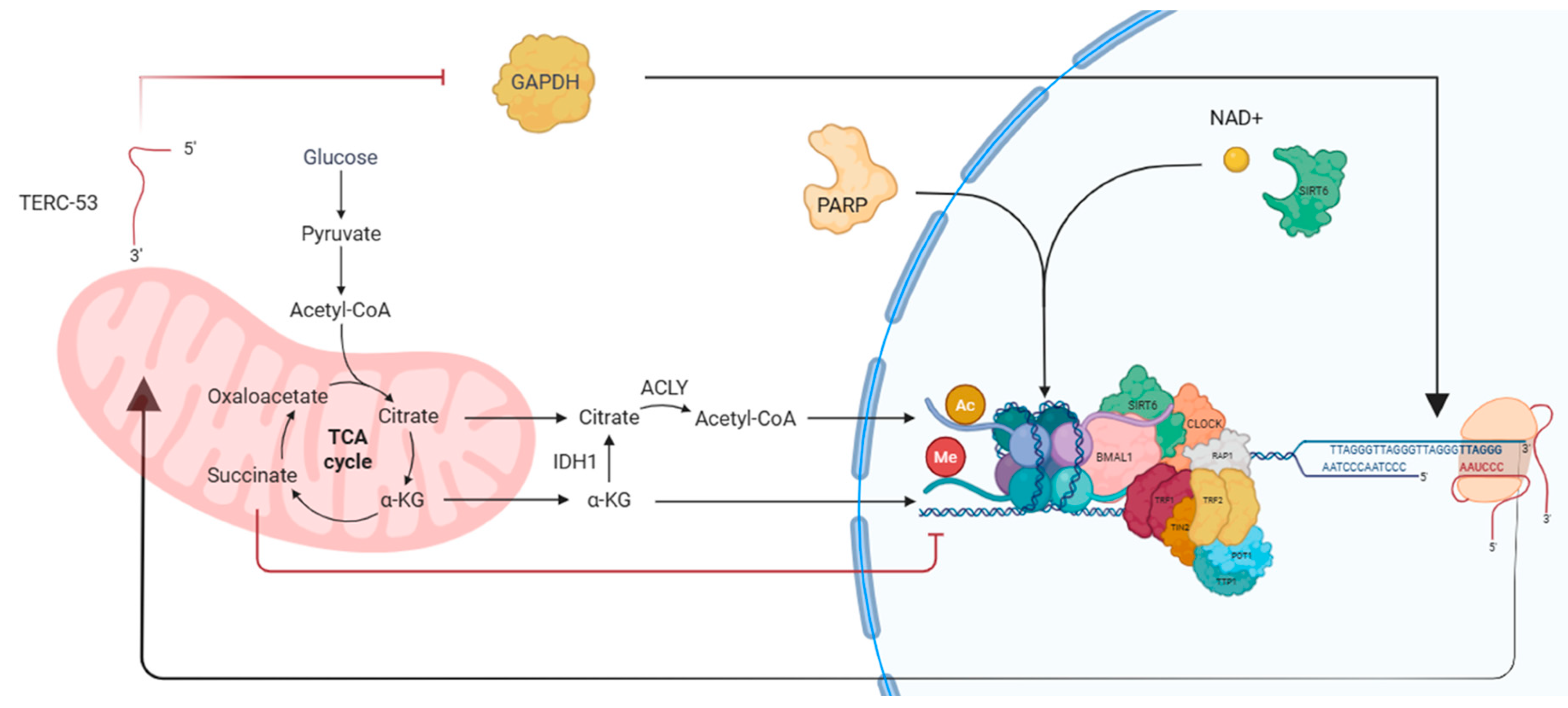
Figure 1: Mitochondrion-nucleus feedback loop. The picture illustrates the crosstalk between cellular metabolism and the structural integrity of nuclear DNA. Within the mitochondrion, the TCA cycle generates key metabolites such as citrate and α-KG. Once transported into the cytosol, citrate is converted into Acetyl-CoA by the enzyme ACLY, providing acetyl groups (Ac) for histone acetylation, while α-KG acts as an essential cofactor for demethylation (Me) modifications, directly influencing the epigenetics of telomeric DNA. In parallel, the NAD+ pool regulates the activity of crucial nuclear enzymes: SIRT6, which promotes telomere deacetylation and stability, and PARPs, involved in DNA damage repair. The Shelterin complex interacts with circadian clock proteins to protect the TTAGGG telomeric repeat sequences. A retrograde signal is represented by TERC-53, a non-coding RNA fragment that bridges mitochondrial distress to nuclear response. The disruption of this homeostatic balance compromises genomic stability and accelerates cellular decline. Abbreviations: TCA, Tricarboxylic Acid; α-KG, α-Ketoglutarate; ACLY, ATP Citrate Lyase; Acetyl-CoA, Acetyl Coenzyme A; Ac, Acetylation; Me, Methylation; NAD+, Nicotinamide Adenine Dinucleotide; SIRT6, Sirtuin 6; PARPs, Poly(ADP-ribose) Polymerases; TERC-53, Telomerase RNA Component 53-nt fragment; TRF1/2, Telomeric Repeat-binding Factor 1/2; TIN2, TRF1-interacting Nuclear Protein 2; RAP1, Repressor/Activator Protein 1; POT1, Protection of Telomeres 1; TPP1, TIN2-interacting Protein 1; BMAL1, Brain and Muscle ARNT-Like 1; CLOCK, Circadian Locomotor Output Cycles Kaput. This figure was created with BioRender.com.
2 Molecular Landscape of the Mito-Nuclear Communication
The metabolic-epigenetic crosstalk is a complex regulatory network where mitochondria act as environmental sensors for the nucleus. This communication relies on a diverse set of intermediates, including TCA cycle metabolites, non-coding RNAs, and transiently translocating proteins that function as molecular messengers. Table 1 provides a comprehensive overview of these actors, their associated metabolites, and their specific roles in maintaining telomeric homeostasis.
Table 1: Molecular mediators of the Mitochondrial-Telomere retrograde signaling axis.
| Abbreviation | Full Name | Associated Metabolite | Functional Role in Telomere Homeostasis | References |
|---|---|---|---|---|
| ACLY | ATP Citrate Lyase | Acetyl-CoA | Converts mitochondrial citrate into Acetyl-CoA, fueling histone acetylation (H3K9ac, H3K27ac) at the hTERT promoter to maintain its expression. | [36,37,38] |
| AMPK | AMP-activated Protein Kinase | ATP/AMP ratio | Master energy sensor; triggers mitochondrial retrograde signaling and activates PGC-1α to promote TERRA expression under metabolic stress. | [30,39,40] |
| ATRX | Alpha-Thalassemia/X-linked | N/A | Chromatin remodeler that maintains telomeric heterochromatin; its loss is a hallmark of the Alternative Lengthening of Telomeres (ALT) pathway. | [41,42,43] |
| EZH2/SUZ12 | PRC2 Complex subunits | SAM | Methyltransferases that interact with TERRA to deposit repressive H3K27me3 marks using SAM as a methyl donor, ensuring telomeric silencing. | [44,45] |
| hTERT | Telomerase Reverse Transcriptase | N/A | Under oxidative stress, it translocates to mitochondria to protect mtDNA and respiratory function. | [46,47,48] |
| ISR/UPRmt | Integrated Stress Response | Mito-stress signals | Signaling cascades that communicate mitochondrial proteotoxic stress to the nucleus to modulate cellular lifespan and adaptation. | [10,11] |
| JHDMs | JmjC-domain Demethylases | α-KG/Succinate | α-KG-dependent enzymes that remove repressive marks (H3K9me3) to maintain telomere plasticity. Competitively inhibited by succinate. | [49,50,51] |
| NAMPT | Nicotinamide phosphoribosyl-transferase | NAD+ | Rate-limiting enzyme in NAD+ metabolism; links the circadian clock to SIRT6-mediated telomere protection. | [52,53] |
| NRF1 | Nuclear Respiratory Factor 1 | N/A | Transcription factor that coordinates mitochondrial biogenesis and promotes TERRA transcription during physiological stress. | [54,55] |
| SDH | Succinate Dehydrogenase | Succinate | Complex II enzyme; its deficiency causes succinate accumulation, leading to epigenetic hardening and telomeric instability. | [34,35,56] |
| SIRT6 | Sirtuin 6 | NAD+ | NAD+-dependent deacetylase; ensures T-loop stability and prevents R-loop formation. | [57,58] |
| TERC-53 | Telomerase RNA Component (fragment 53) | Mito-stress signals | A cytoplasmic fragment of TERC that shuttles to the nucleus as a retrograde signal of mitochondrial distress, bypassing canonical telomerase roles. | [59] |
| TERRA | Telomeric Repeat RNA | N/A | Long non-coding RNA acts as a scaffold for chromatin modifiers (PRC2 and LSD1) and interacts with TERC-53 for mito-nuclear signaling. | [44,60] |
The complexity of this communication lies in its hierarchical decision-making logic. As illustrated in Fig. 2, the cell translates mitochondrial functional states (energetic, oncometabolite, and redox status) into specific nuclear fates. This stepwise transduction ensures that telomere stability is dynamically tuned to the bioenergetic capacity of the cell, transitioning from a permissive state of self-renewal under optimal conditions to epigenetic hardening and senescence when mitochondrial distress signals predominate.
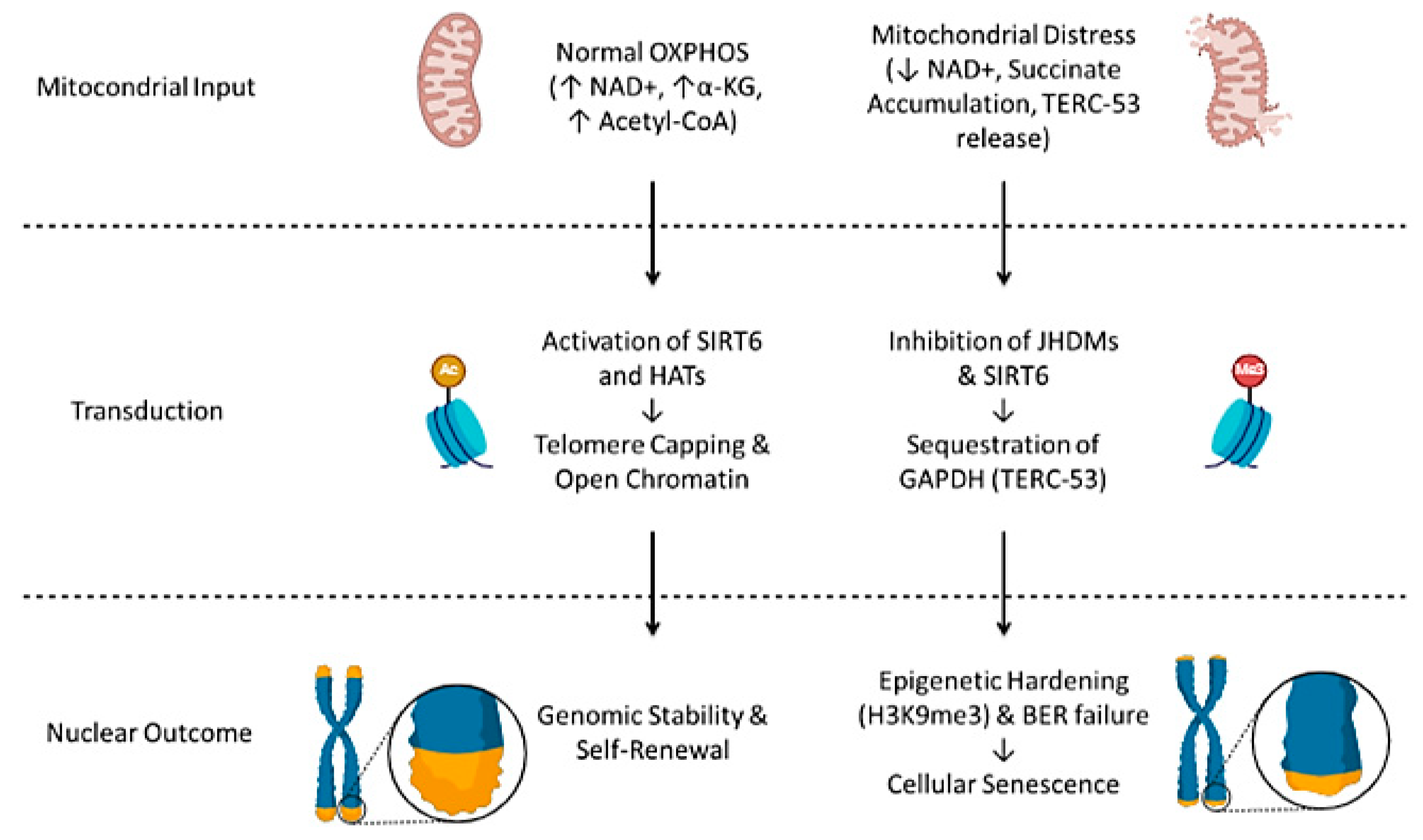
Figure 2: Stepwise functional roadmap of mitochondrial-telomere retrograde signaling. The picture illustrates the causal hierarchy of the mito-nuclear crosstalk, moving from metabolic inputs to genomic outcomes. (Top) Mitochondrial Input: Normal oxidative phosphorylation (OXPHOS) (left) maintains high levels of nicotinamide adenine dinucleotide (NAD+), α-ketoglutarate (α-KG), and Acetyl-CoA, whereas Mitochondrial Distress (right) leads to NAD+ depletion, succinate accumulation, and telomerase RNA component fragment 53 (TERC-53) release. (Middle) Transduction: These metabolic states are sensed by epigenetic modifiers. In homeostatic conditions, sirtuin 6 (SIRT6) and histone acetyltransferases (HATs) ensure telomere capping and an open chromatin state. Conversely, mitochondrial distress triggers the inhibition of histone demethylases (JHDMs) and SIRT6, alongside GAPDH sequestration by TERC-53. (Bottom) Nuclear Outcome: The signaling axis dictates the cellular fate, leading to genomic stability and self-renewal (left) or the epigenetic hardening (H3K9me3), base excision repair (BER) failure, and cellular senescence (right). This figure was created with BioRender.com.
3 The Energetic Axis: Acetyl-CoA and Telomeric Chromatin Accessibility
3.1 Acetyl-CoA Production and Compartmentalization
Acetyl-CoA is the key metabolic intermediate produced by the catabolism of carbohydrates, lipids, and amino acids [61]. The acetyl group is transported out of the mitochondria through an indirect shuttle system: acetyl-CoA inside the mitochondrion condenses with oxaloacetate to form citrate. Citrate passes into the cytoplasm across the inner mitochondrial membrane through the Citrate carrier (SLC25A1) [62]. In the cytosol, citrate is cleaved by citrate lyase (ACLY) to regenerate acetyl-CoA [36]. The cytosolic pool of citrate is also sustained by the reductive carboxylation of α-KG mediated by IDH1. Under conditions of mitochondrial impairment or metabolic shift, IDH1 reverses the canonical flux to generate isocitrate and subsequently citrate, which ACLY then cleaves into Acetyl-CoA [63].
Seminal evidence highlighted that ACLY can translocate into the nucleus, providing a compartmentalized nuclear pool of Acetyl-CoA for histone acetylation [64,65]. Accordingly, ACLY nuclear translocation in fibroblasts upon fibrotic stimulation, facilitating TGF-β-mediated myofibroblast activation by promoting H3K27ac at fibrotic gene loci [37]. On the other hand, in cisplatin-resistant hepatocellular carcinoma cells, the stabilization of nuclear ACLY by NAT10-mediated acetylation at K468 prevents its degradation through SQSTM1-mediated proteasomal pathways. This enhances the nuclear acetyl-CoA levels, increasing H3K27ac at chemoresistance-related genes like CYP2C9 and PIK3R1 [38,66].
Interestingly, in most somatic cells, the hTERT promoter (the on/off switch for telomerase) is typically embedded in a nuclease-resistant, closed chromatin structure that silences gene expression. However, an adequate bioenergetic supply facilitates a switch to an accessible euchromatin state, characterized by markers such as H3K9ac, H3K27ac, and H3K4me2/3, allowing key transcription factors like c-Myc and Sp1 to initiate transcription [67,68]. c-Myc binds to specific E-box sequences (5′-CACGTG-3′) within the hTERT promoter [69]. The convergence of c-Myc/Sp1 serves as a metabolic sensor by recruiting HATs, such as TRRAP and p300/CBP [70,71]. TRRAP is essential for this selectivity, as it bridges nuclear ACLY activity directly to the c-Myc cluster. This process acts as a metabolic sensor: during G1-S phases, the nucleus requires enormous amounts of acetyl-CoA for histone acetylation. While ACLY maintains a basal level of acetylation, translocation of the pyruvate dehydrogenase complex (PDC) produces acetyl-CoA directly from pyruvate within the nucleus, providing a high-flux, spatiotemporal boost of acetyl-CoA directly to chromatin during the high-stress window of DNA replication [64,72,73].
3.2 Metabolic Sensing and hTERT Promoter Regulation
This spatial substrate channeling ensures that Acetyl-CoA is utilized in situ to catalyze the acetylation of surrounding histones, effectively opening the hTERT chromatin while other genomic regions remain unaffected [74]. This is particularly evident in the context of hTERT promoter mutations [75]. Cytosine-to-thymine (C > T) transitions at positions -124 bp (C228T), and -146 bp (C250T) are the most frequent genetic alterations within the hTERT promoter in medulloblastomas [76], hepatocellular carcinomas (HCC) [77], urothelial carcinomas of the bladder [78], melanomas [79], and primary glioblastomas (GBM) [80]. These mutations result in the generation of de novo binding sites (consensus sequence 5′-CCGGAA-3′) for the ETS family of transcription factors [81]. Specifically, the transcription factor GABPA selectively recognizes and binds to the mutated promoter, whereas it shows no affinity for the wild-type sequence [82]. This specific binding recruits the transcriptional machinery, leading to the overexpression of hTERT and subsequently promoting cellular immortalization. Therefore, the oncogenic potential of hTERT mutations is functionally dependent on the bioenergetic state [83,84].
3.3 TERRA Modulation and the Bioenergetic Checkpoint
Consistently, in mammalian cells, the Telomeric Repeat-containing RNA (TERRA) transcript is essential for cis/trans telomere capping to protect the T-loop architecture [44,85,86]. TERRA interacts with H3K9me3 and HP1 (heterochromatin protein 1) proteins [87], the histone methyltransferase SUV39H1 [88], and with chromatin remodeling complexes such as NoRC (nucleolar remodeling complex) [89], MORF4L2 (a component of the NuA histone acetyltransferase complex), and ARID1A [90]. NuA4 is responsible for the acetylation of histone H4 (H4Ac). This specific modification neutralizes the positive charge of the histone tails, reducing DNA torsional tension and favouring chromatin to open. Thus, RTEL1 helicases could remove G-quadruplexes and allow DNA polymerase to complete its work [91,92]. Without this dual-input of Acetyl-CoA, the TERRA-bound MORF4L2 cannot maintain the requisite chromatin flexibility of the subtelomeric region during S-phase [93].
Evidence suggests that telomere length (TL) itself can modulate metabolic signaling, establishing a retrograde feedback loop [94]. The telomere acts as a physical sensor that feedback-regulates the cell’s enzymatic capacity to maintain its own length through the Telomere Position Effect over Long Distance (TPE-OLD) or the non-telomeric binding of TRF2 to the hTERT promoter [95,96,97]. In cases of severe bioenergetic failure, the lack of substrates for the epigenetic machinery triggers a cascade of genomic instability [57,98]. The loss of histone acetylation results in replication fork stalling and the formation of DNA secondary G-quadruplexes [99,100]. These unresolved stalls result in persistent double-strand breaks (DSBs) at telomeric repeats [101]. In this context, the cell is forced to utilize the existing DSBs as substrates for homology-directed repair (HDR), effectively bypassing the need for telomerase through aberrant template switching [102,103,104]. Consequently, the Acetyl-CoA flux serves as a fundamental checkpoint: when the fuel for acetylation is exhausted, the cell is propelled toward senescence or aberrant genomic survival strategies.
4 The Control Axis: α-KG/Succinate and Telomeric Stability
4.1 Competitive Inhibition of 2-OGDD Enzymes by Succinate
Beyond acetylation, the mitochondrial-nuclear axis exerts a second layer of epigenetic control through the α-ketoglutarate (α-KG)/succinate ratio [49]. α-KG, produced in the mitochondrial matrix by isocitrate dehydrogenase (IDH), is the obligatory co-substrate for the 2-oxoglutarate-dependent dioxygenase (2-OGDD) superfamily [50]. This family includes TET (Ten-Eleven Translocation) DNA demethylases, which convert 5-methylcytosine (5-mC) to 5-hydroxymethylcytosine (5-hmC), and JmjC-domain-containing histone demethylases (JHDMs), such as KDM4B/C, which remove repressive marks like H3K9me3 and H3K27me3 [105,106,107]. An accumulation of succinate acts as a potent competitive inhibitor of 2-OGDDs. Specifically, succinate competes with α-KG for binding to the highly conserved Fe2+-containing active site of TET and JmjC enzymes, thereby blocking their catalytic activity [108].
Mitochondrial α-KG is exported to the cytosol through the oxoglutarate carrier SLC25A11 (OGC) in exchange for malate. On the other hand, mitochondrial succinate is exported to the cytosol through the dicarboxylate carrier SLC25A10. Once in the cytosol, their availability in the nucleus directly depends on the cytosolic α-KG/succinate ratio [62]. The α-KG/succinate ratio thus dictates the global methylome: a high ratio (high α-KG) promotes a permissive, hypomethylated state, whereas a low ratio (high succinate) triggers a hypermethylator phenotype. This low-ratio state, often termed CpG Island Methylator Phenotype (CIMP), leads to the hypermethylation of DNA and histones across the genome [109].
At telomeric and subtelomeric regions, this axis is critical for structural plasticity, although through distinct substrates. While subtelomeric regions are regulated by DNA methylation at CpG islands, the telomeric core—which lacks CpG sites—relies exclusively on histone marks [110,111]. TET and JmjC enzymes utilize α-KG to maintain these regions in a metabolically responsive state, facilitating the dynamic turnover of shelterin proteins and fine-tuning the transcription of TERRA [44,86,112,113].
4.2 SDH Deficiency and the Epigenetic Lock
Consistently, TERRA transcripts associate with the histone methyl transferase Polycomb Repressive Complex 2 (PRC2), through a direct interaction with the PRC2 components EZH2 and SUZ12 [44]. Furthermore, TERRA G-quadruplex acts as a scaffold for other telomere-binding proteins, such as the translocated in liposarcoma/fused in sarcoma protein (TLS/FUS) and TRF2 [114,115]. These interactions through the activity of Suv4-20h2, which associates with TLS/FUS, can promote H4K20me3 modification at telomeres [116]. Without this RNA scaffold, the telomere loses its ability to manage and organize its own heterochromatin, leading to structural fragility.
While a systemic lack of S-adenosylmethionine (SAM) could potentially leave methyltransferases catalytically starved, the pathological accumulation of succinate exerts a dominant inhibitory effect on the 2-OGDD family. This disruption shifts the telomeric landscape from a dynamic state to one of epigenetic inertia, leading to replication fork stalling and instability [104]. Mitochondrial compromises, such as loss-of-function mutations in succinate dehydrogenase (SDH), cause a massive accumulation of succinate, leading to an epigenetic silencing of the 2-OGDD family [117]. This process occurs not through mere energy depletion, but through the induction of a state of biochemical pseudohypoxia [118]. The pathological accumulation of succinate, acting as an oncometabolite, inhibits prolyl hydroxylases (PHDs) in the cytosol, leading to the constitutive stabilization of Hypoxia-Inducible Factor 1-alpha (HIF1α) even under normoxic conditions [117,119,120,121]. This stabilization triggers a profound biological paradox that explains the genomic instability observed in SDH-deficient tumors and chronic metabolic diseases. On one hand, stabilized HIF1α translocates to the nucleus and directly binds to the hTERT promoter, attempting to upregulate telomerase expression as an evolutionary rescue mechanism to counteract mitochondrial-induced cellular stress [122,123]. On the other hand, the succinate surplus that stabilizes HIF1α simultaneously functions as a competitive inhibitor of the TET and JmjC enzymes required to maintain telomeric chromatin accessibility [45]. Consequently, the cell faces a metabolic-epigenetic mismatch: it attempts to activate hTERT within a genomic environment that is physically inaccessible. This is driven by subtelomeric DNA hypermethylation and, crucially, by the persistence of H3K9me3 at the telomeric core due to JHDM inhibition [124,125,126]. This epigenetic lock prevents the functional translation of the HIF1α signal into effective telomere repair. Instead, the compromised binding of the shelterin complex to hypermethylated repeats—coupled with the inability of telomerase to access the T-loop—accelerates telomere attrition and favors aberrant DNA template switching [125,127], triggering a transition toward aberrant survival pathways such as the alternative lengthening of telomeres (ALT) pathway [128,129,130]. Moreover, this transition is significantly favored by the loss of ATRX, a chromatin remodeler frequently mutated in SDH-deficient tumors [41,42]. This epigenetic instability promotes the formation of extra-chromosomal telomeric circles (C-circles) and facilitates homology-directed repair (HDR), providing the genetic and metabolic ground for ALT [43,131].
4.3 Mitochondrial Cofactors: Pyrimidine Synthesis and FAD-Dependent Demethylation
Furthermore, the failure of the SDH-CoQ axis disrupts the de novo synthesis of pyrimidines via dihydroorotate dehydrogenase (DHODH), creating a localized depletion of dNTPs that further cripples any residual telomerase-mediated repair [132,133,134].
Complementary to the α-KG-dependent family, telomeric heterochromatin is further fine-tuned by Lysine-specific demethylase 1 (LSD1). Unlike JmjC enzymes, LSD1 does not require α-KG, but is strictly dependent on Flavin Adenine Dinucleotide (FAD) as a catalytic cofactor [61]. LSD1 orchestrates the oxidative demethylation of mono- and di-methylated H3K4 and H3K9, a process that reduces FAD to FADH2. Consequently, in case of ETC compromise, a decline in the mitochondrial FAD/FADH2 ratio limits LSD1 enzymatic turnover [135]. This metabolic bottleneck leads to the aberrant persistence of H3K4 methylation marks, which effectively opens the telomeric chromatin, facilitating the pathological transcription of TERRA and predisposing the locus to the formation of DNA-damaging R-loops [136,137].
5 The Surveillance Axis: NAD+ Bioavailability and SIRT6 Fidelity
5.1 SIRT6 as a Metabolic Sensor of NAD+
The NAD+/SIRT6 axis constitutes the third critical pillar of mitochondrial-nuclear communication. Nicotinamide adenine dinucleotide (NAD+) is the central redox cofactor for the mitochondrial electron transport chain (ETC), facilitating the transfer of electrons to generate the proton gradient necessary for ATP synthesis [138]. Functional mitochondria maintain a high NAD+/NADH ratio, which is essential for maintaining the catalytic activity of NAD+-dependent enzymes [52]. While acetyl-CoA and α-KG provide the building blocks and signals for chromatin remodeling, NAD+ serves as a real-time sensor of bioenergetic integrity that activates nuclear surveillance enzymes. These include the so-called longevity sirtuin, SIRT6 deacetylase, often referred to as the guardian of telomere stability [139,140].
The biochemical fidelity of SIRT6 is strictly governed by NAD+ bioavailability [58,141]. Unlike other sirtuins, SIRT6 possesses a low Kd (~27 μM) and high Km (~270–800 μM) for NAD+, making SIRT6 a true metabolic sensor due to its catalytic activity sensitive to fluctuations in the nuclear NAD+ pool and capable of adapting gene expression and DNA repair based on energy availability [142].
5.2 T-Loop Stability and TERRA Repression
Unlike other members of the sirtuin family, SIRT6 is constitutively tethered to chromatin and is indispensable for telomere stability [58]. Under metabolic homeostasis, mitochondrial Complex I maintains the NAD+/NADH balance, fueling SIRT6 to orchestrate telomeric protection through three primary NAD+-coordinated mechanisms. SIRT6 mediates H3K9 and H3K56 deacetylation to promote a stable heterochromatic structure. This specific deacetylation is a prerequisite for stabilizing the protective T-loop architecture, preventing the formation of aberrant telomeric DNA circles (t-circles) and suppressing illegitimate recombination [143,144]. Moreover, SIRT6 acts as a molecular scaffold for the recruitment of the shelterin complex TRF2 and the coordination of DNA repair machinery. It facilitates the recruitment of the DNA-dependent protein kinase catalytic subunit (DNA-PKcs), ensuring that DBSs at telomeric ends are addressed by non-homologous end joining (NHEJ) [145,146,147]. Finally, in somatic contexts, SIRT6 prevents uncontrolled cellular proliferation and transcriptional leakage of TERRA, thereby protecting telomeres from inappropriate DNA damage response (DDR) activation [148,149]. Mechanistically, the H3K9 residue can be either acetylated or methylated, but never both simultaneously. When SIRT6 is active, it removes the acetyl group (H3K9Ac → H3K9). Thus, SUV39H1 can deposit the methylation mark (H3K9 → H3K9me3). Finally, HP1 binds to the H3K9me3 signal. In this state, TERRA transcription is maintained at basal, physiological levels. Without SIRT6, lysine 9 remains engaged by the acetyl group (H3K9Ac). Consequently, SUV39H1 is unable to act, H3K9me3 fails to form, HP1 is not recruited, and the telomere fails to compact. As a result, RNA polymerase II produces an excessive amount of TERRA. These transcripts then form R-loops (three-stranded DNA: RNA hybrids) on the telomeric DNA. During the S-phase, these structures block the replication fork, leading to double-strand breaks (DSBs) and the subsequent activation of the DDR [150,151].
5.3 The Bioenergetic-Telomeric Trap and PARP-1 Competition
When mitochondrial bioenergetics fail, the resulting reduction in NAD+ bioavailability compromises SIRT6 fidelity. Without sufficient NAD+, SIRT6 can no longer function as an epigenetic brake, leading to the deregulation of metabolic genes through the p53-mediated repression of PGC-1α and PGC-1β [152]. Consequently, the impaired HAT activity due to the localized Acetyl-CoA depletion may lead to reduced histone acetylation, potentially contributing to a transition toward a repressive, closed chromatin state. Without α-KG, TET/JmjC demethylases become blocked (hypermethylation). This bioenergetic-telomeric trap propels the cell toward a point of no return where replicative senescence becomes the inevitable outcome of systemic metabolic exhaustion [57,153]. This vulnerability is exacerbated by the competition for the same NAD+ pool with Poly(ADP-ribose) polymerase 1 (PARP-1) [154]. Activated by DNA damage—often a byproduct of mitochondrial ROS—PARP-1 acts as a metabolic sink, depleting nuclear NAD+ levels due to its higher affinity for the substrate [155]. This creates a lethal vicious cycle: the accumulation of telomeric damage recruits PARP-1, which further starves SIRT6 of NAD+, thereby accelerating telomeric uncapping and preventing efficient DNA repair [156,157].
6 The Extranuclear Translocation of hTERT: Translational Perspectives
6.1 Stress-Induced Nucleocytoplasmic Shuttling of hTERT
Beyond its canonical role within the nucleus, the catalytic subunit hTERT possesses a nucleocytoplasmic shuttling characteristic [46].
In homeostatic conditions, hTERT is predominantly nuclear. However, in response to oxidative stress (i.e., H2O2 exposure), hTERT is transiently exported to the cytosol. This process is mediated by the CRM1 (Exportin 1) pathway and the Ran-GTPase system [47,158]. hTERT possesses an evolutionarily conserved Nuclear Export Signal (NES) located within its C-terminus reverse transcriptase domain (residues 701–900). This signal is conditionally accessible: under basal conditions, the NES is sequestered within the protein tertiary structure. Upon oxidative stress, phosphorylation at specific residues (Tyr707) by Src-kinase or Abl-tyrosine-kinase induces a conformational change that exposes the NES for CRM1 recognition. This mechanism ensures that hTERT export is not a random process but a tightly regulated rescue strategy to protect hTERT from nuclear degradation and to prevent telomerase-induced de novo addition at fragile genomic sites during stress [159].
6.2 Mitochondrial TERT (mtTERT) and the Mitohormetic Trade-Off
Once in the cytosol, hTERT could also be directed to the mitochondria through a specific 20-amino acid amphipathic alpha-helix Mitochondrial Targeting Sequence (MTS), located at the N-terminus (residues 1–20) [160]. Here, it functions as a mitochondrial chaperone (mtTERT) through several integrated mechanisms. While initial studies focused on mtTERT binding to the ND1 and ND2 regions, more extensive mapping has revealed that mtTERT interacts with a broad array of mitochondrial loci, including 12S and 16S rRNA, ND4, ND5, COX I, COX III, and ATP synthase subunits 6 and 8, enhancing its resistance to oxidative damage and reducing the local production of ROS [161]. A compelling hypothesis proposes that mtTERT actively participates in mtDNA replication by utilizing mitochondrial tRNAs as primers. By favoring alternative origins of replication (mt-tRNA genes), mtTERT may limit the traditional strand displacement mode, which is notoriously prone to deletions [162]. Furthermore, mtTERT interacts with the RNA component of mitochondrial RNA processing (RMRP) and improves the activity of the electron transport chain (ETC), particularly Complex I, stabilizing membrane potential and ATP production [163,164]. Finally, mtTERT acts as a protein scaffold for mitochondrial transcripts, stabilizing mitochondrial ribosome function and ensuring efficient translation of respiratory proteins [165].
This movement represents a strategic metabolic pivot where hTERT transitions from a genomic enzyme to a mitochondrial protector. This mitohormetic strategy creates a functional trade-off designed to prioritize immediate cellular survival and bioenergetic maintenance at the direct expense of long-term replicative capacity [48,166]. The pharmacological modulation of this axis offers a promising therapeutic frontier. Molecules capable of stabilizing nuclear hTERT or, conversely, promoting its mitochondrial translocation could be used to fine-tune the balance between genomic stability and metabolic efficiency [167].
6.3 TERC-53 and the Retrograde Signaling of Mitochondrial Distress
The regulatory hierarchy between the nucleus and mitochondria is not a one-way round. The mitochondrial compartment is capable of incorporating and processing non-coding RNAs [168]. The telomerase RNA component (TERC) facilitates this bidirectional feedback loop. TERC-53 is a 53-nucleotide fragment derived from the 5′-end of TERC, and its cytosolic accumulation is dynamically regulated by mitochondrial function [59]. Under conditions of mitochondrial distress, TERC-53 accumulates in the cytosol, acting as a molecular decoy that traps glyceraldehyde 3-phosphate dehydrogenase (GAPDH) in the cytosol, thus preventing its translocation to the nucleus [59]. Nuclear GAPDH is essential for the stabilization and catalytic efficiency of Uracil-DNA Glycosylase (UNG), the primary enzyme responsible for removing pro-mutagenic uracil residues from DNA for the Base Excision Repair (BER) machinery. The resulting failure of the BER pathway leads to the accumulation of single-strand breaks [169]. This finding adds a critical layer to the metabolic axes previously discussed: the mitochondrial-telomere crosstalk is not only mediated by protein translocation (hTERT) or metabolite flux (Acetyl-CoA, NAD+), but also by non-coding RNA signals (TERC-53) that dictate the transition toward replicative arrest [59,169,170,171].
7 Spatiotemporal Regulation and Circadian Clock
The entire system is not static, but it follows a biological circadian rhythm. Telomere maintenance is finally dynamically synchronized within the circadian clock. It regulates the rhythmic availability of NAD+ through the rate-limiting enzyme NAMPT (Nicotinamide phosphoribosyltransferase). NAMPT expression is directly governed by the CLOCK: BMAL1 heterodimer, creating a 24-h oscillation in nuclear NAD+ levels. This temporal fluctuation implies that the surveillance capacity of SIRT6—and consequently the stability of the telomeric T-loop—is subject to a circadian window of vulnerability [53]. However, it should be mentioned that current literature regarding the direct circadian control of telomere-mitochondria crosstalk remains extremely limited. This knowledge gap is due to the significant methodological challenges in chronobiology research.
Finally, under Caloric Restriction (CR), a physiological state of low nutrient intake without malnutrition that shifts cellular priority toward survival and repair, the activation of the AMPK/PGC-1α pathway recruits Nuclear Respiratory Factor 1 (NRF1) to subtelomeric promoters, directly promoting TERRA expression, to ensure that the chromatin environment remains responsive [55,172].
In conclusion, the telomere acts as a metabolic antenna, sensing the state of the mitochondria to decide the cell fate. The transition towards cellular aging is not only a matter of physical DNA wear, but an epigenetic hardening due to the failure of mitochondrial signaling. Deciphering the language of this communication is essential for the generation of therapies in age-related pathologies. Consistently, age-related pathologies would require a holistic approach that views genomic stability as a direct reflection of mitochondrial health.
Acknowledgement:
Funding Statement: The author received no specific funding for this study.
Availability of Data and Materials: Not applicable.
Ethics Approval: Not applicable.
Conflicts of Interest: The author declares no conflicts of interest.
Abbreviations
The following abbreviations are used in this manuscript:
| MFRTA | Mitochondrial Free Radical Theory of Aging |
| ROS | Reactive Oxygen Species |
| DNMTs | DNA Methyltransferases |
| TCA | Tricarboxylic Acid (cycle) |
| SLC25A1 | Citrate Carrier (Solute Carrier Family 25 Member 1) |
| IDH1 | Isocitrate Dehydrogenase 1 |
| NAT10 | N-acetyltransferase 10 |
| CYP2C9 | Cytochrome P450 Family 2 Subfamily C Member 9 |
| PIK3R1 | Phosphoinositide-3-Kinase Regulatory Subunit 1 |
| TRRAP | Transformation/Transcription Domain Associated Protein |
| PDC | Pyruvate Dehydrogenase Complex |
| GABPA | GA Binding Protein Transcription Factor Subunit Alpha |
| HP1 | Heterochromatin Protein 1 |
| SUV39H1 | Suppressor Of Variegation 3-9 Homolog 1 |
| NoRC | Nucleolar Remodeling Complex |
| ARID1A | AT-Rich Interaction Domain 1A |
| RTEL1 | Regulator of Telomere Elongation Helicase 1 |
| TL | Telomere Length |
| TPE-OLD | Telomere Position Effect over Long Distance |
| DSBs | Double-Strand Breaks |
| HDR | Homology-Directed Repair |
| 2-OGDD | 2-Oxoglutarate-Dependent Dioxygenase |
| TET | Ten-Eleven Translocation |
| SLC25A11 (OGC) | Oxoglutarate Carrier |
| SLC25A10 | Dicarboxylate Carrier |
| CIMP | CpG Island Methylator Phenotype |
| PRC2 | Polycomb Repressive Complex 2 |
| TLS/FUS | Translocated in Liposarcoma/Fused in Sarcoma |
| SAM | S-Adenosylmethionine |
| PHDs | Prolyl Hydroxylases |
| HIF1α | Hypoxia-Inducible Factor 1-alpha |
| ALT | Alternative Lengthening of Telomeres |
| DHODH | Dihydroorotate Dehydrogenase |
| dNTPs | Deoxynucleotide Triphosphates |
| LSD1 | Lysine-specific Demethylase 1 |
| ETC | Electron Transport Chain |
| DNA-PKcs | DNA-dependent Protein Kinase catalytic subunit |
| NHEJ | Non-Homologous End Joining |
| DDR | DNA Damage Response |
| PGC-1α/β | Peroxisome proliferator-activated receptor Gamma Coactivator 1-alpha/beta |
| PARP-1 | Poly(ADP-ribose) Polymerase 1 |
| CRM1 | Chromosome Region Maintenance 1 (Exportin 1) |
| NES | Nuclear Export Signal |
| MTS | Mitochondrial Targeting Sequence |
| mtTERT | Mitochondrial hTERT |
| RMRP | RNA component of Mitochondrial RNA Processing |
| UNG | Uracil-DNA Glycosylase |
| BER | Base Excision Repair |
| CLOCK: BMAL1 | Circadian Locomotor Output Cycles Kaput: Brain and Muscle ARNT-Like 1 |
| CR | Caloric Restriction |
References
1. Somasundaram I , Jain SM , Blot-Chabaud M , Pathak S , Banerjee A , Rawat S , et al. Mitochondrial dysfunction and its association with age-related disorders. Front Physiol. 2024; 15: 1384966. doi:10.3389/fphys.2024.1384966. [Google Scholar] [CrossRef]
2. Manganelli M , Stabile G , Scharf C , Podo Brunetti A , Paolino G , Giuffrida R , et al. Skin photodamage and melanomagenesis: a comprehensive review. Cancers. 2025; 17( 11): 1784. doi:10.3390/cancers17111784. [Google Scholar] [CrossRef]
3. Amano H , Chaudhury A , Rodriguez-Aguayo C , Lu L , Akhanov V , Catic A , et al. Telomere dysfunction induces sirtuin repression that drives telomere-dependent disease. Cell Metab. 2019; 29( 6): 1274– 90.e9. doi:10.1016/j.cmet.2019.03.001. [Google Scholar] [CrossRef]
4. Stojanovic B , Jovanovic I , Dimitrijevic Stojanovic M , Stojanovic BS , Kovacevic V , Radosavljevic I , et al. Oxidative stress-driven cellular senescence: mechanistic crosstalk and therapeutic horizons. Antioxidants. 2025; 14( 8): 987. doi:10.3390/antiox14080987. [Google Scholar] [CrossRef]
5. Picca A , Guerra F , Calvani R , Coelho-Júnior HJ , Leeuwenburgh C , Bucci C , et al. The contribution of mitochondrial DNA alterations to aging, cancer, and neurodegeneration. Exp Gerontol. 2023; 178: 112203. doi:10.1016/j.exger.2023.112203. [Google Scholar] [CrossRef]
6. Kim KH , Lee CB . Socialized mitochondria: mitonuclear crosstalk in stress. Exp Mol Med. 2024; 56( 5): 1033– 42. doi:10.1038/s12276-024-01211-4. [Google Scholar] [CrossRef]
7. Chen G , Dong H , Tian Y . Mito-nuclear communication: from cellular responses to organismal health. Mol Cell. 2026; 86( 3): 522– 32. doi:10.1016/j.molcel.2026.01.001. [Google Scholar] [CrossRef]
8. Zhu D , Li X , Tian Y . Mitochondrial-to-nuclear communication in aging: an epigenetic perspective. Trends Biochem Sci. 2022; 47( 8): 645– 59. doi:10.1016/j.tibs.2022.03.008. [Google Scholar] [CrossRef]
9. Walker EM , Pearson GL , Lawlor N , Stendahl AM , Lietzke A , Sidarala V , et al. Retrograde mitochondrial signaling governs the identity and maturity of metabolic tissues. Science. 2025; 388( 6743): eadf2034. doi:10.1126/science.adf2034. [Google Scholar] [CrossRef]
10. Eckl EM , Ziegemann O , Krumwiede L , Fessler E , Jae LT . Sensing, signaling and surviving mitochondrial stress. Cell Mol Life Sci. 2021; 78( 16): 5925– 51. doi:10.1007/s00018-021-03887-7. [Google Scholar] [CrossRef]
11. Torres AK , Fleischhart V , Inestrosa NC . Mitochondrial unfolded protein response (UPRmt): what we know thus far. Front Cell Dev Biol. 2024; 12: 1405393. doi:10.3389/fcell.2024.1405393. [Google Scholar] [CrossRef]
12. Meichsner A , Bader V , Winklhofer KF . Mitochondria as sources and targets of cellular signaling. Mol Cell. 2026; 86( 3): 503– 21. doi:10.1016/j.molcel.2026.01.008. [Google Scholar] [CrossRef]
13. Liu YJ , Sulc J , Auwerx J . Mitochondrial genetics, signalling and stress responses. Nat Cell Biol. 2025; 27( 3): 393– 407. doi:10.1038/s41556-025-01625-w. [Google Scholar] [CrossRef]
14. Donato L , Mordà D , Scimone C , Alibrandi S , D’Angelo R , Sidoti A . From powerhouse to regulator: the role of mitoepigenetics in mitochondrion-related cellular functions and human diseases. Free Radic Biol Med. 2024; 218: 105– 19. doi:10.1016/j.freeradbiomed.2024.03.025. [Google Scholar] [CrossRef]
15. Shyh-Chang N , Ng HH . The metabolic programming of stem cells. Genes Dev. 2017; 31( 4): 336– 46. doi:10.1101/gad.293167.116. [Google Scholar] [CrossRef]
16. Gunawan AL , Liparulo I , Stahl A . Mammalian mitohormesis: from mitochondrial stressors to organismal benefits. EMBO J. 2025; 44( 20): 5640– 61. doi:10.1038/s44318-025-00549-3. [Google Scholar] [CrossRef]
17. Ge T , Gu X , Jia R , Ge S , Chai P , Zhuang A , et al. Crosstalk between metabolic reprogramming and epigenetics in cancer: updates on mechanisms and therapeutic opportunities. Cancer Commun. 2022; 42( 11): 1049– 82. doi:10.1002/cac2.12374. [Google Scholar] [CrossRef]
18. Covarrubias AJ , Perrone R , Grozio A , Verdin E . NAD+ metabolism and its roles in cellular processes during ageing. Nat Rev Mol Cell Biol. 2021; 22( 2): 119– 41. doi:10.1038/s41580-020-00313-x. [Google Scholar] [CrossRef]
19. Paciolla C , Manganelli M , Di Chiano M , Montenegro F , Gallone A , Sallustio F , et al. Valeric acid: a gut-derived metabolite as a potential epigenetic modulator of neuroinflammation in the gut–brain axis. Cells. 2025; 14( 22): 1823. doi:10.3390/cells14221823. [Google Scholar] [CrossRef]
20. Zhang X , Gao Y , Zhang S , Wang Y , Pei X , Chen Y , et al. Mitochondrial dysfunction in the regulation of aging and aging-related diseases. Cell Commun Signal. 2025; 23( 1): 290. doi:10.1186/s12964-025-02308-7. [Google Scholar] [CrossRef]
21. Younesian S , Mohammadi MH , Younesian O , Momeny M , Ghaffari SH , Bashash D . DNA methylation in human diseases. Heliyon. 2024; 10( 11): e32366. doi:10.1016/j.heliyon.2024.e32366. [Google Scholar] [CrossRef]
22. Lee HT , Roe JS , Youn HD . Metabolic oxidoreductases: central regulators of the epigenetic landscapes in stemness. Exp Mol Med. 2026; 58( 4): 1017– 37. doi:10.1038/s12276-026-01687-2. [Google Scholar] [CrossRef]
23. Xu Y , Jian X , Shi L , Shock LS , Chen L , Chow LT , et al. Mitochondria and epigenetic regulation: bidirectional crosstalk and emerging mitochondria-targeted degron tools. Cells. 2026; 15( 2): 95. doi:10.3390/cells15020095. [Google Scholar] [CrossRef]
24. Nam H , Yu G , Ahn SI , Nam S . Mechanical regulation of metabolism, epigenetics, and their interplay. npj Biomed Innov. 2026; 3: 5. doi:10.1038/s44385-025-00059-1. [Google Scholar] [CrossRef]
25. Wei P , Zhang X , Yan C , Sun S , Chen Z , Lin F . Mitochondrial dysfunction and aging: multidimensional mechanisms and therapeutic strategies. Biogerontology. 2025; 26( 4): 142. doi:10.1007/s10522-025-10273-4. [Google Scholar] [CrossRef]
26. Assalve G , Lunetti P , Rocca MS , Cosci I , Di Nisio A , Ferlin A , et al. Exploring the link between telomeres and mitochondria: mechanisms and implications in different cell types. Int J Mol Sci. 2025; 26( 3): 993. doi:10.3390/ijms26030993. [Google Scholar] [CrossRef]
27. Martini H , Passos JF . Cellular senescence: all roads lead to mitochondria. FEBS J. 2023; 290( 5): 1186– 202. doi:10.1111/febs.16361. [Google Scholar] [CrossRef]
28. Li Y , Tian X , Luo J , Bao T , Wang S , Wu X . Molecular mechanisms of aging and anti-aging strategies. Cell Commun Signal. 2024; 22( 1): 285. doi:10.1186/s12964-024-01663-1. [Google Scholar] [CrossRef]
29. Gu Z , Niu Z , Yan Z , Fan Y , Sun J , Zhao X , et al. Chain-mediating effect of interaction between telomeres and mitochondria under oxidative stress in coke oven workers. Environ Pollut. 2023; 331: 121855. doi:10.1016/j.envpol.2023.121855. [Google Scholar] [CrossRef]
30. Chandel NS , Jasper H , Ho TT , Passegué E . Metabolic regulation of stem cell function in tissue homeostasis and organismal ageing. Nat Cell Biol. 2016; 18( 8): 823– 32. doi:10.1038/ncb3385. [Google Scholar] [CrossRef]
31. Sperber H , Mathieu J , Wang Y , Ferreccio A , Hesson J , Xu Z , et al. The metabolome regulates the epigenetic landscape during naive-to-primed human embryonic stem cell transition. Nat Cell Biol. 2015; 17( 12): 1523– 35. doi:10.1038/ncb3264. [Google Scholar] [CrossRef]
32. Dogan F , Forsyth NR . TERT promoter methylation is oxygen-sensitive and regulates telomerase activity. Biomolecules. 2024; 14( 1): 131. doi:10.3390/biom14010131. [Google Scholar] [CrossRef]
33. Gao C . Investigating the association between blood metabolites and telomere length: a Mendelian randomization study. PLoS One. 2024; 19( 3): e0298172. doi:10.1371/journal.pone.0298172. [Google Scholar] [CrossRef]
34. Li W , Quan L , Peng K , Wang Y , Wang X , Chen Q , et al. Succinate dehydrogenase is essential for epigenetic and metabolic homeostasis in hearts. Basic Res Cardiol. 2023; 118( 1): 45. doi:10.1007/s00395-023-01015-z. [Google Scholar] [CrossRef]
35. Wang Q , Li M , Zeng N , Zhou Y , Yan J . Succinate dehydrogenase complex subunit C: Role in cellular physiology and disease. Exp Biol Med. 2023; 248( 3): 263– 70. doi:10.1177/15353702221147567. [Google Scholar] [CrossRef]
36. Ghisletti S , Russo M . TCA-cycle metabolites in the nucleus: drivers of chromatin and epigenetic control. BMC Biol. 2025; 23( 1): 316. doi:10.1186/s12915-025-02423-4. [Google Scholar] [CrossRef]
37. Lazaropoulos MP , Gibb AA , Chapski DJ , Nair AA , Reiter AN , Roy R , et al. Nuclear ATP-citrate lyase regulates chromatin-dependent activation and maintenance of the myofibroblast gene program. Nat Cardiovasc Res. 2024; 3( 7): 869– 82. doi:10.1038/s44161-024-00502-3. [Google Scholar] [CrossRef]
38. Wang Y , Su K , Wang C , Deng T , Liu X , Sun S , et al. Chemotherapy-induced acetylation of ACLY by NAT10 promotes its nuclear accumulation and acetyl-CoA production to drive chemoresistance in hepatocellular carcinoma. Cell Death Dis. 2024; 15: 545. doi:10.1038/s41419-024-06951-9. [Google Scholar] [CrossRef]
39. Mandic M , Misirkic Marjanovic M , Janjetovic K , Bosnjak M , Harhaji-Trajkovic L , Trajkovic V , et al. Multifaceted role of AMPK in autophagy: more than a simple trigger? Am J Physiol Cell Physiol. 2025; 329( 5): C1380– 97. doi:10.1152/ajpcell.01058.2024. [Google Scholar] [CrossRef]
40. Dang K , Muhammad Umer Farooq H , Gao Y , Deng X , Qian A . The role of 5′-adenosine monophosphate-activated protein kinase (AMPK) in skeletal muscle atrophy. Biocell. 2023; 47( 2): 269– 81. doi:10.32604/biocell.2023.023766. [Google Scholar] [CrossRef]
41. Heaphy CM , de Wilde RF , Jiao Y , Klein AP , Edil BH , Shi C , et al. Altered telomeres in tumors with ATRX and DAXX mutations. Science. 2011; 333( 6041): 425. doi:10.1126/science.1207313. [Google Scholar] [CrossRef]
42. Job S , Draskovic I , Burnichon N , Buffet A , Cros J , Lépine C , et al. Telomerase activation and ATRX mutations are independent risk factors for metastatic pheochromocytoma and paraganglioma. Clin Cancer Res. 2019; 25( 2): 760– 70. doi:10.1158/1078-0432.CCR-18-0139. [Google Scholar] [CrossRef]
43. Aguilera P , López-Contreras AJ . ATRX, a guardian of chromatin. Trends Genet. 2023; 39( 6): 505– 19. doi:10.1016/j.tig.2023.02.009. [Google Scholar] [CrossRef]
44. Montero JJ , López-Silanes I , Megías D , F Fraga M , Castells-García Á , Blasco MA . TERRA recruitment of polycomb to telomeres is essential for histone trymethylation marks at telomeric heterochromatin. Nat Commun. 2018; 9( 1): 1548. doi:10.1038/s41467-018-03916-3. [Google Scholar] [CrossRef]
45. Dogan F , Forsyth NR . Telomerase regulation: a role for epigenetics. Cancers. 2021; 13( 6): 1213. doi:10.3390/cancers13061213. [Google Scholar] [CrossRef]
46. Haendeler J , Hoffmann J , Brandes RP , Zeiher AM , Dimmeler S . Hydrogen peroxide triggers nuclear export of telomerase reverse transcriptase via src kinase family-dependent phosphorylation of tyrosine 707. Mol Cell Biol. 2003; 23( 13): 4598– 610. doi:10.1128/MCB.23.13.4598-4610.2003. [Google Scholar] [CrossRef]
47. Marinaccio J , Micheli E , Udroiu I , Di Nottia M , Carrozzo R , Baranzini N , et al. TERT extra-telomeric roles: antioxidant activity and mitochondrial protection. Int J Mol Sci. 2023; 24( 5): 4450. doi:10.3390/ijms24054450. [Google Scholar] [CrossRef]
48. Ebata H , Shima T , Iizuka R , Uemura S . Accumulation of TERT in mitochondria exerts two opposing effects on apoptosis. FEBS Open Bio. 2023; 13( 9): 1667– 82. doi:10.1002/2211-5463.13682. [Google Scholar] [CrossRef]
49. Bénit P , Goncalves J , El Khoury R , Rak M , Favier J , Gimenez-Roqueplo AP , et al. Succinate dehydrogenase, succinate, and superoxides: a genetic, epigenetic, metabolic, environmental explosive crossroad. Biomedicines. 2022; 10( 8): 1788. doi:10.3390/biomedicines10081788. [Google Scholar] [CrossRef]
50. Nakashima Y , Brewitz L , Tumber A , Salah E , Schofield CJ . 2-Oxoglutarate derivatives can selectively enhance or inhibit the activity of human oxygenases. Nat Commun. 2021; 12: 6478. doi:10.1038/s41467-021-26673-2. [Google Scholar] [CrossRef]
51. Wang M , Xue J , Hong W , Chen S , Shi H . JMJD family proteins in cancer and inflammation. Sig Transduct Target Ther. 2022; 7: 304. doi:10.1038/s41392-022-01145-1. [Google Scholar] [CrossRef]
52. Yusri K , Jose S , Vermeulen KS , Tan TCM , Sorrentino V . The role of NAD+ metabolism and its modulation of mitochondria in aging and disease. npj Metab Health Dis. 2025; 3: 26. doi:10.1038/s44324-025-00067-0. [Google Scholar] [CrossRef]
53. Ramsey KM , Yoshino J , Brace CS , Abrassart D , Kobayashi Y , Marcheva B , et al. Circadian clock feedback cycle through NAMPT-mediated NAD+ biosynthesis. Science. 2009; 324( 5927): 651– 4. doi:10.1126/science.1171641. [Google Scholar] [CrossRef]
54. D’Souza AD , Parikh N , Kaech SM , Shadel GS . Convergence of multiple signaling pathways is required to coordinately up-regulate mtDNA and mitochondrial biogenesis during T cell activation. Mitochondrion. 2007; 7( 6): 374– 85. doi:10.1016/j.mito.2007.08.001. [Google Scholar] [CrossRef]
55. Diman A , Boros J , Poulain F , Rodriguez J , Purnelle M , Episkopou H , et al. Nuclear respiratory factor 1 and endurance exercise promote human telomere transcription. Sci Adv. 2016; 2( 7): e1600031. doi:10.1126/sciadv.1600031. [Google Scholar] [CrossRef]
56. Bayley JP , Devilee P . Hypothesis: why different types of SDH gene variants cause divergent tumor phenotypes. Genes. 2022; 13( 6): 1025. doi:10.3390/genes13061025. [Google Scholar] [CrossRef]
57. Tasselli L , Xi Y , Zheng W , Tennen RI , Odrowaz Z , Simeoni F , et al. SIRT6 deacetylates H3K18ac at pericentric chromatin to prevent mitotic errors and cellular senescence. Nat Struct Mol Biol. 2016; 23( 5): 434– 40. doi:10.1038/nsmb.3202. [Google Scholar] [CrossRef]
58. Michishita E , McCord RA , Berber E , Kioi M , Padilla-Nash H , Damian M , et al. SIRT6 is a histone H3 lysine 9 deacetylase that modulates telomeric chromatin. Nature. 2008; 452( 7186): 492– 6. doi:10.1038/nature06736. [Google Scholar] [CrossRef]
59. Zheng Q , Liu P , Gao G , Yuan J , Wang P , Huang J , et al. Mitochondrion-processed TERC regulates senescence without affecting telomerase activities. Protein Cell. 2019; 10( 9): 631– 48. doi:10.1007/s13238-019-0612-5. [Google Scholar] [CrossRef]
60. Rodrigues J , Alfieri R , Bione S , Azzalin CM . TERRA ONTseq: a long-read-based sequencing pipeline to study the human telomeric transcriptome. RNA. 2024; 30( 8): 955– 66. doi:10.1261/rna.079906.123. [Google Scholar] [CrossRef]
61. Wang Y , Yang H , Geerts C , Furtos A , Waters P , Cyr D , et al. The multiple facets of acetyl-CoA metabolism: energetics, biosynthesis, regulation, acylation and inborn errors. Mol Genet Metab. 2023; 138( 1): 106966. doi:10.1016/j.ymgme.2022.106966. [Google Scholar] [CrossRef]
62. Palmieri F . The mitochondrial transporter family SLC25: identification, properties and physiopathology. Mol Aspects Med. 2013; 34( 2–3): 465– 84. doi:10.1016/j.mam.2012.05.005. [Google Scholar] [CrossRef]
63. Gai C , Zeng H , Xu H , Chai X , Zou Y , Zhuang C , et al. Comprehensive exploration of isocitrate dehydrogenase (IDH) mutations: tumorigenesis, drug discovery, and covalent inhibitor advances. Eur J Med Chem. 2025; 282: 117041. doi:10.1016/j.ejmech.2024.117041. [Google Scholar] [CrossRef]
64. Sivanand S , Viney I , Wellen KE . Spatiotemporal control of acetyl-CoA metabolism in chromatin regulation. Trends Biochem Sci. 2018; 43( 1): 61– 74. doi:10.1016/j.tibs.2017.11.004. [Google Scholar] [CrossRef]
65. Yang H , Qin Y , Xu X , Chen W , Zu G , Chen X . Acetyl-CoA: an interplay between metabolism and epigenetics in cancer. Front Mol Med. 2022; 2: 1044585. doi:10.3389/fmmed.2022.1044585. [Google Scholar] [CrossRef]
66. Zhang Y , Jing Y , Wang Y , Tang J , Zhu X , Jin WL , et al. NAT10 promotes gastric cancer metastasis via N4-acetylated COL5A1. Signal Transduct Target Ther. 2021; 6: 173. doi:10.1038/s41392-021-00489-4. [Google Scholar] [CrossRef]
67. Dong S , Li T . Metabolic reprogramming in cancer: signaling pathways and therapeutic targets. Mol Cancer. 2026; 25( 1): 60. doi:10.1186/s12943-026-02582-0. [Google Scholar] [CrossRef]
68. Yuan X , Larsson C , Xu D . Mechanisms underlying the activation of TERT transcription and telomerase activity in human cancer: old actors and new players. Oncogene. 2019; 38( 34): 6172– 83. doi:10.1038/s41388-019-0872-9. [Google Scholar] [CrossRef]
69. Shou S , Maolan A , Zhang D , Jiang X , Liu F , Li Y , et al. Telomeres, telomerase, and cancer: mechanisms, biomarkers, and therapeutics. Exp Hematol Oncol. 2025; 14( 1): 8. doi:10.1186/s40164-025-00597-9. [Google Scholar] [CrossRef]
70. Kyo S , Takakura M , Taira T , Kanaya T , Itoh H , Yutsudo M , et al. Sp1 cooperates with c-Myc to activate transcription of the human telomerase reverse transcriptase gene (hTERT). Nucleic Acids Res. 2000; 28( 3): 669– 77. doi:10.1093/nar/28.3.669. [Google Scholar] [CrossRef]
71. Luo J , Chen Z , Qiao Y , Tien JC , Young E , Mannan R , et al. Targeting histone H2B acetylated enhanceosomes via p300/CBP degradation in prostate cancer. Nat Genet. 2025; 57( 10): 2468– 81. doi:10.1038/s41588-025-02336-6. [Google Scholar] [CrossRef]
72. Sutendra G , Kinnaird A , Dromparis P , Paulin R , Stenson TH , Haromy A , et al. A nuclear pyruvate dehydrogenase complex is important for the generation of acetyl-CoA and histone acetylation. Cell. 2014; 158( 1): 84– 97. doi:10.1016/j.cell.2014.04.046. [Google Scholar] [CrossRef]
73. Wu QJ , Zhang TN , Chen HH , Yu XF , Lv JL , Liu YY , et al. The sirtuin family in health and disease. Signal Transduct Target Ther. 2022; 7: 402. doi:10.1038/s41392-022-01257-8. [Google Scholar] [CrossRef]
74. Lee JV , Carrer A , Shah S , Snyder NW , Wei S , Venneti S , et al. Akt-dependent metabolic reprogramming regulates tumor cell histone acetylation. Cell Metab. 2014; 20( 2): 306– 19. doi:10.1016/j.cmet.2014.06.004. [Google Scholar] [CrossRef]
75. Shanmugam R , Ozturk MB , Low JL , Akincilar SC , Chua JYH , Thangavelu MT , et al. Genome-wide screens identify specific drivers of mutant hTERT promoters. Proc Natl Acad Sci U S A. 2022; 119( 3): e2105171119. doi:10.1073/pnas.2105171119. [Google Scholar] [CrossRef]
76. Remke M , Ramaswamy V , Peacock J , Shih DJH , Koelsche C , Northcott PA , et al. TERT promoter mutations are highly recurrent in SHH subgroup medulloblastoma. Acta Neuropathol. 2013; 126( 6): 917– 29. doi:10.1007/s00401-013-1198-2. [Google Scholar] [CrossRef]
77. Quaas A , Oldopp T , Tharun L , Klingenfeld C , Krech T , Sauter G , et al. Frequency of TERT promoter mutations in primary tumors of the liver. Virchows Arch. 2014; 465( 6): 673– 7. doi:10.1007/s00428-014-1658-7. [Google Scholar] [CrossRef]
78. Kinde I , Munari E , Faraj SF , Hruban RH , Schoenberg M , Bivalacqua T , et al. TERT promoter mutations occur early in urothelial neoplasia and are biomarkers of early disease and disease recurrence in urine. Cancer Res. 2013; 73( 24): 7162– 7. doi:10.1158/0008-5472.CAN-13-2498. [Google Scholar] [CrossRef]
79. Horn S , Figl A , Rachakonda PS , Fischer C , Sucker A , Gast A , et al. TERT promoter mutations in familial and sporadic melanoma. Science. 2013; 339( 6122): 959– 61. doi:10.1126/science.1230062. [Google Scholar] [CrossRef]
80. Killela PJ , Reitman ZJ , Jiao Y , Bettegowda C , Agrawal N , Diaz LA Jr , et al. TERT promoter mutations occur frequently in gliomas and a subset of tumors derived from cells with low rates of self-renewal. Proc Natl Acad Sci U S A. 2013; 110( 15): 6021– 6. doi:10.1073/pnas.1303607110. [Google Scholar] [CrossRef]
81. Sharma S , Chowdhury S . Emerging mechanisms of telomerase reactivation in cancer. Trends Cancer. 2022; 8( 8): 632– 41. doi:10.1016/j.trecan.2022.03.005. [Google Scholar] [CrossRef]
82. Bell RJA , Rube HT , Kreig A , Mancini A , Fouse SD , Nagarajan RP , et al. The transcription factor GABP selectively binds and activates the mutant TERT promoter in cancer. Science. 2015; 348( 6238): 1036– 9. doi:10.1126/science.aab0015. [Google Scholar] [CrossRef]
83. Akıncılar SC , Khattar E , Boon PLS , Unal B , Fullwood MJ , Tergaonkar V . Long-range chromatin interactions drive mutant TERT promoter activation. Cancer Discov. 2016; 6( 11): 1276– 91. doi:10.1158/2159-8290.CD-16-0177. [Google Scholar] [CrossRef]
84. Liu M , Zhang Y , Jian Y , Gu L , Zhang D , Zhou H , et al. The regulations of telomerase reverse transcriptase (TERT) in cancer. Cell Death Dis. 2024; 15: 90. doi:10.1038/s41419-024-06454-7. [Google Scholar] [CrossRef]
85. Rivosecchi J , Cusanelli E . TERRA beyond cancer: the biology of telomeric repeat-containing RNAs in somatic and germ cells. Front Aging. 2023; 4: 1224225. doi:10.3389/fragi.2023.1224225. [Google Scholar] [CrossRef]
86. Manganelli M , Grossi I , Corsi J , D’Agostino VG , Jurikova K , Cusanelli E , et al. Expression of cellular and extracellular TERRA, TERC and TERT in hepatocellular carcinoma. Int J Mol Sci. 2022; 23( 11): 6183. doi:10.3390/ijms23116183. [Google Scholar] [CrossRef]
87. Deng Z , Norseen J , Wiedmer A , Riethman H , Lieberman PM . TERRA RNA binding to TRF2 facilitates heterochromatin formation and ORC recruitment at telomeres. Mol Cell. 2009; 35( 4): 403– 13. doi:10.1016/j.molcel.2009.06.025. [Google Scholar] [CrossRef]
88. Porro A , Feuerhahn S , Delafontaine J , Riethman H , Rougemont J , Lingner J . Functional characterization of the TERRA transcriptome at damaged telomeres. Nat Commun. 2014; 5: 5379. doi:10.1038/ncomms6379. [Google Scholar] [CrossRef]
89. Postepska-Igielska A , Krunic D , Schmitt N , Greulich-Bode KM , Boukamp P , Grummt I . The chromatin remodelling complex NoRC safeguards genome stability by heterochromatin formation at telomeres and centromeres. EMBO Rep. 2013; 14( 8): 704– 10. doi:10.1038/embor.2013.87. [Google Scholar] [CrossRef]
90. Scheibe M , Arnoult N , Kappei D , Buchholz F , Decottignies A , Butter F , et al. Quantitative interaction screen of telomeric repeat-containing RNA reveals novel TERRA regulators. Genome Res. 2013; 23( 12): 2149– 57. doi:10.1101/gr.151878.112. [Google Scholar] [CrossRef]
91. Mitchell L , Lambert JP , Gerdes M , Al-Madhoun AS , Skerjanc IS , Figeys D , et al. Functional dissection of the NuA4 histone acetyltransferase reveals its role as a genetic hub and that Eaf1 is essential for complex integrity. Mol Cell Biol. 2008; 28( 7): 2244– 56. doi:10.1128/MCB.01653-07. [Google Scholar] [CrossRef]
92. Vannier JB , Sandhu S , Petalcorin MIR , Wu X , Nabi Z , Ding H , et al. RTEL1 is a replisome-associated helicase that promotes telomere and genome-wide replication. Science. 2013; 342( 6155): 239– 42. doi:10.1126/science.1241779. [Google Scholar] [CrossRef]
93. Ducker GS , Rabinowitz JD . One-carbon metabolism in health and disease. Cell Metab. 2017; 25( 1): 27– 42. doi:10.1016/j.cmet.2016.08.009. [Google Scholar] [CrossRef]
94. Casagrande S , Hau M . Telomere attrition: metabolic regulation and signalling function? Biol Lett. 2019; 15( 3): 20180885. doi:10.1098/rsbl.2018.0885. [Google Scholar] [CrossRef]
95. Takakura M . Telomerase activation by histone deacetylase inhibitor in normal cells. Nucleic Acids Res. 2001; 29( 14): 3006– 11. doi:10.1093/nar/29.14.3006. [Google Scholar] [CrossRef]
96. Lewis KA , Tollefsbol TO . Regulation of the telomerase reverse transcriptase subunit through epigenetic mechanisms. Front Genet. 2016; 7: 83. doi:10.3389/fgene.2016.00083. [Google Scholar] [CrossRef]
97. Chevalier R , Murcia Pienkowski V , Jullien N , Caron L , Pinard PV , Magdinier F , et al. Telomere position effect-over long distances acts as a genome-wide epigenetic regulator through a common Alu element. Aging Cell. 2025; 24( 6): e70027. doi:10.1111/acel.70027. [Google Scholar] [CrossRef]
98. López-Gil L , Pascual-Ahuir A , Proft M . Genomic instability and epigenetic changes during aging. Int J Mol Sci. 2023; 24( 18): 14279. doi:10.3390/ijms241814279. [Google Scholar] [CrossRef]
99. Alabert C , Groth A . Chromatin replication and epigenome maintenance. Nat Rev Mol Cell Biol. 2012; 13( 3): 153– 67. doi:10.1038/nrm3288. [Google Scholar] [CrossRef]
100. Rhodes D , Lipps HJ . G-quadruplexes and their regulatory roles in biology. Nucleic Acids Res. 2015; 43( 18): 8627– 37. doi:10.1093/nar/gkv862. [Google Scholar] [CrossRef]
101. Martínez P , Blasco MA . Replicating through telomeres: a means to an end. Trends Biochem Sci. 2015; 40( 9): 504– 15. doi:10.1016/j.tibs.2015.06.003. [Google Scholar] [CrossRef]
102. Lee JJ , Kim H , Park H , Lee U , Kim C , Lee M , et al. Disruption of G-quadruplex dynamicity by BRCA2 abrogation instigates phase separation and break-induced replication at telomeres. Nucleic Acids Res. 2024; 52( 10): 5756– 73. doi:10.1093/nar/gkae251. [Google Scholar] [CrossRef]
103. Sobinoff AP , Pickett HA . Alternative lengthening of telomeres: DNA repair pathways converge. Trends Genet. 2017; 33( 12): 921– 32. doi:10.1016/j.tig.2017.09.003. [Google Scholar] [CrossRef]
104. O’Sullivan RJ , Kubicek S , Schreiber SL , Karlseder J . Reduced histone biosynthesis and chromatin changes arising from a damage signal at telomeres. Nat Struct Mol Biol. 2010; 17( 10): 1218– 25. doi:10.1038/nsmb.1897. [Google Scholar] [CrossRef]
105. Tischler J , Gruhn WH , Reid J , Allgeyer E , Buettner F , Marr C , et al. Metabolic regulation of pluripotency and germ cell fate through α-ketoglutarate. EMBO J. 2019; 38( 1): e99518. doi:10.15252/embj.201899518. [Google Scholar] [CrossRef]
106. Tahiliani M , Koh KP , Shen Y , Pastor WA , Bandukwala H , Brudno Y , et al. Conversion of 5-methylcytosine to 5-hydroxymethylcytosine in mammalian DNA by MLL partner TET1. Science. 2009; 324( 5929): 930– 5. doi:10.1126/science.1170116. [Google Scholar] [CrossRef]
107. Black JC , Van Rechem C , Whetstine JR . Histone lysine methylation dynamics: establishment, regulation, and biological impact. Mol Cell. 2012; 48( 4): 491– 507. doi:10.1016/j.molcel.2012.11.006. [Google Scholar] [CrossRef]
108. Koivunen P , Lee S , Duncan CG , Lopez G , Lu G , Ramkissoon S , et al. Transformation by the (R)-enantiomer of 2-hydroxyglutarate linked to EGLN activation. Nature. 2012; 483( 7390): 484– 8. doi:10.1038/nature10898. [Google Scholar] [CrossRef]
109. Figueroa ME , Abdel-Wahab O , Lu C , Ward PS , Patel J , Shih A , et al. Leukemic IDH1 and IDH2 mutations result in a hypermethylation phenotype, disrupt TET2 function, and impair hematopoietic differentiation. Cancer Cell. 2010; 18( 6): 553– 67. doi:10.1016/j.ccr.2010.11.015. [Google Scholar] [CrossRef]
110. Blasco MA . The epigenetic regulation of mammalian telomeres. Nat Rev Genet. 2007; 8( 4): 299– 309. doi:10.1038/nrg2047. [Google Scholar] [CrossRef]
111. Wang K , Liu H , Hu Q , Wang L , Liu J , Zheng Z , et al. Epigenetic regulation of aging: implications for interventions of aging and diseases. Signal Transduct Target Ther. 2022; 7: 374. doi:10.1038/s41392-022-01211-8. [Google Scholar] [CrossRef]
112. Ciccarone F , Vegliante R , Di Leo L , Ciriolo MR . The TCA cycle as a bridge between oncometabolism and DNA transactions in cancer. Semin Cancer Biol. 2017; 47: 50– 6. doi:10.1016/j.semcancer.2017.06.008. [Google Scholar] [CrossRef]
113. Bettin N , Oss Pegorar C , Cusanelli E . The emerging roles of TERRA in telomere maintenance and genome stability. Cells. 2019; 8( 3): 246. doi:10.3390/cells8030246. [Google Scholar] [CrossRef]
114. Collie GW , Haider SM , Neidle S , Parkinson GN . A crystallographic and modelling study of a human telomeric RNA (TERRA) quadruplex. Nucleic Acids Res. 2010; 38( 16): 5569– 80. doi:10.1093/nar/gkq259. [Google Scholar] [CrossRef]
115. Biffi G , Tannahill D , Balasubramanian S . An intramolecular G-quadruplex structure is required for binding of telomeric repeat-containing RNA to the telomeric protein TRF2. J Am Chem Soc. 2012; 134( 29): 11974– 6. doi:10.1021/ja305734x. [Google Scholar] [CrossRef]
116. Takahama K , Takada A , Tada S , Shimizu M , Sayama K , Kurokawa R , et al. Regulation of telomere length by G-quadruplex telomere DNA- and TERRA-binding protein TLS/FUS. Chem Biol. 2013; 20( 3): 341– 50. doi:10.1016/j.chembiol.2013.02.013. [Google Scholar] [CrossRef]
117. Chen L , Huang M . Oncometabolites in cancer: from cancer cells to the tumor microenvironment. Holist Integr Oncol. 2024; 3( 1): 26. doi:10.1007/s44178-024-00096-7. [Google Scholar] [CrossRef]
118. Beyoğlu D , Idle JR . Metabolic rewiring and the characterization of oncometabolites. Cancers. 2021; 13( 12): 2900. doi:10.3390/cancers13122900. [Google Scholar] [CrossRef]
119. Esteban-Amo MJ , Jiménez-Cuadrado P , Serrano-Lorenzo P , de la Fuente MÁ , Simarro M . Succinate dehydrogenase and human disease: novel insights into a well-known enzyme. Biomedicines. 2024; 12( 9): 2050. doi:10.3390/biomedicines12092050. [Google Scholar] [CrossRef]
120. Ősz F , Akbar M , Zsidó BZ , Hetényi C , Orbán TI , Fóthi Á , et al. The Caenorhabditis elegans sdhb-1(R244H) model shows characteristics of human PPGL tumor cells. Int J Mol Sci. 2025; 26( 20): 10185. doi:10.3390/ijms262010185. [Google Scholar] [CrossRef]
121. Atallah R , Olschewski A , Heinemann A . Succinate at the crossroad of metabolism and angiogenesis: roles of SDH, HIF1α and SUCNR1. Biomedicines. 2022; 10( 12): 3089. doi:10.3390/biomedicines10123089. [Google Scholar] [CrossRef]
122. Nishi H , Nakada T , Kyo S , Inoue M , Shay JW , Isaka K . Hypoxia-inducible factor 1 mediates upregulation of telomerase (hTERT). Mol Cell Biol. 2004; 24( 13): 6076– 83. doi:10.1128/MCB.24.13.6076-6083.2004. [Google Scholar] [CrossRef]
123. Yatabe N , Kyo S , Maida Y , Nishi H , Nakamura M , Kanaya T , et al. HIF-1-mediated activation of telomerase in cervical cancer cells. Oncogene. 2004; 23( 20): 3708– 15. doi:10.1038/sj.onc.1207460. [Google Scholar] [CrossRef]
124. Giger OT , ten Hoopen R , Shorthouse D , Abdullahi S , Bulusu VR , Jadhav S , et al. Preferential MGMT hypermethylation in SDH-deficient wild-type GIST. J Clin Pathol. 2024; 77( 1): 34– 9. doi:10.1136/jcp-2022-208462. [Google Scholar] [CrossRef]
125. Wang J , Yuan T , Yang B , He Q , Zhu H . SDH defective cancers: molecular mechanisms and treatment strategies. Cell Biol Toxicol. 2025; 41( 1): 74. doi:10.1007/s10565-025-10022-w. [Google Scholar] [CrossRef]
126. Neves JB , Roberts K , Nguyen JS , El Sheikh S , Tran-Dang MA , Horsfield C , et al. Defining the origin, evolution, and immune composition of SDH-deficient renal cell carcinoma. iScience. 2022; 25( 11): 105389. doi:10.1016/j.isci.2022.105389. [Google Scholar] [CrossRef]
127. Gupta P , Strange K , Telange R , Guo A , Hatch H , Sobh A , et al. Genetic impairment of succinate metabolism disrupts bioenergetic sensing in adrenal neuroendocrine cancer. Cell Rep. 2022; 40( 7): 111218. doi:10.1016/j.celrep.2022.111218. [Google Scholar] [CrossRef]
128. Flynn RL , Centore RC , O’Sullivan RJ , Rai R , Tse A , Zhou S , et al. TERRA and hnRNPA1 orchestrate an RPA-to-POT1 switch on telomeric single-stranded DNA. Nature. 2011; 471( 7339): 532– 6. doi:10.1038/nature09772. [Google Scholar] [CrossRef]
129. Kim W , Ludlow AT , Min J , Robin JD , Stadler G , Mender I , et al. Regulation of the human telomerase gene TERT by telomere position effect-over long distances (TPE-OLD): implications for aging and cancer. PLoS Biol. 2016; 14( 12): e2000016. doi:10.1371/journal.pbio.2000016. [Google Scholar] [CrossRef]
130. Muoio D , Fouquerel E . The alternative lengthening of telomeres pathway through a DNA repair lens: mechanism and therapeutic opportunities. NAR Cancer. 2025; 7( 4): zcaf056. doi:10.1093/narcan/zcaf056. [Google Scholar] [CrossRef]
131. Brosnan-Cashman JA , Yuan M , Graham MK , Rizzo AJ , Myers KM , Davis C , et al. ATRX loss induces multiple hallmarks of the alternative lengthening of telomeres (ALT) phenotype in human glioma cell lines in a cell line-specific manner. PLoS One. 2018; 13( 9): e0204159. doi:10.1371/journal.pone.0204159. [Google Scholar] [CrossRef]
132. Sanford SL , Welfer GA , Freudenthal BD , Opresko PL . Mechanisms of telomerase inhibition by oxidized and therapeutic dNTPs. Nat Commun. 2020; 11: 5288. doi:10.1038/s41467-020-19115-y. [Google Scholar] [CrossRef]
133. Peeters MJW , Aehnlich P , Pizzella A , Mølgaard K , Seremet T , Met Ö , et al. Mitochondrial-linked de novo pyrimidine biosynthesis dictates human T-cell proliferation but not expression of effector molecules. Front Immunol. 2021; 12: 718863. doi:10.3389/fimmu.2021.718863. [Google Scholar] [CrossRef]
134. Hubackova S , Davidova E , Boukalova S , Kovarova J , Bajzikova M , Coelho A , et al. Replication and ribosomal stress induced by targeting pyrimidine synthesis and cellular checkpoints suppress p53-deficient tumors. Cell Death Dis. 2020; 11: 110. doi:10.1038/s41419-020-2224-7. [Google Scholar] [CrossRef]
135. Hino S , Sakamoto A , Nagaoka K , Anan K , Wang Y , Mimasu S , et al. FAD-dependent lysine-specific demethylase-1 regulates cellular energy expenditure. Nat Commun. 2012; 3: 758. doi:10.1038/ncomms1755. [Google Scholar] [CrossRef]
136. Garcia-Exposito L , Bournique E , Bergoglio V , Bose A , Barroso-Gonzalez J , Zhang S , et al. Proteomic profiling reveals a specific role for translesion DNA polymerase η in the alternative lengthening of telomeres. Cell Rep. 2016; 17( 7): 1858– 71. doi:10.1016/j.celrep.2016.10.048. [Google Scholar] [CrossRef]
137. Xu M , Senanayaka D , Zhao R , Chigumira T , Tripathi A , Tones J , et al. TERRA-LSD1 phase separation promotes R-loop formation for telomere maintenance in ALT cancer cells. Nat Commun. 2024; 15: 2165. doi:10.1038/s41467-024-46509-z. [Google Scholar] [CrossRef]
138. Pei Z , Liang F , Wang X , Li H . NAD+ as a central metabolic hub Regulating the hallmarks of aging: mechanisms and therapeutic implications. Mech Ageing Dev. 2026; 231: 112174. doi:10.1016/j.mad.2026.112174. [Google Scholar] [CrossRef]
139. Roichman A , Elhanati S , Aon MA , Abramovich I , Di Francesco A , Shahar Y , et al. Restoration of energy homeostasis by SIRT6 extends healthy lifespan. Nat Commun. 2021; 12: 3208. doi:10.1038/s41467-021-23545-7. [Google Scholar] [CrossRef]
140. Kanfi Y , Naiman S , Amir G , Peshti V , Zinman G , Nahum L , et al. The sirtuin SIRT6 regulates lifespan in male mice. Nature. 2012; 483( 7388): 218– 21. doi:10.1038/nature10815. [Google Scholar] [CrossRef]
141. Mishra K , Kakhlon O . The crucial role of NAD+ in mitochondrial metabolic regulation. Biocell. 2025; 49( 7): 1101– 23. doi:10.32604/biocell.2025.061725. [Google Scholar] [CrossRef]
142. Chen RR , Li YY , Wu JW , Wang Y , Song W , Shao D , et al. SIRT6 in health and diseases: from molecular mechanisms to therapeutic prospects. Pharmacol Res. 2025; 221: 107984. doi:10.1016/j.phrs.2025.107984. [Google Scholar] [CrossRef]
143. Simeoni F , Tasselli L , Tanaka S , Villanova L , Hayashi M , Kubota K , et al. Proteomic analysis of the SIRT6 interactome: novel links to genome maintenance and cellular stress signaling. Sci Rep. 2013; 3: 3085. doi:10.1038/srep03085. [Google Scholar] [CrossRef]
144. Tasselli L , Zheng W , Chua KF . SIRT6: novel mechanisms and links to aging and disease. Trends Endocrinol Metab. 2017; 28( 3): 168– 85. doi:10.1016/j.tem.2016.10.002. [Google Scholar] [CrossRef]
145. Lombard DB . Sirtuins at the breaking point: SIRT6 in DNA repair. Aging. 2008; 1( 1): 12– 6. doi:10.18632/aging.100014. [Google Scholar] [CrossRef]
146. Onn L , Portillo M , Ilic S , Cleitman G , Stein D , Kaluski S , et al. SIRT6 is a DNA double-strand break sensor. eLife. 2020; 9: e51636. doi:10.7554/eLife.51636. [Google Scholar] [CrossRef]
147. Li Y , Jin J , Wang Y . SIRT6 widely regulates aging, immunity, and cancer. Front Oncol. 2022; 12: 861334. doi:10.3389/fonc.2022.861334. [Google Scholar] [CrossRef]
148. Tennen RI , Bua DJ , Wright WE , Chua KF . SIRT6 is required for maintenance of telomere position effect in human cells. Nat Commun. 2011; 2: 433. doi:10.1038/ncomms1443. [Google Scholar] [CrossRef]
149. Robin JD , Ludlow AT , Batten K , Magdinier F , Stadler G , Wagner KR , et al. Telomere position effect: regulation of gene expression with progressive telomere shortening over long distances. Genes Dev. 2014; 28( 22): 2464– 76. doi:10.1101/gad.251041.114. [Google Scholar] [CrossRef]
150. Korotkov A , Seluanov A , Gorbunova V . Sirtuin 6: linking longevity with genome and epigenome stability. Trends Cell Biol. 2021; 31( 12): 994– 1006. doi:10.1016/j.tcb.2021.06.009. [Google Scholar] [CrossRef]
151. Sahin E , Colla S , Liesa M , Moslehi J , Müller FL , Guo M , et al. Telomere dysfunction induces metabolic and mitochondrial compromise. Nature. 2011; 470( 7334): 359– 65. doi:10.1038/nature09787. [Google Scholar] [CrossRef]
152. Sahin E , DePinho RA . Axis of ageing: telomeres, p53 and mitochondria. Nat Rev Mol Cell Biol. 2012; 13( 6): 397– 404. doi:10.1038/nrm3352. [Google Scholar] [CrossRef]
153. Wang Y , Wang X , Flores ER , Yu J , Chang S . Dysfunctional telomeres induce p53-dependent and independent apoptosis to compromise cellular proliferation and inhibit tumor formation. Aging Cell. 2016; 15( 4): 646– 60. doi:10.1111/acel.12476. [Google Scholar] [CrossRef]
154. Lee JH , Hussain M , Kim EW , Cheng SJ , Leung AKL , Fakouri NB , et al. Mitochondrial PARP1 regulates NAD(+)-dependent poly ADP-ribosylation of mitochondrial nucleoids. Exp Mol Med. 2022; 54( 12): 2135– 47. doi:10.1038/s12276-022-00894-x. [Google Scholar] [CrossRef]
155. Murata MM , Kong X , Moncada E , Chen Y , Imamura H , Wang P , et al. NAD+ consumption by PARP1 in response to DNA damage triggers metabolic shift critical for damaged cell survival. Mol Biol Cell. 2019; 30( 20): 2584– 97. doi:10.1091/mbc.E18-10-0650. [Google Scholar] [CrossRef]
156. Jaskelioff M , Muller FL , Paik JH , Thomas E , Jiang S , Adams AC , et al. Telomerase reactivation reverses tissue degeneration in aged telomerase-deficient mice. Nature. 2011; 469( 7328): 102– 6. doi:10.1038/nature09603. [Google Scholar] [CrossRef]
157. Li D , Li W , Liao X , Jiang S , Ma M , Hu Z , et al. NAD+-dependent Sirt6 is a key regulator involved in telomere shortening of in vitro-cultured preimplantation embryos. Commun Biol. 2025; 8: 1275. doi:10.1038/s42003-025-08567-x. [Google Scholar] [CrossRef]
158. Burkatovskii D , Bogorodskiy A , Maslov I , Moiseeva O , Chuprov-Netochin R , Smirnova E , et al. Examining transfer of TERT to mitochondria under oxidative stress. Sci Rep. 2024; 14: 24185. doi:10.1038/s41598-024-75127-4. [Google Scholar] [CrossRef]
159. Lu R , Pickett HA . Telomeric replication stress: the beginning and the end for alternative lengthening of telomeres cancers. Open Biol. 2022; 12( 3): 220011. doi:10.1098/rsob.220011. [Google Scholar] [CrossRef]
160. Cao C , Gong W , Shuai Y , Rasouli S , Ge Q , Khan A , et al. Canonical and non-canonical functions of the non-coding RNA component (TERC) of telomerase complex. Cell Biosci. 2025; 15( 1): 30. doi:10.1186/s13578-025-01367-0. [Google Scholar] [CrossRef]
161. Santos JH , Meyer JN , Skorvaga M , Annab LA , Van Houten B . Mitochondrial hTERT exacerbates free-radical-mediated mtDNA damage. Aging Cell. 2004; 3( 6): 399– 411. doi:10.1111/j.1474-9728.2004.00124.x. [Google Scholar] [CrossRef]
162. Sharma NK , Reyes A , Green P , Caron MJ , Bonini MG , Gordon DM , et al. Human telomerase acts as a hTR-independent reverse transcriptase in mitochondria. Nucleic Acids Res. 2012; 40( 2): 712– 25. doi:10.1093/nar/gkr758. [Google Scholar] [CrossRef]
163. Singhapol C , Pal D , Czapiewski R , Porika M , Nelson G , Saretzki GC . Mitochondrial telomerase protects cancer cells from nuclear DNA damage and apoptosis. PLoS One. 2013; 8( 1): e52989. doi:10.1371/journal.pone.0052989. [Google Scholar] [CrossRef]
164. Fujita T , Yuno M , Okuzaki D , Ohki R , Fujii H . Identification of non-coding RNAs associated with telomeres using a combination of enChIP and RNA sequencing. PLoS One. 2015; 10( 4): e0123387. doi:10.1371/journal.pone.0123387. [Google Scholar] [CrossRef]
165. Haendeler J , Dröse S , Büchner N , Jakob S , Altschmied J , Goy C , et al. Mitochondrial telomerase reverse transcriptase binds to and protects mitochondrial DNA and function from damage. Arterioscler Thromb Vasc Biol. 2009; 29( 6): 929– 35. doi:10.1161/atvbaha.109.185546. [Google Scholar] [CrossRef]
166. Marinaccio J , Rossi E , Micheli E , Udroiu I , Baranzini N , Marcolli G , et al. TERT translocation to mitochondria: exploring its role in mitochondrial homeostasis. PLoS Genet. 2025; 21( 10): e1011923. doi:10.1371/journal.pgen.1011923. [Google Scholar] [CrossRef]
167. López-Otín C , Blasco MA , Partridge L , Serrano M , Kroemer G . Hallmarks of aging: an expanding universe. Cell. 2023; 186( 2): 243– 78. doi:10.1016/j.cell.2022.11.001. [Google Scholar] [CrossRef]
168. Taylor AD , Hathaway QA , Kunovac A , Pinti MV , Newman MS , Cook CC , et al. Mitochondrial sequencing identifies long noncoding RNA features that promote binding to PNPase. Am J Physiol Cell Physiol. 2024; 327( 2): C221– 36. doi:10.1152/ajpcell.00648.2023. [Google Scholar] [CrossRef]
169. Azam S , Jouvet N , Jilani A , Vongsamphanh R , Yang X , Yang S , et al. Human glyceraldehyde-3-phosphate dehydrogenase plays a direct role in reactivating oxidized forms of the DNA repair enzyme APE1. J Biol Chem. 2008; 283( 45): 30632– 41. doi:10.1074/jbc.M801401200. [Google Scholar] [CrossRef]
170. Luhtala N , Parker R . T2 Family ribonucleases: ancient enzymes with diverse roles. Trends Biochem Sci. 2010; 35( 5): 253– 9. doi:10.1016/j.tibs.2010.02.002. [Google Scholar] [CrossRef]
171. Shamloo S , Schloßhauer JL , Tiwari S , Denise Fischer K , Almolla O , Ghebrechristos Y , et al. RNA binding of GAPDH controls transcript stability and protein translation in acute myeloid leukemia. RNA Biol. 2025; 22( 1): 1– 23. doi:10.1080/15476286.2025.2580180. [Google Scholar] [CrossRef]
172. Manganelli M , Mazzoldi EL , Ferraro RM , Pinelli M , Parigi M , Ali Mir Aghel S , et al. Progesterone receptor is constitutively expressed in induced Pluripotent Stem Cells (iPSCs). Stem Cell Rev Rep. 2024; 20( 8): 2303– 17. doi:10.1007/s12015-024-10776-6. [Google Scholar] [CrossRef]
Cite This Article
 Copyright © 2026 The Author(s). Published by Tech Science Press.
This work is licensed under a Creative Commons Attribution 4.0 International License , which permits unrestricted use, distribution, and reproduction in any medium, provided the original work is properly cited.
 Downloads
 Citation Tools
