
 Login
Login Register
 Submit a Paper
 Propose a Special lssue
 Open Access
REVIEW
Implications of KRAS in Molecular Signaling Pathways in Oral Squamous Cell Carcinoma: Interplay with Autophagy, Apoptosis, and Oxidative Stress
1 The Interdisciplinary Center for Dental Research and Development, “Carol Davila” University of Medicine and Pharmacy, 19-21 Jean Louis Calderon, Bucharest, Romania
2 Department of Biochemistry, Faculty of Dental Medicine, “Carol Davila” University of Medicine and Pharmacy, 17-23 Plevnei Street, Bucharest, Romania
* Corresponding Authors: Alexandra Popa. Email: ; Alexandra Ripszky. Email:
(This article belongs to the Special Issue: Autophagy and Oxidative Stress in Cancer: Molecular Crossroads and Cell Fate Decisions)
BIOCELL 2026, 50(10), 8 https://doi.org/10.32604/biocell.2026.082448
Received 16 March 2026; Accepted 15 May 2026; Issue published 22 September 2026
 View Full Text
 Download PDF
Abstract
Oral squamous cell carcinoma (OSCC) is an aggressive malignancy often diagnosed at advanced stages and associated with poor prognosis. This review aims to summarize the role of Kirsten rat sarcoma viral oncogene homolog (KRAS) signaling in OSCC progression, with particular emphasis on its involvement in the regulation of autophagy, apoptosis, and oxidative stress. KRAS contributes to tumor progression despite the low frequency of activating mutations, primarily through increased KRAS expression associated with activation of downstream signaling pathways, including phosphoinositide 3-kinase/protein kinase B/mechanistic target of rapamycin (PI3K/AKT/mTOR) and rapidly accelerated fibrosarcoma/mitogen-activated protein kinase kinase/extracellular signal-regulated kinase (RAF/MEK/ERK). Collectively, KRAS is involved in a complex regulatory network comprising apoptosis, autophagy, and oxidative stress, thereby influencing tumor cell survival and therapeutic resistance. Targeting these pathways may represent a promising strategy to restore apoptotic responses and overcome treatment resistance in OSCC.Keywords
Oral squamous cell carcinoma (OSCC) is the most common malignancy of the oral cavity and represents a significant global health burden due to its high morbidity and mortality [1]. It is often diagnosed at advanced stages and is associated with aggressive progression and poor survival outcomes [1]. Understanding the molecular mechanisms driving OSCC is therefore critical for improving patient prognosis.
At the molecular level, OSCC development and progression involve dysregulation of multiple oncogenic signaling pathways, notably the phosphoinositide 3-kinase/protein kinase B/mechanistic target of rapamycin (PI3K/AKT/mTOR) and rapidly accelerated fibrosarcoma/mitogen-activated protein kinase kinase/extracellular signal-regulated kinase (RAF/MEK/ERK) cascades [2,3]. These pathways regulate key cellular processes, including proliferation, survival, migration, and invasion, and their aberrant activation directly contributes to tumor growth and aggressiveness [2,3]. A central player in these pathways is Kirsten rat sarcoma viral oncogene homolog (KRAS), a small GTPase that transduces signals from cell surface receptors to downstream effectors, including the RAF/MEK/ERK and PI3K/AKT/mTOR pathways [4]. Although RAS mutations, including KRAS, are infrequent in OSCC, aberrant activation of RAS signaling promotes oncogenic pathways such as RAF/MEK/ERK and PI3K/AKT/mTOR, driving cell proliferation, suppressing apoptosis, and supporting survival mechanisms that contribute to therapy resistance [4]. However, considering that KRAS acts as a downstream effector of receptor tyrosine kinase signaling, particularly epidermal growth factor receptor (EGFR)-mediated signaling [5], RAS signaling may still be functionally relevant in OSCC through mutation-independent mechanisms, including increased pathway activation driven by upstream receptor signaling.
In addition to intrinsic molecular alterations, environmental and lifestyle factors strongly influence OSCC development, including tobacco use, alcohol consumption, human papillomavirus (HPV) infection, and chronic inflammation [6]. Tobacco and alcohol together account for 70–80% of OSCC cases and act synergistically to promote carcinogenesis in oral tissues [6], through mechanisms such as oxidative stress, DNA damage, inflammatory responses, and epigenetic alterations [7].
Oxidative stress, defined by the excessive accumulation of reactive oxygen species (ROS), represents a key link between environmental exposures and cellular dysfunction. ROS can induce DNA damage, lipid peroxidation, and mitochondrial dysfunction, and modulate signaling pathways such as MAPK and PI3K/AKT/mTOR. Consequently, oxidative stress influences cellular processes like autophagy and apoptosis, which are critical for maintaining homeostasis and determining cell fate [8].
Autophagy, an essential catabolic process responsible for the recycling of damaged organelles and proteins, plays a key role in maintaining cellular homeostasis [9]. In OSCC, autophagy may function both as an adaptive survival mechanism under metabolic stress and ROS-induced accumulation or as a pathway leading to programmed cell death, depending on the signaling context and microenvironmental conditions [9,10].
Apoptosis is frequently inhibited in many cancers through activation of KRAS and its downstream signaling pathways, which promote cell survival by upregulating anti-apoptotic proteins (e.g., Bcl-2 family members) and suppressing pro-apoptotic molecules [11]. Constitutive KRAS signaling through the PI3K/AKT/mTOR and RAF/MEK/ERK cascades, therefore, contributes to resistance to programmed cell death and treatment failure [11]. In the context of OSCC, similar disruptions of apoptotic regulation promote tumor progression and therapy resistance, with AKT and MAPK-mediated signaling pathways playing central roles in enhancing cell survival [12]. Although KRAS-mediated signaling has attracted increasing attention in OSCC, the complex interplay between KRAS, oxidative stress, autophagy, and apoptosis within the oral tumor microenvironment remains poorly characterized, highlighting a critical gap in our understanding. A deeper insight into these interactions is essential for identifying novel, personalised therapeutic strategies. Accordingly, this review aims to synthesise current knowledge regarding the role of KRAS in OSCC, focusing on its roles in MAPK and PI3K/AKT/mTOR pathways, regulation of autophagy, apoptosis, and emerging therapeutic strategies targeting these pathways.
2 Molecular Normal Functions of KRAS
2.1 Upstream Regulation of KRAS
The KRAS protein plays a crucial role in cell growth and extracellular-to-intracellular signal transduction [13,14,15]. It operates through active (GTP-bound) and inactive (GDP-bound) states. This self-regulatory mechanism ensures that RAS signaling is transient and occurs only in response to specific external signals [16]. Upon growth factor binding to the receptor tyrosine kinases (RTK), including EGFR, upstream signaling events lead to activation of KRAS.
2.2 Downstream Signaling of KRAS
Upon activation, KRAS initiates downstream signaling pathways, including the RAF/MEK/ERK and PI3K/AKT/mTOR cascades. Therefore, the KRAS protein interacts with multiple effector proteins such as RAF, PI3K, AKT, MEK, and MAPK) [16,17].
The RAF/MEK/ERK signaling pathway regulates gene expression and plays a crucial role in controlling cell proliferation and differentiation [18]. In addition, the PI3K/AKT/mTOR pathway regulates cell survival, metabolism, and growth and contributes to the inhibition of apoptosis [19].
Consequently, RAS signaling is essential for normal cellular responses to extracellular stimuli, regulating processes such as cell cycle progression, survival, and differentiation, and thereby contributing to tissue growth, repair, and homeostasis [20]. However, dysregulation of RAS activity, particularly through activating mutations, can lead to constitutive signaling and promote cancer development due to uncontrolled cell proliferation and enhanced cell survival [20,21].
3 KRAS-Mutant Biological Heterogeneity
3.1 RAS Gene Family and Mutation Frequency
The RAS gene family includes three genes—HRAS, KRAS, and NRAS—and is among the most frequently mutated gene families in human cancer. Approximately 19% of patients with malignant tumors have mutations in RAS genes, with KRAS accounting for about 75% of these alterations [22]. Therefore, the RAS family has been of major interest in cancer research for more than 40 years [22]. The human proto-oncogene KRAS is located on chromosome 12p12.1 and is commonly mutated, particularly in pancreatic cancer (80% of cases), colorectal cancer (40%), and lung adenocarcinomas (about 30%) [14].
In cancers associated with RAS genes, missense mutations commonly occur at specific hotspots, particularly at codons glycine 12 (G12), glycine 13 (G13), and glutamine 61 (Q61). Across all cancer types, mutations at glycine 12 (G12) are the most frequent KRAS mutations and are highly oncogenic, representing approximately 83% of KRAS mutations. The most common mutations include G12D (glycine to aspartic acid), G12V (glycine to valine), G12C, G13D (glycine to aspartic acid), and Q61R [21]. Q61 mutations in KRAS are relatively rare (~2%), with Q61H (glutamine to histidine) being the most common substitution, accounting for approximately 57% of Q61 mutations [23].
3.3 Distribution of KRAS Mutations in Cancer
Mutant RAS isoforms are found in various types of cancer. KRAS is commonly associated with lung, pancreatic, and colorectal tumors, NRAS with melanoma, and HRAS with bladder cancers. In non-small cell lung cancer (NSCLC), KRAS is a common oncogenic driver, with the G12C mutation being the most frequent substitution, followed by G12V, G12D, and G12A. KRAS mutations are prevalent in smoking-related NSCLC, occurring more frequently in smokers (30%) than in non-smokers (10%) [24].
3.4 Functional Effects of RAS Mutations
Each of these mutations has distinct effects on the biochemical behavior and oncogenic potential of RAS proteins. RAS mutations often maintain the protein in its active GTP-bound state, resulting in persistent activation of signaling pathways such as the ERK pathway. This leads to continuous signaling that promotes uncontrolled cell division and tumor growth [24].
Understanding the heterogeneity of RAS-mutant tumors is essential for designing personalized treatment approaches. Tailoring therapies to specific mutation subtypes may improve outcomes by addressing the unique biology of each tumor.
4 The Prevalence of KRAS Mutation in OSCC
4.1 Low Prevalence of KRAS Mutations in OSCC
In OSCC, KRAS mutations are relatively rare. A study from 2024 found that none of the nine OSCC samples from Romanian patients exhibited KRAS Q61 mutations when analyzed using droplet digital PCR (ddPCR) [25]. In another study, samples from 26 patients with oropharyngeal squamous cell carcinoma located at the tongue base (10 tumors), tonsil (12 tumors), and floor of the mouth (4 tumors) were analyzed for KRAS and BRAF gene mutations, and none of them showed mutations in these genes [26]. Similarly, in tongue squamous cell carcinoma (TSCC), a high-sensitivity mass spectrometry-based mutation profiling platform was used to analyze 66 samples from Asian patients, and KRAS mutations were not identified [27]. Moreover, in oral carcinoma cell lines (HOC313, TSU, HSC2, HSC3, KOSC2, KOSC3, SCCKN, OSC19, Ca9.22, and Ho1u1) compared with normal gingival fibroblasts (GF12 cells derived from a Japanese population), no KRAS nucleotide substitutions were detected. The nucleotide substitutions were analyzed using single-strand conformation polymorphism analysis [28].
4.2 Occasional Detection of KRAS Mutations in OSCC
However, a recent study from 2023 revealed that stop-codon mutations in KRAS were observed following PCR amplification and DNA sequencing of the first two exons of the KRAS gene. The study also suggested that KRAS mutations may occur more frequently in OSCC compared with HRAS and NRAS in the analysed samples [29]. Additionally, one of the 42 OSCC samples evaluated by direct genomic sequencing of exon 1 of KRAS identified a KRAS G12D mutation [30]. Moreover, in another study involving 100 patients with OSCC from South India, 13% of cases harboured the KRAS G12D mutation, and 9% showed the KRAS G12V mutation, detected using the polymerase chain reaction–restriction fragment length polymorphism (PCR-RFLP) method [31]. In contrast, no KRAS mutations were found in 47 OSCC samples, although two polymorphisms (rs1137282 and rs712) were detected. Individuals with the KRAS SNP rs712 genotypes G/T or T/T showed a reduced risk of OSCC compared with those with the G/G genotype. These data suggest a potential role for the rs712 polymorphism of the KRAS gene in OSCC susceptibility [32].
In conclusion, the literature still offers limited reports on the frequency of KRAS mutations specifically in OSCC. Several studies have analyzed KRAS mutation status across different populations and tumor sites. While most reports found no detectable mutations, a few investigations identified low-to-moderate frequencies of codon 12 mutations, such as G12D and G12V. Variability in reported mutation frequencies likely reflects differences in geographic populations, tumor sites, or detection methods. Table 1 summarizes the reported prevalence, mutation types, and methods used to detect KRAS alterations in OSCC.
Table 1: KRAS mutations in OSCC—summary of studies.
| Population/Location | Sample Size | KRAS Mutation Frequency | Mutation Type/SNP | Method | Reference |
|---|---|---|---|---|---|
| OSCC patients, Romanian | 9 | 0% (No mutations detected) | Nucleotide substitutions (KRAS Q61) | Droplet digital PCR (ddPCR) | [25] |
| Oropharyngeal SCC (tongue base, tonsil, floor of mouth), Turkey | 26 | 0% (No mutations detected) | Nucleotide substitutions | Quantitative fluorescent PCR (QF-PCR) with a multiplex kit and capillary electrophoresis | [26] |
| Tongue SCC, Asian patients | 66 | 0% (No mutations detected) | Nucleotide substitutions | Mass spectrometry-based mutation profiling | [27] |
| Oral carcinoma cell lines vs. normal gingival fibroblasts, Japan | 10 tumor cell lines + 1 normal line | 0% (No mutations detected) | Nucleotide substitutions affecting coding sequence (silent, missense, nonsense) | PCR + Single-strand conformation polymorphism (SSCP) + direct sequencing | [28] |
| OSCC patients | NR | Stop-codon mutations detected | Nucleotide substitutions (point mutation that results in a stop codon) | PCR amplification and DNA sequencing (exons 1–2) | [29] |
| OSCC patients, USA | 42 | 2.4% (1 mutation detected) | Nucleotide substitution (missense, KRAS G12D) | PCR amplification and direct DNA sequencing (exon 1), verified by independent PCR and reverse sequencing | [30] |
| OSCC patients, South India | 100 | 22% total (13% G12D, 9% G12V) | Nucleotide substitutions (KRAS codon 12 point mutations) | PCR-Restriction Fragment Length Polymorphism (RFLP) | [31] |
| OSCC patients, Taiwan | 47 | 0% (No mutations detected) | Single nucleotide polymorphisms (SNPs)—rs1137282, rs712 | DNA sequencing | [32] |
5 KRAS Protein and OSCC Development
Beyond its relatively low mutation frequency, KRAS signaling has also been implicated in OSCC pathogenesis through functional regulatory mechanisms. For example, Wang et al. demonstrated that knockdown of isoprenylcysteine carboxyl methyltransferase (ICMT) disrupts KRAS localization to the cell membrane, leading to reduced tumor growth, inhibition of cell proliferation, induction of apoptosis, and decreased migration and invasion in tongue squamous cell carcinoma (TSCC). ICMT catalyzes the final post-translational methylation of KRAS, which is essential for its activity; ICMT deficiency thus suppresses the protein’s function [33]. Moreover, downregulation of KRAS expression by miR-181d, a microRNA, has been shown to inhibit cell growth and promote apoptosis in OSCC cells [34]. A similar effect has been reported for miR-181a, which directly targets KRAS and downregulates KRAS protein expression. This downregulation of KRAS leads to suppression of OSCC cell growth [35]. These findings suggest that KRAS may contribute to tumor progression in OSCC.
6 The Expression Levels of KRAS in OSCC
Given the importance of alterations in KRAS signaling in OSCC, the expression levels of the KRAS protein may also represent an important aspect of its biological role.
The level of KRAS protein in OSCC was reported to be lower than in oral epithelial precursor lesions (OEPL) [36], but higher than in premalignant epithelial dysplasias [37], suggesting dynamic changes in KRAS expression during tumour progression. An additional study has found that increased KRAS expression correlates with advanced OSCC stages [38]. It is also associated with the male sex [36]. Furthermore, high KRAS expression was observed in 17 out of 40 OSCC cases (42.5%), and this was associated with poor tumor differentiation (grade 3), greater tumor extent, presence of nodal and distant metastasis, higher pathological stage, and the presence of lymphovascular invasion [37].
Additionally, KRAS expression has been reported to be associated with other molecular markers such as Bcl-2, Ki-67, and Cyclin D1, which are involved in cell proliferation and survival. KRAS expression was positively correlated with Bcl-2 and Cyclin D1 expression, suggesting that higher KRAS levels may be associated with increased Bcl-2 expression and consequently reduced apoptosis in cancer cells [37].
These findings suggest that KRAS expression may be associated with invasion and metastasis in OSCC, highlighting its potential as a prognostic marker for disease progression. Consequently, KRAS expression could contribute to the clinical prognosis of OSCC by providing insight into tumor aggressiveness, underscoring the importance of further investigation into KRAS-related mechanisms and potential therapeutic targets. Studies involving larger cohorts are warranted to validate these observations.
Table 2 provides a comparative overview of KRAS expression across different OSCC cohorts, highlighting variations in expression levels, associated clinicopathological features, and correlations with other molecular markers. The table emphasizes the heterogeneity of KRAS expression in OSCC and its potential interactions with key signaling pathways, offering insights into its role as a possible prognostic indicator and therapeutic target.
Table 2: KRAS expression in OSCC and correlation with clinical and molecular parameters.
| Sample Size | KRAS Expression Level | Clinical Correlations | Molecular Correlations | Notes | Reference |
|---|---|---|---|---|---|
| 177 patients (132 OSCC + 45 OEPL) | KRAS expression is significantly lower in OSCC than in OEPLs; males showed relatively higher KRAS expression compared with female OSCC patients | Higher KRAS expression is significantly associated with male sex in OSCC; no significant correlation was observed with age. | Evaluated in the context of MAPK (RAS/RAF/ERK), PI3K/AKT/mTOR, and STAT3 pathways; no significant correlations of KRAS alone with molecular markers reported | KRAS expression decreased in OSCC vs. OEPLs, suggesting a role in early carcinogenesis; higher levels in males may maintain functional activity and contribute to progression | [36] |
| 40 OSCC patients + 20 oral leukoplakia with epithelial dysplasia cases | High KRAS expression (≥50% positive tumor cells) was observed in 42.5% of OSCC cases; low expression (<50% positive tumor cells) in 57.5%. | High KRAS expression is significantly associated with poor histological differentiation (grade 3), larger tumor extent (TNM), nodal metastasis, distant metastasis, advanced pathological stage, and presence of lymphovascular invasion. No significant association with sex, tumor site, or perineural invasion | Positive correlation with Ki-67, Bcl-2, and Cyclin D1 expression | KRAS expression is significantly higher in OSCC compared with oral epithelial dysplasia. Suggests KRAS involvement in tumor progression and potential prognostic value | [37] |
| 41 patients (31 OSCC + 10 normal oral mucosa controls) | KRAS was expressed in all OSCC cases; high expression (score 4, >70% positive cells) in 51.6% cases; moderate expression (score 3, 40–80% positive cells) in 25.8% cases | Higher expression in well-differentiated OSCC and advanced stage tumors (stage IV); no significant correlation with age, sex, histological grade, or clinical stage | Highly significant positive correlation between KRAS and PIK3CB expression | KRAS expression localized in cytoplasm and cell membrane; co-expression with PIK3CB suggests possible cross-talk in advanced OSCC; control mucosa showed minimal expression; study emphasizes potential therapeutic targeting of RAS/PI3K/mTOR pathways | [38] |
Building on these findings, the observed associations between KRAS overexpression and advanced tumor stage, metastasis, and poor differentiation support its potential role as a prognostic biomarker for disease progression and classification of patients according to disease risk. Moreover, given its involvement in key oncogenic pathways such as MAPK and PI3K/AKT/mTOR, KRAS may indirectly influence tumor behavior, particularly through pathway activation, and could be relevant in the context of targeted therapeutic strategies. Although direct evidence linking KRAS expression to treatment response in OSCC remains limited, its association with proliferation- and survival-related markers (e.g., Ki-67, Bcl-2, Cyclin D1) suggests a possible role in modulating therapeutic sensitivity. However, it should be noted that evidence regarding KRAS expression in OSCC remains limited, and this area is still under active investigation. Therefore, further investigation into KRAS-related signaling may help clarify its potential utility in predicting treatment outcomes and guiding personalized management approaches in OSCC patients.
7 KRAS-Related Signaling Pathways
7.1 The KRAS Protein and Downstream RAF/MEK/ERK Signaling Pathway in OSCC
The MAPK signaling pathway has very high complex role in OSCC, being a multifaceted pathway involved in development and metastasis. This pathway is structured as a three-tier cascade involving MAPKKK (e.g., RAF), MAPKK (e.g., MEK), and MAPK (e.g., ERK). The pathway comprises several key members, including ERK (1/2; 3/4; 5; 7/8), Jun N-terminal kinase (JNK 1/2/3), and p38 α/β/γ (ERK6)/δ, each directing distinct cellular outcomes: ERK1/2 primarily promotes survival, whereas JNK and p38 MAPK mediate apoptotic responses [34].
KRAS acts as a pivotal upstream activator of the RAF/MEK/ERK pathway. In this cascade, KRAS activates RAF, which subsequently triggers a kinase cascade involving MEK and ERK [39]. The crucial role of KRAS-driven MAPK signaling is illustrated by Marconi et al., who demonstrated that KRAS mutations upregulate c-Myc through RAF/MEK/ERK activation in OSCC. This, in turn, elevates the expression of Bcl-2, HIF-1α, VEGF, and MMP-9, orchestrating invasive, metastatic, hypoxic, angiogenic, and inflammatory processes within the tumor microenvironment [26].
Other molecular players further highlight the pathway’s importance. Overexpression of miR-654-5p promotes OSCC progression, metastasis, and chemoresistance by activating the RAS/MAPK pathway. It achieves this by downregulating GRAP, a negative regulator of RAS signalling, thus encouraging epithelial–mesenchymal transition (EMT) [40]. Conversely, pharmacological inhibition of MAPK signalling can induce tumour cell death. Semilicoisoflavone B (SFB) prompts apoptosis by raising ROS levels, reducing pro-survival proteins like Survivin, and suppressing RAS/RAF/MEK and MAPK signalling, which ultimately leads to caspase activation [41].
ERK1/2, the most extensively studied branch of the MAPK pathway, exerts its effects by phosphorylating transcription factors such as ELK1, ETS, FOS, Jun, Myc, and Sp1, thereby modulating genes involved in proliferation and the cell cycle [3]. Activated ERK1/2 further promotes tumor growth, migration, and invasion by upregulating MMP2 and MMP9, which degrade the extracellular matrix and basement membrane, facilitating metastatic dissemination [42,43].
Importantly, targeting ERK/MAPK may slow OSCC progression. For example, simultaneous downregulation of ERK1/2 and PI3K/AKT/mTOR, along with suppression of cyclins D1 and E, causes G0/G1 cell cycle arrest and reduces proliferation [44].
Overall, dysregulation of KRAS and ERK1/2 disturbs the balance of proliferation and differentiation, promoting abnormal growth and survival, and ultimately aiding OSCC development. These findings emphasise RAS and ERK1/2 signalling as promising therapeutic targets in managing OSCC.
7.2 KRAS and Apoptosis in OSCC
Apoptosis is a tightly regulated form of programmed cell death characterized by activation of caspases following the release of pro-apoptotic factors such as cytochrome c from mitochondria. This process is controlled by a balance between pro-apoptotic proteins (e.g., BAX, Bim, Puma, Bad) and anti-apoptotic members of the Bcl-2 family (e.g., Bcl-2, Bcl-xL, Mcl-1). Dysregulation of this balance—through upregulation of anti-apoptotic proteins, downregulation of pro-apoptotic factors, or loss of tumor suppressor TP53—is a common mechanism of apoptosis resistance in human cancers [11].
Studies in other cancer models have indicated that KRAS may regulate apoptosis through activation of the PI3K/AKT and RAF/MAPK signaling pathways.
Consequently, KRAS can influence apoptosis by activating the PI3K/AKT/mTOR and RAF/MAPK pathways, which promote cell survival. When KRAS activates PI3K, Rac is subsequently activated, leading to the activation of nuclear factor kappa B (NF-kB), which promotes the transcription of anti-apoptotic genes, including inhibitors of apoptotic proteins (IAPs) [11]. PI3K also activates AKT, a crucial protein in reducing apoptosis. AKT inhibits caspase-9 and forkhead-box transcription factors (Fox), and inactivates the pro-apoptotic protein Bad, which normally inhibits anti-apoptotic proteins Bcl-2 and Bcl-xL [11]. Simultaneously, KRAS signaling via the RAF/MEK/ERK pathway can downregulate pro-apoptotic proteins (e.g., Par-4, Bim) and upregulate anti-apoptotic proteins such as Mcl-1 and Bcl-2, further enhancing apoptosis resistance [11,45].
In OSCC, KRAS and other RAS proteins disrupt apoptosis and promote tumor progression. KRAS activation through the Raf-MEK-ERK/MAPK and PI3K-AKT axes facilitates uncontrolled growth, anti-apoptotic effects, and cellular immortality [46]. Targeting these survival pathways can restore apoptotic responses. For instance, cardamonin has been shown to promote apoptosis in OSCC by inhibiting the PI3K/AKT/mTOR pathway, thereby decreasing cell survival and proliferation [47].
Overall, KRAS-mediated suppression of apoptotic pathways facilitates tumor growth and metastasis, underscoring the importance of targeting KRAS-associated signaling networks in therapeutic strategies [46,48].
7.3 KRAS and Autophagy in OSCC
ROS, whether endogenous or from chemotherapy, radiotherapy, or flavonoids, can promote autophagy [49]. Autophagy counteracts ROS damage by eliminating and recycling cellular components, maintaining cellular homeostasis [50]. Excessive autophagy, however, may lead to apoptosis [51]. Beclin1 is essential for autophagy initiation, forming complexes that promote autophagosome formation. Increased ROS can release Beclin1 from anti-apoptotic proteins such as Bcl-2, initiating autophagy and potentially apoptosis via interaction with pro-apoptotic factors [52].
In OSCC, autophagy is frequently activated, and clinical studies have reported increased expression of autophagy-related markers in tumor tissues compared with normal oral mucosa. Specifically, markers such as LC3B and p62/SQSTM1 are upregulated, and higher expression levels are associated with advanced tumor stage, poorer prognosis, and increased risk of recurrence [53,54]. Mechanistically, stress conditions typical of the OSCC microenvironment, particularly hypoxia, can induce autophagy through the HIF-1α/BNIP3/Beclin-1 signaling pathway, which promotes activation of the autophagic machinery and supports tumor cell adaptation to hypoxic conditions [55].
Autophagy also contributes to chemotherapy resistance. In OSCC cell models, autophagy and extracellular vesicles (EVs) have been shown to promote cisplatin resistance, and the autophagy marker LC3B-II detected in EVs has been proposed as a potential non-invasive predictive biomarker of cisplatin response [56]. Moreover, autophagy blockade has been shown to potentiate the antitumor activity of metformin, leading to reduced tumor growth and increased apoptosis in OSCC xenograft models [57].
Conversely, in certain experimental contexts, excessive activation of autophagy can trigger autophagic cell death, representing an alternative therapeutic strategy. FBXW7 promotes autophagy by regulating Atg7, Beclin1, Bcl-2, BAX, and BAK, reducing OSCC cell proliferation [58]. Surfactin induces autophagy associated with apoptosis in OSCC [59]. PI3K/AKT-mediated autophagy under 16-naphthoquinone treatment correlates with apoptosis [60]. The PI3K/AKT/mTOR pathway is a central inhibitor of autophagy. mTOR prevents autophagosome formation via Atg13 phosphorylation. PI3K and AKT inhibition activate autophagy [61].
In OSCC, autophagy has been suggested to interact with oncogenic RAS signaling in a context-dependent manner, based in part on findings from other cancer types. In early stages of tumorigenesis, RAS activation may induce autophagy through upregulation of BECN1 (Beclin-1), potentially leading to autophagic cell death or cellular senescence and limiting tumor cell proliferation. In more advanced stages, RAS-driven autophagy is thought to support tumor cell survival by promoting metabolic adaptation and maintaining mitochondrial function under stress [62].
In RAS-driven cancers, autophagy supports survival and adaptation to the hostile microenvironment, allowing recycling of cellular debris and maintaining energy levels. Mutated KRAS tumors depend on autophagy for proliferation and survival [11]. For instance, KRAS^G12V increases autophagy [63], which contributes to tumor cell survival and progression [11]. Autophagic cell death can also be induced by KRAS via Beclin-1 and Noxa upregulation [64] or through MAPK-mediated Bcl-2 family regulation in colorectal cancer [65]. Although KRAS can both suppress and promote autophagy depending on the downstream pathway, KRAS-mutant cancers rely on autophagy for survival.
7.4 KRAS Signaling Interconnections with Autophagy, Apoptosis, and Oxidative Stress in OSCC
Evidence from the literature suggests that in OSCC, KRAS may act as a central regulator of cell fate by modulating both apoptosis and autophagy, two interconnected processes that determine cell survival under stress. Through activation of the PI3K/AKT/mTOR and RAF/MEK/ERK pathways, KRAS may promote anti-apoptotic signaling, inhibit pro-apoptotic proteins (such as Bim, Bad, and Par-4), and enhance cell survival, proliferation, and resistance to chemotherapy [11,45,46,47]. Concurrently, in OSCC, KRAS may influence autophagy in a context-dependent manner: it can suppress autophagy via PI3K/AKT/mTOR activation and Beclin-1 inhibition [61,62], but it may also induce autophagy through MAPK signaling under stress conditions [3,26,55]. In this context, KRAS-driven RAF/MEK/ERK activation has been associated with increased c-Myc expression, which in turn contributes to the upregulation of downstream effectors including Bcl-2, HIF-1α, VEGF, and MMP-9, thereby supporting proliferative, anti-apoptotic, angiogenic, and invasive phenotypes in OSCC [26]. In addition, stress conditions, particularly hypoxia, can trigger autophagy through the HIF-1α, which further links stress adaptation to autophagic activation through the BNIP3/Beclin-1 axis, reinforcing tumor cell survival under metabolic stress [55].
The interplay between KRAS-mediated apoptosis inhibition and autophagy modulation creates a pro-survival network that may favour cancer cell persistence, highlighting the therapeutic potential of targeting KRAS and its downstream pathways to restore apoptotic responses and disrupt autophagy-dependent survival mechanisms in OSCC.
Oxidative stress is a key component of this regulatory network in head and neck cancers, including OSCC. Elevated levels of ROS contribute to tumor progression by inducing DNA damage, genomic instability, and proliferative signaling, while also activating survival pathways that counteract apoptosis and alter autophagy regulation [66]. Moderate ROS levels can promote neoplastic transformation and tumor cell survival, whereas excessively high ROS levels can overwhelm antioxidant defences and trigger cell death, highlighting the dual role of ROS in cancer biology [66]. Cancer cells adapt to oxidative stress through activation of antioxidant systems (e.g., Nrf2/KEAP1, glutathione pathways) that help maintain redox balance, support metabolic homeostasis, and contribute to therapy resistance by modulating autophagy and preventing apoptosis [66]. In this context, autophagy may act as a protective mechanism that limits ROS-induced damage by recycling cellular components and maintaining metabolic homeostasis. However, under certain conditions, excessive autophagic activity may contribute to apoptotic cell death.
Recent studies on the natural compound apigenin have reported differential effects on normal epithelial cells versus OSCC cells, revealing vulnerabilities in redox signaling and autophagy that can be therapeutically exploited. In OSCC cells, apigenin treatment reduced autophagy, whereas in normal epithelial cells, autophagy increased, highlighting the interplay between oxidative stress and adaptive autophagic mechanisms [10]. Similarly, sanguinarine has been reported to inhibit autophagy in OSCC cells, further supporting the notion that redox-autophagy pathways represent exploitable vulnerabilities in this cancer type [67].
Overall, the interplay between KRAS signaling, oxidative stress, autophagy, and apoptosis represents a complex regulatory network that contributes to OSCC progression. A better understanding of these interactions, particularly the context-dependent effects of ROS on autophagy and cell survival, may facilitate the identification of novel therapeutic targets and support the development of strategies aimed at disrupting tumor survival mechanisms in OSCC.
Fig. 1 illustrates the role of KRAS in the survival and progression of OSCC cells, highlighting how oxidative stress and signaling through the PI3K/AKT/mTOR and RAF/MEK/ERK pathways influence apoptosis, autophagy, and cellular adaptation.
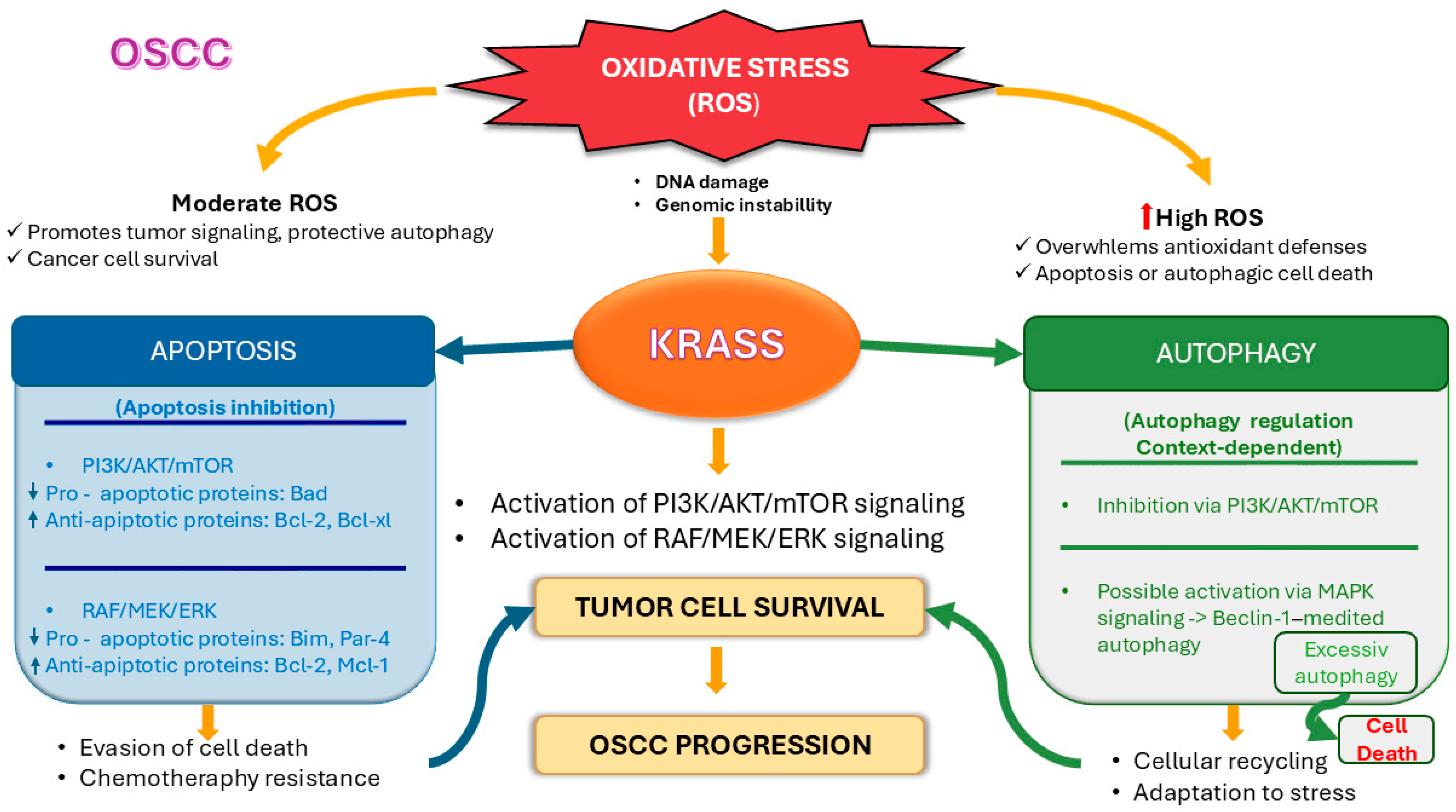
Figure 1: Role of KRAS in OSCC cell survival and progression under oxidative stress. KRAS mediates PI3K/AKT/mTOR and RAF/MEK/ERK signaling pathways, promoting tumor cell survival. Under moderate oxidative stress, tumor cells survive through apoptosis inhibition and autophagy regulation, whereas high oxidative stress can lead to cell death via apoptosis or excessive autophagy. Interactions between KRAS and these signaling pathways contribute to OSCC progression and chemoresistance. ↑ indicates upregulation/activation, and ↓ indicates downregulation/inhibition. Created by the authors in Microsoft PowerPoint (Microsoft 365, Microsoft Corporation, Redmond, WA, USA), based on data from the literature [3,11,26,44,45,46,47,50,51,52,53,54,55,56,57,58,59,60,61,62,66].
In OSCC, the interplay between apoptosis, autophagy, and oxidative stress forms a tightly interconnected regulatory network in which cellular fate is determined by the intensity, duration, and context of stress signals. ROS act as central modulators within this system, exerting concentration-dependent effects: moderate levels of ROS promote tumor cell survival, while excessive or prolonged ROS exposure can trigger cell death. In parallel, ROS-induced dissociation of Beclin-1 from Bcl-2 family proteins provides a mechanistic link between oxidative stress, autophagy initiation, and apoptosis activation. Additionally, beyond the ability of ROS to induce autophagy, autophagy itself protects against ROS. These findings highlight a bidirectional interrelation between ROS and autophagy, whereby ROS induces autophagy, while autophagy modulates ROS levels. In OSCC, ROS influences apoptotic and autophagic processes, which are also regulated by KRAS through the PI3K/AKT/mTOR and RAF/MEK/ERK pathways. KRAS activation promotes apoptosis resistance through PI3K/AKT- and MAPK-dependent upregulation of anti-apoptotic mediators (Bcl-2), while simultaneously influencing autophagy through mTOR-dependent suppression or MAPK-driven induction. Importantly, in OSCC, microenvironmental stress conditions, particularly hypoxia, are associated with the activation of autophagy, which may contribute either to tumor cell survival or, under certain conditions, to cell death. Consequently, in OSCC, cellular outcomes ranging from tumor cell survival and therapy resistance to autophagic or apoptotic cell death are influenced by oxidative stress levels, microenvironmental conditions, and KRAS-driven signaling pathways.
Fig. 2 schematically summarizes the KRAS-driven signaling networks in OSCC and their integrated regulation of autophagy, apoptosis, and ROS homeostasis through the RAF/MEK/ERK and PI3K/AKT/mTOR pathways.
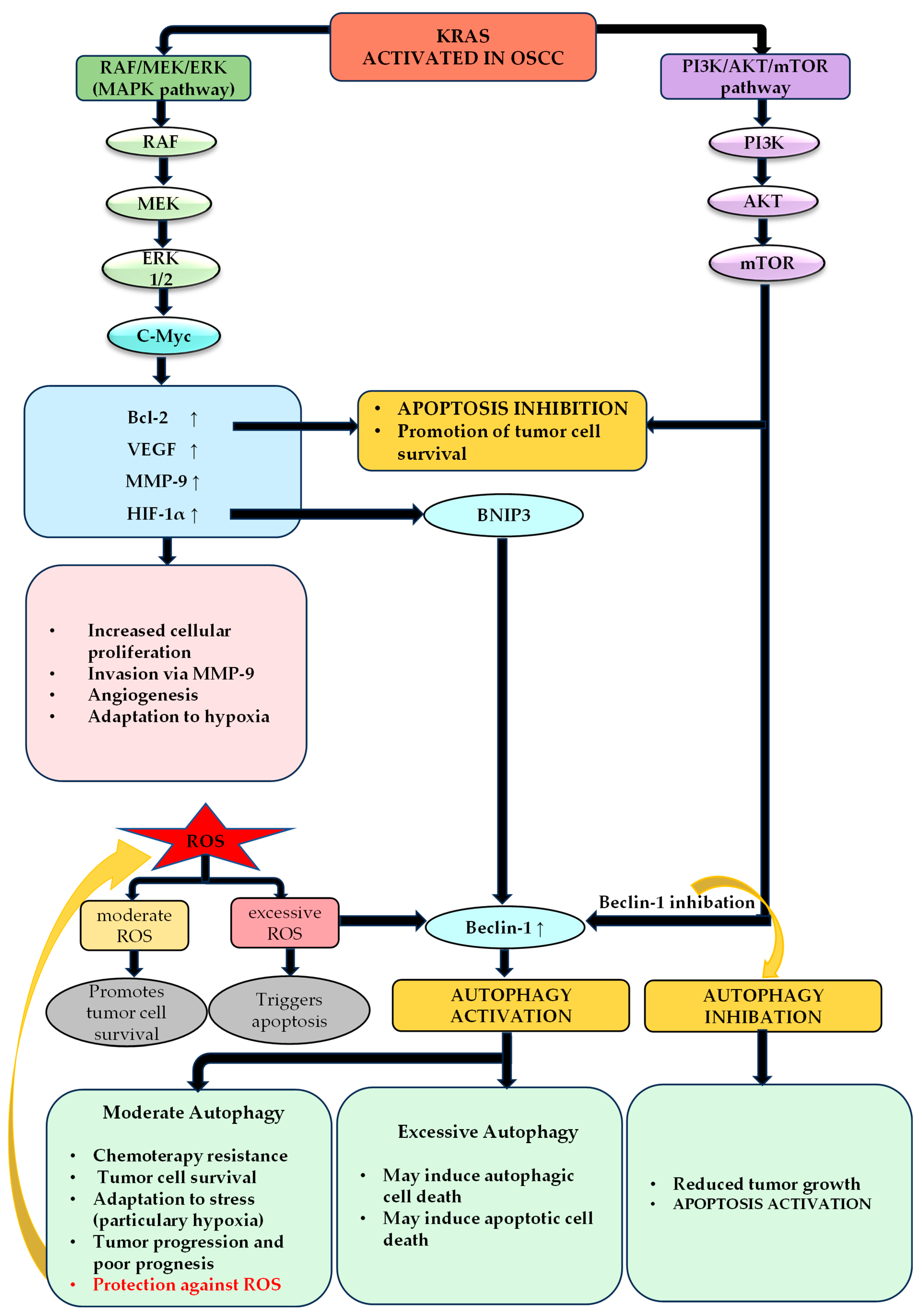
Figure 2: The scheme illustrates the role of KRAS in OSCC and its interrelationship with autophagy and apoptosis, mediated by the main downstream signaling pathways, namely the RAF/MEK/ERK and PI3K/AKT/mTOR pathways. Both pathways contribute to the maintenance of tumor cell survival and the inhibition of apoptosis. Beclin-1 represents a key molecular node that connects these signaling pathways with autophagic mechanisms. Activation of the RAF/MEK/ERK pathway leads to increased Beclin-1 expression and, consequently, stimulation of autophagy, whereas the PI3K/AKT/mTOR pathway inhibits Beclin-1, resulting in suppression of autophagy in OSCC. Autophagy plays an important role in the adaptation of tumor cells to stress, supporting their survival and tumor progression, including through protection against oxidative stress induced by ROS. A bidirectional relationship exists between autophagy and ROS: elevated ROS levels can induce autophagy, while autophagy, in turn, limits excessive ROS accumulation. However, under certain conditions, inhibition of autophagy or, conversely, its excessive activation may lead to cell death, either through mechanisms of autophagic cell death or through the induction of apoptosis. Thus, autophagy exerts a dual role in OSCC, functioning both as a pro-survival mechanism and as a pro-death mechanism when excessively activated, in close interplay with apoptotic pathways. ↑ indicates upregulation/activation. Created by the authors in Microsoft PowerPoint (Microsoft 365, Microsoft Corporation, Redmond, WA, USA), based on data from the literature [3,26,44,46,47,53,54,55,56,57,58,59,60,61,62].
8 Potential Therapeutic Targeting of KRAS and Alternative Pathways
Considering the role of KRAS in regulating downstream signaling pathways, both KRAS and its downstream effectors may represent promising therapeutic targets in OSCC.
8.1 Challenges in Direct KRAS Targeting
Targeting KRAS in OSCC remains challenging due to both biological and clinical factors. Historically, KRAS has been considered “undruggable” because of its high affinity for GTP/GDP and the lack of deep binding pockets suitable for small molecules [68]. Although inhibitors targeting KRAS G12C, such as sotorasib and adagrasib, have been developed, this mutation is exceedingly rare in OSCC, restricting the direct clinical relevance of these drugs [69]. Furthermore, KRAS-driven tumors activate compensatory survival pathways, including PI3K/AKT/mTOR and MAPK, which can reduce the effectiveness of single-agent therapies [70]. Intratumoral heterogeneity and frequent co-occurrence of other genetic alterations further complicate targeted therapy approaches, emphasising the need for combination regimens and detailed molecular profiling.
8.2 Indirect Targeting of KRAS via EGFR
Indirect KRAS inhibition in OSCC can be achieved through EGFR-targeted therapies. EGFR is overexpressed in over 80% of invasive HNSCC, including OSCC, and is associated with tumor progression, metastasis, therapy resistance, and poor prognosis [71]. Therapies targeting EGFR include monoclonal antibodies (mAbs) and tyrosine kinase inhibitors (TKIs). Cetuximab, a chimeric IgG1 antibody, is FDA-approved for use with radiotherapy in advanced HNSCC. While effective in combination regimens, its monotherapy response rates are low (10–13%), and resistance often arises, particularly in tumors with EGFR variants like EGFRvIII [71]. Nimotuzumab, a humanized mAb, has a favorable toxicity profile and shows enhanced efficacy when combined with chemoradiotherapy [71]. TKIs such as gefitinib, erlotinib, and afatinib inhibit the intracellular EGFR kinase domain. Gefitinib and erlotinib show modest activity alone but improved outcomes in combination with chemotherapy or chemoradiotherapy [71]. Afatinib, an irreversible EGFR/HER2 inhibitor, has demonstrated efficacy comparable to cetuximab, including against EGFRvIII variants [71]. Despite these advances, cetuximab remains the only FDA-approved EGFR-targeted therapy for OSCC, highlighting the need for novel agents and combination strategies [3,71].
8.3 Targeting the MAPK Pathway
MEK inhibitors, such as trametinib, have been investigated in OSCC and other HNSCC studies, showing antitumor activity in preclinical and early clinical models [3]. While monotherapy with MAPK inhibitors may face adaptive resistance, combining MAPK targeting with other strategies represents a promising direction for future research in OSCC.
8.4 PI3K/AKT/mTOR Pathway Inhibition
Buparlisib (BKM120), a pan-PI3K inhibitor, has shown preclinical efficacy in OSCC models and is being tested in combination with paclitaxel in the BURAN phase III trial [72]. mTOR inhibitors such as everolimus and temsirolimus show modest effects in early-phase combination trials, while monotherapy often leads to adaptive resistance [73]. KRAS may contribute to resistance against PI3K/AKT/mTOR inhibitors via MAPK crosstalk, supporting combination approaches [74].
Overall, KRAS-targeted therapies face challenges, including resistance mechanisms, highlighting the need for combination strategies and further research to optimise treatment for OSCC. KRAS mutations, despite their rarity, have also been implicated in cetuximab resistance in OSCC cell populations [75].
KRAS plays a central role in OSCC by regulating the interplay between autophagy and apoptosis. Through activation of the RAF/MEK/ERK and PI3K/AKT/mTOR pathways, KRAS promotes cell proliferation, survival, and resistance to apoptosis, contributing to tumor progression and aggressiveness. Elevated levels of ROS influence both autophagy and apoptosis; autophagy can act as a protective mechanism against oxidative stress, but excessive activation may trigger cell death, reflecting the context-dependent role of KRAS in maintaining cellular homeostasis.
Although KRAS mutations are rare in OSCC, KRAS expression and mutation-independent functional activation are associated with advanced tumor stage, invasion, metastasis, and upregulation of anti-apoptotic proteins, suggesting its potential as a prognostic marker. Overall, KRAS involvement in OSCC appears to be driven not only by rare activating mutations but also by signaling activity independent of genetic alterations, including its role as a downstream mediator of other oncogenic pathways. From a therapeutic perspective, KRAS remains a promising target, and indirect therapies targeting EGFR, MAPK, PI3K/AKT/mTOR, or combinations of inhibitors may be effective, especially in resistant or recurrent cases. Understanding the contextual mechanisms by which KRAS modulates apoptosis, autophagy, and oxidative stress responses may guide the development of personalized therapies and provide new insights for the biological and prognostic evaluation of OSCC patients.
Acknowledgement:
Funding Statement: The authors received no specific funding for this study.
Author Contributions: The authors confirm contribution to the paper as follows: Conceptualization, Bianca Voicu Balasea; investigation, Bianca Voicu Balasea, Alexandra Popa; writing—original draft preparation, Bianca Voicu Balasea; writing—review and editing, Alexandra Popa, Florentina Rus, Alexandra Ripszky, Melis Izet, Radu Radulescu; visualization, Melis Izet, Radu Radulescu, Florentina Rus, Alexandra Ripszky; supervision, Alexandra Ripszky. All authors reviewed and approved the final version of the manuscript.
Availability of Data and Materials: Not applicable.
Ethics Approval: Not applicable.
Conflicts of Interest: The authors declare no conflicts of interest.
Abbreviations
The following abbreviations are used in this manuscript:
| AKT | Protein kinase B |
| BAX | Bcl-2-associated X protein |
| Bcl-2 | B-cell lymphoma 2 |
| BNIP3 | BCL2 Interacting Protein 3 |
| ddPCR | Droplet digital polymerase chain reaction |
| EGFR | Epidermal growth factor receptor |
| EGFRvIII | Epidermal growth factor receptor variant III |
| EMT | Epithelial–mesenchymal transition |
| EVs | Extracellular vesicles |
| Fox | Forkhead box transcription factors |
| GAPs | GTPase-activating proteins |
| GF12 | Gingival fibroblasts 12 (cell line) |
| HIF-1α | Hypoxia-inducible factor 1-alpha |
| HPV | Human papillomavirus |
| ICMT | Isoprenylcysteine carboxyl methyltransferase |
| IAPs | Inhibitors of apoptotic proteins |
| JNK | c-Jun N-terminal kinase |
| KRAS | Kirsten rat sarcoma viral oncogene homolog |
| LC3B | Microtubule-associated protein 1 light chain 3 beta |
| LC3B-II | Microtubule-associated protein 1 light chain 3 beta–lipidated form |
| MAPK | Mitogen-activated protein kinase |
| MEK | MAPK/ERK kinase |
| mAbs | Monoclonal antibodies |
| mTOR | Mechanistic target of rapamycin |
| NF-kB | Nuclear factor kappa-light-chain-enhancer of activated B cells |
| NSCLC | Non-small cell lung cancer |
| OEPL | Oral epithelial precursor lesions |
| OSCC | Oral squamous cell carcinoma |
| PAR-4 | Prostate apoptosis response-4 |
| PI3K | Phosphoinositide 3-kinase |
| PIK3CB | Phosphoinositide-3-kinase catalytic subunit beta |
| PCR | Polymerase chain reaction |
| QF-PCR | Quantitative fluorescent polymerase chain reaction |
| RFLP | Restriction fragment length polymorphism |
| ROS | Reactive oxygen species |
| RTK | Receptor tyrosine kinase |
| SFB | Semilicoisoflavone B |
| SSCP | Single-strand conformation polymorphism |
| SNP | Single nucleotide polymorphism |
| TNM | Tumor, Node, Metastasis staging |
| TKI | Tyrosine kinase inhibitor |
| TSCC | Tongue squamous cell carcinoma |
| HNSCC | Head and Neck Squamous Cell Carcinoma |
| G12C | KRAS Glycine 12 to Cysteine mutation |
| G12D | KRAS Glycine 12 to Aspartic acid mutation |
| G12V | KRAS Glycine 12 to Valine mutation |
| G12A | KRAS Glycine 12 to Alanine mutation |
| G13D | KRAS Glycine 13 to Aspartic acid mutation |
| Q61R | KRAS Glutamine 61 to Arginine mutation |
| Q61H | KRAS Glutamine 61 to Histidine mutation |
References
1. Muralidharan S , Nikalje M , Subramaniam T , Koshy JA , Koshy AV , Bangera D . A narrative review on oral squamous cell carcinoma. J Pharm Bioallied Sci. 2025; 17( Suppl 1): S204– 6. doi:10.4103/jpbs.jpbs_593_25. [Google Scholar] [CrossRef]
2. Veerasamy V , Veeran V , Nagini S . Dysregulated PI3K/AKT signaling in oral squamous cell carcinoma: the tumor microenvironment and epigenetic modifiers as key drivers. Oncol Res. 2025; 33( 8): 1835– 60. doi:10.32604/or.2025.064010. [Google Scholar] [CrossRef]
3. Cheng Y , Chen J , Shi Y , Fang X , Tang Z . MAPK signaling pathway in oral squamous cell carcinoma: biological function and targeted therapy. Cancers. 2022; 14( 19): 4625. doi:10.3390/cancers14194625. [Google Scholar] [CrossRef]
4. de Sousa Cabral LG , Martins IM , de Abreu Paulo EP , Pomini KT , Poyet JL , Maria DA . Molecular mechanisms in the carcinogenesis of oral squamous cell carcinoma: a literature review. Biomolecules. 2025; 15( 5): 621. doi:10.3390/biom15050621. [Google Scholar] [CrossRef]
5. Tsai CC , Hsu PC , Kuo CY . Molecular mechanisms in oral squamous cell carcinoma: integrative roles of cancer-associated fibroblasts, immune microenvironment, and precision therapeutic opportunities. Int J Mol Sci. 2026; 27( 7): 2956. doi:10.3390/ijms27072956. [Google Scholar] [CrossRef]
6. Nokovitch L , Maquet C , Crampon F , Taihi I , Roussel LM , Obongo R , et al. Oral cavity squamous cell carcinoma risk factors: state of the art. J Clin Med. 2023; 12( 9): 3264. doi:10.3390/jcm12093264. [Google Scholar] [CrossRef]
7. Ferraguti G , Terracina S , Petrella C , Greco A , Minni A , Lucarelli M , et al. Alcohol and head and neck cancer: updates on the role of oxidative stress, genetic, epigenetics, oral microbiota, antioxidants, and alkylating agents. Antioxidants. 2022; 11( 1): 145. doi:10.3390/antiox11010145. [Google Scholar] [CrossRef]
8. Sendtner N , Seitz R , Brandl N , Müller M , Gülow K . Reactive oxygen species across death pathways: gatekeepers of apoptosis, ferroptosis, pyroptosis, paraptosis, and beyond. Int J Mol Sci. 2025; 26( 20): 10240. doi:10.3390/ijms262010240. [Google Scholar] [CrossRef]
9. Tan Y , Wang Z , Xu M , Li B , Huang Z , Qin S , et al. Oral squamous cell carcinomas: state of the field and emerging directions. Int J Oral Sci. 2023; 15: 44. doi:10.1038/s41368-023-00249-w. [Google Scholar] [CrossRef]
10. Voicu Balasea B , Stan MS , Dinescu M , Imre M , Radulescu R , Cernega A , et al. Differential effects of apigenin on normal and squamous oral epithelial cells reveal redox-autophagy signaling vulnerabilities in OSCC. Int J Mol Sci. 2026; 27( 5): 2091. doi:10.3390/ijms27052091. [Google Scholar] [CrossRef]
11. Ferreira A , Pereira F , Reis C , Oliveira MJ , Sousa MJ , Preto A . Crucial role of oncogenic KRAS mutations in apoptosis and autophagy regulation: therapeutic implications. Cells. 2022; 11( 14): 2183. doi:10.3390/cells11142183. [Google Scholar] [CrossRef]
12. Zhao C . Exploring cell death pathways in oral cancer: mechanisms, therapeutic strategies, and future perspectives. Discov Oncol. 2025; 16( 1): 395. doi:10.1007/s12672-025-02022-3. [Google Scholar] [CrossRef]
13. Tímár J . The clinical relevance of KRAS gene mutation in non-small-cell lung cancer. Curr Opin Oncol. 2014; 26( 2): 138– 44. doi:10.1097/CCO.0000000000000051. [Google Scholar] [CrossRef]
14. Kessler D , Gmachl M , Mantoulidis A , Martin LJ , Zoephel A , Mayer M , et al. Drugging an undruggable pocket on KRAS. Proc Natl Acad Sci U S A. 2019; 116( 32): 15823– 9. doi:10.1073/pnas.1904529116. [Google Scholar] [CrossRef]
15. Hobbs GA , Der CJ , Rossman KL . RAS isoforms and mutations in cancer at a glance. J Cell Sci. 2016; 129( 7): 1287– 92. doi:10.1242/jcs.182873. [Google Scholar] [CrossRef]
16. Ceddia S , Landi L , Cappuzzo F . KRAS-mutant non-small-cell lung cancer: from past efforts to future challenges. Int J Mol Sci. 2022; 23( 16): 9391. doi:10.3390/ijms23169391. [Google Scholar] [CrossRef]
17. Yuan TL , Amzallag A , Bagni R , Yi M , Afghani S , Burgan W , et al. Differential effector engagement by oncogenic KRAS. Cell Rep. 2018; 22( 7): 1889– 902. doi:10.1016/j.celrep.2018.01.051. [Google Scholar] [CrossRef]
18. Roskoski R Jr . ERK1/2 MAP kinases: Structure, function, and regulation. Pharmacol Res. 2012; 66( 2): 105– 43. doi:10.1016/j.phrs.2012.04.005. [Google Scholar] [CrossRef]
19. Khorasani ABS , Delshad M , Sanaei MJ , Pourbagheri-Sigaroodi A , Pirsalehi A , Bashash D . Evaluating oncogenic drivers and therapeutic potential of the PI3K/AKT/mTOR pathway in hepatocellular carcinoma: an overview of clinical trials. Biocell. 2025; 49( 4): 539– 62. doi:10.32604/biocell.2025.059970. [Google Scholar] [CrossRef]
20. Simanshu DK , Nissley DV , McCormick F . RAS proteins and their regulators in human disease. Cell. 2017; 170( 1): 17– 33. doi:10.1016/j.cell.2017.06.009. [Google Scholar] [CrossRef]
21. Cox AD , Fesik SW , Kimmelman AC , Luo J , Der CJ . Drugging the undruggable RAS: mission possible? Nat Rev Drug Discov. 2014; 13( 11): 828– 51. doi:10.1038/nrd4389. [Google Scholar] [CrossRef]
22. Prior IA , Hood FE , Hartley JL . The frequency of RAS mutations in cancer. Cancer Res. 2020; 80( 14): 2969– 74. doi:10.1158/0008-5472.can-19-3682. [Google Scholar] [CrossRef]
23. Huynh MV , Hobbs GA , Schaefer A , Pierobon M , Carey LM , Diehl JN , et al. Functional and biological heterogeneity of KRASQ61 mutations. Sci Signal. 2022; 15( 746): eabn2694. doi:10.1126/scisignal.abn2694. [Google Scholar] [CrossRef]
24. Garrido P , Olmedo ME , Gómez A , Paz Ares L , López-Ríos F , Rosa-Rosa JM , et al. Treating KRAS-mutant NSCLC: latest evidence and clinical consequences. Ther Adv Med Oncol. 2017; 9( 9): 589– 97. doi:10.1177/1758834017719829. [Google Scholar] [CrossRef]
25. Dobre EG , Căruntu A , Munteanu AN , Surcel M , Constantin C , Căruntu C , et al. Investigating EGFR, BRAF, and RAS mutations in oral and cutaneous squamous cell neoplasms: A preliminary report on Romanian patients. Rom J Mil Med. 2024; 127( 5): 341– 50. doi:10.55453/rjmm.2024.127.5.2. [Google Scholar] [CrossRef]
26. Marconi GD , Della Rocca Y , Fonticoli L , Melfi F , Rajan TS , Carradori S , et al. C-Myc expression in oral squamous cell carcinoma: molecular mechanisms in cell survival and cancer progression. Pharmaceuticals. 2022; 15( 7): 890. doi:10.3390/ph15070890. [Google Scholar] [CrossRef]
27. Tan DSW , Wang W , Leong HS , Sew PH , Lau DP , Chong FT , et al. Tongue carcinoma infrequently harbor common actionable genetic alterations. BMC Cancer. 2014; 14: 679. doi:10.1186/1471-2407-14-679. [Google Scholar] [CrossRef]
28. Maemoto S , Yumoto M , Ibata M , Torizuka S , Ozawa N , Tatsumi S , et al. Mutational analysis of HRAS and KRAS genes in oral carcinoma cell lines. Odontology. 2012; 100( 2): 149– 55. doi:10.1007/s10266-011-0032-3. [Google Scholar] [CrossRef]
29. Mustansar T , Mirza T , Hussain M . RAS gene mutations and histomorphometric measurements in oral squamous cell carcinoma. Biotech Histochem. 2023; 98( 6): 382– 90. doi:10.1080/10520295.2023.2196731. [Google Scholar] [CrossRef]
30. Bruckman KC , Schönleben F , Qiu W , Woo VL , Su GH . Mutational analyses of the BRAF KRAS and PIK3CA genes in oral squamous cell carcinoma. Oral Surg Oral Med Oral Pathol Oral Radiol Endod. 2010; 110( 5): 632– 7. doi:10.1016/j.tripleo.2010.05.002. [Google Scholar] [CrossRef]
31. Gadde S , Poda S , Veeravalli S , Addala L . Prevalence of KRAS codon 12 mutation in patients with oral squamous cell carcinoma (OSCC) from South Indian population. Int Res J Nat Appl Sci. 2016; 3( 11): 108– 19. [Google Scholar]
32. Wang WY , Chien YC , Wong YK , Lin YL , Lin JC . Effects of KRAS mutation and polymorphism on the risk and prognosis of oral squamous cell carcinoma. Head Neck. 2012; 34( 5): 663– 6. doi:10.1002/hed.21792. [Google Scholar] [CrossRef]
33. Wang S , Wang W , Zhang S , Yang F , Qiu J , Guo Q , et al. Isoprenylcysteine carboxyl methyltransferase promotes the progression of tongue squamous cell carcinoma via the K-Ras and RhoA signaling pathways. Arch Oral Biol. 2022; 134: 105320. doi:10.1016/j.archoralbio.2021.105320. [Google Scholar] [CrossRef]
34. Shi Y , Li Y , Wu W , Xu B , Ju J . MiR-181d functions as a potential tumor suppressor in oral squamous cell carcinoma by targeting K-ras. Int J Clin Exp Pathol. 2017; 10( 7): 7847– 55. [Google Scholar]
35. Shin KH , Bae SD , Hong HS , Kim RH , Kang MK , Park NH . miR-181a shows tumor suppressive effect against oral squamous cell carcinoma cells by downregulating K-ras. Biochem Biophys Res Commun. 2011; 404( 4): 896– 902. doi:10.1016/j.bbrc.2010.12.055. [Google Scholar] [CrossRef]
36. Tashiro K , Oikawa M , Miki Y , Takahashi T , Kumamoto H . Immunohistochemical assessment of growth factor signaling molecules: MAPK, Akt, and STAT3 pathways in oral epithelial precursor lesions and squamous cell carcinoma. Odontology. 2020; 108( 1): 91– 101. doi:10.1007/s10266-019-00428-4. [Google Scholar] [CrossRef]
37. El Hanbuli HM , Abou Sarie MA . KRAS protein expression in oral squamous cell carcinoma: a potential marker for progression and prognosis. Iran J Pathol. 2022; 17( 4): 469– 79. doi:10.30699/IJP.2022.550727.2856. [Google Scholar] [CrossRef]
38. Al-Rawi N , Merza M , Ghazi A . PIK3CB and K-ras in oral squamous Cell carcinoma. A possible cross-talk! J Orofac Sci. 2014; 6( 2): 99. doi:10.4103/0975-8844.143049. [Google Scholar] [CrossRef]
39. Guo YJ , Pan WW , Liu SB , Shen ZF , Xu Y , Hu LL . ERK/MAPK signalling pathway and tumorigenesis. Exp Ther Med. 2020; 19( 3): 1997– 2007. doi:10.3892/etm.2020.8454. [Google Scholar] [CrossRef]
40. Lu M , Wang C , Chen W , Mao C , Wang J . miR-654-5p targets GRAP to promote proliferation, metastasis, and chemoresistance of oral squamous cell carcinoma through Ras/MAPK signaling. DNA Cell Biol. 2018; 37( 4): 381– 8. doi:10.1089/dna.2017.4095. [Google Scholar] [CrossRef]
41. Hsieh MJ , Ho HY , Lo YS , Lin CC , Chuang YC , Abomughaid MM , et al. Semilicoisoflavone B induces apoptosis of oral cancer cells by inducing ROS production and downregulating MAPK and Ras/Raf/MEK signaling. Int J Mol Sci. 2023; 24( 5): 4505. doi:10.3390/ijms24054505. [Google Scholar] [CrossRef]
42. Lin CW , Chin HK , Lee SL , Chiu CF , Chung JG , Lin ZY , et al. Ursolic acid induces apoptosis and autophagy in oral cancer cells. Environ Toxicol. 2019; 34( 9): 983– 91. doi:10.1002/tox.22769. [Google Scholar] [CrossRef]
43. Dong QZ , Wang Y , Tang ZP , Fu L , Li QC , Wang ED , et al. Derlin-1 is overexpressed in non-small cell lung cancer and promotes cancer cell invasion via EGFR-ERK-mediated up-regulation of MMP-2 and MMP-9. Am J Pathol. 2013; 182( 3): 954– 64. doi:10.1016/j.ajpath.2012.11.019. [Google Scholar] [CrossRef]
44. Wu H , Li L , Ai Z , Yin J , Chen L . Pristimerin induces apoptosis of oral squamous cell carcinoma cells via G(1) phase arrest and MAPK/Erk1/2 and Akt signaling inhibition. Oncol Lett. 2019; 17( 3): 3017– 25. doi:10.3892/ol.2019.9903. [Google Scholar] [CrossRef]
45. Nalca A , Qiu SG , El-Guendy N , Krishnan S , Rangnekar VM . Oncogenic ras sensitizes cells to apoptosis by Par-4. J Biol Chem. 1999; 274( 42): 29976– 83. doi:10.1074/jbc.274.42.29976. [Google Scholar] [CrossRef]
46. He S , Chakraborty R , Ranganathan S . Proliferation and apoptosis pathways and factors in oral squamous cell carcinoma. Int J Mol Sci. 2022; 23( 3): 1562. doi:10.3390/ijms23031562. [Google Scholar] [CrossRef]
47. Wu Y , Wang Y , Liu H , Hu Q , Xie Y , Nan X , et al. Mechanism of apoptosis in oral squamous cell carcinoma promoted by cardamonin through PI3K/AKT signaling pathway. Sci Rep. 2024; 14: 20802. doi:10.1038/s41598-024-71817-1. [Google Scholar] [CrossRef]
48. Kashyap T , Pramanik KK , Nath N , Mishra P , Singh AK , Nagini S , et al. Crosstalk between Raf-MEK-ERK and PI3K-Akt-GSK3β signaling networks promotes chemoresistance, invasion/migration and stemness via expression of CD44 variants (v4 and v6) in oral cancer. Oral Oncol. 2018; 86: 234– 43. doi:10.1016/j.oraloncology.2018.09.028. [Google Scholar] [CrossRef]
49. Pang X , Zhang X , Jiang Y , Su Q , Li Q , Li Z . Autophagy: mechanisms and therapeutic potential of flavonoids in cancer. Biomolecules. 2021; 11( 2): 135. doi:10.3390/biom11020135. [Google Scholar] [CrossRef]
50. Dong L , He J , Luo L , Wang K . Targeting the interplay of autophagy and ROS for cancer therapy: an updated overview on phytochemicals. Pharmaceuticals. 2023; 16( 1): 92. doi:10.3390/ph16010092. [Google Scholar] [CrossRef]
51. Mariño G , Niso-Santano M , Baehrecke EH , Kroemer G . Self-consumption: the interplay of autophagy and apoptosis. Nat Rev Mol Cell Biol. 2014; 15( 2): 81– 94. doi:10.1038/nrm3735. [Google Scholar] [CrossRef]
52. Decuypere JP , Parys JB , Bultynck G . Regulation of the autophagic bcl-2/beclin 1 interaction. Cells. 2012; 1( 3): 284– 312. doi:10.3390/cells1030284. [Google Scholar] [CrossRef]
53. Pangarkar M , Wagh U , Pathak A . Autophagy indicators in oral squamous cell carcinoma. Pathology. 2024; 56( 1): 59– 64. doi:10.1016/j.pathol.2023.08.014. [Google Scholar] [CrossRef]
54. Terabe T , Uchida F , Nagai H , Omori S , Ishibashi-Kanno N , Hasegawa S , et al. Expression of autophagy-related markers at the surgical margin of oral squamous cell carcinoma correlates with poor prognosis and tumor recurrence. Hum Pathol. 2018; 73: 156– 63. doi:10.1016/j.humpath.2017.11.019. [Google Scholar] [CrossRef]
55. Shi F , Luo D , Zhou X , Sun Q , Shen P , Wang S . Combined effects of hyperthermia and chemotherapy on the regulate autophagy of oral squamous cell carcinoma cells under a hypoxic microenvironment. Cell Death Discov. 2021; 7: 227. doi:10.1038/s41420-021-00538-5. [Google Scholar] [CrossRef]
56. Su KY , Khoo XH , Paterson IC , Goh BH , Lee WL . Autophagy and extracellular vesicles mediate cisplatin resistance in oral squamous cell carcinoma with LC3B-II as a potential non-invasive biomarker. Sci Rep. 2025; 15: 15945. doi:10.1038/s41598-025-00703-1. [Google Scholar] [CrossRef]
57. Zhao W , Chen C , Zhou J , Chen X , Cai K , Shen M , et al. Inhibition of autophagy promotes the anti-tumor effect of metformin in oral squamous cell carcinoma. Cancers. 2022; 14( 17): 4185. doi:10.3390/cancers14174185. [Google Scholar] [CrossRef]
58. Qiu B , Sun Y , Nie W , Yang Q , Guo X . FBXW7 promotes autophagy and inhibits proliferation of oral squamous cell carcinoma. Immun Inflamm Dis. 2023; 11( 5): e845. doi:10.1002/iid3.845. [Google Scholar] [CrossRef]
59. Vo TTT , Wee Y , Cheng HC , Wu CZ , Chen YL , Tuan VP , et al. Surfactin induces autophagy, apoptosis, and cell cycle arrest in human oral squamous cell carcinoma. Oral Dis. 2023; 29( 2): 528– 41. doi:10.1111/odi.13950. [Google Scholar] [CrossRef]
60. Borges AA , de Souza MP , da Fonseca ACC , Wermelinger GF , Ribeiro RCB , Amaral AAP , et al. Chemoselective synthesis of Mannich adducts from 1,4-naphthoquinones and profile as autophagic inducers in oral squamous cell carcinoma. Molecules. 2022; 28( 1): 309. doi:10.3390/molecules28010309. [Google Scholar] [CrossRef]
61. Rajendran P , Sekar R , Dhayasankar PS , Ali EM , Abdelsalam SA , Balaraman S , et al. PI3K/AKT signaling pathway mediated autophagy in oral carcinoma—a comprehensive review. Int J Med Sci. 2024; 21( 6): 1165– 75. doi:10.7150/ijms.94566. [Google Scholar] [CrossRef]
62. Abd El-Aziz YS , Leck LYW , Jansson PJ , Sahni S . Emerging role of autophagy in the development and progression of oral squamous cell carcinoma. Cancers. 2021; 13( 24): 6152. doi:10.3390/cancers13246152. [Google Scholar] [CrossRef]
63. Wang X , Li S , Lin S , Han Y , Zhan T , Huang Z , et al. Oncogenic RAS induces a distinctive form of non-canonical autophagy mediated by the P38-ULK1-PI4KB axis. Cell Res. 2025; 35( 6): 399– 422. doi:10.1038/s41422-025-01085-9. [Google Scholar] [CrossRef]
64. Elgendy M , Sheridan C , Brumatti G , Martin SJ . Oncogenic Ras-induced expression of Noxa and Beclin-1 promotes autophagic cell death and limits clonogenic survival. Mol Cell. 2011; 42( 1): 23– 35. doi:10.1016/j.molcel.2011.02.009. [Google Scholar] [CrossRef]
65. Swiderek E , Kalas W , Wysokinska E , Pawlak A , Rak J , Strzadala L . The interplay between epigenetic silencing, oncogenic KRas and HIF-1 regulatory pathways in control of BNIP3 expression in human colorectal cancer cells. Biochem Biophys Res Commun. 2013; 441( 4): 707– 12. doi:10.1016/j.bbrc.2013.10.098. [Google Scholar] [CrossRef]
66. Bani-Ahmad E , Dass J , Dass CR . From carcinogenesis to drug resistance: the multifaceted role of oxidative stress in head and neck cancer. Cancers. 2025; 17( 20): 3295. doi:10.3390/cancers17203295. [Google Scholar] [CrossRef]
67. Peng YC , He ZJ , Yin LC , Pi HF , Jiang Y , Li KY , et al. Sanguinarine suppresses oral squamous cell carcinoma progression by targeting the PKM2/TFEB Aix to inhibit autophagic flux. Phytomedicine. 2025; 136: 156337. doi:10.1016/j.phymed.2024.156337. [Google Scholar] [CrossRef]
68. Moore AR , Rosenberg SC , McCormick F , Malek S . RAS-targeted therapies: is the undruggable drugged? Nat Rev Drug Discov. 2020; 19( 8): 533– 52. doi:10.1038/s41573-020-0068-6. [Google Scholar] [CrossRef]
69. Murugan AK , Munirajan AK , Tsuchida N . Ras oncogenes in oral cancer: the past 20 years. Oral Oncol. 2012; 48( 5): 383– 92. doi:10.1016/j.oraloncology.2011.12.006. [Google Scholar] [CrossRef]
70. Fedele C , Li S , Teng KW , Foster CJR , Peng D , Ran H , et al. SHP2 inhibition diminishes KRASG12C cycling and promotes tumor microenvironment remodeling. J Exp Med. 2021; 218: e20201414. doi:10.1084/jem.20201414. [Google Scholar] [CrossRef]
71. Liu L , Chen J , Cai X , Yao Z , Huang J . Progress in targeted therapeutic drugs for oral squamous cell carcinoma. Surg Oncol. 2019; 31: 90– 7. doi:10.1016/j.suronc.2019.09.001. [Google Scholar] [CrossRef]
72. Xing J , Yang J , Gu Y , Yi J . Research update on the anticancer effects of buparlisib (Review). Oncol Lett. 2021; 21( 4): 266. doi:10.3892/ol.2021.12527. [Google Scholar] [CrossRef]
73. Harsha C , Banik K , Ang HL , Girisa S , Vikkurthi R , Parama D , et al. Targeting AKT/mTOR in oral cancer: mechanisms and advances in clinical trials. Int J Mol Sci. 2020; 21( 9): 3285. doi:10.3390/ijms21093285. [Google Scholar] [CrossRef]
74. Ma Q , Zhang W , Wu K , Shi L . The roles of KRAS in cancer metabolism, tumor microenvironment and clinical therapy. Mol Cancer. 2025; 24( 1): 14. doi:10.1186/s12943-024-02218-1. [Google Scholar] [CrossRef]
75. Lyanova AA , Vladimirova LY , Engibaryan MA , Abramova NA , Popova IL , Ryadinskaya LA , et al. The KRAS mutation status and resistance to cetuximab in patients with squamous cell carcinoma of oral cavity. J Clin Oncol. 2020; 38( 15_suppl): e15514. doi:10.1200/JCO.2020.38.15_suppl.e15514. [Google Scholar] [CrossRef]
Cite This Article
 Copyright © 2026 The Author(s). Published by Tech Science Press.
This work is licensed under a Creative Commons Attribution 4.0 International License , which permits unrestricted use, distribution, and reproduction in any medium, provided the original work is properly cited.
 Downloads
 Citation Tools
