
 Login
Login Register
 Submit a Paper
 Propose a Special lssue
 Open Access
REVIEW

Improving Cancer Therapy: The Strong Synergy of Ginsenosides and Chemotherapy
1 Cancer Biology Laboratory, Department of Biosciences and Bioengineering, Indian Institute of Technology Guwahati (IITG), Guwahati, Assam, India
2 Department of Science in Korean Medicine, Kyung Hee University, 24 Kyungheedae-ro, Dongdaemun-gu, Seoul, Republic of Korea
3 Radiological Sciences Department, College of Applied Medical Sciences, King Khalid University, Abha, Saudi Arabia
4 BioImaging Unit, Space Research Centre, Michael Atiyah Building, University of Leicester, Leicester, UK
5 Electrical Engineering Department, College of Engineering, King Khalid University, Abha, Saudi Arabia
6 Division of Radiology, Department of Medicine, College of Medicine and Surgery, King Khalid University, Abha, Saudi Arabia
7 Department of Pharmacology, College of Osteopathic Medicine, Lake Erie College of Osteopathic Medicine, Bradenton, FL, USA
* Corresponding Authors: Kwang Seok Ahn. Email: ; Ajaikumar B. Kunnumakkara. Email:
# These authors contributed equally to this work
(This article belongs to the Special Issue: Natural Product-Based Anticancer Drug Discovery)
BIOCELL 2026, 50(8), 4 https://doi.org/10.32604/biocell.2026.073397
Received 17 September 2025; Accepted 09 March 2026; Issue published 27 July 2026
 View Full Text
 Download PDF
Abstract
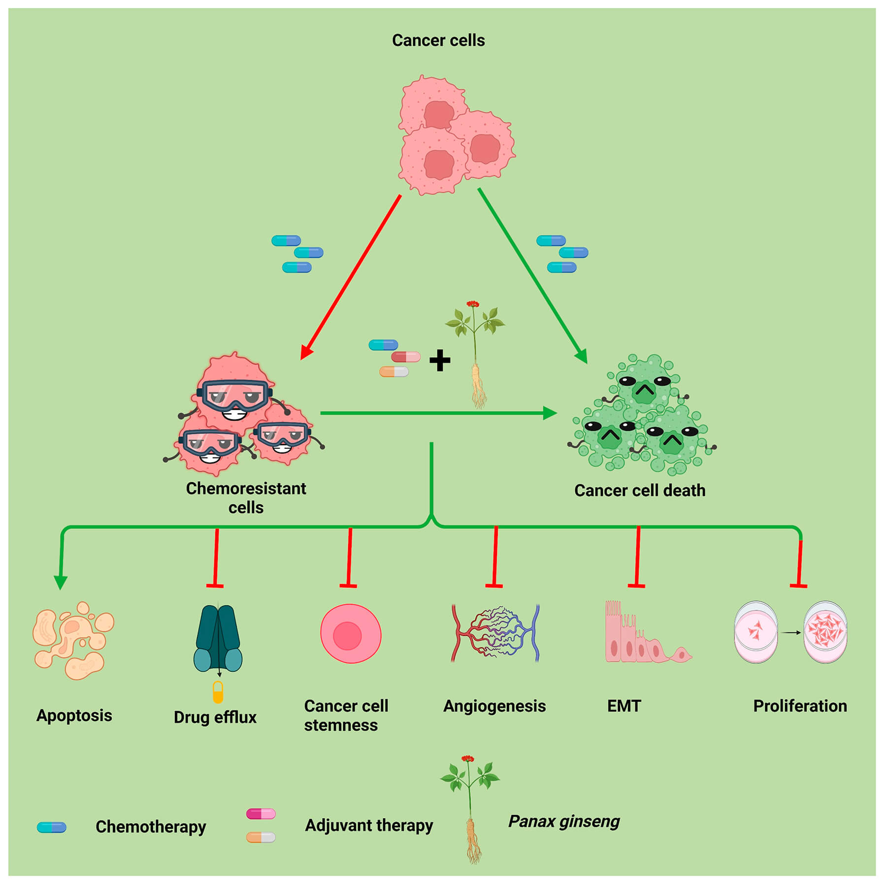
Despite the advancements achieved in chemotherapy, cancer continues to remain a formidable and lethal global threat, ranking as the second leading cause of death worldwide. The development of chemoresistance poses a significant hurdle in cancer treatment. Nonetheless, a therapeutic strategy known as chemosensitization has emerged to counteract cancer cell resistance, wherein the efficacy of one drug is augmented by another. Accumulating evidence suggests that natural products have attracted considerable attention in the cancer therapeutic realm due to their ability to combat multidrug resistance with minimal side effects. Ginsenosides, triterpene saponins extracted from Panax ginseng, have demonstrated significant anticancer activity while exhibiting relatively low toxicity and reduced adverse effects. Co-administration of ginsenosides with chemotherapeutic drugs has been shown to trigger apoptosis, as evidenced by an increased Bcl-2-associated X protein (Bax)/B-cell lymphoma 2 (Bcl-2) ratio, inhibit angiogenesis through suppression of vascular endothelial growth factor (VEGF); and hinder replicative immortality by downregulating stemness-associated markers such as octamer-binding transcription factor 4 (Oct4), Nanog, and sex determining region Y-box 2 (SOX2) in various cancers. Additionally, ginsenosides modulate key chemoresistance pathways, including nuclear factor-kappa B (NF-κB), signal transducers and activators of transcription (STAT), and phosphatidylinositol 3-kinase (PI3K)/protein kinase B (Akt), as well as their downstream targets, thereby rendering cancer cells more susceptible to chemotherapy. Notably, ginsenosides have been shown to modulate the tumor microenvironment and mitigate the side effects associated with chemotherapeutic drugs. This review aims to consolidate findings from preclinical and clinical studies to elucidate the role of ginsenosides as effective chemosensitizing agents.Graphic Abstract
Keywords
Cancer, a prevalent and lethal disease in the modern era, has witnessed substantial advancements in treatment over the past 15 years due to technological progress and extensive insights into molecular and tumor biology [1,2]. Chemotherapy, utilizing various molecules to combat cancer cell proliferation, angiogenesis, and replicative immortality, has emerged as a globally effective treatment modality [3,4]. The FDA approval of multiple anticancer drugs in the current decade highlights significant progress in oncology, marking a shift toward broader therapeutic options after decades of limited advancements [5]. However, the dependence on high drug doses, arising from therapeutic strategies that target one or a few pathways or proteins to kill cancer cells, can potentially worsen the disease state [6]. Additionally, the heterogeneous nature of the cancer cell population gives rise to intrinsic resistance, present from the outset, and to acquired resistance, which develops after multiple treatment cycles [7,8]. In addition, cancer cells become resistant to a single drug or drugs with a similar mechanism by altering drug targets and activating mechanisms that repair drug-induced DNA damage [9]. Moreover, some cancer cell populations exhibit cross-resistance to drugs with distinct mechanisms of action, a phenomenon known as multidrug resistance (MDR) [8,9,10,11]. To address these challenges, researchers are actively seeking compounds with reduced toxicity to non-targeted tissues and exploring combination therapies to overcome drug resistance mechanisms [12,13]. It is well established that phytochemicals, including nutraceuticals, are an alternative for developing novel therapeutic drugs with safety and efficacy [14,15,16].
Nutraceuticals are naturally occurring non-toxic dietary compounds that can offer physiological benefits against chronic diseases [17,18,19]. Carotenoids, flavonoids, and sulfur-containing compounds are among the nutraceuticals known for their significant anticancer properties [20,21]. Additionally, they have been used in combination with conventional therapies which induces apoptosis in cancer cells [13,20,22,23]. Among these, ginsenosides, a group of pharmacologically active triterpenoid saponins derived from Panax ginseng C. A. Meyer, a perennial herb of the Araliaceae family, have been used in traditional oriental medicine for thousands of years [24,25]. It is noteworthy that ginsenosides comprise a diverse group of more than 150 compounds, each exhibiting distinct structural and pharmacological characteristics [26,27]. The genus name, “Panax”, emerged from the Greek word “Pan-acea” meaning “cure for all” [28,29,30]. Most species in the genus Panax, including P. ginseng C. A. Meyer (Asian ginseng or Korean red ginseng), and P. notoginseng (Burk.) F. H. Chen (notoginseng) has been used to treat various chronic diseases in Traditional Chinese Medicine, and P. quinquefolius L. (American ginseng) is used by North Americans as a tonic for stamina and respiratory health [31,32,33]. P. notoginseng was first scientifically described in a Chinese herb encyclopedia, The Compendium of Materia Medica (Bencao Gangmu), written by Li Shi Zhen (1518–1593 AD), and is considered the emperor herb for treating different types of wounds [34,35]. P. ginseng is one of the most studied and commonly used ginsengs and is known to boost psychological and immune function [36]. Moreover, this herb has also been used as a natural remedy in traditional Korean and Chinese medicine for the prevention and treatment of various chronic diseases, including cancer, cardiovascular diseases, diabetes, and obesity [37,38,39,40].
A plethora of studies confirmed the anticancer properties of ginsenosides across multiple malignancies [41,42,43]. However, its role as a chemosensitizing agent is largely underexplored. This gap in the literature is particularly significant given the persistent challenge of chemoresistance, which undermines the efficacy of conventional chemotherapy in many cancer types.
This review seeks to address this gap by comprehensively examining the chemosensitizing potential of ginsenosides when combined with standard chemotherapeutic agents across diverse cancer subtypes. We have summarized the pharmacological properties of ginsenosides as reported in clinical studies and provided an overview of the fundamental molecular mechanisms and downstream pathways underlying their chemosensitizing effects across multiple cancer types. Furthermore, this review highlights current challenges hindering the clinical translation of ginsenosides and discusses future perspectives to guide the development of more effective therapeutic strategies.
2 Bioavailability of Ginsenosides
A growing body of evidence indicates the presence of diverse ginsenosides derived from Panax species. More than 150 ginsenosides have been isolated from the roots, fruits, flower heads, leaves, and stems of the ginseng plant [26]. Besides ginsenosides, other constituents of ginseng include carbohydrates, polysaccharides, peptides, polyacetylenic alcohols, phenolic compounds, etc. However, most of the pharmacological benefits of ginseng are attributed to ginsenosides [25,44,45]. Over 20 ginsenosides possess various biological properties, including analgesic, anti-inflammatory, anti-carcinogenic, anti-diabetic, anti-fatigue, immunomodulatory, and neuroprotective effects [46,47,48]. The structure of various ginsenosides was first identified in 1965 by a Japanese natural medicine chemist, Shibata [49]. Based on their structure, ginsenosides are categorized into dammarane-type and oleanane-type tricyclic triterpenoids. Dammarane type is further divided into protopanaxadiol type (PPD) such as ginsenoside Rb1, Rb2, Rb3, Rc, Rd, Rg3, Rh2, Rs1, and Rk1 (contains a hydrogen atom at C6) and protopanaxatriol type (PPT) such as ginsenoside Re, Rf, Rg1, Rg2, and Rh1, (contains a C6 sugar chain) [46,49,50]. Moreover, the C20 of PPD and PPT can be either 20(S) or 20(R)-type structures, based on the location of the chiral carbon substitution. Notably, it has been found that the anticancer activities of ginsenosides depend on the location of sugar linkages, in the order: C3 > C6 > C20 [50,51]. The minor types of ginsenosides include oleanolic acid type, such as ginsenoside Ro, and ocotillol type, such as pseudo ginsenoside [50]. Rb1, Rb2, Rc, Re, and Rg1 are the major ginsenosides, comprising 70–80% of total ginsenosides, in a fresh ginseng [52]. The structures of various ginsenosides are summarized in Fig. 1.

Figure 1: Various ginsenoside compounds derived from the Panax ginseng plant. It includes Rb1, Rb2, Rb3, Rf, Rc, Rd, Rk1, Rs1, Rg1, Rg2, Rg3, Rh1 and Rh2. Based on their structure, ginsenosides are categorized into dammarane-type and oleanane-type tricyclic triterpenoids. The dammarane-type ginsenosides are further divided into two subtypes: protopanaxadiol (PPD) and protopanaxatriol (PPT). Protopanaxadiol-type ginsenosides, which include Rb1, Rb2, Rb3, Rc, Rd, Rg3, Rh2, Rs1, and Rk1, contain a hydrogen atom at the C6 position. Protopanaxatriol-type ginsenosides, such as Rf, Rg1, Rg2, and Rh1, are characterized by a sugar chain at the C6 position. Created using ChemDraw.
Several clinical trials have explored the pharmacokinetics of ginsenosides due to their biological activities (Table 1) [53]. For instance, an open-label study was conducted in 12 healthy volunteers to investigate the efficacy of intramuscular injection of XueShuanTong, a lyophilized extract of P. notoginseng roots, extensively used in the treatment of ischemic heart and cerebrovascular diseases [54]. It mainly consisted of ginsenosides Rb1 and Rd of 20(S)-protopanaxadiol-type, Rg1, Re, and notoginsenoside R1 of 20(S)-protopanaxatriol-type. Intramuscular injection of XueShuanTong increased the bioavailability of ginsenosides by around 100 to 112% compared to the intravenous method. Moreover, it also demonstrated that the 20(S)-protopanaxadiol-type has a longer half-life than the 20(S)-protopanaxatriol-type [54]. A similar study investigated the pharmacokinetic profile of 20s ginsenoside Rg3 in healthy volunteers. The study was conducted in 2 stages: first, as a single dose of 10–60 mg in 24 healthy volunteers, and second, as multiple doses of 30 g in 9 healthy volunteers [55]. For a single dose, the maximum plasma concentration (Cmax) was 135.4 ± 35.3, 162.1 ± 47.2, and 399.8 ± 217.0 ng/mL for 10, 30, and 60 mg 20(S)-ginsenoside Rg3, respectively [55]. For multiple-dose administration, Cmax was 457.0 ± 165.7 and 770.2 ± 275.4 ng/mL after the first and the last doses of 20(S)-ginsenoside Rg3, respectively [55]. In another study, six healthy volunteers received 10 g of American ginseng root powder, and the amounts of Rb1, Rd, Rg2, and compound K (CK) in their blood were detected using the ultra-performance liquid chromatography/time-of-flight mass spectrometry (UPLC/TOF-MS) method [56]. The result showed that 10 g was well tolerated, the Tmax of Rb1 was 4 h, and the CK level gradually increased from the 7th hour. Moreover, it has also been found that enteric microbiota plays a crucial role in transferring Rb1 to CK [56]. Similarly, American ginseng powder (2 g) was orally administered to six healthy volunteers following either an Asian or a Western diet [57]. In Asian dietary patterns, there was an observed elevation in Rb1 levels, whereas subjects adhering to a Western diet exhibited increased CK levels. This finding provides further confirmation of the direct relationship between ginsenoside biotransformation and the enteric microbiota, which is influenced by diet [57].
Table 1: The bioavailability and safety of ginsenosides in clinical trials conducted in healthy subjects.
| Intervention | Type of Ginsenosides | Dose | No. of Subjects | Effects | References |
|---|---|---|---|---|---|
| XueShuanTong | Rb1, Rg1, Rd, Re, notoginsenosides R1, | 150 mg | 12 | ↑Bioavailability | [54] |
| - | 20(S)-Rg3 | 10–60 mg | 24 | A pharmacokinetic profile suitable for once every 2 days dosing. | [55] |
| - | 20(S)-Rg3 | 30 mg | 9 | A pharmacokinetic profile suitable for once every 2 days dosing | [55] |
| Fermented CK-30 | CK | 600 mg | 22 | ↑Systemic exposure, Bioavailability | [58] |
| - | Rd | 10, 40, 75 mg | 24 | No adverse effect, slowly cleared from plasma | [59] |
| Ginsenoside H dripping pill | Rh2 | 7.8, 31.2 mg | 12 | No adverse effect | [60] |
| Ginsenoside H dripping pill | Rh2 | 15.6 mg | 12 | No obvious accumulation of drug, supported twice a day dosing | [60] |
| TJ-100 | Rg1, Rb1 | 2.5, 5, 10 g | 16 | Observed plateau, kinetics were not proportional to dosage | [61] |
| Daikanchuto | Rb1 | 15 g | 4 | Ginsenosides gradually increased during the sampling period | [62] |
| Fermented ginseng | IH-901 (CK) | 3 g | 24 | No adverse effect other than mild diarrhea | [63] |
| American ginseng root powder | Rb1, Rd, Rg-2, CK | 10 g | 6 | Well tolerated, enteric bacteria converted Rb1 to CK | [56] |
| American ginseng powder | Rb1, CK | 2 g | 6 | ↑CK in the western diet and Rb1 in the Asian diet | [57] |
A randomized, open-label, two-treatment, 2 × 2 crossover study was conducted in 24 healthy volunteers, demonstrating the increased uptake of fermented ginsenoside CK-30 as compared to fresh CK-30 from red ginseng extract [58]. The volunteers were separated into two groups and given an oral dose of either 600 mg of CK-30 or 2.94 g of red ginseng extract. The result showed that the median time to reach Cmax of ginsenoside CK after administration of CK-30 was 3 h; however, for red ginseng extract, it was 10 h [58]. Another open-label, randomized, single-dose, fasting, two-period, crossover, pharmacokinetic study was conducted in 24 Korean healthy male volunteers, demonstrating a similar result [63]. In this study, AUC(t) was 2083.09 ± 91.97 ngh/mL, showing a 15.5-fold increase over that of CK from the non-fermented group (134.50 ± 63.10 ngh/mL), and the mean Cmax was 325.00 ± 91.97 ng/mL in the fermented ginseng group, a 27-fold higher value than that in the non-fermented group (13.88 ± 7.24 ng/mL) [63].
Moreover, in another randomized, open-label, three-way crossover study, 24 healthy volunteers received intravenous infusions of 10, 40, or 75 mg of Rd [59]. The Cmax and AUC0-∞ ranging from 2.8 to 19.3 mg/L and 27.9 to 212.5 mgh/L and for multidose study Cmax, AUC0–∞, and AUCss were 4.0 mg/L, 51.7 mgh/L, and 26.4 mgh/L, respectively. The compound is slowly cleared from plasma, which supports the safety and pharmacokinetics of ginsenoside as a therapeutic regimen [59]. Similarly, another study reported the safety of ginsenoside H dropping pills (GH), a novel clinical-stage adjuvant for the treatment of non-small cell lung cancer (NSCLC), which contains Rh2 as its main ingredient [60]. A total of 24 healthy volunteers were grouped into two and administered with GH pills as a single dose and multiple doses of 7.8 mg, followed by 31.2 mg, and 15.6 mg, respectively [60]. The result showed that the half-life ranged from 9 to 11 h for a single dose and 14.5 h for multiple doses without adverse effects [60].
In another study, Daikenchuto (TJ-100), a pharmaceutical-grade traditional Japanese medicine, was orally administered to Japanese healthy volunteers at doses of 2.5, 5, and 10 g [61]. The six major ingredients of TJ-100 are hydroxy-α-sanshool (HAS), hydroxy-β-sanshool (HBS), [6]-shogaol (6S), [10]-shogaol (10S), ginsenoside Rb1, and ginsenoside Rg1. The plasma concentration of Rb1 reached the maximum concentration at approximately 4 h after administration, with an extremely low plasma concentration (<0.023 ng/mL) [61]. However, in another study, when Daikenchuto was administered orally to 4 healthy volunteers at a dose of 15 g, an increase in the peak level of Rb1 was observed during the sampling period [62].
In summary, ginsenosides have demonstrated a favorable safety profile and are generally well-tolerated in clinical studies, supporting their potential therapeutic use. However, their pharmacokinetics present certain limitations. The diverse array of chemical forms and intricate structures of ginsenosides pose challenges to studying their pharmacokinetics [53]. Additionally, the biotransformation of ginsenosides is highly dependent on the intestinal microbiota, which convert the parent compounds into more pharmacologically active metabolites such as CK. This microbial metabolism plays a crucial role in mediating their bioactivity and systemic absorption. Further, the oral availability of ginsenosides is reported to be very low, which hinders their development as potent therapeutic agents [53]. Furthermore, the stability and composition of ginsenosides are influenced by post-harvest processing methods. Koh et al. reported that thermal processing leads to a marked reduction in total ginsenoside content, accompanied by a transformation in composition, notably, the conversion of ginsenoside Rc to Rg3 during air-drying [64]. Such alterations may affect both the pharmacological profile and consistency of ginseng-based formulations, highlighting the need for optimized processing techniques to preserve or enhance therapeutic efficacy.
3 Anticancer Mechanisms of Ginsenosides
Numerous studies have revealed that ginsenosides such as Rg3, Rg5, Rk1, and Rh2 are novel and potent anticancer agents by instigating apoptosis and retarding cancer cell proliferation and metastasis [65,66,67]. For instance, Rh2, the main component of red ginseng extracts, exerts its anticancer effects by inhibiting cell proliferation by downregulating IL-6-induced STAT-3 activation and expression of matrix metalloproteinases (MMPs) [68]. Similarly, when NSCLC cells are co-cultured with RAW264.7 or THP-1 cells, the proliferation and migration of cancer cells are promoted through the upregulation of vascular endothelial growth factor (VEGF), MMPs, and M2 macrophage activation [69]. However, treatment with ginsenosides Rh2 reversed these effects by shifting M2 macrophages towards an M1 phenotype and reducing VEGF and MMPs levels [69].
A plethora of studies have demonstrated the anticancer properties of ginsenosides Rb2, Rg3, and Rg5 [70,71,72,73]. For instance, an in vitro study investigated the role of Rg3 in lung cancer and found that it can inhibit the migration, invasion, and angiogenesis of lung cancer cells by suppressing VEGF and cyclooxygenase-2 (COX-2) [70]. However, overexpression of COX-2 reversed the protective effect of ginsenosides [70]. Song et al. demonstrated that Rg3 inhibited tumor growth by inhibiting myeloid-derived suppressor cells (MDSCs) in mouse mammary carcinoma [74]. In the same study, Rg3 downregulated stemness and EMT by inhibiting tumor-derived cytokines, STAT-3, and the NOTCH signaling pathway [74]. Notably, a combination study of Rg3 and Rg5 demonstrated potent anticancer activity in lung cancer by inhibiting the phosphatidylinositol 3-kinase (PI3K)/protein kinase B (Akt)/mechanistic target of rapamycin (mTOR) and epidermal growth factor receptor (EGFR)/VEGF signaling pathways [71]. This resulted in a pronounced enhancement of apoptosis and a reduction of metastasis compared to the effects of each compound individually [71]. Another in vitro study highlighted the protective effect of Rg5 and Rk1 in MHCC-97H liver cancer cells [72]. Treatment with these ginsenosides activated endogenous apoptotic pathways, leading to cancer cell death [72]. Jiang et al. reported the tumor-suppressive effects of CK in human prostate cancer cells by showing that it binds to the androgen receptor promoter region and inhibits its signaling [75]. In addition, CK suppressed epithelial-mesenchymal transition (EMT) markers such as vimentin and MMP9, while simultaneously promoting E-cadherin activation. These findings were further supported by consistent results observed in in vivo studies [75]. Further, PPD, another derivative of ginsenoside, suppressed cell viability, migration, colony formation, and invasion in gastric cancer cells [76]. This study also demonstrated that PPD inhibited the phosphorylation of the SRC proto-oncogene and the activation of its downstream targets, ultimately leading to apoptosis and autophagy in these cells [76].
In recent years, novel ginsenosides have been synthesized, and their anticancer mechanisms have been investigated. For example, 20(S)-Rh2E2, a structurally modified derivative of pure 20(S)-Rh2, has been studied for its inhibitory effects on lung cancer by altering the metabolism of cancer cells [77]. In xenograft models, 20(S)-Rh2E2 significantly inhibited tumor growth and metastasis. Notably, animals administered with 20(S)-Rh2E2 at doses up to 320 mg/kg/day survived without significant body weight loss or observable toxicity after 7 days of treatment [77]. Similarly, another compound, 2-deoxy-Rh2, synthesized through the hybridization of 20(S)-Rh2 and 2-deoxy-glucose, demonstrated anticancer activity by triggering apoptosis and inhibiting glycolysis and mitochondrial respiration in human breast cancer cells [78].
Notably, various ongoing clinical trials have also highlighted the anticancer properties of ginsenosides against multiple malignancies. For instance, Lu et al. conducted a clinical trial in patients with NSCLC to investigate the mechanism by which ginsenoside Rg3 (Shenyi Capsule) affects postoperative survival [79]. The results indicated that Rg3 improves survival of postoperative patients, especially when combined with chemotherapeutics [79]. Similarly, evidence has also demonstrated that ginsenosides can induce apoptosis in rectal cancer patients, contributing not only to tumor cell death but also to an improvement in clinical symptoms [80]. Notable symptomatic relief was observed, including reduced defecation frequency, fewer episodes of hematochezia (bloody stools), and alleviation of tenesmus, indicating potential therapeutic benefits beyond cytotoxic effects [80].
In summary, these studies collectively highlight the potent anticancer activity of ginsenosides and the signaling mechanisms underlying it. Ginsenosides have been shown to induce apoptosis and inhibit multiple cancer-associated processes, including cell proliferation, EMT, angiogenesis, and cancer stemness across various cancer types. By modulating key signaling pathways, such as STAT-3, NOTCH, and VEGF, and downregulating oncogenic markers like vimentin and MMPs, ginsenosides demonstrate broad-spectrum therapeutic potential in cancer treatment.
4 Mechanism of Chemoresistance in Cancer Cells
In recent decades, numerous innovative strategies have emerged for cancer treatment. However, the phenomenon of MDR has thwarted many of these interventions [81,82]. Generally, chemotherapeutic drugs enter the body through different influx proteins and lead to DNA damage, cell cycle arrest, apoptosis induction, and inhibition of pro-survival signaling pathways, such as the PI3K/Akt and STAT pathways [83,84]. However, MDR facilitates the efflux of these drugs from cancer cells. Seven major reasons for the development of MDR are increased efflux of the drug by ATP-binding cassette (ABC) transporter, reduced influx, elimination of the drug, blocking apoptosis, elevating adaptability by regulating miRNA and epigenetic mechanisms, mutation in drug targets and signaling pathways, and changes in tumor microenvironment (TME) due to Pasteur and Warburg effects [85,86,87]. ABC transporters are a group of membrane transporter proteins that maintain homeostasis by expelling toxic substances from the body [88,89]. However, cancer cells exploit the ABC transporter function to remove the drugs from the cells [88,90,91]. P-glycoprotein (P-gp, ABCB1), MDR-associated protein 1 (MRP1/ABCC1), and breast cancer resistance protein 2 (BCRP2, ABCG2) are among the major drug transporters that cause efflux of drugs from the cell [92]. P-gp is associated with the transport of molecules, including drugs, cytokines, and steroid hormones [93,94]. Additionally, its expression is regulated by various signaling pathways, including p53, EGR1, Ras, Raf, RARα/β, c-fos, c-jun, protein kinase A (PKA), protein kinase C (PKC), and nuclear factor kappa B (NF-κB) [93]. P-gp-mediated efflux leads to resistance towards drugs such as anthracycline antibiotics, plant alkaloids, epipodophyllotoxins, and taxanes. Moreover, vincristine, vinblastine, doxorubicin, etoposide, mitoxantrone, CPT11, and SN38 efflux were mediated by MRP1 and MRP2, cisplatin by MRP2 alone, and camptothecins (topotecan and SN-38), anthracyclines, methotrexate, and etoposide by BCRP [93].
Moreover, hypoxia and hypoxia-inducible factor (HIF) have also been found to enhance the expression of ABC proteins, ultimately leading to resistance in cancer cells [95,96]. Additionally, genetic mutations, chromosomal rearrangements, and gene amplification can also contribute to the development of drug resistance in cancer cells [97]. Importantly, cancer stem cells, owing to their self-renewal capacity and inherent resistance to conventional therapies, play a crucial role in driving chemoresistance and ultimately contribute to tumor relapse and metastasis [98]. Moreover, exosomes, a type of extracellular vesicle, also play a vital role in the resistance of cancer cells [99]. Several studies have demonstrated the transfer of resistance-associated factors from chemoresistant cells to sensitive cells via exosomes [100,101].
Manipulation of various signaling pathways in cancer cells also resulted in chemoresistance. For example, in human epidermal growth factor receptor 2 (HER2)-positive breast cancer, treatment with anti-HER2 agents inhibits the HER2 signaling pathway [102,103]. However, it also led to the activation of several alternative survival pathways, including hyperactivation of the PI3K/Akt/mTOR pathway and mutations in phosphatidylinositol-4,5-bisphosphate 3-kinase catalytic subunit alpha (PI3KCA) [101,104]. Similarly, in epithelial ovarian cancer, stemness and EMT led to chemoresistance by activating PI3K/Akt/mTOR signaling [105]. Nevertheless, combined treatment of cisplatin with BEZ235, a dual PI3K/mTOR inhibitor, reversed resistance by activating apoptosis, inhibiting EMT, and increasing ROS levels [105].
The Ras pathway is a signature signaling pathway in cancer, where its activation sensitizes breast cancer cells to MEK inhibitors and confers resistance to Akt inhibitors [106,107]. Additionally, activation of NF-κB is a common event in tumor cells, leading to survival and increased aggressiveness [108,109,110]. In pancreatic cancer, it has been observed that miR-146a-5p was downregulated, leading to the activation of tumor necrosis factor receptor-associated factor 6 (TRAF6)/NF-κB p65/P-gp axis, causing resistance to gemcitabine therapy [111]. Besides, doxorubicin-resistant breast cancer cells exhibited increased levels of NF-κB and MDR proteins, such as ABCB1 and ABCC1 [112]. However, combined treatment of doxorubicin with NF-κB inhibitors reversed resistance by downregulating the expression of MDR proteins [112]. Additionally, aldehyde dehydrogenase (ALDH) plays a crucial role in the development of cancer stem cell-related resistance, primarily through two mechanisms. In gastric cancer cells with high drug tolerance, ALDH maintained low ROS levels and inhibited apoptosis induced by the drugs [113]. However, in gynecological malignancy, it detoxifies the toxic compounds induced by chemotherapeutic drugs and scavenges the ROS, leading to cancer stemness and resistance [114].
In summary, MDR is a significant hindrance to the development of safe and efficacious anticancer agents (Fig. 2). It is therefore imperative to identify safe and effective therapeutic strategies to address this challenge. In this context, combining existing chemotherapeutic or targeted agents with natural compounds such as ginsenosides may significantly overcome drug resistance, enhance antitumor efficacy, and simultaneously reduce the adverse side effects associated with conventional chemotherapy.

Figure 2: Mechanisms of chemoresistance in cancer cells. Cancer cells develop resistance to chemotherapeutic drugs through several mechanisms, including the activation of drug efflux proteins such as MRP1, which transport drugs out of the cells; the inhibition of apoptotic proteins; and the upregulation of proteins associated with cancer stemness, survival, and angiogenesis. These mechanisms collectively contribute to chemotherapeutic resistance, increased cell survival, and proliferation. Created using Biorender.com. Abb: Akt: Protein kinase B, Bcl-2: B-cell lymphoma 2, EMT: Epithelial-mesenchymal transition, HIF: Hypoxia-inducible factor, KLF4: Krüppel-like factor 4, Mcl-1: Myeloid cell leukaemia 1, MMP: Matrix metalloproteinase, mTOR: Mechanistic target of rapamycin, NF-κB: Nuclear factor kappa B, Oct4: Octamer-binding transcription factor 4, PDGF: Platelet-derived growth factor, PI3K: Phosphoinositide 3-kinase, SOX2: Sex determining region Y-box 2, VEGF: Vascular endothelial growth factor.
5 Chemosensitizing Action of Ginsenosides
A growing body of evidence has elucidated the anticancer effects of ginsenosides, acting through diverse molecular mechanisms. However, as cancer cells increasingly develop resistance to chemotherapy, as discussed in the previous section, there is a growing focus on natural compounds as alternative strategies. Several studies have detailed the chemosensitizing effects of ginsenosides across various cancer types (Table 2, Fig. 3). In the following sections, we discuss the mechanisms by which ginsenosides enhance chemosensitivity in different cancer types, with a particular emphasis on the signaling pathways involved.
Table 2: Chemosensitizing effects and mechanisms of action of ginsenosides.
| Cancer Types | Combination | In Vitro/In Vivo/Clinical | Mechanism of Action | References |
|---|---|---|---|---|
| Bladder cancer | Rg3 + Cisplatin# | In vitro | ↓Cell viability, Colony formation, PARP, Bcl-2; ↑Cell cycle arrest, Cyt-C, Cleaved caspase-3, -8, -9, p21, Cyclin B1, Bad, Cleaved PARP | [115] |
| Brain cancer | Rg3 + TMZ* | In vitro | ↓Cell growth | [116] |
| Rg3 + LDM TMZ* | In vivo | ↓Tumor size, VEGF-A, MVD, rCBV | [116] | |
| 20(S)-Rg3 + TMZ# | In vitro | ↓Cell viability, Migration, Invasion, Bcl-2; ↑Bax, Cleaved caspase-3, Cleaved PARP | [117] | |
| 20(S)-Rg3 + TMZ# | In vivo | ↓Tumor volume, MGMT | [117] | |
| Paclitaxel-loaded RG3 liposomes* | In vitro | ↓Cell viability; ↑Apoptosis | [118] | |
| Paclitaxel-loaded RG3 liposomes* | In vivo | ↓IL-6, IL-23, p-STAT-3, MDSC, TGFβ; ↑Survival, TNF-α | [118] | |
| Breast cancer | Rh2 + ADR# | In vitro | ↓Cell viability; ↑MDR reversal | [119] |
| 20(S)-Rg3 + Paclitaxel# | In vivo | ↓Tumor growth; ↑Oral bioavailability | [120] | |
| Rg3 + Endostar* | In vivo | ↓Tumor volume, VEGFA, VEGFB, VEGFC, mTOR, PI3K, Akt, JNK, Beclin-1, MMP2, MMP9, p62; ↑LC3-II/LC3-I | [121] | |
| Rg3 + Paclitaxel# | In vitro | ↓Cell viability, NF-κB p65; ↑Bax/Bcl-2 ratio, Caspase-3 | [122] | |
| Rg3 + Paclitaxel# | In vivo | ↓Tumor volume, NF-κB p65; ↑Bax/Bcl-2 ratio | [122] | |
| Rh2 + BA# | In vitro | ↓Cell proliferation, Migration, TRAF2; ↑p-p53, p-p38, p-ASK1 | [123] | |
| GPT + Paclitaxel# | In vitro | ↓Cell viability, Colony formation, Invasion, p-IRAK1, p-p65, p-ERK1/2, CD44, Tumor sphere, SOX2, Nanog, Oct4, Bcl-2, Mcl-1, ALDH-1, IL-6, IL-8, CXCL-1, CCL2; ↑Bax | [124] | |
| Black phosphorous* + Rg3@PLGA | In vitro | ↓Cell proliferation | [125] | |
| Black phosphorous* + Rg3@PLGA | In vivo | ↓Tumor volume, Tumor nodule in the lung | [125] | |
| Ginsenosides + CTX* | In vivo | ↓Tumor weight, NF-κB; ↑Survival, Bax/Bcl-2, IL-17, IFNγ, Caspase-3, Nrf2, ZO-1&-2, Occludin, E-cadherin | [126] | |
| Rg3 + Curcumin* | In vitro | ↓Cell viability; ↑Apoptosis | [127] | |
| Rg3-PNPs + Dox* | In vitro | ↓Cell viability; ↑ATP, CRT, HMGB1 | [128] | |
| Rg3-PNPs + Dox* | In vivo | ↓Tumor volume, Treg; ↑Survival, CRT, HMGB1, CD11c, CTL, Th1, CD86 | [128] | |
| Rg3 + Paclitaxel LP# | In vitro | ↓Cell viability, Tumor spheroids, P-gp, PD-L1; ↑Apoptosis, F4/80+CD86+ | [129] | |
| Rg3 + Paclitaxel LP# | In vivo | ↓Tumor volume, Tumor weight, M2 phenotype, PD-L1, P-gp, IL-6, p-STAT-3, Vessels, Collagen, Fibroblast; ↑M1 phenotype | [129] | |
| Ginsenoside + Doxorubicin* | Clinical | ↓Cardiac dysfunction | [130] | |
| Cervical cancer | SG + Epirubicin* | In vitro | ↓Cell viability; ↑Cyt-C, Cleaved caspase-3, -9, Bax, Bak, Cleaved PARP | [131] |
| SG + Paclitaxel* | In vitro | ↓Cell viability; ↑Cyt-C, Cleaved caspase-3, -9, Bax, Bak, Cleaved PARP | [131] | |
| Rg5 + Paclitaxel# | In vitro | ↓Cell viability, Colony formation, Bcl-2, Cleaved IAP-1, Mcl-1, Cyclin B1, Cyclin D1, Cyclin E, CDK2, 4, p-Akt, p-IKK-α, NF-κB; ↑Bax, Cleaved caspase-9, Bid, Bak, PUMA | [132] | |
| Colorectal cancer | NGF + 5-FU* | In vitro | ↓Cell viability; ↑Apoptosis | [34] |
| Rg3 + Docetaxel# | In vitro | ↓Cell viability, NF-κB, XIAP, C-IAP2, Bcl-2; ↑Cleaved caspase-3, -9, Bax | [40] | |
| 20(S)-Rh2 + Doxorubicin# | In vitro | ↓Cell viability, STAT-3; ↑Cleaved PARP, Cleaved caspase-3 | [68] | |
| Rp1 + ActD# | In vitro | ↓p-Akt, SIRT-1; ↑Cleaved PARP, Ac-p53 | [133] | |
| Rp1 + ActD# | In vivo | ↓Tumor volume | [133] | |
| Ginsenosides + Curcumin* | In vitro | ↓Cell viability | [134] | |
| RG3 + 5-FU* | In vitro | ↓Cell viability, Colony formation, Migration, Invasion, p-Akt, p-PDK1, p-p85, Cyclin D1, MMP9, CDK2, CDK4, N-cadherin; ↑Apaf-1, Cleaved caspase-3, -9, E-cadherin | [135] | |
| RG3 + 5-FU* | In vivo | ↓Tumor weight, Tumor volume, p-PDK1, p-Akt, N-cadherin, MMP9; ↑E-cadherin | [135] | |
| CK + 5-FU* | In vitro | ↓LGR5, c-Myc, Cyclin D1, pro PARP | [136] | |
| CK + Doxorubicin* | In vitro | ↓c-Myc | [136] | |
| G-NLC + Bevacizumab/FOLFIRI* | Clinical | ↑Survival rate | [137] | |
| Rg3 + QTN* | In vitro | ↓Proliferation, Migration, Number of colonies; ↑Apoptosis, CD11c+CD86+ | [138] | |
| Rg3 + QTN + anti PD-L1* | In vivo | ↓IL-4, IL-6, IL-10, M2, Treg, MDSC; ↑Survival, CD4+T, CD8+T, IFNγ, CXCL9, IL-12, CXCL10 | [138] | |
| Esophageal cancer | Ro + 5-FU# | In vitro | ↓Cell viability, MCM-2, -3, -5, -6, -7; ↑LC3-BII, SQSTM1, p-ATM, p-ATR, γH2AFX, DDIT3, Delayed CHEK1 degradation | [139] |
| Gastric cancer | Rg3 + MF* | Clinical | ↓VEGF; ↑Survival rate | [140] |
| Rg3 + Cisplatin# | In vitro | ↓Cell viability, Wound closure, SOX2, PI3K, Akt, mTOR, Bcl-2/Bax; ↑Caspase-3/-7 | [141] | |
| Leukemia | 20(S)-Rg3 + Vp + DOX/VCR# | In vitro | ↑MDR reversal | [142] |
| DR@Lip* | In vitro | ↓Cell viability; ↑Apoptosis, ROS, DC maturation, HMGB1 release, ATP release | [143] | |
| DR@Plip + aPDL1* | In vivo | ↑Survival, CD3+ T cells, CD8+ T cells, TNF-α, IFNγ | [143] | |
| Lung cancer | Rg3 + Gemcitabine* | In vivo | ↓VEGF, MVD, Peak systolic velocity; ↑Tumor necrosis | [144] |
| Rg3 + As2O3* | In vivo | ↓Tumor volume; ↑Apoptosis, survival | [145] | |
| CK + Cisplatin# | In vitro | ↓Cell growth; ↑p53, p21, Apoptosis | [146] | |
| Rg3 + EGFR-TKI# | Clinical | ↑Median PFS & ORR | [146] | |
| Rg3 + Cisplatin# | In vitro | ↓Cell viability, PD-L1, p-Akt, p-p65; ↑Apoptosis | [147] | |
| KG-135 + HCQ* | In vitro | ↓Cell viability, Full-length caspase-8, Bcl-2, Survivin; ↑LC3-I, II, FOXO3a, FasL, Cleaved caspase-9 | [148] | |
| Rg3 + Cisplatin# | In vitro | ↓p-p65, p-IKK, HIF-1α, Colony formation, Bcl-2, Survivin, Vimentin, Snail, N-cadherin, SOX2, Nanog, Oct4, CD44; ↑Bax, Cleaved caspase-3, -8, -9, Cleaved PARP, E-cadherin | [65] | |
| Rg3 + Cisplatin# | In vivo | ↓Tumor weight, Tumor volume, HIF-1α, p-p65, N-cadherin, SOX2, Oct4, Bcl-2, Survivin; ↑E-cadherin, Cleaved caspase-3, Bax | [65] | |
| GH + CTX* | In vivo | ↓Tumor volume, GATA3; ↑CD3, T-bet, TNF-α, IL-2 | [149] | |
| Rg3 + Gefitinib# | In vitro | ↓Cell viability, Bcl-2, Snail, Slug; ↑Bax, E-cadherin, Cleaved caspase-3 | [150] | |
| Rd + Cisplatin# | In vitro | ↓Nrf2, NQO1, MDR1, MRP1; ↑Cell cycle arrest | [151] | |
| Rh2 + Bet A+ HonokiolParthenolide-Liposomes* | In vitro | ↓Migration; ↑G2/M arrest, Apoptosis | [152] | |
| Rh2 + Bet A+ HonokiolParthenolide-Liposomes* | In vivo | ↓Tumor volume, Inflammation, Ki67; ↑Apoptosis | [152] | |
| Rg3 + Osimertinib# | In vitro | ↓Cell viability, Ki67, ABCG2, CD133, ALDH1; ↑Cleaved caspase-3, Cleaved PARP | [153] | |
| Rg3 + 5-FU* | In vitro | ↓Cell viability, Colony number, Migration, Invasion, Tube formation length, VEGF-A, p-p65, p-IKK | [154] | |
| Rh2 + Eve* | In vitro | ↑Cell growth inhibition, Paraptosis, c-Myc, TRIB3, p62 | [155] | |
| Rh2 + Eve* | In vivo | ↓Tumor volume, Hepatic fat | [155] | |
| Liver cancer | 20(S)-Rg3 + Sorafenib* | In vitro | ↓Cell viability, Colony number, p-Akt, p-PDK1; ↑PTEN, Bax, Cleaved caspase-3 | [156] |
| 20(S)-Rg3 + Sorafenib* | In vivo | ↓Tumor volume, Tumor weight, p-Akt; ↑PTEN | [156] | |
| Rd + CA4P* | In vitro | ↓Cell viability; ↑Apoptosis | [157] | |
| Rd + CA4P* | In vivo | ↓Tumor volume, Proliferation, Hypoxia, p-PI3K, p-Akt, p-mTOR, HIF-1α; ↑Apoptosis, Necrosis | [157] | |
| Rg3 + Sorafenib# | In vitro | ↓Cell viability, Glucose consumption, Lactate level, p-PI3K, p-Akt, HK2 | [158] | |
| Rg3 + Artesunate* | In vitro | ↓Cell viability, p-Src, Bcl-2, Mcl-1, p-STAT-3; ↑ROS, Cleaved PARP | [159] | |
| Rg3 + Artesunate* | In vivo | ↓Tumor weight, Tumor volume | [159] | |
| Melanoma | Rh2 + SMI-4a# | In vitro | ↓Cell viability; ↑Caspase-3/-7, LC3-II | [160] |
| Neuroblastoma | CK + Chloroquine* | In vitro | ↓Cell viability, PCNA; ↑Mitochondrial ROS, Loss of mitochondrial membrane potential, Cleaved caspase-3, Cleaved PARP, LC3B-I, -II, p62 | [47] |
| CK + Chloroquine* | In vivo | ↓Tumor weight, Tumor volume; ↑Apoptosis, Cleaved caspase-3 | [47] | |
| Osteosarcoma | Rc + TA3* | In vitro | ↓Cell viability; ↑Apoptosis | [161] |
| Rb1 + TA3* | In vitro | ↓Cell viability; ↑Apoptosis | [161] | |
| CK + TA3* | In vitro | ↓Cell viability | [161] | |
| Rg1 + TA3* | In vitro | ↓Migration, MMP-2, -9, Caspase-3, p-JNK, p-ERK, p-p38, p-CREB, β-Catenin; ↑Apoptosis | [162] | |
| Ovarian cancer | Rh2 + Cisplatin* | In vivo | ↓Tumor volume; ↑Survival | [163] |
| Rh2 + Corilagin* | In vitro | ↑Inhibition rate of cancer cells | [164] | |
| Rg3 + Cyclophosphamide* | In vivo | ↓Tumor weight, VEGF, MVD, PCNALI | [165] | |
| Pancreatic cancer | Rg3 + Erlotinib* | In vitro | ↓Cell viability, Colony number, p-EGFR, p-PI3K, p-Akt; ↑Cleaved caspase-3, -9, Cleaved PARP | [166] |
| Rg3 + Erlotinib* | In vivo | ↓Tumor volume, p-EGFR, p-PI3K, p-Akt | [166] | |
| Rh2 + GEM# | In vitro | ↓TGFβ, VEGF, IL-10, IL-6; ↑Bax, Cleaved caspase-3, CARD9, Bcl-10, MALT1, p-NF-κBp65, ROS | [167] | |
| Rh2 + GEM# | In vivo | ↓PDL-1, Tumor volume; ↑MHC1 | [167] | |
| Prostate cancer | Rh2 + Docetaxel* | In vitro | ↓IC50 | [168] |
| Rh2 + Docetaxel* | In vivo | ↓Tumor volume, Ki67; ↑Apoptosis | [168] | |
| aPPD + Docetaxel* | In vitro | ↓IC50 | [168] | |
| aPPD + Docetaxel* | In vivo | ↓Tumor volume, Ki67; ↑Apoptosis | [168] | |
| Rg3 + Docetaxel# | In vitro | ↓Cell viability, NF-κB, Bcl-2, XIAP, c-IAP2, Cyclin B1, Cyclin D1, Cyclin E, CDK2, CDK4; ↑Cell cycle arrest, Cleaved caspase-3, -9, Cleaved PARP, Bax | [40] | |
| Rh2 + Paclitaxel* | In vitro | ↓Cell viability | [169] | |
| Rh2 + Paclitaxel* | In vivo | ↓Tumor growth, Serum PSA, Ki67; ↑p27kip | [169] | |
| aPPD + Calcitriol* | In vitro | ↓Cell viability, CDK2, AR, PSA, Bcl-2; ↑Bax, Cleaved caspase-3, VDR | [170] | |
| aPPD + Calcitriol* | In vivo | ↓Tumor volume; ↑Cleaved caspase-3, VDR | [171] | |
| Rh2 + Calcitriol* | In vitro | ↓Cell viability, Proliferation, PSA, AR, Bcl-2; ↑VDR, Bax, Caspase-3 | [172] | |
| GK-OCMC-Nps* | In vitro | ↓Cell viability; ↑Caspase-3, -9 | [173] | |
| Hypopharyngeal carcinoma | G-Rb1 + Apatinib* | In vitro | ↓Cell viability, Colony formation, Invasion, migration, Ki67, CD31, VEGFR2, Glut4, HK2, SOX5; ↑Apoptosis | [174] |
| G-Rb1 + Apatinib* | In vivo | ↓Tumor volume, Tumor weight, CD31 VEGFR2; ↑Spleen weight | [174] | |
| Renal cell carcinoma | Rh2 + Sunitinib# | In vitro | ↓Cell viability, Invasion; ↑G2M phase arrest, ROS, γH2AX, p21, p-p53 | [175] |
| Rh2 + Sunitinib# | In vivo | ↓Tumor size, Tumor weight, Ki67; ↑G2M phase arrest, p-ATM, p-ATR, γH2AX, p21, p-p53 | [175] | |
| Ehrlich’s adenocarcinoma | Rh2 + Doxorubicin* | In vitro | ↓Cell adhesion | [176] |
| Rh2 + Doxorubicin* | In vivo | ↓Tumor growth; ↑Survival rate | [176] |

Figure 3: Overview of the anticancer roles of ginsenosides and their chemosensitizing potential across multiple tumor types. This schematic illustrates the broad anticancer activities of ginsenosides when used alone or in combination with conventional therapies. Ginsenosides modulate key cancer-associated processes, including inhibition of proliferation, induction of apoptosis, suppression of metastasis, reduction of angiogenesis, regulation of stemness, and modulation of cell cycle and DNA-repair pathways. The outer ring depicts the range of cancer types in which ginsenosides have demonstrated therapeutic or chemosensitizing effects, including cancers of the brain, esophagus, kidney, liver, lung, pancreas, and prostate. Created using Biorender.com.
Brain cancer is one of the leading causes of cancer-related death in the world [177]. It is considered an aggressive tumor, with treatment difficulties due to the unique intrinsic and microenvironmental properties of neural tissues [178,179]. It contributes about 2% of total cancer globally, and more than 100 subtypes have been found [180,181]. Temozolomide (TMZ), an alkylating agent that causes DNA damage, is the first-line chemotherapy against glioblastoma, the most common and aggressive brain tumor [129,182,183]. Additionally, utilizing low-dose metronomic (LDM) TMZ for treatment offers clinical advantages, yet recurrence remains a significant limitation [116]. Consequently, there is a critical need to augment the efficacy of LDM TMZ. Sun et al. demonstrated that Rg3 significantly enhanced the efficacy of LDM TMZ in glioblastoma by suppressing angiogenesis [116]. In a similar study, TMZ with 20s-Rg3 resulted in reduced cell viability and enhanced apoptosis through the activation of Bax and cleaved caspase-3, as well as downregulation of Bcl-2 [117]. In addition, Rg3 reduced TMZ resistance in glioblastoma cells by downregulating O6-methylguanine DNA-methyltransferase (MGMT), which facilitates the repair of damaged DNA [117]. Moreover, the synergic effect of Rg3 with paclitaxel against glioma leads to an increase in antitumor macrophage (M1), a reduction in pro-tumor macrophage (M2), and inhibition of interleukin (IL)-6/IL-23/STAT-3/p-STAT-3 pathway, which altogether led to the activation of the immune microenvironment and inhibition of cancer cell growth [118]. In essence, chemoresistance significantly contributes to chemotherapy failure in aggressive glioblastoma. Ginsenosides exhibit potential chemosensitizing properties in these cancer cells by decreasing anti-apoptotic and angiogenic factors.
Breast cancer ranks as the most frequently diagnosed cancer among women, with 1 in 8 cancer diagnoses [184]. Recent global data indicate an 11.6% rise in female breast cancer incidence, which is almost 2,308,897 new cases [177]. Chemotherapy, such as docetaxel, doxorubicin, mitomycin C, and paclitaxel, is extensively used and often fails due to resistance. However, combined treatment of Rh2 with adriamycin (ADR) at a non-effective dose in ADR-resistant breast cancer cells resulted in enhanced inhibition efficiency and reduced cell viability due to Rh2 mediated reversal of MDR [119]. Additionally, an in vitro study illustrated the anti-proliferative activities of biochanin A (BA) and ginsenoside Rh2 in breast cancer cell lines [123]. A combination of these two agents synergistically inhibited breast cancer cell invasion and migration compared with the effects of the individual compounds via upregulation of p-p53, p-p38, and p-ASK1, along with downregulation of TNF receptor-associated factor 2 (TRAF2) [123]. A combination treatment of doxorubicin with chitosan and cell-penetrating peptide modified with Rg3 nanoparticle increased antitumor activity by upregulating extracellular ATP, calreticulin, and HMGB1 in 4T1 cells [128]. Additionally, combined treatment with anti-programmed death ligand 1 (PD-L1) inhibited programmed death 1 (PD1)-PD-L1 pathway, which further inhibited tumor progression in 4T1 xenograft model [128]. A combination of Rg2 along with trastuzumab, a humanized monoclonal antibody for HER2-positive breast cancer, suppressed cardiotoxicity by reducing caspase-3, -9, and Bax [185]. Another study conducted by Zhu et al. demonstrated that ginsenoside Rg3-based liposomes loaded with paclitaxel exhibited a significant reversal in multidrug resistance by remodeling TME [186]. Moreover, it was clarified that this restructuring is mediated by the inhibition of the IL-6/STAT-3/p-STAT-3 pathway, activation of immune cells, reduction of TAFs and collagen fibers within the TME, and the induction of apoptosis in tumor cells [186]. Rg3 is also a potential chemosenstizing agent against triple-negative breast cancer (TNBC) as it was shown to enhance the cytotoxicity of paclitaxel through downregulation of the NF-κB pathway and an increase in the Bax/Bcl-2 ratio [122]. Further, 20(s)-ginsenoside Rg3 has been shown to enhance the oral bioavailability and anticancer effects of paclitaxel in the MCF-7 xenograft model [120]. Another study revealed that in vivo administration of Rg3 with recombinant human endostatin (Endostar) suppressed breast cancer invasion, angiogenesis, and promoted autophagy, confirmed by reduced levels of VEGF, and MMP proteins, and an increased LC3-II/LC3-I ratio [121]. The synergistic effect of ginsenosides and cyclophosphamide exhibited an antitumor effect by activating apoptosis through increasing Bcl-2/Bax ratio, IL-7, and IFNγ in the xenograft model [126]. Moreover, gut damage induced by cyclophosphamide can be suppressed by ginsenosides through the downregulation of Nrf-2 and upregulation of the NF-κB pathway in enterocytes [126]. Besides, Wang et al. studied the effect of ginsenoside panaxatriol (GPT) in paclitaxel-resistant MDA-MB-231 cells and showed that GPT activated apoptosis by increasing Bax/Bcl-2 ratio and inhibited interleukin 1 receptor-associated kinase 1 (IRAK1) mediated NF-κB/ERK pathway [124]. Moreover, this combined treatment also reduced the stemness of cancer cells by downregulating Oct4, Nanog, ALDH1, CD44, and SOX2 [124]. Co-treatment of 20s Rg3 with curcumin resulted in a significant increase in apoptosis in MDA-MB-231 cells [127]. Additionally, the co-formulation of Rg3 with black phosphorus exhibited antitumor activity, as confirmed by a reduction in tumor volume and inhibition of metastasis to lungs in orthotropic mice model [125]. Further, in another study using cancer-bearing xenograft nude mice (MDA-MB-231), doxorubicin-induced cardiotoxicity was reversed by Rh2 via reducing the transition from fibroblast to myofibroblast and endothelial-mesenchymal transition [187]. Besides, Rh2 inhibits the apoptotic genes, alpha-smooth muscle actin (α-SMA), and MMPs, which are activated by doxorubicin and enhance the levels of tissue inhibitors of metalloproteinases (TIMPs), p16, and p21, leading to a reduction in cardiac apoptosis, fibrosis, and inflammation [187]. Collectively, it has been demonstrated that ginsenosides, when combined with drugs, show a downregulation of apoptosis, NF-κB, TGFβ/Smad, and STAT signaling pathways in breast cancer, including TNBC.
Cervical cancer is the second most common cancer in women within the reproductive age, both in terms of incidence and mortality [188]. In 2022, 661,021 new cases were reported, with a mortality rate of 3.6%, resulting in approximately 348,189 deaths [177]. Cisplatin is one of the first-line therapeutic options for cervical cancer, exerting its effects by causing DNA damage and regulating several other pathways. However, the reduced accumulation of intracellular platinum compounds, along with an enhanced DNA damage repair capacity, contributed to cisplatin resistance in cervical cancer [189]. Therefore, overcoming therapeutic resistance is critically important in cervical cancer. In line with this, Ramesh et al. demonstrated the efficacy of ginsenoside Rg5 in paclitaxel-resistant HeLa cells. This treatment activated apoptosis by increasing Bax and caspase-9 and inhibiting Bcl-2 and other cell cycle components such as Cyclins and CDKs [132]. Moreover, the co-treatment suppressed the Akt/IKKα/NF-κB pathway, which helps in cancer cell survival [132]. Sun ginseng (SG) is a distinct formulation of red ginseng containing approximately equal quantities of three primary ginsenosides, such as RK1, Rg3, and Rg5 [131]. It has been reported that SG significantly improved apoptosis induced by epirubicin and paclitaxel through enhanced mitochondrial accumulation of both Bax and Bak, resulting in increased Cyt-C release and activation of caspase-3 and -9 [131]. Taken together, these studies have confirmed that apoptosis is activated in cancer cells treated with ginsenosides as an adjuvant, thereby helping to reverse the drug resistance.
Colorectal cancer (CRC) holds the third position in terms of its frequency of occurrence and second in terms of mortality rates in both genders. More than 1.9 million new cases and 0.9 million deaths have been reported in 2022 [177,190]. Drug resistance is the major reason behind the poor prognosis of CRC [11]. One of the major causes of drug resistance in CRC is that the activation of STAT-3, which is associated with the induction of doxorubicin resistance [68]. However, combined treatment of doxorubicin and ginsenosides 20s-Rh2 significantly inhibited STAT-3 and triggered apoptosis as evidenced by increased cleavage of PARP and caspase-3, thereby sensitizing CRC cells to doxorubicin [68]. A combination of notoginseng flower extract (NGF) and 5-fluorouracil (5-FU) exhibited a synergistic antiproliferative effect against HCT116 human colon cancer cells [34]. Moreover, ginsenoside Rg3 was found to synergize with docetaxel in colon cancer cells. It increased the susceptibility of the cancer cells to docetaxel by inhibiting the NF-κB pathway, thus making Rg3 a suitable adjuvant for CRC treatment [40]. Additionally, treatment of ginsenosides alongside modified nanostructure lipid carrier loaded with curcumin increased the uptake of curcumin, leading to reduced cell viability in HCT116 cells [134]. In addition, co-treatment of colon cancer cells with Rp1, a novel ginsenoside derivative, and actinomycin D (Act D) resensitized drug-resistant colon cancer cells via downregulating the Akt/SIRT1 pathway [133]. Moreover, Hong et al. studied the combined effect of Rg3 and 5-FU in preclinical settings [135]. This study showed similar results and demonstrated that co-treatment inhibited cancer cell invasion and migration by downregulating MMPs, N-cadherin, and upregulating E-cadherin. Moreover, it also promoted apoptosis by activating caspase-3, -9, and Apaf1, downregulated cell cycle genes, and suppressed the PI3K/Akt pathway, which helps in cancer cell proliferation [135]. Further, treatment of CK along with 5-FU in HCT116 cells reduced the expression levels of c-Myc, LGR5, Cyclin D1, and pro-PARP [136]. Albeit, the combined effect of polyethylene glycol (PEG)-modified amphiphilic cyclodextrin nanoparticle encapsulated with Rg3 and quercetin targeting folate (FA) triggered apoptosis, inhibited proliferation, and metastasis in CT26 and HCT116 cells [138]. Anti-PD-L1, combined with this coformulation, transformed the TME towards an anticancer immunity by activating CXCL9, CXCL10, IFNγ, IL-12, and downregulating IL-4, IL-6, IL-10, and M2 macrophages in CRC mice [138]. In brief, ginsenosides have been shown to be effective in treating CRC. As chemosensitizing agents, they activate apoptosis by upregulating Bax and caspases while downregulating survival signaling pathways, including PI3K/Akt, MMP9, NF-κB, and c-Myc.
Liver cancer stands as the predominant neoplasm within the digestive tract, characterized by a notably elevated mortality rate [191]. GLOBOCAN reported 865,269 new cases and 757,948 deaths related to liver cancer in 2022 [177]. Sorafenib is a multi-kinase inhibitor used to manage hepatocellular carcinoma (HCC) that activates the PI3K/Akt signaling pathway, leading to drug resistance. However, when it is combined with a steroidal saponin, 20(S)-Ginsenoside Rg3, a synergistic anticancer activity was observed, including increased levels of PTEN, Bax, activated caspase-3, and reduced p-PDK1 and p-Akt levels in HCC cell lines such as HepG2 and Huh7 [156]. Moreover, this combination was found to significantly reduce tumor volume and weight in vivo, thus suggesting a promising approach for HCC treatment [156]. Combined treatment with combretastatin A4 phosphate (C4AP) and ginsenoside Rd reduced tumor growth by inhibiting proliferation and activating apoptosis and necrosis in the xenograft mice. Moreover, this combination also reduced hypoxia by inhibiting hypoxia-induced factor 1 alpha (HIF-1α) and PI3K/Akt/mTOR pathway, providing a novel strategy for the eradication of HCC [157]. Further, the combined treatment of Rg3 with sorafenib inhibited the PI3/Akt pathway in both HepG2 and Bel7404 cells [158]. In sorafenib-resistant HepG2 cells, treatment with Rg3 and artesunate reduced Src/STAT-3 signaling and promoted the production of ROS, resulting in anti-proliferative activity [159]. In summary, these results suggest that ginsenosides combined with drugs inhibited liver tumor growth by downregulating PI3K/Akt, STAT-3 pathway, activating ROS, and triggering apoptosis, which results in sensitizing the tumor cells to the therapeutic agent.
Lung cancer is a frequently diagnosed cancer with an estimated 2,480,301 new cases and 1,817,172 deaths in 2022 [177]. Cisplatin is one of the first-line chemotherapies used for lung cancer treatment; however, hypoxia results in poor drug response [192]. Notably, it has been reported that ginsenoside Rg3 significantly inhibited hypoxia and increased the sensitivity of hypoxic lung cancer cells to cisplatin treatment by inhibiting NF-κB-mediated EMT and stemness [65]. It was also reported that Rg3 could attenuate cisplatin resistance in lung cancer cells by decreasing PD-L1 expression and aid in regaining T cells’ cytotoxicity [147]. Ginsenoside metabolite CK was also found to synergize with cisplatin and induce apoptosis in lung cancer cells in vitro by elevating p53 expression [146]. In NSCLC, ginsenoside Rd has been shown to ameliorate cisplatin resistance by downregulating the nuclear factor erythroid 2-related factor 2 (Nrf2) pathway, demonstrating the targeting efficacy of Nrf2 in cancer cells [151]. Another study demonstrated that co-administration of ginsenoside Rg3 and gemcitabine significantly reduced VEGF and microvessel density (MVD) levels, and increased the tumor necrosis rate, thereby abrogating angiogenesis and tumor growth in mice implanted with Lewis lung carcinoma cells [144]. Moreover, studies have also shown the potency of arsenic trioxide (As2O3) in combination with ginsenoside against solid tumors. For instance, combined use of As2O3 with Rg3 significantly inhibited the proliferation of NCI-H1299 lung cancer cells, while extended survival (number of days), and induced apoptosis in vivo [145]. Similarly, ginsenoside H dripping pills (GH), predominantly composed of ginsenosides Rh1 and Rh2, when administered in combination with CTX, markedly reversed the CTX-induced Th1/Th2 imbalance. This immunomodulatory effect improved paraneoplastic syndrome and inhibited postoperative recurrence in an in vivo NSCLC model [149]. Additionally, a combination of KG-135, a standardized formulation consisting of Rk1, Rg3, and Rg5 ginsenosides with hydroxychloroquine (HCQ), an autophagy inhibitor, induced apoptosis in A549 cells through activation of extrinsic FOXO3a/FasL/caspase-8 and intrinsic caspase-9 pathways [148]. It has also been revealed that ginsenoside Rg3 enhances the potency of gefitinib in NSCLC cells via upregulating anti-migration protein E-cadherin and pro-apoptotic proteins Bax and cleaved caspase-3 while downregulating pro-migratory factors Snail and Slug, thus indicating the potential clinical use of combined gefitinib and ginsenoside Rg3 for NSCLC patients [150]. Recently, Jin et al. reported the synergistic antitumor effects of a cocktail synthesized from BA, Parthenolide, Honokiol, and Rh2 against lung cancer cells in preclinical settings, delivered using liposome systems [152]. It exhibited significant anticancer activity by inducing G2/M phase arrest and promoting apoptotic cell death [152]. Similarly, Osimertinib-resistant H1975 cells were treated with Rg3, resulting in diminished stemness and activation of the Hippo signaling pathway, ultimatley leading to downregulation of Osimertinib-induced resistance in cells [153]. In lung adenocarcinoma cells, treatment of Rg3 combined with 5-FU reduced cell viability, VEGF-A expression, and the NF-κB pathway, with a substantial decrease in p65, p-p65, p-IKK, and IKK [154]. In addition, combined treatment of everolimus with Rh2 increased c-Myc-mediated TRIB3/p62 aggresomes formation and paraptosis in HCC827, A549, and NCI-H1975 cells [155]. Taken together, this evidence indicates that ginsenosides enhance chemosensitization in lung cancer. Combined treatment of ginsenosides with drugs inhibited VEGF-A, Bcl-2, PI3K/Akt, Rad-51 mediated DNA repair, Akt/Nrf-2 pathway, and activated Bax, ROS, G2/M phase arrest, ultimately leading to sensitization of tumor cells to chemotherapies.
Ovarian cancer is considered one of the leading causes of cancer-related death among women [193]. In 2022, an estimated 324,398 new cases and 206,839 deaths were reported worldwide [177]. According to the National Comprehensive Cancer Network (NCCN) (version 1.2023) guidelines, several therapeutic regimens are available for treating resistant ovarian cancer. However, the objective remission rate is still low, with a median survival of less than 12 months [194]. The chemosensitizing effect of ginsenosides against ovarian cancer was first explored two decades ago. In 1991, an in vivo study showed that concurrent administration of cisplatin with ginsenoside Rh2 resulted in synergistic effects by reducing tumor volume and increasing the survival rate of the ovarian cancer model [163]. Moreover, a combination of corilagin (inverse gallic hydrolysable tannin) and Rh2 exhibited a synergistic cytotoxic effect in SKOv3ip and Hey cells [164]. In xenograft models, combined treatment with Rg3 and cyclophosphamide markedly reduced MVD and downregulated VEGF expression, collectively reflecting a significant attenuation of angiogenesis and tumor proliferative capacity [165]. In conclusion, ginsenosides enhance antitumor effects when combined with other drugs, thereby increasing cytotoxicity and reducing tumor growth and angiogenesis.
According to the GLOBOCAN 2022 report, prostate cancer is the world’s second most common cancer and the fifth leading cause of death among men [177]. Therapies may lead to the development of resistance, further complicating prostate cancer management [195]. Numerous studies have shown that combining ginsenosides with chemotherapeutic agents can effectively reduce drug resistance in prostate cancer. For instance, a combination of 20(S)-protopanaxadiol (aPPD), the aglycone ginsenoside, and calcitriol, a dehydroxylated vitamin D3 metabolite, was found to synergistically promote apoptosis in human prostate cancer cells by enhancing Vitamin D receptor (VDR), Bax, and cleaved caspase-3 and decreasing CDK2 level [170]. Similar results were observed in the C4-2 xenograft CRPC mouse [171]. Another study from the same group found that the combination of calcitriol with Rh2 increased apoptosis by upregulating Bax, caspase-3, and downregulating Bcl-2. Moreover, it also reduced prostate cancer antigen (PSA) and androgen receptor while promoting the expression of VDR [172]. Moreover, ginsenoside Rh2 or its aglycone aPPD, when combined with docetaxel, synergistically enhanced apoptosis and reduced tumor size in a prostate cancer model [168]. The synergistic antitumor effect of Rh2 combined with paclitaxel was also observed in prostate cancer LNCaP cells, as evidenced by reduced ED50 and ED75 values and a significant decrease in tumor growth and serum PSA in LNCaP xenografts [169]. Zhang et al. studied the effect of ginsenoside CK entrapped in o-carboxymethyl chitosan, which increased apoptosis by activating caspase-3 and -9 in PC3 cells [173]. Overall, these studies indicated that combined treatment of ginsenosides with other drugs suppressed the expression of NF-κB, CDK2, and Bcl-2 while activating apoptosis through pro-apoptotic protein and other proteins like VDR.
Studies have also shown that the chemosensitizing potential of ginsenosides exists against other cancers as well. For instance, ginsenoside Rg3 synergistically enhanced the therapeutic effect of cisplatin in cisplatin-resistant T24R2 bladder cancer cells by downregulating Bcl-2 and increasing Cyt-C and caspase-3 expressions, thereby promoting intrinsic apoptosis [115]. In pancreatic cancer, erlotinib has been used for its management; however, it has limitations due to the activation of the EGFR-independent PI3K/Akt signaling pathway. It was revealed that Rg3 potentiated the efficacy of erlotinib in pancreatic cancer in preclinical settings by enhancing apoptosis and inhibiting the EGFR/PI3K/Akt pathway [166]. In gemcitabine-resistant pancreatic cells, treatment with Rg3 increased apoptosis by activating the CASC2/PTEN signaling pathway [196]. A recent study found that combined treatment of gemcitabine with Rh2 reduced tumor volume and PD-L1 expression, along with increased MHC1 expression in a pancreatic cancer model. Additionally, Rh2 promoted the CARD9-BCL10-MALT1/NF-κB pathway, which activates dendritic cells in pancreatic cancer [167]. Additionally, in human neuroblastoma cells, co-treatment with ginsenoside CK and chloroquine, an inhibitor of autophagy, instigated ROS-mediated apoptosis and autophagic inhibition and activated caspase-3 in vivo [47]. PIM-1 is a class of serine/threonine kinases that has been reported to augment melanoma cells’ invasion and migration. SMI-4a, an inhibitor of PIM-1 protein, showed a synergistic anti-melanoma effect with Rg3 by enhancing caspase-3 and -7, and repressing tumor growth by inducing autophagy [160]. In acute myeloid leukemia cells, a combination of 20(S)-Rg3 and verapamil reversed the doxorubicin and vincristine resistance with lesser side effects [142]. In another study, a combination of Rg3 with doxorubicin resulted in an increase in apoptosis through elevated ROS levels, ICD upregulation, HMGB1, and ATP production in C1498 cells. Additionally, in AML mice, it increased CD3+ T cells, CD8+ T cells, and TNFα and IFNγ levels, which further confirms the enhanced antitumor activity of doxorubicin [143]. Another study has shown that two ginsenosides, Rb1 and Rc, exhibited synergistic effects on steroidal saponin timosaponin AIII (TA3)-induced apoptosis in MG63 osteosarcoma cells [161]. Similarly, Rg1 with TA3 activated caspase-3 and inhibited MMP2, MMP9, and JNK/ERK/p38 pathway in osteosarcoma [162]. In cisplatin-resistant gastric cancer cells (AGSR-CDDP), treatment with Rg3 suppressed SOX2 and PI3K/Akt/mTOR pathways along with an increase in apoptosis detected by an activation in caspase-3, -7, and a reduction in Bcl-2/Bax ratio [141]. Ginsenoside Ro reversed chemoresistance in esophageal cancer cells by potentiating 5-FU-induced cytotoxicity by delaying checkpoint kinase 1 (CHEK1) degradation and reducing DNA replication, which ultimately delayed DNA repair and accumulated DNA damage [139]. Li et al. investigated the synergetic effect of Rb1 and apatinib in hypopharyngeal carcinoma and found that the combined treatment reduced glycolysis by downregulating GLUT4, HK2, and SOX5 in FUDA and Rca-b cells. This combination also reduced tumor weight, VEGFR2, and CD31 expression in the xenograft mouse model [174]. The synergetic effect of Rh2 with sunitinib causes cell cycle arrest and DNA damage along with enhanced ROS production in clear cell renal carcinoma models, leading to the inhibition of cancer growth and proliferation [175]. In a murine model with Ehrlich’s adenocarcinoma, co-treatment of Rh2 with doxorubicin reduced cell adhesion and increased survival rate [176]. Further, in an oral cancer xenograft, Rb3 ameliorated cisplatin-induced renal toxicity [197]. It also reduced the TGFβ signaling, phosphorylated Smad-2, Smad-3, and inhibited apoptosis in GP-293 cells by downregulating Bax, caspase-3, -8 and -9, and enhanced Bcl-2 expression [197].
In summary, extensive preclinical evidence demonstrated that ginsenosides possess notable chemosensitizing properties, enhancing the efficacy of conventional anticancer therapies. Multiple ginsenoside subtypes have been demonstrated to modulate a broad spectrum of tumor-promoting signaling pathways, including those regulating cell survival, apoptosis, angiogenesis, metastasis, and drug resistance, thereby suppressing cancer progression at multiple molecular levels (Fig. 4). Collectively, these findings explain the therapeutic potential of ginsenosides as multi-targeted adjunct agents in oncology, highlighting the need for further mechanistic studies and well-designed clinical trials to advance their integration into cancer treatment strategies.

Figure 4: Molecular mechanisms through which ginsenosides modulate key signaling pathways involved in cancer progression and therapy response. This schematic illustrates the multi-targeted actions of ginsenosides on major oncogenic and tumor-suppressive pathways. Ginsenosides regulate signaling cascades, including MAPK, PI3K/Akt/mTOR, RAF/MEK/ERK, NF-κB, and STAT-3. Through these pathways, ginsenosides suppress proliferation, inhibit ABC transporter-mediated drug efflux, downregulate survival proteins (e.g., Bcl-2, IAPs), promote mitochondrial Cyt-C release, and activate caspase-dependent apoptosis. Additionally, they influence cell cycle regulators (CDKs, Cyclins, p21, p27, p53), enhance ROS generation, stabilize E-cadherin, and modulate cytokine/chemokine production. Collectively, these interconnected signaling effects illustrate how ginsenosides exert chemosensitizing and anticancer activities across diverse molecular targets. Created using Biorender.com. Apaf: Apoptotic peptidase activating factor 1, Bax: Bcl-2 associated X, Bcl-2: B-cell lymphoma 2, Bid: BH3 interacting domain death agonist, CDK: Cyclin dependent kinase, CDKN1A (p21): Cyclin-dependent kinase inhibitor 1A, CDKN1B (p27): Cyclin dependent kinase inhibitor 1B, CDKN2A (p16): Cyclin dependent kinase inhibitor 2A, CHEK1: Checkpoint kinase 1, Cyt-C: Cytochrome C, MAPK: Mitogen-activated protein kinase, mTOR: Mechanistic target of rapamycin, PI3K: Phosphatidylinositol-4,5-bisphosphate 3-kinase, PTEN: Phosphatase and tensin homolog, STAT-3: Signal transducer and activator of transcription 3, TLR: Toll-like receptor.
Positive results from preclinical studies have encouraged the assessment of the chemosensitizing potential of ginsenosides in clinical trials. In 2016, a clinical trial was conducted with 124 patients with advanced NSCLC/EGFR-mutation to decipher the combinatorial effect of EGFR-tyrosine kinase inhibitor (TKI) and Rg3. It was found that Rg3 improved the median progression-free survival (PFS) and objective response rate (ORR) of EGFR-TKI treatment, thereby providing a novel treatment strategy for delaying acquired EGFR-TKI resistance [198]. Additionally, a clinical trial conducted by Jeon et al. has reported that a combination of bevacizumab/FOLFIRI with a ginsenoside-modified nanostructured lipid carrier containing curcumin (G-NLC) increased the survival of CRC patients [137]. Another clinical trial conducted by Chen et al. on postoperative patients with advanced gastric cancer found that Rg3, combined with mitomycin C and tegafur (MF), resulted in decreased serum VEGF level. Notably, it improved the survival rate of the treated patients [140]. Further, a double-blinded placebo-controlled clinical trial involving 30 patients with metastatic breast cancer exhibited that ginsenosides could attenuate doxorubicin-induced cardiac dysfunction [130]. In summary, limited available clinical studies demonstrated the safety and potential of ginsenoside as a chemosensitizing agent. However, further comprehensive clinical trials are essential to fully understand the therapeutic potential and safety profile of ginsenosides, for advanced treatment and drug development in cancer patients.
6 Challenges and Future Perspectives
Despite substantial evidence demonstrating the anticancer and chemosensitizing effects of ginsenosides, their clinical translation remains restricted by multiple analytical, pharmacokinetic, biochemical, and regulatory challenges.
6.1 Challenges in the Clinical Translation of Ginsenosides
A critical barrier lies in the accurate detection and quantification of ginsenosides in biological systems. High-performance liquid chromatography (HPLC) and liquid chromatography-tandem mass spectrometry (LC-MS) remain the gold standard analytical tools due to their high sensitivity and reproducibility [199]. However, these approaches are not without limitations. HPLC workflows are inherently time-consuming, labor-intensive, and expensive, requiring large volumes of organic solvents, which raises environmental concerns and limits their suitability for high-throughput screening applications. LC-MS offers higher sensitivity but faces challenges when analyzing structurally similar ginsenosides. For example, protopanaxadiol-type ginsenosides such as Rb1, Rc, and Rd share highly similar glycosylation patterns and structural backbones, making them difficult to differentiate solely on the basis of mass-to-charge (m/z) ratios [199]. In addition, matrix effects from co-extracted compounds, including polysaccharides, peptides, phospholipids, and other endogenous metabolites, can reduce ionization efficiency and lead to poor MS sensitivity, particularly when detecting low-abundance ginsenosides in plasma or tissue samples [199]. These analytical challenges limit the accuracy of pharmacokinetic profiling and hinder the development of clinically relevant dosing strategies.
Ginsenosides exhibit extensive structural diversity, with over 150 identified saponins derived from protopanaxadiol, protopanaxatriol, or oleanolic acid backbones. The extensive structural diversity of ginsenosides, coupled with variability arising from different ginseng species and processing methods, leads to inconsistent saponin profiles and hampers standardization across studies [200,201,202]. Minor ginsenosides such as Rg3, Rh2, and CK often exhibit stronger pharmacological activity compared to major ginsenosides, yet they are typically present in trace amounts in raw ginseng [203]. Post-harvest handling and processing also significantly influence ginsenoside stability and composition. Thermal processing, steaming, and drying have been shown to induce degradation, epimerization, or transformation reactions [204]. Koh et al. reported that thermal treatment markedly reduces total ginsenoside content and converts ginsenoside Rc to Rg3 during air drying, altering the pharmacological profiles of ginseng preparations [64]. These inconsistencies create challenges for standardization and quality control, especially when developing clinical-grade formulations.
Another major hurdle that limits the clinical translation is the poor pharmacokinetic profile of ginsenosides. Many native ginsenosides exhibit inherently low aqueous solubility, limited intestinal permeability, and poor membrane permeability, which collectively contribute to their extremely low oral bioavailability. Several studies report bioavailability values of <5% for major ginsenosides such as Rb1, Rg1, and Rc [53,205,206]. This severely restricts their systemic exposure and therapeutic potential. Moreover, the metabolic fate of ginsenosides is strongly influenced by the composition of the gut microbiota, which converts parent molecules into more active metabolites such as CK, F2, and Rg3 [207,208,209]. While microbial transformation enhances pharmacological potency in some cases, inter-individual variability in gut microbiome composition leads to inconsistent metabolite formation and unpredictable therapeutic responses across patients. This together limits the dosage optimization of ginsenosides and their interaction with other drugs [53,210,211].
Although ginsenosides are generally considered safe, minor adverse effects, including anxiety, insomnia, hypertension, and gastrointestinal disturbances such as vomiting and diarrhea, have been reported. Potential interactions with immunological and hematopoietic systems have also been documented [212,213,214]. These safety considerations are crucial for guiding clinical translation and emphasize the importance of rigorous toxicity profiling and well-designed clinical studies.
Future research on ginsenosides is expected to focus on overcoming the pharmacokinetic, analytical, and translational limitations that currently restrict their clinical advancement. One of the most promising directions is the development of advanced drug-delivery systems, including nanoparticles, liposomes, polymeric micelles, etc., which have shown significant improvements in solubility, tumor accumulation, and systemic bioavailability of ginsenosides [212]. Emerging studies are also focusing on the microorganism-mediated biotransformation of major ginsenosides to minor and rare ginsenosides, which exhibited high pharmacological efficacy [203,215]. Further, well-designed clinical trials remain crucial in bridging the gap between promising preclinical evidence and its clinical application. Although several ginsenosides, including Rg3 and CK, have shown safety and efficacy in early-phase clinical studies, large-scale, randomized trials are still lacking. Integration of pharmacokinetic endpoints, metabolite profiling, and microbiome analysis into future clinical studies will enable a deeper understanding of therapeutic responses and facilitate the development of personalized dosing strategies.
Chemoresistance is a significant challenge associated with chemotherapy that can be mitigated through the process of chemosensitization. Moreover, the combination treatment with multiple drugs may increase toxicity to the normal tissues compared to a single drug treatment. In this context, phytochemicals represent a promising option due to their relatively mild side effects and ability to suppress MDR. Thus far, multiple in vitro and in vivo studies have well-demonstrated the anticancer and chemosensitizing actions of various ginsenosides, which comprise more than 150 compounds with diverse structures, through the modulation of different molecular pathways associated with tumorigenesis. It has been proven that ginsenosides activate apoptosis by increasing Bax and inhibiting Bcl-2, downregulating STAT-3, Akt, and NF-κB pathways in various cancer cells. Besides, ginsenosides have also gained increased attention due to their safety and efficacy. Moreover, they help to reduce the toxicity induced by chemotherapy. However, the clinical evidences remain inadequate, rendering further clinical trials imperative to validate the combination of ginsenosides with existing chemotherapy as a feasible therapeutic approach. Recently, ginsenoside nano-formulations have also gained attention due to their excellent results as chemosensitizing agents. Nevertheless, further studies are required to understand their transport across the body, which may provide insights into future therapeutic strategies for the use of ginsenosides as chemosensitization in cancer therapy.
Acknowledgement:
Funding Statement: This work was supported by the National Research Foundation of Korea (NRF) grant funded by the Korean government (MSIP) (NRF-2021R1I1A2060024 and RS-2024-00461726), the Department of Biosciences and Bioengineering, Indian Institute of Technology Guwahati (IITG/BSBE/ABK/PDF/01), and the Deanship of Research and Graduate Studies at King Khalid University through Large Research Project under grant number RGP2/499/46.
Author Contributions: The authors confirm contribution to the paper as follows: Draft manuscript preparation and visualization: Babu Santha Aswani, Bethsebie Lalduhsaki Sailo, Young Yun Jung; Review and editing: Sosmitha Girisa, Mangala Hegde, Mohammed S Alqahtani, Mohamed Abbas, Hassan Ali Almubarak, Anupam Bishayee; Study conception, design and supervision: Ajaikumar B. Kunnumakkara, Kwang Seok Ahn. All authors reviewed and approved the final version of the manuscript.
Availability of Data and Materials: Not applicable.
Ethics Approval: Not applicable.
Conflicts of Interest: The authors declare no conflicts of interest.
Abbreviations
| ADR | Adriamycin |
| As2O3 | Arsenic trioxide |
| BA | Biochanin A |
| Bet A | Betulinic acid |
| CK | Ginsenoside compound K |
| CTX | Cyclophosphamide |
| CA4P | Combretastatin A4 phosphate |
| DDR | DNA damage response |
| ED | Effective dose |
| EGFR-TKI | Epidermal growth factor receptor-tyrosine kinase inhibitor |
| Eve | Everolimus |
| FOXO3a | Forkhead class box O3a |
| GH | Ginsenoside H dripping pills |
| GEM | Gemcitabin |
| GPT | Ginsenoide panaxatriol |
| GRGC | Ginseng rare ginsenoside components |
| HCQ | Hydroxychloroquine |
| MF | Mitomycin C and Tegafur |
| MMC | Mitomycin C |
| MVD | Microvessel density |
| NGF | Notoginseng flower extract |
| Nrf2 | Nuclear factor erythroid 2-related factor 2 |
| ORR | Objective response rate |
| PARP | Poly (ADP-ribose) polymerase |
| PCNA | Proliferating cell nuclear antigen |
| PD-L1 | Programmed death ligand 1 |
| PFS | Progression-free survival |
| PTX | Paclitaxel |
| RAG | Red American ginseng |
| rCBV | Relative cerebral blood volume |
| SG | Sun ginseng |
| TA3 | Timosaponin AIII |
| TGS | Total ginsenosides extract |
| TRAF | TNF receptor associated factor |
| TZM | Trastuzumab |
| VDR | Vitamin D receptor |
| VEGF | Vascular endothelial cell growth factor |
| Vp | Verapamil |
References
1. Li B , Chan HL , Chen P . Immune checkpoint inhibitors: basics and challenges. Curr Med Chem. 2019; 26( 17): 3009– 25. doi:10.2174/0929867324666170804143706. [Google Scholar] [CrossRef]
2. Zugazagoitia J , Guedes C , Ponce S , Ferrer I , Molina-Pinelo S , Paz-Ares L . Current challenges in cancer treatment. Clin Ther. 2016; 38( 7): 1551– 66. doi:10.1016/j.clinthera.2016.03.026. [Google Scholar] [CrossRef]
3. de Oliveira Júnior RG , Christiane Adrielly AF , da Silva Almeida JRG , Grougnet R , Thiéry V , Picot L . Sensitization of tumor cells to chemotherapy by natural products: a systematic review of preclinical data and molecular mechanisms. Fitoterapia. 2018; 129: 383– 400. doi:10.1016/j.fitote.2018.02.025. [Google Scholar] [CrossRef]
4. Naeem A , Hu P , Yang M , Zhang J , Liu Y , Zhu W , et al. Natural products as anticancer agents: current status and future perspectives. Molecules. 2022; 27( 23): 8367. doi:10.3390/molecules27238367. [Google Scholar] [CrossRef]
5. Benjamin DJ , Xu A , Lythgoe MP , Prasad V . Cancer drug approvals that displaced existing standard-of-care therapies, 2016–2021. JAMA Netw Open. 2022; 5( 3): e222265. doi:10.1001/jamanetworkopen.2022.2265. [Google Scholar] [CrossRef]
6. Khatoon E , Banik K , Harsha C , Sailo BL , Thakur KK , Khwairakpam AD , et al. Phytochemicals in cancer cell chemosensitization: current knowledge and future perspectives. Semin Cancer Biol. 2022; 80: 306– 39. doi:10.1016/j.semcancer.2020.06.014. [Google Scholar] [CrossRef]
7. Kunnumakkara AB , Anand P , Aggarwal BB . Nuclear factor-κB and chemoresistance: how intertwined are they? In: Drug resistance in cancer cells. New York, NY, USA: Springer; 2009. p. 177– 208. doi:10.1007/978-0-387-89445-4_8. [Google Scholar] [CrossRef]
8. Lei ZN , Tian Q , Teng QX , Wurpel JND , Zeng L , Pan Y , et al. Understanding and targeting resistance mechanisms in cancer. MedComm. 2023; 4( 3): e265. doi:10.1002/mco2.265. [Google Scholar] [CrossRef]
9. Gottesman MM , Fojo T , Bates SE . Multidrug resistance in cancer: role of ATP-dependent transporters. Nat Rev Cancer. 2002; 2( 1): 48– 58. doi:10.1038/nrc706. [Google Scholar] [CrossRef]
10. Fojo A , Hamilton TC , Young RC , Ozols RF . Multidrug resistance in ovarian cancer. Cancer. 1987; 60( 8 Suppl): 2075– 80. doi:10.1002/1097-0142(19901015)60:8+<2075::aid-cncr2820601521>3.0.co;2-f. [Google Scholar] [CrossRef]
11. Ashique S , Bhowmick M , Pal R , Khatoon H , Kumar P , Sharma H , et al. Multi drug resistance in Colorectal Cancer-approaches to overcome, advancements and future success. Adv Cancer Biol Metastasis. 2024; 10: 100114. doi:10.1016/j.adcanc.2024.100114. [Google Scholar] [CrossRef]
12. Roy NK , Sharma A , Singh AK , Bordoloi D , Sailo BL , Monisha J , et al. Bladder cancer: chemoresistance and chemosensitization. In: Cancer cell chemoresistance and chemosensitization. Singapore: World Scientific; 2018. p. 51– 80. doi:10.1142/9789813208575_0003. [Google Scholar] [CrossRef]
13. Sailo BL , Garhwal A , Mishra A , Hegde M , Vishwa R , Girisa S , et al. Potential of capsaicin as a combinatorial agent to overcome chemoresistance and to improve outcomes of cancer therapy. Biochem Pharmacol. 2025; 236: 116828. doi:10.1016/j.bcp.2025.116828. [Google Scholar] [CrossRef]
14. Khwairakpam AD , Damayenti YD , Deka A , Monisha J , Roy NK , Padmavathi G , et al. Acorus calamus: a bio-reserve of medicinal values. J Basic Clin Physiol Pharmacol. 2018; 29( 2): 107– 22. doi:10.1515/jbcpp-2016-0132. [Google Scholar] [CrossRef]
15. Babu BH , Jayram HN , Nair MG , Ajaikumar KB , Padikkala J . Free radical scavenging, antitumor and anticarcinogenic activity of gossypin. J Exp Clin Cancer Res. 2003; 22( 4): 581– 9. [Google Scholar]
16. Puri V , Nagpal M , Singh I , Singh M , Dhingra GA , Huanbutta K , et al. A comprehensive review on nutraceuticals: therapy support and formulation challenges. Nutrients. 2022; 14( 21): 4637. doi:10.3390/nu14214637. [Google Scholar] [CrossRef]
17. Daimary UD , Parama D , Rana V , Banik K , Kumar A , Harsha C , et al. Emerging roles of cardamonin, a multitargeted nutraceutical in the prevention and treatment of chronic diseases. Curr Res Pharmacol Drug Discov. 2020; 2: 100008. doi:10.1016/j.crphar.2020.100008. [Google Scholar] [CrossRef]
18. Kumar A , Hegde M , Parama D , Kunnumakkara AB . Curcumin: the golden nutraceutical on the road to cancer prevention and therapeutics. A clinical perspective. Crit Rev Oncog. 2022; 27( 3): 33– 63. doi:10.1615/CritRevOncog.2023045587. [Google Scholar] [CrossRef]
19. Fatima G , Khan S , Shukla V , Awaida W , Li D , Gushchina YS . Nutraceutical formulations and natural compounds for the management of chronic diseases. Front Nutr. 2025; 12: 1682590. doi:10.3389/fnut.2025.1682590. [Google Scholar] [CrossRef]
20. Fayez S , Ayoub IM , Mostafa NM , Moussa AY , Gamal ElDin MI , El-Shazly M . Nutraceuticals in cancer therapy: challenges and opportunities. In: Handbook of oxidative stress in cancer: therapeutic aspects. Berlin/Heidelberg, Germany: Springer; 2022. p. 189– 208. doi:10.1007/978-981-16-5422-0_15. [Google Scholar] [CrossRef]
21. Sailo BL , Vishwa R , Girisa S , Aswani BS , Abbas M , Alqahtani MS , et al. Genistein as a chemo-modulatory agent: exploring its potential in chemosensitization and combinatorial therapeutic strategies for cancer treatment. Curr Top Med Chem. 2025; 25( 19): 2240– 73. doi:10.2174/0115680266319955241224073611. [Google Scholar] [CrossRef]
22. Sailo BL , Chauhan S , Hegde M , Girisa S , Alqahtani MS , Abbas M , et al. Therapeutic potential of tocotrienols as chemosensitizers in cancer therapy. Phytother Res. 2025; 39( 4): 1694– 720. doi:10.1002/ptr.8131. [Google Scholar] [CrossRef]
23. Sajeev A , Aswani BS , Alqahtani MS , Abbas M , Sethi G , Kunnumakkara AB . Harnessing liquiritigenin: a flavonoid-based approach for the prevention and treatment of cancer. Cancers. 2025; 17( 14): 2328. doi:10.3390/cancers17142328. [Google Scholar] [CrossRef]
24. Kim EH , Kim W . An insight into ginsenoside metabolite compound K as a potential tool for skin disorder. Evid Based Complement Alternat Med. 2018; 2018: 8075870. doi:10.1155/2018/8075870. [Google Scholar] [CrossRef]
25. Lee JO , Choi E , Shin KK , Hong YH , Kim HG , Jeong D , et al. Compound K, a ginsenoside metabolite, plays an antiinflammatory role in macrophages by targeting the AKT1-mediated signaling pathway. J Ginseng Res. 2019; 43( 1): 154– 60. doi:10.1016/j.jgr.2018.10.003. [Google Scholar] [CrossRef]
26. Christensen LP . Ginsenosides chemistry, biosynthesis, analysis, and potential health effects. Adv Food Nutr Res. 2009; 55: 1– 99. doi:10.1016/S1043-4526(08)00401-4. [Google Scholar] [CrossRef]
27. Yu X , Lu Y , Chen J , Deng Y , Liu H . Unlocking ginsenosides’ therapeutic power with polymer-based delivery systems: current applications and future perspectives. Front Pharmacol. 2025; 16: 1629803. doi:10.3389/fphar.2025.1629803. [Google Scholar] [CrossRef]
28. Unlu A , Nayir E , Kirca O , Ay H , Ozdogan M . Ginseng and cancer. J BUON. 2016; 21( 6): 1383– 7. [Google Scholar]
29. Potenza MA , Montagnani M , Santacroce L , Charitos IA , Bottalico L . Ancient herbal therapy: a brief history of Panax ginseng. J Ginseng Res. 2023; 47( 3): 359– 65. doi:10.1016/j.jgr.2022.03.004. [Google Scholar] [CrossRef]
30. Park JY , Park WY , Song G , Jung SJ , Kim B , Choi M , et al. Panax ginseng C. A. Meyer alleviates benign prostatic hyperplasia while preventing finasteride-induced side effects. Front Pharmacol. 2023; 14: 1039622. doi:10.3389/fphar.2023.1039622. [Google Scholar] [CrossRef]
31. Jia W , Zhang H , Cheng J , Zhang X , Han C , He Y , et al. Extraction, preparation, pharmacological activities, and potential applications of ginsenosides Rk1 and Rg5. J Ethnopharmacol. 2026; 355: 120678. doi:10.1016/j.jep.2025.120678. [Google Scholar] [CrossRef]
32. Park B , Hwang H , Lee J , Sohn SO , Lee SH , Jung MY , et al. Evaluation of ginsenoside bioconversion of lactic acid bacteria isolated from kimchi. J Ginseng Res. 2017; 41( 4): 524– 30. doi:10.1016/j.jgr.2016.10.003. [Google Scholar] [CrossRef]
33. Wang CZ , Anderson S , Du W , He TC , Yuan CS . Red ginseng and cancer treatment. Chin J Nat Med. 2016; 14( 1): 7– 16. doi:10.3724/SP.J.1009.2016.00007. [Google Scholar] [CrossRef]
34. Wang CZ , Luo X , Zhang B , Song WX , Ni M , Mehendale S , et al. Notoginseng enhances anti-cancer effect of 5-fluorouracil on human colorectal cancer cells. Cancer Chemother Pharmacol. 2007; 60( 1): 69– 79. doi:10.1007/s00280-006-0350-2. [Google Scholar] [CrossRef]
35. Yang H , Ni Y , Zhang X , Li J , Chen H , Liu C . The mitochondrial genomes of Panax notoginseng reveal recombination mediated by repeats associated with DNA replication. Int J Biol Macromol. 2023; 252: 126359. doi:10.1016/j.ijbiomac.2023.126359. [Google Scholar] [CrossRef]
36. Kiefer D , Pantuso T . Panax ginseng. Am Fam Physician. 2003; 68( 8): 1539– 42. [Google Scholar]
37. Li JB , Zhang R , Han X , Piao CL . Ginsenoside Rg1 inhibits dietary-induced obesity and improves obesity-related glucose metabolic disorders. Braz J Med Biol Res. 2018; 51( 4): e7139. doi:10.1590/1414-431X20177139. [Google Scholar] [CrossRef]
38. Liu Y , Ju Y , Wang Y , Cui X , Sun Y , Hu P , et al. Ginsenoside in the treatment of type 2 diabetes and its complications: a promising traditional Chinese medicine. Front Pharmacol. 2025; 16: 1593780. doi:10.3389/fphar.2025.1593780. [Google Scholar] [CrossRef]
39. Sarhene M , Ni JY , Duncan ES , Liu Z , Li S , Zhang J , et al. Ginsenosides for cardiovascular diseases; update on pre-clinical and clinical evidence, pharmacological effects and the mechanisms of action. Pharmacol Res. 2021; 166: 105481. doi:10.1016/j.phrs.2021.105481. [Google Scholar] [CrossRef]
40. Kim SM , Lee SY , Yuk DY , Moon DC , Choi SS , Kim Y , et al. Inhibition of NF-kappaB by ginsenoside Rg3 enhances the susceptibility of colon cancer cells to docetaxel. Arch Pharm Res. 2009; 32( 5): 755– 65. doi:10.1007/s12272-009-1515-4. [Google Scholar] [CrossRef]
41. Karimi M , Barjasteh AH , Shariatzadeh M , Taha SR , Fazlollahpour-Naghibi A , Rezaei P , et al. Ginsenosides and gastrointestinal cancers: a novel therapeutic strategy in cancer therapy. Pathol Res Pract. 2025; 272: 156078. doi:10.1016/j.prp.2025.156078. [Google Scholar] [CrossRef]
42. Yang Y , Nan Y , Du Y , Liu W , Ning N , Chen G , et al. Ginsenosides in cancer: proliferation, metastasis, and drug resistance. Biomed Pharmacother. 2024; 177: 117049. doi:10.1016/j.biopha.2024.117049. [Google Scholar] [CrossRef]
43. Bruggink V , Gutjahr C , Decker A , Engelbrecht H , Beekmann U , Kralisch D , et al. Ginsenosides from Panax ginseng modulate lipid mediator profiles in human leukocytes by interference with cellular 5-lipoxygenase activity. Biochem Pharmacol. 2025; 236: 116882. doi:10.1016/j.bcp.2025.116882. [Google Scholar] [CrossRef]
44. Kim BM , Kim DH , Park JH , Na HK , Surh YJ . Ginsenoside Rg3 induces apoptosis of human breast cancer (MDA-MB-231) cells. J Cancer Prev. 2013; 18( 2): 177– 85. doi:10.15430/jcp.2013.18.2.177. [Google Scholar] [CrossRef]
45. Rastogi V , Santiago-Moreno J , Doré S . Ginseng: a promising neuroprotective strategy in stroke. Front Cell Neurosci. 2015; 8: 457. doi:10.3389/fncel.2014.00457. [Google Scholar] [CrossRef]
46. Elshafay A , Tinh NX , Salman S , Shaheen YS , Othman EB , Elhady MT , et al. Ginsenoside Rk1 bioactivity: a systematic review. PeerJ. 2017; 5: e3993. doi:10.7717/peerj.3993. [Google Scholar] [CrossRef]
47. Oh JM , Kim E , Chun S . Ginsenoside compound K induces ros-mediated apoptosis and autophagic inhibition in human neuroblastoma cells in vitro and in vivo. Int J Mol Sci. 2019; 20( 17): 4279. doi:10.3390/ijms20174279. [Google Scholar] [CrossRef]
48. Yi YS . Roles of ginsenosides in inflammasome activation. J Ginseng Res. 2019; 43( 2): 172– 8. doi:10.1016/j.jgr.2017.11.005. [Google Scholar] [CrossRef]
49. Bai L , Gao J , Wei F , Zhao J , Wang D , Wei J . Therapeutic potential of ginsenosides as an adjuvant treatment for diabetes. Front Pharmacol. 2018; 9: 423. doi:10.3389/fphar.2018.00423. [Google Scholar] [CrossRef]
50. Sodrul IMD , Wang C , Chen X , Du J , Sun H . Role of ginsenosides in reactive oxygen species-mediated anticancer therapy. Oncotarget. 2017; 9( 2): 2931– 50. doi:10.18632/oncotarget.23407. [Google Scholar] [CrossRef]
51. Wang KH , Liu JY , Ma LJ , Zhang FX , Wan JB . The rise of dehydrated ginsenosides: Phytochemistry and pharmacology. J Adv Res. 2025. doi:10.1016/j.jare.2025.09.051. [Google Scholar] [CrossRef]
52. Mancuso C , Santangelo R . Panax ginseng and Panax quinquefolius: from pharmacology to toxicology. Food Chem Toxicol. 2017; 107( Pt A): 362– 72. doi:10.1016/j.fct.2017.07.019. [Google Scholar] [CrossRef]
53. Won HJ , Kim HI , Park T , Kim H , Jo K , Jeon H , et al. Non-clinical pharmacokinetic behavior of ginsenosides. J Ginseng Res. 2019; 43( 3): 354– 60. doi:10.1016/j.jgr.2018.06.001. [Google Scholar] [CrossRef]
54. Zhang HY , Niu W , Olaleye OE , Du FF , Wang FQ , Huang YH , et al. Comparison of intramuscular and intravenous pharmacokinetics of ginsenosides in humans after dosing XueShuanTong, a lyophilized extract of Panax notoginseng roots. J Ethnopharmacol. 2020; 253: 112658. doi:10.1016/j.jep.2020.112658. [Google Scholar] [CrossRef]
55. Zhao Q , Li P , Jiang J , Hu P . Pharmacokinetics of single ascending doses and multiple doses of 20(S)-ginsenoside Rg3 in Chinese healthy volunteers. Eur J Drug Metab Pharmacokinet. 2016; 41( 6): 845– 53. doi:10.1007/s13318-015-0304-3. [Google Scholar] [CrossRef]
56. Wang CZ , Kim KE , Du GJ , Qi LW , Wen XD , Li P , et al. Ultra-performance liquid chromatography and time-of-flight mass spectrometry analysis of ginsenoside metabolites in human plasma. Am J Chin Med. 2011; 39( 6): 1161– 71. doi:10.1142/S0192415X11009470. [Google Scholar] [CrossRef]
57. Wan JY , Wang CZ , Zhang QH , Liu Z , Musch MW , Bissonnette M , et al. Significant difference in active metabolite levels of ginseng in humans consuming Asian or Western diet: the link with enteric microbiota. Biomed Chromatogr. 2017; 31( 4): e3851. doi:10.1002/bmc.3851. [Google Scholar] [CrossRef]
58. Ban MS , Kim Y , Lee S , Han B , Yu KS , Jang IJ , et al. Pharmacokinetics of ginsenoside compound K from a compound K fermentation product, CK-30, and from red ginseng extract in healthy Korean subjects. Clin Pharmacol Drug Dev. 2021; 10( 11): 1358– 64. doi:10.1002/cpdd.949. [Google Scholar] [CrossRef]
59. Zeng X , Deng Y , Feng Y , Liu Y , Yang L , Huang Y , et al. Pharmacokinetics and safety of ginsenoside Rd following a single or multiple intravenous dose in healthy Chinese volunteers. J Clin Pharmacol. 2010; 50( 3): 285– 92. doi:10.1177/0091270009344334. [Google Scholar] [CrossRef]
60. Wang Z , Liu R , Chen L , Wang H , Zhou M , Wang Y , et al. Pharmacokinetics of ginsenoside Rh2, the major anticancer ingredient of ginsenoside H dripping pills, in healthy subjects. Clin Pharmacol Drug Dev. 2021; 10( 6): 669– 74. doi:10.1002/cpdd.877. [Google Scholar] [CrossRef]
61. Munekage M , Kitagawa H , Ichikawa K , Watanabe J , Aoki K , Kono T , et al. Pharmacokinetics of daikenchuto, a traditional Japanese medicine (kampo) after single oral administration to healthy Japanese volunteers. Drug Metab Dispos. 2011; 39( 10): 1784– 8. doi:10.1124/dmd.111.040097. [Google Scholar] [CrossRef]
62. Iwabu J , Watanabe J , Hirakura K , Ozaki Y , Hanazaki K . Profiling of the compounds absorbed in human plasma and urine after oral administration of a traditional Japanese (kampo) medicine, daikenchuto. Drug Metab Dispos. 2010; 38( 11): 2040– 8. doi:10.1124/dmd.110.033589. [Google Scholar] [CrossRef]
63. Jin H , Seo JH , Uhm YK , Jung CY , Lee SK , Yim SV . Pharmacokinetic comparison of ginsenoside metabolite IH-901 from fermented and non-fermented ginseng in healthy Korean volunteers. J Ethnopharmacol. 2012; 139( 2): 664– 7. doi:10.1016/j.jep.2011.11.052. [Google Scholar] [CrossRef]
64. Koh E , Jang OH , Hwang KH , An YN , Moon B . Effects of steaming and air-drying on ginsenoside composition of Korean ginseng (Panax ginseng C. A. Meyer). J Food Process Preserv. 2015; 39( 2): 207– 13. doi:10.1111/jfpp.12412. [Google Scholar] [CrossRef]
65. Wang J , Tian L , Khan MN , Zhang L , Chen Q , Zhao Y , et al. Ginsenoside Rg3 sensitizes hypoxic lung cancer cells to cisplatin via blocking of NF-κB mediated epithelial-mesenchymal transition and stemness. Cancer Lett. 2018; 415: 73– 85. doi:10.1016/j.canlet.2017.11.037. [Google Scholar] [CrossRef]
66. Feng L , Liu X , Sun K , Sun Y , Wu W , Chen C , et al. Ginsenoside Rb1 inhibits the proliferation of lung cancer cells by inducing the mitochondrial-mediated apoptosis pathway. Anticancer Agents Med Chem. 2024; 24( 12): 928– 41. doi:10.2174/0118715206299212240304142047. [Google Scholar] [CrossRef]
67. Zhao M , Che EX , Wang LJ , Wang L , Liu BL , Chen QF . Ginsenosides Rh2 and Rg3 exert their anti-cancer effects on non-small cell lung cancer by regulating cell autophagy and choline-phosphatidylcholine metabolism. Front Pharmacol. 2025; 16: 1507990. doi:10.3389/fphar.2025.1507990. [Google Scholar] [CrossRef]
68. Han S , Jeong AJ , Yang H , Bin Kang K , Lee H , Yi EH , et al. Ginsenoside 20(S)-Rh2 exerts anti-cancer activity through targeting IL-6-induced JAK2/STAT3 pathway in human colorectal cancer cells. J Ethnopharmacol. 2016; 194: 83– 90. doi:10.1016/j.jep.2016.08.039. [Google Scholar] [CrossRef]
69. Li H , Huang N , Zhu W , Wu J , Yang X , Teng W , et al. Modulation the crosstalk between tumor-associated macrophages and non-small cell lung cancer to inhibit tumor migration and invasion by ginsenoside Rh2. BMC Cancer. 2018; 18( 1): 579. doi:10.1186/s12885-018-4299-4. [Google Scholar] [CrossRef]
70. Lv Q , Xia Z , Huang Y , Ruan Z , Wang J , Huang Z . Ginsenoside Rg3 alleviates the migration, invasion, and angiogenesis of lung cancer cells by inhibiting the expressions of cyclooxygenase-2 and vascular endothelial growth factor. Chem Biol Drug Des. 2023; 101( 4): 937– 51. doi:10.1111/cbdd.14203. [Google Scholar] [CrossRef]
71. Yu X , Wang Q , Dai Z . Ginsenosides inhibit the proliferation of lung cancer cells and suppress the rate of metastasis by modulating EGFR/VEGF signaling pathways. J Oleo Sci. 2024; 73( 2): 219– 30. doi:10.5650/jos.ess23120. [Google Scholar] [CrossRef]
72. Chen C , Lv Q , Li Y , Jin YH . The anti-tumor effect and underlying apoptotic mechanism of ginsenoside Rk1 and Rg5 in human liver cancer cells. Molecules. 2021; 26( 13): 3926. doi:10.3390/molecules26133926. [Google Scholar] [CrossRef]
73. Wu L , Bai L , Dai W , Wu Y , Xi P , Zhang J , et al. Ginsenoside Rg3: a review of its anticancer mechanisms and potential therapeutic applications. Curr Top Med Chem. 2024; 24( 10): 869– 84. doi:10.2174/0115680266283661240226052054. [Google Scholar] [CrossRef]
74. Song JH , Eum DY , Park SY , Jin YH , Shim JW , Park SJ , et al. Inhibitory effect of ginsenoside Rg3 on cancer stemness and mesenchymal transition in breast cancer via regulation of myeloid-derived suppressor cells. PLoS One. 2020; 15( 10): e0240533. doi:10.1371/journal.pone.0240533. [Google Scholar] [CrossRef]
75. Jiang M , Sun Y , Song K . Ginsenoside compound K suppresses tumour growth in the 22Rv1 xenograft model and inhibits androgenic responses via the transcriptional mechanism in human prostate cancer cells. FEBS J. 2023; 290( 7): 1840– 54. doi:10.1111/febs.16676. [Google Scholar] [CrossRef]
76. Song C , Shen T , Kim HG , Hu W , Cho JY . 20(S)-protopanaxadiol from Panax ginseng induces apoptosis and autophagy in gastric cancer cells by inhibiting src. Am J Chin Med. 2023; 51( 1): 205– 21. doi:10.1142/S0192415X2350012X. [Google Scholar] [CrossRef]
77. Huang Q , Zhang H , Bai LP , Law BYK , Xiong H , Zhou X , et al. Novel ginsenoside derivative 20(S)-Rh2E2 suppresses tumor growth and metastasis in vivo and in vitro via intervention of cancer cell energy metabolism. Cell Death Dis. 2020; 11( 8): 621. doi:10.1038/s41419-020-02881-4. [Google Scholar] [CrossRef]
78. Gao H , Liang D , Li C , Xu G , Jiang M , Li H , et al. 2-Deoxy-Rh2: a novel ginsenoside derivative, as dual-targeting anti-cancer agent via regulating apoptosis and glycolysis. Biomed Pharmacother. 2020; 124: 109891. doi:10.1016/j.biopha.2020.109891. [Google Scholar] [CrossRef]
79. Lu P , Su W , Miao ZH , Niu HR , Liu J , Hua QL . Effect and mechanism of ginsenoside Rg3 on postoperative life span of patients with non-small cell lung cancer. Chin J Integr Med. 2008; 14( 1): 33– 6. doi:10.1007/s11655-007-9002-6. [Google Scholar] [CrossRef]
80. Xing JH , Chen YQ , Ji MX . Clinical study on effect of ginsenoside in inducing rectal cancer cell apoptosis. Zhongguo Zhong Xi Yi Jie He Za Zhi. 2001; 21( 4): 260– 1. [Google Scholar]
81. Halder J , Pradhan D , Kar B , Ghosh G , Rath G . Nanotherapeutics approaches to overcome P-glycoprotein-mediated multi-drug resistance in cancer. Nanomedicine. 2022; 40: 102494. doi:10.1016/j.nano.2021.102494. [Google Scholar] [CrossRef]
82. Shabnam B , Padmavathi G , Banik K , Girisa S , Monisha J , Sethi G , et al. Sorcin a potential molecular target for cancer therapy. Transl Oncol. 2018; 11( 6): 1379– 89. doi:10.1016/j.tranon.2018.08.015. [Google Scholar] [CrossRef]
83. Longley DB , Johnston PG . Molecular mechanisms of drug resistance. J Pathol. 2005; 205( 2): 275– 92. doi:10.1002/path.1706. [Google Scholar] [CrossRef]
84. Liu YP , Zheng CC , Huang YN , He ML , Xu WW , Li B . Molecular mechanisms of chemo- and radiotherapy resistance and the potential implications for cancer treatment. MedComm. 2021; 2( 3): 315– 40. doi:10.1002/mco2.55. [Google Scholar] [CrossRef]
85. Milane L , Duan Z , Amiji M . Role of hypoxia and glycolysis in the development of multi-drug resistance in human tumor cells and the establishment of an orthotopic multi-drug resistant tumor model in nude mice using hypoxic pre-conditioning. Cancer Cell Int. 2011; 11: 3. doi:10.1186/1475-2867-11-3. [Google Scholar] [CrossRef]
86. Li H , Yang BB . Friend or foe: the role of microRNA in chemotherapy resistance. Acta Pharmacol Sin. 2013; 34( 7): 870– 9. doi:10.1038/aps.2013.35. [Google Scholar] [CrossRef]
87. Li YJ , Lei YH , Yao N , Wang CR , Hu N , Ye WC , et al. Autophagy and multidrug resistance in cancer. Chin J Cancer. 2017; 36( 1): 52. doi:10.1186/s40880-017-0219-2. [Google Scholar] [CrossRef]
88. Vaidya FU , Sufiyan Chhipa A , Mishra V , Gupta VK , Rawat SG , Kumar A , et al. Molecular and cellular paradigms of multidrug resistance in cancer. Cancer Rep. 2022; 5( 12): e1291. doi:10.1002/cnr2.1291. [Google Scholar] [CrossRef]
89. Raza A , Williams AR , Abeer MM . Importance of ABC transporters in the survival of parasitic nematodes and the prospect for the development of novel control strategies. Pathogens. 2023; 12( 6): 755. doi:10.3390/pathogens12060755. [Google Scholar] [CrossRef]
90. Fan J , To KKW , Chen ZS , Fu L . ABC transporters affects tumor immune microenvironment to regulate cancer immunotherapy and multidrug resistance. Drug Resist Updat. 2023; 66: 100905. doi:10.1016/j.drup.2022.100905. [Google Scholar] [CrossRef]
91. Yuan Y , Xiang Z , Xia Y , Xie J , Jiang X , Lu Z . The role of ATP binding cassette (ABC) transporters in breast cancer: evaluating prognosis, predicting immunity, and guiding treatment. Channels. 2023; 17( 1): 2273247. doi:10.1080/19336950.2023.2273247. [Google Scholar] [CrossRef]
92. Zhang W , Zhu Z , Liu Y . The impact of the ATP-binding cassette (ABC) transporter family on multidrug resistance in head and neck tumors. Mol Biol Rep. 2025; 52( 1): 256. doi:10.1007/s11033-025-10321-9. [Google Scholar] [CrossRef]
93. Gatti L , Zunino F . Overview of tumor cell chemoresistance mechanisms. Methods Mol Med. 2005; 111: 127– 48. doi:10.1385/1-59259-889-7:127. [Google Scholar] [CrossRef]
94. Shchulkin AV , Abalenikhina YV , Kosmachevskaya OV , Topunov AF , Yakusheva EN . Regulation of P-glycoprotein during oxidative stress. Antioxidants. 2024; 13( 2): 215. doi:10.3390/antiox13020215. [Google Scholar] [CrossRef]
95. Li Z , Rich JN . Hypoxia and hypoxia inducible factors in cancer stem cell maintenance. Curr Top Microbiol Immunol. 2010; 345: 21– 30. doi:10.1007/82_2010_75. [Google Scholar] [CrossRef]
96. Lee PWT , Koseki LR , Haitani T , Harada H , Kobayashi M . Hypoxia-inducible factor-dependent and independent mechanisms underlying chemoresistance of hypoxic cancer cells. Cancers. 2024; 16( 9): 1729. doi:10.3390/cancers16091729. [Google Scholar] [CrossRef]
97. Duan C , Yu M , Xu J , Li BY , Zhao Y , Kankala RK . Overcoming Cancer Multi-drug Resistance (MDR): reasons, mechanisms, nanotherapeutic solutions, and challenges. Biomed Pharmacother. 2023; 162: 114643. doi:10.1016/j.biopha.2023.114643. [Google Scholar] [CrossRef]
98. Lee H , Kim B , Park J , Park S , Yoo G , Yum S , et al. Cancer stem cells: landscape, challenges and emerging therapeutic innovations. Signal Transduct Target Ther. 2025; 10( 1): 248. doi:10.1038/s41392-025-02360-2. [Google Scholar] [CrossRef]
99. Patra SK , Sahoo RK , Biswal S , Panda SS , Biswal BK . Enigmatic exosomal connection in lung cancer drug resistance. Mol Ther Nucleic Acids. 2024; 35( 2): 102177. doi:10.1016/j.omtn.2024.102177. [Google Scholar] [CrossRef]
100. Zhong Y , Li H , Li P , Chen Y , Zhang M , Yuan Z , et al. Exosomes: a new pathway for cancer drug resistance. Front Oncol. 2021; 11: 743556. doi:10.3389/fonc.2021.743556. [Google Scholar] [CrossRef]
101. Chen B , Qiu X , Li Y . Exosomes in ovarian cancer: impact on drug resistance and advances in SERS detection techniques. J Pharm Anal. 2025; 15( 7): 101170. doi:10.1016/j.jpha.2024.101170. [Google Scholar] [CrossRef]
102. Chandarlapaty S , Sakr RA , Giri D , Patil S , Heguy A , Morrow M , et al. Frequent mutational activation of the PI3K-AKT pathway in trastuzumab-resistant breast cancer. Clin Cancer Res. 2012; 18( 24): 6784– 91. doi:10.1158/1078-0432.CCR-12-1785. [Google Scholar] [CrossRef]
103. Bou Antoun N , Chioni AM . Dysregulated signalling pathways driving anticancer drug resistance. Int J Mol Sci. 2023; 24( 15): 12222. doi:10.3390/ijms241512222. [Google Scholar] [CrossRef]
104. Pernas S , Tolaney SM . HER2-positive breast cancer: new therapeutic frontiers and overcoming resistance. Ther Adv Med Oncol. 2019; 11: 1758835919833519. doi:10.1177/1758835919833519. [Google Scholar] [CrossRef]
105. Deng J , Bai X , Feng X , Ni J , Beretov J , Graham P , et al. Inhibition of PI3K/Akt/mTOR signaling pathway alleviates ovarian cancer chemoresistance through reversing epithelial-mesenchymal transition and decreasing cancer stem cell marker expression. BMC Cancer. 2019; 19( 1): 618. doi:10.1186/s12885-019-5824-9. [Google Scholar] [CrossRef]
106. McCubrey JA , Abrams SL , Fitzgerald TL , Cocco L , Martelli AM , Montalto G , et al. Roles of signaling pathways in drug resistance, cancer initiating cells and cancer progression and metastasis. Adv Biol Regul. 2015; 57: 75– 101. doi:10.1016/j.jbior.2014.09.016. [Google Scholar] [CrossRef]
107. Yang X , Wu H . RAS signaling in carcinogenesis, cancer therapy and resistance mechanisms. J Hematol Oncol. 2024; 17( 1): 108. doi:10.1186/s13045-024-01631-9. [Google Scholar] [CrossRef]
108. Mao H , Zhao X , Sun SC . NF-κB in inflammation and cancer. Cell Mol Immunol. 2025; 22( 8): 811– 39. doi:10.1038/s41423-025-01310-w. [Google Scholar] [CrossRef]
109. Pavitra E , Kancharla J , Gupta VK , Prasad K , Sung JY , Kim J , et al. The role of NF-κB in breast cancer initiation, growth, metastasis, and resistance to chemotherapy. Biomed Pharmacother. 2023; 163: 114822. doi:10.1016/j.biopha.2023.114822. [Google Scholar] [CrossRef]
110. Cao Y , Yi Y , Han C , Shi B . NF-κB signaling pathway in tumor microenvironment. Front Immunol. 2024; 15: 1476030. doi:10.3389/fimmu.2024.1476030. [Google Scholar] [CrossRef]
111. Meng Q , Liang C , Hua J , Zhang B , Liu J , Zhang Y , et al. A miR-146a-5p/TRAF6/NF-kB p65 axis regulates pancreatic cancer chemoresistance: functional validation and clinical significance. Theranostics. 2020; 10( 9): 3967– 79. doi:10.7150/thno.40566. [Google Scholar] [CrossRef]
112. Abdin SM , Tolba MF , Zaher DM , Omar HA . Nuclear factor-κB signaling inhibitors revert multidrug-resistance in breast cancer cells. Chem Biol Interact. 2021; 340: 109450. doi:10.1016/j.cbi.2021.109450. [Google Scholar] [CrossRef]
113. Raha D , Wilson TR , Peng J , Peterson D , Yue P , Evangelista M , et al. The cancer stem cell marker aldehyde dehydrogenase is required to maintain a drug-tolerant tumor cell subpopulation. Cancer Res. 2014; 74( 13): 3579– 90. doi:10.1158/0008-5472.CAN-13-3456. [Google Scholar] [CrossRef]
114. Muralikrishnan V , Hurley TD , Nephew KP . Targeting aldehyde dehydrogenases to eliminate cancer stem cells in gynecologic malignancies. Cancers. 2020; 12( 4): 961. doi:10.3390/cancers12040961. [Google Scholar] [CrossRef]
115. Lee YJ , Lee S , Ho JN , Byun SS , Hong SK , Lee SE , et al. Synergistic antitumor effect of ginsenoside Rg3 and cisplatin in cisplatin-resistant bladder tumor cell line. Oncol Rep. 2014; 32( 5): 1803– 8. doi:10.3892/or.2014.3452. [Google Scholar] [CrossRef]
116. Sun C , Yu Y , Wang L , Wu B , Xia L , Feng F , et al. Additive antiangiogenesis effect of ginsenoside Rg3 with low-dose metronomic temozolomide on rat glioma cells both in vivo and in vitro. J Exp Clin Cancer Res. 2016; 35: 32. doi:10.1186/s13046-015-0274-y. [Google Scholar] [CrossRef]
117. Chen Z , Wei X , Shen L , Zhu H , Zheng X . 20(S)-ginsenoside-Rg3 reverses temozolomide resistance and restrains epithelial-mesenchymal transition progression in glioblastoma. Cancer Sci. 2019; 110( 1): 389– 400. doi:10.1111/cas.13881. [Google Scholar] [CrossRef]
118. Zhu Y , Liang J , Gao C , Wang A , Xia J , Hong C , et al. Multifunctional ginsenoside Rg3-based liposomes for glioma targeting therapy. J Control Release. 2021; 330: 641– 57. doi:10.1016/j.jconrel.2020.12.036. [Google Scholar] [CrossRef]
119. Zhou B , Xiao X , Xu L , Zhu L , Tan L , Tang H , et al. A dynamic study on reversal of multidrug resistance by ginsenoside Rh2 in adriamycin-resistant human breast cancer MCF-7 cells. Talanta. 2012; 88: 345– 51. doi:10.1016/j.talanta.2011.10.051. [Google Scholar] [CrossRef]
120. Yang LQ , Wang B , Gan H , Fu ST , Zhu XX , Wu ZN , et al. Enhanced oral bioavailability and anti-tumour effect of paclitaxel by 20(s)-ginsenoside Rg3 in vivo. Biopharm Drug Dispos. 2012; 33( 8): 425– 36. doi:10.1002/bdd.1806. [Google Scholar] [CrossRef]
121. Zhang Y , Liu QZ , Xing SP , Zhang JL . Inhibiting effect of Endostar combined with ginsenoside Rg3 on breast cancer tumor growth in tumor-bearing mice. Asian Pac J Trop Med. 2016; 9( 2): 180– 3. doi:10.1016/j.apjtm.2016.01.010. [Google Scholar] [CrossRef]
122. Yuan Z , Jiang H , Zhu X , Liu X , Li J . Ginsenoside Rg3 promotes cytotoxicity of Paclitaxel through inhibiting NF-κB signaling and regulating Bax/Bcl-2 expression on triple-negative breast cancer. Biomed Pharmacother. 2017; 89: 227– 32. doi:10.1016/j.biopha.2017.02.038. [Google Scholar] [CrossRef]
123. Ren G , Shi Z , Teng C , Yao Y . Antiproliferative activity of combined biochanin A and ginsenoside Rh2 on MDA-MB-231 and MCF-7 human breast cancer cells. Molecules. 2018; 23( 11): 2908. doi:10.3390/molecules23112908. [Google Scholar] [CrossRef]
124. Wang P , Song D , Wan D , Li L , Mei W , Li X , et al. Ginsenoside panaxatriol reverses TNBC paclitaxel resistance by inhibiting the IRAK1/NF-κB and ERK pathways. PeerJ. 2020; 8: e9281. doi:10.7717/peerj.9281. [Google Scholar] [CrossRef]
125. Xiong J , Yuan H , Wu H , Cheng J , Yang S , Hu T . Black phosphorus conjugation of chemotherapeutic ginsenoside Rg3: enhancing targeted multimodal nanotheranostics against lung cancer metastasis. Drug Deliv. 2021; 28( 1): 1748– 58. doi:10.1080/10717544.2021.1966129. [Google Scholar] [CrossRef]
126. Zhu H , He YS , Ma J , Zhou J , Kong M , Wu CY , et al. The dual roles of ginsenosides in improving the anti-tumor efficiency of cyclophosphamide in mammary carcinoma mice. J Ethnopharmacol. 2021; 265: 113271. doi:10.1016/j.jep.2020.113271. [Google Scholar] [CrossRef]
127. Changizi V , Gharekhani V , Motavaseli E . Co-treatment with ginsenoside 20(S)-Rg3 and curcumin increases radiosensitivity of MDA-MB-231 cancer cell line. Iran J Med Sci. 2021; 46( 4): 291– 7. doi:10.30476/ijms.2020.83977.1334. [Google Scholar] [CrossRef]
128. Wu H , Wei G , Luo L , Li L , Gao Y , Tan X , et al. Ginsenoside Rg3 nanoparticles with permeation enhancing based chitosan derivatives were encapsulated with doxorubicin by thermosensitive hydrogel and anti-cancer evaluation of peritumoral hydrogel injection combined with PD-L1 antibody. Biomater Res. 2022; 26( 1): 77. doi:10.1186/s40824-022-00329-8. [Google Scholar] [CrossRef]
129. Zhu Y , Chen Z , Kim SN , Gan C , Ryl T , Lesjak MS , et al. Characterization of temozolomide resistance using a novel acquired resistance model in glioblastoma cell lines. Cancers. 2022; 14( 9): 2211. doi:10.3390/cancers14092211. [Google Scholar] [CrossRef]
130. Hamidian M , Foroughinia F , Haghighat S , Attar A , Haem E . Protective effects of Panax ginseng against doxorubicin-induced cardiac toxicity in patients with non-metastatic breast cancer: a randomized, double-blind, placebo-controlled clinical trial. J Oncol Pharm Pract. 2023; 29( 6): 1306– 16. doi:10.1177/10781552221118530. [Google Scholar] [CrossRef]
131. Lin Y , Jiang D , Li Y , Han X , Yu D , Park JH , et al. Effect of Sun ginseng potentiation on epirubicin and paclitaxel-induced apoptosis in human cervical cancer cells. J Ginseng Res. 2015; 39( 1): 22– 8. doi:10.1016/j.jgr.2014.08.001. [Google Scholar] [CrossRef]
132. Ramesh J , Thilakan RC , Gopalakrishnan RM , Vijayapoopathi S , Dorschel A , Venugopal B . Ginsenoside Rg5 sensitizes paclitaxel-resistant human cervical-adeno-carcinoma cells to paclitaxel-and enhances the anticancer effect of paclitaxel. Genes. 2022; 13( 7): 1142. doi:10.3390/genes13071142. [Google Scholar] [CrossRef]
133. Yun UJ , Lee IH , Lee JS , Shim J , Kim YN . Ginsenoside Rp1, a ginsenoside derivative, augments anti-cancer effects of actinomycin D via downregulation of an AKT-SIRT1 pathway. Cancers. 2020; 12( 3): 605. doi:10.3390/cancers12030605. [Google Scholar] [CrossRef]
134. Vijayakumar A , Baskaran R , Baek JH , Sundaramoorthy P , Yoo BK . In vitro cytotoxicity and bioavailability of ginsenoside-modified nanostructured lipid carrier containing curcumin. AAPS PharmSciTech. 2019; 20( 2): 88. doi:10.1208/s12249-019-1295-1. [Google Scholar] [CrossRef]
135. Hong S , Cai W , Huang Z , Wang Y , Mi X , Huang Y , et al. Ginsenoside Rg3 enhances the anticancer effect of 5-FU in colon cancer cells via the PI3K/AKT pathway. Oncol Rep. 2020; 44( 4): 1333– 42. doi:10.3892/or.2020.7728. [Google Scholar] [CrossRef]
136. Pak JN , Jung JH , Park JE , Hwang J , Lee HJ , Shim BS , et al. p53 dependent LGR5 inhibition and caspase 3 activation are critically involved in apoptotic effect of compound K and its combination therapy potential in HCT116 cells. Phytother Res. 2020; 34( 10): 2745– 55. doi:10.1002/ptr.6717. [Google Scholar] [CrossRef]
137. Jeon Y , Sym SJ , Yoo BK , Baek JH . Long-term survival, tolerability, and safety of first-line bevacizumab and FOLFIRI in combination with ginsenoside-modified nanostructured lipid carrier containing curcumin in patients with unresectable metastatic colorectal cancer. Integr Cancer Ther. 2022; 21: 15347354221105498. doi:10.1177/15347354221105498. [Google Scholar] [CrossRef]
138. Sun D , Zou Y , Song L , Han S , Yang H , Chu D , et al. A cyclodextrin-based nanoformulation achieves co-delivery of ginsenoside Rg3 and quercetin for chemo-immunotherapy in colorectal cancer. Acta Pharm Sin B. 2022; 12( 1): 378– 93. doi:10.1016/j.apsb.2021.06.005. [Google Scholar] [CrossRef]
139. Zheng K , Li Y , Wang S , Wang X , Liao C , Hu X , et al. Inhibition of autophagosome-lysosome fusion by ginsenoside Ro via the ESR2-NCF1-ROS pathway sensitizes esophageal cancer cells to 5-fluorouracil-induced cell death via the CHEK1-mediated DNA damage checkpoint. Autophagy. 2016; 12( 9): 1593– 613. doi:10.1080/15548627.2016.1192751. [Google Scholar] [CrossRef]
140. Chen ZJ , Cheng J , Huang YP , Han SL , Liu NX , Zhu GB , et al. Effect of adjuvant chemotherapy of ginsenoside Rg3 combined with mitomycin C and tegafur in advanced gastric cancer. Chin J Gastrointest Surg. 2007; 10( 1): 64– 6. [Google Scholar]
141. Wang X , He R , Geng L , Yuan J , Fan H . Ginsenoside Rg3 alleviates cisplatin resistance of gastric cancer cells through inhibiting SOX2 and the PI3K/Akt/mTOR signaling axis by up-regulating miR-429. Front Genet. 2022; 13: 823182. doi:10.3389/fgene.2022.823182. [Google Scholar] [CrossRef]
142. Kim SS , Seong S , Kim SY . Synergistic effect of ginsenoside Rg3 with verapamil on the modulation of multidrug resistance in human acute myeloid leukemia cells. Oncol Lett. 2014; 7( 4): 1265– 9. doi:10.3892/ol.2014.1826. [Google Scholar] [CrossRef]
143. Chen M , Qiao Y , Cao J , Ta L , Ci T , Ke X . Biomimetic doxorubicin/ginsenoside co-loading nanosystem for chemoimmunotherapy of acute myeloid leukemia. J Nanobiotechnol. 2022; 20( 1): 273. doi:10.1186/s12951-022-01491-w. [Google Scholar] [CrossRef]
144. Liu TG , Huang Y , Cui DD , Huang XB , Mao SH , Ji LL , et al. Inhibitory effect of ginsenoside Rg3 combined with gemcitabine on angiogenesis and growth of lung cancer in mice. BMC Cancer. 2009; 9: 250. doi:10.1186/1471-2407-9-250. [Google Scholar] [CrossRef]
145. Che JB , Liu ZH , Ma HB , Li Y , Zhao H , Li XH , et al. Influence of As2O3 combined with ginsenosides Rg3 on inhibition of lung cancer NCI-H1299 cells and on subsistence of nude mice bearing hepatoma. Asian Pac J Trop Med. 2014; 7( 10): 772– 5. doi:10.1016/S1995-7645(14)60134-6. [Google Scholar] [CrossRef]
146. Li Y , Zhou T , Ma C , Song W , Zhang J , Yu Z . Ginsenoside metabolite compound K enhances the efficacy of cisplatin in lung cancer cells. J Thorac Dis. 2015; 7( 3): 400– 6. doi:10.3978/j.issn.2072-1439.2015.01.03. [Google Scholar] [CrossRef]
147. Jiang Z , Yang Y , Yang Y , Zhang Y , Yue Z , Pan Z , et al. Ginsenoside Rg3 attenuates cisplatin resistance in lung cancer by downregulating PD-L1 and resuming immune. Biomed Pharmacother. 2017; 96: 378– 83. doi:10.1016/j.biopha.2017.09.129. [Google Scholar] [CrossRef]
148. Yao CJ , Chow JM , Chuang SE , Chang CL , Yan MD , Lee HL , et al. Induction of Forkhead Class box O3a and apoptosis by a standardized ginsenoside formulation, KG-135, is potentiated by autophagy blockade in A549 human lung cancer cells. J Ginseng Res. 2017; 41( 3): 247– 56. doi:10.1016/j.jgr.2016.04.003. [Google Scholar] [CrossRef]
149. Chen F , Li X , Wang J , Ma X , Song Z , Sun L , et al. Combination of Ginsenoside H dripping pills and cyclophosphamide improve paraneoplastic syndrome and inhibit postoperative recurrence via the reversion of Th1/Th2 shift. Biomed Pharmacother. 2018; 108: 865– 75. doi:10.1016/j.biopha.2018.09.085. [Google Scholar] [CrossRef]
150. Dai Y , Wang W , Sun Q , Tuohayi J . Ginsenoside Rg3 promotes the antitumor activity of gefitinib in lung cancer cell lines. Exp Ther Med. 2019; 17( 1): 953– 9. doi:10.3892/etm.2018.7001. [Google Scholar] [CrossRef]
151. Song C , Zhao Y , Xu M , Yu X , Ke X , Gao R , et al. Ginsenoside Rd reverses cisplatin resistance in non-small-cell lung cancer A549 cells by downregulating the nuclear factor erythroid 2-related factor 2 pathway. Anticancer Drugs. 2019; 30( 8): 838– 45. doi:10.1097/CAD.0000000000000781. [Google Scholar] [CrossRef]
152. Jin X , Yang Q , Cai N , Zhang Z . A cocktail of betulinic acid, parthenolide, honokiol and ginsenoside Rh2 in liposome systems for lung cancer treatment. Nanomedicine. 2020; 15( 1): 41– 54. doi:10.2217/nnm-2018-0479. [Google Scholar] [CrossRef]
153. Tan Q , Lin S , Zeng Y , Yao M , Liu K , Yuan H , et al. Ginsenoside Rg3 attenuates the osimertinib resistance by reducing the stemness of non-small cell lung cancer cells. Environ Toxicol. 2020; 35( 6): 643– 51. doi:10.1002/tox.22899. [Google Scholar] [CrossRef]
154. Shan K , Deng Y , Du Z , Yue P , Yang S . Examination of combined treatment of ginsenoside Rg3 and 5-fluorouracil in lung adenocarcinoma cells. Comput Math Methods Med. 2022; 2022: 2813142. doi:10.1155/2022/2813142. [Google Scholar] [CrossRef]
155. Su MX , Xu YL , Jiang XM , Huang MY , Zhang LL , Yuan LW , et al. C-MYC-mediated TRIB3/P62(+) aggresomes accumulation triggers paraptosis upon the combination of everolimus and ginsenoside Rh2. Acta Pharm Sin B. 2022; 12( 3): 1240– 53. doi:10.1016/j.apsb.2021.09.014. [Google Scholar] [CrossRef]
156. Lu M , Fei Z , Zhang G . Synergistic anticancer activity of 20(S)-Ginsenoside Rg3 and Sorafenib in hepatocellular carcinoma by modulating PTEN/Akt signaling pathway. Biomed Pharmacother. 2018; 97: 1282– 8. doi:10.1016/j.biopha.2017.11.006. [Google Scholar] [CrossRef]
157. Yang X , Gao M , Miao M , Jiang C , Zhang D , Yin Z , et al. Combining combretastatin A4 phosphate with ginsenoside Rd synergistically inhibited hepatocellular carcinoma by reducing HIF-1α via PI3K/AKT/mTOR signalling pathway. J Pharm Pharmacol. 2021; 73( 2): 263– 71. doi:10.1093/jpp/rgaa006. [Google Scholar] [CrossRef]
158. Wei Q , Ren Y , Zheng X , Yang S , Lu T , Ji H , et al. Ginsenoside Rg3 and sorafenib combination therapy relieves the hepatocellular carcinomaprogression through regulating the HK2-mediated glycolysis and PI3K/Akt signaling pathway. Bioengineered. 2022; 13( 5): 13919– 28. doi:10.1080/21655979.2022.2074616. [Google Scholar] [CrossRef]
159. Chen YJ , Wu JY , Deng YY , Wu Y , Wang XQ , Li AS , et al. Ginsenoside Rg3 in combination with artesunate overcomes sorafenib resistance in hepatoma cell and mouse models. J Ginseng Res. 2022; 46( 3): 418– 25. doi:10.1016/j.jgr.2021.07.002. [Google Scholar] [CrossRef]
160. Lv DL , Chen L , Ding W , Zhang W , Wang HL , Wang S , et al. Ginsenoside G-Rh2 synergizes with SMI-4a in anti-melanoma activity through autophagic cell death. Chin Med. 2018; 13: 11. doi:10.1186/s13020-018-0168-y. [Google Scholar] [CrossRef]
161. Jung O , Lee SY . Synergistic anticancer effects of timosaponin AIII and ginsenosides in MG63 human osteosarcoma cells. J Ginseng Res. 2019; 43( 3): 488– 95. doi:10.1016/j.jgr.2018.11.002. [Google Scholar] [CrossRef]
162. Lee SY . Ginsenoside Rg1 drives stimulations of timosaponin AIII-induced anticancer effects in human osteosarcoma cells. Evid Based Complement Alternat Med. 2020; 2020: 8980124. doi:10.1155/2020/8980124. [Google Scholar] [CrossRef]
163. Kikuchi Y , Sasa H , Kita T , Hirata J , Tode T , Nagata I . Inhibition of human ovarian cancer cell proliferation in vitro by ginsenoside Rh2 and adjuvant effects to cisplatin in vivo. Anticancer Drugs. 1991; 2( 1): 63– 7. doi:10.1097/00001813-199102000-00009. [Google Scholar] [CrossRef]
164. Li N , Lin Z , Chen W , Zheng Y , Ming Y , Zheng Z , et al. Corilagin from Longan seed: identification, quantification, and synergistic cytotoxicity on SKOv3ip and hey cells with ginsenoside Rh2 and 5-fluorouracil. Food Chem Toxicol. 2018; 119: 133– 40. doi:10.1016/j.fct.2018.05.018. [Google Scholar] [CrossRef]
165. Xu TM , Xin Y , Cui MH , Jiang X , Gu LP . Inhibitory effect of ginsenoside Rg3 combined with cyclophosphamide on growth and angiogenesis of ovarian cancer. Chin Med J. 2007; 120( 7): 584– 8. doi:10.1097/00029330-200704010-00011. [Google Scholar] [CrossRef]
166. Jiang J , Yuan Z , Sun Y , Bu Y , Li W , Fei Z . Ginsenoside Rg3 enhances the anti-proliferative activity of erlotinib in pancreatic cancer cell lines by downregulation of EGFR/PI3K/Akt signaling pathway. Biomed Pharmacother. 2017; 96: 619– 25. doi:10.1016/j.biopha.2017.10.043. [Google Scholar] [CrossRef]
167. Li Q , He J , Li S , Tian C , Yang J , Yuan H , et al. The combination of gemcitabine and ginsenoside Rh2 enhances the immune function of dendritic cells against pancreatic cancer via the CARD9-BCL10-MALT1/NF-κB pathway. Clin Immunol. 2023; 248: 109217. doi:10.1016/j.clim.2022.109217. [Google Scholar] [CrossRef]
168. Musende AG , Eberding A , Jia W , Ramsay E , Bally MB , Guns ET . Rh2 or its aglycone aPPD in combination with docetaxel for treatment of prostate cancer. Prostate. 2010; 70( 13): 1437– 47. doi:10.1002/pros.21179. [Google Scholar] [CrossRef]
169. Xie X , Eberding A , Madera C , Fazli L , Jia W , Goldenberg L , et al. Rh2 synergistically enhances paclitaxel or mitoxantrone in prostate cancer models. J Urol. 2006; 175( 5): 1926– 31. doi:10.1016/S0022-5347(05)00891-8. [Google Scholar] [CrossRef]
170. Ben-Eltriki M , Deb S , Adomat H , Tomlinson Guns ES . Calcitriol and 20(S)-protopanaxadiol synergistically inhibit growth and induce apoptosis in human prostate cancer cells. J Steroid Biochem Mol Biol. 2016; 158: 207– 19. doi:10.1016/j.jsbmb.2015.12.002. [Google Scholar] [CrossRef]
171. Ben-Eltriki M , Deb S , Shankar G , Meckling G , Hassona M , Yamazaki T , et al. Anti-tumor effects of ginsenoside 20(S)-protopanaxadiol and 1, 25-dihydroxyvitamin D3 combination in castration resistant prostate cancer. Medicines. 2021; 8( 6): 28. doi:10.3390/medicines8060028. [Google Scholar] [CrossRef]
172. Ben-Eltriki M , Deb S , Guns EST . 1α, 25-Dihydroxyvitamin D(3) synergistically enhances anticancer effects of ginsenoside Rh2 in human prostate cancer cells. J Steroid Biochem Mol Biol. 2021; 209: 105828. doi:10.1016/j.jsbmb.2021.105828. [Google Scholar] [CrossRef]
173. Zhang J , Zhou J , Yuan Q , Zhan C , Shang Z , Gu Q , et al. Characterization of ginsenoside compound K loaded ionically cross-linked carboxymethyl chitosan-calcium nanoparticles and its cytotoxic potential against prostate cancer cells. J Ginseng Res. 2021; 45( 2): 228– 35. doi:10.1016/j.jgr.2020.01.007. [Google Scholar] [CrossRef]
174. Li Y , He F , Zhang Y , Pan Z . Apatinib and ginsenoside-Rb1 synergetically control the growth of hypopharyngeal carcinoma cells. Dis Markers. 2022; 2022: 3833489. doi:10.1155/2022/3833489. [Google Scholar] [CrossRef]
175. Hwang HJ , Hong SH , Moon HS , Yoon YE , Park SY . Ginsenoside Rh2 sensitizes the anti-cancer effects of sunitinib by inducing cell cycle arrest in renal cell carcinoma. Sci Rep. 2022; 12( 1): 19752. doi:10.1038/s41598-022-20075-0. [Google Scholar] [CrossRef]
176. Popov A , Klimovich A , Styshova O , Tsybulsky A , Hushpulian D , Osipyants A , et al. Probable mechanisms of doxorubicin antitumor activity enhancement by ginsenoside Rh2. Molecules. 2022; 27( 3): 628. doi:10.3390/molecules27030628. [Google Scholar] [CrossRef]
177. Bray F , Laversanne M , Sung H , Ferlay J , Siegel RL , Soerjomataram I , et al. Global cancer statistics 2022: GLOBOCAN estimates of incidence and mortality worldwide for 36 cancers in 185 countries. CA Cancer J Clin. 2024; 74( 3): 229– 63. doi:10.3322/caac.21834. [Google Scholar] [CrossRef]
178. Aldape K , Brindle KM , Chesler L , Chopra R , Gajjar A , Gilbert MR , et al. Challenges to curing primary brain tumours. Nat Rev Clin Oncol. 2019; 16( 8): 509– 20. doi:10.1038/s41571-019-0177-5. [Google Scholar] [CrossRef]
179. Mishra J , Dickinson JD , Maurya SK . Brain microenvironment orchestrates highly aggressive tumor variants: current trends and therapeutic approaches. Front Aging Neurosci. 2025; 17: 1666837. doi:10.3389/fnagi.2025.1666837. [Google Scholar] [CrossRef]
180. Girisa S , Saikia Q , Bordoloi D , Banik K , Monisha J , Daimary UD , et al. Xanthohumol from Hop: hope for cancer prevention and treatment. IUBMB Life. 2021; 73( 8): 1016– 44. doi:10.1002/iub.2522. [Google Scholar] [CrossRef]
181. BharathwajChetty B , Kumar A , Deevi P , Abbas M , Alqahtani A , Liang L , et al. Gut microbiota and their influence in brain cancer milieu. J Neuroinflamm. 2025; 22( 1): 129. doi:10.1186/s12974-025-03434-2. [Google Scholar] [CrossRef]
182. Jezierzański M , Nafalska N , Stopyra M , Furgoł T , Miciak M , Kabut J , et al. Temozolomide (TMZ) in the treatment of glioblastoma multiforme—a literature review and clinical outcomes. Curr Oncol. 2024; 31( 7): 3994– 4002. doi:10.3390/curroncol31070296. [Google Scholar] [CrossRef]
183. Thanh HD , Lee S , Nguyen TT , Huu TN , Ahn EJ , Cho SH , et al. Temozolomide promotes matrix metalloproteinase 9 expression through p38 MAPK and JNK pathways in glioblastoma cells. Sci Rep. 2024; 14( 1): 14341. doi:10.1038/s41598-024-65398-2. [Google Scholar] [CrossRef]
184. Fu M , Peng Z , Wu M , Lv D , Li Y , Lyu S . Current and future burden of breast cancer in Asia: a GLOBOCAN data analysis for 2022 and 2050. Breast. 2025; 79: 103835. doi:10.1016/j.breast.2024.103835. [Google Scholar] [CrossRef]
185. Liu G , Zhang J , Sun F , Ma J , Qi X . Ginsenoside Rg2 attenuated trastuzumab-induced cardiotoxicity in rats. Biomed Res Int. 2022; 2022: 8866660. doi:10.1155/2022/8866660. [Google Scholar] [CrossRef]
186. Zhu Y , Wang A , Zhang S , Kim J , Xia J , Zhang F , et al. Paclitaxel-loaded ginsenoside Rg3 liposomes for drug-resistant cancer therapy by dual targeting of the tumor microenvironment and cancer cells. J Adv Res. 2023; 49: 159– 73. doi:10.1016/j.jare.2022.09.007. [Google Scholar] [CrossRef]
187. Hou J , Yun Y , Cui C , Kim S . Ginsenoside Rh2 mitigates doxorubicin-induced cardiotoxicity by inhibiting apoptotic and inflammatory damage and weakening pathological remodelling in breast cancer-bearing mice. Cell Prolif. 2022; 55( 6): e13246. doi:10.1111/cpr.13246. [Google Scholar] [CrossRef]
188. Wu J , Jin Q , Zhang Y , Ji Y , Li J , Liu X , et al. Global burden of cervical cancer: current estimates, temporal trend and future projections based on the GLOBOCAN 2022. J Natl Cancer Cent. 2025; 5( 3): 322– 9. doi:10.1016/j.jncc.2024.11.006. [Google Scholar] [CrossRef]
189. Zhu H , Luo H , Zhang W , Shen Z , Hu X , Zhu X . Molecular mechanisms of cisplatin resistance in cervical cancer. Drug Des Devel Ther. 2016; 10: 1885– 95. doi:10.2147/DDDT.S106412. [Google Scholar] [CrossRef]
190. Manickasamy MK , Jayaprakash S , Girisa S , Kumar A , Lam HY , Okina E , et al. Delineating the role of nuclear receptors in colorectal cancer, a focused review. Discov Oncol. 2024; 15( 1): 41. doi:10.1007/s12672-023-00808-x. [Google Scholar] [CrossRef]
191. Zhou Y , Li Y , Zhou T , Zheng J , Li S , Li HB . Dietary natural products for prevention and treatment of liver cancer. Nutrients. 2016; 8( 3): 156. doi:10.3390/nu8030156. [Google Scholar] [CrossRef]
192. Devarajan N , Manjunathan R , Ganesan SK . Tumor hypoxia: the major culprit behind cisplatin resistance in cancer patients. Crit Rev Oncol Hematol. 2021; 162: 103327. doi:10.1016/j.critrevonc.2021.103327. [Google Scholar] [CrossRef]
193. Sajeev A , BharathwajChetty B , Manickasamy MK , Alqahtani MS , Abbas M , Shakibaei M , et al. Nuclear receptors in ovarian cancer: changing paradigms in cancer therapeutics. Front Oncol. 2024; 14: 1383939. doi:10.3389/fonc.2024.1383939. [Google Scholar] [CrossRef]
194. Wang L , Wang X , Zhu X , Zhong L , Jiang Q , Wang Y , et al. Drug resistance in ovarian cancer: from mechanism to clinical trial. Mol Cancer. 2024; 23( 1): 66. doi:10.1186/s12943-024-01967-3. [Google Scholar] [CrossRef]
195. Hashemi M , Zandieh MA , Talebi Y , Rahmanian P , Shafiee SS , Nejad MM , et al. Paclitaxel and docetaxel resistance in prostate cancer: molecular mechanisms and possible therapeutic strategies. Biomed Pharmacother. 2023; 160: 114392. doi:10.1016/j.biopha.2023.114392. [Google Scholar] [CrossRef]
196. Zou J , Su H , Zou C , Liang X , Fei Z . Ginsenoside Rg3 suppresses the growth of gemcitabine-resistant pancreatic cancer cells by upregulating lncRNA-CASC2 and activating PTEN signaling. J Biochem Mol Toxicol. 2020; 34( 6): e22480. doi:10.1002/jbt.22480. [Google Scholar] [CrossRef]
197. Wu WJ , Tang YF , Dong S , Zhang J . Ginsenoside Rb3 alleviates the toxic effect of cisplatin on the kidney during its treatment to oral cancer via TGF-β-mediated mitochondrial apoptosis. Evid Based Complement Alternat Med. 2021; 2021: 6640714. doi:10.1155/2021/6640714. [Google Scholar] [CrossRef]
198. Li Y , Wang Y , Niu K , Chen X , Xia L , Lu D , et al. Clinical benefit from EGFR-TKI plus ginsenoside Rg3 in patients with advanced non-small cell lung cancer harboring EGFR active mutation. Oncotarget. 2016; 7( 43): 70535– 45. doi:10.18632/oncotarget.12059. [Google Scholar] [CrossRef]
199. Liu A , Zhou J , Luo Y , Li Y , Yang Y , Meng Z , et al. Separation and detection of ginsenosides: challenges, industrial implications, and developments. Food Res Int. 2025; 221: 117069. doi:10.1016/j.foodres.2025.117069. [Google Scholar] [CrossRef]
200. Nag SA , Qin JJ , Wang W , Wang MH , Wang H , Zhang R . Ginsenosides as anticancer agents: in vitro and in vivo activities, structure-activity relationships, and molecular mechanisms of action. Front Pharmacol. 2012; 3: 25. doi:10.3389/fphar.2012.00025. [Google Scholar] [CrossRef]
201. Geng X , Wang J , Liu Y , Liu L , Liu X , Zhao Y , et al. Research progress on chemical diversity of saponins in Panax ginseng. Chin Herb Med. 2024; 16( 4): 529– 47. doi:10.1016/j.chmed.2024.08.005. [Google Scholar] [CrossRef]
202. Ye XW , Li CS , Zhang HX , Li Q , Cheng SQ , Wen J , et al. Saponins of ginseng products: a review of their transformation in processing. Front Pharmacol. 2023; 14: 1177819. doi:10.3389/fphar.2023.1177819. [Google Scholar] [CrossRef]
203. Kim SY , Lee HN , Hong SJ , Kang HJ , Cho JY , Kim D , et al. Enhanced biotransformation of the minor ginsenosides in red ginseng extract by Penicillium decumbens β-glucosidase. Enzyme Microb Technol. 2022; 153: 109941. doi:10.1016/j.enzmictec.2021.109941. [Google Scholar] [CrossRef]
204. Guo B , Liang Y , Fu B , Luo J , Zhou X , Ji R , et al. Integrated analysis of ginsenoside content and biomarker changes in processed ginseng: implications for anti-cancer mechanisms. Foods. 2024; 13( 16): 2497. doi:10.3390/foods13162497. [Google Scholar] [CrossRef]
205. Hu QR , Hong H , Zhang ZH , Feng H , Luo T , Li J , et al. Methods on improvements of the poor oral bioavailability of ginsenosides: pre-processing, structural modification, drug combination, and micro- or nano-delivery system. J Ginseng Res. 2023; 47( 6): 694– 705. doi:10.1016/j.jgr.2023.07.005. [Google Scholar] [CrossRef]
206. Zhuang S , Shi F , Cannella N , Ubaldi M , Ciccocioppo R , Li H , et al. Pharmacological mechanism and drug research prospects of ginsenoside Rb1 as an antidepressant. Antioxidants. 2025; 14( 2): 238. doi:10.3390/antiox14020238. [Google Scholar] [CrossRef]
207. Kim DH . Gut microbiota-mediated pharmacokinetics of ginseng saponins. J Ginseng Res. 2018; 42( 3): 255– 63. doi:10.1016/j.jgr.2017.04.011. [Google Scholar] [CrossRef]
208. Kim HJ , Oh TK , Kim YH , Lee J , Moon JM , Park YS , et al. Pharmacokinetics of ginsenoside Rb1, Rg3, Rk1, Rg5, F2, and compound K from red ginseng extract in healthy Korean volunteers. Evid Based Complement Alternat Med. 2022; 2022: 8427519. doi:10.1155/2022/8427519. [Google Scholar] [CrossRef]
209. Zhao L , Sui M , Zhang T , Zhang K . The interaction between ginseng and gut microbiota. Front Nutr. 2023; 10: 1301468. doi:10.3389/fnut.2023.1301468. [Google Scholar] [CrossRef]
210. Kim JK , Choi MS , Park HS , Kee KH , Kim DH , Yoo HH . Pharmacokinetic profiling of ginsenosides, Rb1, Rd, and Rg3, in mice with antibiotic-induced gut microbiota alterations: implications for variability in the therapeutic efficacy of red ginseng extracts. Foods. 2023; 12( 23): 4342. doi:10.3390/foods12234342. [Google Scholar] [CrossRef]
211. Wei W , Li M , Pan L , Shao M , He X , Li Y , et al. Inter-species and individualized biotransformation of five saponins by human being- and mouse-derived fecal microbiota. Chin Med. 2025; 20( 1): 132. doi:10.1186/s13020-025-01190-2. [Google Scholar] [CrossRef]
212. Sana SS , Chandel AKS , Raorane CJ , Aly Saad Aly M , Kim SC , Raj V , et al. Recent advances in nano and micro formulations of Ginsenoside to enhance their therapeutic efficacy. Phytomedicine. 2024; 134: 156007. doi:10.1016/j.phymed.2024.156007. [Google Scholar] [CrossRef]
213. He M , Wang N , Zheng W , Cai X , Qi D , Zhang Y , et al. Ameliorative effects of ginsenosides on myelosuppression induced by chemotherapy or radiotherapy. J Ethnopharmacol. 2021; 268: 113581. doi:10.1016/j.jep.2020.113581. [Google Scholar] [CrossRef]
214. Kang S , Min H . Ginseng, the ‘immunity boost’: the effects of Panax ginseng on immune system. J Ginseng Res. 2012; 36( 4): 354– 68. doi:10.5142/jgr.2012.36.4.354. [Google Scholar] [CrossRef]
215. Gao Y , Feng Y , Chang Y , Zhu Z , Zhao H , Xu W , et al. Biotransformation of ginsenoside Rb1 to ginsenoside rd and 7 rare ginsenosides using Irpex lacteus with HPLC-HRMS/MS identification. ACS Omega. 2024; 9( 21): 22744– 53. doi:10.1021/acsomega.4c00837. [Google Scholar] [CrossRef]
Cite This Article
 Copyright © 2026 The Author(s). Published by Tech Science Press.
This work is licensed under a Creative Commons Attribution 4.0 International License , which permits unrestricted use, distribution, and reproduction in any medium, provided the original work is properly cited.
 Downloads
 Citation Tools
