
 Login
Login Register
 Submit a Paper
 Propose a Special lssue
 Open Access
ARTICLE
Opioids Promote Autophagy and Attenuate LPS-Induced Cellular Senescence-Associated Changes in Microglia
1 Department of Physiology, Faculty of Science, Charles University, Prague, Czechia
2 Department of Pharmacology, First Faculty of Medicine, Charles University and General University Hospital, Prague, Czechia
* Corresponding Author: Jiri Novotny. Email:
BIOCELL 2026, 50(9), 9 https://doi.org/10.32604/biocell.2026.080886
Received 17 February 2026; Accepted 11 June 2026; Issue published 26 August 2026
 View Full Text
 Download PDF
Abstract
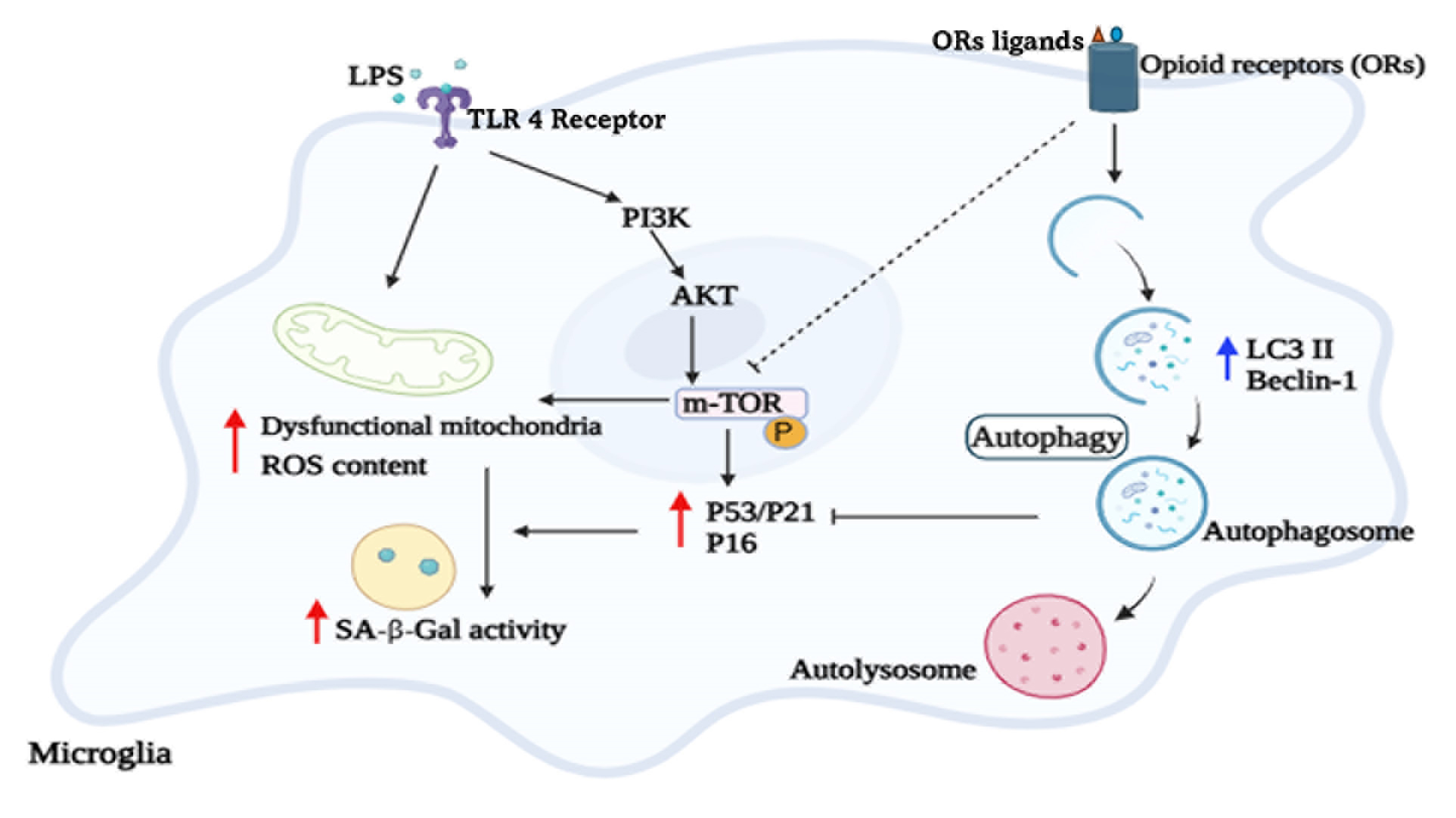
Background: Opioids can modulate mitochondrial redox homeostasis and autophagy and are implicated in the regulation of key physiological and pathological processes, including aging, cellular metabolism, and tumorigenesis. The study aimed to investigate how opioid receptor agonists influence lipopolysaccharide-induced senescence in microglia. Methods: C8-B4 microglial cells were either left untreated or pretreated with different opioid agonists and subsequently exposed to lipopolysaccharide (LPS). Colorimetric assays, fluorescence microscopy, flow cytometry, and Western blotting were used to assess cellular senescence, autophagy-associated changes, reactive oxygen species, and calcium levels, as well as the expression of selected marker proteins and signaling molecules. Results: Treatment with DAMGO, DADLE, and U-50488 significantly attenuated LPS-induced increases in intracellular calcium levels and reduced the expression of cellular senescence markers, including p53, p16, p21, SA-β-Gal activity, and mitochondrial ROS (mtROS), while enhancing total antioxidant capacity (p < 0.05). Notably, opioid treatment was associated with changes consistent with increased autophagy-related activity, as demonstrated by the upregulation of autophagy-related markers Autophagy-related proteins 5 and 7, beclin-1, and microtubule-associated proteins 1A/1B light chain 3B (MAP-LC3). It reversed LPS-induced impairment of autophagy-related activity, evidenced by increased degradation of p62 (p < 0.05). Furthermore, opioids inhibited LPS-induced activation of the phosphatidylinositol 3-kinase/Protein Kinase B/mechanistic Target of Rapamycin signaling pathway (p < 0.05), thereby promoting autophagy. Conclusions: Taken together, these findings suggest that opioids may support cell survival, attenuate LPS-associated senescence markers, and be accompanied by changes consistent with increased autophagy-related activity.Graphic Abstract
Keywords
Cellular senescence; opioids; autophagy; microglia; neuroinflammation
Supplementary Material
Supplementary Material FileCite This Article
APA Style
Mali, A.S., Das, D., Greco, D., Telensky, P., Novotny, J. (2026). Opioids Promote Autophagy and Attenuate LPS-Induced Cellular Senescence-Associated Changes in Microglia. BIOCELL, 50(9), 9. https://doi.org/10.32604/biocell.2026.080886
Vancouver Style
Mali AS, Das D, Greco D, Telensky P, Novotny J. Opioids Promote Autophagy and Attenuate LPS-Induced Cellular Senescence-Associated Changes in Microglia. BIOCELL. 2026;50(9):9. https://doi.org/10.32604/biocell.2026.080886
IEEE Style
A. S. Mali, D. Das, D. Greco, P. Telensky, and J. Novotny, “Opioids Promote Autophagy and Attenuate LPS-Induced Cellular Senescence-Associated Changes in Microglia,” BIOCELL, vol. 50, no. 9, pp. 9, 2026. https://doi.org/10.32604/biocell.2026.080886
 Copyright © 2026 The Author(s). Published by Tech Science Press.
This work is licensed under a Creative Commons Attribution 4.0 International License , which permits unrestricted use, distribution, and reproduction in any medium, provided the original work is properly cited.
 Downloads
 Citation Tools
