
 Submit a Paper
 Propose a Special lssue
 Open Access
ARTICLE

Deep Convolutional Neural Networks for South Indian Mango Leaf Disease Detection and Classification
School of Computer Science and Engineering, VIT-AP University, Amaravati, Vijayawada, 522237, India
* Corresponding Author: S. Sudhakar Ilango. Email:
Computers, Materials & Continua 2023, 77(3), 3593-3618. https://doi.org/10.32604/cmc.2023.042496
Received 01 June 2023; Accepted 26 September 2023; Issue published 26 December 2023
 View Full Text
 Download PDF
Abstract
The South Indian mango industry is confronting severe threats due to various leaf diseases, which significantly impact the yield and quality of the crop. The management and prevention of these diseases depend mainly on their early identification and accurate classification. The central objective of this research is to propose and examine the application of Deep Convolutional Neural Networks (CNNs) as a potential solution for the precise detection and categorization of diseases impacting the leaves of South Indian mango trees. Our study collected a rich dataset of leaf images representing different disease classes, including Anthracnose, Powdery Mildew, and Leaf Blight. To maintain image quality and consistency, pre-processing techniques were employed. We then used a customized deep CNN architecture to analyze the accuracy of South Indian mango leaf disease detection and classification. This proposed CNN model was trained and evaluated using our collected dataset. The customized deep CNN model demonstrated high performance in experiments, achieving an impressive 93.34% classification accuracy. This result outperformed traditional CNN algorithms, indicating the potential of customized deep CNN as a dependable tool for disease diagnosis. Our proposed model showed superior accuracy and computational efficiency performance compared to other basic CNN models. Our research underscores the practical benefits of customized deep CNNs for automated leaf disease detection and classification in South Indian mango trees. These findings support deep CNN as a valuable tool for real-time interventions and improving crop management practices, thereby mitigating the issues currently facing the South Indian mango industry.Keywords
Mango, sometimes called the “King of Fruits,” is a precious fruit crop grown in many nations. It is widely consumed and valued for its economic and nutritional significance. India is a major producer; approximately 40% of the world’s mangoes come from India, making it the leading country. However, mango crops face significant challenges due to pests and diseases, which result in substantial yield losses estimated at around 30%–40% [1]. Mango leaves, in particular, are susceptible to various diseases that significantly impact mango production. Mango cultivation is a vital agricultural activity in South India, contributing significantly to the region’s economy. However, the growth and productivity of mango trees are often hampered by various leaf diseases. These diseases can lead to significant crop losses and reduced fruit quality if not detected and managed promptly. Manual inspection and diagnosis of leaf diseases are time-consuming, labor-intensive, and prone to errors, necessitating the development of automated and accurate disease detection systems [2]. Thus, the need for autonomous, precise, rapid, and cost-effective plant disease identification technology is developing. Image processing and machine learning classify leaf diseases. Deep learning, a machine learning branch, has garnered attention and found practical applications. It uses deep neural networks to provide a helpful tool for diagnosing and categorizing plant diseases. Deep learning, especially Deep Convolutional Neural Networks (CNNs), has benefited image analysis tasks like disease diagnosis and plant categorization. CNNs can automatically learn the discriminative features required for complex pattern recognition tasks from the raw input images [3].
The fundamental goal of this study is to use Deep Convolutional Neural Networks to identify and categorize mango tree illnesses affecting leaves in the South Indian climate. By utilizing these advanced machine learning algorithms, we aim to develop an efficient and accurate system to aid farmers and agricultural experts in timely disease identification and management. The proposed system will involve the collection of a comprehensive dataset of mango leaf images, encompassing various disease classes prevalent in South Indian mango trees, such as Anthracnose, Powdery Mildew, and Leaf Blight. The dataset will be pre-processed to enhance the quality and consistency of the images, ensuring optimal input for the CNN model.
We plan to develop and fine-tune a customized deep CNN architecture to detect and categorize mango leaf diseases. After numerous convolutional and pooling layers, the CNN model will employ fully connected layers to classify the input. After prolonged training, the model could correctly identify the various diseases present in mango leaves by extracting essential features from the photos. We will do extensive experiments on the amassed dataset to assess the efficacy of our proposed approach. Classification accuracy and loss will be used to evaluate the customized deep CNN model’s disease detection and classification performance. Our proposed method will also be measured against other CNN approaches to leaf disease detection and classification through comparative analyses. This will shed light on the accuracy, robustness, and computing efficiency advantages of deep CNNs. This study’s anticipated conclusion is an automated and trustworthy approach for identifying leaf diseases in South Indian mango trees. Improved crop output, fewer economic losses, and more sustainable agricultural practices are all possible thanks to early and precise disease detection that allows farmers to execute tailored disease management measures. Basic preliminaries and associated work are discussed in Section 2, an optimized CNN model is offered in Section 3, results and discussion are described in Section 4, and the conclusion and future work are presented in Section 5, followed by references.
2 Basic Preliminaries and Related Works
Several diseases may infect mango trees and cause serious harm to the plants, reducing the yield and quality of the mango fruit produced [4]. Fungal, bacterial, or viral pathogens and environmental factors can cause these diseases. Mango leaf diseases manifest as visual symptoms on the leaves, such as discoloration, spots, lesions, deformities, or wilting. These symptoms vary by disease and infection stage. Some common mango leaf diseases include Anthracnose, powdery mildew, leaf blight, bacterial black spot, and mango malformation. Anthracnose is a common and economically significant disease in mango trees, caused by the fungal pathogen Colletotrichum gloeosporioides. It results in leaf spots, fruit rot, and blossom blight. Powdery mildew, caused by Oidium mangiferae, leads to a white powdery growth on the leaves, affecting photosynthesis. Leaf blight, caused by Pestalotiopsis mangiferae, causes irregular-shaped lesions, leading to defoliation and reduced vigor. Bacterial black spot, caused by Xanthomonas campestris pv. mangiferaeindicae causes dark necrotic spots on leaves and can spread to fruits.
Mango malformation, caused by Fusarium mangiferae, affects tree growth and development, causing abnormal inflorescences and leaf malformation. Mango leaf infections must be detected quickly and accurately to be managed effectively. Image processing and machine learning algorithms are examples of cutting-edge technologies. have been increasingly used to develop automated systems for disease detection and classification [4]. These systems analyze digital images of mango leaves to identify diseases, enabling timely intervention and precise management. Farmers and plant pathologists can use these technologies to implement appropriate practices such as pruning, sanitation, and targeted chemical applications. This proactive approach improves crop health and productivity by controlling the spread and severity of mango leaf diseases. Farmers can minimize crop losses, ensure better fruit quality, and enhance mango orchards’ overall productivity and profitability by effectively detecting and managing mango leaf diseases.
2.1 Types of Mango Leaf Diseases
Numerous diseases can impact mango leaves [4], and here are a few common ones: Numerous diseases can affect mango leaves [4], and here are a few common ones:
1) Anthracnose: This fungal disease is caused by Colletotrichum gloeosporioides, leading to dark, sunken lesions on mango leaves. It can also affect other parts of the tree, including fruits and flowers.
2) Powdery Mildew: The fungus Oidium mangiferae is responsible for powdery mildew. It results in a white, powdery growth on the surface of mango leaves, affecting their ability to carry out photosynthesis.
3) Leaf Spot: Leaf spot diseases are caused by various fungal pathogens, like Cercospora mangiferae and Pestalotiopsis mangiferae. They cause the formation of small to large spots on the leaves, which may be brown, black, or gray.
4) Bacterial Black Spot: This bacterial disease is caused by Xanthomonas campestris pv. mangiferaeindicae. It leads to the development of dark, necrotic spots on mango leaves, which can also affect the fruits.
5) Mango Malformation: Mango malformation is a disease caused by a phytoplasma pathogen called Fusarium mangiferae. It results in abnormal growth patterns, distorted leaves, and deformed inflorescences.
6) Rust: Mango rust is caused by the fungus Puccinia mangiferae. It leads to rust-colored pustules on the undersides of mango leaves.
7) Dieback: Dieback is characterized by the progressive death of branches or twigs. Various factors, including fungal infections, nutrient deficiencies, or environmental stress, can cause it.
8) Sooty Mold: This fungal disease causes a black, sooty coating on the leaves and stems of mango trees, often as a result of insect infestations.
9) Phoma Blight: Phoma blight is a plant disease that affects various crops, causing significant damage and yield loss.
These are just a few examples of the many diseases affecting mango leaves [5]. Proper disease identification and timely management are crucial for maintaining the health and productivity of mango trees. Fig. 1 shows the sample images of South Indian mango tree leaf diseases.

Figure 1: South Indian mango tree leaf diseases
2.2 Literature Review on Mango Leaf Disease Detection and Classification
Investigating and detecting plant diseases using machine vision is a significant area of study, particularly in agriculture. Traditional approaches rely on conventional algorithms; manual feature creation CNNs have changed that and revolutionized crop disease identification. However, identifying diseases in complex natural environments poses challenges due to lesion size, type, and low contrast variations. Nevertheless, modern techniques like machine learning and computer vision offer accurate and consistent results, reducing the risk of misdiagnosis. This improves decision-making for farmers, leading to higher profits and a more sustainable agriculture industry.
Plant disease detection is crucial for effective agricultural disease management [6]. Automated leaf disease identification can reduce monitoring efforts on large farms and enable early and accurate detection of mango leaf diseases, which is vital for plant nutrition [7]. Traditional disease management methods involving manual diagnosis and pesticide application are time-consuming, challenging, and prone to errors [7]. CNNs are inspired by biological nerve and visual systems, offering supervised deep-learning classification with high accuracy [2]. Their complex structure with multiple layers enables feature learning from training data, although they require substantial data for training [2,8].
A recent study [9] suggested employing a deep CNN model to identify and classify leaf diseases—54,306 Plant Village pictures in 38 classes, including 14 crop species and 26 disease variations. ReLU(Rectified Linear Unit) activation and batch normalization gave the CNN model accurate and efficient recognition. We checked mango leaves for Leaf Webber, Alternaria leaf spots, leaf gall, leaf burn, and Anthracnose. CNN extracted and categorized probable features [10].
Their article [11] used CNNs to detect and categorize mango leaf diseases. Deep learning extracted attributes from mango leaf images to classify diseases accurately. This research contributes to the advancement of automated systems for the early and precise detection of mango leaf diseases, facilitating effective disease management strategies and enhancing crop yield.
The study's authors published as [12] looked into the feasibility of applying machine learning, particularly CNNs, to disease detection and diagnosis in mango leaves. This research compared and contrasted various machine learning algorithms to determine which performed best. The outcomes demonstrated the usefulness of CNNs in correctly categorizing mango leaf diseases. This study illuminates the potential of machine-learning algorithms for speedy and accurate mango leaf disease diagnosis, advancing mango disease management tools.
The research study introduced a deep learning method to automate mango leaf disease detection and categorization [13]. The training dataset was expanded using data augmentation techniques, while Convolutional Neural Networks (CNNs) served as the central architecture. The accuracy of disease detection and classification was improved through their tests, which is cause for optimism. This research showed that CNNs are a promising tool for automatic disease detection and classification in mango leaves, which could improve disease control in mango farming.
To detect and categorize mango leaf illnesses, the study’s authors titled [14] transfer learning with deep CNNs. Using their mango leaf dataset, the authors refined pre-trained CNN models. They were able to successfully classify diseases by using transfer learning. This research emphasizes the utility of mango leaf disease categorization with transfer learning, demonstrating its potential to enhance the speed and accuracy with which diseases are identified in mango crops.
The work [15] presented a deep learning-based method for detecting and categorizing mango tree leaf diseases. To extract useful and distinguishing information from photographs of leaves, the authors used Convolutional Neural Networks (CNNs). The results of their tests demonstrated the usefulness of deep learning methods for accurately categorizing diseases. This research indicates the promise of Convolutional Neural Networks (CNNs) to aid in effective disease control in mango crops by automating the identification and categorization of mango leaf diseases.
Kumar et al. [16] study classifies mango leaves with Anthracnose. A classification model for Anthracnose-infected mango leaves was created using Convolutional Neural Networks (CNNs). Training a CNN model using mango leaf photos enabled the network to extract features and patterns automatically. Deep learning proved effective for early detection and efficient control of Anthracnose in mango crops by demonstrating its efficacy in accurately recognizing and classifying mango leaves afflicted by the disease. This study contributes significantly to plant pathology by demonstrating the utility of deep learning approaches for tackling the difficulties of disease diagnosis and categorization in farming.
Research conducted in 2020 used a Radial Basis Function (RBF) Neural Network to create a web-based method for segmenting Anthracnose disease from mango tree leaves. By automating the segmentation process, the study provided a convenient and efficient tool for plant pathologists and farmers to detect and monitor Anthracnose disease in mango trees. This research contributes to agricultural technology by introducing a novel approach for disease segmentation using neural networks, which can help diagnose and treat Anthracnose disease in mango crops.
Various innovative methods were introduced in a series of studies focusing on enhancing the detection and prevention of plant diseases and pests. The convolutional ensemble network Es-MbNet, utilizing a two-stage training strategy with three lightweight CNNs [17], achieved remarkable accuracy in identifying plant lesions. Another study introduced a two-part method using a hybrid segmentation algorithm and convolutional neural network, achieving significantly higher validation accuracy than conventional methods for detecting leaf diseases [18]. Further research led to the creation of SE-MobileNet, a fusion of MobileNet with squeeze-and-excitation block, that showed substantial efficiency in identifying rice disease types [19]. A novel approach was also proposed for automatically identifying plant diseases, using enhanced artificial neural networks and CNNs, achieving impressive performance in experimental analyses [20]. Finally, a CNN-based method with an attention mechanism and two-stage transfer learning was introduced to identify insect pests, achieving excellent accuracy even in heterogeneous background conditions [21]. These innovative approaches represent significant advancements in agricultural technology, contributing to improved crop yield and food security.
The goal of the 2020 study [2] was to use SVM(Support Vector Machine) and Neural Networks to create a system to identify mango leaf diseases. They made a dataset of mango leaf images representing various disease conditions and used it to train and evaluate the models [22]. Neural Network and SVM algorithms were employed to classify mango leaf diseases accurately. The study showcased the effectiveness of these techniques in diagnosing and managing leaf diseases, benefiting mango farmers and plant pathologists by improving crop health and yield. This study advances agricultural technology in the field of mango cultivation.
In their 2020 study [7], the researchers developed a system for early disease classification in mango leaves. They collected a dataset of mango leaf images, extracted relevant features, and applied a hybrid metaheuristic feature selection algorithm. The study demonstrated the effectiveness of this approach in achieving early disease classification, enabling timely interventions for disease management in mango orchards, and minimizing crop losses. This research contributes to agricultural technology by providing a valuable tool for farmers to detect and address mango leaf diseases [23] early. Table 1 presents the summary of the various deep learning models’ outcomes on mango leaf detection and classification.
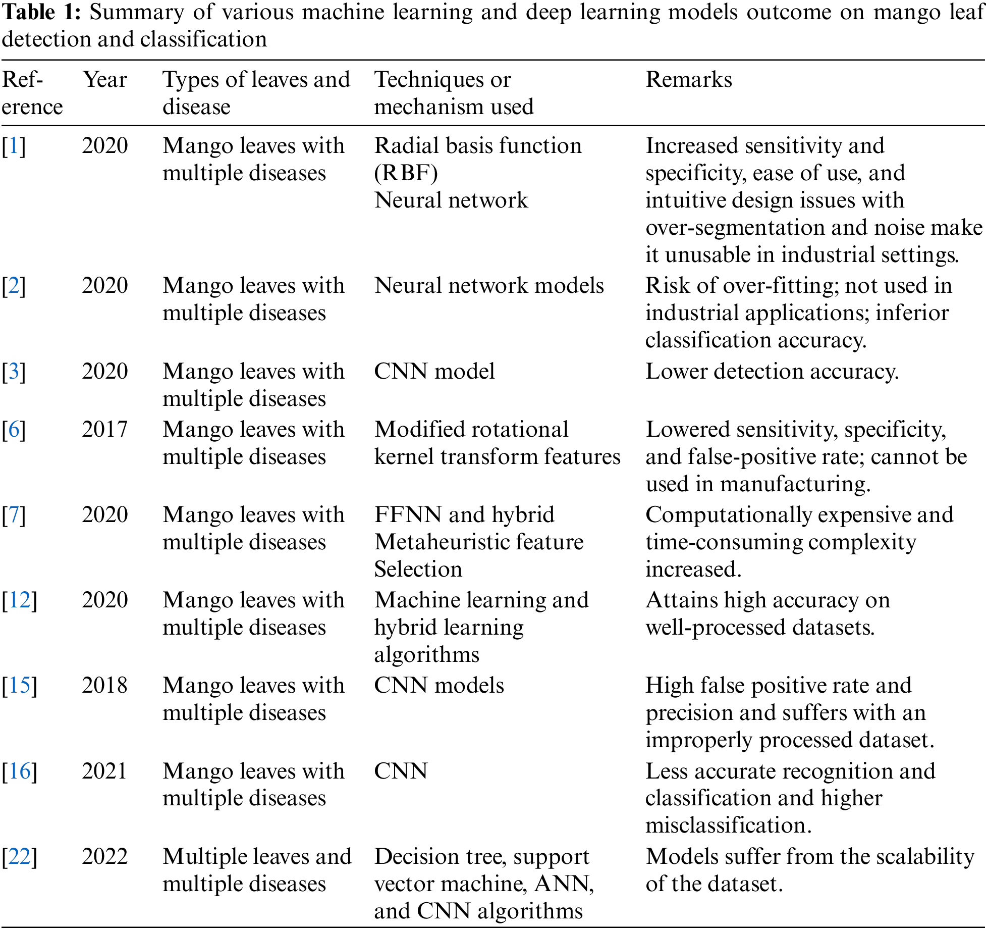
These related works demonstrate the growing interest in leveraging deep learning techniques, specifically CNNs, for leaf disease detection and classification in mango trees. They highlight the potential of deep CNNs in achieving accurate and automated disease identification, which can significantly benefit the agricultural sector in South India by enabling timely interventions and improved crop management practices.
3 Customized Deep CNN for Mango Leaf Disease Detection and Classification
Mango leaf disease detection and classification involves a collection of mango leaf images, pre-processing, image processing, and classification. This section elaborates on implementing customized deep CNN for Mango leaf disease detection and classification.
Deep learning methods have been extensively researched to identify and categorize leaf diseases. However, these methods come with several complications, including the potential for misdiagnosis due to variations in diseases, varieties, and environmental factors. Early identification and treatment of leaf diseases are crucial, but the lack of agricultural expertise in rural areas can be time-consuming and hindered. With autonomous feature selection and reduced reliance on labor-intensive picture pre-processing, Convolutional Neural Networks (CNNs) have proven effective in image-based recognition tasks. However, one of the challenges is the availability of large and diverse datasets to train these models effectively. Acquiring such datasets remains a difficult task. Fig. 2 depicts the suggested method’s flowchart.

Figure 2: Flowchart for the proposed customized deep CNN model for mango leaf disease detection and classification
3.1 Dataset Description and Augmentation
A collection of high-quality pictures of diverse mango plant leaves with various diseases is collected from different locations in the south Indian region. The dataset includes healthy and sick leaf photos, including those with rust, bacterial spots, and powdery mildew. The dataset contains a complete 1275 images with 14 different types of diseases. The ratio between training, validation, and testing is 80:20.
To address the data imbalance, we have performed augmentation to increase the images in each class. The augmentation is a complex data preprocessing task specifically geared towards augmenting a dataset containing images of various plants and their corresponding diseases. It utilizes Keras’s ‘ImageDataGenerator’ class to define a sequence of on-the-fly transformations, including a 30-degree rotation range, width and height shifts of 20%, shear transformation of 20%, zooming by 20%, and horizontal flipping, filling missing pixels using the “nearest” neighbor method.
Each image is read, checked for validity, resized to 256 × 256 pixels, and appended to a list after being converted to an array using the. These images are then normalized to the range [0,1] by dividing by 255.0. After preprocessing, the images are augmented using the specified transformations. For each disease type, a total of 320 images including original images are generated and saved to a specified directory, retaining the hierarchical structure of plants and diseases. The original and augmented image counts are printed to monitor the progress. Fig. 3 displays the variety of leaf images after augmentation from each of the 14 categories.

Figure 3: The distribution of mango leaf images across the 14 classes after augmentation
Image processing is vital for mango leaf disease detection in deep learning. Techniques like pre-processing and enhancement of raw leaf images improve model accuracy [24], while compression methods reduce image size without losing crucial information. Our work used Lossless [25] and Hybrid compression [26] to create low-resolution images, aiding efficient storage, transmission, and faster processing for deep-learning applications.
At first, the training and test photos underwent pre-processing to improve contrast and resize them to a 300 × 300 pixel resolution. For resizing the images, nearest neighbor interpolation (Eq. (1)) and for rescaling, the traditional method (Eq. (2)) and (Eq. (3)) are used along with image crop.
By incorporating image processing techniques into the pipeline of deep learning for mango leaf disease detection, the models can benefit from enhanced image quality, improved feature representation, and better generalization capabilities, leading to more effective and accurate disease classification.
3.3 Deep Convolutional Neural Networks to Detect and Classify Mango Leaf Diseases
The literature on deep learning approaches has many issues, such as misdiagnosis of leaf illnesses, variety in diseases, varieties, and environmental influences. Early identification and treatment of such diseases are therefore helpful. However, doing such is challenging. A time-consuming process, agricultural expert knowledge is unavailable in rural locations. Convolutional neural networks have recently achieved significant advancements in picture-based recognition by eliminating requirements for image pre-processing and enabling built-in feature selection [4]. Finding massive datasets for such challenges is another quite challenging issue. To handle data with a grid-like structure, like photographs, CNNs were developed [3]. The pixels in an image are arranged in a grid, and the value of each pixel determines its hue and luminance. Likewise, each neuron in a CNN processes information within its receptive field. Like how the human brain processes visual information, CNN layers detect simpler patterns first, then more complex ones as the layer progresses. Convolutional neural networks have input, hidden, and output layers. Convolution, normalization, pooling, and fully-connected layers lie between the output and input layers [3]. The convolutional layer’s filters create classification feature maps. Image processing uses ReLU [27]. An enhanced fine-grained robust CNN model is proposed for this study’s classification of leaf diseases. At the first level, pre-processing techniques are used to reduce the size of the leaf image. To detect diseases from photos of leaves, a customized deep CNN learning model has been created at the second level utilizing a convolutional neural network [28]. Fig. 4 shows the implementation of the customized deep CNN model for mango leaf disease detection and classification.
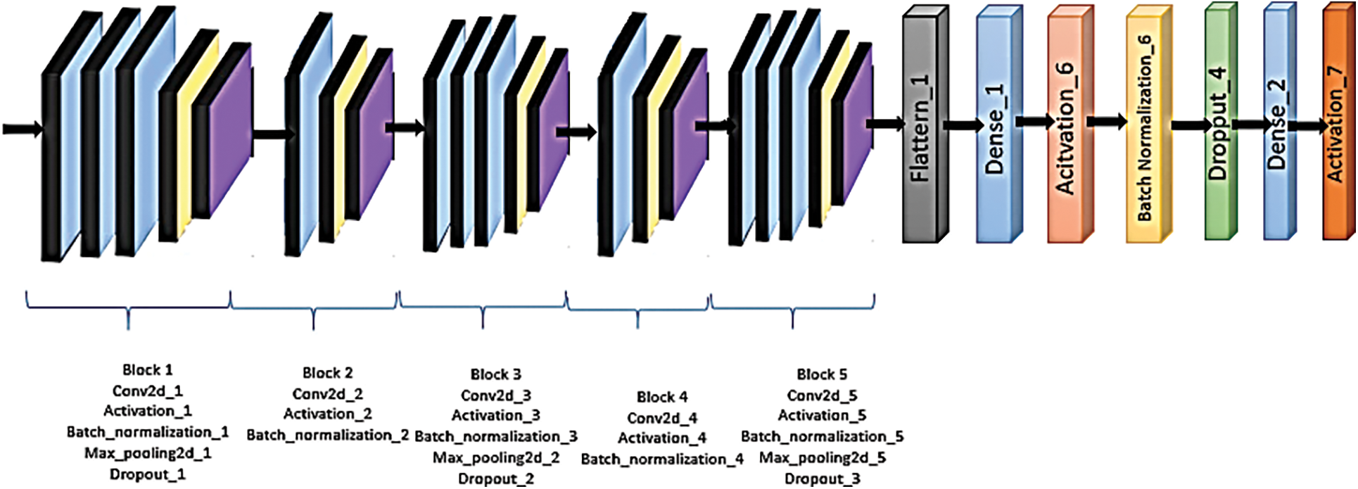
Figure 4: Architecture of the proposed customized deep CNN model for mango leaf disease detection and classification
The description of the proposed model visualizes each layer type with a specific color. The Conv2D, activation, and batch normalization layers are denoted in blue, signifying these stages’ feature extraction and normalization operations. The yellow color represents the max-pooling layers, indicating the down-sampling process. Dropout layers are marked in violet, essential for reducing overfitting by randomly dropping out neurons. The Flatten layer, which prepares the data for the fully connected layers, is shown in black. Following the Flatten layer, there is a Dense layer represented in blue. An activation function (ReLU) follows, shown in orange, which introduces non-linearity to the model. Afterward, another batch normalization layer is presented in yellow, and a Dropout layer in green. Finally, the last Dense layer and a Softmax activation function are depicted in blue and red, respectively, marking the classification stage of the model.
Deep CNN models have numerous convolutional, pooling, and fully linked layers. Because of its high complexity, a deep neural network can develop hierarchical representations of the input data, which are crucial for achieving precise categorization. Algorithm 1 presents a detailed pseudo code for the proposed customized deep CNN model for mango leaf disease detection and classification. The deep Convolutional Neural Network (CNN) model consists of multiple layers, including Conv2D, Batch Normalization, Max Pooling, and Activation functions. Here is a description of the model architecture:
The convolutional operation distinguishes a CNN from other neural networks. The basic form of convolution consists of two functions that take real numbers as arguments. To explain convolution, we can pretend that it is possible to track where a car is using a laser that gives an output: x(t), where x is the car’s position in time step t. Several measurements can be taken to reduce possible noise during the measurements, and an average value can be used as the measurement value. Later measurements have more excellent value than the older ones; therefore, a weight function, w(a), is used, where a represents how old a measurement is. The weight function w must be a valid density function. If these weighted average measurements are performed every time step, it can be described with a function, s known as the Convolution function.
In CNN terminology, the first argument in the convolution function is called the input, and the second is called the kernel; what is returned is called the feature map.
For the example with the car above to be realistic, the data cannot be collected in each time step when the amount had become too large, but in regular intervals, for example, every second or minute. In such a case, the time variable t would only be of integer type. Likewise, the variables x and w, then the mathematical discrete convolution can be defined as,
The model includes 5 Conv2D layers. Conv2D performs convolution operations on the input image to extract features. Each Conv2D layer consists of a set of learnable filters that scan the input image and
3.3.2 Batch Normalization Layers
The batch normalized activation is
where
Max Pooling is performed after each Conv2D layer. It reduces the spatial dimensions of the feature maps by selecting the maximum value within a defined pool size. Max Pooling helps down-sampling the feature maps and extract the essential features while reducing computational complexity.
The model utilizes 7 activation functions throughout its layers. Activation functions introduce non-linearity to the model, enabling it to learn complex patterns and make non-linear decisions. ReLU, sigmoid, and tanh activation functions are frequently employed in CNNs—the layer’s output before it is fed into the following activation function, element by element.
Combining Conv2D layers, Batch Normalization, Max Pooling, and Activation functions helps the deep CNN model extract and learn intricate features from the input data. This allows the model to capture the information for accurate classification or detection tasks, such as mango leaf disease identification.
The proposed model is a Convolutional Neural Network (CNN) comprising several layers arranged sequentially. The output of one layer serves as the input to the next, creating a chain-like interconnection. The description of the phase wised interconnections is described as,
1) Input and First Convolutional Layer: The model begins by taking an input of shape (height, width, depth), which corresponds to the dimensions of the image and the color channels. This is then passed through the first Conv2D layer, which applies 32 filters of size (3, 3) to the input, where each filter detects specific features in the image. The padding is set to “same,” meaning the output size matches the input size, preserving the spatial dimensions of the input. The ReLU activation function is applied element-wise to introduce non-linearity to the model.
2) Batch Normalization and Pooling: The output is then normalized using Batch Normalization to stabilize learning and reduce training time. The MaxPooling2D layer reduces the spatial dimensions of the output, effectively summarizing the presence of features in the image and reducing computational complexity. A Dropout layer is added to randomly set 25% of the input units to 0 at each update during training time to prevent overfitting.
3) Additional Convolutional Layers: This pattern repeats with more Conv2D layers, each having an increasing number of filters (64, 128), with each filter extracting more complex features. Batch Normalization and MaxPooling2D layers follow each convolution operation, repeating the normalization and dimension reduction process. Dropout layers are interspersed to prevent overfitting.
4) Flattening and Fully Connected Layer: After the convolutions, normalizations, poolings, and dropouts, the high-dimensional output is flattened into a 1D array before passing into a Dense (fully connected) layer with 1024 neurons. The Dense layer learns non-linear combinations of features extracted from the previous layers.
5) Final Classification: A final Dropout layer is included before the last Dense layer, which outputs probability scores for each class via the softmax activation function.

3.4 Training, Testing, and Validation Description
The dataset was divided into training and test sets. The test dataset tested the proposed model’s performance, whereas the training dataset fine-tuned CNN models. As a result, we segmented the datasets into three categories: training, validation, and testing, into equal halves of 80%, 10%, and 10%, respectively. Backpropagation was carried out in the opposite direction in the event of an incorrect forecast. As a result, the current research used the backpropagation technique to update the model weights for a better prognosis appropriately. One epoch was the collective term for forwarding and backpropagation. For the investigation, the model made use of the Adam optimizing algorithm. The training images for the current study were obtained while retaining the 80% image. Each dataset was examined using the remaining 20% of unaltered photos [22].
The performance of the proposed model is evaluated using multiple metrics, including accuracy, precision, recall, and F-measure, calculated with standard formulas involving true positive (TP), false positive (FP), true negative (TN), and false negative (FN). These metrics comprehensively assess the model’s performance [14].
Accuracy: Classification Accuracy is determined by the correct prediction ratio to total predictions [29].
Precision: Precision [14] determines with what precision the network places images in the positive category. Precision is calculated as follows:
Recall: Recall [14] indicates how many positive images the network recorded. The recall is calculated as follows:
F-measure: F-measure [14] is a combination of Precision and Recall. The calculation is as follows:
Python libraries are used to implement the proposed research and TensorFlow [30], and Keras [30] are used for the optimized CNN model experiments presented. It employed the Adam optimizer for training, which possessed a learning rate and a built-in loss function.
A dataset of 1275 high-quality images of various mango plant leaves, both healthy and diseased, was assembled from the South Indian region. The collection includes images of leaves affected by diseases such as rust, bacterial spots, and powdery mildew. Utilizing different image processing techniques, the pictures were thoroughly pre-processed. The images were then categorized into 14 distinct groups based on the type of disease and the plant species, with 127 images set aside specifically for validation purposes.
The augmentation is performed using Keras’s ‘ImageDataGenerator’ class defines a sequence of on-the-fly transformations, including a 30-degree rotation range, width and height shifts of 20%, shear transformation of 20%, zooming by 20%, and horizontal flipping, filling empty pixels with the “nearest” neighbor approach. After being transformed to an array using each image is read, validated, scaled to 256 × 256 pixels, and appended to a list. Images are transformed after preprocessing. 320 images, including original images, are generated for each disease kind and saved to a directory to maintain plant and disease hierarchies. Printing original and augmented image counts tracks progress.
The objective is to learn how to pre-process images for Convolutional Neural Networks. Tensors can be considered multi-dimensional arrays simply because they are meant to hold knowledge rather than actual data. Load the dataset after importing the required libraries. Every data analysis procedure must include this step. Images come in a variety of sizes and shapes. Data pre-processing starts with uniformly sizing all of the images. With the help of ImageDataGenerator in Keras [30], several enhancing techniques are used to the collected photos, creating new images with a resolution of 300 × 300 pixels. Fig. 5 represents the pre-processed image with a resolution of 300 × 300.
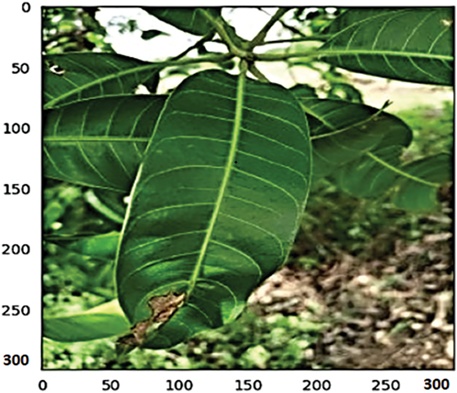
Figure 5: Represents the pre-processed image with a resolution of 300 × 300
4.3 Performance of the Proposed Customized Deep CNN Model on Dataset
The performance measures, such as accuracy and loss, of a deep CNN model trained on the South Indian Mango Tree Leaf Disease Dataset may be examined with the number of epochs used in the training process. Mango leaf disease detection customized deep CNN model parameters are shown in Table 2. During training, the deep CNN model is exposed to the dataset and iteratively updates its internal parameters to improve performance. As the training progresses through multiple epochs, the model learns to extract relevant features and make accurate predictions for classifying mango tree leaf diseases. Monitoring the accuracy and loss values at each epoch provides insights into the model’s performance. Accuracy measures the percentage of correctly classified instances, while loss quantifies the dissimilarity between the predicted outputs and the actual labels. Figs. 6–8 represents the accuracy of the proposed deep CNN model and basic CNN model on mango leaf disease detection and classification.
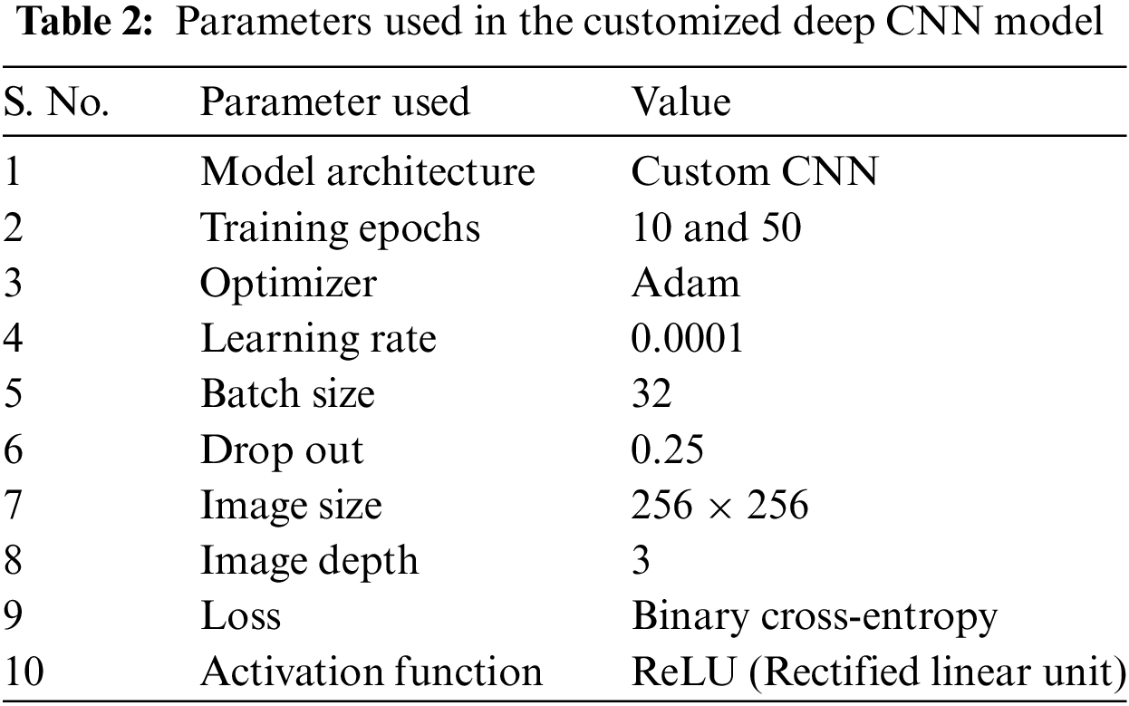
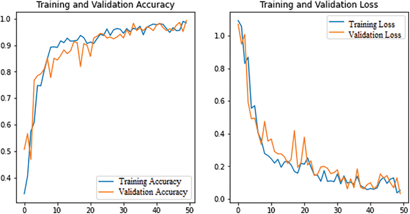
Figure 6: Shows the accuracy of a customized deep CNN model on the South Indian Mango Leaf Disease Dataset for 50 epochs after augmentation
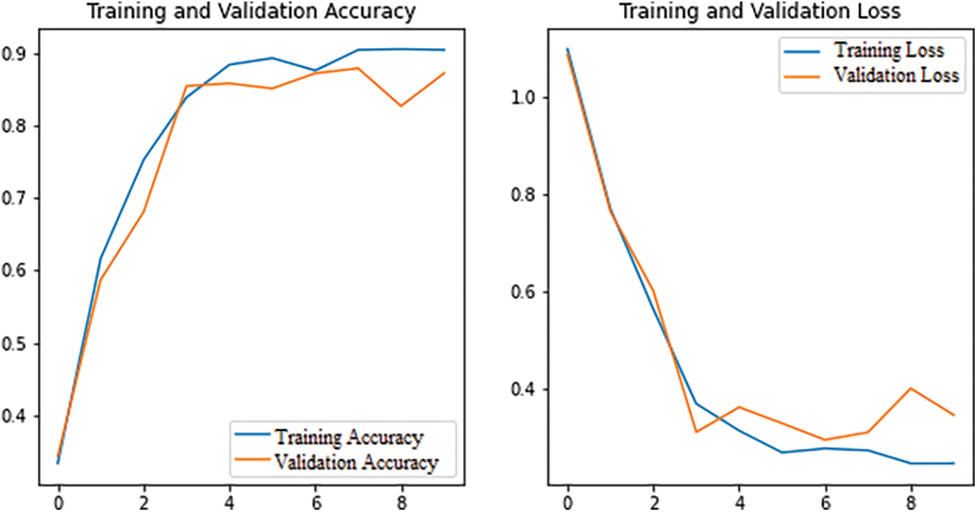
Figure 7: The accuracy of a customized deep CNN model on the South Indian Mango Leaf Disease Dataset for 10 epochs after augmentation
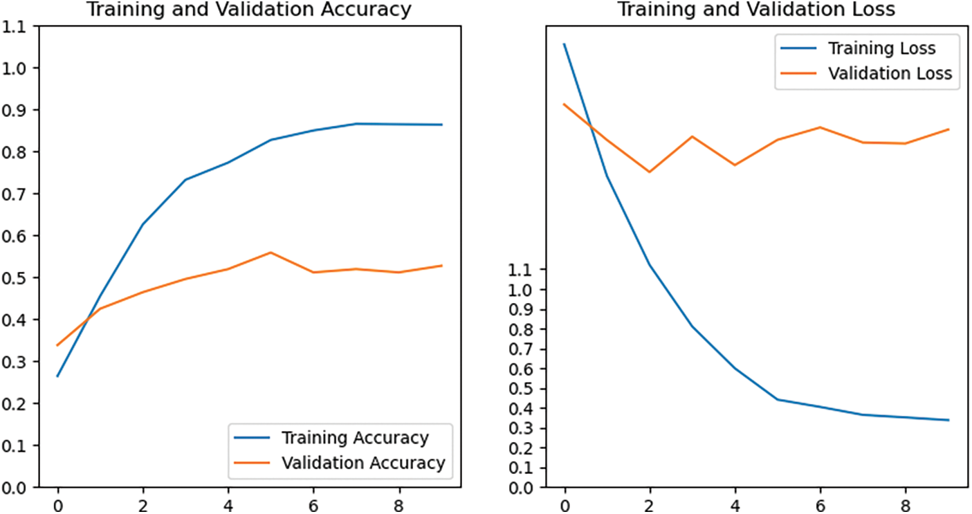
Figure 8: The accuracy of a standard CNN model on South Indian mango tree leaf disease for 10 epochs without augmentation
Each parameter in this table has a significant role in the model. The model is a custom Convolutional Neural Network (CNN) designed for this task. We trained our model for both 10 and 50 epochs to compare performance. Adam Optimizer was utilized because it efficiently handles sparse gradients on noisy problems. A relatively low learning rate 0.0001 was set to ensure gradual and stable learning. We employed a batch size of 32, balancing the need for stochastic gradient approximation and computational efficiency.
A Dropout rate of 0.25 was applied to prevent overfitting by reducing the complexity of the model. The input images were resized to 256 × 256 pixels and were composed of three color channels (Red, Green, and Blue). The Binary Cross-Entropy loss was utilized due to the nature of our problem being a binary classification task. Finally, the ReLU activation function was used because of its efficiency and effectiveness in deep learning models.
Fig. 6 demonstrates the deep CNN model’s accuracy over 50 epochs using the South Indian Mango Tree Leaf Disease Dataset after augmentation. The model appears to be getting better in the beginning as both training and validation accuracy increase and the corresponding losses decline. However, there are notable variations in the accuracy and validation loss as the epochs advance, notably after about Epoch 15. These variations include some sizable spikes in the validation loss. These are caused by gummosis, the initial Weaver Ant and Good Leaves photos are less than the batch size, and even when augmentation is used and the image count rises, a slight amount of overfitting may be seen throughout the model training process. The validation loss begins to stabilize somewhat toward the conclusion of training, although there are still significant variances that could lead to overfitting of the model. A training accuracy of roughly 94.73% and a validation accuracy of roughly 91.43% are obtained in the last epoch.
Fig. 7 shows model performance over ten epochs after augmentation. The model’s training accuracy gradually increases throughout training, going from 89.48% in the first epoch to 92.42% in the tenth, demonstrating that the model is picking up new information from the training data. Although the validation accuracy is consistently around 91%, the validation loss fluctuates and rises generally from 0.5125 to 0.4358. As the validation performance does not regularly increase along with the training performance, this may suggest that the model may have overfitted the training data. The model may be learning patterns particular to the training data and may not generalize well to unknown data, according to the rising validation loss and varying validation accuracy.
In comparison, a standard CNN model, which typically has a shallower architecture with fewer layers, achieves an accuracy of 85.80% as shown in Fig. 8. While still respectable, the lower accuracy suggests that the standard CNN model may need help to capture the complexity of the dataset and accurately differentiate between different types of mango tree leaf diseases. The significant difference in accuracy between the customized deep CNN model and the standard CNN model highlights the importance of model depth and complexity in achieving higher accuracy. The more profound architecture of the customized deep CNN model allows it to learn more intricate data representations, leading to improved classification performance. It is worth noting that accuracy is just one performance metric and should be considered to understand the models’ performance comprehensively. Nonetheless, the reported accuracy values of 93.34% for the customized deep CNN model and 85.80% for the standard CNN model indicate that the customized deep CNN model outperforms the standard CNN model in accurately classifying the South Indian Mango Tree Leaf Disease Dataset.
The results of both the CNN and customized deep CNN architectures on the mango leaf are compared in Table 3. Convolutional Neural Networks (CNNs) and Deep Convolutional Neural Networks (Deep CNNs) were tested for their ability to classify mango leaves. This research aimed to compare and contrast the performance of these architectures to several metrics, such as training accuracy, test accuracy, loss, and the number of epochs needed for convergence. Both models were trained on a mango leaf image dataset and evaluated using separate test datasets. The results revealed that the customized deep CNN architecture outperformed the CNN architecture in terms of both training and test accuracy. The customized deep CNN model achieved higher accuracy scores, indicating its ability to learn and generalize from the training data better. The customized deep CNN architecture also exhibited lower loss values, demonstrating its superior capability in minimizing prediction errors during training. Furthermore, the customized deep CNN model required fewer epochs to converge, indicating its faster learning and convergence rate than the CNN architecture. These findings highlight the superior performance of customized deep CNN architectures in mango leaf classification tasks, showcasing their potential for accurate and efficient disease identification and classification.
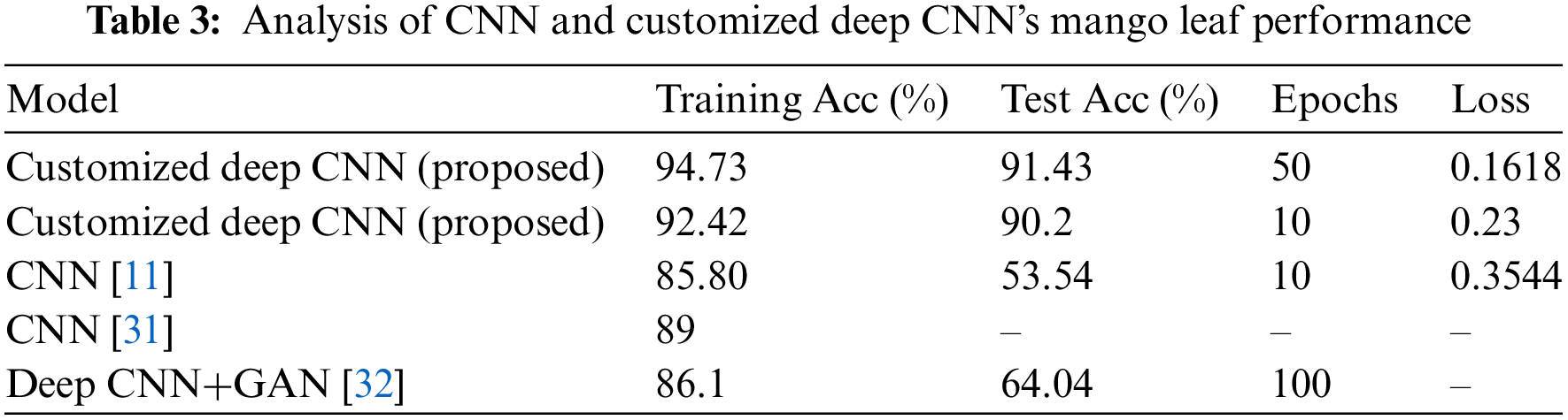
The values in Table 3, are updated after training the model with the augmented and fully balanced dataset. The performance measures are significantly improved due to augmentation. From Table 3, it is evident to note that little overfitting persists due to the original image falling below the ideal batch size. Due to this, the model average TPR is 81%, which correctly classified the positive instances. Also, the FPR, rate is high on Multi disease and Red Rust, where the model is misclassified with the original instances.
4.4 Model Performance on Correctly Predicted Images
The model performance on correctly predicted images refers to how well the customized deep CNN model correctly classifies images of healthy and diseased leaves. In this case, a correctly predicted image is one where the model accurately classifies an image of a vigorous or sickly plant leaf. The model’s performance on correctly predicted images is typically evaluated using accuracy. These metrics provide a measure of how well the model is performing in terms of classifying images correctly. The performance of correctly predicted images is important because it indicates how well the model can detect healthy and diseased plant leaves in real-world scenarios. Additionally, it assists in identifying areas where the model may be functioning well and areas where it may require improvement. Fig. 9 demonstrates The effectiveness of a customized deep CNN model for accurately predicted images.
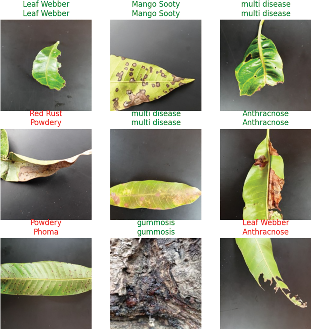
Figure 9: The effectiveness of a customized deep CNN model for accurately predicted images
4.5 Model Performance on Incorrectly Predicted Images
Model performance on incorrectly predicted images can provide valuable insights into the limitations of the model and areas that require improvement. The incorrectly predicted images are the ones where the model has predicted a wrong class label. By analyzing these images, we can identify the patterns or features that need to be included in the model, leading to an incorrect prediction. This analysis can help improve the model architecture to capture the missed patterns or features. Fig. 10 shows The effectiveness of a Deep CNN model on incorrectly predicted images. Table 4 represents the most confused mango leaf disease detection and classification predictions.
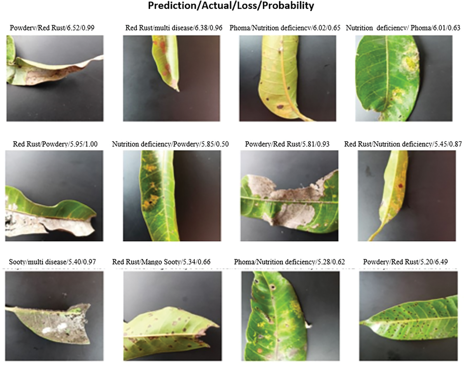
Figure 10: The Performance of a customized deep CNN model on incorrectly predicted images

In deep learning, models are trained to make predictions based on complex patterns and relationships in the input data. While deep learning models have achieved remarkable success in various domains, they are flexible and sometimes produce confused or erroneous predictions. It is due to the Out-of-distribution data, Lack of representative training data, Noisy or ambiguous input, and Overfitting. Table 4 represents some of the most confusing predictions while training the Deep CNN model. Due to the data set, the mango leaf images were collected from a real environment from the south Indian region. There is a need to adhere to the standardization of the dataset to minimize confused predictions.
4.6 Performance Measures on Individual Diseases Prediction and Classification
When evaluating the performance of prediction and classification models for individual diseases of mango leaves, performance measures Precision, Recall, and F1-score are used to assess the accuracy and effectiveness of the customized deep CNN Model. These performance measures provide insights into the prediction and classification accuracy of individual diseases of mango leaves. Different measures may be prioritized depending on the specific goals and requirements of the study. Considering these metrics collectively is essential to understand the model’s performance comprehensively. Table 5 shows the performance measures of individual disease classes on mango leaves. Table 5 shows that a few diseases, like good leaves, Nutrition deficiency, and Phoma, are not predicted accurately due to Out-of-distribution data, Lack of representative training data, Noisy or ambiguous input, and Overfitting. Few diseases are predicted more accurately with the customized Deep CNN model. Fig. 11 shows the confusion matrix for the proposed deep CNN model on incorrectly predicted individual disease classes.

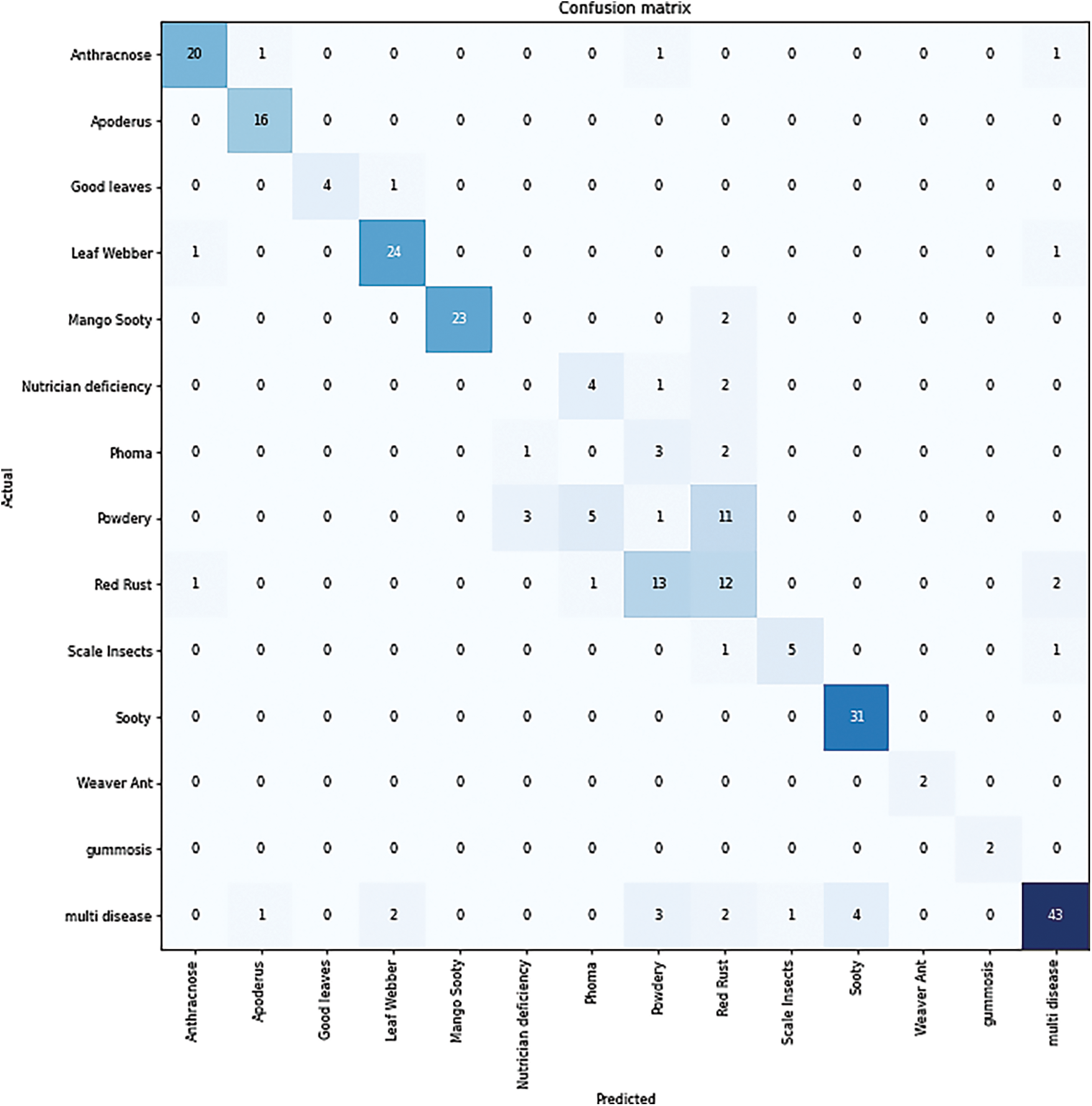
Figure 11: Confusion matrix for the customized deep CNN model on incorrectly predicted individual disease classes
The experiment results show that the customized deep CNN model achieved an accuracy of 94.73% on the South Indian Mango Tree Leaf Disease Dataset. In comparison, the standard CNN model achieved an accuracy of 85.80%. These accuracy values provide insights into the performance of the two models and their ability to accurately classify mango tree leaf diseases. The higher accuracy of the customized deep CNN model suggests that its deeper architecture and increased complexity enable it to learn more intricate and representative features from the dataset. This allows the model to make more accurate predictions and precisely distinguish between different types of mango tree leaf diseases.
On the other hand, the standard CNN model, with its shallower architecture and fewer layers, achieves a lower accuracy of 85.80%. This indicates that the model may need help to capture the dataset’s complexity and cannot extract the distinguishing features required for accurate classification. The significant difference in accuracy between the two models emphasizes the importance of model depth and complexity in achieving higher accuracy. The customized deep CNN model’s ability to learn hierarchical representations and capture intricate patterns in the data contributes to its superior performance compared to the standard CNN model.
These results suggest that employing deeper architectures and increasing model complexity can positively impact the accuracy of mango leaf disease classification. The customized deep CNN model demonstrates its effectiveness in accurately identifying and classifying different types of leaf diseases in South Indian Mango Trees. In conclusion, the experimental results highlight that the superior CNN model performance for classifying South Indian mango tree leaf diseases was 94.73%, while that of a regular CNN model was 85.80%. The findings support using deep architectures and increased complexity to improve the accuracy of mango leaf disease detection and classification.
Our study shows that when classifying South Indian mango tree leaf diseases accurately, the customized deep CNN model is superior to the regular CNN model. The customized deep CNN model outperformed the baseline CNN model by a significant margin of 94.73% against 85.80%. The significant difference in accuracy between the two models suggests that the deeper architecture and increased complexity of the customized deep CNN model enable it to learn more intricate and representative features from the dataset, allowing for a more precise and accurate classification of different types of mango tree leaf diseases. The results highlight the importance of leveraging deep learning techniques, such as customized deep CNNs, for improved accuracy in mango leaf disease detection and classification. The higher accuracy of the customized deep CNN model indicates its potential for assisting in the early detection and effective management of diseases in South Indian mango trees. These results have real-world applications in agriculture, as correct diagnosis of diseases aids farmers in taking preventative steps to boost mango tree health and yields. The higher accuracy of the customized Deep CNN model suggests its suitability for deployment in real-world scenarios, where accurate and efficient disease detection is crucial. Expanding the dataset to include photos of a wider variety of diseases and exploring transfer learning techniques to utilize pre-trained CNN models are potential avenues for further study. Integrating other advanced technologies like hyperspectral imaging can enhance disease detection capabilities. Overall, our research supports using customized Deep CNN models for accurately classifying South Indian Mango Tree Leaf Diseases, providing valuable insights for developing effective disease management strategies in the agricultural sector.
Acknowledgement: None.
Funding Statement: The authors declare that they have no known competing financial & non-financial interests and personal relationships that could have appeared to influence the work reported in this paper.
Author Contributions: Study conception and design: Shaik Thaseentaj, S. Sudhakar Ilango; data collection: Shaik Thaseentaj; analysis and interpretation of results: Shaik Thaseentaj, S. Sudhakar Ilango; draft manuscript preparation: Shaik Thaseentaj, S. Sudhakar Ilango. All authors reviewed the results and approved the final version of the manuscript.
Availability of Data and Materials: The authors believe that this data should be available for others to use and will provide the concerned material to anyone who contacts the corresponding author.
Conflicts of Interest: The authors declare they have no conflicts of interest to report regarding the present study.
References
1. S. S. Chouhan, U. P. Singh and S. Jain, “Web facilitated anthracnose disease segmentation from the leaf of mango tree using radial basis function (RBF) neural network,” Wireless Personal Communication, vol. 113, pp. 1279–1296, 2020. [Google Scholar]
2. M. Mia, S. Roy, S. K. Das and M. Rahman, “Mango leaf disease recognition using neural network and support vector machine,” Iran Journal of Computer Science, vol. 3, pp. 185–193, 2020. [Google Scholar]
3. Y. Nagaraju, T. S. Sahana, S. Swetha and S. U. Hegde, “Transfer learning based convolutional neural network model for classification of mango leaves infected by anthracnose,” in Proc. of INOCON, Bengaluru, India, pp. 1–7, 2020. [Google Scholar]
4. U. P. Singh, S. S. Chouhan, S. Jain and S. Jain, “Multilayer convolution neural network for the classification of mango leaves infected by anthracnose disease,” IEEE Access, vol. 7, pp. 43721–43729, 2019. [Google Scholar]
5. “Mango disease dataset,” [Online]. Available: https://www.kaggle.com/sriramr/applemango-leaf-disease-dataset (accessed on 10/05/2023) [Google Scholar]
6. S. B. Ullagaddi and S. V. Raju, “Disease recognition in Mango crop using modified rotational kernel transform features,” in Proc. of ICACCS, Coimbatore, India, pp. 1–8, 2017. [Google Scholar]
7. T. N. Pham, L. van Tran and S. V. T. Dao, “Early disease classification of mango leaves using feed-forward neural network and hybrid metaheuristic feature selection,” IEEE Access, vol. 8, pp. 189960–189973, 2020. [Google Scholar]
8. C. Trongtorkid and P. Pramokchon, “Expert system for diagnosis mango diseases using leaf symptoms analysis,” in Proc. of ICDAMT, Phayao, Thailand, pp. 59–64, 2018. [Google Scholar]
9. X. Guan, “A novel method of plant leaf disease detection based on deep learning and convolutional neural network,” in Proc. of ICSP, Xi’an, China, pp. 816–819, 2021. [Google Scholar]
10. S. P. Mohanty, D. Hughes and M. Salathe, “Using deep-learning for image base plant disease detection,” Frontirs Plant Science, vol. 7, pp. 1419, 2016. [Google Scholar]
11. A. Kumar, S. Sharma and A. Verma, “Automated detection and classification of mango leaf diseases using deep learning techniques,” International Journal of Computer Science and Information Technologies, vol. l, no. 2, pp. 987–992, 2019. [Google Scholar]
12. M. Rajeshwari, V. Pradeepa and R. Srinivasan, “Leaf disease identification and classification of mango leaves using machine learning algorithms,” International Journal of Advanced Science and Technology, vol. 29, no. 7, pp. 607–615, 2020. [Google Scholar]
13. A. Sharma, A. Gupta and P. Verma, “Automatic detection of mango leaf diseases using deep learning,” International Journal of Scientific Research in Computer Science, Engineering and Information Technology, vol. 7, no. 2, pp. 11–15, 2021. [Google Scholar]
14. S. Gupta, R. Sharma and P. Singh, “Deep learning-based mango leaf disease detection and classification using transfer learning,” International Journal of Computer Applications, vol. 179, no. 11, pp. 15–20, 2020. [Google Scholar]
15. A. Prasad, V. Kumar, P. Choudhary and P. Singh, “Leaf disease identification and classification in mango leaves using deep learning techniques,” International Journal of Advanced Research in Computer science, vol. 9, no. 5, pp. 127–132, 2018. [Google Scholar]
16. P. Kumar, S. Ashtekar, S. S. Jayakrishna, K. P. Bharath, Vanathi et al., “Classification of mango leaves infected by fungal disease anthracnose using deep learning,” in Proc. of ICCMC, Erode, India, pp. 1723–1729, 2021. [Google Scholar]
17. J. Chen, A. Zeb and Y. A. Nanehkaran, “Stacking ensemble model of deep learning for plant disease recognition,” Journal of Ambient Intelligence and Humanized Computing, vol. 14, pp. 12359–12372, 2023. [Google Scholar]
18. P. Kottapapple, N. Reddy, S. Chaganti, D. Himaja and L. P. Barenkala, “A compartive study on deep learnig models for plant disease detection and organic solutions,” International Journal for Research in Applied Science and Engineering Technology, vol. 11, no. VI, pp. 3706–371, 2023. [Google Scholar]
19. J. Chen, D. Zhang, M. Suzauddola, Y. A. Nanehkaran and Y. Sun, “Identification of plant disease images via a squeeze-and-excitation MobileNet model and twice transfer learning,” IET Image Processing, vol. 15, no. 5, pp. 1115–1127, 2021. [Google Scholar]
20. J. Chen, J. Chen, D. Zhang, Y. A. Nanehkaran and Y. Sun, “A cognitive vision method for the detection of plant disease images,” Machine Vision and Applications, vol. 32, no. 31, 2021. [Google Scholar]
21. J. Chen, W. Chen, A. Zeb, D. Zhang and Y. A. Nanehkaran, “Crop pest recognition using attention-embedded lightweight network under field conditions,” Applied Entomology and Zoology, vol. 56, no. 4, pp. 427–442, 2021. [Google Scholar]
22. C. Jackulin and S. Murugavalli, “A comprehensive review on detection of plant disease using machine learning and deep learning approaches,” Measurement: Sensors, vol. 24, no. 100441, 2022. [Google Scholar]
23. “Mango plant disease detection dataset,” [Online]. Available: https://github.com/pratikpv/mango-plant-disease-detection (accessed on 10/05/2023) [Google Scholar]
24. S. K. Mishra and S. Srivastava, “Computer vision-based automated identification and classification of mango leaf diseases,” Journal of Imaging, vol. 5, no. 3, pp. 38, 2019. [Google Scholar]
25. E. Hernández-Valencia, F. J. Gallegos-Funes, J. Olivares-Mercado and V. Medina-Bañuelos, “Lossless compression for multispectral images of agricultural products based on 3D DCT,” Journal of Applied Research and Technology, vol. 18, no. 4, pp. 301–309, 2020. [Google Scholar]
26. X. Yao, X. Zhou, S. Lu and R. R. Zhang, “Hybrid compression algorithm for remote sensing images based on JPEG and fractal compression,” in Proc. of ICMTMA, Phuket, Thailand, pp. 128–132, 2020. [Google Scholar]
27. J. Liu and X. Wang, “Plant diseases and pests detection based on deep learning: A review,” Plant Methods, vol. 17, no. 22, 2021. [Google Scholar]
28. J. Bergstra and Y. Bengio, “Random search for hyper-parameter optimization,” Journal of Machine Learning Research, vol. 13, no. 10, pp. 281–305, 2012. [Google Scholar]
29. J. Rashid, I. Khan, G. Ali, S. H. Almortiri, M. A. AlGhamdi et al., “Multi-level deep learning model for potato leaf disease recognition,” Electronics, vol. 10, pp. 2064, 2021. [Google Scholar]
30. P. Sermanet, D. Eigen, X. Zhang, M. Mathieu, R. Fergus et al., “Overfeat: Integrated recognition, localization, and detection using convolutional networks,” arXiv:1312.6229, 2013. [Google Scholar]
31. U. Sanath Rao, R. Swathi, V. Sanjana, L. Arpitha, K. Chandrasekhar et al., “Deep learning precision farming: Grapes and mango leaf disease detection by transfer learning,” Global Transitions Proceedings, vol. 2, no. 2, pp. 535–544, 2021. [Google Scholar]
32. H. Nazki, S. Yoon, A. Fuentes and D. S. Park, “Unsupervised image translation using adversarial networks for improved plant disease recognition,” Computers and Electronics in Agriculture, vol. 168, pp. 105117, 2020. [Google Scholar]
Cite This Article
 Copyright © 2023 The Author(s). Published by Tech Science Press.
This work is licensed under a Creative Commons Attribution 4.0 International License , which permits unrestricted use, distribution, and reproduction in any medium, provided the original work is properly cited.
 Downloads
 Citation Tools