
 Login
Login Register
 Submit a Paper
 Propose a Special lssue
 Open Access
REVIEW
Renewable Polymeric Tissue Scaffolding: Trends in Structural Development
1 Department of Polymer Engineering, University of Bayreuth, Bayreuth, Germany
2 Department of Environmental and Biological Sciences, University of Eastern Finland, Kuopio, Finland
3 Instituto de Macromoléculas Eloisa Mano, Federal University of Rio de Janeiro, Rio de Janeiro, Brazil
4 Faculty of Engineering Sciences, University of Bayreuth, Bayreuth, Germany
5 Neue Materialien Bayreuth GmbH, Bayreuth, Germany
* Corresponding Authors: Emmanuel Abu-Danso. Email: ; Holger Ruckdäschel. Email:
Journal of Renewable Materials 2026, 14(8), 3 https://doi.org/10.32604/jrm.2026.02025-0198
Received 03 December 2025; Accepted 29 May 2026; Issue published 26 August 2026
 View Full Text
 Download PDF
Abstract
Polymeric scaffolds are now widely used in biomedical engineering to reconstruct tissues as well as other corrective medical applications. They are designed to promote the proliferation of the host cell and provide load-bearing capabilities. Through tailored fabrication methods and material compatibility, polymeric scaffolds can be applied either temporarily or permanently, and successful applications have resulted in commercialized products. Currently, there is a high interest in tissue scaffolds that combine both effective mechanical performance with efficient surface response functionalities. In this work, we performed an in-depth comparative analysis of tissue scaffolds made from renewable parent materials, their production routes, and performance at laboratory scale, patents, and commercialized products. From the literature reviewed, natural polymer-based scaffolds demonstrate superior vascularization whereas the partially biosourced constructs exhibit mechanical advantage. These suggest that combining optimized features from both material classes could yield scaffolds with enhanced stress-strain properties, high vascularization and porosity. To tailor scaffold performance, we found that the flexibility of 3-Dimensional (3D) printing particularly precision laser writing technology, offers a more efficient fabrication approach as it enables the production of hierarchical scaffold capable of performing different functions across different size scales. We found a gap in (i) the knowledge of leveraging the similarities between plant and animal cellularized systems, (ii) the use of self-healing and shape-memory smart materials, and (iii) ex vivo culturing in bioreactor systems and subsequent seeding. To date, most tissue scaffold engineering research work remains at the laboratory scale, therefore, more effort is needed in upscaling, as well as in research and development. Diverse methodologies and materials still need to be explored for fit-for-purpose tissue scaffolds that can meet future needs.Graphic Abstract
Keywords
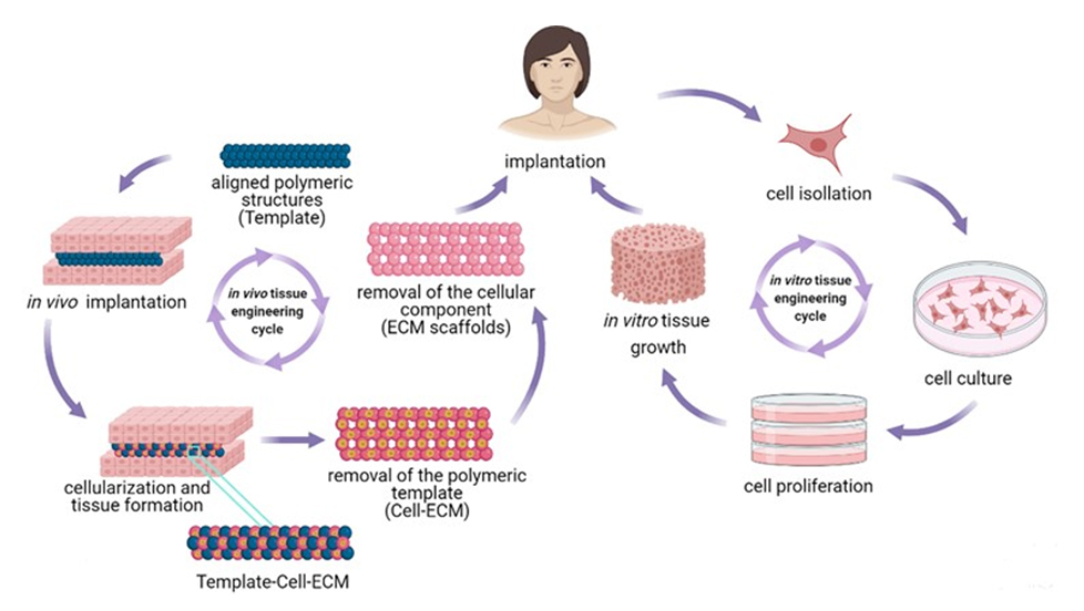
Tissue engineering has emerged as a promising approach in regenerating tissues and organs. Compared to traditional therapies, such as drug administration and transplantation, tissue engineering enables autografts and tuneable cell growth [1]. Tissues can be engineered by both in vivo, where an implanted scaffold augments cell generation and growth, leading to tissue formation, and in vitro, where cells are seeded onto a scaffold under controlled conditions to promote proliferation and migration [2,3].
The term “tissue engineering” was officially introduced at a workshop by the United States National Science Foundation and formally recognized as an engineering discipline in 1987 [4]. The concept of modern tissue engineering was later introduced by Vacanti and colleagues [5]. However, the first successful tissue engineering occurred in 1962, when open-cell porous polyvinyl alcohol (IVALON®) was used to cover burn wounds and provide a template for skin tissue regeneration [6]. Following this pioneering work, tissue engineering has evolved significantly to include both bio and synthetic polymers as critical materials. Studies involving natural collagen [7–10], polysaccharides (e.g., alginate and agarose), chitin, and chitosan [11–15] have been reported, with performance assessments covering biocompatibility, toxicity, mechanical properties, hydrophilicity and specific cell interaction leading to cell adhesion and seeding [16–18]. Other studies also investigated the improvement of reported drawbacks in biopolymeric scaffolds by upscaling with chemical and physical modification aimed at improving the structural stability and mechanical properties [19,20]. In this regard, alternative biosourced, partially biosourced polymers and synthetic collagen have also been studied [21].
In general, functionalized as well as biosourced polymers are of great interest to the tissue engineering research community because their molecular weight and chain architecture can be readily modified to tailor their physico-chemical properties offering greater control compared to unmodified natural polymers. However, the potential release of acidic byproducts during hydrolytic degradation and inherent hydrophobicity are considered as the main drawbacks [22]. Various precursor materials and their composites have been studied as potential candidates for scaffolds in tissue engineering. Nonetheless, functionalized polymeric materials have been identified as capable of meeting set objectives and therefore a suitable choice over other materials [23,24]. Notwithstanding this, researchers are generally in agreement on key criteria for selecting materials for tissue engineering scaffolds as;
i. Biocompatibility to ensure a successful seeding.
ii. Biodegradability and degradation products that cause minimal immune or inflammatory responses, easy metabolism and release from the body.
iii. Mechanical strength for support and protection.
iv. High porosity (>90%) and well-interconnected open pore structure to ensure high cell seeding density, tissue in-growth, cell migration, and nutrient transportation.
To this end, some published research works have reported optimum pore sizes of scaffolds that enabled efficient tissue engineering performance in both human and animal cells. These are summarized in Table 1 showing the reported range of pore sizes of the developed scaffolds for specific cell types and tissue function.

The pore sizes of scaffolds in Table 1 show both narrow and wide ranges depending on their specific application. Thus, most precursors have been selected and developed with a strong emphasis on targeted performance goals, enhanced fabrication route, process parameters and optimization [35–37].
This study collates and analyzes the significant findings in polymeric scaffold from selected literature published over the years in journals listed in the Web of Science portal. It highlights advances in tissue engineering using renewable, biosourced, and partially biosourced polymers as well as their composite materials. The information presented herein compiles and discusses research covering methodological approaches to microstructure-porosity relationships that optimize proliferation and cell migration. Not limited to a specific time frame, this review discusses emerging trends, promising developments and relevant patents. Finally, future directions within the scaffold engineering community that are likely to cascade towards mass production are identified and presented. Our keyword search was limited to “renewable polymeric scaffold” and “Tissue engineering” while we limited our literature search to journals indexed in the Web of Science data.
2.1 Natural and Biosourced Polymers
Natural biopolymers have been used in polymeric scaffolds over the years due to their inherent properties. They have water uptake-and-release capabilities which is critical in the control of moisture in a wound environment and clotting for effective healing [38]. As mesoscale materials, their wide fibre size range makes them amenable to various modification processes. As a result, natural biopolymer-based scaffolds have been engineered through surface modification, grafting, copolymerization and hydrogelation. The outcome of a biopolymeric scaffold usually depends on the sequence of the monomers, molecular weight, chain length and geometry, all of which influence surface morphology, solubility, and gel formation. These parameters invariably affect the scaffold’s response to modification [39]. Natural biopolymeric scaffolds reported in the literature by researchers have been applied both in vitro and in vivo tissue engineering processes. From these favorable attributes, a major bottleneck is the difficulty associated with purification which can lead to contamination [40]. The most common class of natural biopolymer in tissue engineering applications are the polysaccharides which generally consist of repeating units linked by glycosidic bonds [41].
Cellulose is the most abundant polymer and the success of cellulose in the fabrication of scaffolds is due to the variable molecular weight, degree of polymerization, and the numerous hydroxy (OH) groups [42]. Cellulose has been used as a tissue regeneration material through various methodologies and material compositions. Gao et al. developed a modified algal nanocellulosic scaffold for skin tissue remediation [43]. To enhance the skin healing capabilities of the algal nanocellulose, organic rectorite and quaternized chitin were used to modify the cellulose and were treated in an antibacterial suspension. The resultant composite was recovered through freeze-drying. The skin regeneration performance was attributed to the formation of functional neovascularization and enhanced response to collagen formation. The potential of cellulose as a polymeric scaffold material is also demonstrated by the synthesis of cellulose/polyvinyl alcohol composite [44]. The inclusion of polyvinyl alcohol improved the swelling, thermal and mechanical properties of the resultant scaffold due to the combined effect of the physicochemical characteristics of the parent materials. The pore structure also improved showing extensive interconnectivity after freeze-drying.
In another study, Wang et al. investigated the influence of different amounts of cellulose nanofibers synthesized from Apoacynum venetum on the behaviour of chitosan hydrogels. The results revealed an effective cellulose-chitosan hydrogel interconnectivity, improved swelling and antibacterial capacity due to the hydrogen bonding capability of cellulose [45]. Various nanomaterials have been used to augment celluloses for tissue scaffolding. In these approaches, nanoscale ZnO, TiO2, AgZ, and AgNO3, Ag nanoclusters, and graphene have been used as modifiers [46–50].
Chitin is the second most ubiquitous polymer. It consists of long interchanging nanocrystalline and crystalline fibrils of a repeating chain of N-acetylglucosamine and each unit of the acetylglucosamine has an acetamide and OH groups [51]. Chitin can be found extensively in the exoskeleton of arthropods, fungal cell walls and in mollusks, hence, highly biocompatible [52]. Between the N-acetylglucosamine units and OH groups is a rigid network of intramolecular and intermolecular hydrogen bonds which gives chitin a characteristic high strength and insolubility leading to low to poor water solubility and uptake [53]. Nonetheless, successful modification of this feature can transform chitin into a base material that can be used to construct a hierarchical polymeric scaffold.
One of the earliest modifications of chitin for skin tissue regeneration was reported by Muzzarelli et al., who prepared a chitosan glycolate-based nanofibrilated chitin using crystal chitin aqueous suspensions, recovered by freeze-drying at −93°C. They found that the chitin scaffold applied when as a gauze, performed better in epithelial differentiation and keratinization than other assessed parameters. Furthermore, the chitin scaffold demonstrated superior performance in scarless epidermis restoration [54]. More recently, Peng et al. used a combination of additive manufacturing and gas foaming to prepare a chitin/PLA composite scaffold for bone repair. They focused on the mechanical properties of the scaffold due to its intended application and evaluated different precursor compositions. Their results showed good in vitro biodegradation and biocompatibility demonstrating that the composite could normalize body fluid acidity [55]. Studies by Mutsenko et al. utilized poriferan chitin from Ianthella basta to repair human mesenchymal stromal cells [56]. The engineered chitin scaffold showed the capacity to differentiate into fat cells, along with high cell attachment ability and viability. Narayanan et al. electrospun chitin composite made from fungal mycelia into three-dimensional (3D) chitin-glucan polysaccharide cell scaffolds. They incorporated β-mercaptoethanol to enhance keratinocytes proliferation. Their results showed favorable attributes of the engineered composite scaffold including a low degradation rate, relatively fast in vitro cytocompatibility and deposition of extracellular matrix (ECM) after keratinocytes seeding [57]. In a study by Cheng et al., silk fibroin and TGF-β1reinforced chitin mimicking ECM was synthesized for cartilage regeneration. The material selection and fabrication were all to a specific functional goal beyond the overall biocompatibility. For example, TGF-β1 incorporation promoted chondrogenic cell growth, silk fibroin and chitin enhanced elasticity and mechanical strength, and a rough morphology facilitated rapid chondrocytes attachment leading to proliferation. With regard to porosity, the volume of silk fibroin influenced scaffold porosity, with 2% and 3% pure silk fibroin scaffolds exhibiting porosities of 95.28 ± 0.34% and 94.89 ± 0.30% respectively. TGF-β1 catalyzed the regeneration process in both in vitro and in vivo studies [58]. These reported modifications effectively overcame the inherent limitations of chitin as a tissue scaffold material.
Chitosan is a derivative of deacetylated chitin. Unlike cellulose, chitosan is a heteropolymer having both N and C, H, O groups with inherent antimicrobial capabilities and high skin compatibility [59–63]. Due to this favorable characteristic, chitosan has received wide acknowledgement as an ideal precursor in polymeric scaffold fabrication and has been widely used in skin tissue regeneration procedures [64–66]. The various developed chitosan scaffolds developed over time have confirmed chitosan as a malleable material that can be tailored towards specified tissue regeneration goals.
Silver nanoparticles-laden chitosan with enhanced mechanical properties has been used effectively as an antimicrobial polymeric scaffold in wound healing applications [67]. Researchers such as Cardoso et al. prepared phenytoin-modified chitosan at pH values ranging from 4.9 to 5.6, exhibiting non-Newtonian pseudoplastic rheological behavior, for effective wound healing in both in vivo and in vitro studies [68]. The work revealed improved wound healing due to enhanced collagen fibre formation and increased fibroblast content attributed to the presence of a phenytoin carrier in chitosan. This finding emphasized the importance of chitosan as a tissue scaffolding material. Additionally, nanoencapsulation likely reduced the risks associated with absorption during in vivo application. A stimuli-responsive chitosan/N-vinyl-2-pyrrolidone composite scaffold was developed by Rasool et al. for wound healing [69]. Attributes from this developed scaffold include high water uptake capacity, varying degrees of drug delivery, thermal resilience and optimum swelling at neutral pH. These results, however, could not be achieved when chitosan was studied without the modifications. Furthermore, a skin healing polymeric scaffold with enhanced keratinocyte maturation and neovascularization rates was synthesized by Patil et al. using fluorinated methacrylamide modified chitosan [70]. In this study, the chitosan composite scaffold wound dressing significantly improved blood vessel formation and keratinocyte maturation compared to other treatments, a capability attributed to superior re-epithelialization.
Protein is made of distinct chains of amino acid units [71]. This difference in molecular makeup can potentially be a drawback in tissue engineering. However, circumventing this drawback can also be beneficial as the contrasting molecular composition can bridge cells requiring specific biochemical characteristic. It must be noted, however, that the majority of researchers in tissue engineering have opted for protein augmentation ease of application rather than relying on the contrasting units as the sole functional component.
Studies such as Tupone et al. have made a case for structural contrasts in protein and demonstrated the effective exploitation of molecular variations for tissue engineering applications [72]. The most common protein-derived polymeric scaffolds in tissue engineering over the years have been synthesized from gelatin, fibrinogen, collagen, silk fibroin, elastin, or soy protein. Protein-based scaffolds provide stabilization of encapsulated cells during cellular regeneration due to their inherent potential to mimic the ECM. This characteristic is favorable for cellular proliferation and has been capitalized on to produce numerous commercialized proteinaceous scaffolds, including Biomend®, Apligraf®, Surgifoam®, CultiSpher-G®, Tisseel VH® (Baxter Deerfield, IL), CryoSeal® GeniaBeads® etc. These commercial scaffolds have been developed through biosynthesis procedures that depend on the molecular structure of the parent protein and have been applied in tissue scaffolding ranging from skin grafting to bone restructuring [73]. Beyond these commercial scaffolds, strategies such as the regulation of cell proliferation using protein-enriched magnetic hydroxyapatite have been explored. Zhu et al. studied the promotion of MC3T3-E1 cell, an important event for bone tissue engineering and reported significant activation of major signalling pathways, including mitogen-activated protein kinase kinases1/2 (MEK1/2) and extracellular signal regulated kinase 1/2 (ERK1/2). These pathways promoted MC3T3-E1 cell proliferation. Findings from this study do not only highlight mechanistic insights of cellular proliferation on magnetized scaffolds but also provide an important foundation for future studies in the design of scaffolds from magnetic constructs for osteocytes engineering [74]. A three-dimensional protein scaffold with adhesion and proliferation potential for L929 cells was engineered by Tan et al. using a high internal phase emulsion-assisted construction approach to fabricate a hierarchical porous scaffold composed of gelatin and their nano particles [75]. The effectiveness of this scaffold was attributed to the aggregation and agglomeration capability of gelatin and the gelatin nanoparticles which altered the wetting behaviour of the gelatin particles. The aim of cross-linking in this study was to preserve the hierarchical porous structure to enable effective cell spreading. In another study by Szymkowiak et al., a porous silk fibroin protein was used to fabricate a protein scaffold mimicking kidney cells and seeded with immortalized renal proximal tubule epithelial cells [76]. The scaffold was engineered using directional freezing which enhanced the integration and support of the synthesized porous silk sponge with the native cells. The method resulted in high porosity ranging from 6−50 μm aligning with native cell pore dimensions and increased moisture uptake by two-fold.
Overall, results demonstrated that the fabricated protein scaffold could support higher metabolic activity and an increased cell count. Another protein scaffold hierarchically patterned porous honey-incorporated silk fibroin was fabricated by Mukhopadhyay et al. to promote the proliferation of adipose-derived mesenchymal stem cells [77]. A combination of soft lithography and freeze-drying was used. By parameterizing material composition, criteria including surface roughness, swelling behaviour, and degradation rate were assessed for optimization. The authors found that a 2% honey/silk fibroin composition achieved higher pore interconnectivity, porosity, and mechanical strength, resulting in optimal cell proliferation. Compared with other variants, the 2% honey/silk fibroin was most effective for stem cell adhesion and proliferation because it was the composition comparable to the native environment.
Collagen
Collagen is a form of protein found in both hard and soft tissues [78]. Proteins in the vertebrate body consist of up to 30% collagen [79], making it highly biocompatible. Collagen is formed by three polypeptide α-chains assembled into a triple-helix structure, with the fibrillar type being the most abundant form in the human body [80,81]. Twenty-eight different types of collagen have been identified with approximately 1400 amino acids in each α-chain held together mainly by hydrogen bonds. The resulting triple helix typically has a diameter of 1.5 nm. Each triple helix aligns with adjacent helices via strong electrostatic interactions ultimately forming a stable fibrillar structure [82]. Due to naturally occurring crosslinks, mature collagen normally exhibits high tensile strength along the axis of the triple helix. Nevertheless, additional crosslinking is often employed to improve overall mechanical properties. Physical crosslinking via dehydrothermal treatment and chemical crosslinking involving amide groups have been widely used [83]. Cross-linked collagen also prevents rapid scaffold degradation by slowing solubility. Hence, the production of commercialized medical-grade collagen has received significant attention for scaffolding applications, with freeze-drying being the most extensively studied fabrication method for collagen-based scaffolds [84].
Another advantage of collagen is the production of gelatin which is obtained through collagen hydrolysis. Gelatin is widely used in pharmaceuticals and has become a core material in tissue engineering. It is produced by thermal denaturation of the collagen during which cross-links between the polypeptide chains are disrupted and the triple helix structure is separated [85]. Gelatin’s attraction as a tissue scaffold material is biocompatibility and biodegradability, however, it possesses weak mechanical strength. This drawback can be mitigated by blending gelatin with synthetic polymers. For example, Hwang et al. engineered a porous poly(ε-caprolactone) (PCL)/gelatin composite scaffold via electrospinning followed by gas foaming/salt leaching process to create crater-like surface topography [86]. This strategy achieved higher porosity than the conventional electrospun scaffolds. This designed scaffold’s demonstrated superior human mesenchymal stem cell proliferation and infiltration due to enhanced mimicking of the tissue’s microenvironment and improved biochemical and structural attributes.
To obtain hydrolyzed collagen (HC) from gelatin, gelatin is subjected to enzymatic hydrolysis to break collagen chains into small peptides. The low molecular weight of the peptides does not permit scaffold formation independently. However, when HC is used in combination with other polymers, it offers notable advantages. In particular, its low molecular weight increases material solubility, facilitating easier absorption by the body [87]. Fig. 1 illustrates the process of denaturation and hydrolysis of collagen.

Figure 1: Collagen denaturation and hydrolysis.
Polyhydroxyalkanoates (PHAs) are a class of biodegradable and renewable polyesters accumulated intracellularly by certain microorganisms as a carbon source [88]. PHAs have been widely used as reinforcement agents in polymeric composites via copolymerization. Various PHAs have been used in tissue engineering due to their biocompatibility, however, the selection of a particular PHA depends on the intended application of the scaffold. In this regard, Poly(β-hydroxybutyrate) (PHB) and poly(hydroxybutyrate-co-valerate) (PHBV) and their modifications have attracted considerable interest in polymeric scaffold engineering [89,90]. These materials have found extensive applications in both in vivo and in vitro studies over the years.
For example, Foster et al. used BioPEGylation of PHB to fabricate composite scaffolds via electrospinning for the repair of olfactory ensheathing cells [91]. The incorporation of BioPEGylation was intended to enhance hydrophilicity which in turn influenced moisture retention. The authors reported significant variations in fiber and pore diameters of 5.5 and 14.1 μm, respectively under 30% relative humidity conditions. BioPEGylated PHB also significantly reduced hydrophobicity and increase in olfactory ensheathing cells from 70% to 108%. Additionally, the fabrication method influenced cell proliferation, with BioPEGylated PHB scaffolds outperforming solvent-cast composites of identical composition. Poly[(R)-3-hydroxybutyrate-co-(R)-3-hydroxy-10-undecenoate] (PHBU) from Escherichia coli has also been studied. Cross-linking via thiol-ene click chemistry was used to improve tensile strength and reduce cytotoxicity for applications involving human mesenchymal stem cells repair [92]. The results demonstrated a 200% tensile strength alongside low cell toxicity confirming the suitability of the material for tissue engineering. Furthermore, Mendibil et al. prepared a PHA-poly(ε-caprolactone) composite scaffold using melt extrusion to combine the favorable processability of PHAs with the mechanical properties of PCL [93]. The composite supported mammalian cell growth and proliferation and exhibited improved in vivo neuroregenerative properties and bioresorption rate. Melt extrusion process resulted in porous tubes with mechanical properties comparable to rat sciatic nerve tissue. After implantation to treat a 10 mm long sciatic nerve defect, the scaffold demonstrated excellent mechanical performance for the intended biomedical application.
PLA is a hydrophobic, biocompatible and biodegradable synthetic polymer whose degradation occurs through the hydrolysis of its ester bonds, forming nontoxic lactic acid. As a result, PLA has been successfully applied in both in vitro and in vivo scaffold applications. Poly(L-lactic acid) (PLLA), Poly(D-lactic acid) (PDLA), Poly (D, L-lactic acid) (PDLLA) are stereoisomers of PLA. PLLA and PDLA are semicrystalline, with melting temperatures (Tm) around 180°C whereas PDLLA is amorphous and exhibits a glass transition temperature (Tg) of approximately 60°C. The crystallinity and molecular weight of PLA significantly influence its degradation rate and mechanical properties. High molecular weight prolongs in vivo degradation and may cause inflammation in surrounding tissues [94]. Therefore, achieving a balance between degradation rate and mechanical performance is critical in the fabrication of PLA scaffolds.
Advances in the use of PLA for tissue scaffolds have mainly targeted applications requiring mechanical strength or load-bearing capability [95]. Haddad et al. reported electrospun PLA scaffolds chemically functionalize with different amino groups, achieving porosity levels of up to 87% for neural tissue engineering applications. Their study focused on maintaining the mechanical profile of neural stems while promoting amine group formation through chemical modification with aminated polymers. This approach resulted in average fiber diameters of 443 ± 28 and 408 ± 77 nm, respectively. The scaffold demonstrated enhanced cell viability by maintaining cells in a proliferative state [96]. PLA scaffold fabrication methods are diverse, and their functionality can be tailored for operation across multiple size scales. It is also noteworthy that early PLA-based scaffolds were predominantly applied in bone regeneration.
Over time, however, PLA has been successfully used to regenerate both hard and soft musculoskeletal tissues. Key parameters influencing success include the tuning of mechanical properties, crystallinity, and degradation kinetics through adjustment of L and D lactic acid ratios. For cartilage regeneration, Sonthithai et al. fabricated a 3D-printed PEG–PLA/Gelatin hydrogel scaffold and evaluated porcine articular chondrocyte adhesion and proliferation by varying PLA chain length, material composition, crosslinker concentration, swelling behavior, and degradability [97]. They observed no significant effect on chondrogenic function when PLA chain length and cross-linker content were varied. However, increasing gelatin content improved chondrogenic redifferentiation due to reduced compressive modulus. Glycosaminoglycan secretion was significantly enhanced in hydrogels with higher gelatin content. Immunofluorescence analysis revealed clustered cells with dense type II collagen networks, confirming the scaffold’s suitability for cartilage tissue engineering. Paunovic et al. have demonstrated successful high resolution 3D printing of near infrared (NIR) light responsive elastomers based on poly(DLLA co CL) methacrylate (MA) and gold nanorods (AuNRs). These scaffolds exhibited photothermal responsiveness, tunable shape memory properties, and controlled cell death behavior, suggesting potential applications in photothermal therapy [98].
2.2 Partially Biosourced Polymers
These are polymers with varying degrees of biobased content. Interest in partially biosourced polymers has grown steadily over the few years as a suitable substitute for biopolymers as well as their conventional synthetic counterparts in tissue re-engineering. Partially biosourced polymeric materials are chemically synthesized to tailor properties such as hydrophilicity, biocompatibility, degradation rate, pH stability, thermal resilience, and mechanical strength. Polymerization and radical based reactions can be adjusted during synthesis to suit specific applications. Furthermore, physical properties such as porosity and scalability are easier to control in partially biosourced polymers. This section provides an overview of the most used biosourced polymers in tissue engineering.
PGA is degradable under in vivo conditions and has been extensively studied for tissue engineering applications. It is a hydrophilic and semicrystalline (45%–55%), with low solubility in organic solvents [99]. Compared to PLA PGA exhibits a higher degradation rate and typically loses its mechanical integrity within 1–2 months after implantation [100]. Hydrolytic degradation of PGA occurs via scission of the ester bond cleavage to produce glycine which is eliminated through urine or converted into CO2 and H2O; degradation rate is largely determined by molecular weight [101]. From the study by Ribeiro et al., PGA/chitosan composite nanoparticles treated with ibuprofen (Ibuprofen Ch/PGA) were synthesized as scaffolds for cartilage regeneration under osteoarthritic conditions. The ibuprofen modified scaffolds were fabricated using a coacervation process and evaluated for their ability to modulate inflammation and promote chondrogenesis under pro inflammatory conditions using cultured mesenchymal stem/stromal cells. The resulting nano particle-based scaffold (average pore size ~200 nm) showed enhanced type II collagen deposition, indicating its potential for cartilage repair.
Earlier work by Patrascu et al. reported the fabrication of PGA–hyaluronan (PGA HYA) scaffolds via freeze drying and subsequent loading with mesenchymal stem cells (MSCs). These cell laden scaffolds demonstrated chondrogenic differentiation in vitro and successfully regenerated hyaline cartilage defects following implantation in rabbit models [102]. This shows the effectiveness of PGA materials in tissue scaffolding. Zhang et al. fabricated porous PGA scaffolds using a batch foaming process using supercritical CO2. By optimizing temperature and pressure, scaffolds with porosities ranging from 45% to 74%, average pore sizes of 10–38 μm, and interconnectivity above 90% were produced [103]. Scaffold with the highest porosity (74%) and average pore size of 38 μm were selected for evaluation for subcutaneous implantation in rats. Fibroblasts seeded on the scaffold showed high proliferation rates and the in vivo implantation showed good histocompatibility, tissue ingrowth, and neovascularization.
2.2.2 Poly(Lactic-Co-Glycolic Acid) (PLGA)
PLGA is a copolymer of lactic acid (LA) and glycolic acid (GA). Because LA exists as D- and L-enantiomers, PLGA can be synthesized in D-, L-, or D, L-forms. Unlike PLA and PGA, PLGA is amorphous [104]. The degradation rate and mechanical properties of PLGA can be tailored by adjusting the LA and GA ratio, allowing intermediate properties between PLA and PGA. Higher LA content generally lowers the degradation rate [105]. Although PLGA degradation byproducts are eliminated through natural metabolic pathways, excessive accumulation may alter local biological responses, posing a challenge for some tissue engineering applications [106].
Kim et al. evaluated solvent cast, particle leached PLGA scaffolds for intervertebral disc degeneration treatment. Scaffolds with pore sizes of 169 ± 7.8, 200 ± 3.3, 328 ± 17.7 and 410 ± 19.3 μm were fabricated to enhance compressive strength and cell growth [107]. The production method did not result in a well-organized porous structure, however, it enabled effective cell interconnectivity. Smaller pore sizes yielded higher compressive strength, while scaffolds with pore sizes ≤ 200 μm supported better nucleus pulposus cell proliferation, with ECM secretion exhibiting suitability for intervertebral disc repair. Ducrocq et al. developed dextran-coated PLGA nanoparticle scaffolds using a high internal phase emulsion combined with photocuring. PLGA acted as both a structural component and a co stabilizer, improving scaffold stability and interconnectivity. The resulting scaffolds exhibited low Young’s moduli comparable to soft tissues such as the kidney and liver, along with good cell viability and proliferation [108].
Due to PLGA’s hydrophobicity, cellular interactions may be limited. It also has the potential to bioaccumulate leading to reduced cell function. To address this limitation, organ-derived ECM was incorporated into PLGA using the ice particle leaching method followed by freeze-drying. The resulting scaffolds exhibited an interconnected and uniform pore structure, with size distribution ranging from 108.21 to 189.64 μm and porosity higher than 95%. Both in vitro and in vivo studies demonstrated enhanced renal cortical epithelial cell growth with increasing ECM concentration, supporting the use of PLGA–ECM composites for chronic kidney disease treatment [109]. Tian et al. conducted tissue engineering studies with PLGA to repair stromal cells. Their main objective was to design a scaffold that could sustain tissue microstructure, possess superior cell adhesion properties, enhanced biological signals that promote cell differentiation. They reported tumor penetrating iRGD modified PEG–PLGA nanoparticles (100–110 nm diameter) encapsulating FN1 siRNA for targeted stromal modulation. The platform significantly reduced tumor burden and reversed resistance to gemcitabine by remodeling ECM driven stromal barriers [110].
PCL has been widely investigated for bone tissue engineering due to its slow degradation rate, which provides long-term scaffold stability [111]. To enhance osseointegration, bioactive agents such as β tricalcium phosphate (β TCP), bioactive glass, and hydroxyapatite (HA) are commonly incorporated to improve cell attachment and proliferation. PCL hydrophobicity is a critical drawback and affects its interaction with the surrounding tissue. Besides its common application, PCL has also been investigated for tissue engineering applications including cartilage, nerve, skin and cardiovascular regeneration [112]. A major limitation of PCL for bone repair is rigidity, as a result, it often requires modification that can improve stiffness in the fabricated scaffold.
For example, Ressler et al. fabricated a porous PCL-coated hydroxyapatite composite scaffold using hydrothermal and vacuum impregnation procedures for bone regeneration. The hydroxyapatite composed of silicates of calcium and phosphate was synthesized from cuttlefish bone. The fabricated scaffold had 10 w/v% of the PCL component. This approach resulted in an enhanced, well-aligned and interconnected porosity (approx. 78%) and non observable toxicity. The open porous network supported human mesenchymal stem cells adhesion and proliferation. However, the inorganic component decreased the compressive modulus of the scaffold due to the lowering of PCL crystallinity [113]. In the study by Hwang et al. on gelatin-based scaffolds (Collagen section), their results demonstrated enhanced mechanical stability of the fabricated scaffold due to the modification of PCL with gelatin. Additionally, gelatin improved cell proliferation compared to a neat PCL scaffold, creating a scaffold that combines mechanical stability with superior cell affinity. These results are comparable to those found by [114].
Freeze-drying (FD) based on sublimation, is one of the most used fabrication methods for fabricating porous scaffolds and has been applied to most polymeric materials [115,116]. It enables the production of scaffolds with porosity up to 90% and pore sizes ranging from 20 to 400 μm by sublimating solvent from the solid to the gaseous phase under vacuum. Beyond drying, FD also stabilizes biological materials by preventing degradation. Despite drawbacks such as residual solvents and long processing times, FD offers tunable processing parameters that allow control over scaffold microstructure. The basic work procedures of freeze-drying are shown in Fig. 2.

Figure 2: Basic work mechanism of freeze-drying technology.
The first application of FD in tissue engineering was the fabrication of biodegradable PU/PLLA composite scaffold. The results demonstrated a pore size distribution ranging between 100 and 300 μm attributed to porogen loss during drying [117]. The processing parameters of FD can be adjusted for a specific intent, hence, the microstructure of scaffold can be controlled to fabricate the targeted scaffold. Parameters including freezing temperature, freezing rate, molecular weight, and glass transition temperature of materials can also influence the microstructure of the fabricated scaffold.
3.2 Thermally Induced Phase Separation (TIPS)
The main difference between FD and thermally induced phase separation (TIPS) technologies is that, for TIPS applications, the components must be miscible; therefore, controlled thermal variation is required to induce phase separation. Generally, TIPS proceeds by lowering the temperature to the freezing point of the solvent (Tk) to trigger phase separation: the polymer-rich phase solidifies, while the polymer-poor phase crystallizes. The separation is subsequently finalized through extraction or evaporation. The basic procedure of TIPS is shown in Fig. 3.

Figure 3: General operation of a TIPS setup.
The first reported use of TIPS for porous scaffold fabrication was by Schugens et al., who prepared PLA scaffolds and reported porosity values ranging from 76% to 91%, with pore sizes between 10 and 300 μm [118]. Since then, extensive use of TIPS has been reported. Although high porosity is achievable with TIPS, the lack of large pore size (>300 μm) limits its application in bone tissue regeneration [119]. Several approaches have been employed to address this issue, including porogen leaching techniques, and the induction of polymer-poor droplet coalescence [120]. Thermal conditions and the miscible nature of the constituent materials are key parameters that enable control over pore morphology in TIPS.
This technique is widely used in industry to produce foam products devoid of solvents and is capable of fabricating scaffolds with varied pore size ranging from 100 to 500 μm and porosity exceeding 90% [121]. The gas foaming (GF) process involves saturating a compact polymer sample with high pressure gas and optimized temperature followed by the rapid release of gas. During the degassing process, the nucleation and growth of gas bubbles take place in the polymer hence the compact polymer sample is foamed. In some cases, gas-generating components such as water [122] or ammonium bicarbonate [123], are used instead of external gases, however, CO2 and N2 are the most commonly used. The schematic process of GF is shown in Fig. 4.

Figure 4: Different stages of gas foaming process.
Deponti first reported GF-assisted scaffold fabrication using biodegradable polymers and CO2 for gas saturation [124]. The resulting scaffolds possessed porosity up to 93% and a pore size of approximately 100 μm. However, solid skin layers were observed on the scaffold surfaces due to rapid gas diffusion from the sample exterior. Subsequently, Nam et al. addressed this issue by employing ammonium bicarbonate as a foaming agent, reporting PLA scaffolds with porosity up to 95.12% and pore size ranging from 100 to 500 μm. A well-interconnected porous structure without solid skin layers was achieved because ammonium bicarbonate functions as both a leaching particle and gas-generator [125]. In general, the processing variables such as temperature, pressure, saturation time, and material properties govern the scaffolds structure in GF. Nevertheless, high porosity may compromise the mechanical strength, which can limit the use of GF for fabricating scaffolds [126].
Electrospinning (ES) is a simple, inexpensive and widely used fabrication method in preparing non-woven scaffold meshes at different size scales [127,128]. In this technology, a high voltage (10–20 KV) is applied to the polymer solution, generating an electric field between the needle tip and a grounded collector. As the solution is ejected from the needle tip, the droplets are stretched and elongated once a critical potential difference is reached. Due to random fiber deposition on the collector, pore sizes vary from a few micrometres to several hundred micrometres, while fiber diameters range from less than 100 nm to several micrometres. The basic working scheme of ES is shown in Fig. 5.

Figure 5: Electrospinning device and mechanism of operation.
Cooley et al. first used and patented an electrically driven fluid-dispersion apparatus, which later became known as electrospinning [129]. Subsequently, Li et al. reported nanofibrous PLGA scaffolds with fiber diameters between 500 and 800 nm, a wide pore size distribution, high porosity, and mechanical properties comparable to those of skin and cartilage [130]. Importantly, electrospun nanofibrous scaffolds closely resemble the architecture of human extracellular matrix (ECM), particularly collagen structures [131–133]. Fiber deposition in ES influences mesh thickness and cell infiltration; therefore, strategies such as multiaxial electrospinning (coaxial and triaxial ES) have been developed to improve fiber deposition [134]. Multiaxial ES enables the fabrication of fibers with specific structural features—including core–shell, hollow, and triaxial-channel morphologies—resulting in higher surface area, porosity, and enhanced mechanical properties. Triaxial ES is more complex, as it involves simultaneous feeding of three polymer solutions, but it allows precise tuning of hydrophobicity and mechanical strength, which are essential for cell migration [135]. A major limitation of ES is the presence of residual solvents or materials, which may negatively affect scaffold performance. To address this issue, Panzavolta et al. employed water-soluble polymers, using an electrospinning solution containing 30 wt.% gelatin in 60/40 acetic acid/water (v/v), followed by crosslinking with a 5% genipin solution. They reported favorable preliminary in vitro adhesion and proliferation of mesenchymal stem cells. However, the resulting scaffolds exhibited an elastic modulus and stress at break in the range of 9.90–21 MPa. It is noteworthy that the crosslinking strategy also contributed to water stability [136].
To date, ES remains a prominent and efficient processing method for fabricating polymer scaffolds and their blends. Over time, ES has evolved through numerous improvements, increasing its adaptability for customized scaffold fabrication. These advancements include encapsulation formulations, ECM-mimicking scaffolds, and delivery systems for bioactive components. Current ES research primarily focuses on achieving high surface-to-volume ratios and high-porosity architectures to facilitate active agent loading and mass transfer, although challenges persist [137,138].
3.5 Additive Manufacturing (AM)
Additive manufacturing is among the most advanced technologies for fabricating complex three-dimensional (3D) structures. All 3D printing methods rely on local or combined deposition techniques to enhance printing accuracy. Each printing technology is compatible with specific materials and enables the fabrication of distinct structural features. Local deposition techniques allow the fabrication of multicomponent structures using materials such as gels, polymers, ceramics, metals, and composites. Crosslinked materials such as gels and rubbers must be printed using fluid precursors, specifically reactive (macro)monomers. The resolution of each 3D printing technique depends on material viscosity and the applied deposition force. For melt-extrusion printing, the typical resolution is approximately 100 μm using polymer melts or solutions with viscosities around 106 mPa × s. Inkjet printing achieves resolutions near 10 μm using diluted polymer solutions with viscosities around 103 mPa × s, while melt electrospinning can reach resolutions of approximately 1 μm under high voltage (several kilovolts) to extrude polymer melts into fibers [139].
A primary limitation of these technologies is the need for support structures when printing hollow or overhanging scaffolds to counteract gravitational effects. This is particularly critical for vascularized scaffolds and hydrogel printing. Low-viscosity materials used in inkjet printing pose challenges for structures with suspended features, limiting their use in bioprinting. Conversely, extrusion printing is widely used for fabricating both cell-free and cell-laden scaffolds. While extrusion printing allows deposition of low-aspect-ratio features in the X–Y plane, melt electrospinning enables deposition of long, horizontally aligned fibers, though printing small squares or circles remains challenging [140].
Melt electrowriting (MEW) produces fibers capable of mimicking the fibrous architecture of native tissues, offering high surface area and effective cellular support [141]. Although MEW enables high-resolution printing of complex patterns, cell-laden fabrication typically requires a combination with other 3D printing techniques and remains limited by insufficient mechanical compatibility with soft tissues [142–144].
Stereolithography (SLA) is a laser-induced, site-selective crosslinking technique offering high resolution (≥10 μm) and excellent scaffold uniformity. However, its application is limited by material constraints, particularly composite inks [145]. Mechanical and swelling properties can be adjusted by controlling light dosage. Digital light processing (DLP) overcomes some resolution and size limitations of SLA by employing a digital light projector with a digital micromirror device (DMD), enabling faster layer formation and improved scalability. Liquid crystal display (LCD) printing uses UV light from LED arrays for polymer crosslinking. Although SLA can process low-viscosity photopolymerizable materials, its tissue engineering applications are typically restricted to water-soluble polymers.
Laser sintering is a versatile 3D printing technique utilizing localized powder fusion under laser irradiation, accommodating polymers, metals, and ceramics. Like SLA, it does not require support structures, as sintering occurs in a powder bed. However, fabrication of multicomponent or cell-containing structures is challenging due to high processing temperatures.
Four-dimensional (4D) printing enables complex shape transformations, including hollow geometries, which are difficult to achieve using conventional 3D printing [146]. Shape transformation is typically achieved using materials with differential responses—either distinct materials or a single material with gradient properties such as swelling or thickness. Common transformation modes include bending and twisting, with more complex folding achieved by combining multiple transformation elements.
While numerous scaffold fabrication techniques exist, chemical modification methods strongly influence scaffold microstructure and performance. Chemical processing can enhance mechanical properties, suturability, viscoelasticity, host compatibility, and cell proliferation, yielding more stable scaffold materials [147]. Such enhancements are achieved when chemical modifiers alter the polymer structure either in situ or ex situ. The general mechanism involves radical or nucleophile-mediated reactions producing ionized or covalently crosslinked scaffolds. Catalyst-mediated reaction pathways have also been reported to further improve scaffold properties [148].
For example, Michael-type addition reactions involving thiol interactions with unsaturated bonds provide an effective, cost-efficient crosslinking approach with high yields under mild conditions, suitable for both natural and synthetic polymers [149,150]. These reactions proceed via the strong nucleophilicity of the thiolate (S−) group interacting with electron-deficient species. A general cross-linking scheme of thiols with cellulose, PLA and PHA is shown in Fig. 6.

Figure 6: General thiol-based cross-linking of (a) cellulose, (b) PLA, and (c) PHA polymeric materials.
Facile processes such as photo-induced radicalization of unsaturated polymer bonds, to yield a thioether have been studied [151,152]. These reactions induce affinity for water, and therefore are attractive in the synthesis of hydrogels for scaffolds applications. One limitation of photo-radicalized polymeric materials is the potential degradation of photo-sensitive components. This can possibly result in the formation of radicals detrimental to chemically sensitive environments such as organ systems [153]. The chemical processes are also usually not reversible due to the formation of permanent chemical bonds/moieties. During the chemical modification of polymeric scaffolds, chemoselectivity and compatibility are critical since the scaffold functions in a living system. However, the success or otherwise of the modification process is determined by reaction parameters, hence, the reaction environment is also critical.
Palomares et al. demonstrated the potential of patterned collector system for electrospinning poly(lactic-co-glycolic) acid (PLGA) scaffold [154]. The modified collector was designed as an enhanced system to increase cell adhesion based on high surface area fibrous constructs having residual patterned openings. The configured collector contributed to the overall performance (258 ± 31 cells) compared to the non-patterned collector (150 ± 15 cells). The UV exposure and the other chemical treatments contributed to the successful seeding of the human umbilical artery smooth muscle cells on the prepared scaffold. Tajik et al. reported a scaffold developed through a process where β-TCP was used to coat PCL by infiltrating 3D printed PCL scaffold using thermomechanical means [155]. The β-TCP tethered PCL revealed a stronger bonding interface compared to the biomimetically-coated scaffold. It also enabled a significantly higher dental pulp stem cells seeding (DPSCs) over the non-functionalized and biomimetically functionalized 3D PCL scaffolds. The seeding difference was in the compatibility of DPSCs with β-TCP. Studies on DPSCs have revealed varied cellular differentiation capability on one hand, and proliferation on phosphate moieties on the other hand [156,157]. Yu et al. modified collagen with 2-dodecen-1-yl succinic anhydride (DDSA) to study swelling, gel formation and structure of collagen scaffold [158]. The study reported multiple folds swelling of collagen compared to the control. Additionally, the collagen showed curled up structural domains, and formed phase separation after DDSA chemical treatment for 60 min. The modified microstructure further showed a higher suture retention as well as elongation strength. The chemically modified collagen revealed superior cell growth and subcutaneous biocompatibility during in vitro cell growth and in vivo studies respectively. The enhanced structural domains formed by chemical modification induced superior cell proliferation.
Photo-grafting designed to enhance bioactivity and mechanical properties was studied by Ke et al. [159], here, PAM was grafted on PHVB scaffolds with an initial porosity of approximately 90%, which was then reduced to 75.4%–78.6%, due to the partial occupation of the pore volume. The pore size of PHVB/PAM scaffolds was also reduced, however, the network formed by the PAM chains built an interconnected pore structure while the hydrophilic PAM chains improved the initial cell adhesion on the modified scaffolds and compressive modulus. Surface coating of scaffolds with inorganic materials has also been used to improve cellular responses in studies that used inorganic coating particles such as HA, MgO, bioactive glass and calcium have been studied all with favorable cellular activities due to increased wettability of the scaffold, which affects cellular adhesion and proliferation as well as improves antibacterial activity [160,161].
4 Advances and Future Perspectives
Polymeric foams have been extensively used in tissue engineering scaffold fabrication, with a wide range of techniques developed. However, continuous efforts are required to identify innovative materials and processes to further enhance scaffold performance. This is especially important since scaffolds also serve as platforms for drug delivery and other therapeutic applications. Strategic manipulation of material composition remains critical for meeting performance and commercialization needs in advancing materials engineering [162].
Smart materials are increasingly favored owing to their responsiveness to external stimuli or local biological changes. These materials can respond to variations in pH, chemical composition, temperature, or light to promote effective cellular activity [163–165]. Advanced smart biomaterials, such as shape-memory and self-healing materials, can restore original geometry or function after exposure to stimuli such as mechanical stress or thermal changes, making them promising scaffold candidates. For example, adaptive scaffolds capable of matching ECM remodeling during tissue regeneration can be realized using smart materials [166]. Smart materials may also be engineered to reduce adverse immune responses through precise chemical modification [167]. Increasing attention has been given to bioactive molecule delivery, cell-regeneration design, and molecular self-assembly, suggesting that novel strategies for scaffold engineering using smart materials will continue to emerge. One example of a natural smart material is plant tissue.
Murphy et al. demonstrated the use of decellularized plant tissues as scaffolds, leveraging similarities between plant and human vascular systems to support human cell culture [168]. The similarities of plant vascular system and the human vascular system have enabled the culture of human cell on a decellularized plant scaffold. This approach offers promising scalability for scaffold fabrication. The application of vascularized scaffolds using arteriovenous vessel loops has demonstrated clinically relevant outcomes in mammalian models [169]. Although species-specific growth factor compatibility remains a limitation [170], corrective and enhanced modifications may enhance applicability.
In vivo cell culture remains critical yet challenging for scaffold evaluation. To accurately replicate dynamic biochemical and mechanical conditions, bioreactor systems offer a potential [171]. Bioreactors allow controlled ex vivo monitoring of cellular responses under variable environments while reducing labor, cost, and inter-laboratory variability. Literature addressing non-uniform parent materials remains limited.
Polymeric scaffolds combining rigid and soft regions offer the advantage of supporting both cell proliferation and mechanical integrity. Progress includes collagen scaffolds with spatial variation in stiffness, ensuring suturability and improved clinical handling [172]. The use of aligned superparamagnetic particles in tissue engineering has shown promise for neural regeneration by facilitating nerve signal conduction and directional cell growth, as reported by Dapprich et al. [173]. With no reported cell deaths, this strategy serves a potential in future nervous system repair.
One emerging driver of next-generation scaffold development is the use of collaborative robots (cobots). Cobots offer precision, scalability, and reduced labor demand in tissue engineering and medical device manufacturing [174]. As a natural evolution of artificial intelligence, ergonomics, and cognitive sciences, cobots can operate collaboratively with humans, enhancing productivity and reducing costs. Growing industry and academic interest in collaborative workspaces aims to integrate robotic accuracy with human adaptability, further advancing scaffold manufacturing. Despite significant efforts devoted to the development of tissue engineering scaffolds, challenges remain.
For example, additive manufacturing for scaffold fabrication faces mechanical anisotropy, structural integrity and vascularization challenges. These limitations have hindered upscaling and clinical translation till date. Overall, the development of tissue engineering scaffolds requires multidisciplinary advancement across materials science, technology, and biology. It is the combined efforts from these fields that can result in standardized and reproducible scaffolds to facilitate commercialization and meet market demands.
The relationship between structure and performance is a key parameter in polymeric scaffold development and its sustainability within industry. Factors such as material selection, compatibility, mechanical durability, chemical composition, morphology, and fabrication technologies require critical consideration by researchers to advance scaffold constructs. Over the years, these parameters have been extensively investigated at the laboratory scale, with the most successful materials and production routes being patented or commercialized. This review reveals that specific materials are better suited to particular fabrication technologies for producing efficient polymeric scaffolds, and that various modifications have been applied to optimize scaffold performance. Most materials discussed require structural enhancement to maximize their potential for long-term applications. These modifications may be physical, chemical, or a combination of both, with varying degrees of adaptability depending on the intended scaffold function. For example, physical modifications are often achieved through device-based processes and are generally reversible, whereas chemical modifications are typically permanent. Based on the reviewed studies, microstructure plays a vital role in polymeric scaffold development. Significant efforts have been devoted to engineering scaffold porosity and pore size. Soft tissues are typically reconstructed using scaffolds with pore sizes below 500 μm, whereas hard tissues may require pore sizes up to 1500 μm. This indicates that optimal pore-size ranges are tissue-specific. However, failed scaffold applications related to inadequate porosity or pore size have rarely been reported in the literature. Common fabrication technologies that influence structural dimensions include 3D printing, TIPS, gas foaming, and electrospinning. Freeze-drying is also widely used for inducing phase changes in materials with minimal impact on composition. Nevertheless, several drawbacks have been associated with these technologies. For instance, solvent residues in freeze-drying and electrospinning remain operational limitations. Additionally, gas foaming is constrained by the formation of surface skin layers during expansion, which poses challenges for scaffold preparation, while pore formation in TIPS often requires additional processing steps. The growing demand for commercial-scale production has intensified efforts toward scaffold upscaling. However, challenges such as achieving homogeneous oxygen distribution and adequate vascularization continue to limit large-scale fabrication. Various strategies have been developed over recent decades, including intrinsically vascularized scaffolds, modular 3D-printed hierarchical scaffolds, and computer-aided modeling of oxygen diffusion. Precision laser writing in 3D printing has recently emerged as a scalable technique capable of maximizing porosity across repeated layers.
Further attention is required in leveraging the similarities between plant and animal cellular systems, as plant-derived scaffolds offer a low-cost and widely available alternative for complex applications. To overcome compatibility challenges, future research should focus on developing materials with environmentally responsive, self-tunable properties. In particular, advancing smart materials with self-healing and shape-memory capabilities may improve scaffold adaptation to extracellular environments and mechanical deformation. Alternatively, challenges associated with ex vivo cell culture and post-fabrication cell seeding have been partially addressed by monitoring cellular expression within controlled environments. Although this strategy requires further investigation, several related patents have already been registered. Increased scientific collaboration is necessary to improve bioreactor designs, which hold significant potential for large-scale scaffold production. Interest in collaborative robots (cobots) is expected to increase due to their capacity to enhance production efficiency; however, sustained investment will be necessary to fully realize their adaptive potential in biomedical manufacturing. Beyond automation, the integration of artificial intelligence into scaffold fabrication and performance assessment is anticipated to be effectively facilitated by cobots. The resulting data repositories will enable rapid retrieval of material performance indices, supporting scaffold optimization and informed decision-making.
Acknowledgement: Authors acknowledge M.Sc. Sruthi Sunder (Department of Polymer Eng., University of Bayreuth) for administrative support.
Funding Statement: Author Emmanuel Abu-Danso was supported by funding from Suomen Kulttuurirahasto, grant number 00210061, UEF Saastamoinen Säätiö and Department of Polymer Eng., University of Bayreuth. Authors Huan Long and Mônica Rufino Senra were supported by funding from Department of Polymer Eng., University of Bayreuth. The authors Leonid Ionov, Holger Ruckdäschel and Jussi V. K. Kukkonen received no specific funding for this study.
Author Contributions: The authors confirm contribution to the paper as follows: Conceptualization, Huan Long and Emmanuel Abu-Danso; formal analysis, Huan Long, Emmanuel Abu-Danso and Mônica Rufino Senra; writing—original draft preparation, Huan Long, Emmanuel Abu-Danso, Mônica Rufino Senra and Leonid Ionov; writing—review and editing, Emmanuel Abu-Danso and Holger Ruckdäschel; supervision, Holger Ruckdäschel and Jussi V. K. Kukkonen. All authors reviewed and approved the final version of the manuscript.
Availability of Data and Materials: Data available on request from corresponding authors.
Ethics Approval: Not applicable.
Conflicts of Interest: The authors declare no conflicts of interest.
References
1. di Berardino C, Liverani L, Boccaccini AR, Rojo-Fleming CC, Sacchetti G, Peserico A, et al. Scaffold-based biomaterials in ovarian tissue engineering. RSC Adv. 2026;16(13):11530–42. doi:10.1039/d6ra00380j. [Google Scholar] [CrossRef]
2. Reinke A, Ward G, Feldman J, Whiteside EJ, Polly Paulomi B, Windus LCE. Bioengineered in vitro bone scaffolds to investigate bone metastases: a systematic review of mechanical and biological model validation. Prog Biomed Eng. 2026;8(2):022003. doi:10.1088/2516-1091/ae3f6a. [Google Scholar] [CrossRef]
3. Kolliopoulos VK, Jammalamadaka U, Mikos AG. Decellularized extracellular matrix biomaterials for osteoarthritis study and treatment. Regen Eng Transl Med. 2025;11(4):922–34. doi:10.1007/s40883-025-00520-y. [Google Scholar] [CrossRef]
4. Parveen S, Krishnakumar K, Sahoo S. New era in health care: tissue engineering. J Stem Cells Regen Med. 2006;1(1):8–24. doi:10.46582/jsrm.0101003. [Google Scholar] [PubMed] [CrossRef]
5. Vacanti JP, Morse MA, Saltzman WM, Domb AJ, Perez-Atayde A, Langer R. Selective cell transplantation using bioabsorbable artificial polymers as matrices. J Pediatr Surg. 1988;23(1 Pt 2):3–9. doi:10.1016/s0022-3468(88)80529-3. [Google Scholar] [PubMed] [CrossRef]
6. Chardack WM, Bruseske DA, Santomauro AP, Fazekas G. Experimental studies on synthetic substitutes for skin and their use in the treatment of burns. Ann Surg. 1962;155(1):127–39. doi:10.1097/00000658-196201000-00015. [Google Scholar] [PubMed] [CrossRef]
7. Becerra J, Rodriguez M, Leal D, Noris-Suarez K, Gonzalez G. Chitosan-collagen-hydroxyapatite membranes for tissue engineering. J Mater Sci Mater Med. 2022;33(2):18. doi:10.1007/s10856-022-06643-w. [Google Scholar] [PubMed] [CrossRef]
8. Leonard AR, Cumming MH, Ali MA, Cabral JD. Fish collagen cross-linking strategies to improve mechanical and bioactive capabilities for tissue engineering and regenerative medicine. Adv Funct Mater. 2024;34(45):2405335. doi:10.1002/adfm.202405335. [Google Scholar] [CrossRef]
9. Coe M, Pallack EL, Patel DD, Kaplan DL, Iyer NR. A modular silk-collagen scaffold to model dorsoventral spinal cord organization using human pluripotent stem cells. J Mater Chem B. 2026;14(5):1562–73. doi:10.1039/d5tb02074c. [Google Scholar] [PubMed] [CrossRef]
10. Coppola A, Oliviero M, de Cesare N, Russo N, Nappo N, Buonocore C, et al. Marine collagen from European sea bass (Dicentrarchus labrax) waste for the development of chitosan/collagen scaffolds in skin tissue engineering. Mar Drugs. 2025;23(10):375. doi:10.3390/md23100375. [Google Scholar] [CrossRef]
11. Samani S, Nazbar A, Vasei M, Bonakdar S, Azami M. A cell-laden methacrylated chitosan-based photocrosslinkable hydrogel for bone tissue engineering and its in vitro structural and biological characterization. Sci Rep. 2026;16(1):3999. doi:10.1038/s41598-025-34127-8. [Google Scholar] [CrossRef]
12. Garud B, Gour N, Vohra H, Gholap A, Hatvate N. Advances in Chitosan-polyethyleneimine composite scaffolds in tissue engineering: a comprehensive review on the challenges and opportunities. Next Mater. 2026;10:101365. doi:10.1016/j.nxmate.2025.101365. [Google Scholar] [CrossRef]
13. Chen F, Wang L, Liu H, Wang Y, Tang B, Bao N. Multiscale stimuli-responsive nanocomposite scaffolds for neural regeneration and neurodegenerative disease therapy. Mater Today Bio. 2026;24(13):102822. doi:10.1016/j.mtbio.2026.102822. [Google Scholar] [CrossRef]
14. Barbalinardo M, Falini G, Montroni D. Sustainable 3D scaffolds based on β-chitin and collagen I for wound dressing applications. Polymers. 2025;17(2):140. doi:10.3390/polym17020140. [Google Scholar] [CrossRef]
15. Olza S, Hadj Bouzidi NM, Rubatat L, Pellerin V, Montejo U, Alonso-Varona A, et al. Mineralized chitin nanocrystals enhance osteoinductive ability of chitosan 3D porous biohybrid scaffolds for bone tissue regeneration. Carbohydr Polym. 2025;366(3):123911. doi:10.1016/j.carbpol.2025.123911. [Google Scholar] [PubMed] [CrossRef]
16. Borges J, Zeng J, Liu XQ, Chang H, Monge C, Garot C, et al. Recent developments in layer-by-layer assembly for drug delivery and tissue engineering applications. Adv Healthc Mater. 2024;13(8):2302713. doi:10.1002/adhm.202302713. [Google Scholar] [PubMed] [CrossRef]
17. Ortega JA, Soares de Aguiar GP, Chandravanshi P, Levy N, Engel E, Álvarez Z. Exploring the properties and potential of the neural extracellular matrix for next-generation regenerative therapies. Wiley Interdiscip Rev Nanomed Nanobiotechnol. 2024;16(3):e1962. doi:10.1002/wnan.1962. [Google Scholar] [PubMed] [CrossRef]
18. Demott CJ, Grunlan MA. Emerging polymeric material strategies for cartilage repair. J Mater Chem B. 2022;10(46):9578–89. doi:10.1039/D2TB02005J. [Google Scholar] [PubMed] [CrossRef]
19. Brasinika D, Koumoulos EP, Kyriakidou K, Gkartzou E, Kritikou M, Karoussis IK, et al. Mechanical enhancement of cytocompatible 3D scaffolds, consisting of hydroxyapatite nanocrystals and natural biomolecules, through physical cross-linking. Bioengineering. 2020;7(3):96. doi:10.3390/bioengineering7030096. [Google Scholar] [PubMed] [CrossRef]
20. Johnston A, Callanan A. Recent methods for modifying mechanical properties of tissue-engineered scaffolds for clinical applications. Biomimetics. 2023;8(2):205. doi:10.3390/biomimetics8020205. [Google Scholar] [PubMed] [CrossRef]
21. Chen Y, Xu W, Shafiq M, Song D, Xie X, Yuan Z, et al. Chondroitin sulfate cross-linked three-dimensional tailored electrospun scaffolds for cartilage regeneration. Biomater Adv. 2022;134:112643. doi:10.1016/j.msec.2022.112643. [Google Scholar] [PubMed] [CrossRef]
22. Degli Esposti M, Changizi M, Salvatori R, Chiarini L, Cannillo V, Morselli D, et al. Comparative study on bioactive filler/biopolymer scaffolds for potential application in supporting bone tissue regeneration. ACS Appl Polym Mater. 2022;4(6):4306–18. doi:10.1021/acsapm.2c00270. [Google Scholar] [CrossRef]
23. Cyr JA, Colzani M, Bayraktar S, Köhne M, Bax DV, Graup V, et al. Extracellular macrostructure anisotropy improves cardiac tissue-like construct function and phenotypic cellular maturation. Biomater Adv. 2023;155:213680. doi:10.1016/j.bioadv.2023.213680. [Google Scholar] [PubMed] [CrossRef]
24. Sumner EL, Cameron RE, Best SM, Owens RM. Porous scaffolds for in vitro modelling and monitoring of the extracellular matrix. Curr Opin Biomed Eng. 2026;37:100633. doi:10.1016/j.cobme.2025.100633. [Google Scholar] [CrossRef]
25. Gao Y, Bate TSR, Callanan A. A unification of nanotopography and extracellular matrix in electrospun scaffolds for bioengineered hepatic models. ACS Appl Bio Mater. 2023;6(6):2158–71. doi:10.1021/acsabm.3c00032. [Google Scholar] [PubMed] [CrossRef]
26. Sawadkar P, Mandakhbayar N, Patel KD, Buitrago JO, Kim TH, Rajasekar P, et al. Three dimensional porous scaffolds derived from collagen, elastin and fibrin proteins orchestrate adipose tissue regeneration. J Tissue Eng. 2021;12(1):20417314211019238. doi:10.1177/20417314211019238. [Google Scholar] [PubMed] [CrossRef]
27. Browe DC, Díaz-Payno PJ, Freeman FE, Schipani R, Burdis R, Ahern DP, et al. Bilayered extracellular matrix derived scaffolds with anisotropic pore architecture guide tissue organization during osteochondral defect repair. Acta Biomater. 2022;143(2):266–81. doi:10.1016/j.actbio.2022.03.009. [Google Scholar] [PubMed] [CrossRef]
28. Oh SH, Kim TH, Im GI, Lee JH. Investigation of pore size effect on chondrogenic differentiation of adipose stem cells using a pore size gradient scaffold. Biomacromolecules. 2010;11(8):1948–55. doi:10.1021/bm100199m. [Google Scholar] [PubMed] [CrossRef]
29. Wang F, Xia D, Wang S, Gu R, Yang F, Zhao X, et al. Photocrosslinkable Col/PCL/Mg composite membrane providing spatiotemporal maintenance and positive osteogenetic effects during guided bone regeneration. Bioact Mater. 2022;13(1):53–63. doi:10.1016/j.bioactmat.2021.10.019. [Google Scholar] [PubMed] [CrossRef]
30. Akay G, Birch MA, Bokhari MA. Microcellular polyHIPE polymer supports osteoblast growth and bone formation in vitro. Biomaterials. 2004;25(18):3991–4000. doi:10.1016/j.biomaterials.2003.10.086. [Google Scholar] [CrossRef]
31. Karageorgiou V, Kaplan D. Porosity of 3D biomaterial scaffolds and osteogenesis. Biomaterials. 2005;26(27):5474–91. doi:10.1016/j.biomaterials.2005.02.002. [Google Scholar] [PubMed] [CrossRef]
32. Mandal BB, Kundu SC. Cell proliferation and migration in silk fibroin 3D scaffolds. Biomaterials. 2009;30(15):2956–65. doi:10.1016/j.biomaterials.2009.02.006. [Google Scholar] [PubMed] [CrossRef]
33. Lien SM, Ko LY, Huang TJ. Effect of pore size on ECM secretion and cell growth in gelatin scaffold for articular cartilage tissue engineering. Acta Biomater. 2009;5(2):670–9. doi:10.1016/j.actbio.2008.09.020. [Google Scholar] [PubMed] [CrossRef]
34. Murphy CM, Haugh MG, O’Brien FJ. The effect of mean pore size on cell attachment, proliferation and migration in collagen-glycosaminoglycan scaffolds for bone tissue engineering. Biomaterials. 2010;31(3):461–6. doi:10.1016/j.biomaterials.2009.09.063. [Google Scholar] [PubMed] [CrossRef]
35. Negrescu AM, Mocanu AC, Miculescu F, Mitran V, Constantinescu AE, Cimpean A. In vitro studies on 3D-printed PLA/HA/GNP structures for bone tissue regeneration. Biomimetics. 2024;9(1):55. doi:10.3390/biomimetics9010055. [Google Scholar] [CrossRef]
36. Kantaros A, Ganetsos T, Petrescu FIT, Ungureanu LM, Munteanu IS. Post-production finishing processes utilized in 3D printing technologies. Processes. 2024;12(3):595. doi:10.3390/pr12030595. [Google Scholar] [CrossRef]
37. Hashemi A, Ezati M, Zumberg I, Chmelíková L, Fohlerová Z, Provazník V. In vitro evaluation of 3D-printed conductive chitosan-polyaniline scaffolds with exosome release for enhanced angiogenesis and cardiomyocyte protection. RSC Adv. 2025;15(21):16826–44. doi:10.1039/d5ra02940f. [Google Scholar] [CrossRef]
38. Bajgiran NK, Ahmadi Y, Majidi S, Ahmadi B. Smart polymer-based membranes for biomedical applications: mechanisms, fabrication, and advances in controlled drug delivery and tissue engineering. Mater Today Commun. 2026;50(9):114443. doi:10.1016/j.mtcomm.2025.114443. [Google Scholar] [CrossRef]
39. Al Kayal T, Buscemi M, Cavallo A, Foffa I, Soldani G, Losi P. Plasminogen-loaded fibrin scaffold as drug delivery system for wound healing applications. Pharmaceutics. 2022;14(2):251. doi:10.3390/pharmaceutics14020251. [Google Scholar] [PubMed] [CrossRef]
40. ter Horst B, Moiemen NS, Grover LM. Natural polymers. In: Biomaterials for skin repair and regeneration. Amsterdam, The Netherlands: Elsevier; 2019. p. 151–92. doi:10.1016/b978-0-08-102546-8.00006-6. [Google Scholar] [CrossRef]
41. Zinovieva DN, Zakharova VA, Nikolskaya ED, Romashkin IV, Mollaeva MR, Sokol MB, et al. Alginate hydrogels: methods of preparation and application in tissue engineering and targeted drug delivery. Biochem Mosc. 2026;91(1):S225–50. doi:10.1134/S0006297925604071. [Google Scholar] [PubMed] [CrossRef]
42. Abu-Danso E, Srivastava V, Sillanpää M, Bhatnagar A. Pretreatment assisted synthesis and characterization of cellulose nanocrystals and cellulose nanofibers from absorbent cotton. Int J Biol Macromol. 2017;102:248–57. doi:10.1016/j.ijbiomac.2017.03.172. [Google Scholar] [PubMed] [CrossRef]
43. Gao H, Zhong Z, Xia H, Hu Q, Ye Q, Wang Y, et al. Construction of cellulose nanofibers/quaternized chitin/organic rectorite composites and their application as wound dressing materials. Biomater Sci. 2019;7(6):2571–81. doi:10.1039/c9bm00288j. [Google Scholar] [PubMed] [CrossRef]
44. Lam NT, Chollakup R, Smitthipong W, Nimchua T, Sukyai P. Utilizing cellulose from sugarcane bagasse mixed with poly(vinyl alcohol) for tissue engineering scaffold fabrication. Ind Crops Prod. 2017;100:183–97. doi:10.1016/j.indcrop.2017.02.031. [Google Scholar] [CrossRef]
45. Wang C, Wang L, Zhang Q, Cheng L, Yue H, Xia X, et al. Preparation and characterization of apoacynum venetum cellulose nanofibers reinforced chitosan-based composite hydrogels. Colloids Surf B Biointerfaces. 2021;199(5):111441. doi:10.1016/j.colsurfb.2020.111441. [Google Scholar] [PubMed] [CrossRef]
46. Liu W, He S, Zhou H. Effect of octa(epoxycyclohexyl) POSS on thermal, rheology property, and foaming behavior of PLA composites. J Appl Polym Sci. 2018;135(25):46399. doi:10.1002/app.46399. [Google Scholar] [CrossRef]
47. Batool S, Hussain Z, Niazi MBK, Liaqat U, Afzal M. Biogenic synthesis of silver nanoparticles and evaluation of physical and antimicrobial properties of Ag/PVA/starch nanocomposites hydrogel membranes for wound dressing application. J Drug Deliv Sci Technol. 2019;52(3):403–14. doi:10.1016/j.jddst.2019.05.016. [Google Scholar] [CrossRef]
48. Morena AG, Stefanov I, Ivanova K, Pérez-Rafael S, Sánchez-Soto M, Tzanov T. Antibacterial polyurethane foams with incorporated lignin-capped silver nanoparticles for chronic wound treatment. Ind Eng Chem Res. 2020;59(10):4504–14. doi:10.1021/acs.iecr.9b06362. [Google Scholar] [CrossRef]
49. Polakova V, Matulova J, Brtnikova J, Fohlerova Z, Smerkova K, Kaiser J, et al. In situ homogeneous generation of copper nanoparticles in collagen-cellulose freeze-dried foams using natural reduction agents to enhance their stability, antibacterial properties, and cytocompatibility. ACS Omega. 2025;10(35):39799–813. doi:10.1021/acsomega.5c03661. [Google Scholar] [CrossRef]
50. Liu Y, Wang S, Wang Z, Yao Q, Fang S, Zhou X, et al. The in situ synthesis of silver nanoclusters inside a bacterial cellulose hydrogel for antibacterial applications. J Mater Chem B. 2020;8(22):4846–50. doi:10.1039/d0tb00073f. [Google Scholar] [CrossRef]
51. Street RM, Huseynova T, Xu X, Chandrasekaran P, Han L, Shih WY, et al. Variable piezoelectricity of electrospun chitin. Carbohydr Polym. 2018;195(5):218–24. doi:10.1016/j.carbpol.2018.04.086. [Google Scholar] [PubMed] [CrossRef]
52. Zhang Z, Wang Z, Li X, Zheng Y, Yang Z. Design and manufacturing of piezoelectric biomaterials for bioelectronics and biomedical applications. Chem Rev. 2025;125(20):9875–929. doi:10.1021/acs.chemrev.5c00399. [Google Scholar] [PubMed] [CrossRef]
53. da Rosa RR, Fernandes SN, Mitov M, Godinho MH. Cellulose and chitin twisted structures: from nature to applications. Adv Funct Mater. 2024;34(35):2304286. doi:10.1002/adfm.202304286. [Google Scholar] [CrossRef]
54. Otsuka T, Kan HM, Laurencin CT. Regenerative engineering approaches to scar-free skin regeneration. Regen Eng Transl Med. 2022;8(2):225–47. doi:10.1007/s40883-021-00229-8. [Google Scholar] [CrossRef]
55. Peng Z, Du Y, Song M, Sun C, Xu Y, Qian J, et al. Dual-functionalized injectable pH-responsive hydrogels loaded with magnesium peroxide for the treatment of implant-associated infection. ACS Appl Mater Interfaces. 2024;16(50):68794–815. doi:10.1021/acsami.4c10912. [Google Scholar] [CrossRef]
56. Mutsenko VV, Gryshkov O, Lauterboeck L, Rogulska O, Tarusin DN, Bazhenov VV, et al. Novel chitin scaffolds derived from marine sponge Ianthella basta for tissue engineering approaches based on human mesenchymal stromal cells: biocompatibility and cryopreservation. Int J Biol Macromol. 2017;104(3):1955–65. doi:10.1016/j.ijbiomac.2017.03.161. [Google Scholar] [CrossRef]
57. Narayanan KB, Zo SM, Han SS. Novel biomimetic chitin-glucan polysaccharide nano/microfibrous fungal-scaffolds for tissue engineering applications. Int J Biol Macromol. 2020;149:724–31. doi:10.1016/j.ijbiomac.2020.01.276. [Google Scholar] [PubMed] [CrossRef]
58. Cheng G, Dai J, Dai J, Wang H, Chen S, liu Y, et al. Extracellular matrix imitation utilizing nanofibers-embedded biomimetic scaffolds for facilitating cartilage regeneration. Chem Eng J. 2021;410(2):128379. doi:10.1016/j.cej.2020.128379. [Google Scholar] [CrossRef]
59. Douglas A, Chen Y, Elloso M, Levschuk A, Jeschke MG. Bioprinting-by-design of hydrogel-based biomaterials for in situ skin tissue engineering. Gels. 2025;11(2):110. doi:10.3390/gels11020110. [Google Scholar] [CrossRef]
60. Nicolle L, Journot CMA, Gerber-Lemaire S. Chitosan functionalization: covalent and non-covalent interactions and their characterization. Polymers. 2021;13(23):4118. doi:10.3390/polym13234118. [Google Scholar] [PubMed] [CrossRef]
61. Youn J, Patel KD, Perriman AW, Sung JS, Patel M, Bouchard LS, et al. Tissue adhesives based on chitosan for biomedical applications. J Mater Chem B. 2024;12(41):10446–65. doi:10.1039/d4tb01362j. [Google Scholar] [PubMed] [CrossRef]
62. Gholap AD, Rojekar S, Kapare HS, Vishwakarma N, Raikwar S, Garkal A, et al. Chitosan scaffolds: expanding horizons in biomedical applications. Carbohydr Polym. 2024;323(2):121394. doi:10.1016/j.carbpol.2023.121394. [Google Scholar] [PubMed] [CrossRef]
63. Mawazi SM, Kumar M, Ahmad N, Ge Y, Mahmood S. Recent applications of chitosan and its derivatives in antibacterial, anticancer, wound healing, and tissue engineering fields. Polymers. 2024;16(10):1351. doi:10.3390/polym16101351. [Google Scholar] [PubMed] [CrossRef]
64. Dsouza MV, Dodamani S, Kurangi B, Kumbar V, Hussain M. Functionalized nanoscaffolds for drug delivery using biomaterials: a glimmer of hope in diabetic wound management. Regen Eng Transl Med. 2025:1–20. doi:10.1007/s40883-025-00415-y. [Google Scholar] [CrossRef]
65. Najafian-Najafabadi A, Salehi M, Amani AM, Ebrahiminezhad A, Vojoudi E, Dortaj H, et al. Biomimetic skin regeneration using a dual-layer scaffold/hydrogel:polycaprolactone-gelatin electrospun scaffold incorporated with bromelain-silver nanoparticles and alginate hydrogel enriched with selenium-doped bioglass. Int J Biol Macromol. 2025;311(3):143733. doi:10.1016/j.ijbiomac.2025.143733. [Google Scholar] [PubMed] [CrossRef]
66. Pramanik S, Aggarwal A, Kadi A, Alhomrani M, Alamri AS, Alsanie WF, et al. Chitosan alchemy: transforming tissue engineering and wound healing. RSC Adv. 2024;14(27):19219–56. doi:10.1039/D4RA01594K. [Google Scholar] [PubMed] [CrossRef]
67. Alven S, Aderibigbe BA. Chitosan-based scaffolds incorporated with silver nanoparticles for the treatment of infected wounds. Pharmaceutics. 2024;16(3):327. doi:10.3390/pharmaceutics16030327. [Google Scholar] [PubMed] [CrossRef]
68. Cardoso AM, de Oliveira EG, Coradini K, Bruinsmann FA, Aguirre T, Lorenzoni R, et al. Chitosan hydrogels containing nanoencapsulated phenytoin for cutaneous use: skin permeation/penetration and efficacy in wound healing. Mater Sci Eng C Mater Biol Appl. 2019;96(24):205–17. doi:10.1016/j.msec.2018.11.013. [Google Scholar] [PubMed] [CrossRef]
69. Rasool A, Ata S, Islam A. Stimuli responsive biopolymer (chitosan) based blend hydrogels for wound healing application. Carbohydr Polym. 2019;203:423–9. doi:10.1016/j.carbpol.2018.09.083. [Google Scholar] [PubMed] [CrossRef]
70. Patil PS, Evancho-Chapman MM, Li H, Huang H, George RL, Shriver LP, et al. Fluorinated methacrylamide chitosan hydrogel dressings enhance healing in an acute porcine wound model. PLoS One. 2018;13(9):e0203371. doi:10.1371/journal.pone.0203371. [Google Scholar] [PubMed] [CrossRef]
71. Douglas J, Bouckaert R, Carter CW, Wills PR. Reduced amino acid substitution matrices find traces of ancient coding alphabets in modern day proteins. Mol Biol Evol. 2025;42(9):msaf197. doi:10.1093/molbev/msaf197. [Google Scholar] [PubMed] [CrossRef]
72. Tupone MG, D’Angelo M, Castelli V, Catanesi M, Benedetti E, Cimini A. A state-of-the-art of functional scaffolds for 3D nervous tissue regeneration. Front Bioeng Biotechnol. 2021;9:639765. doi:10.3389/fbioe.2021.639765. [Google Scholar] [PubMed] [CrossRef]
73. Chen J, Fan Y, Dong G, Zhou H, Du R, Tang X, et al. Designing biomimetic scaffolds for skin tissue engineering. Biomater Sci. 2023;11(9):3051–76. doi:10.1039/D3BM00046J. [Google Scholar] [PubMed] [CrossRef]
74. Ribeiro TP, Flores M, Madureira S, Zanotto F, Monteiro FJ, Laranjeira MS. Magnetic bone tissue engineering: reviewing the effects of magnetic stimulation on bone regeneration and angiogenesis. Pharmaceutics. 2023;15(4):1045. doi:10.3390/pharmaceutics15041045. [Google Scholar] [PubMed] [CrossRef]
75. Tan H, Tu Z, Jia H, Gou X, Ngai T. Hierarchical porous protein scaffold templated from high internal phase emulsion costabilized by gelatin and gelatin nanoparticles. Langmuir. 2018;34(16):4820–9. doi:10.1021/acs.langmuir.7b04047. [Google Scholar] [PubMed] [CrossRef]
76. Szymkowiak S, Sandler N, Kaplan DL. Aligned silk sponge fabrication and perfusion culture for scalable proximal tubule tissue engineering. ACS Appl Mater Interfaces. 2021;13(9):10768–77. doi:10.1021/acsami.1c00548. [Google Scholar] [PubMed] [CrossRef]
77. Mukhopadhyay A, Das A, Mukherjee S, Rajput M, Gope A, Chaudhary A, et al. Improved mesenchymal stem cell proliferation, differentiation, epithelial transition, and restrained senescence on hierarchically patterned porous honey silk fibroin scaffolds. ACS Appl Bio Mater. 2021;4(5):4328–44. doi:10.1021/acsabm.1c00115. [Google Scholar] [PubMed] [CrossRef]
78. Grover R, Verma HG, Tripathy SP, Arsath H, Choudhary P. Process engineering for scalable collagen production: upstream and downstream strategies for tissue regeneration. In: Industrial microbiology and biotechnology: trends in microbial biotechnology. Singapore: Springer; 2026. p. 85–115. doi:10.1007/978-981-95-5378-5_4. [Google Scholar] [CrossRef]
79. Friess W. Collagen-biomaterial for drug delivery 1 Dedicated to Professor Dr. Eberhard Nürnberg, Friedrich-Alexander-Universität Erlangen-Nürnberg, on the occasion of his 70th birthday. Eur J Pharm Biopharm. 1998;45(2):113–36. doi:10.1016/S0939-6411(98)00017-4. [Google Scholar] [CrossRef]
80. Dong C, Lv Y. Application of collagen scaffold in tissue engineering: recent advances and new perspectives. Polymers. 2016;8(2):42. doi:10.3390/polym8020042. [Google Scholar] [PubMed] [CrossRef]
81. Avila Rodríguez MI, Rodríguez Barroso LG, Sánchez ML. Collagen: a review on its sources and potential cosmetic applications. J Cosmet Dermatol. 2018;17(1):20–6. doi:10.1111/jocd.12450. [Google Scholar] [PubMed] [CrossRef]
82. Li X, Zhang Q, Yu SM, Li Y. The chemistry and biology of collagen hybridization. J Am Chem Soc. 2023;145(20):10901–16. doi:10.1021/jacs.3c00713. [Google Scholar] [PubMed] [CrossRef]
83. Song R, Murphy M, Li C, Ting K, Soo C, Zheng Z. Current development of biodegradable polymeric materials for biomedical applications. Drug Des Dev Ther. 2018;12:3117–45. doi:10.2147/dDDT.s165440. [Google Scholar] [PubMed] [CrossRef]
84. Bardakova KN, Grebenik EA, Minaev NV, Churbanov SN, Moldagazyeva Z, Krupinov GE, et al. Tailoring the collagen film structural properties via direct laser crosslinking of star-shaped polylactide for robust scaffold formation. Mater Sci Eng C Mater Biol Appl. 2020;107:110300. doi:10.1016/j.msec.2019.110300. [Google Scholar] [PubMed] [CrossRef]
85. Sionkowska A, Skrzyński S, Śmiechowski K, Kołodziejczak A. The review of versatile application of collagen. Polym Adv Technolog. 2017;28(1):4–9. doi:10.1002/pat.3842. [Google Scholar] [CrossRef]
86. Hwang PT, Murdock K, Alexander GC, Salaam AD, Ng JI, Lim DJ, et al. Poly(ε-caprolactone)/gelatin composite electrospun scaffolds with porous crater-like structures for tissue engineering. J Biomed Mater Res A. 2016;104(4):1017–29. doi:10.1002/jbm.a.35614. [Google Scholar] [CrossRef]
87. Bartoletti E, Cavallini M, Klinger M, Lim T, Llorca Pérez V, Raichi M. Hydrolyzed marine collagen: emerging evidence of benefits via the oral route-review and insights for medical aesthetics practitioners. Clin Cosmet Investig Dermatol. 2025;18:3339–56. doi:10.2147/ccid.s562518. [Google Scholar] [PubMed] [CrossRef]
88. Thijssen Q, Quaak A, Bijleveld B, Li B, Daele LV, Heise A, et al. Light-based 3D printing of polyesters: from synthesis to fabrication. Chem Rev. 2026;126(2):1258–93. doi:10.1021/acs.chemrev.5c00611. [Google Scholar] [PubMed] [CrossRef]
89. Abate T, Amabile C, Muñoz R, Chianese S, Musmarra D. Polyhydroxyalkanoate recovery overview: properties, characterizations, and extraction strategies. Chemosphere. 2024;356:141950. doi:10.1016/j.chemosphere.2024.141950. [Google Scholar] [PubMed] [CrossRef]
90. Attenborough E, Yazdan Parast F, Nosrati R, Banaszak Holl MM, van ‘t Hag L. Bacterial species-structure-property relationships of polyhydroxyalkanoate biopolymers produced on simple sugars for thin film applications. Microb Cell Fact. 2025;24(1):204. doi:10.1186/s12934-025-02833-7. [Google Scholar] [PubMed] [CrossRef]
91. Foster LJR, Chan RTH, Russell RA, Holden PJ. Using humidity to control the morphology and properties of electrospun BioPEGylated polyhydroxybutyrate scaffolds. ACS Omega. 2020;5(41):26476–85. doi:10.1021/acsomega.0c02993. [Google Scholar] [PubMed] [CrossRef]
92. Levine AC, Sparano A, Twigg FF, Numata K, Nomura CT. Influence of cross-linking on the physical properties and cytotoxicity of polyhydroxyalkanoate (PHA) scaffolds for tissue engineering. ACS Biomater Sci Eng. 2015;1(7):567–76. doi:10.1021/acsbiomaterials.5b00052. [Google Scholar] [PubMed] [CrossRef]
93. Mendibil X, González-Pérez F, Bazan X, Díez-Ahedo R, Quintana I, Rodríguez FJ, et al. Bioresorbable and mechanically optimized nerve guidance conduit based on a naturally derived medium chain length polyhydroxyalkanoate and poly(ε-caprolactone) blend. ACS Biomater Sci Eng. 2021;7(2):672–89. doi:10.1021/acsbiomaterials.0c01476. [Google Scholar] [CrossRef]
94. Gazzotti S, Ortenzi MA, Farina H, Silvani A. Synthesis of fluorine-containing, UV-responsive PLA-based materials by means of functionalized DOX monomer. Macromol Chem Phys. 2022;223(14):2100436. doi:10.1002/macp.202100436. [Google Scholar] [CrossRef]
95. Guessasma S, Stephant N, Durand S, Belhabib S. Digital light processing route for 3D printing of acrylate-modified PLA/lignin blends: microstructure and mechanical performance. Polymers. 2024;16(10):1342. doi:10.3390/polym16101342. [Google Scholar] [PubMed] [CrossRef]
96. Haddad T, Noel S, Liberelle B, El Ayoubi R, Ajji A, de Crescenzo G. Fabrication and surface modification of poly lactic acid (PLA) scaffolds with epidermal growth factor for neural tissue engineering. Biomatter. 2016;6(1):e1231276. doi:10.1080/21592535.2016.1231276. [Google Scholar] [PubMed] [CrossRef]
97. Sonthithai P, Kaewkong P, Channasanon S, Tanodekaew S. 3D-printed PEG-PLA/gelatin hydrogel: characterization toward in vitro chondrocyte redifferentiation. ACS Biomater Sci Eng. 2025;11(4):2157–66. doi:10.1021/acsbiomaterials.4c02409. [Google Scholar] [CrossRef]
98. Paunović N, Marbach J, Bao Y, Berger V, Klein K, Schleich S, et al. Digital light 3D printed bioresorbable and NIR-responsive devices with photothermal and shape-memory functions. Adv Sci. 2022;9(27):e2200907. doi:10.1002/advs.202200907. [Google Scholar] [PubMed] [CrossRef]
99. Ivanisova D, Bevizova K, Vach Agocsova S, Danisovic L, Culenova M. Implications of tissue engineering for tendon repair and regeneration. J Funct Biomater. 2025;16(11):403. doi:10.3390/jfb16110403. [Google Scholar] [PubMed] [CrossRef]
100. Cantürk Bamyacı M, Çetin D, Cengiz C, Belen SN, Mert O, Cengiz U, et al. Synthesis, hydrolytic degradation behavior, and surface properties of poly(alkyl glycolide)-polyglycolide copolymers. ACS Omega. 2025;10(8):8499–511. doi:10.1021/acsomega.4c10768. [Google Scholar] [PubMed] [CrossRef]
101. Collins CE, Hassanein RT, Poulose BK. Absorbable synthetic mesh options. In: Atlas of abdominal wall reconstruction. Amsterdam, The Netherlands: Elsevier; 2026. p. 46–52. doi:10.1016/b978-0-443-11850-0.00011-7. [Google Scholar] [CrossRef]
102. Patrascu JM, Krüger JP, Böss HG, Ketzmar AK, Freymann U, Sittinger M, et al. Polyglycolic acid-hyaluronan scaffolds loaded with bone marrow-derived mesenchymal stem cells show chondrogenic differentiation in vitro and cartilage repair in the rabbit model. J Biomed Mater Res Part B Appl Biomater. 2013;101(7):1310–20. doi:10.1002/jbm.b.32944. [Google Scholar] [CrossRef]
103. Zhang J, Yang S, Yang X, Xi Z, Zhao L, Cen L, et al. Novel fabricating process for porous polyglycolic acid scaffolds by melt-foaming using supercritical carbon dioxide. ACS Biomater Sci Eng. 2018;4(2):694–706. doi:10.1021/acsbiomaterials.7b00692. [Google Scholar] [PubMed] [CrossRef]
104. Mayer JB, Patil SM, Shin SH, Yoo J, Won YY. Internal water-induced acceleration, chemical pathways, and contributing factors in the degradation of poly(lactic-co-glycolic acid) (PLGA) microparticles and devices. ACS Biomater Sci Eng. 2025;11(7):3932–48. doi:10.1021/acsbiomaterials.5c00419. [Google Scholar] [PubMed] [CrossRef]
105. Costanzo A, Caponio C, Coiai S, Cicogna F, Lorenzetti G, Pitzalis E, et al. Integrating water-exfoliated phosphorene into poly(lactic-co-glycolic) acid: stability and photoactivity potential. Next Mater. 2026;10:101529. doi:10.1016/j.nxmate.2025.101529. [Google Scholar] [CrossRef]
106. Sandoval-Vásquez C, Cárcamo I, Lagos P, Muñoz A, Zavala F, Colil V, et al. Effectiveness of drug-loaded poly(ethylene glycol) and poly(lactic-co-glycolic-acid) nanoparticles in the in vitro treatment of breast cancer: a systematic review. Front Pharmacol. 2025;16:1710176. doi:10.3389/fphar.2025.1710176. [Google Scholar] [CrossRef]
107. Kim HY, Kim HN, Lee SJ, Song JE, Kwon SY, Chung JW, et al. Effect of pore sizes of PLGA scaffolds on mechanical properties and cell behaviour for nucleus pulposus regeneration in vivo. J Tissue Eng Regen Med. 2017;11(1):44–57. doi:10.1002/term.1856. [Google Scholar] [CrossRef]
108. Ducrocq M, Rinaldi A, Halgand B, Veziers J, Guihard P, Boury F, et al. Bioactive dextran-based scaffolds from emulsion templates co-stabilized by poly(lactic-co-glycolic acid) nanocarriers. Colloids Surf B Biointerfaces. 2025;245:114342. doi:10.1016/j.colsurfb.2024.114342. [Google Scholar] [PubMed] [CrossRef]
109. Lih E, Park KW, Chun SY, Kim H, Kwon TG, Joung YK, et al. Biomimetic porous PLGA scaffolds incorporating decellularized extracellular matrix for kidney tissue regeneration. ACS Appl Mater Interfaces. 2016;8(33):21145–54. doi:10.1021/acsami.6b03771. [Google Scholar] [PubMed] [CrossRef]
110. Tian M, Meng X, Song W, Wu Z, Liu Y, Dong X, et al. Targeting FN1 to overcome gemcitabine resistance in gallbladder cancer: mechanistic insights and an iRGD-modified PEG-PLGA nanoparticle delivery strategy. Mater Today Bio. 2026;37:102877. doi:10.1016/j.mtbio.2026.102877. [Google Scholar] [PubMed] [CrossRef]
111. Johansen Å, Yamada S, Hutchinson DJ, Yassin MA, Mohamed-Ahmed S, Gjerde C, et al. Triazine-trione thermosets with high processability for scaffold applications in bone tissue engineering. Adv Healthc Mater. 2026;15(7):e04163. doi:10.1002/adhm.202504163. [Google Scholar] [PubMed] [CrossRef]
112. Ramírez-Ruiz F, Núñez-Tapia I, Pérez-Sánchez L, Piña-Barba MC, Sanchez-Leal I, Alvarez-Perez MA, et al. Crosslinked gelatin-polycaprolactone scaffolds: balancing biocompatibility and inflammatory response for tissue engineering applications. Polym Eng Sci. 2026;66(4):2537–56. doi:10.1002/pen.70363. [Google Scholar] [CrossRef]
113. Ressler A, Bauer L, Prebeg T, Ledinski M, Hussainova I, Urlić I, et al. PCL/Si-doped multi-phase calcium phosphate scaffolds derived from cuttlefish bone. Materials. 2022;15(9):3348. doi:10.3390/ma15093348. [Google Scholar] [PubMed] [CrossRef]
114. Shen G, Gao B, Guo J, Xu W, Chen G, Huang S, et al. Dynamic culturing of large cell-loaded PCL/gelatin methacryloyl scaffolds for bone critical size defect repair. Int J Biol Macromol. 2025;298(3):139906. doi:10.1016/j.ijbiomac.2025.139906. [Google Scholar] [PubMed] [CrossRef]
115. Wang X, Li Y, Ren W, Hou R, Liu H, Li R, et al. PEI-modified diatomite/chitosan composites as bone tissue engineering scaffold for sustained release of BMP-2. J Biomater Sci Polym Ed. 2021;32(10):1337–55. doi:10.1080/09205063.2021.1916868. [Google Scholar] [PubMed] [CrossRef]
116. Patra R, Ghosal K, Saha R, Sarkar P, Chattopadhyay S, Sarkar K. Advances in the development of biodegradable polymeric materials for biomedical applications with respect to their synthesis procedures, degradation properties, toxicity, stability and applications. In: Encyclopedia of materials: plastics and polymers. Amsterdam: Elsevier; 2022. p. 567–92. doi:10.1016/b978-0-12-820352-1.00252-2. [Google Scholar] [CrossRef]
117. de Groot JH, Nijenhuis AJ, Bruin P, Pennings AJ, Veth RPH, Klompmaker J, et al. Use of porous biodegradable polymer implants in Meniscus reconstruction. 1) preparation of porous biodegradable polyurethanes for the reconstruction of Meniscus lesions. Colloid Polym Sci. 1990;268(12):1073–81. doi:10.1007/BF01410672. [Google Scholar] [CrossRef]
118. Schugens C, Maquet V, Grandfils C, Jerome R, Teyssie P. Polylactide macroporous biodegradable implants for cell transplantation. II. Preparation of polylactide foams by liquid-liquid phase separation. J Biomed Mater Res. 1996;30(4):449–61. doi:10.1002/(SICI)1097-4636(199604)30:. [Google Scholar] [CrossRef]
119. Wei G, Ma PX. Structure and properties of nano-hydroxyapatite/polymer composite scaffolds for bone tissue engineering. Biomaterials. 2004;25(19):4749–57. doi:10.1016/j.biomaterials.2003.12.005. [Google Scholar] [PubMed] [CrossRef]
120. Kim J, Hollinger JO. Recombinant human bone morphogenetic protein-2 released from polyurethane-based scaffolds promotes early osteogenic differentiation of human mesenchymal stem cells. Biomed Mater. 2012;7(4):045008. doi:10.1088/1748-6041/7/4/045008. [Google Scholar] [PubMed] [CrossRef]
121. Pan H, Zhao J, Fan R, McCarthy AD, Wan W, Chen S. 3D hierarchically aligned nanofiber scaffolds promote cell migration for tissue regeneration. Nat Protoc. 202:1–22. doi:10.1038/s41596-026-01339-9. [Google Scholar] [PubMed] [CrossRef]
122. Park G, Yang S, Baek J, Kim D, Youn J, Cho Y, et al. Flow channels-integrated porous media and foam-based gas diffusion layer-less flow fields as next-generation strategies for proton exchange membrane fuel cells: a review of recent advances. Int J Precis Eng Manuf Green Technol. 2026. doi:10.1007/s40684-026-00850-4. [Google Scholar] [CrossRef]
123. Mooney DJ, Baldwin DF, Suh NP, Vacanti JP, Langer R. Novel approach to fabricate porous sponges of poly(d, l-lactic-co-glycolic acid) without the use of organic solvents. Biomaterials. 1996;17(14):1417–22. doi:10.1016/0142-9612(96)87284-X. [Google Scholar] [PubMed] [CrossRef]
124. Deponti R. Use of supercritical fluids to obtain porous sponges of biodegradable polymers. 1991. [Google Scholar]
125. Nam YS, Yoon JJ, Park TG. A novel fabrication method of macroporous biodegradable polymer scaffolds using gas foaming salt as a porogen additive. J Biomed Mater Res. 2000;53(1):1–7. doi:10.1002/(SICI)1097-4636(2000)53:1<1::AID-JBM1>3.0.CO;2-R. [Google Scholar] [CrossRef]
126. Azimi H, Javanparast A. Biofunctional polymeric foams for mechanically and biochemically active tissue scaffolds. J Thermoplast Compos Mater. 2026;39(4):1398–426. doi:10.1177/08927057251384969. [Google Scholar] [CrossRef]
127. Karbowniczek JE, İlhan P, Szewczyk PK, Namli EM, Polak M, Morçimen ZG, et al. A sustainable alternative to PVDF for neural tissue engineering via piezoelectric PHBV and cellulose acetate fibers. ACS Biomater Sci Eng. 2026;12(5):2684–99. doi:10.1021/acsbiomaterials.5c01876. [Google Scholar] [PubMed] [CrossRef]
128. Tören E. Advancements in portable electrospinning technology for wound healing applications: a comprehensive review. Biomed Mater Devices. 2026;4(2):2548–71. doi:10.1007/s44174-025-00409-y. [Google Scholar] [CrossRef]
129. Cooley JF inventor, Eastman A, Farquhar CS assignees. Apparatus for electrically dispersing fluids. United States patent US692631A; 1902 Feb 4. [Google Scholar]
130. Li WJ, Laurencin CT, Caterson EJ, Tuan RS, Ko FK. Electrospun nanofibrous structure: a novel scaffold for tissue engineering. J Biomed Mater Res. 2002;60(4):613–21. doi:10.1002/jbm.10167. [Google Scholar] [PubMed] [CrossRef]
131. Cheng F, Song D, Li H, Ravi SK, Tan SC. Recent progress in biomedical scaffold fabricated via electrospinning: design, fabrication and tissue engineering application. Adv Funct Mater. 2025;35(1):2406950. doi:10.1002/adfm.202406950. [Google Scholar] [CrossRef]
132. Giaconia MA, dos Passos Ramos S, Araújo TA, de Almeida Cruz M, Renno AC, Braga ARC. Scaffold production and bone tissue healing using electrospinning: trends and gap of knowledge. Regen Eng Transl Med. 2022;8(4):506–22. doi:10.1007/s40883-022-00260-3. [Google Scholar] [CrossRef]
133. Abdullah KK, Molnár K. Current trends and future prospects of integrating electrospinning with 3D printing techniques for mimicking bone extracellular matrix scaffolds. J Polym Sci. 2025;63(6):1481–504. doi:10.1002/pol.20241010. [Google Scholar] [CrossRef]
134. Elfawal GF, Šišková AO, Andicsová AE. Electrospinning: a game-changer in fiber production and practical applications. Fibres Polym. 2025;26(10):4133–60. doi:10.1007/s12221-025-01105-w. [Google Scholar] [CrossRef]
135. Tabakoglu S, Kołbuk D, Sajkiewicz P. Multifluid electrospinning for multi-drug delivery systems: pros and cons, challenges, and future directions. Biomater Sci. 2022;11(1):37–61. doi:10.1039/d2bm01513g. [Google Scholar] [PubMed] [CrossRef]
136. Panzavolta S, Gioffrè M, Focarete ML, Gualandi C, Foroni L, Bigi A. Electrospun gelatin nanofibers: optimization of genipin cross-linking to preserve fiber morphology after exposure to water. Acta Biomater. 2011;7(4):1702–9. doi:10.1016/j.actbio.2010.11.021. [Google Scholar] [PubMed] [CrossRef]
137. Dai X, Nie W, Shen H, Machens HG, Böker K, Taheri S, et al. Electrospinning based biomaterials for biomimetic fabrication, bioactive protein delivery and wound regenerative repair. Regen Biomater. 2025;12:rbae139. doi:10.1093/rb/rbae139. [Google Scholar] [PubMed] [CrossRef]
138. Concha VOC, Bahú JO, Crivellin S, Khouri NG, Munoz FL, Souza SDA, et al. Harnessing electrospinning for improvement of polymeric drug delivery systems. Polym Bull. 2025;82(11):5909–43. doi:10.1007/s00289-025-05710-w. [Google Scholar] [CrossRef]
139. Culbreath CJ, Taylor MS, McCullen SD, Mefford OT. A review of additive manufacturing in tissue engineering and regenerative medicine. Biomed Mater Devices. 2025;3(1):237–58. doi:10.1007/s44174-024-00183-3. [Google Scholar] [CrossRef]
140. Zhang F, Cao K, Zaeri A, Zgeib R, Chang RC. Effects of scaffold design parameters on the printing accuracy for melt electrowriting. J Manuf Process. 2022;81:177–90. doi:10.1016/j.jmapro.2022.06.070. [Google Scholar] [CrossRef]
141. Loewner S, Heene S, Baroth T, Heymann H, Cholewa F, Blume H, et al. Recent advances in melt electro writing for tissue engineering for 3D printing of microporous scaffolds for tissue engineering. Front Bioeng Biotechnol. 2022;10:896719. doi:10.3389/fbioe.2022.896719. [Google Scholar] [PubMed] [CrossRef]
142. Jia J, Lei Q, Liu Z, Zhang X, Guan X, Bai Z, et al. Advances in melt-electrowriting of fibrous bioscaffolds: a high-resolution manufacturing strategy for tissue regeneration. Adv Fiber Mater. 2026;8(2):499–525. doi:10.1007/s42765-025-00658-y. [Google Scholar] [CrossRef]
143. Snow F, De Rauch D, Aguilar LMC, Millett D, McBride JW, Nisbet DR, et al. Melt electrowriting high resolution poly(ethylene-co-vinyl acetate) scaffolds for soft tissue engineering. Adv Healthc Mater. 2026;15(17):e04945. doi:10.1002/adhm.202504945. [Google Scholar] [PubMed] [CrossRef]
144. Devlin B. Advancing melt electrowriting for biomedical engineering: process innovations, material versatility and cellular growth optimisation [Dissertation]. Brisbane, Australia: Queensland University of Technology; 2024. doi:10.5204/thesis.eprints.255160. [Google Scholar] [CrossRef]
145. Miri AK, Nieto D, Iglesias L, Goodarzi Hosseinabadi H, Maharjan S, Ruiz-Esparza GU, et al. Microfluidics-enabled multimaterial maskless stereolithographic bioprinting. Adv Mater. 2018;30(27):1800242. doi:10.1002/adma.201800242. [Google Scholar] [PubMed] [CrossRef]
146. Kirillova A, Maxson R, Stoychev G, Gomillion CT, Ionov L. 4D biofabrication using shape-morphing hydrogels. Adv Mater. 2017;29(46):1703443. doi:10.1002/adma.201703443. [Google Scholar] [PubMed] [CrossRef]
147. Kirillova A, Yeazel TR, Asheghali D, Petersen SR, Dort S, Gall K, et al. Fabrication of biomedical scaffolds using biodegradable polymers. Chem Rev. 2021;121(18):11238–304. doi:10.1021/acs.chemrev.0c01200. [Google Scholar] [PubMed] [CrossRef]
148. Wilson JA, Luong D, Kleinfehn AP, Sallam S, Wesdemiotis C, Becker ML. Magnesium catalyzed polymerization of end functionalized poly(propylene maleate) and poly(propylene fumarate) for 3D printing of bioactive scaffolds. J Am Chem Soc. 2018;140(1):277–84. doi:10.1021/jacs.7b09978. [Google Scholar] [PubMed] [CrossRef]
149. Berne D, Ladmiral V, Leclerc E, Caillol S. Thia-Michael reaction: the route to promising covalent adaptable networks. Polymers. 2022;14(20):4457. doi:10.3390/polym14204457. [Google Scholar] [PubMed] [CrossRef]
150. Younas F, Zaman M, Aman W, Farooq U, Raja MAG, Amjad MW. Thiolated polymeric hydrogels for biomedical applications: a review. CPD. 2023;29(40):3172–86. doi:10.2174/1381612829666230825100859. [Google Scholar] [PubMed] [CrossRef]
151. Cramer NB, Reddy SK, O’Brien AK, Bowman CN. Thiol-ene photopolymerization mechanism and rate limiting step changes for various vinyl functional group chemistries. Macromolecules. 2003;36(21):7964–9. doi:10.1021/ma034667s. [Google Scholar] [CrossRef]
152. Lowe AB. Thiol-yne ‘click’/coupling chemistry and recent applications in polymer and materials synthesis and modification. Polymer. 2014;55(22):5517–49. doi:10.1016/j.polymer.2014.08.015. [Google Scholar] [CrossRef]
153. Wang DA, Varghese S, Sharma B, Strehin I, Fermanian S, Gorham J, et al. Multifunctional chondroitin sulphate for cartilage tissue-biomaterial integration. Nat Mater. 2007;6(5):385–92. doi:10.1038/nmat1890. [Google Scholar] [PubMed] [CrossRef]
154. Palomares D, Ammann KR, Saldana Perez JJ, Gomez A, Barreda A, Russell-Cheung A, et al. Patterned electrospinning: a method of generating defined fibrous constructs influencing cell adhesion and retention. ACS Appl Bio Mater. 2021;4(5):4084–93. doi:10.1021/acsabm.0c01311. [Google Scholar] [PubMed] [CrossRef]
155. Tajik S, Yadegari A, Momtaz M, Tabatabaei FS, Tongas N, Rasoulianboroujeni M. Pressure-assisted coating of ceramics on 3D-printed polymeric scaffolds. ACS Appl Bio Mater. 2021;4(8):6462–72. doi:10.1021/acsabm.1c00608. [Google Scholar] [PubMed] [CrossRef]
156. Atari M, Caballé-Serrano J, Gil-Recio C, Giner-Delgado C, Martínez-Sarrà E, García-Fernández DA, et al. The enhancement of osteogenesis through the use of dental pulp pluripotent stem cells in 3D. Bone. 2012;50(4):930–41. doi:10.1016/j.bone.2012.01.005. [Google Scholar] [PubMed] [CrossRef]
157. Goto N, Fujimoto K, Fujii S, Ida-Yonemochi H, Ohshima H, Kawamoto T, et al. Role of MSX1 in osteogenic differentiation of human dental pulp stem cells. Stem Cells Int. 2016;2016(1):8035759. doi:10.1155/2016/8035759. [Google Scholar] [PubMed] [CrossRef]
158. Yu C, Sharma S, Fang CH, Jeong H, Li J, Joice G, et al. Aliphatic chain modification of collagen type I: development of elastomeric, compliant, and suturable scaffolds. ACS Appl Bio Mater. 2020;3(3):1331–43. doi:10.1021/acsabm.9b00781. [Google Scholar] [PubMed] [CrossRef]
159. Ke Y, Wang YJ, Ren L, Zhao QC, Huang W. Modified PHBV scaffolds by in situ UV polymerization: structural characteristic, mechanical properties and bone mesenchymal stem cell compatibility. Acta Biomater. 2010;6(4):1329–36. doi:10.1016/j.actbio.2009.10.026. [Google Scholar] [CrossRef]
160. Lao J, Dieudonné X, Benbakkar M, Jallot É. Bioactive glass coating on gelatin scaffolds at ambient temperature: easy route to make polymer scaffolds become bioactive. J Mater Sci. 2017;52(15):9129–39. doi:10.1007/s10853-017-0781-7. [Google Scholar] [CrossRef]
161. Armenise V, Gristina R, Favia P, Cosmai S, Fracassi F, Sardella E. Plasma-assisted deposition of magnesium-containing coatings on porous scaffolds for bone tissue engineering. Coatings. 2020;10(4):356. doi:10.3390/coatings10040356. [Google Scholar] [CrossRef]
162. Simões S. High-performance advanced composites in multifunctional material design: state of the art, challenges, and future directions. Materials. 2024;17(23):5997. doi:10.3390/ma17235997. [Google Scholar] [PubMed] [CrossRef]
163. Chandra PK, Soker S, Atala A. Tissue engineering: current status and future perspectives. In: Principles of tissue engineering. Amsterdam, The Netherlands: Elsevier; 2020. p. 1–35. doi:10.1016/b978-0-12-818422-6.00004-6. [Google Scholar] [CrossRef]
164. Infantiya S, Anbuselvi D, Vanitha N. Emerging trends and future developments in smart materials and their applications: a comprehensive review. In: Prakash K, Ranganayakulu S, Jagannatha Rao K, editors. New frontiers in materials science. 1st edition, Hoboken, NJ, USA: John Wiley & Sons, Inc.; 2025. p. 215–40. doi:10.1002/9781394314935.ch11. [Google Scholar] [CrossRef]
165. Oyervides-Guajardo VG, Claudio-Rizo JA, Cabrera-Munguia DA, Cano-Salazar LF, León-Campos MI, Tamayo-Ordoñez MC. Protein-polysaccharide hydrogels: a review on smart materials for the future of biotechnology. Polym Adv Technolog. 2025;36(8):e70295. doi:10.1002/pat.70295. [Google Scholar] [CrossRef]
166. Sun S, Chen C, Zhang J, Hu J. Biodegradable smart materials with self-healing and shape memory function for wound healing. RSC Adv. 2023;13(5):3155–63. doi:10.1039/d2ra07493a. [Google Scholar] [PubMed] [CrossRef]
167. Zhang W, Zeng X, Deng X, Yang F, Ma X, Gao W. Smart biomaterials: as active immune modulators to shape pro-regenerative microenvironments. Front Cell Dev Biol. 2025;13:1669399. doi:10.3389/fcell.2025.1669399. [Google Scholar] [PubMed] [CrossRef]
168. Murphy WL, Fontana G, Gershlak J, Gaudette G, Weathers P, Dominko T inventors, et al. Worcester polytechnic institute, Arkansas State University, Wisconsin Alumni Research Foundation, assignees. Functionalization of plant tissues for human cell expansion. United States patent US20220054708A1; 2022 Feb 24. [Google Scholar]
169. Salehi A, Ernez M, Salido GL, Cattaneo G. Toward green vessels: characterization of microstructure, mechanics, and endothelial cell interaction on three macro-tubular plants for vascular tissue engineering applications. Adv Mater Technolog. 2025;10(7):2401129. doi:10.1002/admt.202401129. [Google Scholar] [CrossRef]
170. Sillmann YM, Eber P, Orbeta E, Wilde F, Gross AJ, Guastaldi FPS. Milestones in mandibular bone tissue engineering: a systematic review of large animal models and critical-sized defects. J Clin Med. 2025;14(8):2717. doi:10.3390/jcm14082717. [Google Scholar] [PubMed] [CrossRef]
171. Ritter P, Cai A, Reischl B, Fiedler M, Prölß G, Frieß B, et al. MyoBio: an automated bioreactor system technology for standardized perfusion-decellularization of whole skeletal muscle. IEEE Trans Biomed Eng. 2022;69(7):2305–13. doi:10.1109/TBME.2022.3142317. [Google Scholar] [PubMed] [CrossRef]
172. Frey P, Hubbell JA, Larsson HM, Vardar E, Balet EM, Vythilingam G, et al. inventors; Regenosca SA, assignee. Non-uniformly stiff polymeric scaffolds and methods for producing thereof. United States patent US20190117838A1; 2019 Apr 25. [Google Scholar]
173. Dapprich J, Dresdner KP Jr., inventors, Neuropair Inc., assignee. Superparamagnetic particle scaffold for regenerating damaged neural tissue. United States patent US11083907B2; 2021 Aug 10. [Google Scholar]
174. Palanisamy C, Perumal L, Chin CW. A comprehensive review of collaborative robotics in manufacturing. Eng Technol Appl Sci Res. 2025;15(2):21970–5. doi:10.48084/etasr.9709. [Google Scholar] [CrossRef]
Cite This Article
 Copyright © 2026 The Author(s). Published by Tech Science Press.
This work is licensed under a Creative Commons Attribution 4.0 International License , which permits unrestricted use, distribution, and reproduction in any medium, provided the original work is properly cited.
 Downloads
 Citation Tools
