
 Submit a Paper
 Propose a Special lssue
 Open Access
REVIEW
Plasticity of myeloid-derived suppressor cells in cancer and cancer therapy
Department of Hematology and Oncology, Geriatric Hospital of Nanjing Medical University, Jiangsu Province Geriatric Hospital, Nanjing, 210000, China
* Corresponding Author: WEIFEI FAN. Email:
(This article belongs to the Special Issue: New Insights in Drug Resistance of Cancer Therapy: A New Wine in an Old Bottle)
Oncology Research 2025, 33(7), 1581-1592. https://doi.org/10.32604/or.2025.060063
Received 23 October 2024; Accepted 07 February 2025; Issue published 26 June 2025
 View Full Text
 Download PDF
Abstract
The tumor microenvironment (TME) is a complex and dynamic network comprised of tumor cells, surrounding cellular components, various signaling molecules, and the stroma. Myeloid-derived suppressor cells (MDSCs) are pivotal players in the immunosuppressive landscape of the TME, effectively hindering antitumor immune responses and facilitating tumor progression. Originating from pathologically activated myeloid precursors and relatively immature myeloid cells, MDSCs retain plasticity to further differentiate into other myeloid cells, such as macrophages or dendritic cells, which underpins their heterogeneity and adaptability in response to the TME. In this review, we delve into the plasticity of MDSCs in the tumor microenvironment and illuminate the underlying mechanisms that enable them to modulate immune responses. Furthermore, we explore the implications of MDSCs plasticity for cancer therapy, particularly its role in enhancing the efficiency of combination treatments.Keywords
Hematopoietic stem cells typically undergo a series of processes to differentiate into mature cells, including macrophages, dendritic cells, and granulocytes [1]. Myeloid differentiation constitutes the primary mechanism of host protection. The classical activation of mature myeloid cells is driven by danger-associated molecular patterns (DAMPs), pathogen-associated molecular patterns (PAMPs), and Toll-like receptors (TLRs), resulting in a robust and temporary response. In the hypoxic, acidic, and high reactive oxygen species (ROS) tumor microenvironment (TME), tumor-derived factors provide a continuous stimulus that alters myeloid differentiation, leading to the generation of immunosuppressive cell subsets [2]. The accumulation of unfolded proteins in the endoplasmic reticulum (ER) triggers ER stress, which drives the pathological activation of the immunosuppressive phenotype in myeloid-derived suppressor cells (MDSCs) [3,4]. MDSCs can be classified into two subtypes: monocytic MDSCs (M-MDSCs) and polymorphonuclear MDSCs (PMN-MDSCs).
The definition of MDSCs remains controversial. MDSCs are not a single actual myeloid subset but rather a heterogeneous population of myeloid cells, consisting of pathologically activated myeloid precursors and relatively immature myeloid cells with immunosuppressive properties [4,5]. Recent studies suggest that not all MDSCs are undifferentiated cells. For example, Francesca Pettinella et al. identified mature PMN-MDSCs that retain immunosuppressive functions in the TME, regulating T and Natural Killer cell (NK cell) activity to promote tumor immune evasion [6]. This underscores the diverse roles of MDSCs in tumor immunity, which are influenced by their differentiation status and the surrounding settings. Mouse CD11b+Gr-1+ myeloid cells were first found immunosuppressive and defined according to cell-surface markers [7]. Based on the expression of Ly6C and Ly6G, M-MDSCs were defined as CD11b+Ly6GloLy6Chi, and PMN-MDSCs were defined as CD11b+Ly6GhiLy6Clo. Moreover, Ly6Chi monocytes are often regarded as pro-inflammatory monocytes and share a common phenotype with MDSCs. Many studies have shown that Ly6Chi monocytes have immunosuppressive activity and function as a subset of MDSCs within the TME [2].
MDSCs exhibit significant plasticity, allowing them to adapt their phenotype and function, and differentiate into various immune cell subsets, depending on environmental cues such as hypoxia, cytokines, and inflammation within the tumor microenvironment [8]. This inherent flexibility allows MDSCs to respond to changes within the TME and participate in a variety of immune suppression processes. MDSCs can differentiate into mature myeloid cells, such as tumor-associated macrophages (TAMs) and dendritic cells (DCs), as well as osteoclasts [9], fibroblasts [10], or phenotypes that activate CD8+ T cells through stimulator of interferon genes (STING)-dependent induction of type I interferons (IFNs) [11]. Recent studies have highlighted the differentiation of MDSCs into TAMs and DCs. This review discusses the plasticity of MDSCs in cancers and their role in cancer therapy, particularly in the context of targeting MDSCs and combination treatments.
Differentiation, recruitment, and activation of MDSCs
The generation of MDSCs occurs in two main stages: expansion and activation. Typically, MDSCs expand in the bone marrow or spleen and are activated within the TME, where they exert immunosuppressive effects. These two stages may partially overlap [2]. In both tumor-bearing mice and cancer patients, MDSCs exhibit a functional gradient, with less suppression in the bone marrow and more potent ones in the spleen, blood, and tumor. This progression highlights the critical role of the TME in activating and differentiating MDSCs, enhancing their immunosuppressive activity [12].
The differentiation of MDSCs is mainly regulated by signal transducer and activator of transcription3 (STAT3), CCAAT/enhancer-binding protein β (C/EBPβ), and interferon regulatory factor 8 (IRF8). STAT3 is a major regulator of MDSCs differentiation via the CD39/CD73-adenosine pathway [13]. Conversely, STAT3 inhibition can induce MDSCs apoptosis. V-domain suppressor of T cell activation (VISTA), a major factor in regulating myeloid differentiation, promotes MDSCs differentiation by maintaining STAT3 activation and polyamine synthesis which activates the JAK/STAT3 pathway by enhancing the activity of casein kinase 2 [14]. Crucial in myeloid differentiation, IFR8 mainly promotes monocyte-DCs differentiation and impresses immature myeloid cell differentiation to PMN-MDSCs [15]. Highly expressed C/EBPβ encourages the expansion of MDSCs by activating factors such as interleukin-6 (IL-6), colony stimulating factors (CSFs), CSFRs, and Matrix Metalloproteinases (MMPs) [16]. In addition, cytokines such as granulocyte-macrophage colony-stimulating factor (GM-CSF), granulocyte colony-stimulating factor (G-CSF), IL-1β, IL-4, IL-6, IL-13, tumor necrosis factor (TNF), IFN-γ, and vascular endothelial growth factor (VEGF), also promote the accumulation of MDSCs. These cytokines act as inducers of the above signaling pathways [2,12].
Bone marrow MDSCs are mainly recruited into the TME via the CCL2/CCL12-CCR2, CCL3/4/5-CCR5, CCL15-CCR1, and so on pathways [17,18]. The TME is hypoxia, adenosine accumulation, low pH, and low tryptophan. Hypoxia recruits MDSCs and inhibits T cells and NK cells in the TME. The acid environment caused by hypoxia not only favors tumor cell survival and metastasis but also affects the activity and function of other immune cells [19,20]. Multiple factors contribute to MDSCs activation, including IFN-γ, IL-1β, IL-4, IL-13, GM-CSF, prostaglandin E2 (PGE2), cyclooxygenase2 (COX2), STAT1, STAT6, and nuclear factor κB (NF-κB). Activated MDSCs promote tumor angiogenesis and metastasis, and cooperate with or transform into other immune cells [21].
The differentiation, recruitment, and activation of MDSCs form the basis for their immunosuppressive functions. MDSCs can further differentiate into mature myeloid cells, such as TAMs and DCs. While these immune cells are essential components of the cellular immune response, their functions tend to lean to immunosuppression in this environment [22].
Function of MDSCs in cancer immunosuppression
In the TME, MDSCs secrete soluble factors such as arginase1 (ARG1), inducible NO synthase (iNOS), nitric oxide (NO), ROS, transforming growth factorβ (TGFβ), adenosine, IL-10, and PGE2 to inhibit T cells and NK cells activity, and suppress antitumor immunity, leading to tumor progression [2,23]. COX2 catalyzes the conversion of arachidonic acid (AA) to PGE2. In myeloid cells, PGE2 enhances the immunosuppressive functions of MDSCs by activating E-prostanoid receptors EP2 and EP4 [24]. Through the p50 NF-κB signaling pathway, tumor-derived PGE2 facilitates the binding of STAT1 to the regulatory regions of IFNγ-dependent genes, such as inducible nitric oxide synthase (Nos2), thereby promoting NO production and contributing to immune suppression [25]. MDSCs facilitate the transformation of ATP to adenosine by expressing ectonucleotidases (CD39 and CD73). Adenosine induces the differentiation of MDSCs, indirectly destroys the cytotoxicity of T cells and NK cells, and activates Regulatory T cells (Tregs) through binding to A2A receptor (A2AR) and A2BR [13]. MDSCs can deplete nutrients like cysteine, L-arginine, and tryptophan, thereby blocking T cell activation and function [20]. MDSCs promote the release of indoleamine 2,3-dioxygenase (IDO) by activating the STAT3 and NF-κB pathways [26,27]. IDO can produce immunosuppressive metabolite kynurenine, thereby inhibiting T cells, NK cells, and DCs, and promoting M2 polarization (Fig. 1).
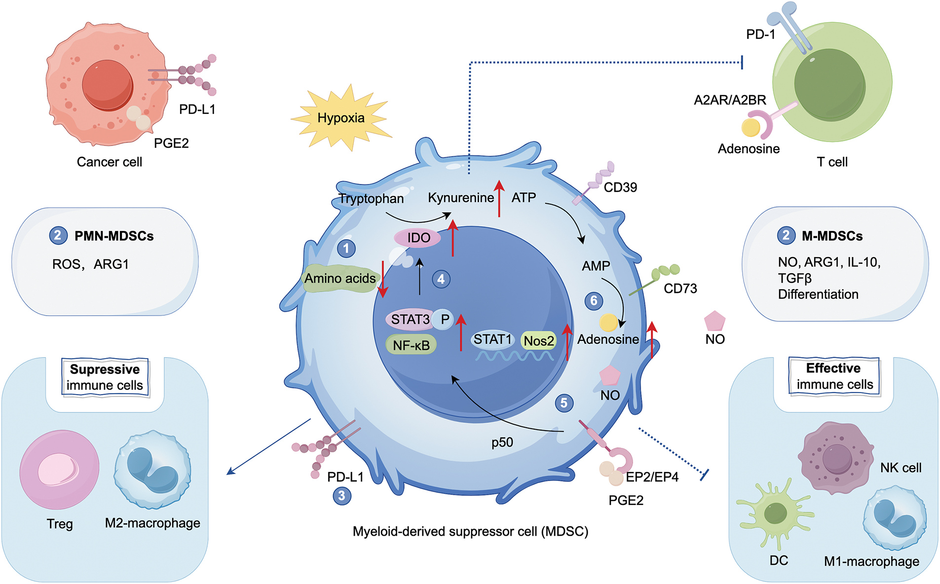
Figure 1: Function of myeloid-derived suppressor cells (MDSCs) in cancer immunosuppression. MDSCs play a pivotal role in the tumor microenvironment (TME) by suppressing effective immune cells such as Natural Killer cells (NK cells), M1 macrophages, and dendritic cells (DCs) and cooperating with other immunosuppressive cells, including Regulatory T cells (Tregs) and M2 macrophages, with T cells serving as the primary target. MDSCs suppress T cell activity through the following mechanisms: (1) depletion of amino acids, (2) secretion of cytokines that inhibit T cell proliferation and function: Polymorphonuclear MDSCs (PMN-MDSCs) primarily secrete reactive oxygen species (ROS) and arginase1 (ARG1), while monocytic MDSCs (M-MDSCs) secrete NO, ARG1, IL-10, and TGFβ, (3) expression of PD-L1, (4) activation of STAT3 and NF-κB signaling pathways, leading to the release of indoleamine 2,3-dioxygenase (IDO), which promotes the conversion of tryptophan to kynurenine, (5) promotion of nitric oxide (NO): Tumor-derived prostaglandin E2 (PGE2)-mediated induction of nuclear p50 nuclear factor κB (NF-κB) facilitates signal transducer and activator of transcription 1 (STAT1) binding to the regulatory regions of IFNγ-dependent genes, such as inducible nitric oxide synthase (Nos2), (6) upregulation of CD39/CD73, facilitating ATP dephosphorylation to adenosine. Adenosine can bind to A2A receptor (A2AR) and A2BR receptors on T cells, suppressing their activity and activating Tregs. Created with FigDraw.
MDSCs exhibit significant plasticity throughout tumor progression and in response to therapeutic stress, adapting their phenotype and function to the TME. PMN-MDSCs are key mediators of immune suppression, promoting T-cell tolerance via ROS and ARG1. In contrast, M-MDSCs secrete immunosuppressive factors like NO, ARG1, IL-10, and TGFβ, inhibiting both antigen-specific and non-specific T-cell responses. Moreover, M-MDSCs, similar to monocytes, can differentiate into other immune cells, enhancing their immune modulatory functions [28].
Differentiation of MDSCs into TAMs
Macrophages are vital to both innate immunity and adaptive immunity. They are highly plastic and adaptable in their functions. The phenotypes of macrophages remain challenging to distinguish. Therefore, most studies use the concept of polarization to divide them into two extremes: M1 macrophages and M2 macrophages [29]. However, it is still insufficient to differentiate macrophages only by polarization. Like a color palette, different combinations of spectral patterns give rise to various macrophage subtypes. M-MDSCs can be distinguished from TAMs by higher F4/80 and CSF receptor CD115 expression, lower to moderate Ly6C, and reduced or undetectable S100A9 and IRF8 levels [7]. Single-cell RNA sequencing (scRNA-seq) analysis revealed the presence of cells expressing markers for M-MDSCs, such as IL10, CD14, and VEGFA, as well as markers for PMN-MDSCs, including IL6, oxidized low-density lipoprotein receptor 1 (OLR1), and TGFB1, during the transition from monocytes to M2-like cells. These findings highlight that MDSCs are molecularly distinct from M1/M2 macrophages [30]. Specially, MDSC-derived macrophages have stronger immunosuppressive functions, whereas monocyte-derived macrophages (mo-Macs) tend to exhibit limited immunosuppressive activity, possibly related to S100A8/A9 [22,31].
The differentiation of MDSCs into macrophages is tissue-dependent. MDSCs differentiate into macrophages in mouse spleen, whereas they rapidly differentiate into TAMs in tumor tissue. This process is regulated by hypoxia which functions in the development of MDSCs and TAMs. In the TME, hypoxia activates CD45 protein tyrosine phosphatases (CD45PTP), which inhibit STAT3 and promote the differentiation of MDSCs to TAMs [32]. STAT3 promotes the amplification of MDSCs. IL-6 activates the JAK/STAT3 pathway, leading to the induction of PMN-MDSCs to produce and secrete miR-93-5p. This miRNA subsequently inhibits STAT3, drives the differentiation of M-MDSCs into M2 macrophages, and accelerates the transition from colitis to cancer [33]. This indicates that STAT3 dephosphorylation is the prerequisite of the further differentiation of MDSCs. IRF8 and PU.1 are important transcription factors in macrophage differentiation. Retinoic-acid-related orphan receptor (RORC1), a critical driver of tumor-induced emergency hematopoiesis, regulates MDSCs differentiation by inhibiting negative regulators and promoting positive regulators such as C/EBPβ. Through IRF8 and PU.1, RORC1 promotes MDSCs differentiate into macrophages [34]. Furthermore, GM-CSF and M-CSF drive the differentiation of MDSCs into M2-TAMs [35]. Extracellular ROS promote monocyte differentiation into macrophages and M2 polarization, while high ROS levels induced by NADPH oxidase 2 inhibit MDSCs differentiation [19]. Earlier studies have demonstrated that the differentiation of immature myeloid cells (IMCs) into TAMs and DCs is enhanced in mice cultured after removing ROS [36] (Fig. 2).
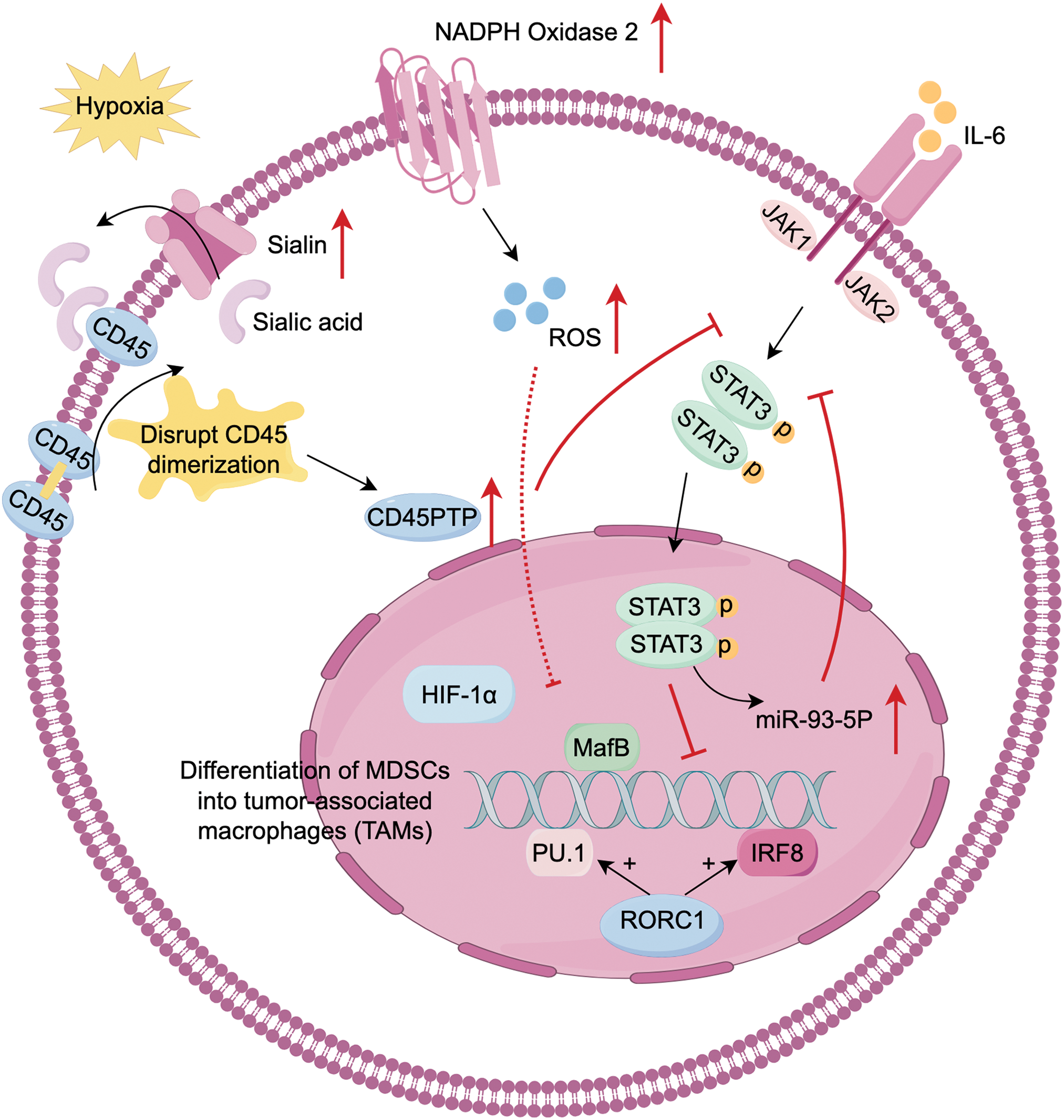
Figure 2: Differentiation of MDSCs into tumor-associated macrophages (TAMs). MDSCs plasticity is strongly influenced by the hypoxic TME, with STAT3 downregulation serving as a prerequisite for differentiation. (1) In hypoxia conditions, MDSCs upregulate sialin, facilitating the transport of sialic acid to the cell surface. Sialic acid binds with CD45 and disrupt CD45 dimerization. These activates CD45 protein tyrosine phosphatase (CD45PTP), leading to STAT3 dephosphorylation. (2) IL-6/JAK/STAT3 produce miR-93-5p, which inhibits STAT3 phosphorylation. (3) IRF8, PU.1, and musculoaponeurotic fibrosarcoma oncogene homolog B (MafB) are key transcription factors driving myeloid progenitor differentiation to the monocytic/macrophage lineage. Retinoic-acid-related orphan receptor (RORC1), a driver of emergency hematopoiesis in cancer, enhances IRF8 and PU.1 expression. (4) Extracellular reactive oxygen species (ROS) promotes monocyte differentiation and M2 polarization, but high ROS levels from NADPH oxidase 2 (NOX2) in MDSCs inhibit their differentiation into TAMs. Created with FigDraw.
Lactate notably impacts the differentiation and development of MDSCs and TAMs. Lactate promotes hypoxia-inducible factor 1 subunit alpha (HIF-1α)-mediated polarization of M2-TAMs and decreases the presence of M1 macrophage markers [37,20]. Zhao et al. identify the Notch/RBP-J pathway, which regulates lactic acid metabolism in myeloid cells to promote M1 macrophage polarization. However, subsequent research indicates that lactate promotes the differentiation of M-MDSCs into PMN-MDSCs, rather than M1 macrophages [38]. In prostate cancer model, the acidic environment induced by lactate favors M2 polarization [39]. In general, the metabolic activities of MDSCs, especially lactic acid metabolism, significantly influence macrophage polarization, driving their shift to TAMs. However, specific mechanisms may differ due to aspects like cell type, tissue environment, and disease state.
Differentiation of MDSCs into DCs
Dendritic cells are the most functional antigen-presenting cells (APCs) affecting intrinsic and adaptive immune systems. DCs present protein fragments from bacteria, viruses, and tumors on the cell surface major histocompatibility complex I (MHCI) molecules, which are recognized by cytotoxic T lymphocytes and NK cells to initiate adaptive neo-immune responses. DCs are broadly divided into two primary types according to their origin: plasmacytoid DCs (pDCs) and conventional DCs (cDCs). cDCs have unique differentiation and APC properties. The differentiation of cDC1 is dependent on IRF8, which primarily regulates their cell-killing effects on CTLs [40]. Conversely, cDC2 differentiation relies on IRF4, which preferentially activates CD4+ T cells [41].
Transcription factors such as STAT3, PU.1, and the IRF family regulate DC differentiation. In the presence of M-CSF, immature monocytes naturally differentiate into mo-Macs. However, when IRF4 is induced by IL-4 and TNF-α, and aryl hydrocarbon receptor (AHR) ligands are bound, these monocytes instead differentiate into monocyte-derived DCs (mo-DCs). The AHR is necessary but not sufficient for DCs differentiation [35,42]. In murine sarcoma models, tumor cells produce retinoic acid, which promotes monocyte differentiation into TAMs not DCs by inhibiting DCs-promoting transcription factor IRF4 [41]. STAT3 activation inhibits myeloid cells from developing into mature forms, which is evident in the expansion of MDSCs and reduced differentiation into DCs in the TME [43]. In vitro in bone marrow cultures and tumor-bearing mice, STAT3 inhibition due to VISTA deletion results in decreased differentiation of monocytes into MDSCs and expansion of DCs [14]. The differentiation of DCs is dependent on FMS-like tyrosine kinase 3 (Flt3) and FMS-like tyrosine kinase 3 ligand (Flt3L) [44]. PU.1 promotes Flt3 expression and synergistically regulates the development of DCs. In immune checkpoint blockade (ICB)-resistant non-small cell lung cancer (NSCLC), PU.1 and Flt3 expression are reduced, leading to inhibited differentiation of classical Ly6C+ monocytes into DCs/TAMs [45].
DC metabolism in TME is distinguished by a substantial accumulation of neutral lipids linked to the reduced ability to cross-present exogenous antigens, including tumor-specific ones. MDSCs produce abundant lipid peroxidation in the TME. PMN-MDSCs upregulate fatty acid transporter protein 2 (FATP2), and produce oxidized lipids via myeloperoxidase (MPO) and ROS, leading to the cross-presentation function of DCs inhibited [46]. DCs present tumor cell antigens to CD4+ T cells through the Jak/STAT1 signaling pathway. NO produced by MDSCs induces nitration of STAT1 at Tyrosine701 and inhibits STAT1 phosphorylation by reducing immune reactivity to IFN [47] (Fig. 3).
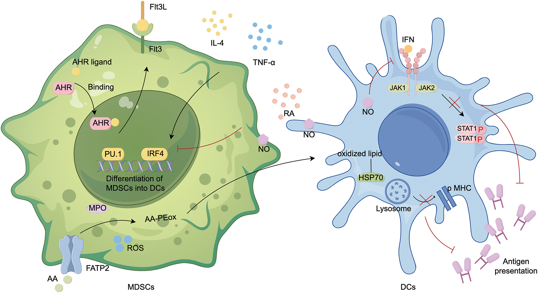
Figure 3: Differentiation of MDSCs into DCs. MDSCs differentiate into DCs under two key conditions: IL-4 or TNF-α activation of IRF4, and AHR binding to ligands. In addition, PU.1 and IRF4 are critical transcription factors that regulate this process. PU.1 promotes Flt3 expression and synergistically regulates the development of DCs. MDSCs are more likely to differentiate into TAMs than DCs, and their primary role is to inhibit DCs antigen presentation: (1) PMN-MDSCs upregulate fatty acid transport protein 2 (FATP2), leading to the uptake of arachidonic acid (AA). In the presence of ROS and myeloperoxidase (MPO), AA undergoes lipid peroxidation to form AA-PEox. These oxidized lipids are then transferred to DCs, where they bind to heat shock proteins (HSPs), preventing the release of peptide-major histocompatibility complex (pMHC) on the cell surface. (2) NO from MDSCs reduces DCs sensitivity to IFN, inhibiting the JAK/STAT1 pathway and preventing DCs from presenting antigens to CD4+ T cells. PE, phosphatidylethanolamine; ox, oxidized; created with FigDraw.
In conclusion, MDSCs employ various mechanisms, including producing oxidized lipids or increasing NO, to inhibit DCs from recognizing and presenting antigens. Even when MDSCs differentiate into DCs under the impact of specific cytokines, the antigen presentation capacity of MDSC-DCs will be reduced, which is an adaptation to the immunosuppressive settings [48].
Plasticity of MDSCs in Cancer Therapy
The advent of immunotherapies like immune checkpoint inhibitors (ICIs), CAR-T cell therapies, and tumor vaccines has markedly improved outcomes for patients with malignant neoplasms. However, drug resistance and suboptimal responses persist. MDSCs are crucial in tumor immunosuppression, though their specific markers remain unclear. The ability to multidirectional differentiation, metabolic reprogramming, and crosstalk with other cells in the TME has spurred therapeutic strategies for direct targeting and combination therapies.
All-trans retinoic acid (ARTA), the major bioactive isoform of retinoic acid [41], decreases immunosuppression and improves antitumor response by reducing the level of ARG1, iNOS, IDO, ROS, and S100A8/A9 to inhibit the function of MDSCs [49]. In addition, ARTA indirectly promotes the accumulation of glutathione by activating the extracellular signal-regulated kinase 1/2 (ERK1/2) pathway, thereby neutralizing the high levels of ROS in MDSCs and facilitating their differentiation [50]. In metastatic melanoma, ATRA significantly reduces PMN-MDSCs and increases HLA−DR+ myeloid cells, enhancing the sensitivity and efficiency of pembrolizumab [51]. In mouse cervical cancer, ATRA inhibits the accumulation of MDSCs and increases the infiltration of cytotoxic CD8+ T cells [52]. In LKB1-deficent murine tumors, ATRA inhibits immunosuppressive MDSCs, enhances T cell cytotoxicity, and increases sensitivity to PD-1 inhibition, thereby increasing tumor sensitivity to ICBs [53]. These findings collectively indicate that ATRA has considerable potential as an immunomodulator and may serve as an adjunctive therapy for immunotherapy.
In metastatic renal cell carcinoma, very small size particle (VSSP), a new immune modulator, lowers the occurrence of PMN-MDSCs and elevates the levels of monocytes and DCs, with high expression of IRF8 and PU.1 [54]. Glutamine metabolism is essential for nucleotide synthesis, amino acid production, and redox balance. Besides IDO inhibition, targeting glutamine metabolism promotes the differentiation of MDSCs into pro-inflammatory TAMs [55]. These suggest that targeting specific molecules or remodeling TME metabolism can promote the differentiation of MDSCs, which is beneficial to enhance theoretical effectiveness. Multiple proceeding or completed clinical trials explore the effects of promoting MDSCs differentiation into TAMs or DCs (Table 1).
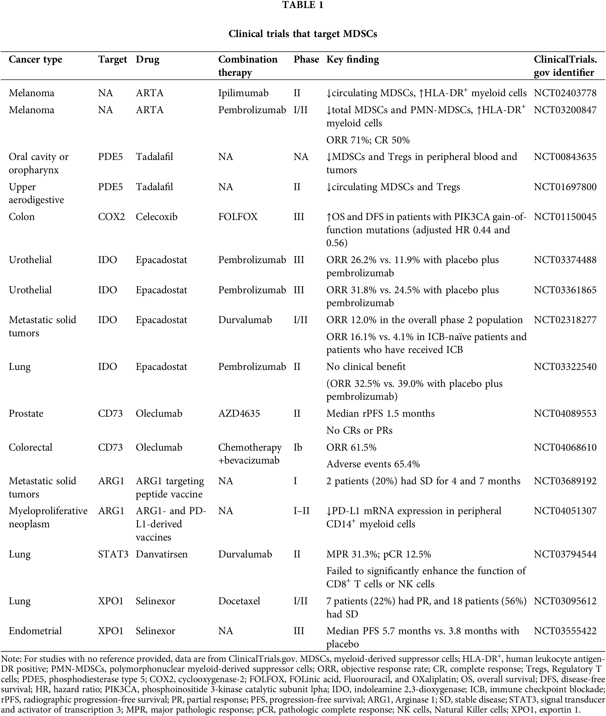
Although the particular markers for MDSCs are still unidentified, targeting certain signaling pathways or cytokines has demonstrated distinct effects on MDSCs. IDO1, CD39/CD73, Phosphodiesterase-5 (PDE5), ARG1, COX2 and STAT3 are important therapeutic targets for MDSCs. IDO is a key factor influencing MDSCs function. The IDO/PD-L1 vaccine combined with nivolumab shows sustained and promising efficacy in long-term follow-up, with an overall response rate (ORR) of 80%, and 50% of patients achieving a complete response (CR) [56]. Epacadostat (IDO inhibitor) has shown promise when combined with PD-1 inhibitors in cancers such as melanoma, NSCLC, and urogenital cancers [57–59]. However, in some trials, the results have fallen short. For example, a phase II NSCLC study (NCT03322540) finds no improvement in progression-free survival (PFS) or ORR compared to placebo. These could be due to a small sample size, short follow-up time, and epacadostat doses.
Targeting CD73 or adenosine receptors can block adenosine activity but may also enhance the chemotaxis of MDSCs. For example, in murine pancreatic ductal adenocarcinoma models, AB680 (CD73 inhibitor) increases the chemotaxis of MDSCs through CXCL5 in an AMP-dependent manner [60]. This suggests that chemokine inhibitors are essential to inhibit the aggregation of MDSCs and improve drug response. In a mouse triple-negative breast cancer model, dual blockade of CD73 and TGFβ reprograms the tumor microenvironment by reducing MDSCs and M2 macrophages, while increasing activated Tregs, CD8+ T cells, and B cells [61]. Currently, targeting CD73 or adenosine could be a supplement to immunotherapy, but targeting it alone may increase the chemotaxis of MDSCs, leading to limited significant efficacy.
PDE5 inhibitors, such as tadalafil, inhibit MDSCs by lowering levels of iNOS and ARG1. In addition, tadalafil prevents M2 polarization and inhibits polyamine metabolism in TAMs and MDSCs, effectively reversing the immunosuppressive TME and improving the efficacy of ICB in hepatocellular carcinoma [62]. Tadalafil has shown promising clinical results in trials for head and neck squamous cell carcinoma (HNSCC), glioblastoma, and melanoma [63–65]. STAT3 is crucial in MDSCs differentiation, expansion, and activation. Therefore, targeting STAT3 offers a promising avenue for targeting MDSCs. In melanoma model, napabucasin (STAT3 inhibitor) promotes apoptosis of MDSCs in human peripheral blood and mouse bone marrow, thereby enhancing antitumor immunity and prognosis [43]. Furthermore, exportin-1 (XPO1), a key nuclear transport protein, mediates the export of various cargo proteins from the nucleus. Due to its central role in cellular regulation, blocking XPO1-mediated nuclear export has emerged as a promising therapeutic strategy in cancer. IL-6 activates both the JAK/STAT3 and JAK/MAPK signaling pathways in MDSCs. Selinexor (XPO1 inhibitor) exerts a dual effect on MDSCs differentiation. On one hand, it promotes the conversion of MDSCs into neutrophil-like cells with immune-activating properties by intervening in the ERK1/2 signaling within the MAPK pathway. On the other hand, it decreases the expression of immunosuppressive markers and reduces the immunosuppressive function of MDSCs [66]. Selinexor has demonstrated efficacy in clinical trials (NCT03147885, NCT03555422, NCT02269293), particularly in hematologic malignancies and advanced solid tumors (e.g., endometrial and ovarian cancers), especially when used in combination therapies.
In conclusion, modulating signaling pathways to remove MDSCs is increasingly central to cancer immunotherapy. The new target EP2/4 is currently being evaluated in several clinical trials (NCT06129604, NCT05940571). There are active clinical trials targeting MDSCs, and a deeper understanding of their role in combination therapies is needed (Table 1).
Chemotherapy kills tumor cells and surrounding cells, including MDSCs. Moreover, some chemotherapeutic drugs trigger cancer cells to release signaling molecules that activate APCs and induce immunogenic cell death (ICD) [67]. Chemotherapeutic drugs suppress MDSCs, enhance T-cell responses, and improve the immunosuppression of the TME. Cisplatin represses MDSCs expansion by suppressing STAT3/COX-2 signaling and enhances T-cell responses in melanoma and HNSCC patients [68]. Gemcitabine and celecoxib (COX2 inhibitors) induce apoptosis in MDSCs (9.9% and 6.9%), with apoptosis rates increasing to 21.0% after combined application [67]. This shows that chemotherapy and immunotherapy have synergistic effects in depleting MDSCs.
However, the immunosuppressive nature of the TME decreases such immune response. Chemotherapeutic agents can also generate tumor resistance by upregulating MDSCs. Cisplatin, oxaliplatin, and adriamycin can increase oxidized 1-palmitoyl-2-arachidonoyl-sn-glycero-3-phosphocholine (oxPAPC) release in tumors. In WT LL2-bearing mice, a significant up-regulation of MDSCs in the TME is observed after oxPAPC injection [69]. In a ovarian cancer mouse model, carboplatin increases M-MDSCs and M2 TAMs, while carboplatin-paclitaxel do not reveal any immunological alterations [70]. Nevertheless, Paclitaxel resistance hinders its effectiveness in breast cancer. Combing with Rg3-based liposomes not only targets tumor cells but also repolarizes pro-tumor M2 macrophages to the antitumor M1 phenotype and suppress MDSCs, thereby remodeling the TME [71]. Different impacts of MDSCs from chemotherapeutic drugs may result from tumor-specific immunobiology and population immune heterogeneity.
Radiotherapy and ICD in various cancers are aimed directly at inducing DNA damage and tumor cell apoptosis in the TME [72]. The YTHN6-methyladenosine RNA binding protein 2 (YTHDF2), a major m6A RNA-binding protein, is deleted after ionizing radiation (IR) in mouse M-MDSCs. This results in M-MDSCs differentiating more into M1 macrophages and enhanced antitumor immunity [73]. Radiotherapy has contradictory effects on the suppression and promotion of tumor progression. Local irradiation increases systemic MDSCs, enhances immune suppression, and promotes tumor metastasis [74]. STING activation by radiation promotes the recruitment of MDSCs through the CCR2 pathway and enhanced antitumor immunity [75]. Together, extrinsic radio resistance is associated with MDSCs. MDSCs are a critical factor in the generation of immune tolerance. Targeting MDSCs is beneficial to improving therapeutic efficacy. However, the mechanism by which radiotherapy affects MDSCs, and the clinical benefit of combination therapy still need further experimental studies to validate.
Targeted therapy aims to inhibit tumor progression by targeting specific genetic mutations or molecules while minimizing side effects. Targeted therapies can modify the function and phenotype of MDSCs to improve antitumor immunity, especially in combination with ICIs. For example, demethylation inhibitor decitabine promotes MDSCs differentiation into M1 macrophages by upregulating IRF7. Decitabine also activates innate immune pathways, including NOD-like signaling, thereby enhancing antitumor immune responses [76]. Guadecitabine is a dinucleotide prodrug of decitabine that inhibits DNA Methyltransferase 1 (DNMT1) and induces hypomethylation. In a breast cancer mouse model, guadecitabine eliminates most MDSCs and induces a subset to express APC markers and co-stimulatory molecules like major histocompatibility complex II (MHCII) and CD80/86 [77]. Furthermore, histone deacetylase inhibitors CN133 inhibits the recruitment of PMN-MDSCs by downregulating genes associated with their migration, which restores a positive immune environment and significantly enhances the efficacy of PD-1 therapy in prostate cancer [78]. In a mouse lung metastasis model, low doses DNA methyltransferase, histone deacetylase inhibitors, 5-azacytidine and entinostat promote the differentiation of MDSCs into macrophage, disrupting the pre-metastatic immunosuppressive environment [79]. These findings highlight that epigenetic mechanisms can reshape MDSCs function and improve immune responses in the TME.
In addition, Bruton’ s tyrosine kinase (BTK) inhibitor ibrutinib, increases the frequency of M-MDSCs and reduces chemokines such as CCL2, CCL3 involved in MDSCs recruitment [80]. However, acalabrutinib (another BTK inhibitor) has shown limited efficacy in pancreatic cancer patients. While it reduces PMN-MDSCs and induces T cell activation, ORR and disease control rate (DCR) for monotherapy were only 0% and 14.3%. BTK inhibitors may require combination with other immune therapies to achieve optimal therapeutic outcomes [81]. Sunitinib (multi-target tyrosine kinase inhibitor) has been associated with MDSCs expansion and immune resistance. In mouse models, the combination of sorafenib and tazemetostat (enhancer of zeste homolog 2 inhibitor) significantly reduces MDSCs and Treg populations, promotes T cell infiltration, and reverses immune resistance in HNSCC [82].
In summary, the plasticity of MDSCs in targeted therapy represents a promising avenue for improving antitumor immunity. By remodulating MDSCs function and phenotype, these therapies offer potential to enhance the efficacy of immune therapies and reverse immune resistance, providing new insights for future cancer treatment strategies.
The plasticity of MDSCs plays a crucial role in tumor immune evasion. In the TME, MDSCs respond to a complex network of cytokines and signaling pathways that regulate their differentiation, phenotype, and function. Especially, this plasticity enables MDSCs to differentiate into various immune cell types, such as TAMs and DCs, depending on the specific conditions of the tissue or microenvironment. Hypoxia and acidic environment, along with the downregulation of STAT3 activity, promote MDSCs differentiation into TAMs, while MDSCs differentiation into DCs is less frequent and associated with a diminished antigen-presenting capacity. Additionally, metabolic factors, including glutamine metabolism and ROS levels, along with epigenetic modifications such as m6A methylation, significantly influence MDSCs plasticity and their immunosuppressive function. Despite the therapeutic potential of targeting MDSCs to overcome tumor immune evasion, the clinical translation of such therapies faces several challenges. These include the lack of specific targeting markers, the complexity of the TME, and immune resistance. Therefore, future research should focus on further elucidating the molecular mechanisms driving MDSCs plasticity, particularly the signaling and metabolic pathways involved, as well as developing more precise targeting strategies. The rise of single-cell analysis and high-throughput sequencing technologies offers promising avenues for a deeper understanding of MDSCs heterogeneity and the identification of novel therapeutic targets. In conclusion, MDSCs plasticity is a critical driver of tumor immune escape and a promising target for therapeutic intervention. By precisely modulating MDSCs differentiation and function, it may be possible to overcome current treatment limitations and enhance the efficacy of immune therapies. Future research should aim to translate these findings into clinical strategies to improve cancer treatment outcomes.
Acknowledgement: None.
Funding Statement: This work was supported by the Scientific Research Project of Jiangsu Commmission of Health (No. M2021108).
Author Contributions: The authors confirm their contribution to the paper as follows: study conception and design: Jiajia Lv, Xiaoyou Zhong; draft manuscript preparation: Jiajia Lv, Xiaoyou Zhong; review and editing: Lin Wang, Weifei Fan; visualization: Jiajia Lv; supervision: Weifei Fan. All authors reviewed the results and approved the final version of the manuscript.
Availability of Data and Materials: Not applicable.
Ethics Approval: Not applicable.
Conflicts of Interest: The authors declare no conflicts of interest to report regarding the present study.
References
1. Farahzadi R, Valipour B, Montazersaheb S, Fathi E. Targeting the stem cell niche micro-environment as therapeutic strategies in aging. Front Cell Dev Biol. 2023;11:1162136. doi:10.3389/fcell.2023.1162136. [Google Scholar] [PubMed] [CrossRef]
2. Lasser SA, Ozbay Kurt FG, Arkhypov I, Utikal J, Umansky V. Myeloid-derived suppressor cells in cancer and cancer therapy. Nat Rev Clin Oncol. 2024;21(2):147–64. doi:10.1038/s41571-023-00846-y. [Google Scholar] [PubMed] [CrossRef]
3. Veglia F, Perego M, Gabrilovich D. Myeloid-derived suppressor cells coming of age. Nat Immunol. 2018;19(2):108–19. doi:10.1038/s41590-017-0022-x. [Google Scholar] [PubMed] [CrossRef]
4. Grover A, Sanseviero E, Timosenko E, Gabrilovich DI. Myeloid-derived suppressor cells: a propitious road to clinic. Cancer Discov. 2021;11(11):2693–706. doi:10.1158/2159-8290.CD-21-0764. [Google Scholar] [PubMed] [CrossRef]
5. Zhou J, Nefedova Y, Lei A, Gabrilovich D. Neutrophils and PMN-MDSC: their biological role and interaction with stromal cells. Semin Immunol. 2018;35(3):19–28. doi:10.1016/j.smim.2017.12.004. [Google Scholar] [PubMed] [CrossRef]
6. Pettinella F, Mariotti B, Lattanzi C, Bruderek K, Donini M, Costa S, et al. Surface CD52, CD84, and PTGER2 mark mature PMN-MDSCs from cancer patients and G-CSF-treated donors. Cell Rep Med. 2024;5(2):101380. doi:10.1016/j.xcrm.2023.101380. [Google Scholar] [PubMed] [CrossRef]
7. Bronte V, Brandau S, Chen SH, Colombo MP, Frey AB, Greten TF, et al. Recommendations for myeloid-derived suppressor cell nomenclature and characterization standards. Nat Commun. 2016;7(1):12150. doi:10.1038/ncomms12150. [Google Scholar] [PubMed] [CrossRef]
8. Nepal MR, Shah S, Kang KT. Dual roles of myeloid-derived suppressor cells in various diseases: a review. Arch Pharm Res. 2024;47(7):597–616. doi:10.1007/s12272-024-01504-2. [Google Scholar] [PubMed] [CrossRef]
9. Wang P, Xu L, Bai M, Zheng X, Song J, Xie Y, et al. MDSCs are important osteoclast precursors primed by B cells in rheumatoid arthritis. Eur J Immunol. 2024;54(9):e2350823. doi:10.1002/eji.202350823. [Google Scholar] [PubMed] [CrossRef]
10. Zhao Q, Huang L, Qin G, Qiao Y, Ren F, Shen C, et al. Cancer-associated fibroblasts induce monocytic myeloid-derived suppressor cell generation via IL-6/exosomal miR-21-activated STAT3 signaling to promote cisplatin resistance in esophageal squamous cell carcinoma. Cancer Lett. 2021;518:35–48. doi:10.1016/j.canlet.2021.06.009. [Google Scholar] [PubMed] [CrossRef]
11. Mohamed E, Sierra RA, Trillo-Tinoco J, Cao Y, Innamarato P, Payne KK, et al. The unfolded protein response mediator PERK governs myeloid cell-driven immunosuppression in tumors through inhibition of STING signaling. Immunity. 2020;52(4):668–82.e7. doi:10.1016/j.immuni.2020.03.004. [Google Scholar] [PubMed] [CrossRef]
12. van Vlerken-Ysla L, Tyurina YY, Kagan VE, Gabrilovich DI. Functional states of myeloid cells in cancer. Cancer Cell. 2023;41(3):490–504. doi:10.1016/j.ccell.2023.02.009. [Google Scholar] [PubMed] [CrossRef]
13. Cui H, Lan Z, Zou K-L, Zhao Y-Y, Yu G-T. STAT3 promotes differentiation of monocytes to MDSCs via CD39/CD73-adenosine signal pathway in oral squamous cell carcinoma. Cancer Immunol, Immunother. 2023;72(5):1315–26. doi:10.1007/s00262-022-03336-9. [Google Scholar] [PubMed] [CrossRef]
14. Zhang K, Zakeri A, Alban T, Dong J, Ta HM, Zalavadia AH, et al. VISTA promotes the metabolism and differentiation of myeloid-derived suppressor cells by STAT3 and polyamine-dependent mechanisms. Cell Rep. 2024;43(1):113661. doi:10.1016/j.celrep.2023.113661. [Google Scholar] [PubMed] [CrossRef]
15. Liang KL, Laurenti E, Taghon T. Circulating IRF8-expressing CD123+CD127+ lymphoid progenitors: key players in human hematopoiesis. Trends Immunol. 2023;44(9):678–92. doi:10.1016/j.it.2023.07.004. [Google Scholar] [PubMed] [CrossRef]
16. Hashimoto A, Sarker D, Reebye V, Jarvis S, Sodergren MH, Kossenkov A, et al. Upregulation of C/EBPα inhibits suppressive activity of myeloid cells and potentiates antitumor response in mice and patients with cancer. Clin Cancer Res. 2021;27(21):5961–78. doi:10.1158/1078-0432.CCR-21-0986. [Google Scholar] [PubMed] [CrossRef]
17. Li BH, Garstka MA, Li ZF. Chemokines and their receptors promoting the recruitment of myeloid-derived suppressor cells into the tumor. Molecular Immunol. 2020;117(4):201–15. doi:10.1016/j.molimm.2019.11.014. [Google Scholar] [PubMed] [CrossRef]
18. Reschke R, Enk AH, Hassel JC. Chemokines and cytokines in immunotherapy of melanoma and other tumors: from biomarkers to therapeutic targets. Int J Mol Sci. 2024;25(12):6532. doi:10.3390/ijms25126532. [Google Scholar] [PubMed] [CrossRef]
19. Yu Y, Liu S, Yang L, Song P, Liu Z, Liu X, et al. Roles of reactive oxygen species in inflammation and cancer. MedComm. 2024;5(4):e519. doi:10.1002/mco2.519. [Google Scholar] [PubMed] [CrossRef]
20. Goldmann O, Medina E. Metabolic pathways fueling the suppressive activity of myeloid-derived suppressor cells. Front Immunol. 2024;15:1461455. doi:10.3389/fimmu.2024.1461455. [Google Scholar] [PubMed] [CrossRef]
21. Veglia F, Sanseviero E, Gabrilovich DI. Myeloid-derived suppressor cells in the era of increasing myeloid cell diversity. Nat Rev Immunol. 2021;21(8):485–98. doi:10.1038/s41577-020-00490-y. [Google Scholar] [PubMed] [CrossRef]
22. Kwak T, Wang F, Deng H, Condamine T, Kumar V, Perego M, et al. Distinct populations of immune-suppressive macrophages differentiate from monocytic myeloid-derived suppressor cells in cancer. Cell Rep. 2020;33(13):108571. doi:10.1016/j.celrep.2020.108571. [Google Scholar] [PubMed] [CrossRef]
23. He ZN, Zhang CY, Zhao YW, He SL, Li Y, Shi BL, et al. Regulation of T cells by myeloid-derived suppressor cells: emerging immunosuppressor in lung cancer. Discov Oncol. 2023;14(1):185. doi:10.1007/s12672-023-00793-1. [Google Scholar] [PubMed] [CrossRef]
24. Cuenca-Escalona J, Subtil B, Garcia-Perez A, Cambi A, De Vries IJM, Flórez-Grau G. EP2 and EP4 blockade prevents tumor-induced suppressive features in human monocytic myeloid-derived suppressor cells. Front Immunol. 2024;15:1355769. doi:10.3389/fimmu.2024.1355769. [Google Scholar] [PubMed] [CrossRef]
25. Porta C, Consonni FM, Morlacchi S, Sangaletti S, Bleve A, Totaro MG, et al. Tumor-derived prostaglandin E2 promotes p50 NF-κB-dependent differentiation of monocytic MDSCs. Cancer Res. 2020;80(13):2874–88. doi:10.1158/0008-5472.CAN-19-2843. [Google Scholar] [PubMed] [CrossRef]
26. Nandre R, Verma V, Gaur P, Patil V, Yang X, Ramlaoui Z, et al. IDO vaccine ablates immune-suppressive myeloid populations and enhances antitumor effects independent of tumor cell IDO status. Cancer Immunol Res. 2022;10(5):571–80. doi:10.1158/2326-6066.CIR-21-0457. [Google Scholar] [PubMed] [CrossRef]
27. Pagni RL, Souza PDC, Pegoraro R, Porchia BFMM, da Silva JR, Aps LRMM, et al. Interleukin-6 and indoleamine-2, 3-dioxygenase as potential adjuvant targets for papillomavirus-related tumors immunotherapy. Front Immunol. 2022;13:1005937. doi:10.3389/fimmu.2022.1005937. [Google Scholar] [PubMed] [CrossRef]
28. Gabrilovich DI, Nagaraj S. Myeloid-derived-suppressor cells as regulators of the immune system. Nature Rev Immunol. 2009;9(3):162–74. doi:10.1038/nri2506. [Google Scholar] [PubMed] [CrossRef]
29. Li M, Yang Y, Xiong L, Jiang P, Wang J, Li C. Metabolism, metabolites, and macrophages in cancer. J Hematol Oncol. 2023;16(1):80. doi:10.1186/s13045-023-01478-6. [Google Scholar] [PubMed] [CrossRef]
30. Song Q, Hawkins GA, Wudel L, Chou PC, Forbes E, Pullikuth AK, et al. Dissecting intratumoral myeloid cell plasticity by single cell RNA-seq. Cancer Med. 2019;8(6):3072–85. doi:10.1002/cam4.2113. [Google Scholar] [PubMed] [CrossRef]
31. Tang X, Gao L, Jiang X, Hou Z, Wang Y, Hou S, et al. Single-cell profiling reveals altered immune landscape and impaired NK cell function in gastric cancer liver metastasis. Oncogene. 2024;43(35):2635–46. doi:10.1038/s41388-024-03114-0. [Google Scholar] [PubMed] [CrossRef]
32. Kumar V, Cheng P, Condamine T, Mony S, Languino LR, McCaffrey JC, et al. CD45 phosphatase inhibits STAT3 transcription factor activity in myeloid cells and promotes tumor-associated macrophage differentiation. Immunity. 2016;44(2):303–15. doi:10.1016/j.immuni.2016.01.014. [Google Scholar] [PubMed] [CrossRef]
33. Wang Y, Liu H, Zhang Z, Bian D, Shao K, Wang S, et al. G-MDSC-derived exosomes mediate the differentiation of M-MDSC into M2 macrophages promoting colitis-to-cancer transition. J Immunother Cancer. 2023;11(6):e006166. doi:10.1136/jitc-2022-006166. [Google Scholar] [PubMed] [CrossRef]
34. Strauss L, Sangaletti S, Consonni FM, Szebeni G, Morlacchi S, Totaro MG, et al. RORC1 regulates tumor-promoting emergency granulo-monocytopoiesis. Cancer Cell. 2015;28(2):253–69. doi:10.1016/j.ccell.2015.07.006. [Google Scholar] [PubMed] [CrossRef]
35. Goudot C, Coillard A, Villani AC, Gueguen P, Cros A, Sarkizova S, et al. Aryl hydrocarbon receptor controls monocyte differentiation into dendritic cells versus macrophages. Immunity. 2017;47(3):582–96. doi:10.1016/j.immuni.2017.08.016. [Google Scholar] [PubMed] [CrossRef]
36. Kusmartsev S, Gabrilovich DI. Inhibition of myeloid cell differentiation in cancer: the role of reactive oxygen species. J Leukocyte Biol. 2003;74(2):186–96. doi:10.1189/jlb.0103010. [Google Scholar] [PubMed] [CrossRef]
37. Colegio OR, Chu N-Q, Szabo AL, Chu T, Rhebergen AM, Jairam V, et al. Functional polarization of tumour-associated macrophages by tumour-derived lactic acid. Nature. 2014;513(7519):559–63. doi:10.1038/nature13490. [Google Scholar] [PubMed] [CrossRef]
38. Zhao J-L, Ye Y-C, Gao C-C, Wang L, Ren K-X, Jiang R, et al. Notch-mediated lactate metabolism regulates MDSC development through the Hes1/MCT2/c-Jun axis. Cell Rep. 2022;38(10):110451. doi:10.1016/j.celrep.2022.110451. [Google Scholar] [PubMed] [CrossRef]
39. El-Kenawi A, Gatenbee C, Robertson-Tessi M, Bravo R, Dhillon J, Balagurunathan Y, et al. Acidity promotes tumour progression by altering macrophage phenotype in prostate cancer. Br J Cancer. 2019;121(7):556–66. doi:10.1038/s41416-019-0542-2. [Google Scholar] [PubMed] [CrossRef]
40. Waller K, Scott CL. Who on IRF are you? IRF8 deficiency redirects cDC1 lineage commitment. Trends Immunol. 2022;43(9):687–9. doi:10.1016/j.it.2022.07.007. [Google Scholar] [PubMed] [CrossRef]
41. Devalaraja S, To TKJ, Folkert IW, Natesan R, Alam MZ, Li M, et al. Tumor-derived retinoic acid regulates intratumoral monocyte differentiation to promote immune suppression. Cell. 2020;180(6):1098–114. doi:10.1016/j.cell.2020.02.042. [Google Scholar] [PubMed] [CrossRef]
42. Avancini D, Testori A, Fresolone L, Andolfi G, Vuono M, Martinelli V, et al. Aryl hydrocarbon receptor activity downstream of IL-10 signaling is required to promote regulatory functions in human dendritic cells. Cell Rep. 2023;42(3):112193. doi:10.1016/j.celrep.2023.112193. [Google Scholar] [PubMed] [CrossRef]
43. Bitsch R, Kurzay A, Özbay Kurt F, De La Torre C, Lasser S, Lepper A, et al. STAT3 inhibitor napabucasin abrogates MDSC immunosuppressive capacity and prolongs survival of melanoma-bearing mice. J Immunother Cancer. 2022;10(3):e004384. doi:10.1136/jitc-2021-004384. [Google Scholar] [PubMed] [CrossRef]
44. Waskow C, Liu K, Darrasse-Jèze G, Guermonprez P, Ginhoux F, Merad M, et al. The receptor tyrosine kinase Flt3 is required for dendritic cell development in peripheral lymphoid tissues. Nat Immunol. 2008;9(6):676–83. doi:10.1038/ni.1615. [Google Scholar] [PubMed] [CrossRef]
45. Rodriguez BL, Chen L, Li Y, Miao S, Peng DH, Fradette JJ, et al. Targeting immunosuppressive Ly6C+ classical monocytes reverses anti-PD-1/CTLA-4 immunotherapy resistance. Front Immunol. 2023;14:1161869. doi:10.3389/fimmu.2023.1161869. [Google Scholar] [PubMed] [CrossRef]
46. Ugolini A, Tyurin VA, Tyurina YY, Tcyganov EN, Donthireddy L, Kagan VE, et al. Polymorphonuclear myeloid-derived suppressor cells limit antigen cross-presentation by dendritic cells in cancer. JCI Insight. 2020;5(15):e138581. doi:10.1172/jci.insight.138581. [Google Scholar] [PubMed] [CrossRef]
47. Mundy-Bosse BL, Lesinski GB, Jaime-Ramirez AC, Benninger K, Khan M, Kuppusamy P, et al. Myeloid-derived suppressor cell inhibition of the IFN response in tumor-bearing mice. Cancer Res. 2011;71(15):5101–10. doi:10.1158/0008-5472.CAN-10-2670. [Google Scholar] [PubMed] [CrossRef]
48. Tcyganov E, Mastio J, Chen E, Gabrilovich DI. Plasticity of myeloid-derived suppressor cells in cancer. Curr Opin Immunol. 2018;51:76–82. doi:10.1016/j.coi.2018.03.009. [Google Scholar] [PubMed] [CrossRef]
49. Li X, Luo X, Chen S, Chen J, Deng X, Zhong J, et al. All-trans-retinoic acid inhibits hepatocellular carcinoma progression by targeting myeloid-derived suppressor cells and inhibiting angiogenesis. Int Immunopharmacol. 2023;121(1):110413. doi:10.1016/j.intimp.2023.110413. [Google Scholar] [PubMed] [CrossRef]
50. Nefedova Y, Fishman M, Sherman S, Wang X, Beg AA, Gabrilovich DI. Mechanism of all-trans retinoic acid effect on tumor-associated myeloid-derived suppressor cells. Cancer Res. 2007;67(22):11021–8. doi:10.1158/0008-5472.CAN-07-2593. [Google Scholar] [PubMed] [CrossRef]
51. Tobin RP, Cogswell DT, Cates VM, Davis DM, Borgers JSW, Van Gulick RJ, et al. Targeting MDSC differentiation using ATRA: a phase I/II clinical trial combining pembrolizumab and all-trans retinoic acid for metastatic melanoma. Clin Cancer Res. 2023;29(7):1209–19. doi:10.1158/1078-0432.CCR-22-2495. [Google Scholar] [PubMed] [CrossRef]
52. Liang Y, Wang W, Zhu X, Yu M, Zhou C. Inhibition of myeloid-derived suppressive cell function with all-trans retinoic acid enhanced anti-PD-L1 efficacy in cervical cancer. Sci Rep. 2022;12(1):9619. doi:10.1038/s41598-022-13855-1. [Google Scholar] [PubMed] [CrossRef]
53. Li R, Salehi-Rad R, Crosson W, Momcilovic M, Lim RJ, Ong SL, et al. Inhibition of granulocytic myeloid-derived suppressor cells overcomes resistance to immune checkpoint inhibition in LKB1-deficient non-small cell lung cancer. Cancer Res. 2021;81(12):3295–308. doi:10.1158/0008-5472.CAN-20-3564. [Google Scholar] [PubMed] [CrossRef]
54. Oliver L, Alvarez R, Diaz R, Valdes A, Colligan SH, Nemeth MJ, et al. Mitigating the prevalence and function of myeloid-derived suppressor cells by redirecting myeloid differentiation using a novel immune modulator. J Immunother Cancer. 2022;10(9):e004710. doi:10.1136/jitc-2022-004710. [Google Scholar] [PubMed] [CrossRef]
55. Oh MH, Sun IH, Zhao L, Leone RD, Sun IM, Xu W, et al. Targeting glutamine metabolism enhances tumor-specific immunity by modulating suppressive myeloid cells. J Clin Invest. 2020;130(7):3865–84. doi:10.1172/JCI131859. [Google Scholar] [PubMed] [CrossRef]
56. Lorentzen CL, Kjeldsen JW, Ehrnrooth E, Andersen MH, Marie Svane I. Long-term follow-up of anti-PD-1 naïve patients with metastatic melanoma treated with IDO/PD-L1 targeting peptide vaccine and nivolumab. J Immunother Cancer. 2023;11(5):e006755. doi:10.1136/jitc-2023-006755. [Google Scholar] [PubMed] [CrossRef]
57. Kjeldsen JW, Lorentzen CL, Martinenaite E, Ellebaek E, Donia M, Holmstroem RB, et al. A phase 1/2 trial of an immune-modulatory vaccine against IDO/PD-L1 in combination with nivolumab in metastatic melanoma. Nat Med. 2021;27(12):2212–23. doi:10.1038/s41591-021-01544-x. [Google Scholar] [PubMed] [CrossRef]
58. Boyer M, Hui R, Urban D, Clingan P, Su WC, Devaux C, et al. Pembrolizumab with platinum-based chemotherapy with or without epacadostat as first-line treatment for metastatic non-small cell lung cancer: a randomized, partially double-blind, placebo-controlled phase II study. BMC Cancer. 2024;23(Suppl 1):1250. doi:10.1186/s12885-022-10427-4. [Google Scholar] [PubMed] [CrossRef]
59. Necchi A, Van der Heijden MS, Trukhin D, Peer A, Gurney H, Alekseev BY, et al. Pembrolizumab plus either epacadostat or placebo for cisplatin-ineligible urothelial carcinoma: results from the ECHO-307/KEYNOTE-672 study. BMC Cancer. 2024;23(Suppl 1):1252. doi:10.1186/s12885-023-10727-3. [Google Scholar] [PubMed] [CrossRef]
60. Chen Q, Yin H, He J, Xie Y, Wang W, Xu H, et al. Tumor microenvironment responsive CD8+ T cells and myeloid-derived suppressor cells to trigger CD73 inhibitor AB680-based synergistic therapy for pancreatic cancer. Adv Sci. 2023;10(33):e2302498. doi:10.1002/advs.202302498. [Google Scholar] [PubMed] [CrossRef]
61. Xing Y, Ren ZQ, Jin R, Liu L, Pei JP, Yu K. Therapeutic efficacy and mechanism of CD73-TGFβ dual-blockade in a mouse model of triple-negative breast cancer. Acta Pharmacol Sin. 2022;43(9):2410–8. doi:10.1038/s41401-021-00840-z. [Google Scholar] [PubMed] [CrossRef]
62. Wang X, Zhang Q, Zhou J, Xiao Z, Liu J, Deng S, et al. T cell-mediated targeted delivery of tadalafil regulates immunosuppression and polyamine metabolism to overcome immune checkpoint blockade resistance in hepatocellular carcinoma. J Immunother Cancer. 2023;11(2):e006493. doi:10.1136/jitc-2022-006493. [Google Scholar] [PubMed] [CrossRef]
63. Weed DT, Zilio S, Reis IM, Sargi Z, Abouyared M, Gomez-Fernandez CR, et al. The reversal of immune exclusion mediated by tadalafil and an anti-tumor vaccine also induces PDL1 upregulation in recurrent head and neck squamous cell carcinoma: interim analysis of a phase I clinical trial. Front Immunol. 2019;10:1206. doi:10.3389/fimmu.2019.01206. [Google Scholar] [PubMed] [CrossRef]
64. Ghosh S, Johanns TM, Chheda MG, Liu E, Butt O, Abraham C, et al. A pilot phase Ib study to evaluate tadalafil to overcome immunosuppression during chemoradiotherapy for IDH-wild-type glioblastoma. Neurooncol Adv. 2023;5(1):vdad088. doi:10.1093/noajnl/vdad088. [Google Scholar] [PubMed] [CrossRef]
65. Hassel JC, Jiang H, Bender C, Winkler J, Sevko A, Shevchenko I, et al. Tadalafil has biologic activity in human melanoma. Results of a pilot trial with tadalafil in patients with metastatic melanoma (TaMe). Oncoimmunology. 2017;6(9):e1326440. doi:10.1080/2162402X.2017.1326440. [Google Scholar] [PubMed] [CrossRef]
66. Daneshmandi S, Yan Q, Choi JE, Katsuta E, MacDonald CR, Goruganthu M, et al. Exportin 1 governs the immunosuppressive functions of myeloid-derived suppressor cells in tumors through ERK1/2 nuclear export. Cell Mol Immunol. 2024;21(8):873–91. doi:10.1038/s41423-024-01187-1. [Google Scholar] [PubMed] [CrossRef]
67. Zhang X, Liang Q, Cao Y, Yang T, An M, Liu Z, et al. Dual depletion of myeloid-derived suppressor cells and tumor cells with self-assembled gemcitabine-celecoxib nano-twin drug for cancer chemoimmunotherapy. J Nanobiotechnol. 2024;22(1):319. doi:10.1186/s12951-024-02598-y. [Google Scholar] [PubMed] [CrossRef]
68. Van Wigcheren GF, De Haas N, Mulder TA, Horrevorts SK, Bloemendal M, Hins-Debree S, et al. Cisplatin inhibits frequency and suppressive activity of monocytic myeloid-derived suppressor cells in cancer patients. Oncoimmunology. 2021;10(1):1935557. doi:10.1080/2162402X.2021.1935557. [Google Scholar] [PubMed] [CrossRef]
69. Nie J, Ai J, Hong W, Bai Z, Wang B, Yang J, et al. Cisplatin-induced oxPAPC release enhances MDSCs infiltration into LL2 tumour tissues through MCP-1/CCL2 and LTB4/LTB4R pathways. Cell Prolif. 2024;57(4):e13570. doi:10.1111/cpr.13570. [Google Scholar] [PubMed] [CrossRef]
70. Vankerckhoven A, Baert T, Riva M, De Bruyn C, Thirion G, Vandenbrande K, et al. Type of chemotherapy has substantial effects on the immune system in ovarian cancer. Transl Oncol. 2021;14(6):101076. doi:10.1016/j.tranon.2021.101076. [Google Scholar] [PubMed] [CrossRef]
71. Zhu Y, Wang A, Zhang S, Kim J, Xia J, Zhang F, et al. Paclitaxel-loaded ginsenoside Rg3 liposomes for drug-resistant cancer therapy by dual targeting of the tumor microenvironment and cancer cells. J Adv Res. 2023;49(11):159–73. doi:10.1016/j.jare.2022.09.007. [Google Scholar] [PubMed] [CrossRef]
72. Zhu M, Yang M, Zhang J, Yin Y, Fan X, Zhang Y, et al. Immunogenic cell death induction by ionizing radiation. Front Immunol. 2021;12:705361. doi:10.3389/fimmu.2021.705361. [Google Scholar] [PubMed] [CrossRef]
73. Wang L, Dou X, Chen S, Yu X, Huang X, Zhang L, et al. YTHDF2 inhibition potentiates radiotherapy antitumor efficacy. Cancer Cell. 2023;41(7):1294–308. doi:10.1016/j.ccell.2023.04.019. [Google Scholar] [PubMed] [CrossRef]
74. Hou Y, Yang K, Wang L, Wang J, Huang X, Piffkó A, et al. Radiotherapy enhances metastasis through immune suppression by inducing PD-L1 and MDSC in distal sites. Clin Cancer Res. 2024;30(9):1945–58. doi:10.1158/1078-0432.CCR-23-3206. [Google Scholar] [PubMed] [CrossRef]
75. Liang H, Deng L, Hou Y, Meng X, Huang X, Rao E, et al. Host STING-dependent MDSC mobilization drives extrinsic radiation resistance. Nat Commun. 2017;8(1):1736. doi:10.1038/s41467-017-01566-5. [Google Scholar] [PubMed] [CrossRef]
76. Zhang Z, Wang T, Fang G, Xiao X, Zhang Z, Zhao J. Decitabine suppresses MDSC-induced immunosuppression through dual functional mechanism and inhibits melanoma metastasis. Med Oncol. 2024;41(7):165. doi:10.1007/s12032-024-02320-w. [Google Scholar] [PubMed] [CrossRef]
77. Luker AJ, Graham LJ, Smith TMJr., Camarena C, Zellner MP, Gilmer JS, et al. The DNA methyltransferase inhibitor, guadecitabine, targets tumor-induced myelopoiesis and recovers T cell activity to slow tumor growth in combination with adoptive immunotherapy in a mouse model of breast cancer. BMC Immunol. 2020;21(1):8. doi:10.1186/s12865-020-0337-5. [Google Scholar] [PubMed] [CrossRef]
78. Chen Z, Yang X, Chen Z, Li M, Wang W, Yang R, et al. A new histone deacetylase inhibitor remodels the tumor microenvironment by deletion of polymorphonuclear myeloid-derived suppressor cells and sensitizes prostate cancer to immunotherapy. BMC Med. 2023;21(1):402. doi:10.1186/s12916-023-03094-0. [Google Scholar] [PubMed] [CrossRef]
79. Lu Z, Zou J, Li S, Topper MJ, Tao Y, Zhang H, et al. Epigenetic therapy inhibits metastases by disrupting premetastatic niches. Nature. 2020;579(7798):284–90. doi:10.1038/s41586-020-2054-x. [Google Scholar] [PubMed] [CrossRef]
80. Schwarz E, Benner B, Wesolowski R, Quiroga D, Good L, Sun SH, et al. Inhibition of Bruton’s tyrosine kinase with PD-1 blockade modulates T cell activation in solid tumors. JCI Insight. 2024;9(21):402. doi:10.1172/jci.insight.169927. [Google Scholar] [PubMed] [CrossRef]
81. Overman M, Javle M, Davis RE, Vats P, Kumar-Sinha C, Xiao L, et al. Randomized phase II study of the Bruton tyrosine kinase inhibitor acalabrutinib, alone or with pembrolizumab in patients with advanced pancreatic cancer. J Immunother Cancer. 2020;8(1):e000587. doi:10.1136/jitc-2020-000587. [Google Scholar] [PubMed] [CrossRef]
82. Liu J, Lin WP, Su W, Wu ZZ, Yang QC, Wang S, et al. Sunitinib attenuates reactive MDSCs enhancing anti-tumor immunity in HNSCC. Int Immunopharmacol. 2023;119(8):110243. doi:10.1016/j.intimp.2023.110243. [Google Scholar] [PubMed] [CrossRef]
Cite This Article
 Copyright © 2025 The Author(s). Published by Tech Science Press.
This work is licensed under a Creative Commons Attribution 4.0 International License , which permits unrestricted use, distribution, and reproduction in any medium, provided the original work is properly cited.
 Downloads
 Citation Tools