
 Login
Login Register
 Submit a Paper
 Propose a Special lssue
 Open Access
REVIEW
From Tumor Biology to Clinical Perspectives: Novel Biomarkers and Therapeutic Insights in Gastric Cancer
1 Department of Medical Affairs, Huadong Hospital, Fudan University, Shanghai, China
2 Hepatobiliary Surgery, Department of General Surgery, Huashan Hospital & Cancer Metastasis Institute, Fudan University, Shanghai, China
3 Department of Digestive Diseases, Huadong Hospital, Fudan University, Shanghai, China
4 Department of Neurology, Huashan Hospital, Fudan University, Shanghai, China
5 Department of Endocrinology, Huadong Hospital, Fudan University, Shanghai, China
6 Gastrointestinal Surgery, Department of General Surgery, Huashan Hospital, Fudan University, Shanghai, China
* Corresponding Authors: Xiaoming Tao. Email: ; Jing Zhao. Email:
; Peizhun Du. Email:
# These authors contributed equally to this work
Oncology Research 2026, 34(10), 12 https://doi.org/10.32604/or.2026.083832
Received 11 April 2026; Accepted 04 August 2026; Issue published 14 September 2026
 View Full Text
 Download PDF
Abstract
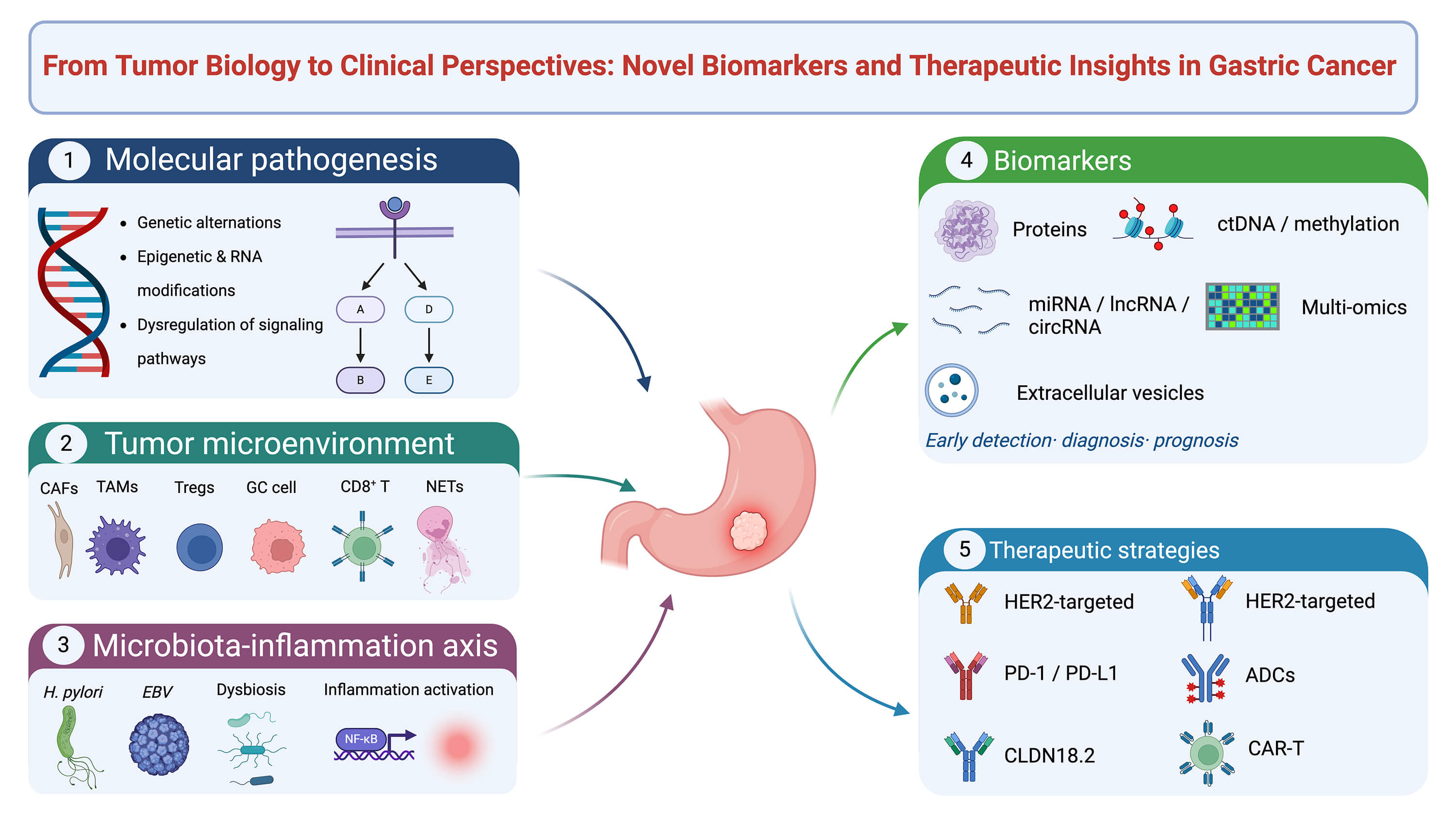
Gastric cancer (GC) is a leading cause of cancer-related mortality worldwide. Accurate early detection, timely diagnostic stratification, and robust prognostic risk assessment are essential for optimizing clinical outcomes and improving survival duration. However, conventional serological biomarkers demonstrate limited diagnostic performance owing to suboptimal sensitivity and specificity, while standard chemotherapy and targeted therapies provide only modest survival benefits in GC. Marked inter- and intratumoral heterogeneity further characterizes GC as a biologically complex and treatment-resistant malignancy. To date, significant progress has been made in comprehensively delineating the complex molecular pathogenesis of GC, providing a strong rationale for the development of novel biomarkers and promising therapeutic targets. The aim of this review is to systematically summarize recent advances ranging from aberrant genetic and epigenetic alterations; dysregulation of oncogenic signaling pathways; sophisticated crosstalk within the tumor microenvironment; intricate microbiota-inflammation interactions; fundamental discoveries to potential clinical translation, including novel biomarkers for early detection, diagnosis and prognostic stratification; and emerging therapeutic strategies for GC. Collectively, this work provides a conceptual framework for advancing precision medicine in GC.Graphic Abstract
Keywords
Gastric cancer (GC) remains a major global health challenge. According to the latest GLOBOCAN 2024 statistics, there were an estimated 980,000 new cases and 642,000 deaths worldwide, making it the fifth most frequently diagnosed cancer and the fifth leading cause of cancer-related mortality [1]. Despite steady improvements in surgical techniques and conventional chemotherapy regimens, the overall prognosis for patients with advanced GC remains poor, largely due to profound intra- and intertumoral heterogeneity. The classical Correa cascade provides a fundamental histopathological framework for understanding the stepwise progression from chronic gastritis to invasive adenocarcinoma [2]. At the molecular level, GC is driven by a highly complex and dynamic interplay of genetic mutations, epigenetic reprogramming, tumor microenvironment evolution, and microbiota-inflammation interactions.
In recent years, unprecedented advances in multi-omics and high-resolution sequencing technologies have enabled the dissection of the multilayered molecular pathogenesis underlying GC progression and therapeutic resistance. For example, single-cell RNA sequencing (scRNA-seq) and spatial transcriptomics have revolutionized our understanding of the highly plastic ecosystem within the tumor microenvironment (TME) [3]. We now recognize that GC progression is strongly influenced by immunosuppressive crosstalk orchestrated by cancer-associated fibroblasts (CAFs), tumor-associated macrophages (TAMs), regulatory T cells (Tregs), exhausted CD8+ T cells, and local microbial communities. These insights have, in turn, provided a strong biological rationale for the identification, validation, and clinical translation of next-generation biomarkers and actionable therapeutic targets.
Concurrently, the clinical management of GC is undergoing a paradigm shift toward biomarker-guided precision medicine. The continuous discovery of novel non-invasive liquid biopsy biomarkers—ranging from circulating tumor DNA (ctDNA) and circular RNAs to extracellular vesicles (EVs)—has demonstrated significant potential for early detection, accurate diagnostic and prognostic stratification, and real-time monitoring of disease progression in GC [4,5,6]. Cancer treatment is moving toward an era of precision oncology. Targeted therapies, pioneered by trastuzumab for HER2-positive tumors, have revolutionized the treatment paradigm from organ-based to biomarker-driven selection [7]. Recently, the advent of immune checkpoint inhibitors, antibody-drug conjugates (ADCs), bispecific T-cell engagers (BiTEs), and chimeric antigen receptor (CAR) T-cell therapies has further diversified the oncological armamentarium, offering unprecedented opportunities for treating refractory malignancies including GC. Nowaday, the clinical success of targeted therapies and immunotherapies, has profoundly improved the first-line therapeutic landscape of GC.
In this comprehensive review, we systematically summarize the latest breakthroughs in GC biology and clinical oncology. Specifically, this review aims to integrate current evidence on the molecular pathogenesis, tumor microenvironment, microbiota–inflammation axis, emerging biomarkers, and biomarker-guided therapies in GC, and to clarify their implications for precision diagnosis and individualized treatment. We first examine the complex molecular pathogenesis driven by dysregulated signaling cascades, genomic instability, epigenetic alterations, and RNA modifications. Afterward, we explore the sophisticated regulation of the TME and the pivotal microbiota-inflammation axis. We then discuss the emergence of novel liquid biopsy biomarkers and multi-omics panels for precision diagnostics. Finally, we review the current landscape and future directions of biomarker-guided therapeutic strategies. This work provides a hierarchical framework that links molecular mechanisms to cellular behaviors and, ultimately, to clinical implications in GC.
2 Complex Molecular Pathogenesis in GC
Gastric carcinogenesis is a multistep and multifactorial process. The classical Correa cascade delineates the sequential progression from chronic gastritis to atrophic gastritis, intestinal metaplasia, dysplasia, and ultimately invasive carcinoma, providing a fundamental histopathological framework [8]. This process involves complex molecular mechanisms, including genetic mutations, genomic instability, epigenetic alterations, and activation of oncogenic signaling pathways. These aberrations confer marked tumor heterogeneity and drive cancer initiation and progression. Understanding this complex pathogenesis is essential for precision subtyping and the discovery of novel biomarkers and therapeutic targets in GC.
2.1 Genetic Alterations and Dysregulated Oncogenic Pathways
GC is characterized not only by recurrent driver mutations but also by a multi-pathway activation pattern, in which multiple oncogenic cascades are simultaneously engaged. The Cancer Genome Atlas (TCGA) classified GC into four principal molecular subtypes—Epstein–Barr virus-positive (EBV), microsatellite instability (MSI), genomically stable, and chromosomal instability (CIN) [9]. CIN tumors are enriched in TP53 alterations and receptor tyrosine kinase (RTK) amplification, whereas genomically stable tumors more often harbor abnormalities in cell adhesion and cytoskeletal regulation, including CDH1 and RHOA alterations, as well as rearrangements involving CLDN18–ARHGAP26 or ARHGAP6 fusions [9]. EBV-positive and MSI tumors are more frequently associated with hypermutation, PIK3CA mutations, and ARID1A loss [9,10,11].
It has been well documented that loss-of-function TP53 mutations impair genomic surveillance and facilitate chromosomal instability, thereby creating a permissive environment for additional oncogenic events [9]. In addition, several growth-promoting pathways are frequently activated, particularly the RTK/RAS/RAF/MEK/ERK and PI3K/AKT/mTOR cascades [9,11,12]. Amplification or activation of upstream receptors such as ERBB2, EGFR, MET, and FGFR2 sustains downstream proliferative and survival signaling, promoting cell growth, resistance to apoptosis, and metabolic adaptation [9,11]. The Wnt/β-catenin pathway is another key oncogenic pathway in gastric carcinogenesis. Mutations in APC or inactivation of negative regulators such as AXIN can lead to abnormal nuclear accumulation of β-catenin, which activates transcriptional programs associated with stemness, proliferation, and cellular plasticity [13]. The PI3K/AKT/mTOR pathway, driven by PIK3CA mutations or PTEN loss, enhances survival signaling, metabolic reprogramming, and treatment resistance in GC [9,11].
Disruption of epithelial cohesion represents another major route of tumorigenesis. Germline CDH1 mutations establish E-cadherin loss as a central mechanism in hereditary diffuse GC [14], and similar defects are also observed in sporadic diffuse-type tumors. In the genomically stable subtype of GC, concurrent CDH1 and RHOA alterations impair cell-cell adhesion and cytoskeletal regulation, facilitating diffuse infiltration and invasive growth [9]. In addition, recurrent ARID1A mutations link oncogenic signaling to chromatin remodeling and reshape transcriptional programs involved in differentiation, DNA repair, and stress responses, particularly in the EBV-positive GC subtype [9,15].
Clinically, some of these genetic alterations are already actionable. ERBB2 amplification has established therapeutic relevance and is targetable with trastuzumab, while CLDN18.2 targeting with zolbetuximab has demonstrated promising clinical efficacy in advanced GC.
2.2 Epigenetic Alterations and RNA Modifications in GC Progression
Epigenetic alterations and RNA modifications play key roles in regulating gene expression without changing DNA or RNA sequences. These modifications are highly dynamic and reversible. DNA methylation, histone modifications, and chromatin remodeling represent the major forms of epigenetic regulation. RNA modifications mainly include N6-methyladenosine (m6A), 5-methylcytosine (m5C), and N4-acetylcytidine (ac4C). Collectively, these molecular alterations have significant effects on GC cellular functions, including malignant proliferation, epithelial-mesenchymal transition (EMT), metastasis and invasion, metabolic reprogramming, and therapy resistance.
2.2.1 Epigenetic Silencing and Early Initiation of GC
Promoter hypermethylation-mediated silencing of tumor suppressor genes is a critical driver of tumorigenesis in the early stages of GC development. Promoter hypermethylation of genes such as p16, MLH1, and APC is commonly observed in precancerous lesions, including chronic atrophic gastritis and intestinal metaplasia [16,17]. For example, the CDH1 promoter is methylated in 30–50% of precancerous lesions, leading to disruption of cell adhesion [18]. Helicobacter pylori infection activates DNMT1 and NF-κB signaling pathways, triggering genome-wide aberrant DNA methylation and establishing an “epigenetic memory” that can persist even after pathogen eradication, thereby facilitating tumor initiation [19,20,21]. Additionally, the EBV-positive GC subtype exhibits an extreme CpG island methylator phenotype, characterized by hypermethylation of nearly 20% of promoter CpG islands. EBV-induced hypermethylation silences key tumor suppressor genes and differentiation factors, thereby promoting a highly proliferative and poorly differentiated phenotype that favors early tumorigenesis in GC [9,22]. Promoter hypermethylation of genes such as DAPK, CDH1, GSTP1, p15, and p16 can be detected in serum from GC patients, suggesting their potential as biomarkers [23]. Recently, methylated ctDNA has become a major focus in the development of novel GC biomarkers [24,25,26].
2.2.2 Epigenetic Modifications Regulate Malignant Cell Proliferation, Metastasis, and Invasion of GC
Dysregulation of chromatin remodeling factors spans the entire trajectory of GC, from local invasion to systemic dissemination. CHD4, a core subunit of the Mi-2/NuRD complex, exhibits non-canonical functions by interacting with MYH9 to induce GSK3β degradation. This stabilizes β-catenin, leading to sustained activation of Wnt signaling and EMT in GC [27].
M6A modification is the most prevalent RNA epigenetic modification, orchestrated by a range of writers, readers, and erasers. METTL3, a well-documented m6A writer, enhances the stability of the oncogenic CENPF mRNA transcript, thereby activating the FAK/MAPK signaling pathway and promoting GC metastasis [28]. By contrast, another m6A writer, METTL14, suppresses GC growth and invasion by inducing m6A modification of circORC5, thereby regulating the miR-30c-2-3p/AKT1S1 axis [29]. M6A readers, including YTHDF1/2/3, IGF2BP1/2/3, and HNRNPA2B1, recognize methylation marks to determine RNA fate. For example, the m6A reader YTHDF1 enhances the translation of m6A-modified FZD7, thereby activating the Wnt/β-catenin signaling axis and promoting malignant proliferation and metastasis in GC [30]. ALKBH5 and FTO function as m6A erasers. ALKBH5-mediated demethylation of WRAP53 reduces RALBP1 expression and suppresses activation of the PI3K/AKT/mTOR pathway, thereby inhibiting GC metastasis [31].
In addition, m5C modification has been implicated in the regulation of alternative splicing. The RNA-binding protein NONO recruits NSUN2 to PTEN pre-mRNA, altering its methylation pattern and leading to aberrant splicing and reduced expression of functional PTEN. This ultimately activates the PI3K/AKT signaling pathway and promotes proliferation, migration, and invasion in GC [32]. NAT10-mediated ac4C modification has also been reported to enhance the stability of the splicing factor SRSF2 in GC, thereby promoting exon skipping of YTHDF1 and generating oncogenic transcript variants, which in turn stimulate proliferation and metastasis [33,34].
2.2.3 Epigenetic Alterations Modulate Metabolic Reprogramming of GC
Epigenetic modifications play critical roles in regulating metabolic reprogramming in GC. A recent study demonstrated that the synergistic effects of H3K27ac and H3K4me3 drive lncRNA DLEU1 expression, which promotes glycolysis and activates G6PD to increase NADPH production via the ASCC2/ALKBH3 axis. This enhanced NADPH production helps maintain redox homeostasis and supports metabolic adaptation [35]. P300-mediated H3K27 acetylation can also increase METTL3 transcription, thereby promoting m6A modification of HDGF mRNA and enhancing its stability. The resulting nuclear HDGF subsequently activates GLUT4 and ENO2 expression, thereby increasing glycolysis in GC [36]. Beyond m6A, other RNA modifications also contribute to GC progression. NSUN2-mediated m5C modification enhances the stability of lncRNA NR_033928, which acts as a molecular scaffold to recruit the IGF2BP3/HUR complex and stabilize GLS mRNA, thereby promoting glutamine metabolism reprogramming and tumor growth in GC [37]. Natural product brusatol can inhibit glycolysis-driven H3K18la and H3K9la, thereby blocking GC progression [38]. Targeting epigenetic alterations to remodel metabolism offers a promising therapeutic strategy for GC.
2.2.4 Epitranscriptomic Plasticity Contributes to Therapy Resistance in GC
RNA modifications confer high molecular plasticity to GC cells and have a crucial impact on chemoresistance. For example, the m6A methyltransferase METTL3 enhances the stability of transcripts such as ARF6 and PARP1 through m6A modification, thereby promoting resistance to chemotherapeutic agents, including cisplatin and 5-fluorouracil (5-FU) in GC [39,40]. The m6A reader HNRNPA2B1 can also induce chemoresistance by stabilizing lncRNA NEAT1 and maintaining GC stemness [41]. By contrast, another m6A reader, YTHDF2, has been reported to suppress cancer stemness and improve chemosensitivity to oxaliplatin in GC by promoting the degradation of ONECUT2 mRNA [42]. In addition, m6A demethylases (e.g., ALKBH5, FTO) and the m5C methyltransferase NSUN2 are also involved in resistance regulation [43,44,45]. Knockdown of NSUN2 can increase the sensitivity of GC cells to cisplatin and 5-FU [44].
Additionally, histone modifications can also contribute to therapy resistance in GC and have emerged as promising targets for improving treatment efficacy. The novel HDAC inhibitor chidamide, which specifically targets HDAC3, increases HNF4A acetylation levels and downregulates TYMS, thereby effectively reversing 5-FU chemoresistance in GC cells. EZH2 is a well-known histone methyltransferase that catalyzes H3K27me3-mediated transcriptional repression. EZH2 deficiency leads to loss of H3K27 methylation, triggering activation of Tfap2c and inducing aberrant squamous differentiation and chemotherapy resistance in GC [46].
The schematic illustration in Fig. 1 depicts the complex pathogenesis of GC. Integrating GC molecular subtypes into clinical practice enables more refined prognostic stratification and facilitates the delivery of targeted therapies. Emerging epigenetic and epitranscriptomic markers—such as methylated ctDNA and RNA modifications—show promising potential as novel biomarkers. The reversibility of these epigenetic modifications further supports the development of novel therapeutics to overcome treatment resistance. However, prospective validation and standardized detection platforms are still required to enable their clinical translation in GC.

Figure 1: Integrated molecular landscape of gastric cancer (GC) pathogenesis and therapeutic targets. The schematic summarizes the major classical genomic subtypes, including Epstein–Barr virus (EBV)–positive tumors, microsatellite instability (MSI), chromosomal instability (CIN), and genomically stable (GS) GCs, and links Helicobacter pylori–driven chronic inflammation to the histological progression from gastritis to intestinal metaplasia, dysplasia, and carcinoma. The figure also highlights key oncogenic signaling cascades, epigenetic alterations, such as DNA methylation, histone modification, lactylation, and chromatin remodeling, together with RNA modifications including m6A, m5C, and ac4C, which play essential roles in GC progression. Clinically actionable targets, such as ERBB2 amplification and CLDN18.2, are shown with representative antibodies trastuzumab and zolbetuximab. Created with BioRender.com. Abbreviations: CIN, chromosomal instability; EBV, Epstein-Barr virus; GS, genomically stable; MSI, microsatellite instability.
3 Sophisticated Regulations of TME in GC Progression
Accumulating evidence has demonstrated that the TME plays a critical role in GC. The TME is a complex ecosystem composed of diverse immune cells, stromal cells, endothelial cells, and the extracellular matrix (ECM). These components engage in intricate crosstalk, collectively promoting immunosuppression and immune evasion, and ultimately driving GC cell proliferation, metastasis, and therapeutic resistance.
3.1 Multifunctional Roles of CAFs in GC Development
CAFs can directly activate pro-oncogenic signaling pathways in GC cells by secreting factors such as IL-6, IL-8, and TGF-β [47,48]. Moreover, CAFs are key drivers of ECM remodeling, promoting tumor invasion and metastasis [49]. Recent large-scale scRNA-seq data have characterized CAF heterogeneity in GC and identified a distinct INHBA- and FAP-high CAF subpopulation associated with poor prognosis, as INHBA can increase FAP expression and induce collagen transcription to mediate stromal remodeling [50]. Lu et al. reported that CAF-secreted SEMA7A binds to ITGB1 on GC cells, triggering GAL3ST1-mediated H3Y99 sulfation (H3Y99sulf), which recruits KAT2A to establish H3K56ac modification. This cascade activates the β-catenin pathway, driving EMT and metastasis [51]. Additionally, CAF-derived lactate induces increased H3K18la levels in GC cells, activating the ASPM/NCAPG/STAT3 axis and upregulating PD-L1, which contributes to immunotherapy resistance [52]. Recent research has also revealed a unique nicotinamide “metabolic face-off” between macrophages and CAFs [53]. Specifically, macrophages are enriched in nicotinamide phosphoribosyltransferase (NAMPT), whereas CAFs highly express Notch-regulated nicotinamide N-methyltransferase (NNMT). This competitive expression of rate-limiting enzymes dynamically alters nicotinamide availability within the TME, which in turn directly influences the antitumor activity and exhaustion status of local CD8+ T cells. As a counter-regulatory mechanism, macrophages can release NAMPT-enriched EVs that inhibit NNMT transcription in CAFs via the SIRT1/NICD axis [53].
3.2 Complicated Crosstalk of TAMs in GC
TAMs, predominantly exhibiting M2-like polarization, are a major component of the TME in GC. They foster an immunosuppressive niche, promote therapeutic resistance, and accelerate cancer progression. Recent studies have identified DKK1 as a key factor driving macrophage recruitment through binding to the CKAP4 receptor on the macrophage surface, thereby activating the PI3K/AKT pathway to induce M2 polarization and suppress CD8+ T-cell and Natural Killer (NK) cell function in GC [54]. Blockade of DKK1 not only reduces TAM infiltration but also synergizes with anti-PD-1 therapy, overcoming primary resistance [54]. Exosomes mediate bidirectional communication between GC cells and TAMs. GC cells highly express SERPINE1 through autocrine JAK2/STAT3 signaling and subsequently release let-7g-5p-enriched exosomes to macrophages, which potently drive M2 polarization by inhibiting SOCS7 and activating STAT3 [55]. Another study indicated that TAMs can release MALAT1-enriched exosomes to GC cells, activating the β-catenin/HIF-1α pathway to promote the Warburg effect, thereby enhancing tumor proliferation and chemotherapy resistance [56]. lncRNA NR-109 is highly expressed in TAMs and can block FUBP1 degradation while activating c-Myc to promote M2-like macrophage polarization in GC [57]. Reciprocally, c-Myc can function as an upstream transcription factor to stimulate NR-109 expression, forming a positive feedback loop that drives pro-oncogenic M2 polarization in GC [57].
3.3 Oncogenic Functions of Tumor-Associated Neutrophils (TANs) and Neutrophil Extracellular Traps (NETs) in GC Aggravation
The role of TANs in GC has become increasingly prominent. Recent investigations have shown that microbial communities within the TME can recruit TANs. Fusobacterium nucleatum, enriched in GC tissues, can recruit large numbers of TANs by activating the IL-17/NF-κB/RelB pathway and inducing PD-L1 expression, leading to CD8+ T-cell exhaustion and immune evasion [58]. Polymorphonuclear (PMN) Myeloid-Derived Suppressor Cells (MDSCs) are often regarded as neutrophils in a pathological state and exhibit potent immunosuppressive functions. Their abundance in the TME is frequently associated with poor prognosis in GC [59]. Previous studies have shown that PMN-MDSCs exert dual pro-tumor effects by secreting the S100A8/A9 heterodimer, which simultaneously affects GC cells and CD8+ T cells. S100A8/A9 binds to TLR4 on GC cells and activates the p38 MAPK/NF-κB pathway to increase CXCL1 expression, thereby promoting PMN-MDSC recruitment. Meanwhile, S100A8/A9 directly induces CD8+ T-cell exhaustion via the TLR4/AKT/mTOR pathway, suppressing T-cell glycolysis, proliferation, and production of TNF-α and IFN-γ [59].
Notably, overactivated neutrophils can release large amounts of DNA-rich NETs, which have emerged as an attractive research target in GC. The hypoxic TME in GC can recruit neutrophils and induce NET formation via the HMGB1/TLR4/p38 MAPK pathway, thereby promoting invasion, migration, angiogenesis, and tumor growth [60]. Mouse model studies indicate that infection-induced NET formation in peripheral blood and ascites may facilitate GC cell extravasation and dissemination to the liver and peritoneum [61]. Zhang et al. further demonstrated that activation of the TGF-β/Smad/LIF signaling axis is critical for mediating NET formation, thereby driving peritoneal metastasis of GC [61]. Targeting NET formation is therefore emerging as a promising strategy for suppressing GC metastasis [60].
3.4 T-Cell Dysfunction Exacerbates GC Progression
T cells serve as the primary effector cells of antitumor immunity, and their functional status directly influences tumor progression and immunotherapy outcomes. However, the immunosuppressive TME impairs T-cell activation, infiltration, and cytotoxic function, thereby reducing the efficacy of immunotherapies. As described above, CAFs, TAMs, TANs, NETs, and MDSCs are major cellular components that shape an immunosuppressive TME, promoting CD8+ T-cell exhaustion and suppressing antitumor immunity. In addition, metabolic competition between tumor cells and immune cells is a key mechanism underlying T-cell dysfunction. Gut microbial metabolites can also modulate the cytotoxic function of CD8+ T cells in GC. Yu et al. demonstrated that patients with GC commonly exhibit reduced abundance of butyrate-producing gut microbiota and decreased circulating butyrate levels [62]. Exogenous butyrate supplementation significantly enhances the antitumor cytotoxicity of CD8+ T cells by activating the GPR109A/HOPX signaling axis [62].
Tregs are critical drivers of immunosuppression and immune evasion in GC. GC-derived IL-33 enables mast cells to secrete IL-2, which induces Tregs to express ICOS. These ICOS+ Tregs can potently suppress CD8+ T-cell proliferation and effector molecule production, thereby promoting GC progression [63].
3.5 The Landscape of TME Spatiotemporal Evolution in GC
The rapid advancement of single-cell RNA sequencing (scRNA-seq) and spatial transcriptomics (ST) technologies has provided a high-resolution framework for dissecting cellular plasticity and the spatial evolution of the TME in GC [3]. In early EGC, single-cell atlases have revealed the enrichment of CH25H+ CD4+ T cells, ADAMTSL2+ endothelial cells, and other subsets. Notably, IL-33+ endothelial cells not only enhance angiogenesis but also directly upregulate KRT17 expression in GC cells, thereby promoting tumor growth [64]. Spatial transcriptomics further optimizes the characterization of molecular dynamics with high spatial precision. Spatiotemporal multi-omics analyses of endoscopic submucosal dissection specimens from EGC have identified, for the first time, precancerous microenvironment (PMC_P) and PMC_2 cell subpopulations that drive carcinogenic initiation [65]. Combined with single-cell analysis, Zhang et al. demonstrated that GC stem cells originate from mature gastric chief cells through spasmolytic polypeptide-expressing metaplasia (SPEM) transdifferentiation, maintaining stemness and drug resistance via the iCAF-secreted AREG–ERBB2 signaling axis and establishing an immune-evasive microenvironment [66]. Currently, the abundance of tertiary lymphoid structures (TLSs) has become a key indicator for assessing therapeutic efficacy. Wang et al. found that high TLS abundance and increased infiltration of intratumoral CXCL13+ CD160+ CD8+ T cells prior to treatment in advanced GC patients are significantly associated with a favorable treatment response [67]. Mechanistically, intracellular vitamin B6 in CD160+ CD8+ T cells may inhibit MDM2-mediated HIF-1α ubiquitination and degradation, leading to upregulation of CXCL13 expression and promoting CXCR5+ B cell recruitment and TLS maturation [67].
Taken together, a comprehensive understanding of TME characteristics is of profound importance for developing novel therapeutic strategies that target not only GC cells but also the immunosuppressive microenvironment. These findings provide a solid theoretical foundation for therapeutic interventions, including targeting key stromal-immune axes (e.g., DKK1–CKAP4, SEMA7A–ITGB1, and NETosis pathways), reversing metabolic dysregulation through taurine and butyrate supplementation or SLC6A6 inhibition, and leveraging spatial transcriptomic signatures alongside TLS-based stratification to guide personalized combination immunotherapies. Such integrated approaches hold promise for overcoming treatment resistance and improving clinical outcomes in GC.
Fig. 2 delineates diverse ligand-receptor axes and secreted factors mediating crosstalk among GC cells, cancer-associated fibroblasts, myeloid suppressor cells and cytotoxic T lymphocytes, which collaboratively induce CD8+ T cell exhaustion and accelerate malignant progression.

Figure 2: Overview of cellular interactions in the GC tumor microenvironment. This schematic illustrates the crosstalk between GC cells and major components of the tumor microenvironment, including CAFs, TAMs, TANs, MDSCs, and CD8+ T cells. These interactions activate multiple signaling pathways that promote tumor cell proliferation, migration, metastasis, metabolic reprogramming, immune suppression, and CD8+ T cell dysfunction. Created with BioRender.com. Abbreviations:GC, gastric cancer; CAF, cancer-associated fibroblast; TAN, tumor-associated neutrophil; MDSC, myeloid-derived suppressor cell; TAM, tumor-associated macrophage.
4 The Microbiota-Inflammation Axis in GC Development
The microbiota-inflammation axis has emerged as a key factor in gastric carcinogenesis, integrating microbial dysbiosis, host immune responses, and tumor-promoting processes into a dynamic and interconnected network. Beyond the classical H. pylori-driven model, accumulating evidence highlights the contribution of a broader microbial ecosystem that collectively sustains chronic inflammation and promotes cancer development [68]. Notably, this axis spans multiple stages of GC, from early mucosal injury to disease progression and therapeutic response, with some microbiota-based interventions showing translational potential [69,70]. In this section, we summarize the initiation of dysbiosis-driven inflammation, the underlying molecular networks, key microbial drivers, and their clinical and therapeutic implications in GC.
4.1 Dysbiosis-Driven Inflammatory Initiation of Gastric Carcinogenesis
The intricate interplay between the gastric microbiota and chronic inflammation represents a fundamental axis in gastric carcinogenesis. This “inflammation-microbiota-cancer” axis is initiated by microbial dysbiosis, which disrupts gastric homeostasis and establishes a self-perpetuating cycle of mucosal injury and inflammatory response. Persistent inflammation, in turn, further reshapes the microbial community and reinforces dysbiosis [71]. In the highly acidic gastric environment, H. pylori remains the dominant pathogenic driver; however, emerging evidence implicates non-H. pylori microbiota in sustaining this inflammatory cascade and promoting premalignant progression, highlighting a shift from a single-pathogen model to a complex microbial ecosystem perspective [72,73]. This dysbiosis-driven chronic inflammation ultimately creates a procarcinogenic milieu that facilitates genomic instability and neoplastic transformation [74].
4.2 Molecular Signaling Networks Linking Microbiota and Inflammation
At the molecular level, the crosstalk between the microbiota and host inflammatory responses is orchestrated through a complex signaling network centered on the NF-κB pathway, which serves as a critical hub linking microbial dysbiosis to gastric tumorigenesis [75]. Specific microbial components and toxins activate inflammatory signaling cascades, including the NF-κB and MAPK pathways, leading to the transcription of pro-inflammatory cytokines that contribute to gastrointestinal carcinogenesis [74].
In parallel, additional signaling pathways, including STAT3 and PI3K/AKT, are activated by microbial stimuli to regulate cell survival, proliferation, and inflammatory responses [76]. The NLRP3 inflammasome serves as another key effector activated by bacterial virulence factors such as H. pylori CagA, leading to caspase-1-dependent maturation of IL-1β and IL-18 and amplification of inflammatory responses [77]. Systemic inflammatory indices, including the neutrophil-to-lymphocyte ratio (NLR) and systemic inflammation response index, are strongly associated with cancer cachexia in patients with GC [78].
Conversely, microbiota-derived metabolites, particularly short-chain fatty acids, exert anti-inflammatory effects by inhibiting histone deacetylases (HDACs), thereby suppressing NF-κB activation and preventing excessive inflammation [79]. Dynamic alterations in the microbiota during gastric carcinogenesis are associated with both epigenetic and genetic changes, including DNA methylation and TP53 mutations [80].
Collectively, these pathways form a dynamic regulatory network through which the microbiota can both initiate and restrain inflammatory responses, thereby maintaining or disrupting gastric homeostasis.
4.3 Microbial Drivers and Pro-Tumorigenic Mechanisms
Bacteria often exhibit distinct pro-inflammatory and tumor-promoting mechanisms. H. pylori employs a sophisticated virulence arsenal: the CagA oncoprotein activates NF-κB and β-catenin signaling, while VacA induces epithelial vacuolation and disrupts autophagy. In addition, the HtrA polymorphism (171S/L) enhances cleavage of E-cadherin and occludin, thereby compromising epithelial barrier integrity [81,82,83]. H. pylori-secreted Gamma-Glutamyl Transferase (GGT) also depletes glutamine and α-KG in gastric epithelial cells and increases histone H3K9me3 and H3K27me3 levels. This epigenetic reprogramming activates the Wnt signaling pathway and contributes to gastric carcinogenesis [84]. H. pylori CagA can also upregulate transcription of the RNA demethylase FTO via JUN, leading to demethylation and stabilization of HBEGF mRNA, thereby facilitating EMT and metastasis [85]. Large-scale community-based eradication trials have shown that H. pylori infection eradication significantly reduces long-term GC risk, particularly in individuals without precancerous lesions [86,87].
Beyond H. pylori, several non-H. pylori commensals also function as direct contributors to carcinogenesis. Streptococcus anginosus activates the TMPC/ANXA2/MAPK pathway, promoting cell proliferation and epithelial barrier disruption [88]. In addition, the microbiota can modulate the TME in GC. Streptococcus anginosus has been shown to promote immune evasion through arginine-ornithine metabolism, thereby suppressing CD8+ T-cell infiltration [89], while Fusobacterium nucleatum further contributes to protumoral neutrophil recruitment via the IL-17/NF-κB/RelB axis [58]. These microbial interactions support a broader gastric microbial axis that sustains a pro-inflammatory network driving gastric carcinogenesis [90]. Probiotic and prebiotic interventions also show therapeutic potential. Engineered probiotics can alleviate inflammation by scavenging reactive oxygen species and modulating microbial composition [91], while prebiotics such as inulin enhance antitumor immunity [69].
4.4 EBV Infection Promotes GC Development
In addition to bacterial infection, EBV represents a distinct carcinogenic driver in GC. EBV-associated GC is characterized by widespread CpG island hypermethylation and recurrent PIK3CA and ARID1A alterations [15,92]. A recent study reported that EBV can upregulate KDM5B through cooperation between latent and lytic viral proteins, leading to transcriptional repression of the tumor suppressor PLK2 and activation of the PI3K/AKT/mTOR pathway, thereby promoting malignant GC proliferation [93]. In parallel, EBV-encoded non-coding RNAs critically reshape the tumor microenvironment and epigenetic landscape. EBV-circLMP2A enhances hypoxia-driven angiogenesis via a HIF1α/VEGFA positive feedback loop, contributing to tumor vascularization and GC progression [94], while EBV-circRPMS1 promotes GC growth and invasion by activating METTL3-dependent m6A modification [95]. Immune escape is another central hallmark of EBV-driven carcinogenesis, as viral miRNAs (BART11 and BART17-3p) upregulate PD-L1 expression by targeting FOXP1 and PBRM1 [96], while LMP2A induces platelet aggregation and TGF-β release, thereby suppressing NK cell cytotoxicity [97]. EBV-associated GC is characterized by a highly inflamed tumor microenvironment with abundant CD8+ T-cell infiltration [98]. Consistent with this feature, EBV-positive GC demonstrates remarkable sensitivity to immune checkpoint blockade in both advanced and neoadjuvant settings [99,100].
4.5 Clinical Implications of the Microbiota-Inflammation Axis
Accumulating evidence increasingly highlights the prognostic and therapeutic relevance of microbiota-inflammation interactions. Microbiota-based signatures may provide valuable diagnostic and prognostic utility. For example, deep learning models incorporating oral taxa such as Aggregatibacter outperform conventional TNM staging in predicting GC prognosis [101]. Importantly, microbiota composition also influences therapeutic response, as prior antibiotic exposure has been shown to impair immunotherapy efficacy by promoting CD8+ T-cell exhaustion [102]. In addition, fecal microbiota transplantation strategies are gaining increasing attention. Clinical studies have demonstrated their safety and potential to overcome immunotherapy resistance [103]. Together, these findings underscore the translational potential of targeting the microbiota-inflammation axis as a promising complementary strategy for GC prevention and treatment.
5 Novel Biomarkers for Early Detection, Diagnosis, and Prognosis in GC
Early detection, accurate diagnosis, and precise prognostic prediction are the cornerstones for improving outcomes in GC. Traditional endoscopic screening is invasive, costly, and often inconvenient for patients. With the advent of high-throughput sequencing and multi-omics technologies, a growing number of non-invasive liquid biopsy biomarkers have emerged, demonstrating improved sensitivity and specificity. These novel biomarkers include proteins, circulating nucleic acids (DNA, miRNA, lncRNA, and circRNA), and EVs, which can be detected in both blood and gastric juice.
Conventional blood-based biomarkers such as CEA, CA19-9, and CA72-4 exhibit limited sensitivity and specificity in GC. Accordingly, numerous novel serum proteins have been extensively investigated as potential diagnostic biomarkers. Recent studies have reported that interleukin enhancer-binding factor 2 (ILF2) is markedly increased in the serum of GC patients and demonstrates strong diagnostic performance, with an Area Under the Curve (AUC) of 0.915 for GC versus healthy controls, 0.854 for GC versus benign gastric disease, and 0.888 for stage I GC versus benign gastric disease plus healthy controls—outperforming conventional blood-based indicators in the same cohort [104]. Similarly, insulin-like growth factor binding protein 7 (IGFBP7) has shown promise for early diagnosis, with an AUC of 0.773 (95% CI: 0.701–0.845) in early GC [105]. SNCG (γ-synuclein) is another promising biomarker for early detection in GC; it is significantly upregulated in both gastric juice and serum of GC patients, achieving an AUC of 0.923 with high sensitivity and specificity [106].
5.2 Circulating Tumor DNA (ctDNA) Biomarkers
ctDNA has emerged as an ideal liquid biopsy biomarker over the past decade, as it can reflect tumor-derived molecular alterations earlier than conventional diagnostic modalities.
Copy number variation (CNV) of the HER2 gene (also known as ERBB2) represents a frequent genomic abnormality in gastric carcinogenesis. Increased HER2 CNV can be detected in serum ctDNA from GC patients and can distinguish GC cases from healthy controls with a specificity of 98% [4]. ctDNA detection is also valuable for guiding therapeutic selection. In advanced GC, EGFR-amplified ctDNA is associated with better response to EGFR inhibitors, whereas FGFR2 and MET co-amplification is associated with limited benefit from EGFR blockade [107]. Additionally, longitudinal postoperative ctDNA monitoring can detect GC recurrence earlier than radiographic evidence of relapse [108,109]. Methylated DNA is another promising liquid biopsy biomarker for GC. A landmark marker discovery study identified a three-marker plasma methylation panel consisting of ELMO1, ZNF569, and C13orf18 (now standardized as C13orf18/ZNF569), which detected 86% of GC cases at 95% specificity [110]. Another plasma-based methylation panel combining ELMO1, ZNF582, and TFPI2 has also been proposed for the detection of upper gastrointestinal cancers [111].
Additionally, serial monitoring of cfDNA methylation profiles has emerged as a powerful tool for tracking disease dynamics and guiding therapeutic decision-making. A recent pan-cancer methylation analysis of cfDNA demonstrated that a universal cancer-only methylation marker panel enables multi-organ cancer detection and longitudinal monitoring through simple blood-based assays, highlighting the clinical potential of integrating methylation-based liquid biopsy into routine surveillance protocols [112]. In GC, such dynamic cfDNA monitoring may facilitate real-time assessment of treatment response, early detection of molecular residual disease, and identification of emerging resistance, thus guiding adaptive treatment modifications throughout the course of GC progression.
5.3 Circulating Non-Coding RNAs as Biomarkers
Circulating non-coding RNAs mainly include circulating miRNAs, long non-coding RNAs (lncRNAs), and circular RNAs (circRNAs), which are stable in bodily fluids and have been widely investigated as diagnostic, prognostic, and therapeutic biomarkers in GC [113].
5.3.1 Circulating RNAs as Promising Diagnostic Biomarkers in GC
For early GC detection, serum miR-181 and miR-652 both show promising diagnostic value, with AUCs of 0.820 and 0.842, respectively. When combined with the conventional marker CA72-4, the three-marker model achieved an AUC of 0.917 (95% CI: 0.856–0.975), with 92.5% sensitivity and 86.8% specificity [114]. An optimized three-miRNA signature (miR-18a, miR-181b, and miR-335) for early GC detection was established based on genome-wide expression profiling in a large cohort of 598 patients [115]. In an independent prospective validation cohort of 349 patients, this serum signature demonstrated strong diagnostic accuracy across all GC stages (I–IV), with an AUC of 0.86 (95% CI: 0.83–0.90). This performance surpasses conventional biomarkers such as CEA and CA19-9 and offers greater cost-effectiveness than endoscopic screening, highlighting its strong potential for clinical translation. The DESTINEX multicenter study constructed a 10-miRNA signature that distinguished GC patients from controls with AUCs of 0.958 and 0.948 in the training and validation cohorts, respectively, and notably achieved an AUC of 0.968 for detecting pT1-stage GC [116]. In a plasma-based study, circ-KIAA1244 was significantly downregulated in GC and achieved an AUC of 0.748, with 77.42% sensitivity and 68.00% specificity for distinguishing GC from healthy controls; lower expression was also associated with lymphatic metastasis and poorer survival [117]. A systematic discovery approach identified an 8-circRNA panel that robustly distinguished GC patients from non-disease controls, with AUCs of 0.87 and 0.83 in the training and validation cohorts, respectively. Notably, this panel effectively identified early-stage GC (stage I/II) and exhibited high specificity for GC compared with other gastrointestinal cancers [5].
A multiphase study demonstrated that exosomal lncRNA-GC1 can detect early-stage GC and monitor disease progression more effectively than conventional markers such as CEA, CA19-9, and CA72-4 [118]. A multicenter study further validated that EV-GClnc1 shows appropriate diagnostic performance for early-stage (I/II) GC, effectively distinguishing early GC from precancerous lesions, including chronic atrophic gastritis and intestinal metaplasia [119]. In the field of early-onset GC (EOGC), a multicenter study established a 3-EV-lncRNA signature (NALT1, PTENP1, and HOTTIP) through genome-wide transcriptomic profiling. This signature robustly identified EOGC patients with an AUC of 0.924 in the training cohort and maintained high accuracy in two external validation cohorts (AUCs of 0.911 and 0.933). Moreover, it effectively distinguished resectable EOGC (stage I/II) and early-stage EOGC (stage I) from precancerous lesions, outperforming traditional GC-related biomarkers [120].
5.3.2 Circulating RNAs as Potential Prognostic Biomarkers in GC
In addition to their diagnostic value, circulating RNAs can also serve as prognostic biomarkers. Genome-wide plasma lncRNA microarray profiling identified a panel of five circulating lncRNAs (TINCR, CCAT2, AOC4P, BANCR, and LINC00857) that effectively distinguished GC patients from healthy controls, with an AUC of 0.91 (95% CI: 0.88–0.95). Moreover, this lncRNA-based index decreased significantly on postoperative day 14 and correlated with tumor size, depth of invasion, lymph node metastasis, and advanced TNM stage, supporting its utility for both diagnosis and monitoring tumor dynamics [121]. Another study using a two-phase validation approach identified three plasma lncRNAs (FAM49B-AS, GUSBP11, and CTDHUT) that were significantly upregulated in GC patients, achieving a combined AUC of 0.818 (95% CI: 0.772–0.864). The expression levels of these lncRNAs decreased markedly on postoperative day 10, and functional assays confirmed that FAM49B-AS promotes GC cell proliferation and invasion, further supporting their tumor-derived origin [122]. Additionally, serum HOXA11-AS is significantly increased in GC patients and can distinguish GC from healthy controls with an AUC of 0.924 (95% CI: 0.881–0.967). Higher HOXA11-AS levels are positively associated with tumor size, TNM stage, and lymph node metastasis, while patients with lower expression exhibit better overall survival, highlighting its diagnostic and prognostic potential [123]. Integrative approaches can also identify candidate biomarkers. For example, integrative transcriptomic mining has identified CHST14, which may link stromal remodeling, immune infiltration, and adverse prognosis in GC [124].
Another study focusing on plasma circERBB2, derived from the ERBB2 gene locus, demonstrated its potential as a prognostic biomarker. The presence of circERBB2 in preoperative plasma was significantly associated with lymph node metastasis and poor prognosis, and high expression was identified as an independent predictor of relapse-free survival. Furthermore, longitudinal monitoring of postoperative plasma circERBB2 levels showed potential for detecting GC recurrence [125].
5.3.3 Circulating RNAs as Candidate Therapeutic Biomarkers
Circulating RNAs have been extensively investigated as therapeutic biomarkers. For example, a plasma-based five-exosomal miRNA panel (miR-10a-5p, miR-25-5p, miR-125a-5p, miR-139-5p, and miR-450a-5p) was developed to predict response to paclitaxel plus ramucirumab in advanced GC, with high-risk patients showing significantly shorter survival [126]. In addition, a four-circRNA panel (hsa_circ_0009594, hsa_circ_0002019, hsa_circ_0001789, and hsa_circ_0003192) was developed for the diagnosis of stage III GC. This panel achieved an AUC of 0.81 in distinguishing stage III from stage I/II disease; when combined with clinical imaging, it further improved pre-treatment diagnostic accuracy. Notably, among patients clinically diagnosed as stage III but pathologically confirmed as stage I/II, 86% were accurately identified by this circRNA-based approach, highlighting its potential to refine patient selection for neoadjuvant chemotherapy [127]. In addition, EV-GClnc1 levels were significantly upregulated in early-stage GC and declined markedly after surgery, suggesting a tumor-specific origin. Furthermore, EV-derived lncRNA-GC1 can predict immunotherapeutic outcomes in GC, and low levels of EV-lncRNA-GC1 are strongly associated with an active antitumor microenvironment [128].
The above-mentioned biomarkers are listed in Table 1.
5.4 Critical Limitations in the Development of GC Biomarkers
Despite substantial progress in multi-omics technologies and liquid biopsy-based biomarker discovery, the clinical translation of novel biomarkers remains challenging. A key issue is that biomarker discovery and clinical utility are often not distinguished. Many studies report promising diagnostic performance with high AUC values in discovery or internal validation cohorts; however, such results do not necessarily indicate that a biomarker is ready for real-world screening, diagnosis, prognostic stratification, or treatment selection. Clinical utility requires not only statistical accuracy but also reproducibility, standardization, cost-effectiveness, prospective validation, and superiority over existing clinical approaches.
Batch effects represent a major source of technical variation, particularly in sequencing-, proteomics-, metabolomics-, and EV-based studies. Differences in sample collection time, anticoagulant type, storage conditions, freeze-thaw cycles, extraction kits, library preparation, sequencing depth, mass spectrometry settings, and bioinformatic pipelines can introduce systematic variation unrelated to true biological differences. If samples are not properly randomized and batch effects are not adequately corrected, these artifacts may artificially inflate diagnostic performance and yield biomarker signatures that fail to validate in independent cohorts. Platform heterogeneity further limits reproducibility and cross-study comparability. EV-derived biomarkers are particularly affected by differences in isolation and characterization methods, which can yield EV preparations with varying purity, yield, and levels of contamination by lipoproteins or protein aggregates. Similar concerns apply to ctDNA methylation assays, circulating nucleic acid detection, proteomic profiling, and metabolomic platforms. Even for the same biomarker, variations in assay sensitivity, normalization strategies, internal controls, and cutoff definitions may lead to inconsistent results. Reproducibility remains a major limitation in GC biomarker research. Many candidate biomarkers are identified in small, retrospective, or single-center cohorts with limited external validation, predefined diagnostic thresholds, blinded testing, or head-to-head comparisons with established indicators such as CEA, CA19-9, CA72-4, endoscopy, and histopathological diagnosis. Cohort bias may also lead to overestimation of biomarker performance. In many studies, GC patients are compared with healthy controls, whereas real-world screening populations include individuals with chronic gastritis, gastric ulcers, intestinal metaplasia, dysplasia, H. pylori infection, and other gastrointestinal malignancies. Tumor stage, histological subtype, Lauren classification, treatment history, ethnicity, geographic region, age, sex, and comorbidities may all influence circulating molecular profiles. As a result, a biomarker that distinguishes advanced GC from healthy individuals may not reliably detect stage I disease or differentiate early GC from premalignant lesions.
Future studies should adopt a clinically oriented translational framework. Pre-analytical procedures, assay platforms, quality control criteria, normalization methods, and bioinformatic workflows should be harmonized using unified internal controls, reference materials, and prespecified cutoff-setting strategies. Cost-effectiveness should be considered early in development, and broad multi-omics discoveries should be optimized into minimal but informative marker panels suitable for scalable clinical application. Prospective multicenter studies with predefined endpoints, locked algorithms, blinded testing, and independent validation cohorts are essential. Moreover, integration across omics layers should follow biological and clinical rationale rather than simple statistical aggregation. Genomic and epigenetic alterations may define upstream tumor evolution, transcriptomic and non-coding RNA changes may reflect regulatory programs, proteomic and metabolomic profiles may capture functional pathway activity, and EV-derived signals may represent tumor-host communication. Such biologically informed integration, combined with real-world clinical evaluation, will be essential for translating GC biomarkers from research discovery into routine clinical practice.
6 Emerging Therapeutic Strategies and Translational Advances in GC
The management of GC has shifted from empiric chemotherapy toward a biomarker-guided therapeutic framework. In advanced disease, routine testing for ERBB2 (HER2), programmed death-ligand 1 (PD-L1), claudin-18.2 (CLDN18.2), and mismatch repair deficiency/microsatellite instability-high (dMMR/MSI-H) has become central to treatment selection, enabling therapy to be aligned with tumor biology rather than clinical stage alone [9,129,130]. This transition reflects the clinical implementation of molecular stratification and has redefined first-line management. A major unresolved issue, however, is biomarker co-expression, particularly in tumors that are simultaneously HER2-positive, PD-L1-positive, or CLDN18.2-positive, for which evidence-based sequencing strategies remain limited [129].
6.1 First-Line Biomarker-Defined Strategies
HER2-positive GC was the first molecular subtype to demonstrate a survival benefit from targeted therapy. The ToGA trial established trastuzumab plus fluoropyrimidine-platinum chemotherapy as the standard first-line regimen, improving median overall survival from 11.1 to 13.8 months [131]. In patients with HER2-positive and PD-L1-positive tumors, the addition of pembrolizumab has further improved outcomes. In KEYNOTE-811, pembrolizumab combined with trastuzumab and chemotherapy prolonged median overall survival to 20.0 months compared with 16.8 months in the control arm, supporting dual HER2 and PD-1 blockade in this subgroup [132].
6.1.2 PD-L1-Positive, HER2-Negative Disease
For HER2-negative tumors with PD-L1 expression, anti-PD-1 therapy combined with fluoropyrimidine-platinum chemotherapy has become a standard first-line option. CheckMate 649 demonstrated that nivolumab plus chemotherapy improved overall survival, particularly in patients with a combined positive score (CPS) ≥ 5 [133]. Similar findings were reported in KEYNOTE-859 and RATIONALE-305, confirming that immune checkpoint inhibition has been integrated into first-line treatment across multiple clinical trial platforms [134]. Nevertheless, PD-L1 remains an imperfect biomarker due to assay variability, heterogeneous expression, and uncertainty regarding optimal cutoff thresholds [129].
However, PD-L1 should be interpreted as a context-dependent predictive biomarker rather than a simple binary indicator of benefit [135]. Its clinical utility is complicated by spatial heterogeneity, dynamic changes in expression following therapy [136,137], interobserver variability in Combined Positive Score (CPS) assessment [137], and differences among immunohistochemical assays such as 22C3, 28-8, and SP263 [138,139]. In addition, different clinical trials and regulatory settings have applied varying CPS thresholds, which may affect patient selection and cross-trial comparability [133,140,141]. Therefore, harmonized staining protocols, pathologist training, external quality assessment, and prospective validation across clinically relevant CPS cutoffs are needed to improve the reproducibility and clinical utility of PD-L1 testing in GC.
6.1.3 CLDN18.2-Positive Disease
CLDN18.2 has emerged as one of the most clinically relevant novel targets in GC. As a gastric lineage-associated tight junction protein exposed during malignant transformation, it provides a biologically plausible and therapeutically actionable target [142]. The phase III SPOTLIGHT and GLOW trials demonstrated that zolbetuximab combined with chemotherapy significantly improved progression-free and overall survival in patients with CLDN18.2-positive, HER2-negative advanced disease [143,144]. These studies established CLDN18.2-directed therapy as a new first-line option and validated CLDN18.2 as a key biomarker in routine clinical practice.
6.1.4 dMMR/MSI-H and EBV-Positive Disease
dMMR/MSI-H GC is characterized by a high mutational burden and marked immune infiltration, features associated with increased sensitivity to immune checkpoint blockade. Pooled analyses of pembrolizumab studies have demonstrated substantial activity in metastatic MSI-H disease [145]. In localized disease, the NEONIPIGA study reported a pathological complete response rate of 58.6% with neoadjuvant nivolumab plus ipilimumab, supporting a shift toward immunotherapy-based strategies in resectable dMMR/MSI-H tumors [146]. EBV-positive tumors also represent an immunologically distinct subgroup with frequent PD-L1 overexpression, although prospective biomarker-driven evidence remains less mature [129].
6.2 Second-Line and Later-Line Targeted Therapies
6.2.1 Trastuzumab Deruxtecan in HER2-Positive Disease
After progression on trastuzumab-based therapy, trastuzumab deruxtecan (T-DXd) has emerged as the most important HER2-directed advance. In DESTINY-Gastric01, T-DXd significantly improved median overall survival to 12.5 months compared with 8.4 months with the physician’s choice chemotherapy [147]. Its activity is particularly relevant in GC, where HER2 expression is often heterogeneous; the antibody-drug conjugate design and bystander effect may help overcome this limitation [129]. More recent confirmatory data have further reinforced its role as the preferred subsequent HER2-directed therapy [148].
6.2.2 Ramucirumab-Based Therapy in Biomarker-Unselected Disease
For patients without a targetable biomarker or after failure of biomarker-directed therapy, ramucirumab plus paclitaxel remains the standard second-line regimen. In the RAINBOW trial, this combination improved median overall survival from 7.4 to 9.6 months and also preserved functional outcomes and quality of life [149,150]. Although the absolute benefit is modest compared with some biomarker-selected therapies, ramucirumab-based treatment remains clinically important in routine practice.
6.3 Emerging Targets and Investigational Platforms
FGFR2b has emerged as a promising biomarker-defined target in GC. In the phase II FIGHT trial, bemarituzumab plus mFOLFOX6 improved progression-free and overall survival in FGFR2b-selected disease [151]. Because bemarituzumab selectively targets the FGFR2b isoform, it may offer a more favorable therapeutic index than non-selective FGFR inhibition. Ongoing phase III studies will determine whether FGFR2b joins HER2 and CLDN18.2 as a standard actionable target [152].
6.3.2 Novel Antigens and Antibody-Drug Conjugates
The therapeutic pipeline is expanding beyond established oncogenic drivers. TROP2-targeted antibody-drug conjugates, such as SKB264, have shown encouraging activity in previously treated GC, while CAPRIN1- and DKK1-directed strategies are also entering clinical evaluation [153,154,155]. Although these approaches remain investigational, they reflect a broader shift toward exploiting tumor-associated antigens and lineage-specific vulnerabilities.
6.3.3 CLDN18.2-Directed Cellular Therapy
CLDN18.2 is also being developed as a platform for engineered immune therapies. In a phase I study, CLDN18.2-specific CAR T-cell therapy demonstrated encouraging response rates in heavily pretreated gastrointestinal cancers, supporting the feasibility of cellular immunotherapy in GC [156]. This platform extends the therapeutic relevance of CLDN18.2 beyond monoclonal antibody-based strategies.
Resistance to PD-1 blockade has driven interest in additional immune checkpoint targets. TIGIT inhibition combined with PD-1 blockade has been extensively evaluated in GC. Although phase 2 EDGE-Gastric data and mechanistic studies supported dual TIGIT/PD-1 co-blockade [157,158], the phase 3 STAR-221 trial was discontinued in December 2025 after a futility analysis, and its final results showed no overall survival benefit with domvanalimab plus zimberelimab and chemotherapy in HER2-negative advanced gastric or gastroesophageal junction adenocarcinoma. These negative results highlight the need for biomarker-driven patient selection in TIGIT-directed combinations [159]. Likewise, combinations of checkpoint inhibitors with multikinase inhibitors such as regorafenib or lenvatinib are being explored based on their potential to modulate the tumor microenvironment and promote vascular normalization [160,161]. These studies reflect a broader strategy of overcoming immune resistance through rational combination therapy rather than simple treatment intensification.
6.5 Extending Precision Therapy to Operable Disease
Perioperative FLOT remains the standard backbone for fit patients with resectable locally advanced GC, following the FLOT4 trial, which demonstrated a substantial survival advantage over older epirubicin-based regimens [162]. More recently, perioperative immunotherapy has begun to reshape this setting. In MATTERHORN, the addition of durvalumab to perioperative FLOT improved 2-year event-free survival, suggesting a potential role for checkpoint inhibition beyond metastatic disease [163]. This benefit is likely to be most pronounced in biomarker-selected populations, particularly dMMR/MSI-H tumors, in which neoadjuvant immunotherapy has already demonstrated exceptionally high pathological response rates [146].
Biomarker-directed ongoing clinical trials involving targeted therapies are listed in Table 2. Despite substantial progress, several translational barriers remain. First, biomarker overlap complicates treatment prioritization in co-positive tumors [129]. Second, intratumoral and temporal heterogeneity limit the reliability of single baseline biopsies, particularly for HER2 and potentially other therapeutic markers [164]. Third, many emerging strategies still lack robust predictive biomarkers and remain in early-phase development [151,153,154,155,156]. Finally, improved integration of liquid biopsy approaches, adaptive trial designs, and biologically faithful preclinical models will be essential for refining treatment sequencing and extending precision medicine across disease stages [165,166].
6.6 Resistance to Biomarker-Guided Therapy
Treatment resistance significantly limits the durability and efficacy of biomarker-guided therapy. In HER2-positive GC, intrinsic and acquired resistance to trastuzumab represents a major clinical challenge. Extensive molecular profiling across tumor types has revealed that co-occurring genomic alterations in the PIK3CA and KRAS pathways can drive resistance by sustaining downstream PI3K/AKT/mTOR and RAS/MAPK signaling despite effective HER2 blockade [167]. The dual pathway co-activation phenotype underscores the importance of comprehensive genomic profiling rather than single-marker testing when evaluating HER2-targeted therapy eligibility. Furthermore, HER2 heterogeneity, both spatial within a single tumor and temporal under therapeutic pressure, also contributes to incomplete target inhibition. The emergence of HER2-low or HER2-negative clones following trastuzumab-based therapy, has been documented by longitudinal biopsy studies in the GASTHER3 trial [164]. The development of antibody-drug conjugates such as trastuzumab deruxtecan (T-DXd), which exploits the bystander killing effect to overcome heterogeneous HER2 expression, represents a rational strategy to address this limitation [147]. Nevertheless, resistance to T-DXd has also been observed, mediated by mechanisms including reduced HER2 expression, alterations in the internalization and trafficking of the ADC complex, and acquisition of mutations in the payload target. These findings collectively emphasize that effective targeting of HER2-driven GC requires not only assessing binary HER2 status but also characterizing the broader genomic context and longitudinally monitoring clonal dynamics under therapeutic pressure.
Similarly, although CLDN18.2-directed therapy with zolbetuximab has recently entered the first-line treatment landscape, the mechanisms underlying primary and secondary resistance to CLDN18.2 targeting remain poorly characterized in GC. Potential resistance mechanisms include heterogeneous CLDN18.2 expression within and between tumor lesions, clonal selection of CLDN18.2-negative or low-expressing subpopulations under therapeutic pressure, compensatory activation of alternative tight junction proteins or growth factor signaling pathways, and modulation of antibody-dependent cellular cytotoxicity (ADCC) effector function in the TME [142,143]. The long-term efficacy of CLDN18.2-targeted agents may depend on longitudinal biomarker surveillance, and the development of next-generation therapeutics such as CLDN18.2-directed ADCs, bispecific antibodies, CAR T-cell platforms and rational combination strategies.
In addition, a substantial proportion of GC patients harbor tumors that are simultaneously positive for two or more actionable biomarkers, most commonly HER2, PD-L1, and CLDN18.2, which poses a difficult clinical challenge in determining optimal treatment prioritization and sequencing [129]. Translation from molecular discovery to clinical utility therefore requires not merely identifying individual biomarkers but establishing a hierarchical framework that accounts for biomarker co-occurrence, mutual exclusivity, and context-dependent predictive value [168]. Currently, evidence-based guidelines for managing co-positive tumors are lacking, and treatment decisions are often extrapolated from single-biomarker clinical trials that systematically excluded patients with overlapping actionable alterations. However, these biomarkers frequently show complex interactions and dynamic expression. For instance, HER2 expression can be lost under therapeutic pressure, PD-L1 expression fluctuates in response to chemotherapy and inflammatory signals, and CLDN18.2 expression may vary with tumor differentiation status [136,164]. A principled clinical approach would thus integrate comprehensive baseline profiling of all actionable biomarkers, longitudinal reassessment at disease progression, and biologically informed algorithms that incorporate co-expression patterns with tumor-intrinsic biology (e.g., MSI status, tumor-mutational burden, and genomic co-alterations) to guide sequential or combinatorial therapy. The biomarker framework should function as a dynamic decisionsupport system, not a static classification tool.
7 Conclusion and Future Perspectives
In summary, the transition from traditional morphological classification to a deeper molecular and microenvironmental understanding has fundamentally reshaped GC research, propelled by the rapid advances in multi-omics technologies. The distinctive value of multi-omics lies in integrative interrogation rather than parallel analysis of individual molecular layers. By covering genomic, transcriptomic, proteomic, and epigenomic data via computational platforms such as IntOGen, reseachers can systermatically identify cancer driver genes, functional interactions, and tumor-specific vulnerabilities that would remain undetectable through single-layer profiling alone [169]. In GC, such multi-omics integration reveals how genetic alterations in TP53, ARID1A, and CDH1 intersect with epigenetic and RNA modifications to shape transcriptional programs driving immune evasion and therapeutic resistance. This provides a strong biological rationale for moving beyond descriptive multi-omics cataloging toward a mechanistically informed, and clinically actionable integration paradigm. As discussed throughout this review, the advent of high-throughput multi-omics and advanced liquid biopsy technologies has generated an expanding repertoire of biomarkers for early detection, prognostic stratification, and dynamic disease monitoring. Concurrently, elucidating the complex interplay among genetic driver mutations, reversible epigenetic alterations, dynamic RNA modifications, and the tumor microenvironment has provided critical mechanistic insights into GC development and opened new opportunities for precision medicine.
Despite these advances, substantial challenges remain before these discoveries can be fully translated into routine clinical practice. The profound intertumoral and intratumoral heterogeneity of GC, together with its temporal and spatial evolution under therapeutic pressure, continues to complicate biomarker interpretation and treatment selection. In addition, methodological variability, insufficient prospective validation, and the lack of standardized clinical frameworks still limit the widespread implementation of many emerging biomarkers and targeted strategies.
Looking ahead, further progress will rely on integrating biomarker discovery with biologically grounded patient stratification and clinically actionable treatment decision-making. Large-scale prospective studies, longitudinal liquid biopsy surveillance, and the continued application of single-cell, spatial, and artificial intelligence-driven approaches are expected to refine disease classification and accelerate translational implementation. Ultimately, a deeper understanding of GC biology will be essential to enable earlier detection, more precise therapy selection, and improved survival outcomes for patients with GC.
Table 1: Overview of GC biomarker in this review.
| Stratification | Representative Biomarkers | Current Evidence Status | Clinical Utility | |||||||
|---|---|---|---|---|---|---|---|---|---|---|
| Actionable | HER2, PD-L1 | Incorporated into Clinical Guidelines | Guide Diagnosis/1st-Line/2nd-Line Treatment Decisions | |||||||
| Ref. | Main Finding | Circulating Biomarker(s) | Sample Type | GC Patients (N) | Control Group (N) | Control Type | Sensitivity | Specificity | AUC | 95% CI |
| [4] | HER2 gene assessment in liquid biopsy of gastric and esophagogastric junction cancer patients qualified for surgery | HER2 gene copy number variation (CNV) | Serum (liquid biopsy) | 87 | 40 | Healthy donors | 58% | 98% | 0.707 | 0.593–0.821 |
| [170] | Prognostic value of soluble PD-L1 and exosomal PD-L1 in advanced gastric cancer patients receiving systemic chemotherapy | sPD-L1 (soluble PD-L1) + exoPD-L1 (exosomal PD-L1) | Plasma (sPD-L1) + Serum-derived exosomes (exoPD-L1) | 99 | N/A—prognostic study, no control group | N/A—prognostic study (all had advanced GC receiving chemotherapy) | Not applicable | Not applicable | Not applicable | N/A |
| Stratification | Representative Biomarkers | Clinical Utility | ||||||||
| Near-Clinical | ctDNA MRD, Methylation Panels, EV-lncRNA-GC1 | Candidates for Recurrence Monitoring/Early Diagnosis Stratification | ||||||||
| Ref. | Main Finding | Circulating Biomarker(s) | Sample Type | GC Patients (N) | Control Group (N) | Control Type | Sensitivity | Specificity | AUC | 95% CI |
| [108] | Monitoring circulating tumor DNA by analyzing personalized cancer-specific rearrangements to detect recurrence in gastric cancer | ctDNA (personalized cancer-specific rearrangements) | Plasma | 21 | 4 | Nonrecurrent patients | Not applicable | Not applicable | N/A | N/A |
| [109] | Deep sequencing of circulating tumor DNA detects molecular residual disease and predicts recurrence in gastric cancer | ctDNA (MRD detection by deep sequencing) | Plasma | 46 | N/A (all patients had GC; MRD monitoring, no control group) | N/A—MRD/recurrence monitoring study | Not applicable | Not applicable | Not reported (prognostic, not diagnostic) | N/A |
| [107] | Circulating Tumor DNA Analysis Detects FGFR2 Amplification and Co-occurring Resistance Mechanisms in Advanced Gastric Cancer | ctDNA (FGFR2 amplification) | Plasma | 365 (ctDNA screening); 44 paired tissue-plasma | N/A—biomarker detection study, not case-control diagnostic | N/A—detection of FGFR2 amplification, not diagnostic | Not applicable | Not applicable | Not applicable | N/A |
| [118] | Circulating Exosomal Gastric Cancer-Associated Long Noncoding RNA1 as a Biomarker for Early Detection and Monitoring Progression of Gastric Cancer | lncRNA-GC1 (exosomal) | Plasma-derived exosomes | 522 | 85 (precancerous lesions) + 219 (healthy donors) = 304 | Healthy donors; Gastric precancerous lesions | 88.24% | 82.29% | 0.8905 | 0.8371–0.9438 |
| [119] | A Liquid Biopsy Signature for the Early Detection of Gastric Cancer in Patients | EV-derived GClnc1 (extracellular vesicle long noncoding RNA) | Plasma-derived EVs | 888 | 158 (chronic atrophic gastritis) + 193 (intestinal metaplasia) + 501 (healthy donors) + 401 (other GI cancers) = 1253 | Healthy donors; Chronic atrophic gastritis; Intestinal metaplasia; Other GI cancers | 87.42% (vs. HD); 91.04% (vs. CAG); 93.02% (vs. IM); 90.71% (vs. Controls) | 84.82% (vs. HD); 80.36% (vs. CAG); 74. 11% (vs. IM); 80.36% (vs. Controls) | 0.9369 (vs. HD); 0.9272 (vs. CAG); 0.9101 (vs. IM); 0.9274 (vs. Controls) | 0.9073–0.9664 (vs. HD); 0.8916–0.9629 (vs. CAG); 0.8714–0.9489 (vs. IM); 0.8969–0.9579 (vs. Controls) |
| [128] | Extracellular vesicle-derived lncRNA-GC1 serves as a novel biomarker for predicting and monitoring the immunotherapeutic outcomes of patients with gastric cancer | EV-derived lncRNA-GC1 | Plasma-derived EVs | 760 | N/A—biomarker for immunotherapy response prediction, not diagnostic | N/A—prognostic/predictive biomarker for ICI treatment outcomes | Not applicable | Not applicable | Not applicable | N/A |
| [110] | Detection of Gastric Cancer with Novel Methylated DNA Markers: Discovery, Tissue Validation, and Pilot Testing in Plasma | ELMO1, ZNF569, C13orf18 (3-marker MDM panel) | Plasma | 36 | 38 | healthy controls | 86% | 95% | N/A | N/A |
| [111] | A Novel Plasma-Based Methylation Panel for Upper Gastrointestinal Cancer Early Detection | ELMO1, ZNF582, TFPI2 (3-methylated gene panel) | Plasma | 186 (UGC patients, includes gastric + other upper GI) | 190 | Control subjects (non-UGC) | 71% | 90% | 0.870 | 0.832–0.902 |
| Stratification | Representative Biomarkers | Clinical Utility | ||||||||
| Validating | miRNA/circRNA/lncRNA Panels | Requires Prospective Validation | ||||||||
| Ref. | Main Finding | Circulating Biomarker(s) | Sample Type | GC Patients (N) | Control Group (N) | Control Type | Sensitivity | Specificity | AUC | 95% CI |
| [115] | Assessment of the Diagnostic Efficiency of a Liquid Biopsy Assay for Early Detection of Gastric Cancer | miR-18a, miR-181b, miR-335 (3-miRNA signature) | Serum | 586 (retrospective) + 349 (prospective) = 935 total | 598 (discovery); normal tissue controls used in discovery | Normal tissues (discovery); no separate healthy control in validation | 71.6% | 87.9% | 0.86 | 0.83–0.90 |
| [116] | Exosomal Liquid Biopsy for the Early Detection of Gastric Cancer: The DESTINEX Multicenter Study | 10-miRNA Destinex signature (8 cell-free + 9 exosomal miRNAs → final 10 overlapping) | Serum | 161 | 102 | Controls without disease | 95.0% (Early stages); 86.0% (Late stages) | 93.9% (Early stages); 82.9% (Late stages) | 0.968 (Early stages); 0.939 (Late stages) | 0.935–1.00 (Early stages); 0.907–0.972 (Late stages) |
| [126] | A machine-learning powered liquid biopsy predicts response to paclitaxel plus ramucirumab in advanced gastric cancer: results from the prospective IVY trial | miR-10a-5p, miR-25-5p, miR-125a-5p, miR-139-5p, miR-450a-5p (5-exo-miRNA panel) | Serum-derived exosomes | 115 (all advanced GC receiving PTX + RAM) | N/A—predictive biomarker for treatment response (controlled vs. progressive disease) | N/A—predictive: controlled disease vs. progressive disease (not HC comparison) | 66% | 88% | 0.84 | 0.76–0.92 |
| [5] | Diagnostic efficacy of circular RNAs as noninvasive, liquid biopsy biomarkers for early detection of gastric cancer | 8-circRNA panel | Serum | 194 | 94 | Non-disease controls | 78.3% (training); 89.0% (validation) | 78.3% (training); 62.0% (validation) | 0.87 (training); 0.83 (validation) | 0.82–0.93 (training); 0.77–0.90 (validation) |
| [114] | Diagnosis value of miR-181, miR-652, and CA72-4 for gastric cancer | miR-181, miR-652, CA72-4 (combined) | Serum | 112 (60 early GC + 52 advanced GC) | 50 (benign gastric lesions) + 40 (healthy controls) = 90 | Gastric benign lesions; Healthy controls | 92.5% | 86.8% | 0.917 | 0.856–0.975 |
| [121] | Genome-Wide lncRNA Microarray Profiling Identifies Novel Circulating lncRNAs for Detection of Gastric Cancer | TINCR, CCAT2, AOC4P, BANCR, LINC00857 (5-plasma-lncRNA panel) | Plasma | 167 | 110 | healthy controls | 82% | 87% | 0.91 | 0.88–0.95 |
| [122] | Genome-wide long non-coding RNAs identified a panel of novel plasma biomarkers for gastric cancer diagnosis | FAM49B-AS, GUSBP11, CTDHUT (3-lncRNA panel) | Plasma | 173 | 173 | Healthy controls | 77.5% | 73.9% | 0.818 | 0.772–0.864 |
| [127] | Enhancing Preoperative Diagnosis Accuracy of Stage III Gastric Cancer with Circulating circRNAs | 4-circRNA panel (hsa_circ_0001789 + hsa_circ_0002019 + hsa_circ_0003192 + hsa_circ_0009594) | Plasma | 83 (16 with stage III) | 67 | with stage I/II | 56% | 89% | 0.91 | 0.84–0.99 |
| [120] | Diagnostic efficacy of an extracellular vesicle-derived lncRNA-based liquid biopsy signature for the early detection of early-onset gastric cancer | NALT1, PTENP1, HOTTIP (3-EV-lncRNA signature) | Plasma-derived EVs | 43 Early-onset gastric cancer | 37 | Age-matched non-disease controls | 82.6% | 88.9% | 0.924 | 0.889–0.953 |
| Stratification | Representative Biomarkers | Clinical Utility | ||||||||
| Exploratory | Single Proteins, Single circRNA/lncRNA | Requires Further Validation in Large Cohorts | ||||||||
| Ref. | Main Finding | Circulating Biomarker(s) | Sample Type | GC Patients (N) | Control Group (N) | Control Type | Sensitivity | Specificity | AUC | 95% CI |
| [117] | CircRNA microarray profiling identifies a novel circulating biomarker for detection of gastric cancer | circ-KIAA1244 | Plasma | 10 | 5 | Healthy individuals | 77.42% | 68.0% | 0.7481 | 0.6388–0.8573 |
| [123] | Long noncoding RNA HOXA11-AS promotes gastric cancer cell proliferation and invasion | HOXA11-AS | Serum | 94 | 40 | Healthy controls | 78.7% | 97.8% | 0.924 | 0.881–0.967 |
| [125] | Circulating circERBB2 as a potential prognostic biomarker for gastric cancer | circERBB2 | Plasma | 70 (preoperative); 37 had detectable circERBB2 | N/A—prognostic study, no healthy control group | N/A—prognostic biomarker study | Not applicable | Not applicable | Not reported (prognostic, not diagnostic) | N/A |
| [106] | A new biomarker for the early diagnosis of gastric cancer: gastric juice- and serum-derived SNCG | SNCG (γ-synuclein) | Gastric juice + Serum | 87 | 38 (precancerous lesions) + 44 (healthy volunteers) = 82 | Gastric precancerous lesions; Healthy volunteers | 95.4% (vs. control); 93.1% (vs. GPL) | 86.4% (vs. control); 50.0% (vs. GPL) | 0.924 (vs. control); 0.776 (vs. GPL) | 0.871–0.976 (vs. control); 0.691–0.862 (vs. GPL) |
| [105] | The diagnostic value of serum insulin-like growth factor binding protein 7 in gastric cancer | IGFBP7 (insulin-like growth factor binding protein 7) | Serum | 169 (training) + 55 (validation) = 224 | 100 (training) + 55 (validation) = 155 | Normal controls | 36.7% (training); 34.5% (validation) | 90.0% (training); 85.5% (validation) | 0.774 (training); 0.758 (validation) | 0.713–0.836 (training); 0.664–0.852 (validation) |
| [104] | ILF2 protein is a promising serum biomarker for early detection of gastric cancer | ILF2 (interleukin enhancer binding factor 2) | Serum | 99 | 49 (benign gastric disease) + 51 (healthy controls) = 100 | Benign gastric disease; Healthy controls | 69.7% (GC vs. HC); 69.7% (GC vs. BGD); 69.7% (GC vs. BGD + HC) | 91.0% (GC vs. HC); 85.7% (GC vs. BGD); 96.1% (GC vs. BGD + HC) | 0.915 (GC vs. HC); 0.854 (GC vs. BGD); 0.885 (GC vs. BGD + HC) | 0.873–0.957 (GC vs. HC); 0.793–0.915 (GC vs. BGD); 0.841–0.929 (GC vs. BGD + HC) |
Table 2: Biomarker-directed ongoing clinical trials with targeted therapy.
| NCT ID | Brief Title | Phase | Enrollment (n) | Drug Combination | Contains Chemotherapy | Contains ICB | ICB Name(s) | Study Start | Primary Completion | Study Completion |
|---|---|---|---|---|---|---|---|---|---|---|
| NCT03563326 | Intraperitoneal Infusion of EpCAM CAR-T Cell in Advanced Gastric Cancer with Peritoneal Metastasis (WCH-GC-CART) | Phase 1 | 40 | EpCAM CAR-T cells (intraperitoneal infusion); Chemotherapy | Yes | No | NA | 30 August 2018 | 31 December 2019 | 31 December 2022 |
| NCT06821048 | Study of CEA Targeting CAR-T (PTC13) in the Treatment of CEA-Positive Advanced Malignant Solid Tumors | Phase 1 | 18-36 | FAST CEA-targeted CAR-T (PTC13); Cyclophosphamide; Fludarabine (lymphodepletion) | Yes | No | NA | 24 July 2024 | 01 June 2026 | 01 December 2027 |
| NCT07311408 | SHR-1701 + Rivoceranib (± SHR-2554) in Advanced GC After First-Line Immunotherapy Failure | Phase 2 | 40 | SHR-1701 (anti-PD-L1/TGF-β); Rivoceranib (anti-VEGFR2); ± SHR-2554 (EZH2 inhibitor) | No | Yes | SHR-1701 (PD-L1/TGF-β dual antibody) | 01 January 2026 | 01 January 2028 | 01 December 2029 |
| NCT05942573 | Continuation of Serplulimab Plus Chemotherapy After First Progression in Advanced Gastric or Gastro-oesophageal Junction Adenocarcinoma | Phase 2 | 107 | Serplulimab (anti-PD-1); Chemotherapy (various regimens) ± Apatinib/Ramucirumab (anti-VEGFR2) ± Serplulimab (anti-PD-1) | Yes | Yes | Serplulimab | 24 December 2022 | 01 October 2023 | 31 December 2024 |
| NCT06376773 | Molecular Subtype-guided Neoadjuvant Therapy for Gastric Cancer | Observational | 234 | Camrelizumab (anti-PD-1); Apatinib (anti-VEGFR2); neoadjuvant chemotherapy | Yes | Yes | Camrelizumab | 01 June 2022 | 01 July 2023 | 30 November 2023 |
| NCT05608785 | Genotype-based First-line Treatment for Unresectable LA/Advanced Adenocarcinoma of the Stomach or Gastroesophageal Junction | Phase 1/2 | 45 | IBI315 (anti-PD-1/HER2 bispecific antibody); TST001 (anti-Claudin18.2 monoclonal antibody); TQB2450 (anti-PD-L1 monoclonal antibody); anrotinib (anti-angiogenic tyrosine kinase inhibitor); oxaliplatin; capecitabine | Yes | Yes | IBI315 (HER2/PD-1 bispecific); TQB2450 (PD-L1 inhibitor) | 01 January 2023 | 31 December 2023 | 01 October 2024 |
| NCT06881017 | Precision Targeted Therapy Based on Novel Molecular Subtyping in Advanced GC | Phase 2 | 140 | Trastuzumab (anti-HER2); Adebrelimab (anti-PD-L1); Apatinib (anti-VEGFR2); S-1 (chemotherapy); Capecitabine (chemotherapy); Oxaliplatin (chemotherapy); Zolbetuximab (anti-Claudin18.2); SHR-A1811 (anti-HER2 ADC); SHR-A1904 (anti-Claudin18.2 ADC); SHR-1701 (anti-PD-L1/TGF-β) | Yes | Yes | Adebrelimab (PD-L1); SHR-1701 (PD-L1/TGF-β) | 01 May 2025 | 01 August 2029 | 01 November 2029 |
| NCT07257380 | ctDNA-based MRD-Guided Adjuvant Therapy in LA Gastric Cancer (MRD-ATLAS) | Phase 2 | 90 | Standard neoadjuvant therapy; Radical gastrectomy; Standard adjuvant therapy; ctDNA-MRD-guided adjuvant therapy | Yes | No | NA | 30 December 2025 | 30 December 2029 | 30 December 2029 |
| NCT05216237 | Sintilimab + Apatinib + Chemo in HER-2 Negative MSS Advanced GC/GEJ | Phase 2 | 31 | Sintilimab (anti-PD-1); Apatinib (anti-VEGFR2); Oxaliplatin + Tegafur (capeox-like chemotherapy) | Yes | Yes | Sintilimab | 15 February 2022 | 01 June 2024 | 01 June 2025 |
| NCT06415669 | Paclitaxel + Apatinib + Adebrelimab in Gastric/GEJ Adenocarcinoma | Phase 2 | 30 | Paclitaxel (chemotherapy); Apatinib (anti-VEGFR2); Adebrelimab (anti-PD-L1) | Yes | Yes | Adebrelimab | 19 May 2024 | 19 May 2026 | 19 May 2027 |
| NCT03505320 | Zolbetuximab (IMAB362) plus chemotherapy and/or immunotherapy for metastatic/locally advanced unresectable or locoregional gastric/GEJ adenocarcinoma | Phase 2 | 143 | Zolbetuximab (anti-Claudin18.2); Oxaliplatin; Leucovorin (biochemical modulator); Fluorouracil (chemotherapy); Pembrolizumab (anti-PD-1); Folinic acid (biochemical modulator); Nivolumab (anti-PD-1); Docetaxel | Yes | Yes | Pembrolizumab; Nivolumab | 29 June 2018 | 31 July 2026 | 31 May 2027 |
Acknowledgement:
Funding Statement: This work was supported by the Bethune Charitable Foundation, the Hospital Management Research Project of Shanghai Hospital Association (Youth Program; No. Q2024033), and the National Natural Science Foundation of China (Nos. 82173093, 82103375).
Author Contributions: Conceptualization, Xiya Cheng, Yunshu Ma, Xiaoming Tao, Jing Zhao and Peizhun Du; writing—original draft, Xiya Cheng and Yunshu Ma; visualization, Xiya Cheng and Yunshu Ma; writing—review & editing, Yizhe Zhang, Jinglu Yan, Riguge Su, Xiaoming Tao, Jing Zhao and Peizhun Du; data curation, Yizhe Zhang, Riguge Su, Jinglu Yan and Xiya Cheng; supervision, Xiaoming Tao, Jing Zhao and Peizhun Du; funding acquisition, Xiaoming Tao, Jing Zhao and Peizhun Du. All authors reviewed and approved the final version of the manuscript.
Availability of Data and Materials: This is a review article, and no original datasets were generated or analyzed in this study. Therefore, a data availability statement is not applicable.
Ethics Approval: Not applicable.
Conflicts of Interest: The authors declare no conflicts of interest.
Abbreviations
| 5-FU | 5-fluorouracil |
| ac4C | N4-acetylcytidine |
| ADC | antibody-drug conjugate |
| ADCC | antibody-dependent cellular cytotoxicity |
| α-KG | α-ketoglutarate |
| AUC | area under the curve |
| BGD | benign gastric disease |
| BiTE | bispecific T-cell engager |
| CA19-9 | carbohydrate antigen 19-9 |
| CA72-4 | carbohydrate antigen 72-4 |
| CAF | cancer-associated fibroblast |
| CAG | chronic atrophic gastritis |
| CAR-T | chimeric antigen receptor T-cell |
| CEA | carcinoembryonic antigen |
| cfDNA | cell-free DNA |
| CI | confidence interval |
| CIN | chromosomal instability |
| circRNA | circular RNA |
| CLDN18.2 | claudin-18.2 |
| CNV | copy number variation |
| CPS | combined positive score |
| ctDNA | circulating tumor DNA |
| EBV | Epstein–Barr virus |
| ECM | extracellular matrix |
| EGC | early gastric cancer |
| EMT | epithelial–mesenchymal transition |
| EOGC | early-onset gastric cancer |
| EpCAM | epithelial cell adhesion molecule |
| EV | extracellular vesicle |
| FAP | fibroblast activation protein |
| FGFR2b | fibroblast growth factor receptor 2b |
| FLOT | fluorouracil, leucovorin, oxaliplatin, and docetaxel |
| GC | gastric cancer |
| GEJ | gastroesophageal junction |
| GGT | gamma-glutamyl transferase |
| GI | gastrointestinal |
| GPL | gastric precancerous lesion |
| GS | genomically stable |
| HC | healthy control |
| HD | healthy donor |
| HDAC | histone deacetylase |
| HER2 | human epidermal growth factor receptor 2 |
| HIF-1α | hypoxia-inducible factor 1α |
| ICB | immune checkpoint blockade |
| ICI | immune checkpoint inhibitor |
| ICOS | inducible T-cell costimulator |
| IFN-γ | interferon-γ |
| IL | interleukin |
| IM | intestinal metaplasia |
| LA | locally advanced |
| lncRNA | long non-coding RNA |
| m5C | 5-methylcytosine |
| m6A | N6-methyladenosine |
| MDSC | myeloid-derived suppressor cell |
| miRNA | microRNA |
| MRD | minimal/molecular residual disease |
| MSI | microsatellite instability |
| MSI-H | microsatellite instability-high |
| MSS | microsatellite stable |
| NADPH | nicotinamide adenine dinucleotide phosphate |
| NET | neutrophil extracellular trap |
| NF-κB | nuclear factor-κB |
| NK | natural killer |
| NLR | neutrophil-to-lymphocyte ratio |
| PD-1 | programmed cell death protein 1 |
| PD-L1 | programmed death-ligand 1 |
| PMN | polymorphonuclear |
| RTK | receptor tyrosine kinase |
| scRNA-seq | single-cell RNA sequencing |
| SPEM | spasmolytic polypeptide-expressing metaplasia |
| ST | spatial transcriptomics |
| TAM | tumor-associated macrophage |
| TAN | tumor-associated neutrophil |
| TCGA | the cancer genome atlas |
| TGF-β | transforming growth factor-β |
| TIGIT | T-cell immunoreceptor with Ig and ITIM domains |
| TLS | tertiary lymphoid structure |
| TME | tumor microenvironment |
| TNF-α | tumor necrosis factor-α |
| TNM | tumor-node-metastasis |
| Treg | regulatory T cell |
| TROP2 | trophoblast cell surface antigen 2 |
| UGC | upper gastrointestinal cancer |
| VEGFR2 | vascular endothelial growth factor receptor 2 |
References
1. Sung H , Filho AM , Laversanne M , Ferlay J , Siegel RL , Soerjomataram I , et al. Global cancer statistics 2024: GLOBOCAN estimates of incidence and mortality worldwide for 34 cancers in 186 countries. CA Cancer J Clin. 2026; 76( 4): e70090. doi:10.3322/caac.70090. [Google Scholar] [CrossRef]
2. Li W , Zhang T . Precancerous pathways to gastric cancer: A review of experimental animal models recapitulating the correa cascade. Front Cell Dev Biol. 2025; 13: 1620756. doi:10.3389/fcell.2025.1620756. [Google Scholar] [CrossRef]
3. Weng J , Ju F , Lyu Z , Fan N , Smit DJ , Xu W , et al. Single-cell insights into tumor microenvironment heterogeneity and plasticity: Transforming precision therapy in gastrointestinal cancers. J Exp Clin Cancer Res. 2025; 44( 1): 314. doi:10.1186/s13046-025-03567-5. [Google Scholar] [CrossRef]
4. Grenda A , Wojas-Krawczyk K , Skoczylas T , Krawczyk P , Sierocińska-Sawa J , Wallner G , et al. HER2 gene assessment in liquid biopsy of gastric and esophagogastric junction cancer patients qualified for surgery. BMC Gastroenterol. 2020; 20( 1): 382. doi:10.1186/s12876-020-01531-5. [Google Scholar] [CrossRef]
5. Roy S , Kanda M , Nomura S , Zhu Z , Toiyama Y , Taketomi A , et al. Diagnostic efficacy of circular RNAs as noninvasive, liquid biopsy biomarkers for early detection of gastric cancer. Mol Cancer. 2022; 21( 1): 42. doi:10.1186/s12943-022-01527-7. [Google Scholar] [CrossRef]
6. Wang M , Shu H , Cheng X , Xiao H , Jin Z , Yao N , et al. Exosome as a crucial communicator between tumor microenvironment and gastric cancer. Int J Oncol. 2024; 64( 3): 28. doi:10.3892/ijo.2024.5616. [Google Scholar] [CrossRef]
7. Sonkin D , Thomas A , Teicher BA . Cancer treatments: Past, present, and future. Cancer Genet. 2024; 286: 18– 24. doi:10.1016/j.cancergen.2024.06.002. [Google Scholar] [CrossRef]
8. Correa P . Human gastric carcinogenesis: A multistep and multifactorial process—First American cancer society award lecture on cancer epidemiology and prevention. Cancer Res. 1992; 52( 24): 6735– 40. [Google Scholar]
9. The Cancer Genome Atlas Research Network. Comprehensive molecular characterization of gastric adenocarcinoma. Nature. 2014; 513( 7517): 202– 9. doi:10.1038/nature13480. [Google Scholar] [CrossRef]
10. Cristescu R , Lee J , Nebozhyn M , Kim KM , Ting JC , Wong SS , et al. Molecular analysis of gastric cancer identifies subtypes associated with distinct clinical outcomes. Nat Med. 2015; 21( 5): 449– 56. doi:10.1038/nm.3850. [Google Scholar] [CrossRef]
11. Tan P , Yeoh KG . Genetics and molecular pathogenesis of gastric adenocarcinoma. Gastroenterology. 2015; 149( 5): 1153– 62.e3. doi:10.1053/j.gastro.2015.05.059. [Google Scholar] [CrossRef]
12. Smyth EC , Nilsson M , Grabsch HI , van Grieken NC , Lordick F . Gastric cancer. Lancet. 2020; 396( 10251): 635– 48. doi:10.1016/S0140-6736(20)31288-5. [Google Scholar] [CrossRef]
13. Chiurillo MA . Role of the Wnt/β-catenin pathway in gastric cancer: An in-depth literature review. World J Exp Med. 2015; 5( 2): 84. doi:10.5493/wjem.v5.i2.84. [Google Scholar] [CrossRef]
14. Blair VR , McLeod M , Carneiro F , Coit DG , D’Addario JL , van Dieren JM , et al. Hereditary diffuse gastric cancer: Updated clinical practice guidelines. Lancet Oncol. 2020; 21( 8): e386– 97. doi:10.1016/S1470-2045(20)30219-9. [Google Scholar] [CrossRef]
15. Wang K , Kan J , Yuen ST , Shi ST , Chu KM , Law S , et al. Exome sequencing identifies frequent mutation of ARID1A in molecular subtypes of gastric cancer. Nat Genet. 2011; 43( 12): 1219– 23. doi:10.1038/ng.982. [Google Scholar] [CrossRef]
16. Ceccon C , Maddalena G , Angerilli V , Nappo F , Gasparello J , Sabbadin M , et al. DNA methylation in gastric cancer and preneoplastic lesions: Emerging insights and future directions. Cancers. 2026; 18( 7): 1075. doi:10.3390/cancers18071075. [Google Scholar] [CrossRef]
17. Zeng R , Chen J . Advances in epigenetics of gastric cancer. Oncol Rev. 2026; 20: 1656621. doi:10.3389/or.2026.1656621. [Google Scholar] [CrossRef]
18. Bayat M , Shirgir A , Kazemi Veisari A , Najjar Sadeghi R . Detection of CDH1 gene promoter hypermethylation in gastric cancer and chronic gastritis. Pract Lab Med. 2024; 40: e00406. doi:10.1016/j.plabm.2024.e00406. [Google Scholar] [CrossRef]
19. Lin S , Zhang Y , Hu Y , Yang B , Cui J , Huang J , et al. Epigenetic downregulation of MUC17 by H. pylori infection facilitates NF-κB-mediated expression of CEACAM1-3S in human gastric cancer. Gastric Cancer. 2019; 22( 5): 941– 54. doi:10.1007/s10120-019-00932-0. [Google Scholar] [CrossRef]
20. Zhang Y , Zhang XR , Park JL , Kim JH , Zhang L , Ma JL , et al. Genome-wide DNA methylation profiles altered by Helicobacter pylori in gastric mucosa and blood leukocyte DNA. Oncotarget. 2016; 7( 24): 37132– 44. doi:10.18632/oncotarget.9469. [Google Scholar] [CrossRef]
21. Yamada H , Abe S , Charvat H , Ando T , Maeda M , Murakami K , et al. Precision risk stratification of primary gastric cancer after eradication of H. pylori by a DNA methylation marker: A multicentre prospective study. Gut. 2025; 74( 9): 1410– 8. doi:10.1136/gutjnl-2025-335039. [Google Scholar] [CrossRef]
22. Matsusaka K , Kaneda A , Nagae G , Ushiku T , Kikuchi Y , Hino R , et al. Classification of Epstein–Barr virus-positive gastric cancers by definition of DNA methylation epigenotypes. Cancer Res. 2011; 71( 23): 7187– 97. doi:10.1158/0008-5472.can-11-1349. [Google Scholar] [CrossRef]
23. Lee TL , Leung WK , Chan MWY , Ng EKW , Tong JHM , Lo KW , et al. Detection of gene promoter hypermethylation in the tumor and serum of patients with gastric carcinoma. Clin Cancer Res. 2002; 8( 6): 1761– 6. [Google Scholar]
24. Qi J , Hong B , Wang S , Wang J , Fang J , Sun R , et al. Plasma cell-free DNA methylome-based liquid biopsy for accurate gastric cancer detection. Cancer Sci. 2024; 115( 10): 3426– 38. doi:10.1111/cas.16284. [Google Scholar] [CrossRef]
25. Dai Z , Jiang J , Chen Q , Bai M , Sun Q , Feng Y , et al. Combining methylated RNF180 and SFRP2 plasma biomarkers for noninvasive diagnosis of gastric cancer. Transl Oncol. 2025; 51: 102190. doi:10.1016/j.tranon.2024.102190. [Google Scholar] [CrossRef]
26. Lee YY , An J , Han J , Moon Y , Lee SI . Plasma-based digital PCR assay for early detection of gastric cancer using multiple methylation biomarkers. Sci Rep. 2025; 16( 1): 1727. doi:10.1038/s41598-025-31314-5. [Google Scholar] [CrossRef]
27. Shi Y , Zhao Z , Zhou S , Zhou Z , Huang Z , Zhou Z , et al. CHD4 drives gastric cancer metastasis via MYH9/GSK3β/β-catenin axis and WNT/EMT pathway activation. Cancer Lett. 2025; 628: 217813. doi:10.1016/j.canlet.2025.217813. [Google Scholar] [CrossRef]
28. Xu P , Yang J , Chen Z , Zhang X , Xia Y , Wang S , et al. N6-methyladenosine modification of CENPF mRNA facilitates gastric cancer metastasis via regulating FAK nuclear export. Cancer Commun. 2023; 43( 6): 685– 705. doi:10.1002/cac2.12443. [Google Scholar] [CrossRef]
29. Fan HN , Chen ZY , Chen XY , Chen M , Yi YC , Zhu JS , et al. METTL14-mediated m6A modification of circORC5 suppresses gastric cancer progression by regulating miR-30c-2-3p/AKT1S1 axis. Mol Cancer. 2022; 21( 1): 51. doi:10.1186/s12943-022-01521-z. [Google Scholar] [CrossRef]
30. Pi J , Wang W , Ji M , Wang X , Wei X , Jin J , et al. YTHDF1 promotes gastric carcinogenesis by controlling translation of FZD7. Cancer Res. 2021; 81( 10): 2651– 65. doi:10.1158/0008-5472.can-20-0066. [Google Scholar] [CrossRef]
31. Zheng Z , Lin F , Zhao B , Chen G , Wei C , Chen X , et al. ALKBH5 suppresses gastric cancer tumorigenesis and metastasis by inhibiting the translation of uncapped WRAP53 RNA isoforms in an M6A-dependent manner. Mol Cancer. 2025; 24( 1): 19. doi:10.1186/s12943-024-02223-4. [Google Scholar] [CrossRef]
32. Zhao G , Liu R , Ge L , Qi D , Wu Q , Lin Z , et al. NONO regulates m5C modification and alternative splicing of PTEN mRNAs to drive gastric cancer progression. J Exp Clin Cancer Res. 2025; 44( 1): 81. doi:10.1186/s13046-024-03260-z. [Google Scholar] [CrossRef]
33. Liu S , Lin C , Lin X , Lin P , He R , Pan X , et al. NAT10 phase separation regulates YTHDF1 splicing to promote gastric cancer progression. Cancer Res. 2024; 84( 19): 3207– 22. doi:10.1158/0008-5472.CAN-23-4062. [Google Scholar] [CrossRef]
34. Chen C , Wang Z , Lin Q , Li M , Xu L , Fu Y , et al. NAT10 promotes gastric cancer liver metastasis by modulation of M2 macrophage polarization and metastatic tumor cell hepatic adhesion. Adv Sci. 2025; 12( 15): e2410263. doi:10.1002/advs.202410263. [Google Scholar] [CrossRef]
35. Zhang X , Wang X , Wang Q , Wang X , Sun H , Liu Y , et al. Histone modification-regulated LncRNA DLEU1 interacts with ASCC2/ALKBH3 complex to drive DNA repair, antioxidant homeostasis and glucose metabolism in gastric cancer. Biomark Res. 2026; 14( 1): 1. doi:10.1186/s40364-025-00867-y. [Google Scholar] [CrossRef]
36. Wang Q , Chen C , Ding Q , Zhao Y , Wang Z , Chen J , et al. METTL3-mediated m6A modification of HDGF mRNA promotes gastric cancer progression and has prognostic significance. Gut. 2020; 69( 7): 1193– 205. doi:10.1136/gutjnl-2019-319639. [Google Scholar] [CrossRef]
37. Fang L , Huang H , Lv J , Chen Z , Lu C , Jiang T , et al. M5C-methylated lncRNA NR_033928 promotes gastric cancer proliferation by stabilizing GLS mRNA to promote glutamine metabolism reprogramming. Cell Death Dis. 2023; 14( 8): 520. doi:10.1038/s41419-023-06049-8. [Google Scholar] [CrossRef]
38. Zhang K , Ni X , Wang C , Guo J , Fan W , Jiang T , et al. Brusatol inhibits gastric cancer by targeting P4HA2 and suppressing glycolysis-driven histone lactylation. Phytomedicine. 2026; 150: 157585. doi:10.1016/j.phymed.2025.157585. [Google Scholar] [CrossRef]
39. Song H , Sun X , Wang X , Xie T , Zheng Z , Ji Y , et al. β-elemene ameliorates cisplatin resistance of gastric cancer via regulating exosomal METTL3-M6A-ARF6 axis. Cell Biochem Biophys. 2025; 83( 2): 2047– 58. doi:10.1007/s12013-024-01615-z. [Google Scholar] [CrossRef]
40. Wang Y , Hong Z , Song J , Zhong P , Lin L . METTL3 promotes drug resistance to oxaliplatin in gastric cancer cells through DNA repair pathway. Front Pharmacol. 2023; 14: 1257410. doi:10.3389/fphar.2023.1257410. [Google Scholar] [CrossRef]
41. Wang J , Zhang J , Liu H , Meng L , Gao X , Zhao Y , et al. N6-methyladenosine reader hnRNPA2B1 recognizes and stabilizes NEAT1 to confer chemoresistance in gastric cancer. Cancer Commun. 2024; 44( 4): 469– 90. doi:10.1002/cac2.12534. [Google Scholar] [CrossRef]
42. Fan X , Han F , Wang H , Shu Z , Qiu B , Zeng F , et al. YTHDF2-mediated M6A modification of ONECUT2 promotes stemness and oxaliplatin resistance in gastric cancer through transcriptionally activating TFPI. Drug Resist Updat. 2025; 79: 101200. doi:10.1016/j.drup.2024.101200. [Google Scholar] [CrossRef]
43. Chen C , Zhai E , Liu Y , Qian Y , Zhao R , Ma Y , et al. ALKBH5-mediated CHAC1 depletion promotes malignant progression and decreases cisplatin-induced oxidative stress in gastric cancer. Cancer Cell Int. 2023; 23( 1): 293. doi:10.1186/s12935-023-03129-9. [Google Scholar] [CrossRef]
44. Shen X , Sun H , Shu S , Tang W , Yuan Y , Su H , et al. Suppression of NSUN2 enhances the sensitivity to chemosensitivity and inhibits proliferation by mediating cell apoptosis in gastric cancer. Pathol Res Pract. 2024; 253: 154986. doi:10.1016/j.prp.2023.154986. [Google Scholar] [CrossRef]
45. Zhang Y , Gao LX , Wang W , Zhang T , Dong FY , Ding WP . M6 A demethylase fat mass and obesity-associated protein regulates cisplatin resistance of gastric cancer by modulating autophagy activation through ULK1. Cancer Sci. 2022; 113( 9): 3085– 96. doi:10.1111/cas.15469. [Google Scholar] [CrossRef]
46. Zhang M , Zhong A , Liu H , Zhao L , Wang Y , Lu Z , et al. EZH2 loss promotes gastric squamous cell carcinoma. Nat Commun. 2025; 16: 6032. doi:10.1038/s41467-025-61024-5. [Google Scholar] [CrossRef]
47. Wang Z , Wang X , Zhang T , Su L , Liu B , Zhu Z , et al. LncRNA MALAT1 promotes gastric cancer progression via inhibiting autophagic flux and inducing fibroblast activation. Cell Death Dis. 2021; 12( 4): 368. doi:10.1038/s41419-021-03645-4. [Google Scholar] [CrossRef]
48. Sun H , Wang X , Wang X , Xu M , Sheng W . The role of cancer-associated fibroblasts in tumorigenesis of gastric cancer. Cell Death Dis. 2022; 13( 10): 874. doi:10.1038/s41419-022-05320-8. [Google Scholar] [CrossRef]
49. Zhang T , Li X , He Y , Wang Y , Shen J , Wang S , et al. Cancer-associated fibroblasts-derived HAPLN1 promotes tumour invasion through extracellular matrix remodeling in gastric cancer. Gastric Cancer. 2022; 25( 2): 346– 59. doi:10.1007/s10120-021-01259-5. [Google Scholar] [CrossRef]
50. Kumar V , Ramnarayanan K , Sundar R , Padmanabhan N , Srivastava S , Koiwa M , et al. Single-cell atlas of lineage states, tumor microenvironment, and subtype-specific expression programs in gastric cancer. Cancer Discov. 2022; 12( 3): 670– 91. doi:10.1158/2159-8290.CD-21-0683. [Google Scholar] [CrossRef]
51. Lu Y , Wu X , Li B , Yao L , Hou J , Yu B , et al. GAL3ST1-mediated histone tyrosine sulfation induced by cancer-associated fibroblasts promotes gastric cancer metastasis. Cancer Res. 2026; 86( 10): 2429– 46. doi:10.1158/0008-5472.can-25-2452. [Google Scholar] [CrossRef]
52. Zhou S , Xiao L , Hu L , Zuo F , Wang Y , Fei B , et al. CAFs promote immune evasion in gastric cancer through histone lactylation-mediated suppression of NCAPG ubiquitination. J Transl Med. 2025; 23( 1): 989. doi:10.1186/s12967-025-07013-0. [Google Scholar] [CrossRef]
53. Jiang Y , Wang Y , Chen G , Sun F , Wu Q , Huang Q , et al. Nicotinamide metabolism face-off between macrophages and fibroblasts manipulates the microenvironment in gastric cancer. Cell Metab. 2024; 36( 8): 1806– 22.e11. doi:10.1016/j.cmet.2024.05.013. [Google Scholar] [CrossRef]
54. Shi T , Zhang Y , Wang Y , Song X , Wang H , Zhou X , et al. DKK1 promotes tumor immune evasion and impedes anti-PD-1 treatment by inducing immunosuppressive macrophages in gastric cancer. Cancer Immunol Res. 2022; 10( 12): 1506– 24. doi:10.1158/2326-6066.CIR-22-0218. [Google Scholar] [CrossRef]
55. Ye Z , Yi J , Jiang X , Shi W , Xu H , Cao H , et al. Gastric cancer-derived exosomal let-7 g-5p mediated by SERPINE1 promotes macrophage M2 polarization and gastric cancer progression. J Exp Clin Cancer Res. 2025; 44( 1): 2. doi:10.1186/s13046-024-03269-4. [Google Scholar] [CrossRef]
56. Wang Y , Zhang J , Shi H , Wang M , Yu D , Fu M , et al. M2 tumor-associated macrophages-derived exosomal MALAT1 promotes glycolysis and gastric cancer progression. Adv Sci. 2024; 11( 24): 2309298. doi:10.1002/advs.202309298. [Google Scholar] [CrossRef]
57. Zhang C , Wei S , Dai S , Li X , Wang H , Zhang H , et al. The NR_109/FUBP1/c-Myc axis regulates TAM polarization and remodels the tumor microenvironment to promote cancer development. J Immunother Cancer. 2023; 11( 5): e006230. doi:10.1136/jitc-2022-006230. [Google Scholar] [CrossRef]
58. Zhang T , Li Y , Zhai E , Zhao R , Qian Y , Huang Z , et al. Intratumoral Fusobacterium Nucleatum recruits tumor-associated neutrophils to promote gastric cancer progression and immune evasion. Cancer Res. 2025; 85( 10): 1819– 41. doi:10.1158/0008-5472.CAN-24-2580. [Google Scholar] [CrossRef]
59. Zhou X , Fang D , Liu H , Ou X , Zhang C , Zhao Z , et al. PMN-MDSCs accumulation induced by CXCL1 promotes CD8+ T cells exhaustion in gastric cancer. Cancer Lett. 2022; 532: 215598. doi:10.1016/j.canlet.2022.215598. [Google Scholar] [CrossRef]
60. Li J , Xia Y , Sun B , Zheng N , Li Y , Pang X , et al. Neutrophil extracellular traps induced by the hypoxic microenvironment in gastric cancer augment tumour growth. Cell Commun Signal. 2023; 21( 1): 86. doi:10.1186/s12964-023-01112-5. [Google Scholar] [CrossRef]
61. Zhang F , Yan Y , Cao X , Guo C , Wang K , Lv S . TGF-β-driven LIF expression influences neutrophil extracellular traps (NETs) and contributes to peritoneal metastasis in gastric cancer. Cell Death Dis. 2024; 15( 3): 218. doi:10.1038/s41419-024-06594-w. [Google Scholar] [CrossRef]
62. Yu X , Ou J , Wang L , Li Z , Ren Y , Xie L , et al. Gut microbiota modulate CD8+ T cell immunity in gastric cancer through Butyrate/GPR109A/HOPX. Gut Microbes. 2024; 16( 1): 2307542. doi:10.1080/19490976.2024.2307542. [Google Scholar] [CrossRef]
63. Lv Y , Tian W , Teng Y , Wang P , Zhao Y , Li Z , et al. Tumor-infiltrating mast cells stimulate ICOS+ regulatory T cells through an IL-33 and IL-2 axis to promote gastric cancer progression. J Adv Res. 2024; 57: 149– 62. doi:10.1016/j.jare.2023.04.013. [Google Scholar] [CrossRef]
64. Zhou L , Yang M , Deng C , Hu M , Wu S , Lai K , et al. Single-cell sequencing reveals the role of IL-33+ endothelial subsets in promoting early gastric cancer progression. IMeta. 2025; 4( 4): e70050. doi:10.1002/imt2.70050. [Google Scholar] [CrossRef]
65. Gao P , Zuo C , Yuan W , Cai J , Chai X , Gong R , et al. Spatiotemporal multi-omics analysis uncovers NAD-dependent immunosuppressive niche triggering early gastric cancer. Signal Transduct Target Ther. 2025; 10( 1): 313. doi:10.1038/s41392-025-02390-w. [Google Scholar] [CrossRef]
66. Zhang G , Zhang X , Pan W , Chen X , Wan L , Liu C , et al. Dissecting the spatial and single-cell transcriptomic architecture of cancer stem cell niche driving tumor progression in gastric cancer. Adv Sci. 2025; 12( 18): 2413019. doi:10.1002/advs.202413019. [Google Scholar] [CrossRef]
67. Wang J , Liang Y , Xue A , Xiao J , Zhao X , Cao S , et al. Intratumoral CXCL13+ CD160+ CD8+ T cells promote the formation of tertiary lymphoid structures to enhance the efficacy of immunotherapy in advanced gastric cancer. J Immunother Cancer. 2024; 12( 9): e009603. doi:10.1136/jitc-2024-009603. [Google Scholar] [CrossRef]
68. Zeng R , Gou H , Lau HCH , Yu J . Stomach microbiota in gastric cancer development and clinical implications. Gut. 2024; 73( 12): 2062– 73. doi:10.1136/gutjnl-2024-332815. [Google Scholar] [CrossRef]
69. Zhao LY , Mei JX , Yu G , Lei L , Zhang WH , Liu K , et al. Role of the gut microbiota in anticancer therapy: From molecular mechanisms to clinical applications. Signal Transduct Target Ther. 2023; 8( 1): 201. doi:10.1038/s41392-023-01406-7. [Google Scholar] [CrossRef]
70. Jiang XW , Zhang L , Liu ZC , Zhou T , Li WQ , Liu WD , et al. Integrative metabolomics and microbiomics analysis reveals distinctive microbiota-metabolites interactions in gastric carcinogenesis. Int J Cancer. 2025; 156( 12): 2389– 400. doi:10.1002/ijc.35392. [Google Scholar] [CrossRef]
71. Rugge M , Genta RM , Malfertheiner P , Dinis-Ribeiro M , El-Serag H , Graham DY , et al. RE.GA.IN.: The real-world gastritis Initiative–updating the updates. Gut. 2024; 73( 3): 407– 41. doi:10.1136/gutjnl-2023-331164. [Google Scholar] [CrossRef]
72. Kwon SK , Park JC , Kim KH , Yoon J , Cho Y , Lee B , et al. Human gastric microbiota transplantation recapitulates premalignant lesions in germ-free mice. Gut. 2022; 71( 7): 1266– 76. doi:10.1136/gutjnl-2021-324489. [Google Scholar] [CrossRef]
73. Jandl B , Dighe S , Baumgartner M , Makristathis A , Gasche C , Muttenthaler M . Gastrointestinal biofilms: Endoscopic detection, disease relevance, and therapeutic strategies. Gastroenterology. 2024; 167( 6): 1098– 112.e5. doi:10.1053/j.gastro.2024.04.032. [Google Scholar] [CrossRef]
74. Kashyap S , Pal S , Chandan G , Saini V , Chakrabarti S , Saini NK , et al. Understanding the cross-talk between human microbiota and gastrointestinal cancer for developing potential diagnostic and prognostic biomarkers. Semin Cancer Biol. 2022; 86( Pt 3): 643– 51. doi:10.1016/j.semcancer.2021.04.020. [Google Scholar] [CrossRef]
75. Tang K , McLeod L , Livis T , West AC , Dawson R , Yu L , et al. Toll-like receptor 9 promotes initiation of gastric tumorigenesis by augmenting inflammation and cellular proliferation. Cell Mol Gastroenterol Hepatol. 2022; 14( 3): 567– 86. doi:10.1016/j.jcmgh.2022.06.002. [Google Scholar] [CrossRef]
76. Gagnaire A , Nadel B , Raoult D , Neefjes J , Gorvel JP . Collateral damage: Insights into bacterial mechanisms that predispose host cells to cancer. Nat Rev Microbiol. 2017; 15( 2): 109– 28. doi:10.1038/nrmicro.2016.171. [Google Scholar] [CrossRef]
77. Zhang X , Li C , Chen D , He X , Zhao Y , Bao L , et al. H. pylori CagA activates the NLRP3 inflammasome to promote gastric cancer cell migration and invasion. Inflamm Res. 2022; 71( 1): 141– 55. doi:10.1007/s00011-021-01522-6. [Google Scholar] [CrossRef]
78. Huang Y , Zhang J , Ge Y , Wang C , Wang X , Zuo J . Association between systemic inflammation biomarkers and cancer cachexia in patients with gastric cancer: A cross-sectional study. Front Nutr. 2026; 13: 1737375. doi:10.3389/fnut.2026.1737375. [Google Scholar] [CrossRef]
79. Peng K , Xiao S , Xia S , Li C , Yu H , Yu Q . Butyrate inhibits the HDAC8/NF-κB pathway to enhance Slc26a3 expression and improve the intestinal epithelial barrier to relieve colitis. J Agric Food Chem. 2024; 72( 44): 24400– 16. doi:10.1021/acs.jafc.4c04456. [Google Scholar] [CrossRef]
80. Shimogama T , Tahara T , Shijimaya T , Yamazaki J , Kobayashi S , Nakamura N , et al. Gastric microbiome in gastric cancer sequence depicts diverse microbial structures associated with cancer risk and prognosis. J Transl Med. 2025; 23( 1): 1039. doi:10.1186/s12967-025-07046-5. [Google Scholar] [CrossRef]
81. Duan Y , Xu Y , Dou Y , Xu D . Helicobacter pylori and gastric cancer: Mechanisms and new perspectives. J Hematol Oncol. 2025; 18( 1): 10. doi:10.1186/s13045-024-01654-2. [Google Scholar] [CrossRef]
82. Malfertheiner P , Camargo MC , El-Omar E , Liou JM , Peek R , Schulz C , et al. Helicobacter pylori infection. Nat Rev Dis Primers. 2023; 9: 19. doi:10.1038/s41572-023-00431-8. [Google Scholar] [CrossRef]
83. Sharafutdinov I , Tegtmeyer N , Linz B , Rohde M , Vieth M , Tay AC , et al. A single-nucleotide polymorphism in Helicobacter Pylori promotes gastric cancer development. Cell Host Microbe. 2023; 31( 8): 1345– 58.e6. doi:10.1016/j.chom.2023.06.016. [Google Scholar] [CrossRef]
84. Jiang X , Wang W , Wang Z , Wang Z , Shi H , Meng L , et al. Gamma-glutamyl transferase secreted by Helicobacter Pylori promotes the development of gastric cancer by affecting the energy metabolism and histone methylation status of gastric epithelial cells. Cell Commun Signal. 2024; 22( 1): 402. doi:10.1186/s12964-024-01780-x. [Google Scholar] [CrossRef]
85. He B , Hu Y , Wu Y , Wang C , Gao L , Gong C , et al. Helicobacter pylori CagA elevates FTO to induce gastric cancer progression via a “hit-and-run” paradigm. Cancer Communications. 2025; 45( 5): 608– 31. doi:10.1002/cac2.70004. [Google Scholar] [CrossRef]
86. Pan KF , Li WQ , Zhang L , Liu WD , Ma JL , Zhang Y , et al. Gastric cancer prevention by community eradication of Helicobacter Pylori: A cluster-randomized controlled trial. Nat Med. 2024; 30( 11): 3250– 60. doi:10.1038/s41591-024-03153-w. [Google Scholar] [CrossRef]
87. Yan L , Chen Y , Chen F , Tao T , Hu Z , Wang J , et al. Effect of Helicobacter Pylori eradication on gastric cancer prevention: Updated report from a randomized controlled trial with 26.5 years of follow-up. Gastroenterology. 2022; 163( 1): 154– 62.e3. doi:10.1053/j.gastro.2022.03.039. [Google Scholar] [CrossRef]
88. Xia K , Zhou Y , Wang W , Cai Y . Streptococcus anginosus: The potential role in the progression of gastric cancer. J Cancer Res Clin Oncol. 2025; 151( 4): 143. doi:10.1007/s00432-025-06201-1. [Google Scholar] [CrossRef]
89. Yuan L , Pan L , Wang Y , Zhao J , Fang L , Zhou Y , et al. Characterization of the landscape of the intratumoral microbiota reveals that Streptococcus Anginosus increases the risk of gastric cancer initiation and progression. Cell Discov. 2024; 10( 1): 117. doi:10.1038/s41421-024-00746-0. [Google Scholar] [CrossRef]
90. Xia M , Lei L , Zhao L , Xu W , Zhang H , Li M , et al. The dynamic oral-gastric microbial axis connects oral and gastric health: Current evidence and disputes. npj Biofilms Microbiomes. 2025; 11( 1): 1. doi:10.1038/s41522-024-00623-4. [Google Scholar] [CrossRef]
91. Guo P , Wang W , Xiang Q , Pan C , Qiu Y , Li T , et al. Engineered probiotic ameliorates ulcerative colitis by restoring gut microbiota and redox homeostasis. Cell Host Microbe. 2024; 32( 9): 1502– 18.e9. doi:10.1016/j.chom.2024.07.028. [Google Scholar] [CrossRef]
92. Kang GH , Lee S , Kim WH , Lee HW , Kim JC , Rhyu MG , et al. Epstein–Barr virus-positive gastric carcinoma demonstrates frequent aberrant methylation of multiple genes and constitutes CpG island methylator phenotype-positive gastric carcinoma. Am J Pathol. 2002; 160( 3): 787– 94. doi:10.1016/S0002-9440(10)64901-2. [Google Scholar] [CrossRef]
93. Zhou YQ , Jiang JX , He S , Li YQ , Cheng XX , Liu SQ , et al. Epstein–Barr virus hijacks histone demethylase machinery to drive epithelial malignancy progression through KDM5B upregulation. Signal Transduct Target Ther. 2025; 10: 83. doi:10.1038/s41392-025-02163-5. [Google Scholar] [CrossRef]
94. Du Y , Zhang JY , Gong LP , Feng ZY , Wang D , Pan YH , et al. Hypoxia-induced ebv-circLMP2A promotes angiogenesis in EBV-associated gastric carcinoma through the KHSRP/VHL/HIF1α/VEGFA pathway. Cancer Lett. 2022; 526: 259– 72. doi:10.1016/j.canlet.2021.11.031. [Google Scholar] [CrossRef]
95. An YR , Jung J , Kwon KM , Kim JY , Lee MH , Lee JY , et al. Epstein–Barr virus promotes gastric cancer progression by modulating m6A-dependent YTHDF1–TSC22D1 axis. Microorganisms. 2025; 13( 12): 2820. doi:10.3390/microorganisms13122820. [Google Scholar] [CrossRef]
96. Wang J , Ge J , Wang Y , Xiong F , Guo J , Jiang X , et al. EBV miRNAs BART11 and BART17-3p promote immune escape through the enhancer-mediated transcription of PD-L1. Nat Commun. 2022; 13: 866. doi:10.1038/s41467-022-28479-2. [Google Scholar] [CrossRef]
97. Duan X , Chen H , Zhou X , Liu P , Zhang X , Zhu Q , et al. EBV infection in epithelial malignancies induces resistance to antitumor natural killer cells via F3-mediated platelet aggregation. Cancer Res. 2022; 82( 6): 1070– 83. doi:10.1158/0008-5472.can-21-2292. [Google Scholar] [CrossRef]
98. McMiller TL , Besharati S , Yarchoan M , Zhu Q , Ünsal-Kaçmaz K , Xu K , et al. Immune microenvironment of Epstein–Barr virus (EBV)-negative compared to EBV-associated gastric cancers: Implications for immunotherapy. J Immunother Cancer. 2024; 12( 11): e010201. doi:10.1136/jitc-2024-010201. [Google Scholar] [CrossRef]
99. Bai Y , Xie T , Wang Z , Tong S , Zhao X , Zhao F , et al. Efficacy and predictive biomarkers of immunotherapy in Epstein–Barr virus-associated gastric cancer. J Immunother Cancer. 2022; 10( 3): e004080. doi:10.1136/jitc-2021-004080. [Google Scholar] [CrossRef]
100. Zhao L , Liu H , Hu Y , Yu J , Yuan S , Xue F , et al. Neoadjuvant toripalimab plus CapeOX in locally advanced Epstein–Barr virus-associated gastric or gastroesophageal junction adenocarcinoma: A phase II trial. Nat Commun. 2026; 17: 5649. doi:10.1038/s41467-026-72059-7. [Google Scholar] [CrossRef]
101. Gao XF , Zhang CG , Huang K , Zhao XL , Liu YQ , Wang ZK , et al. An oral microbiota-based deep neural network model for risk stratification and prognosis prediction in gastric cancer. J Oral Microbiol. 2025; 17( 1): 2451921. doi:10.1080/20002297.2025.2451921. [Google Scholar] [CrossRef]
102. Kim CG , Koh JY , Shin SJ , Shin JH , Hong M , Chung HC , et al. Prior antibiotic administration disrupts anti-PD-1 responses in advanced gastric cancer by altering the gut microbiome and systemic immune response. Cell Rep Med. 2023; 4( 11): 101251. doi:10.1016/j.xcrm.2023.101251. [Google Scholar] [CrossRef]
103. Zhang Y , Xu X , Wang S , Yin X , Zhang B , Zhu Z , et al. Fecal microbiota transplantation combined with anti-PD-1 therapy in refractory microsatellite-stable gastric cancer: A phase I feasibility and safety study. J Immunother Cancer. 2026; 14( 3): e013823. doi:10.1136/jitc-2025-013823. [Google Scholar] [CrossRef]
104. Liu SS , Wang JK , Liu MS , Guo DF , Wen Q , Liang YH , et al. ILF2 protein is a promising serum biomarker for early detection of gastric cancer. BMC Cancer. 2024; 24( 1): 1447. doi:10.1186/s12885-024-13205-6. [Google Scholar] [CrossRef]
105. Liu CT , Wu FC , Zhuang YX , Huang XY , Li XH , Qu QQ , et al. The diagnostic value of serum insulin-like growth factor binding protein 7 in gastric cancer. PeerJ. 2023; 11: e15419. doi:10.7717/peerj.15419. [Google Scholar] [CrossRef]
106. Pan Y , Zheng Y , Yang J , Wei Y , Wu H , Liu S , et al. A new biomarker for the early diagnosis of gastric cancer: Gastric juice- and serum-derived SNCG. Future Oncol. 2022; 18( 28): 3179– 90. doi:10.2217/fon-2022-0253. [Google Scholar] [CrossRef]
107. Jogo T , Nakamura Y , Shitara K , Bando H , Yasui H , Esaki T , et al. Circulating tumor DNA analysis Detects FGFR2 Amplification and concurrent genomic alterations associated with FGFR inhibitor efficacy in advanced gastric cancer. Clin Cancer Res. 2021; 27( 20): 5619– 27. doi:10.1158/1078-0432.ccr-21-1414. [Google Scholar] [CrossRef]
108. Kim YW , Kim YH , Song Y , Kim HS , Sim HW , Poojan S , et al. Monitoring circulating tumor DNA by analyzing personalized cancer-specific rearrangements to detect recurrence in gastric cancer. Exp Mol Med. 2019; 51( 8): 1– 10. doi:10.1038/s12276-019-0292-5. [Google Scholar] [CrossRef]
109. Yang J , Gong Y , Lam VK , Shi Y , Guan Y , Zhang Y , et al. Deep sequencing of circulating tumor DNA detects molecular residual disease and predicts recurrence in gastric cancer. Cell Death Dis. 2020; 11( 5): 346. doi:10.1038/s41419-020-2531-z. [Google Scholar] [CrossRef]
110. Anderson BW , Suh YS , Choi B , Lee HJ , Yab TC , Taylor WR , et al. Detection of gastric cancer with novel methylated DNA markers: Discovery, tissue validation, and pilot testing in plasma. Clin Cancer Res. 2018; 24( 22): 5724– 34. doi:10.1158/1078-0432.ccr-17-3364. [Google Scholar] [CrossRef]
111. Peng C , Zhao G , Pei B , Wang K , Li H , Fei S , et al. A novel plasma-based methylation panel for upper gastrointestinal cancer early detection. Cancers. 2022; 14( 21): 5282. doi:10.3390/cancers14215282. [Google Scholar] [CrossRef]
112. Dong W , Lau CH , Li J , Huang Z , Li J , Wu W , et al. Pan-cancer methylation analysis of circulating cell-free DNA. Cancer Genet. 2025; 296–7: 182– 95. doi:10.1016/j.cancergen.2025.07.014. [Google Scholar] [CrossRef]
113. Ma L , Guo H , Zhao Y , Liu Z , Wang C , Bu J , et al. Liquid biopsy in cancer current: Status, challenges and future prospects. Signal Transduct Target Ther. 2024; 9( 1): 336. doi:10.1038/s41392-024-02021-w. [Google Scholar] [CrossRef]
114. Tian W , Pang X , Luan F . Diagnosis value of miR-181, miR-652, and CA72-4 for gastric cancer. J Clin Lab Anal. 2022; 36( 6): e24411. doi:10.1002/jcla.24411. [Google Scholar] [CrossRef]
115. Izumi D , Zhu Z , Chen Y , Toden S , Huo X , Kanda M , et al. Assessment of the diagnostic efficiency of a liquid biopsy assay for early detection of gastric cancer. JAMA Netw Open. 2021; 4( 8): e2121129. doi:10.1001/jamanetworkopen.2021.21129. [Google Scholar] [CrossRef]
116. Sui S , Xu C , Kanda M , Okugawa Y , Toiyama Y , Park JO , et al. Exosomal liquid biopsy for the early detection of gastric cancer: The DESTINEX multicenter study. JAMA Surg. 2025; 160( 9): 973. doi:10.1001/jamasurg.2025.2493. [Google Scholar] [CrossRef]
117. Tang W , Fu K , Sun H , Rong D , Wang H , Cao H . CircRNA microarray profiling identifies a novel circulating biomarker for detection of gastric cancer. Mol Cancer. 2018; 17( 1): 137. doi:10.1186/s12943-018-0888-8. [Google Scholar] [CrossRef]
118. Guo X , Lv X , Ru Y , Zhou F , Wang N , Xi H , et al. Circulating exosomal gastric cancer-associated long noncoding RNA1 as a biomarker for early detection and monitoring progression of gastric cancer: A multiphase study. JAMA Surg. 2020; 155( 7): 572– 9. doi:10.1001/jamasurg.2020.1133. [Google Scholar] [CrossRef]
119. Guo X , Peng Y , Song Q , Wei J , Wang X , Ru Y , et al. A liquid biopsy signature for the early detection of gastric cancer in patients. Gastroenterology. 2023; 165( 2): 402– 13.e13. doi:10.1053/j.gastro.2023.02.044. [Google Scholar] [CrossRef]
120. Guo X , Wang W , Cheng X , Song Q , Wang X , Wei J , et al. Diagnostic efficacy of an extracellular vesicle-derived lncRNA-based liquid biopsy signature for the early detection of early-onset gastric cancer. Gut. 2025; 74( 8): 1209– 18. doi:10.1136/gutjnl-2024-333657. [Google Scholar] [CrossRef]
121. Zhang K , Shi H , Xi H , Wu X , Cui J , Gao Y , et al. Genome-wide lncRNA microarray profiling identifies novel circulating lncRNAs for detection of gastric cancer. Theranostics. 2017; 7( 1): 213– 27. doi:10.7150/thno.16044. [Google Scholar] [CrossRef]
122. Zheng R , Liang J , Lu J , Li S , Zhang G , Wang X , et al. Genome-wide long non-coding RNAs identified a panel of novel plasma biomarkers for gastric cancer diagnosis. Gastric Cancer. 2019; 22( 4): 731– 41. doi:10.1007/s10120-018-00915-7. [Google Scholar] [CrossRef]
123. Liu Y , Zhang YM , Ma FB , Pan SR , Liu BZ . Long noncoding RNA HOXA11-AS promotes gastric cancer cell proliferation and invasion via SRSF1 and functions as a biomarker in gastric cancer. World J Gastroenterol. 2019; 25( 22): 2763– 75. doi:10.3748/wjg.v25.i22.2763. [Google Scholar] [CrossRef]
124. Liu H , Ou L . Deciphering the role of CHST14 in gastric cancer through in silico analysis. J Cancer Metastasis Treat. 2025; 11: 23. doi:10.20517/2394-4722.2025.04. [Google Scholar] [CrossRef]
125. Nanishi K , Konishi H , Shoda K , Arita T , Kosuga T , Komatsu S , et al. Circulating circERBB2 as a potential prognostic biomarker for gastric cancer: An investigative study. Cancer Sci. 2020; 111( 11): 4177– 86. doi:10.1111/cas.14645. [Google Scholar] [CrossRef]
126. Shoda K , Xu C , Nagasaka T , Ichikawa D , Goel A . A machine-learning powered liquid biopsy predicts response to paclitaxel plus ramucirumab in advanced gastric cancer: Results from the prospective IVY trial. Mol Cancer. 2025; 25( 1): 30. doi:10.1186/s12943-025-02526-0. [Google Scholar] [CrossRef]
127. Matsutoka K , Shoda K , Higuchi Y , Nakayama T , Saito R , Maruyama S , et al. Enhancing preoperative diagnosis accuracy of stage III gastric cancer with circulating circRNAs. Ann Surg Oncol. 2025; 32( 1): 333– 41. doi:10.1245/s10434-024-16387-2. [Google Scholar] [CrossRef]
128. Wei J , Wang X , Dong D , Ru Y , Chen L , Cheng X , et al. Extracellular vesicle-derived lncRNA-GC1 serves as a novel biomarker for predicting and monitoring the immunotherapeutic outcomes of patients with gastric cancer. BMC Med. 2025; 23( 1): 499. doi:10.1186/s12916-025-04270-0. [Google Scholar] [CrossRef]
129. Nakamura Y , Kawazoe A , Lordick F , Janjigian YY , Shitara K . Biomarker-targeted therapies for advanced-stage gastric and gastro-oesophageal junction cancers: An emerging paradigm. Nat Rev Clin Oncol. 2021; 18( 8): 473– 87. doi:10.1038/s41571-021-00492-2. [Google Scholar] [CrossRef]
130. Ajani JA , D’Amico TA , Bentrem DJ , Chao J , Cooke D , Corvera C , et al. Gastric cancer, version 2.2022, NCCN clinical practice guidelines in oncology. J Natl Compr Canc Netw. 2022; 20( 2): 167– 92. doi:10.6004/jnccn.2022.0008. [Google Scholar] [CrossRef]
131. Bang YJ , Van Cutsem E , Feyereislova A , Chung HC , Shen L , Sawaki A , et al. Trastuzumab in combination with chemotherapy versus chemotherapy alone for treatment of HER2-positive advanced gastric or gastro-oesophageal junction cancer (ToGA): A phase 3, open-label, randomised controlled trial. Lancet. 2010; 376( 9742): 687– 97. doi:10.1016/S0140-6736(10)61121-X. [Google Scholar] [CrossRef]
132. Janjigian YY , Kawazoe A , Bai Y , Xu J , Lonardi S , Metges JP , et al. Pembrolizumab plus trastuzumab and chemotherapy for HER2-positive gastric or gastro-oesophageal junction adenocarcinoma: Interim analyses from the phase 3 KEYNOTE-811 randomised placebo-controlled trial. Lancet. 2023; 402( 10418): 2197– 208. doi:10.1016/S0140-6736(23)02033-0. [Google Scholar] [CrossRef]
133. Janjigian YY , Shitara K , Moehler M , Garrido M , Salman P , Shen L , et al. First-line nivolumab plus chemotherapy versus chemotherapy alone for advanced gastric, gastro-oesophageal junction, and oesophageal adenocarcinoma (CheckMate 649): A randomised, open-label, phase 3 trial. Lancet. 2021; 398( 10294): 27– 40. doi:10.1016/S0140-6736(21)00797-2. [Google Scholar] [CrossRef]
134. Qiu MZ , Oh DY , Kato K , Arkenau T , Tabernero J , Correa MC , et al. Tislelizumab plus chemotherapy versus placebo plus chemotherapy as first line treatment for advanced gastric or gastro-oesophageal junction adenocarcinoma: RATIONALE-305 randomised, double blind, phase 3 trial. BMJ. 2024; 385: e078876. doi:10.1136/bmj-2023-078876. [Google Scholar] [CrossRef]
135. Schoemig-Markiefka B , Eschbach J , Scheel AH , Pamuk A , Rueschoff J , Zander T , et al. Optimized PD-L1 scoring of gastric cancer. Gastric Cancer. 2021; 24( 5): 1115– 22. doi:10.1007/s10120-021-01195-4. [Google Scholar] [CrossRef]
136. Zhou KI , Peterson B , Serritella A , Thomas J , Reizine N , Moya S , et al. Spatial and temporal heterogeneity of PD-L1 expression and tumor mutational burden in gastroesophageal adenocarcinoma at baseline diagnosis and after chemotherapy. Clin Cancer Res. 2020; 26( 24): 6453– 63. doi:10.1158/1078-0432.CCR-20-2085. [Google Scholar] [CrossRef]
137. Yamashita K , Iwatsuki M , Harada K , Koga Y , Kiyozumi Y , Eto K , et al. Can PD-L1 expression evaluated by biopsy sample accurately reflect its expression in the whole tumour in gastric cancer? Br J Cancer. 2019; 121( 3): 278– 80. doi:10.1038/s41416-019-0515-5. [Google Scholar] [CrossRef]
138. Klempner SJ , Cowden ES , Cytryn SL , Fassan M , Kawakami H , Shimada H , et al. PD-L1 immunohistochemistry in gastric cancer: Comparison of combined positive score and tumor area positivity across 28-8, 22C3, and SP263 assays. JCO Precis Oncol. 2024; 8: e2400230. doi:10.1200/po.24.00230. [Google Scholar] [CrossRef]
139. Park Y , Koh J , Na HY , Kwak Y , Lee KW , Ahn SH , et al. PD-L1 testing in gastric cancer by the combined positive score of the 22C3 PharmDx and SP263 assay with clinically relevant cut-offs. Cancer Res Treat. 2020; 52( 3): 661– 70. doi:10.4143/crt.2019.718. [Google Scholar] [CrossRef]
140. Rha SY , Oh DY , Yañez P , Bai Y , Ryu MH , Lee J , et al. Pembrolizumab plus chemotherapy versus placebo plus chemotherapy for HER2-negative advanced gastric cancer (KEYNOTE-859): A multicentre, randomised, double-blind, phase 3 trial. Lancet Oncol. 2023; 24( 11): 1181– 95. doi:10.1016/S1470-2045(23)00515-6. [Google Scholar] [CrossRef]
141. U.S. Food and Drug Administration. FDA Amends pembrolizumab’s gastric cancer indication. Silver Spring, MD, USA: U.S. Food and Drug Administration; 2024. [Google Scholar]
142. Nakayama I , Qi C , Chen Y , Nakamura Y , Shen L , Shitara K . Claudin 18.2 as a novel therapeutic target. Nat Rev Clin Oncol. 2024; 21( 5): 354– 69. doi:10.1038/s41571-024-00874-2. [Google Scholar] [CrossRef]
143. Shitara K , Shah MA , Lordick F , Van Cutsem E , Ilson DH , Klempner SJ , et al. Zolbetuximab in gastric or gastroesophageal junction adenocarcinoma. N Engl J Med. 2024; 391( 12): 1159– 62. doi:10.1056/nejmc2409512. [Google Scholar] [CrossRef]
144. Shah MA , Shitara K , Ajani JA , Bang YJ , Enzinger P , Ilson D , et al. Zolbetuximab plus CAPOX in CLDN18.2-positive gastric or gastroesophageal junction adenocarcinoma: The randomized, phase 3 GLOW trial. Nat Med. 2023; 29( 8): 2133– 41. doi:10.1038/s41591-023-02465-7. [Google Scholar] [CrossRef]
145. Chao J , Fuchs CS , Shitara K , Tabernero J , Muro K , Van Cutsem E , et al. Assessment of pembrolizumab therapy for the treatment of microsatellite instability–high gastric or gastroesophageal junction cancer among patients in the KEYNOTE-059, KEYNOTE-061, and KEYNOTE-062 clinical trials. JAMA Oncol. 2021; 7( 6): 895. doi:10.1001/jamaoncol.2021.0275. [Google Scholar] [CrossRef]
146. André T , Tougeron D , Piessen G , de la Fouchardière C , Louvet C , Adenis A , et al. Neoadjuvant nivolumab plus ipilimumab and adjuvant nivolumab in localized deficient mismatch repair/microsatellite instability-high gastric or esophagogastric junction adenocarcinoma: The GERCOR NEONIPIGA phase II study. J Clin Oncol. 2023; 41( 2): 255– 65. doi:10.1200/JCO.22.00686. [Google Scholar] [CrossRef]
147. Shitara K , Bang YJ , Iwasa S , Sugimoto N , Ryu MH , Sakai D , et al. Trastuzumab deruxtecan in previously treated HER2-positive gastric cancer. N Engl J Med. 2020; 382( 25): 2419– 30. doi:10.1056/nejmoa2004413. [Google Scholar] [CrossRef]
148. Van Cutsem E , di Bartolomeo M , Smyth E , Chau I , Park H , Siena S , et al. Trastuzumab deruxtecan in patients in the USA and Europe with HER2-positive advanced gastric or gastroesophageal junction cancer with disease progression on or after a trastuzumab-containing regimen (DESTINY-Gastric02): Primary and updated analyses from a single-arm, phase 2 study. Lancet Oncol. 2023; 24( 7): 744– 56. doi:10.1016/S1470-2045(23)00215-2. [Google Scholar] [CrossRef]
149. Wilke H , Muro K , Van Cutsem E , Oh SC , Bodoky G , Shimada Y , et al. Ramucirumab plus paclitaxel versus placebo plus paclitaxel in patients with previously treated advanced gastric or gastro-oesophageal junction adenocarcinoma (RAINBOW): A double-blind, randomised phase 3 trial. Lancet Oncol. 2014; 15( 11): 1224– 35. doi:10.1016/S1470-2045(14)70420-6. [Google Scholar] [CrossRef]
150. Al-Batran SE , Van Cutsem E , Oh SC , Bodoky G , Shimada Y , Hironaka S , et al. Quality-of-life and performance status results from the phase III RAINBOW study of ramucirumab plus paclitaxel versus placebo plus paclitaxel in patients with previously treated gastric or gastroesophageal junction adenocarcinoma. Ann Oncol. 2016; 27( 4): 673– 9. doi:10.1093/annonc/mdv625. [Google Scholar] [CrossRef]
151. Wainberg ZA , Enzinger PC , Kang YK , Qin S , Yamaguchi K , Kim IH , et al. Bemarituzumab in patients with FGFR2b-selected gastric or gastro-oesophageal junction adenocarcinoma (FIGHT): A randomised, double-blind, placebo-controlled, phase 2 study. Lancet Oncol. 2022; 23( 11): 1430– 40. doi:10.1016/S1470-2045(22)00603-9. [Google Scholar] [CrossRef]
152. Catenacci DVT , Rasco D , Lee J , Rha SY , Lee KW , Bang YJ , et al. Phase I escalation and expansion study of bemarituzumab (FPA144) in patients with advanced solid tumors and FGFR2b-selected gastroesophageal adenocarcinoma. J Clin Oncol. 2020; 38( 21): 2418– 26. doi:10.1200/jco.19.01834. [Google Scholar] [CrossRef]
153. Kelly RJ , Rha SY , Lee J , Okano F , Shitara K . A randomized, multicenter, open-label, phase 2 study of TRK-950 in combination with ramucirumab and paclitaxel as second-line therapy in patients with CAPRIN-1 positive gastric or gastroesophageal junction (G/GEJ) adenocarcinoma. J Clin Oncol. 2024; 42( 3 suppl): TPS420. doi:10.1200/jco.2024.42.3_suppl.tps420. [Google Scholar] [CrossRef]
154. Rodon J , Wainberg ZA , Zhang M , Liu T , Liu B , Yu G , et al. Abstract CT038: Preliminary efficacy and safety results of anti-TROP2 ADC SKB264 (MK-2870) in patients (pts) with previously treated advanced gastric (G) or gastroesophageal junction (GEJ) cancer from a Phase 2 study. Cancer Res. 2024; 84( 7 Suppl): CT038. doi:10.1158/1538-7445.am2024-ct038. [Google Scholar] [CrossRef]
155. Klempner SJ , Sonbol MB , Wainberg ZA , Uronis HE , Chiu VK , Scott AJ , et al. DKN-01 in combination with tislelizumab and chemotherapy as first-line therapy in advanced gastric or gastroesophageal junction adenocarcinoma: Distinguish. J Clin Oncol. 2025; 43( 3): 339– 49. doi:10.1200/jco.24.00410. [Google Scholar] [CrossRef]
156. Qi C , Liu C , Gong J , Liu D , Wang X , Zhang P , et al. Claudin18.2-specific CAR T cells in gastrointestinal cancers: Phase 1 trial final results. Nat Med. 2024; 30( 8): 2224– 34. doi:10.1038/s41591-024-03037-z. [Google Scholar] [CrossRef]
157. Janjigian YY , Oh DY , Pelster M , Wainberg ZA , Prusty S , Nelson S , et al. Domvanalimab and zimberelimab in advanced gastric, gastroesophageal junction or esophageal cancer: A phase 2 trial. Nat Med. 2025; 31( 12): 4274– 80. doi:10.1038/s41591-025-04022-w. [Google Scholar] [CrossRef]
158. Banta KL , Xu X , Chitre AS , Au-Yeung A , Takahashi C , O’Gorman WE , et al. Mechanistic convergence of the TIGIT and PD-1 inhibitory pathways necessitates co-blockade to optimize anti-tumor CD8+ T cell responses. Immunity. 2022; 55( 3): 512– 26.e9. doi:10.1016/j.immuni.2022.02.005. [Google Scholar] [CrossRef]
159. Luo D , Zhou J , Ruan S , Zhang B , Zhu H , Que Y , et al. Overcoming immunotherapy resistance in gastric cancer: Insights into mechanisms and emerging strategies. Cell Death Dis. 2025; 16( 1): 75. doi:10.1038/s41419-025-07385-7. [Google Scholar] [CrossRef]
160. Fukuoka S , Hara H , Takahashi N , Kojima T , Kawazoe A , Asayama M , et al. Regorafenib plus nivolumab in patients with advanced gastric or colorectal cancer: An open-label, dose-escalation, and dose-expansion phase Ib trial (REGONIVO, EPOC1603). J Clin Oncol. 2020; 38( 18): 2053– 61. doi:10.1200/JCO.19.03296. [Google Scholar] [CrossRef]
161. Kawazoe A , Fukuoka S , Nakamura Y , Kuboki Y , Wakabayashi M , Nomura S , et al. Lenvatinib plus pembrolizumab in patients with advanced gastric cancer in the first-line or second-line setting (EPOC1706): An open-label, single-arm, phase 2 trial. Lancet Oncol. 2020; 21( 8): 1057– 65. doi:10.1016/S1470-2045(20)30271-0. [Google Scholar] [CrossRef]
162. Al-Batran SE , Homann N , Pauligk C , Goetze TO , Meiler J , Kasper S , et al. Perioperative chemotherapy with fluorouracil plus leucovorin, oxaliplatin, and docetaxel versus fluorouracil or capecitabine plus cisplatin and epirubicin for locally advanced, resectable gastric or gastro-oesophageal junction adenocarcinoma (FLOT4): A randomised, phase 2/3 trial. Lancet. 2019; 393( 10184): 1948– 57. doi:10.1016/S0140-6736(18)32557-1. [Google Scholar] [CrossRef]
163. Janjigian YY , Al-Batran SE , Wainberg ZA , Muro K , Molena D , Van Cutsem E , et al. Perioperative durvalumab in gastric and gastroesophageal junction cancer. N Engl J Med. 2025; 393( 3): 217– 30. doi:10.1056/nejmoa2503701. [Google Scholar] [CrossRef]
164. Seo S , Ryu MH , Park YS , Ahn JY , Park Y , Park SR , et al. Loss of HER2 positivity after anti-HER2 chemotherapy in HER2-positive gastric cancer patients: Results of the GASTric cancer HER2 reassessment study 3 (GASTHER3). Gastric Cancer. 2019; 22( 3): 527– 35. doi:10.1007/s10120-018-0891-1. [Google Scholar] [CrossRef]
165. Ococks E , Frankell AM , Masque Soler N , Grehan N , Northrop A , Coles H , et al. Longitudinal tracking of 97 esophageal adenocarcinomas using liquid biopsy sampling. Ann Oncol. 2021; 32( 4): 522– 32. doi:10.1016/j.annonc.2020.12.010. [Google Scholar] [CrossRef]
166. Huffman BM , Aushev VN , Budde GL , Chao J , Dayyani F , Hanna D , et al. Analysis of circulating tumor DNA to predict risk of recurrence in patients with esophageal and gastric cancers. JCO Precis Oncol. 2022; 6: e2200420. doi:10.21203/rs.3.rs-1581524/v1. [Google Scholar] [CrossRef]
167. Chao WR , Lee MY , Lee YJ , Sheu GT , Chiu HH , Shen HP , et al. Profiling of HER2, KRAS, and PIK3CA mutations in uterine cervical neuroendocrine carcinoma and implications for oncogenic driver targeting therapy. Cancer Genet. 2025; 296–7: 9– 14. doi:10.1016/j.cancergen.2025.05.005. [Google Scholar] [CrossRef]
168. Liu H , Karsidag I , Golin R , Wu G . Bridging discovery and treatment: Cancer biomarker. Cancers. 2025; 17( 22): 3720. doi:10.3390/cancers17223720. [Google Scholar] [CrossRef]
169. Ubaid S , Kushwaha R , Kashif M , Singh V . Comprehensive analysis of oncogenic determinants across tumor types via multi-omics integration. Cancer Genet. 2025; 298–9: 44– 62. doi:10.1016/j.cancergen.2025.08.010. [Google Scholar] [CrossRef]
170. Shin K , Kim J , Park SJ , Lee MA , Park JM , Choi MG , et al. Prognostic value of soluble PD-L1 and exosomal PD-L1 in advanced gastric cancer patients receiving systemic chemotherapy. Sci Rep. 2023; 13: 6952. doi:10.1038/s41598-023-33128-9. [Google Scholar] [CrossRef]
Cite This Article
 Copyright © 2026 The Author(s). Published by Tech Science Press.
This work is licensed under a Creative Commons Attribution 4.0 International License , which permits unrestricted use, distribution, and reproduction in any medium, provided the original work is properly cited.
 Downloads
 Citation Tools
