
 Login
Login Register
 Submit a Paper
 Propose a Special lssue
 Open Access
REVIEW
Senescence-associated secretory phenotype in urological cancers: molecular mechanisms, tumor microenvironment modulation, and therapeutic targeting
Department of Urology, The First Affiliated Hospital of Zhejiang Chinese Medical University (Zhejiang Provincial Hospital of Chinese Medicine), Hangzhou, China
* Corresponding Author: Yunqiu Gao. Email:
(This article belongs to the Special Issue: Targeting Non-Canonical Cytokine Sources in the Tumor Microenvironment: From Biology to Novel Antitumor Strategies)
European Cytokine Network 2026, 37(3), 281-292. https://doi.org/10.32604/ecn.2026.087229
Received 12 June 2026; Accepted 07 September 2026; Issue published 28 September 2026
 View Full Text
 Download PDF
Abstract
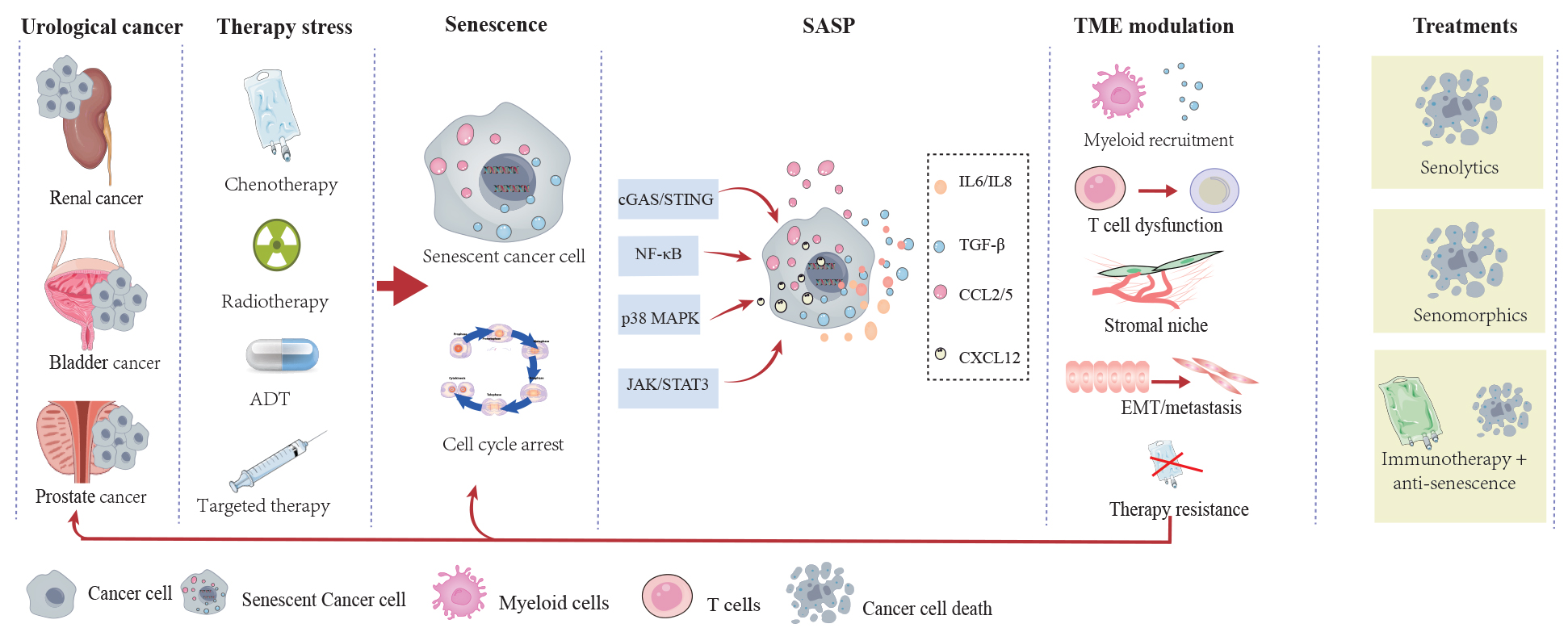
Cellular senescence is a durable stress response with double effects on cancer. Although stable cell-cycle arrest may initially restrict tumor growth, senescent tumor and stromal cells remain metabolically active and can remodel the tumor microenvironment through the senescence-associated secretory phenotype (SASP). This review synthesizes current evidence on therapy-induced and tumor-intrinsic senescence in prostate, bladder, and kidney cancers, focusing on molecular regulation and context-dependent SASP effects. We discuss how NF-κB, JAK/STAT3, cGAS-STING, and p38 MAPK regulate SASP composition. These pathways help determine the composition of the SASP, including the cytokines, chemokines, growth factors, and matrix-remodeling enzymes released by senescent cells. These SASP factors can recruit immunosuppressive myeloid cells, impair T-cell function, alter extracellular matrix organization, and support invasion, metastasis, and treatment response. The available evidence indicates that outcome depends on the inducing stimulus, cellular source, immune context, and duration of the response. Senolytics, senomorphics, and combinations with immunotherapy are promising, but evidence in urological cancers remains largely preclinical. Clinical translation will require validated multi-marker assays, longitudinal sampling, and prospective trials that distinguish transient immunogenic senescence from persistent immunosuppressive senescence.Graphic Abstract
Keywords
Prostate, bladder, and kidney cancers are a growing health burden worldwide [1,2]. Prostate cancer (PC) alone accounts for over 1.4 million new diagnoses annually and is the fifth leading cause of cancer-related death among men worldwide. Bladder cancer (BC), with approximately 600,000 new cases per year, is characterized by a high recurrence rate that necessitates lifelong surveillance, while renal cell carcinoma (RCC) incidence continues to rise, induced in part by the obesity epidemic and improved cross-sectional imaging. A bibliometric synthesis identified aging as a recurring theme in cancer research [3]. The incidence rises sharply after the sixth decade, implicating aging-associated biological processes in both tumor initiation and progression [2]. Separate reviews have examined oxidative stress-senescence interactions and circulating tumor-cell plasticity in cancer [4,5]. How the senescence-associated secretory phenotype (SASP) converts a cell-autonomous proliferative arrest into non-cell-autonomous tumor-promoting effects remains incompletely understood in urological cancers, as summarized in a recent review and supported by original experimental work [6,7]. This knowledge gap has direct clinical consequences, as therapies that inadvertently induce senescence may paradoxically promote the disease they are intended to eradicate.
The SASP is not a single, stereotyped secretory program. Separate reviews have addressed SASP context dependence and broader immune-regulatory mechanisms of treatment resistance across cancer types [8,9]. Studies indicate that senescence can influence progression and treatment outcomes in urological cancers. For instance, in PC, androgen deprivation therapy (ADT) induced a distinct senescence state via IL-6 and IL-8 secretion that was mechanistically linked to castration-resistant progression [10]. In RCC, tyrosine kinase inhibitors (such as sunitinib) triggered P53/DEC1-dependent senescence via VEGF and IL-8 release, while axitinib induced endothelial cell senescence through ROS accumulation and ATM activation [11,12]. In BC models, pemigatinib induced G1 arrest, cellular stress, and senescence, whereas decitabine activated NOTCH1, increased IL-6 release, and promoted differentiation [13,14]. Single-cell and spatial transcriptomic analyses identified club-like epithelial cells with a SASP and immunosuppressive interactions with myeloid cells in PC [15]. Research over the past five years has focused increasingly on why the SASP can either restrain or promote tumors and how it affects cancer progression, the TME, and treatment responses in urological cancers. The discovery that mitochondrial DNA released by senescent cells activated the cGAS-STING pathway and induced PMN-MDSC recruitment has linked SASP biology to innate immune sensing [16]. Metformin has been experimentally shown to suppress the SASP through IKK/NF-κB inhibition [17]. These findings support the SASP as a candidate therapeutic target, but its clinical relevance in urological cancers remains unproven.
This review synthesizes the molecular mechanisms governing SASP regulation in urological cancers, with a focus on transcriptional control of the SASP, immune remodeling of the TME, and the contribution of senescence to disease progression and therapy resistance. This review integrates findings from experimental studies in PC, RCC, and BC to identify common principles as well as cancer-type-specific features, and critically evaluates the therapeutic strategies (senolytics, senomorphics, and senescence-immunotherapy combinations) and identifies the questions that must be resolved before senescence-targeted approaches could be tested clinically in urological cancers.
2 Cellular Senescence in Urological Cancers
2.1 Therapy-Induced Senescence
Therapy-induced senescence (TIS) is a durable cell-cycle arrest caused by chemotherapy, radiotherapy, androgen deprivation, or targeted treatment. Unlike apoptosis, TIS allows damaged cells to remain metabolically active and to produce SASP factors that can support immune clearance or, if the cells persist, promote inflammation and treatment resistance. Therefore, TIS should be identified with a combination of durable arrest, DNA damage, lysosomal, and SASP markers rather than a single assay. The timing of measurement is also important because transient and persistent states may have different biological effects, as reviewed in [18].
Non-urological studies show that impaired DNA repair can alter chemotherapy response and that tumor cells can escape p53-dependent senescence [19,20]. For example, docetaxel, a standard-of-care agent for metastatic castration-resistant PC (mCRPC), induced a robust senescence response in RB1-deficient PC cells [21]. Elimination of these senescent cells via Bcl-2 inhibition attenuated malignant progression, establishing a direct causal link between therapy-induced senescence and disease development. In RCC, sunitinib promoted senescent tumor cell formation through P53/DEC1 transcriptional activation, with consequent upregulation of IL-6 and IL-8 that reshaped the local cytokine milieu [11]. The related VEGFR-targeted TKI axitinib induced endothelial cell senescence through ROS accumulation and the ATM-dependent DNA damage pathway [12]. Radiotherapy produced comparable effects across tissue compartments. In an irradiated bone model, osteoblasts and bone-marrow stromal cells acquired an early pro-inflammatory senescent signature, and navitoclax reduced chronic structural bone damage [22]. This non-urological study did not examine renal cell carcinoma [22]. Gefitinib inhibited the bidirectional crosstalk between mesenchymal stem cells and PC cells (a paracrine circuit that otherwise promoted tumor cell proliferation and blunted docetaxel activity) through the disruption of the CCL2/CCL5-mediated pathway [23]. In BC, the FGFR inhibitor pemigatinib induced G1 phase cell cycle arrest accompanied by cellular stress responses and the upregulation of tumor-suppressor microRNAs, resulting in a senescence-like state that might contribute to its antitumor activity [13]. Decitabine activated NOTCH1, increased IL-6 release, and induced senescence-like morphology without increased SA-β-gal activity or p16 expression in muscle-invasive BC cells [14]. IL-4, a cytokine typically associated with Th2 immunity, was found to directly induce cellular senescence in human RCC cell lines through STAT6 and p38 MAPK activation, revealing an unexpected connection between the type 2 immune pathway and cancer cell senescence [24]. Photobiomodulation with blue laser light inhibited BC progression in association with MMP modulation and senescence induction, representing a non-pharmacological approach to senescence engagement [25]. An IL-12-producing cellular vaccine suppressed growth promoted by chemotherapy-induced senescent cells in murine models that included TRAMP-C2 prostate cancer [26]. Together, these findings indicate that conventional urological cancer treatments, such as chemotherapy, radiotherapy, and targeted kinase inhibitors, create a biologically significant reservoir of senescent cells whose SASP can undermine therapeutic durability. Whether treatment-induced senescence should be actively mitigated through concurrent senolytic or senomorphic intervention, and at what time point relative to therapy such intervention would be optimally deployed, remain unresolved questions for prospective clinical evaluation.
2.2 Tumor-Intrinsic and Oncogene-Induced Senescence
Similarly, oncogenic stress and tumor suppressor loss can activate an intrinsic senescence barrier. A bibliometric analysis of research on aging, circadian rhythms, and cancer identified oxidative stress, prostate cancer, and NF-κB among the major research themes [3]. However, this literature does not specifically establish a senescence mechanism in urological malignancies. In PC, NFATC1 promoted tumorigenesis by allowing cells to escape PTEN loss-induced senescence [27]. These observations show that senescence can restrain early tumor growth, whereas escape from or persistence of this state may support later progression.
2.3 Androgen Deprivation Therapy as a Senescence Inducer in Prostate Cancer
ADT is a prostate-specific inducer of cellular senescence and SASP generation. The broader clinical context of resistance to ADT has been reviewed, while treatment-related outcomes have been examined in clinical studies [28,29]. For example, ADT triggered senescence in androgen-sensitive PC cells through AR suppression, which led to SKP2 downregulation and consequent stabilization of the cyclin-dependent kinase inhibitor P27, resulting in a durable proliferation arrest with characteristic SA-β-gal activity and SASP factor secretion [30]. ADT-induced senescence is permissive for the development of castration resistance. Senescent LNCaP cells persisted after ADT, and their SASP (enriched in IL-6, IL-8, and CXCL12) created a microenvironment conducive to the emergence of androgen-independent clones [10]. ADT-senescent PC cells also upregulated anti-apoptotic Bcl-2 family proteins, particularly BCL-XL, and subsequent treatment with navitoclax selectively eliminated these cells, suppressing castration-resistant progression [10,31]. The same compound, navitoclax (ABT-263), produced distinct responses in AR agonist- and antagonist-induced senescent LNCaP cells, indicating that the inducing stimulus shapes senolytic sensitivity [32]. Kawata et al. [33] further confirmed that ADT stimulated SASP secretion, including IL-6, IL-8, and antioxidant response genes, in human PC specimens. Combined high-dose androgen modulation with hypofractionated radiotherapy induced DNA damage-dependent senescence that modified the TME, suggesting the senescence-AR axis could be therapeutically manipulated [34]. A spatio-temporal mathematical model combining ADT, chemotherapy, and senolytic treatment has predicted optimal sequencing strategies for metastatic PC, providing a theoretical framework for clinical trial design [35]. Together, these results define ADT-induced senescence as a double-edged phenomenon. Senescence initially restrains tumor growth through proliferative arrest, but the accompanying SASP factors facilitate castration-resistant progression, leading to a paradox that can potentially be resolved by the timely addition of senolytic therapy.
3 Molecular Regulation of the SASP
The SASP comprises cytokines and chemokines such as IL-6, IL-8, CCL2, CCL5, CXCL1, CXCL12, growth and differentiation factors such as TGF-β, and matrix-remodeling enzymes including MMPs. During an early, transient response, these factors may recruit immune cells that remove damaged cells and reinforce tumor suppression. If senescent cells persist, overlapping factors can sustain inflammation, recruit myeloid-derived suppressor cells and regulatory T cells, impair cytotoxic lymphocytes, remodel extracellular matrix, and support invasion or treatment resistance. SASP composition alone therefore cannot define biological outcome; cellular source, concentration, duration, receptor availability, and immune context must also be considered. In summary, senescence in urological cancers is not a uniform state. Its effect depends on the inducing stress, tumor genotype, cellular source of the SASP, and whether immune clearance occurs.
3.1 NF-κB and Pro-Inflammatory SASP Transcription
NF-κB is a central regulator of the pro-inflammatory SASP. Metformin inhibited IKK/NF-κB signaling and reduced IL-6, IL-8, and CXCL1 secretion without reversing growth arrest [17]. In PC, monoacylglycerol lipase inhibition also reduced NF-κB-dependent SASP activity and improved docetaxel response [36]. These findings support NF-κB as a senomorphic target, although systemic inhibition may disrupt protective immune responses.
3.2 The JAK/STAT Pathway and IL-6/IL-8 Production
In pleural mesothelioma, palbociclib-induced pseudo-senescence was accompanied by increased STAT3 phosphorylation but did not improve sensitivity to senolytics [37]. In PC, Zhou et al. [38] identified a population of p21-positive senescent stromal cells in the TME that promoted immune suppression through paracrine STAT3-dependent signaling, and demonstrated that this immunosuppressive program could be reversed via the senolytic navitoclax, directly linking STAT3-mediated SASP to immune evasion. In a PTEN-deficient prostate cancer model, STAT3-dependent ARF expression was reported to reinforce the senescence barrier and suppress metastatic progression. A related commentary and the original genetic-model study both stress that this effect is context specific [39,40]. Osalmid sensitized clear cell RCC to navitoclax-mediated senolysis through a STAT3/BCL-XL-dependent mechanism, identifying a candidate combination for further preclinical evaluation [41]. In PC, the IL-6/STAT3 axis also engaged the AR signaling network bidirectionally, with SASP-derived IL-6 capable of ligand-independent AR activation, leading to ADT resistance [10]. Interleukin-4-induced senescence in RCC proceeded through STAT6 and p38 MAPK, indicating that multiple STAT family members contributed to senescence regulation in a stimulus-specific manner [24]. Together, these studies suggest that JAK/STAT signaling can regulate SASP output and senolytic sensitivity in specific experimental settings.
3.3 DNA Damage Response and the p53/p21 Axis
The DNA damage response (DDR) is a common proximal trigger of senescence and a determinant of SASP composition. Oxidative stress-senescence interactions have been reviewed, while hypoxia-driven lactylation has been studied experimentally as a bladder-cancer resistance mechanism [4,42]. MnTE-2-PyP protected primary mouse prostate fibroblasts from radiation-induced activation and senescence by limiting ROS and TGF-β signaling [43]. CMTM6, a ubiquitously expressed regulator of PD-L1 stability, restrained DNA damage-induced senescence and suppressed antitumor immunity, while its loss promoted senescence and simultaneously enhanced T-cell-mediated anti-tumor function in RCC [44]. TIMP1 deficiency was shown to reprogram senescence in PC. Specifically, in the absence of TIMP1, senescent PC cells shifted their secretome toward a matrix-metalloproteinase-enriched, pro-metastatic SASP that promoted cancer cell invasion and distant metastasis [45]. The broader therapeutic potential of natural products in urological cancers has been reviewed in [46]. In CRPC models, atraric acid suppressed androgen-regulated neo-angiogenesis through angiopoietin-2 [47]. MiRNA-106a promoted PC radioresistance by targeting LITAF, increasing ATM expression, and reducing radiation-induced senescence [48]. Persistent DNA-damage signaling through ATM and ATR can stabilize p53 and induce p21-mediated cell-cycle arrest. This response can restrict tumor growth when damaged cells are cleared, but persistent senescent cells may maintain a SASP through parallel NF-κB and p38 MAPK signaling. The p53/p21 axis therefore helps establish TIS but does not, by itself, determine whether the resulting microenvironment is tumor suppressive or tumor promoting. Together, these studies show that DDR-severity shapes SASP quality, and that interventions targeting DDR proximal signaling can alter the senescence phenotype without necessarily eliminating senescent cells.
3.4 Innate Immune Sensing, Inflammasomes, and cGAS-STING Signaling
Mitochondrial DNA is one damage-associated molecular pattern, while the broader DAMP response includes nuclear DNA fragments, micronuclei, chromatin, ATP, and other stress signals. This crosstalk has been reviewed in a non-urological disease context [49]. Transient cGAS-STING activation can enhance antigen presentation and immune clearance, whereas persistent signaling can maintain inflammatory SASP production and recruit suppressive myeloid cells, as reviewed in [18]. The pathway should therefore be interpreted as a context-dependent immune regulator rather than a uniformly beneficial or harmful signal. For instance, ALDH1A3 reduced cellular senescence and SASP in PC through the cGAS-STING pathway modulation, adding a metabolic dimension to the innate immune regulation of senescence [50]. In BC, STING-mediated immune senescence was associated with distinct immune microenvironment configurations and prognostic characteristics [51]. These findings support a context-dependent role for STING signaling in urological cancers. The broader links between microbial sensing pathways and urinary tumor immunity have been reviewed in [52]. For example, TLR4 activation by lipopolysaccharide could confer a survival advantage to growth factor-deprived PC cells in association with CCL2/CCL5 secretion [53]. The RNA-binding protein ROCK2 promoted RCC progression through regulation of the PAI-1/NLRP3 inflammasome axis, linking senescence escape to inflammasome activation [54]. In the aged bladder, preexisting senescent fibroblasts created a tumor-permissive niche through CXCL12 secretion [55]. Cancer-associated senescence gene patterns distinguished clinically relevant clear cell RCC subtypes and were associated with prognosis and an inferred immunotherapy-response phenotype [56]. Together, this evidence supports a context-dependent role for cGAS-STING in shaping SASP output according to the DAMP involved, the responding cell type, and the tumor immune context. These pathways form an interacting regulatory network rather than separate linear cascades. Their duration and relative activity shape SASP composition and help explain why similar senescence markers can accompany different immune outcomes.
4 SASP-Mediated Remodeling of the Tumor Microenvironment
4.1 Myeloid-Cell Recruitment and Immunosuppression
A review has summarized senescence-associated immune remodeling, while a clear cell RCC cohort linked mitochondrial DNA copy-number enrichment to prognosis [6,57]. In PC, Di Mitri et al. [58] found that tumor-infiltrating Gr-1-positive myeloid cells antagonized senescence via paracrine mechanisms, providing evidence for bidirectional crosstalk between myeloid cells and senescence. Building on this foundation, the same group demonstrated that CXCR2 blockade re-educated tumor-associated macrophages from an M2-like immunosuppressive phenotype and, in turn, induced tumor cells into senescence, revealing a myeloid-senescence regulatory loop amenable to pharmacological intervention [59]. The release of mitochondrial DNA from senescent tumor cells activated the cGAS-STING pathway in myeloid cells and stimulated PMN-MDSC-mediated immunosuppression [16]. Apolipoprotein E, identified as a SASP component, induced a pathogenic senescent-like state in myeloid cells infiltrating PC, indicating that the senescence phenotype can propagate from cancer cells to immune cells in a paracrine, feedforward manner [60]. Single-cell and spatial transcriptomic analyses of human PC have identified club-like epithelial cells that engaged in immunosuppressive interactions with myeloid populations, providing spatial context for SASP-immune crosstalk [15]. The chemokine CXCL1, a canonical SASP factor, promoted PC cell growth and invasion [61]. CXCL16 and its receptor CXCR6 were prognostic in PC, although that study did not directly measure a SASP program [62]. Gefitinib-mediated inhibition of the crosstalk between mesenchymal stem cells and PC cells operated in part through the disruption of CCL2/CCL5-mediated signaling [23]. Together, these studies implicate SASP-associated chemokine networks, including CCL2/CCL5, CXCL1, CXCL12, CXCL16, and CXCR2-associated signaling, in myeloid-cell recruitment, retention, or functional polarization. Elucidating which specific chemokine-receptor axes predominate in each urological cancer, and whether these axes can be selectively interrupted, represents a priority for translational development.
4.2 T-Cell Dysfunction and Immune-Checkpoint Regulation
The context-dependent effects of SASP factors on T-cell function and immune-checkpoint responses, including both immunostimulatory and immunosuppressive programs, have been reviewed in [63,64]. For instance, tumor-intrinsic MAVS deficiency disrupted MAVS–CMTM6 mitochondrial stabilization, triggering ROS-driven senescence and a CCL3-rich SASP that recruited CD8+ T cells, synergizing with PD-1 blockade [65]. Another study also reported that CMTM6 restrained DNA damage-induced senescence and suppressed antitumor immunity, while its loss promoted senescence and enhanced T-cell-mediated anti-tumor function, improving immunotherapy responses [44]. RAR activation reprogrammed the senescence response in PC, converting a prototypical immunosuppressive SASP into one that enhanced NK cell anti-tumor activity, indicating that the senescence phenotype can be pharmacologically redirected from immune-evasive to immune-stimulatory [66]. DNA ligase 4 inhibition sensitized PC to immune checkpoint blockade in vivo through enhanced DNA damage and immunogenic senescence, suggesting that therapeutic induction of a specific senescence subtype could potentiate immunotherapy [67]. Conversely, radiotherapy-resistant PC cells escaped immune checkpoint blockade through the senescence-related ATR kinase, which suppressed the immunogenic features of senescence that would otherwise trigger antitumor immunity [68]. These studies show that the immune effect of the SASP depends on both the inducing pathway and the factors released.
A single-center series evaluated immunotherapy outcomes in older patients with cancer, but it did not establish a senescence-specific effect [69]. In clear cell RCC, multi-omics predictors of disease aggressiveness were associated with immunotherapy outcomes, although senescence was not directly measured [70]. The net immunological impact of the SASP thus depends on the balance between chemokines that recruit immunosuppressive populations (CXCL1/2/5/8/12, CCL2) and those that recruit effector lymphocytes (CCL3, CCL5), as well as on the pre-existing immune contexture of the tumor. SENEX was identified as a regulator of peripheral regulatory T cell accumulation in aged urinary BC, linking a specific senescence gene to immune cell trafficking [71].
4.3 Stromal Senescence and the Tumor-Promoting Niche
Fibro-adipogenic progenitors have been reviewed in muscle aging, and tumor-microenvironment diagnostics have been reviewed more broadly in [72,73]. Zhou et al. [38] identified p21-positive senescent stromal cells in the prostate TME that actively suppressed antitumor immunity and promoted tumor progression, and demonstrated their selective elimination by navitoclax, resulting in restored immune surveillance. In the aging bladder, Meguro et al. demonstrated that preexisting senescent fibroblasts accumulated with chronological age and created a tumor-permissive niche through CXCL12 secretion, a finding that mechanistically linked organismal aging to BC susceptibility [55]. Senescent stromal cells also contribute to TME metabolic reprogramming. Senescent fibroblasts from PC secreted glutamine that facilitated neighboring cancer cell proliferation and invasion [74]. Therapy-induced stromal senescence promoted the aggressiveness of prostate cancer cells through paracrine SASP factors including IL-6, IL-8, and MMPs [75]. The SASP from mesenchymal stromal cells impaired the growth of immortalized prostate epithelial cells while having no significant effect on metastatic PC cells, demonstrating cell-context-dependent differential responses to the senescence secretome [76]. Together, these studies indicate that stromal senescence can actively shape a tumor-promoting microenvironment, but its effects remain dependent on cell type and tissue context.
4.4 Temporal Evolution from Immune Surveillance to Immune Suppression
The immune effect of senescence can change over time. An early response may include interferon signaling, antigen-presentation cues, and chemokines that recruit natural killer and CD8+ T cells. If clearance fails, persistent NF-κB, JAK/STAT, and DNA-sensing signals can maintain IL-6, IL-8, CCL2, CXCL chemokines, TGF-β, and matrix-remodeling enzymes. This later state favors MDSC and regulatory T-cell recruitment, macrophage polarization, and T-cell dysfunction. Because this transition does not follow a universal clock, treatment timing should be guided by serial tumor and circulating measurements rather than a fixed interval. The immune effect of the SASP therefore depends on both cell type and time. Early immune recruitment may support clearance, whereas persistent signaling can establish a suppressive stromal and myeloid niche.
5 SASP in Disease Progression and Therapy Resistance
5.1 Epithelial-Mesenchymal Transition, Invasion, and Metastasis
Outside urological oncology, NLRP3 regulation has been studied in hepatocellular carcinoma, whereas senescence-EMT coupling has been examined in hypertensive nephropathy [77,78]. In BC, HINFP downregulation induced a non-cell-autonomous SASP that promoted metastasis [79]. Specifically, SASP factors secreted by HINFP-low cells, including IL-6 and MMPs, enhanced the migratory and invasive capacity of neighboring HINFP-proficient cells, demonstrating how a subpopulation of senescent-like cancer cells promoted the collective malignant behavior of the tumor [79]. The calcium sensor STIM1 simultaneously accelerated senescence and enhanced EMT in PC through NF-κB and calcium-dependent transcription factors, co-regulating two seemingly contradictory programs (stable arrest and enhanced invasion) through shared upstream signaling [80]. TIMP1 deficiency reprogrammed the senescence secretome toward a pro-metastatic, MMP-enriched SASP that promoted PC invasion and distant metastasis [45]. In lung epithelial models, MMP-induced Rac1b promoted EMT and bypassed oncogene-induced senescence; its relevance to urological SASP biology remains unproven [81]. SPARC induced stromal BMP7 signaling that promoted senescence-associated dormancy of PC cells in bone [82]. Matairesinol inhibited TGF-β/Wnt-associated EMT in PC3 cells, while transcriptomic analysis indicated activation of p53 and senescence programs [83]. Together, the urological studies indicate that SASP-associated chemokine, MMP, and TGF-β pathways can support EMT or invasion in defined models.
5.2 Castration-Resistant Progression and the Senescence-Androgen Receptor Axis
The transition from androgen-sensitive to castration-resistant PC is the major cause of PC mortality, and therapy-induced senescence is increasingly recognized as a contributor to this transition. ADT-induced senescence generated a SASP enriched in IL-6, IL-8, and growth factors that could activate AR in a ligand-independent manner, enabling the outgrowth of androgen-independent clones [10]. STAT3 directly modulated AR transcriptional output bidirectionally. AR suppression by ADT can induce senescence, and SASP factors can reactivate AR in the absence of androgens. This feedforward loop created a challenging therapeutic scenario in which the treatment that manages androgen-driven growth simultaneously promotes androgen-independent recurrence. Preclinical evidence that the addition of senolytics to ADT could suppress castration-resistant progression provides a therapeutic rationale for interrupting this loop at the senescence node [10,31].
5.3 Therapy-Resistance Feedback Loops
The relationship between senescence and therapy resistance is context-dependent. In prostate cancer, CCL5/CCR5 signaling within tumor-stromal crosstalk has been linked to cisplatin resistance and neuroendocrine differentiation [84]. Senescence can create a therapeutic vulnerability while also supporting resistance. In TFE3-rearranged RCC, TFE3 fusions increased p21 expression; cytoplasmic p21 promoted survival and migration, whereas nuclear p21 induced senescence and SASP secretion [85]. Radiotherapy-resistant PC cells escaped immune checkpoint blockade through senescence-related ATR signaling that suppressed immunogenic features of senescence [68]. DNA damage-induced senescence, but not enzalutamide-induced senescence, increased sensitivity to Bcl-xL inhibition, indicating stimulus-specific senolytic dependencies [86]. This apparent discrepancy indicates that androgen deprivation and direct AR antagonism can generate distinct senescence states with different BCL-XL dependencies. The concept of “senescence subtype” therefore has direct clinical implications. For example, a tumor with DNA damage-induced senescence may respond differently to a senolytic than a tumor with ADT-induced senescence. PTEN-TP53-deficient PC cells exhibited specific vulnerabilities to combined PARP-PI3K inhibition that intersected with senescence and DDR signaling pathways [87]. Understanding the molecular taxonomy of senescence states and matching specific senescence subtypes to specific interventions represents an important direction for precision senescence medicine in urological oncology.
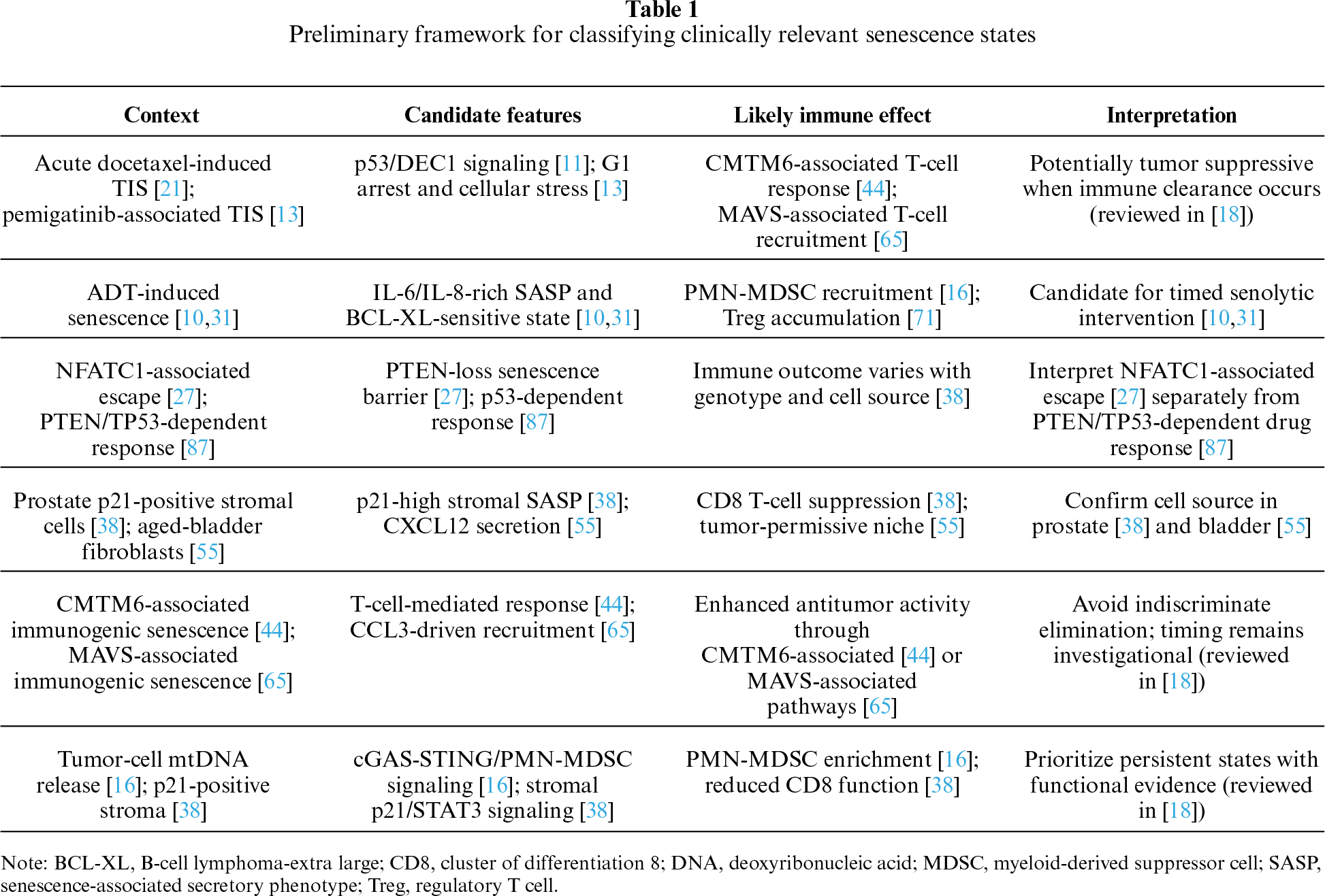
Table 1 shows the key gene/pathway. Across these disease settings, persistent senescence can create feedback between tumor cells, stromal cells, and immune cells. This feedback may promote invasion and treatment resistance even when the initial senescence response restricted proliferation.
6 Therapeutic Targeting of Senescence and the SASP
Senescence-targeted therapies include senolytics, senomorphics, and immune-based approaches that promote senescent-cell clearance. Their context-dependent effects have been reviewed in [18]. Specifically, senolytics selectively eliminate senescent cells by exploiting their acquired dependency on anti-apoptotic pathways. Senomorphics suppress the SASP without killing the senescent cell. For instance, the Bcl-2 family inhibitor navitoclax (ABT-263) showed senolytic activity in preclinical prostate cancer models. Navitoclax eliminated ADT-induced senescent PC cells and delayed castration-resistant progression in preclinical models [10,31]. In RCC, osalmid sensitized clear cell RCC to navitoclax via a STAT3/BCL-XL pathway [41]. DNA damage-induced senescent PC cells were sensitive to Bcl-xL inhibition, whereas enzalutamide-induced senescent cells were not [86]. The BH3 mimetic ABT-737, a predecessor of navitoclax, was shown to itself induce cancer cell senescence before promoting apoptosis, revealing a sequential pharmacodynamic process of senescence induction followed by senolytic elimination [88]. Senolytic compounds such as ABT-263 regulated a distinct fate of AR agonist- and antagonist-induced senescence [32].
Several questions remain unresolved. It is unclear whether senolytics should be given concurrently with senescence-inducing therapy or after senescence is established, how senescent-cell burden should be monitored to guide treatment, whether navitoclax-associated thrombocytopenia can be reduced through tumor-directed delivery or more selective BCL-2-family inhibitors, and whether senomorphic combinations with immune-checkpoint inhibitors can convert immunologically cold tumors into treatment-responsive tumors. Other treatment modalities may also interact with senescence biology. In a non-urological preclinical study, a B7-H3-targeted antibody-drug conjugate showed greater activity in TP53-deficient tumors when combined with ferroptosis induction; this was not evidence of senolytic efficacy [89]. Dietary factors may also affect senescence biology. In kidney transplant recipients, marine omega-3 polyunsaturated fatty acids reduced several circulating SASP components, but tissue senescence was not assessed [90]. Natural-product studies provide mechanistic and preclinical evidence, but most candidates lack mature structure-activity relationships, clinically achievable exposure data, pharmacokinetic characterization, and toxicological evaluation. In a castration-resistant prostate cancer model, verbascoside inhibited epithelial-mesenchymal transition and mitochondrial biogenesis [91]. Recent activity-guided studies have combined compound isolation with in vitro, in vivo, and in silico assessment to prioritize anticancer candidates [92]. This is a methodological precedent, not direct evidence of senolytic efficacy in urological cancers.
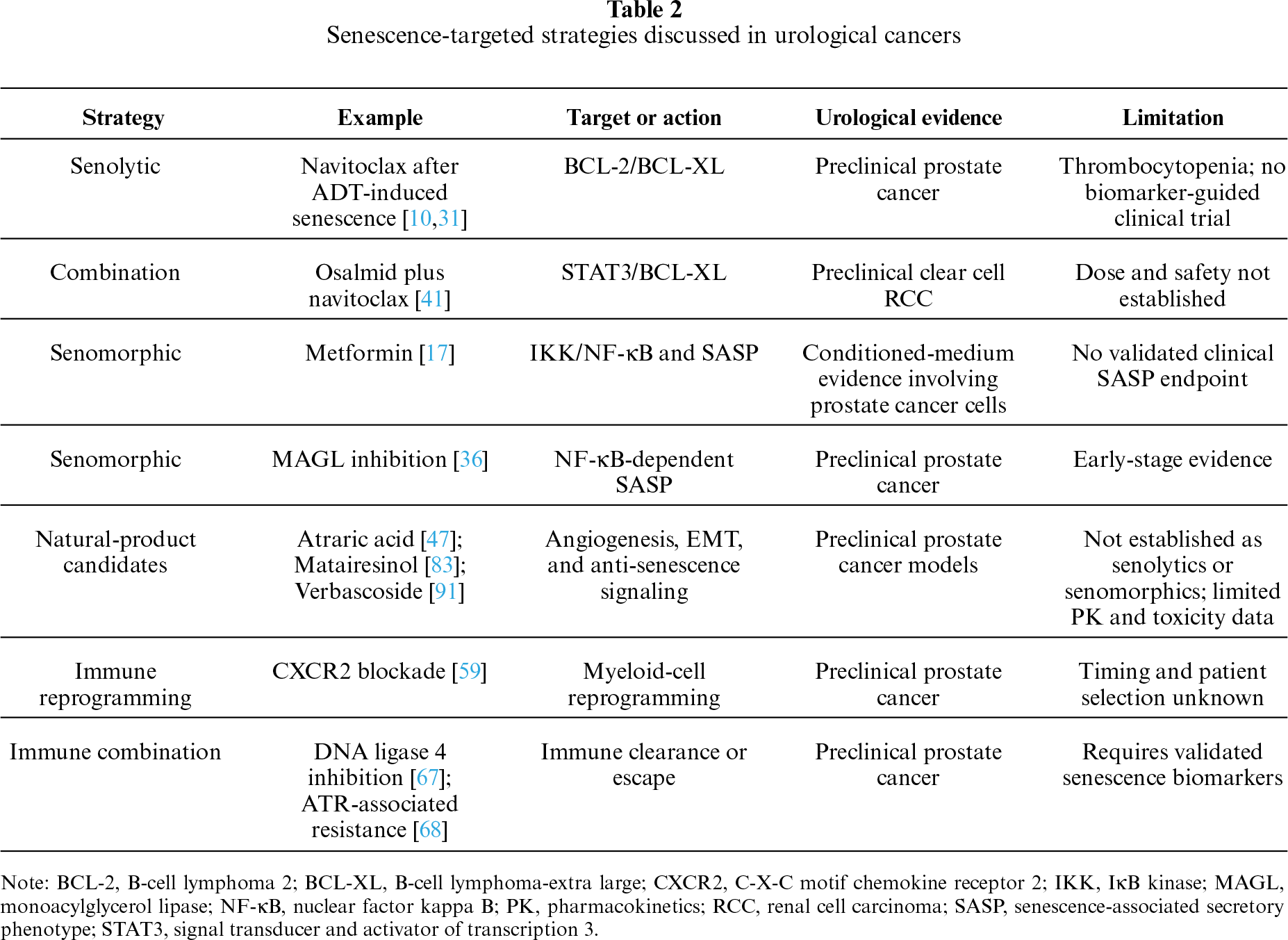
Table 2 provides key information of above discussions. Current senescence-targeted strategies remain largely preclinical in urological cancers. Translation will require biomarker-defined patient selection, pharmacodynamic confirmation of target engagement, and treatment schedules that preserve beneficial immune clearance.
7 Clinical Evidence and Monitoring
Direct clinical evidence for senescence-targeted treatment in urological cancers remains limited. Human PC specimens have shown senescence-associated changes after androgen deprivation therapy, but no prospective trial has established that a senolytic or senomorphic strategy improves outcomes by targeting senescent cells in prostate, bladder, or kidney cancer. In a preliminary trial in diabetic kidney disease, dasatinib plus quercetin reduced markers of senescent-cell burden in adipose tissue and skin [93]. In a separate open-label pilot study of patients with idiopathic pulmonary fibrosis, dasatinib plus quercetin was associated with improved physical function, and exploratory pharmacodynamic assessments included serum levels of circulating SASP factors [94]. Neither study established clinical efficacy in RCC or any other urological cancer.
A practical monitoring strategy should combine tissue and circulating measurements. Tissue assessment could include p16, p21, loss of Lamin B1, persistent DNA-damage foci, lysosomal activity, and a prespecified SASP panel. Serial plasma measurements of IL-6, IL-8, GDF15, CXCL chemokines, and matrix-remodeling proteins may provide a less invasive readout, but these factors are not specific to senescence and are influenced by age, infection, treatment, renal function, and tumor burden. A multi-marker approach may be more informative than a single analyte, but it requires prospective validation in urological cancer [95]. Prospective studies should collect tumor tissue and blood before treatment, shortly after senescence-inducing therapy, and at later time points. A persistent rise in a validated composite score, together with tissue evidence of durable arrest and a suppressive immune state, could identify a window for senolytic or senomorphic intervention. Imaging probes and liquid-biopsy approaches remain investigational and require analytical and clinical validation. Figure 1 summarizes this proposed monitoring framework.
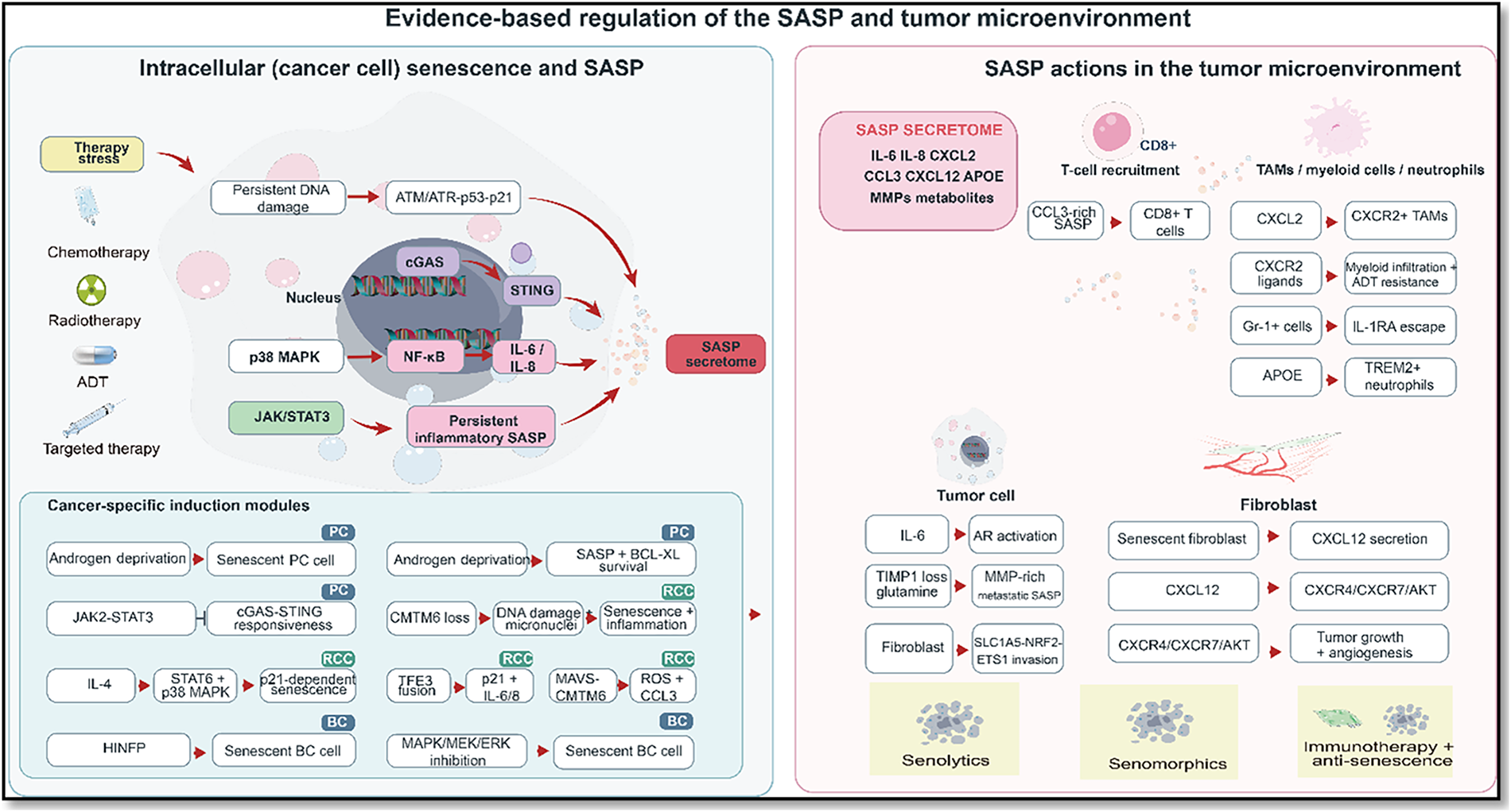
Figure 1: Context-dependent regulation of the senescence-associated secretory phenotype (SASP) in urological cancers. Therapy-related and tumor-intrinsic stresses engage DNA-damage, NF-κB, JAK/STAT3, cGAS-STING, and p38 MAPK signaling to shape SASP factor composition. Depending on the inducing stimulus, cellular source, immune context, and persistence, these factors can support immune clearance or promote suppressive myeloid recruitment, T-cell dysfunction, stromal remodeling, epithelial-mesenchymal transition, metastasis, and treatment resistance. ADT, androgen deprivation therapy; AR, androgen receptor; ATM, ataxia-telangiectasia mutated; BC, bladder cancer; cGAS, cyclic GMP-AMP synthase; CCL, C-C motif chemokine ligand; CD8, cluster of differentiation 8; CXCL, C-X-C motif chemokine ligand; EMT, epithelial-mesenchymal transition; JAK, Janus kinase; MAPK, mitogen-activated protein kinase; MMP, matrix metalloproteinase; NF-κB, nuclear factor kappa B; PC, prostate cancer; RCC, renal cell carcinoma; ROS, reactive oxygen species; SASP, senescence-associated secretory phenotype; STAT3, signal transducer and activator of transcription 3; STING, stimulator of interferon genes; TAM, tumor-associated macrophage.
Current evidence shows that cellular senescence has context-dependent effects in urological cancers. Growth arrest can restrict tumor expansion, whereas a persistent SASP can promote immune suppression, stromal remodeling, metastasis, and treatment resistance. The balance depends on the inducing treatment, tumor genotype, cellular source, and effectiveness of immune clearance.
Clinical translation requires a practical definition of harmful persistent senescence. Prospective studies should combine serial tissue and blood sampling with multi-marker panels, record the timing and duration of the senescence response, and test senolytics or senomorphics in biomarker-defined settings. Trials should distinguish pharmacodynamic evidence of senescent-cell reduction from conventional antitumor outcomes. Single-cell and spatial studies have resolved epithelial, stromal, and immune interactions in PC and recurrent BC, although only the PC study directly identified a SASP-associated niche [15,96]. Senescence-targeted treatment in prostate, bladder, and kidney cancers remains experimental. The next step is prospective testing to determine which patients, senescence states, and treatment schedules are most likely to benefit.
Acknowledgement: None.
Funding Statement: The authors received no specific funding for this study.
Author Contributions: Yan Zhu and Yunqiu Gao contributed to the conception, design, and drafting of the manuscript. Wenhao Zhang provided critical suggestions to improve the content and interpretation of the data. Yunqiu Gao supervised the study, offered expert guidance, and contributed to the manuscript’s revision. Yan Zhu and Yunqiu Gao were responsible for the overall conceptual framework, coordinated the research activities, and provided final approval of the manuscript. All authors reviewed and approved the final version of the manuscript.
Availability of Data and Materials: Data sharing is not applicable to this article because no new datasets were generated or analyzed in the current study.
Ethics Approval: Not applicable.
Conflicts of Interest: The authors declare no conflicts of interest.
References
1. Bray F, Laversanne M, Sung H, et al. Global cancer statistics 2022: GLOBOCAN estimates of incidence and mortality worldwide for 36 cancers in 185 countries. CA Cancer J Clin 2024;74(3):229–63. doi:10.3322/caac.21834. [Google Scholar] [CrossRef]
2. Feng DC, Li DX, Wu RC, et al. Global burden and cross-country inequalities in urinary tumors from 1990 to 2021 and predicted incidence changes to 2046. Military Med Res 2025;12(1):12. doi:10.1186/s40779-025-00599-y. [Google Scholar] [CrossRef]
3. Feng D, Xiao Y, Wang J, et al. Unraveling links between aging, circadian rhythm and cancer: insights from evidence-based analysis. Chin J Cancer Res 2024;36(2):341–50. doi:10.21147/j.issn.1000-9604.2024.03.09. [Google Scholar] [CrossRef]
4. Li D, Yu Q, Wu R, et al. Interactions between oxidative stress and senescence in cancer: mechanisms, therapeutic implications, and future perspectives. Redox Biol 2024;73(6):103208. doi:10.1016/j.redox.2024.103208. [Google Scholar] [CrossRef]
5. Linkner TR, Nagy ZB, Kalmár A, et al. Circulating tumor cells: indicators of cancer progression, plasticity and utility for therapies. Pathol Oncol Res 2025;31:1612181. doi:10.3389/pore.2025.1612181. [Google Scholar] [CrossRef]
6. Li D, Yu Q, Shao F, et al. Decoding the crossroads of aging and cancer through single-cell analysis: implications for precision oncology. Int J Cancer 2025;157(5):807–18. doi:10.1002/ijc.35456. [Google Scholar] [CrossRef]
7. Joshi CS, Salazar AM, Wang C, et al. D-Mannose reduces cellular senescence and NLRP3/GasderminD/IL-1β-driven pyroptotic uroepithelial cell shedding in the murine bladder. Dev Cell 2024;59(1):33–47.e5. doi:10.1016/j.devcel.2023.11.017. [Google Scholar] [CrossRef]
8. Saha S, Mahapatra S, Khanra S, et al. Decoding breast cancer treatment resistance through genetic, epigenetic, and immune-regulatory mechanisms: from molecular insights to translational perspectives. Cancer Drug Resist 2025;8:36. doi:10.20517/cdr.2025.69. [Google Scholar] [CrossRef]
9. Cao L, Li K, Li Q, Tong Q, Wang Y, Huang L. The controversial role of senescence-associated secretory phenotype (SASP) in cancer therapy. Mol Cancer 2025;24(1):283. doi:10.1186/s12943-025-02475-8. [Google Scholar] [CrossRef]
10. Carpenter V, Saleh T, Min Lee S, et al. Androgen-deprivation induced senescence in prostate cancer cells is permissive for the development of castration-resistance but susceptible to senolytic therapy. Biochem Pharmacol 2021;193(6):114765. doi:10.1016/j.bcp.2021.114765. [Google Scholar] [CrossRef]
11. Zhu Y, Xu L, Zhang J, et al. Sunitinib induces cellular senescence via p53/Dec1 activation in renal cell carcinoma cells. Cancer Sci 2013;104(8):1052–61. doi:10.1111/cas.12176. [Google Scholar] [CrossRef]
12. Mongiardi MP, Radice G, Piras M, et al. Axitinib exposure triggers endothelial cells senescence through ROS accumulation and ATM activation. Oncogene 2019;38(27):5413–24. doi:10.1038/s41388-019-0798-2. [Google Scholar] [CrossRef]
13. Pace A, Scirocchi F, Napoletano C, et al. Targeting FGFRs by pemigatinib induces G1 phase cell cycle arrest, cellular stress and upregulation of tumor suppressor microRNAs. J Transl Med 2023;21(1):626. doi:10.1186/s12967-023-04450-7. [Google Scholar] [CrossRef]
14. Ramakrishnan S, Hu Q, Krishnan N, et al. Decitabine, a DNA-demethylating agent, promotes differentiation via NOTCH1 signaling and alters immune-related pathways in muscle-invasive bladder cancer. Cell Death Dis 2017;8(12):3217. doi:10.1038/s41419-017-0024-5. [Google Scholar] [CrossRef]
15. Kiviaho A, Eerola SK, Kallio HML, et al. Single cell and spatial transcriptomics highlight the interaction of club-like cells with immunosuppressive myeloid cells in prostate cancer. Nat Commun 2024;15(1):9949. doi:10.1038/s41467-024-54364-1. [Google Scholar] [CrossRef]
16. Lai P, Liu L, Bancaro N, et al. Mitochondrial DNA released by senescent tumor cells enhances PMN-MDSC-driven immunosuppression through the cGAS-STING pathway. Immunity 2025;58(4):811–25.e7. doi:10.1016/j.immuni.2025.03.005. [Google Scholar] [CrossRef]
17. Moiseeva O, Deschênes-Simard X, St-Germain E, et al. Metformin inhibits the senescence-associated secretory phenotype by interfering with IKK/NF-κB activation. Aging Cell 2013;12(3):489–98. doi:10.1111/acel.12075. [Google Scholar] [CrossRef]
18. Schmitt CA, Wang B, Demaria M. Senescence and cancer—role and therapeutic opportunities. Nat Rev Clin Oncol 2022;19(10):619–36. doi:10.1038/s41571-022-00668-4. [Google Scholar] [CrossRef]
19. Li J, Meng Y, Ouyang X, Lin X, Wu Y, Kwok HF. RCC1 knockdown sensitizes drug-resistant colorectal cancer to 5-fluorouracil or doxorubicin by impairing DNA repair. Cancer Drug Resist 2025;8:58. doi:10.20517/cdr.2025.159. [Google Scholar] [CrossRef]
20. Buko T, Staniak K, Dudkowska M, et al. Anillin recedes in p53-dependent senescence of tumor cells and reappears in cells escaping from senescence. Aging Dis 2025;17(3):1590–603. doi:10.14336/AD.2025.0402. [Google Scholar] [CrossRef]
21. Su H, Huang L, Xia W, Tang DG, Zhou J, Yang G. Elimination of docetaxel-induced senescence attenuates malignant progression in RB1-deficient CRPC. Cell Oncol 2025;48(6):2055–72. doi:10.1007/s13402-025-01126-w. [Google Scholar] [CrossRef]
22. Achudhan D, Orme J, Sharma R, et al. Suppression of early pro-inflammatory senescent signature post-radiotherapy mitigates chronic bone damage. bioRxiv 2026. doi:10.64898/2026.03.01.708630. [Google Scholar] [CrossRef]
23. Borghese C, Cattaruzza L, Pivetta E, et al. Gefitinib inhibits the cross-talk between mesenchymal stem cells and prostate cancer cells leading to tumor cell proliferation and inhibition of docetaxel activity. J Cell Biochem 2013;114(5):1135–44. doi:10.1002/jcb.24456. [Google Scholar] [CrossRef]
24. Kim HD, Yu SJ, Kim HS, et al. Interleukin-4 induces senescence in human renal carcinoma cell lines through STAT6 and p38 MAPK. J Biol Chem 2013;288(40):28743–54. doi:10.1074/jbc.m113.499053. [Google Scholar] [CrossRef]
25. Xia Y, Yu W, Cheng F, et al. Photobiomodulation with blue laser inhibits bladder cancer progression. Front Oncol 2021;11:701122. doi:10.3389/fonc.2021.701122. [Google Scholar] [CrossRef]
26. Simova J, Sapega O, Imrichova T, et al. Tumor growth accelerated by chemotherapy-induced senescent cells is suppressed by treatment with IL-12 producing cellular vaccines. Oncotarget 2016;7(34):54952–64. doi:10.18632/oncotarget.10712. [Google Scholar] [CrossRef]
27. Manda KR, Tripathi P, Hsi AC, et al. NFATc1 promotes prostate tumorigenesis and overcomes PTEN loss-induced senescence. Oncogene 2016;35(25):3282–92. doi:10.1038/onc.2015.389. [Google Scholar] [CrossRef]
28. Khorasanchi A, Hong F, Yang Y, et al. Overcoming drug resistance in castrate-resistant prostate cancer: current mechanisms and emerging therapeutic approaches. Cancer Drug Resist 2025;8:9. doi:10.20517/cdr.2024.173. [Google Scholar] [CrossRef]
29. Huangfu X, Zheng J, Fan Z. Clinical study on thromboembolic events in prostate cancer patients receiving radiation therapy with or without androgen deprivation therapy. Ann Ital Chir 2025;96(4):533–42. doi:10.62713/aic.3946. [Google Scholar] [CrossRef]
30. Zhuang D, Kang J, Luo H, Tian Y, Liu X, Shao C. ARv7 promotes the escape of prostate cancer cells from androgen deprivation therapy-induced senescence by mediating the SKP2/p27 axis. BMC Biol 2025;23(1):66. doi:10.1186/s12915-025-02172-4. [Google Scholar] [CrossRef]
31. Carpenter V, Saleh T, Chakraborty E, et al. Androgen deprivation-induced senescence confers sensitivity to a senolytic strategy in prostate cancer. Biochem Pharmacol 2024;226(11):116385. doi:10.1016/j.bcp.2024.116385. [Google Scholar] [CrossRef]
32. Pungsrinont T, Sutter MF, Ertingshausen MCCM, et al. Senolytic compounds control a distinct fate of androgen receptor agonist- and antagonist-induced cellular senescent LNCaP prostate cancer cells. Cell Biosci 2020;10(1):59. doi:10.1186/s13578-020-00422-2. [Google Scholar] [CrossRef]
33. Kawata H, Kamiakito T, Nakaya T, et al. Stimulation of cellular senescent processes, including secretory phenotypes and anti-oxidant responses, after androgen deprivation therapy in human prostate cancer. J Steroid Biochem Mol Biol 2017;165(6):219–27. doi:10.1016/j.jsbmb.2016.06.007. [Google Scholar] [CrossRef]
34. Zamagni A, Zanoni M, Cortesi M, et al. Investigating the benefit of combined androgen modulation and hypofractionation in prostate cancer. Int J Mol Sci 2020;21(22):8447. doi:10.3390/ijms21228447. [Google Scholar] [CrossRef]
35. Lazebnik T, Friedman A. Spatio-temporal model of combining ADT and chemotherapy with senolytic treatment in metastatic prostate cancer. J Theor Biol 2025;602–603(2):112069. doi:10.1016/j.jtbi.2025.112069. [Google Scholar] [CrossRef]
36. Yu J, Zhang M, Li T, et al. Monoacylglycerol lipase blockades the senescence-associated secretory phenotype by interfering with NF-κB activation and promotes docetaxel efficacy in prostate cancer. Oncogene 2024;43(38):2835–49. doi:10.1038/s41388-024-03132-y. [Google Scholar] [CrossRef]
37. Sreeram I, Plans-Marin S, Cruz-Rodríguez M, et al. Pseudo-senescence induced by palbociclib does not sensitise pleural mesothelioma cells to combinations with senolytics. Cell Death Dis 2026;17(1):388. doi:10.1038/s41419-026-08696-z. [Google Scholar] [CrossRef]
38. Zhou L, DeMarco KD, Murphy KC, et al. p21-positive senescent stromal cells promote prostate cancer immune suppression and progression that can be reversed by senolytic therapy. Cancer Discov 2026;16(3):571–91. doi:10.1158/2159-8290.cd-25-1212. [Google Scholar] [CrossRef]
39. Culig Z, Pencik J, Merkel O, Kenner L. Breaking a paradigm: IL-6/STAT3 signaling suppresses metastatic prostate cancer upon ARF expression. Mol Cell Oncol 2016;3(2):e1090048. doi:10.1080/23723556.2015.1090048. [Google Scholar] [CrossRef]
40. Pencik J, Schlederer M, Gruber W, et al. STAT3 regulated ARF expression suppresses prostate cancer metastasis. Nat Commun 2015;6(1):7736. doi:10.1038/ncomms8736. [Google Scholar] [CrossRef]
41. Xue Y, Chen T, Ma Z, et al. Osalmid sensitizes clear cell renal cell carcinoma to navitoclax through a STAT3/BCL-XL pathway. Cancer Lett 2025;613(5):217514. doi:10.1016/j.canlet.2025.217514. [Google Scholar] [CrossRef]
42. Sun J, Shi Y, Yu K, et al. Hypoxia-induced histone lactylation drives cisplatin resistance in bladder cancer by promoting RBM15-dependent m 6 a methylation of IGFBP3. Research 2025;8(10):970. doi:10.34133/research.0970. [Google Scholar] [CrossRef]
43. Chatterjee A, Kosmacek EA, Oberley-Deegan RE. MnTE-2-PyP treatment, or NOX4 inhibition, protects against radiation-induced damage in mouse primary prostate fibroblasts by inhibiting the TGF-beta 1 signaling pathway. Radiat Res 2017;187(3):367–81. doi:10.1667/RR14623.1. [Google Scholar] [CrossRef]
44. Wang H, Fan Y, Chen W, et al. Loss of CMTM6 promotes DNA damage-induced cellular senescence and antitumor immunity. OncoImmunology 2022;11(1):2011673. doi:10.1080/2162402x.2021.2011673. [Google Scholar] [CrossRef]
45. Guccini I, Revandkar A, D’Ambrosio M, et al. Senescence reprogramming by TIMP1 deficiency promotes prostate cancer metastasis. Cancer Cell 2021;39(1):68–82.e9. doi:10.1016/j.ccell.2020.10.012. [Google Scholar] [CrossRef]
46. Li D, Wang J, Tuo Z, et al. Natural products and derivatives in renal, urothelial and testicular cancers: targeting signaling pathways and therapeutic potential. Phytomedicine 2024;127(3):155503. doi:10.1016/j.phymed.2024.155503. [Google Scholar] [CrossRef]
47. Ehsani M, Bartsch S, Rasa SMM, et al. The natural compound atraric acid suppresses androgen-regulated neo-angiogenesis of castration-resistant prostate cancer through angiopoietin 2. Oncogene 2022;41(23):3263–77. doi:10.1038/s41388-022-02333-7. [Google Scholar] [CrossRef]
48. Hoey C, Ray J, Jeon J, et al. miRNA-106a and prostate cancer radioresistance: a novel role for LITAF in ATM regulation. Mol Oncol 2018;12(8):1324–41. doi:10.1002/1878-0261.12328. [Google Scholar] [CrossRef]
49. Jindal D, Chopra V, Garg K, Sharma S. Mitochondrial DNA-mediated cGAS-STING activation and its crosstalk with NLRP3 inflammasome in chronic obstructive pulmonary disease. Cytokine Growth Factor Rev 2026;90:12–25. doi:10.1016/j.cytogfr.2026.05.001. [Google Scholar] [CrossRef]
50. Wang S, Wang L, Zhao Y. ALDH1A3 regulates cellular senescence and senescence-associated secretome in prostate cancer. Cancers 2025;17(7):1184. doi:10.3390/cancers17071184. [Google Scholar] [CrossRef]
51. Yao Z, Yang L, Yang X, Liu F, Fu B, Xiong J. Stimulator of interferon genes mediated immune senescence reveals the immune microenvironment and prognostic characteristics of bladder cancer. Heliyon 2024;10(9):e28803. doi:10.1016/j.heliyon.2024.e28803. [Google Scholar] [CrossRef]
52. Li D, Wu R, Yu Q, et al. Microbiota and urinary tumor immunity: mechanisms, therapeutic implications, and future perspectives. Chin J Cancer Res 2024;36(6):596–615. doi:10.21147/j.issn.1000-9604.2024.06.03. [Google Scholar] [CrossRef]
53. Jain S, Suklabaidya S, Das B, Raghav SK, Batra SK, Senapati S. TLR4 activation by lipopolysaccharide confers survival advantage to growth factor deprived prostate cancer cells. Prostate 2015;75(10):1020–33. doi:10.1002/pros.22983. [Google Scholar] [CrossRef]
54. Hong Z, Zeng Y, Zhou Q, et al. ROCK2 promotes renal cell carcinoma progression by functioning as an RNA-binding protein regulating the PAI-1/NLRP3 axis and senescence escape. Cell Signal 2025;136(1):112154. doi:10.1016/j.cellsig.2025.112154. [Google Scholar] [CrossRef]
55. Meguro S, Johmura Y, Wang TW, et al. Preexisting senescent fibroblasts in the aged bladder create a tumor-permissive niche through CXCL12 secretion. Nat Aging 2024;4(11):1582–97. doi:10.1038/s43587-024-00704-1. [Google Scholar] [CrossRef]
56. Zhu Z, Cao Q, Chen J, et al. Expression pattern of cancer-associated cellular senescence genes in clear cell renal cell carcinoma distinguishes tumor subclasses with clinical implications. Sci Rep 2025;15(1):442. doi:10.1038/s41598-024-84620-9. [Google Scholar] [CrossRef]
57. Bellal S, Rolley C, Richard J, et al. MtDNA copy number enrichment is associated with poor prognosis and eosinophilic morphology in clear cell renal cell carcinoma. Pathol Oncol Res 2025;31:1612172. doi:10.3389/pore.2025.1612172. [Google Scholar] [CrossRef]
58. Di Mitri D, Toso A, Chen JJ, et al. Tumour-infiltrating Gr-1+ myeloid cells antagonize senescence in cancer. Nature 2014;515(7525):134–7. doi:10.1038/nature13638. [Google Scholar] [CrossRef]
59. Di Mitri D, Mirenda M, Vasilevska J, et al. Re-education of tumor-associated macrophages by CXCR2 blockade drives senescence and tumor inhibition in advanced prostate cancer. Cell Rep 2019;28(8):2156–68.e5. doi:10.1016/j.celrep.2019.07.068. [Google Scholar] [CrossRef]
60. Bancaro N, Calì B, Troiani M, et al. Apolipoprotein E induces pathogenic senescent-like myeloid cells in prostate cancer. Cancer Cell 2023;41(3):602–19.e11. doi:10.1016/j.ccell.2023.02.004. [Google Scholar] [CrossRef]
61. Benelli R, Stigliani S, Minghelli S, Carlone S, Ferrari N. Impact of CXCL1 overexpression on growth and invasion of prostate cancer cell. Prostate 2013;73(9):941–51. doi:10.1002/pros.22640. [Google Scholar] [CrossRef]
62. Richardsen E, Ness N, Melbø-Jørgensen C, et al. The prognostic significance of CXCL16 and its receptor C-X-C chemokine receptor 6 in prostate cancer. Am J Pathol 2015;185(10):2722–30. doi:10.1016/j.ajpath.2015.06.013. [Google Scholar] [CrossRef]
63. Faget DV, Ren Q, Stewart SA. Unmasking senescence: context-dependent effects of SASP in cancer. Nat Rev Cancer 2019;19(8):439–53. doi:10.1038/s41568-019-0156-2. [Google Scholar] [CrossRef]
64. Liu Y, Yu Y, Chen Y, Zhuang J, Sun C. Tumor immunosenescence driven by chronic inflammation: mechanisms, microenvironment remodeling and therapeutic strategies. Aging Dis 2025;17(3):1347–70. doi:10.14336/AD.2025.0471. [Google Scholar] [CrossRef]
65. Wang H, Fan Y, Liang Q, et al. MAVS/CMTM6 axis couples mitochondrial homeostasis to immunogenic senescence via CCL3-driven T-cell recruitment in renal carcinoma. J Immunother Cancer 2025;13(12):e011477. doi:10.1136/jitc-2025-011477. [Google Scholar] [CrossRef]
66. Colucci M, Zumerle S, Bressan S, et al. Retinoic acid receptor activation reprograms senescence response and enhances anti-tumor activity of natural killer cells. Cancer Cell 2024;42(4):646–61.e9. doi:10.1016/j.ccell.2024.02.004. [Google Scholar] [CrossRef]
67. Wu J, Lagunas AM, Crowe DL. DNA ligase 4 inhibition sensitizes prostate cancer to immune checkpoint blockade in vivo. Anticancer Res 2025;45(3):883–96. doi:10.21873/anticanres.17476. [Google Scholar] [CrossRef]
68. Shao C, Zhang Y, Li H, et al. Radiotherapy-resistant prostate cancer cells escape immune checkpoint blockade through the senescence-related Ataxia telangiectasia and Rad3-related protein. Cancer Commun 2025;45(3):218–44. doi:10.1002/cac2.12636. [Google Scholar] [CrossRef]
69. Ramos MJ, Mendes AS, Romão R, Febra J, Araújo A. Immunotherapy in elderly patients—single-center experience. Cancers 2024;16(1):145. doi:10.3390/cancers16010145. [Google Scholar] [CrossRef]
70. Kamal Y, Cheng C, Frost HR, Amos CI. Predictors of disease aggressiveness influence outcome from immunotherapy treatment in renal clear cell carcinoma. Oncoimmunology 2019;8(1):e1500106. doi:10.1080/2162402X.2018.1500106. [Google Scholar] [CrossRef]
71. Chen T, Wang H, Zhang Z, et al. A novel cellular senescence gene, SENEX is involved in peripheral regulatory T cells accumulation in aged urinary bladder cancer. PLoS One 2014;9(2):e87774. doi:10.1371/journal.pone.0087774. [Google Scholar] [CrossRef]
72. Zhang FM, Zhang XZ, Yu Z, Zhuang CL, Han LL. Rethinking muscle aging through the lens of fibro-adipogenic progenitors. Aging Dis 2025. doi:10.14336/ad.2025.1162. [Google Scholar] [CrossRef]
73. Fan B, Zhang J, Li C, Zhang X. New trend in molecular diagnostics: insights from the tumor microenvironment. Interdiscip Med 2025;3(6):e70048. doi:10.1002/inmd.70048. [Google Scholar] [CrossRef]
74. Lori G, Mancini C, Paffetti C, et al. Senescent stroma-derived glutamine: a driver of aggressiveness in prostate and ovarian cancer cells. Cells 2026;15(9):770. doi:10.3390/cells15090770. [Google Scholar] [CrossRef]
75. Pardella E, Pranzini E, Nesi I, et al. Therapy-induced stromal senescence promoting aggressiveness of prostate and ovarian cancer. Cells 2022;11(24):4026. doi:10.3390/cells11244026. [Google Scholar] [CrossRef]
76. Alessio N, Aprile D, Squillaro T, et al. The senescence-associated secretory phenotype (SASP) from mesenchymal stromal cells impairs growth of immortalized prostate cells but has no effect on metastatic prostatic cancer cells. Aging 2019;11(15):5817–28. doi:10.18632/aging.102172. [Google Scholar] [CrossRef]
77. Gong Z, Li Y, Nie Y, et al. USP50-mediated NLRP3 deubiquitination enhances NLRP3 inflammasome activation to suppress HCC metastasis. J Pharm Anal 2025;15(11):101380. doi:10.1016/j.jpha.2025.101380. [Google Scholar] [CrossRef]
78. Sun MC, Zuo FW, Wang YZ, et al. CYFIP2 deficiency ameliorates renal interstitial fibrosis through attenuating tubular senescence in hypertensive nephropathy. Acta Pharmacol Sin 2026;47(9):2554–68. doi:10.1038/s41401-026-01793-x. [Google Scholar] [CrossRef]
79. Zheng X, Liu Z, Zhong J, et al. Downregulation of HINFP induces senescence-associated secretory phenotype to promote metastasis in a non-cell-autonomous manner in bladder cancer. Oncogene 2022;41(28):3587–98. doi:10.1038/s41388-022-02371-1. [Google Scholar] [CrossRef]
80. Xu Y, Zhang S, Niu H, et al. STIM1 accelerates cell senescence in a remodeled microenvironment but enhances the epithelial-to-mesenchymal transition in prostate cancer. Sci Rep 2015;5(1):11754. doi:10.1038/srep11754. [Google Scholar] [CrossRef]
81. Stallings-Mann ML, Waldmann J, Zhang Y, et al. Matrix metalloproteinase induction of Rac1b, a key effector of lung cancer progression. Sci Transl Med 2012;4(142):142ra95. doi:10.1126/scitranslmed.3004062. [Google Scholar] [CrossRef]
82. Sharma S, Xing F, Liu Y, et al. Secreted protein acidic and rich in cysteine (SPARC) mediates metastatic dormancy of prostate cancer in bone. J Biol Chem 2016;291(37):19351–63. doi:10.1074/jbc.M116.737379. [Google Scholar] [CrossRef]
83. Rajadnya R, Patil P, Deotare A, Mali A. Matairesinol regulates TGF-β-induced EMT and TGF-β associated crosstalk with Wnt Signaling in PC3 prostate cancer cells: an omics-guided therapeutic insight. Toxicol Appl Pharmacol 2026;506:117638. doi:10.1016/j.taap.2025.117638. [Google Scholar] [CrossRef]
84. Liu B, Zhang W, Ji Y, et al. Targeting the CCL5/CCR5 axis in tumor-stromal crosstalk to overcome cisplatin resistance in neuroendocrine prostate cancer. J Exp Clin Cancer Res 2025;44(1):296. doi:10.1186/s13046-025-03552-y. [Google Scholar] [CrossRef]
85. Lu Y, Ma W, Chen Y, et al. Transcriptionally up-regulation of CDKN1A/p21 by TFE3 fusion proteins worsened TFE3-rearranged renal cell carcinoma. Sci Rep 2025;15(1):30067. doi:10.1038/s41598-025-13302-x. [Google Scholar] [CrossRef]
86. Malaquin N, Vancayseele A, Gilbert S, et al. DNA damage- but not enzalutamide-induced senescence in prostate cancer promotes senolytic bcl-xL inhibitor sensitivity. Cells 2020;9(7):1593. doi:10.3390/cells9071593. [Google Scholar] [CrossRef]
87. González-Billalabeitia E, Seitzer N, Song SJ, et al. Vulnerabilities of PTEN–TP53-deficient prostate cancers to compound PARP–PI3K inhibition. Cancer Discov 2014;4(8):896–904. doi:10.1158/2159-8290.cd-13-0230. [Google Scholar] [CrossRef]
88. Song JH, Kandasamy K, Zemskova M, Lin YW, Kraft AS. The BH3 mimetic ABT-737 induces cancer cell senescence. Cancer Res 2011;71(2):506–15. doi:10.1158/0008-5472.CAN-10-1977. [Google Scholar] [CrossRef]
89. Leo J, Chen F, Shi W, et al. Improve the efficacy of B7-H3-targeting antibody-drug conjugate DS-7300a in TP53-deficient tumors by inducing ferroptosis. bioRxiv 2026. doi:10.64898/2026.05.05.722958. [Google Scholar] [CrossRef]
90. Chan J, Eide IA, Tannæs TM, Waldum-Grevbo B, Jenssen T, Svensson M. Marine n-3 polyunsaturated fatty acids and cellular senescence markers in incident kidney transplant recipients: the omega-3 fatty acids in renal transplantation (ORENTRA) randomized clinical trial. Kidney Med 2021;3(6):1041–9. doi:10.1016/j.xkme.2021.07.010. [Google Scholar] [CrossRef]
91. Mai HC, Hsieh PF, Wu CH, et al. Verbascoside suppresses epithelial–mesenchymal transition and mitochondrial biogenesis by targeting anti-senescence signaling in castration-resistant prostate cancer. Cancer Genom Proteom 2025;22(6):1044–60. doi:10.21873/cgp.20554. [Google Scholar] [CrossRef]
92. Güleç M, Şenol H, Tan N. Novel nordaucane sesquiterpenoid and sesquiterpene lactone from Laserpitium species: isolation, structure elucidation, in vitro, in vivo, and in silico evaluation as anticancer agents. Phytochem Anal 2025;36(3):846–65. doi:10.1002/pca.3472. [Google Scholar] [CrossRef]
93. Hickson LJ, Langhi Prata LGP, Bobart SA, et al. Senolytics decrease senescent cells in humans: preliminary report from a clinical trial of Dasatinib plus Quercetin in individuals with diabetic kidney disease. eBioMedicine 2019;47:446–56. doi:10.1016/j.ebiom.2019.08.069. [Google Scholar] [CrossRef]
94. Justice JN, Nambiar AM, Tchkonia T, et al. Senolytics in idiopathic pulmonary fibrosis: results from a first-in-human, open-label, pilot study. eBioMedicine 2019;40:554–63. doi:10.1016/j.ebiom.2018.12.052. [Google Scholar] [CrossRef]
95. St Sauver JL, Weston SA, Atkinson EJ, et al. Biomarkers of cellular senescence and risk of death in humans. Aging Cell 2023;22(12):e14006. doi:10.1111/acel.14006. [Google Scholar] [CrossRef]
96. Shi ZD, Sun Z, Zhu ZB, et al. Integrated single-cell and spatial transcriptomic profiling reveals higher intratumour heterogeneity and epithelial–fibroblast interactions in recurrent bladder cancer. Clin Transl Med 2023;13(7):e1338. doi:10.1002/ctm2.1338. [Google Scholar] [CrossRef]
Cite This Article
 Copyright © 2026 The Author(s). Published by Tech Science Press.
This work is licensed under a Creative Commons Attribution 4.0 International License , which permits unrestricted use, distribution, and reproduction in any medium, provided the original work is properly cited.
 Downloads
 Citation Tools
