
 Submit a Paper
 Propose a Special lssue
 Open Access
ARTICLE

Dioscin Regulates Mitochondrial Autophagy and Cell Cycle to Promote Pulpal Stem Cell Differentiation and Mineralization
1 Department of Stomatology, Naval Medical University, Shanghai, China
2 Department of Stomatology, Wusong Central Hospital, Shanghai, China
3 Department of Stomatology, The First Affiliated Hospital of Naval Medical University, Shanghai, China
* Corresponding Author: Qiang Zhu. Email:
BIOCELL 2026, 50(5), 10 https://doi.org/10.32604/biocell.2026.076758
Received 26 November 2025; Accepted 12 February 2026; Issue published 13 May 2026
 View Full Text
 Download PDF
Abstract
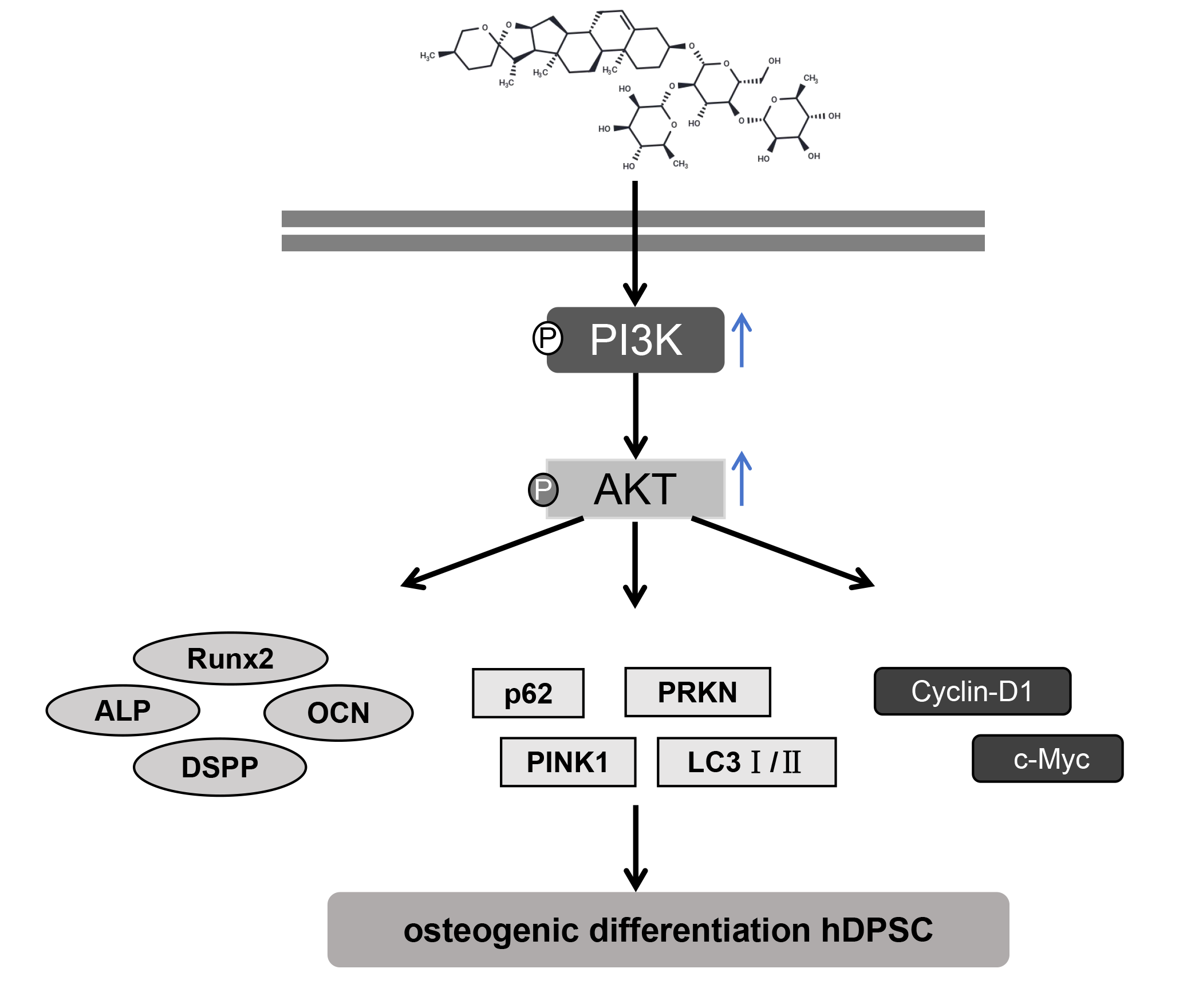
Background: Human dental pulp stem cells (hDPSCs) are promising for dental tissue regeneration. Dioscin (Dio), a natural compound, has various biological activities, but its effects on hDPSCs are unclear. This study aims to systematically elucidate the effects of Dio on promoting the osteogenic differentiation of hDPSCs and the underlying molecular mechanisms. Methods: Characterized hDPSCs were treated with Dio. Cell viability, proliferation, osteogenic differentiation (alkaline phosphatase (ALP) activity, Alizarin Red S (ARS)), and migration (Transwell) were assessed. Mitophagy (fluorescence, Western blot for PTEN-induced kinase 1 (PINK1), parkin RBR E3 ubiquitin-protein ligase (PRKN), microtubule-associated protein 1 light chain 3-II/I (LC3II/I), sequestosome 1 (p62)) and cell cycle (flow cytometry, cyclin D1 (Cyclin-D1), cellular myelocytomatosis oncogene (c-Myc)) were evaluated. The phosphatidylinositol 3-kinase (PI3K) inhibitor LY294002 and runt-related transcription factor 2 (Runx2) overexpression were used to investigate the PI3K/protein kinase B (AKT)/Runx2 pathway. Results: hDPSCs displayed mesenchymal stem cell characteristics. Dio treatment enhanced hDPSC viability, significantly increased ALP activity and ARS staining intensity, and promoted cell migration. It also increased mitophagy (increased colocalization of mitochondria and lysosomes, upregulated protein expression of PINK1 and PRKN, an increased LC3II/I ratio) and promoted cell cycle progression (increased S-phase cells, Cyclin-D1, c-Myc). Dio activated the PI3K/AKT pathway, upregulating Runx2. LY294002 reversed Dio’s effects, while Runx2 overexpression enhanced them. Conclusion: Dio is associated with enhanced hDPSC proliferation, osteogenesis, migration, mitophagy, and cell cycle progression, partly through activation of the PI3K/AKT pathway and upregulation of Runx2. These findings support the potential application of Dio in dental pulp regeneration and bone tissue engineering.Graphic Abstract
Keywords
Dental pulp stem cells; Dioscin; phosphatidylinositol 3-kinase (PI3K)/protein kinase B (AKT) signaling pathway; mitophagy; cell cycle
Supplementary Material
Supplementary Material FileCite This Article
APA Style
Zhou, Z., Chen, J., Zhu, Q. (2026). Dioscin Regulates Mitochondrial Autophagy and Cell Cycle to Promote Pulpal Stem Cell Differentiation and Mineralization. BIOCELL, 50(5), 10. https://doi.org/10.32604/biocell.2026.076758
Vancouver Style
Zhou Z, Chen J, Zhu Q. Dioscin Regulates Mitochondrial Autophagy and Cell Cycle to Promote Pulpal Stem Cell Differentiation and Mineralization. BIOCELL. 2026;50(5):10. https://doi.org/10.32604/biocell.2026.076758
IEEE Style
Z. Zhou, J. Chen, and Q. Zhu, “Dioscin Regulates Mitochondrial Autophagy and Cell Cycle to Promote Pulpal Stem Cell Differentiation and Mineralization,” BIOCELL, vol. 50, no. 5, pp. 10, 2026. https://doi.org/10.32604/biocell.2026.076758
 Copyright © 2026 The Author(s). Published by Tech Science Press.
This work is licensed under a Creative Commons Attribution 4.0 International License , which permits unrestricted use, distribution, and reproduction in any medium, provided the original work is properly cited.
 Downloads
 Citation Tools