
 Login
Login Register
 Submit a Paper
 Propose a Special lssue
 Open Access
MINI REVIEW
Dysregulated Mechanotransduction in Brain Pathologies: How Physical Forces Contribute to Neurodegeneration
1 Department of Global Innovative Drugs, Graduate School of Chung-Ang University, Seoul, Republic of Korea
2 Department of Anatomy and Cell Biology, College of Medicine, Chung-Ang University, Seoul, Republic of Korea
* Corresponding Author: Hyun Jung Lee. Email:
(This article belongs to the Special Issue: Advanced Cell Signaling Pathways in Health and Disease)
BIOCELL 2026, 50(10), 3 https://doi.org/10.32604/biocell.2026.081123
Received 24 February 2026; Accepted 20 May 2026; Issue published 22 September 2026
 View Full Text
 Download PDF
Abstract
Aging and brain injury remodel the central nervous system (CNS) physical microenvironment, yet the contribution of these mechanical changes to neurodegenerative disease remains underappreciated. While traditional models emphasize biochemical mechanisms, emerging evidence indicates that altered tissue stiffness, extracellular matrix composition, and interstitial fluid dynamics actively reprogram intracellular signaling via dysregulated mechanotransduction. This review describes key physical cues shaping the brain microenvironment, including substrate rigidity and fluid flow within the cerebrospinal fluid (CSF) and glymphatic system. We discuss how aging and injury-induced alterations disrupt mechanotransductive signaling compared to physiological conditions. Although candidate mechanosensors remain incompletely characterized, several are highlighted, including stretch-activated calcium channels like Piezo1 and Transient Receptor Potential Vanilloid 4 (TRPV4) and stiffness-responsive pathways involving focal adhesion kinase and Yes-associated protein 1 (YAP1)/Transcriptional co-activator with PDZ-binding motif (TAZ). We examine how these altered pathways influence the progression of neurodegenerative disorders and brain injury–associated pathologies. By integrating mechanobiology into neurodegenerative research, this review establishes physical forces as active disease drivers and identifies mechanotransduction as a promising frontier for the diagnosis and treatment of various brain pathologies.Keywords
As societies worldwide are aging rapidly, extensive research efforts have been devoted to delaying or preventing the aging process. In particular, the incidence of neurodegenerative diseases accompanied by cognitive decline has increased markedly with brain aging, emerging as a major public health challenge that imposes substantial social and economic burdens [1,2]. Consequently, large-scale drug development programs and clinical studies aimed at treating or preventing neurodegenerative disorders, such as Alzheimer’s disease (AD) and Parkinson’s disease, have been actively pursued [3,4]. Age-associated neurodegenerative diseases, however, cannot be attributed to damage to a single cell type or to simple pathogenic mechanisms. Rather, they arise from complex, integrated alterations in the brain microenvironment, including reduced cerebral blood flow [5], consequent impairment of cerebrospinal fluid (CSF) circulation [6], dysfunction of the glymphatic system [7,8], diminished waste clearance due to astrocytic functional decline [9,10,11], development of a chronic inflammatory state, and widespread disruption of neuronal signaling networks [6,12,13] (Fig. 1). These multifactorial changes collectively shape the pathological landscape of the aging brain.
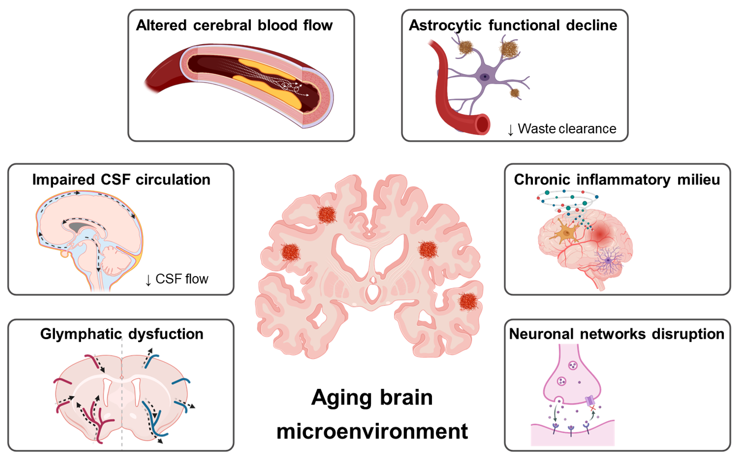
Figure 1: Multifactorial alterations in the aging brain microenvironment. The aging brain undergoes a complex and integrated process of physical remodeling, which collectively shapes the pathological landscape of neurodegeneration. As illustrated, age-associated vascular decline leads to altered hemodynamic patterns within the brain. These vascular changes, along with structural alterations, result in impaired cerebrospinal fluid (CSF) circulation and significant glymphatic dysfunction. Such disruption impairs the essential exchange between the CSF and interstitial fluid (ISF), leading to the diminished clearance of neurotoxic metabolic waste products. Concurrently, an age-dependent decline in astrocytic health further exacerbates this failure in waste clearance and results in the loss of vital homeostasis support. These multifaceted physical changes, operating within a chronic inflammatory milieu, ultimately culminate in widespread neuronal network disruption and synaptic loss. Together, this state of mechanical and biochemical dysregulation establishes a feed-forward loop that accelerates the initiation and progression of neurodegenerative diseases. This figure was created with BioRender (Toronto, ON, Canada, biorender.com).
Current therapeutic strategies have largely focused on preserving neuronal survival by preventing the accumulation of neurotoxic proteins, such as amyloid-β, or inhibiting tau hyperphosphorylation, both of which are key drivers of neuronal degeneration. In parallel, approaches aimed at modulating glial cells to attenuate neuroinflammation have been actively developed [14,15]. Notably, anti-amyloid-β antibody therapies have recently received FDA approval [16,17], and additional treatments targeting components of the complement system to reduce immune-mediated neuronal damage [18] are being applied in the context of neurodegenerative disease therapy.
Nevertheless, age-related neurodegenerative diseases are not only multifactorial in onset and progression but also reported to be predominantly sporadic rather than solely genetically driven [19]. Notably, even among patients who exhibit similar pathological hallmarks, such as amyloid-β or tau accumulation, the clinical course and symptom severity vary considerably, suggesting that factors beyond pathological protein burden contribute to disease heterogeneity [20]. Rather than arising solely from discrete molecular abnormalities, these disorders emerge from complex, system-wide alterations in the brain microenvironment [21]. Importantly, these microenvironmental alterations are accompanied by dysregulated mechanotransduction, wherein changes in tissue stiffness, fluid flow, and cellular tension are converted into aberrant intracellular signaling. Such mechanically driven signaling imbalances may actively accelerate neurodegenerative processes by amplifying inflammatory cascades, impairing synaptic stability, and disrupting neuronal survival pathways. In particular, age-dependent decline in circulatory system function induces alterations in cerebral blood flow, which in turn lead to reduced CSF circulation and disruption of the glymphatic system. These changes collectively drive global physical remodeling of the brain, accompanied by increased inflammatory burden and neuronal loss [22,23]. Accumulating evidence further suggests that such processes are associated with alterations in the mechanical properties of brain parenchyma, including changes in tissue stiffness [24,25]. This review evaluates the state of research on pathological remodeling of the brain’s physical microenvironment and details existing evidence regarding the relationship between this remodeling and dysregulated mechanotransductive signaling, which in turn may establish feed-forward loops that contribute to neurodegeneration.
2 Components of the Mechanical Microenvironment in the Healthy Brain
All organs reside within distinct physical microenvironments defined by tissue stiffness and interstitial fluid (ISF) dynamics, both of which are essential for maintaining normal physiological function. In the vascular system, for example, endothelial cells are continuously exposed to laminar shear stress on the order of tens of dyne/cm2 [26], while vascular smooth muscle cells experience circumferential stretching along with additional mechanical stimuli [27]. Thus, under normal physiological conditions, most tissues are persistently subjected to organ-specific physical forces that actively regulate cellular behavior.
Similarly, the brain parenchyma—composed of neurons and glial cells—is an exceptionally delicate tissue whose physical microenvironment is shaped by a combination of mechanical, viscoelastic, and fluid-dynamic properties [28]. Under physiological conditions, the brain exists within a defined range of parenchymal stiffness that plays a critical role in regulating cellular function, development, and disease susceptibility [29]. This stiffness is influenced by factors such as neuronal myelination and extracellular matrix (ECM) composition [30] and varies across distinct brain regions [31]. In parallel with its mechanical properties, the brain’s physical microenvironment is dynamically maintained by continuous fluid transport mediated by CSF and ISF, collectively referred to as the glymphatic system.
Although the existence of a tightly regulated physical microenvironment in the brain is established, its functional significance in controlling neuronal and glial behavior has only recently begun to attract substantial attention. In this context, Hu et al. (2021) demonstrated that astrocytes cultured on collagen I- and alginate-based hydrogels tuned to approximately 1 kPa-a stiffness that closely mimics healthy brain tissue-exhibited increased nuclear translocation of the mechanosensor Yes-associated protein 1 (YAP1), accompanied by reduced astrocyte activation [32]. Consistently, Benincasa et al. (2024) reported that astrocytes cultured on brain-mimetic substrates (~1 kPa) display a smaller cell area, fewer processes, reduced GFAP expression, and slower proliferation, collectively resembling the phenotype of healthy adult astrocytes. These findings indicate that physiological tissue stiffness maintains a balanced mechanotransductive state in glial cells, restraining excessive inflammatory activation and preserving cellular homeostasis [33].
Beyond these morphological and functional changes, astrocytes residing in a healthy brain-mimetic mechanical environment show increased expression of the chondroitin sulfate proteoglycan (CSPG) aggrecan. In silico analyses further revealed that aggrecan expression is correlated with mechanotransduction-related molecules, including tyrosine phosphatases, annexin A2, and type IV and XIII collagens. Together, these findings indicate that physical characteristics such as brain tissue stiffness can directly regulate astrocyte function through mechanosensor-mediated signal transduction pathways, underscoring the importance of the physical microenvironment as a key regulator of glial physiology [33].
Shear stress generated by ISF flow represents another critical physical cue within the brain microenvironment that influences both neuronal and glial function. Gaub et al. demonstrated that mouse cortical and hippocampal neurons exposed to mechanically induced stimuli mimicking shear stress exhibited distinct calcium channel activation patterns depending on the magnitude of the applied force. These results suggest that shear-induced membrane tension activates mechanosensitive calcium channels, leading to either transient or sustained calcium signaling and the engagement of divergent downstream pathways [34].
Astrocytes similarly respond to fluid shear stress. Compared with static culture conditions, astrocytes exposed to shear stress exhibit enhanced phagocytic activity [35]. Shear stress induces pronounced cytoskeletal remodeling and alters the directionality of astrocyte migration, indicating that astrocytes sense both the magnitude and direction of fluid flow through intrinsic mechanosensors. Also, mechanosensors, including Piezo1 and TRPV4 in astrocytes, lead to Ca2+ influx and oscillatory Ca2+ signaling. These Ca2+ signals regulate gliotransmitter release (e.g., ATP and glutamate) and modulate neuron-glia communications, thereby contributing to brain function and homeostasis [36]. This ability to translate fluid-derived physical cues into structural reorganization and functional outputs suggests that astrocytes exposed to physiological fluid flow in the healthy brain engage homeostatic signaling pathways that are disrupted under pathological conditions.
3 Pathological Remodeling of the Brain’s Physical Microenvironment
While the healthy brain maintains a tightly regulated mechanical and fluid-dynamic microenvironment, pathological conditions profoundly disrupt these physical parameters. Traumatic brain injury (TBI), neuroinflammatory states, and chronic neurodegenerative disorders such as AD are all associated with measurable alterations in brain stiffness, ISF flow, and glymphatic function [6,7,37,38,39]. These physical changes are not merely secondary consequences of tissue damage but actively reshape intracellular signaling networks through altered mechanotransduction [40,41,42]. Abrupt fluctuations in tissue stiffness and shear stress following injury may hyperactivate integrin clustering, enhance focal adhesion kinase (FAK) signaling, and promote RhoA/ROCK-dependent cytoskeletal contractility, thereby amplifying pro-inflammatory NF-κB and MAPK pathways [43,44,45]. Such mechanically driven signaling cascades may establish self-reinforcing loops that perpetuate glial activation and neuronal vulnerability.
In the context of TBI, the mechanical properties of brain tissue undergo rapid and dynamic remodeling. Magnetic resonance elastography (MRE) studies have demonstrated region-specific alterations in brain stiffness following injury, including acute softening associated with edema and tissue disruption [46,47]. In contrast, chronic stages of TBI are characterized by reactive gliosis, extracellular matrix deposition, and scar formation, resulting in focal stiffening of affected regions [48,49] and altered signaling. Particularly, increased tissue stiffness can promote microglial migration. According to Chen et al., elevated stiffness of the brain parenchyma activates Piezo1 channels in microglia, leading to an increase in intracellular calcium levels. This calcium influx facilitates the conversion of phosphorylated cofilin to its active form, cofilin, thereby enhancing F-actin reassembly and ultimately promoting microglial migration [50]. These bidirectional changes in parenchymal stiffness alter the mechanical cues perceived by neurons and glial cells. Simultaneously, TBI disrupts cerebrovascular integrity and blood–brain barrier function, resulting in altered interstitial pressure gradients and impaired CSF circulation [51,52]. Inflammatory cell infiltration and astrocytic swelling further restrict ISF flow, thereby reducing glymphatic clearance [6,7]. Reduced fluid dynamics modify shear stress experienced by parenchymal cells, potentially altering mechanosensitive calcium signaling and cytoskeletal organization [53,54,55]. Notably, astrocytes exposed to increased fluid dynamics exhibit significantly enhanced phagocytic activity compared to those under static conditions, primarily through cytoskeleton reassembly. These findings suggest that a pathological reduction in fluid dynamics may contribute to impaired astrocyte function [35]. Thus, mechanical remodeling following TBI may perpetuate neuroinflammatory signaling and maladaptive glial activation through sustained mechanotransductive alterations [56,57].
In AD, mechanical and fluid-dynamic disturbances occur in a more gradual yet persistent manner. Glymphatic dysfunction in AD has been widely reported, characterized by reduced CSF–ISF exchange and impaired clearance of amyloid-β and other metabolic waste products [7,8]. Age-related vascular decline and astrocytic AQP4 depolarization further compromise directional fluid flow [58,59]. Concurrently, accumulating evidence suggests that parenchymal stiffness is altered in AD. MRE studies have revealed region-specific reductions in brain stiffness in patients with AD, likely reflecting neurodegeneration and microstructural disorganization [25,60]. Conversely, focal stiffening associated with amyloid plaque deposition and reactive gliosis has also been reported at the microscale level [30,61].
The heterogeneity of mechanical properties within the CNS creates spatially distinct microenvironments that differentially regulate cellular mechanosensors. Specifically, reduced mechanical tension in degenerating regions may impair YAP/TAZ-mediated transcriptional programs necessary for cellular resilience, whereas focal stiffening associated with gliosis may enhance integrin-dependent inflammatory signaling [62,63]. These mechanical alterations directly contribute to the impairment of cellular regenerative and homeostatic mechanisms, thereby accelerating neurodegenerative progression. The importance of mechanical cues in maintaining cellular youthfulness is particularly evident in the context of YAP/TAZ signaling. Astrocytes lacking YAP exhibit premature phenotypes, where reduced CDK6 expression contributes to cellular senescence [64]. This loss of YAP-mediated resilience is also observed in aged neurons; Lei et al. reported that decreased YAP1 expression in aged animals is strongly associated with Alzheimer’s disease pathology, a process mediated in part through the AKT/GSK-3β signaling pathway [65]. Collectively, these findings underscore that the modulation of the mechanosensors YAP/TAZ is a fundamental regulator of cellular senescence within the physical landscape [66]. In addition to regulating senescence, mechanosensors dictate the regenerative capacity of CNS progenitor cells. Segel et al. demonstrated that the diminished regenerative capacity of aged oligodendrocyte progenitor cells (OPC) in stiff, aged-like environments can be restored when cultured in neonatal-like mechanical niches. This rejuvenation is mediated by the activation of the mechanosensitive ion channel Piezo1, highlighting its critical role in sensing and responding to changes in tissue rigidity [67]. However, chronic pathological conditions such as AD, the upregulation of mechanosensors can drive maladaptive responses. For instance, astrocytes located near amyloid plaques exhibit increased Piezo1 expression—a phenomenon more pronounced in aged AD models [68]—which enhances Wnt7b expression and activates calcium-dependent signaling, including PLCβ2, protein kinase C (PKC), and CaMK signaling cascades [69]. Similarly, microglia surrounding amyloid plaques upregulate Piezo1 expression, leading to increased intracellular calcium influx. While this initially facilitates actin cytoskeletal remodeling, microglial clustering, and Aβ clearance [61], sustained activation may be detrimental. Excessive calcium entry through Piezo1 can induce NADPH oxidase 2 (NOX2), resulting in increased extracellular oxidative stress and the eventual impairment of phagocytic function [70].
The coexistence of these opposing mechanical states may create spatially distinct zones of dysregulated mechanotransduction, thereby accelerating synaptic dysfunction and neuronal loss. Moreover, pathological shifts in stiffness and fluid dynamics are likely to exert notable effects by reprogramming intracellular signaling pathways, thus reinforcing chronic neuroinflammation and synaptic dysfunctions [15,71,72].
4 Mechanopharmacology: Targeting Mechanosensitive Pathways in the Brain
Despite growing recognition of mechanotransduction as a critical regulator of cellular function in the CNS, most current therapeutic strategies for neurodegenerative diseases remain largely ‘mechanical blind’, focusing primarily on molecular and biochemical targets. In this context, the concept of mechanopharmacology-the targeted modulation of mechanosensitive signaling pathways-has emerged as a promising yet underexplored therapeutic avenue.
Several key mechanosensors discussed in this review, including Piezo1, TRPV4, and YAP/TAZ, represent potential pharmacological targets. For example, Piezo1 activity can be modulated by small molecules such as the agonist Yoda1 [73] and inhibitors including GsMTx4 [74], which have been widely used in preclinical studies to investigate mechanosensitive calcium signaling [75,76,77]. Similarly, TRPV4 can be pharmacologically activated or inhibited using compounds such as GSK2798745 [78] and HC-067047 [79,80], respectively, both of which have been explored in the context of vascular and neuroinflammatory regulation. In addition, YAP/TAZ signaling can be indirectly modulated by compounds such as verteporfin, which disrupts YAP-TEAD interactions [81] and has been investigated in cancer and fibrosis models [82,83].
However, the translation of mechanopharmacological strategies into effective CNS therapeutics remains challenging. One major obstacle is the delivery of these agents across the blood-brain barrier (BBB), which restricts the penetration of many small molecules and biologics. Emerging approaches, including focused ultrasound-mediated BBB opening [84] and nanoparticle-based delivery systems [85], may offer potential solutions, but their safety and long-term effects require further evaluation. In addition, because mechanosensitive pathways are widely involved in essential physiological processes such as vascular homeostasis, sensory transduction, and tissue integrity, systemic modulation of these pathways carries the risk of off-target effects [86,87]. Another critical challenge lies in achieving cell-type-specific targeting. As highlighted in this review, mechanosensors such as Piezo1 and YAP/TAZ can exert distinct and sometimes opposing roles across different CNS cell types. For instance, Piezo1-mediated calcium signaling may promote beneficial phagocytic activity in microglia under acute conditions [61], while chronic activation may drive oxidative stress and neuroinflammation [70]. Similarly, YAP/TAZ signaling can support astrocytic resilience and tissue repair [64], yet dysregulation may contribute to aberrant proliferation or fibrosis-like responses [88]. Therefore, precise spatial and temporal control of mechanosensory activity will be essential for the development of safe and effective therapies.
Taken together, these considerations highlight both the therapeutic potential and the complexity of targeting mechanotransduction in the brain. Future efforts should focus on developing selective modulators, improving CNS delivery strategies, and establishing a deeper understanding of cell-type-specific mechanosensitive signaling networks. Integrating mechanopharmacology into current drug development pipelines may ultimately enable more effective interventions for neurodegenerative diseases by addressing the physical as well as molecular dimensions of the brain microenvironment.
This review summarizes accumulating evidence indicating that physical remodeling of the brain microenvironment in traumatic injury and neurodegenerative disorders can profoundly alter mechanotransduction in neurons and glial cells, thereby accelerating disease initiation and progression. We propose that dysregulated mechanosignaling serves as a critical mechanistic bridge linking altered tissue stiffness, impaired fluid dynamics, and chronic neuroinflammatory signaling to progressive neuronal dysfunction.
Historically, research on neurodegenerative diseases has focused predominantly on immunological and biochemical mechanisms. However, considering that previous research has not sufficiently explored the mechanics of neurological aging and degeneration, we highlight the emerging concept that mechanically driven alterations in intracellular signaling represent an additional and underappreciated layer of disease regulation. The field of CNS mechanobiology is still in its infancy. Although mechanosensors such as Piezo1, YAP/TAZ, integrins, and TRPV4 have been identified, the precise signaling hierarchies and cell–type–specific responses remain incompletely understood. Further investigation is required to define how mechanical cues are integrated at the molecular level and how their dysregulation contributes to progressive neurodegeneration.
CNS mechanobiology offers a transformative perspective on neurodegenerative diseases by incorporating the dynamic interplay between physical forces and cellular responses into current research on biochemical signaling. Despite significant progress in identifying mechanosensitive pathways, several critical challenges and opportunities remain.
First, current drug discovery strategies for neurodegeneration have been largely “mechanically blind.” Most existing therapies for brain disorders remain insufficient in addressing the regulatory functions of mechanosensors. For example, physical stimulation using ultrasound has been explored to activate TRPV4 channels and transiently open the blood-brain barrier (BBB) [89], and focused ultrasound has been employed to enhance the delivery of Alzheimer’s therapeutics into the brain [84,90,91]. Nevertheless, these approaches target only a limited subset of mechanosensitive pathways. Future therapeutic approaches should explore the concept of mechanopharmacology—the targeted modulation of mechanosensitive signaling pathways. For example, selective regulation of Piezo1 activity or controlled modulation of YAP/TAZ signaling may help decouple pathological tissue stiffening from downstream inflammatory cascades. However, because mechanotransduction is essential for normal physiological processes such as vascular regulation and sensory perception, achieving cell–type–specific or region-specific targeting remains a major challenge.
Second, there is a growing need for diagnostic tools capable of detecting early mechanical alterations in the brain. Although amyloid-β and tau PET imaging remain the current gold standards, their correlation with clinical symptoms is often incomplete. Advanced imaging modalities such as high-resolution MRE and Brillouin microscopy may enable the identification of “mechanical biomarkers” that precede overt protein aggregation. Determining whether regional stiffness alterations can serve as early indicators of sporadic AD or TBI progression represents an important frontier in translational research.
Finally, experimental models should be refined to better represent the mechanical complexity of the brain. Traditional 2D plastic substrates impose supraphysiological stiffness that likely obscures key mechanobiological insights. The development of three-dimensional brain-on-a-chip systems and organoids with tunable viscoelastic properties will be essential for validating the feed-forward mechanotransductive loops evaluated in this review. Such platforms allow precise control of ISF flow, matrix tension, and mechanical heterogeneity, thereby enabling systematic testing of mechanotherapeutic interventions.
In summary, dysregulated mechanotransduction represents a unifying framework that links physical remodeling of the brain to chronic inflammation, synaptic instability, and neuronal loss. Integrating mechanical biology into mainstream neuroscience research may not only refine our understanding of disease mechanisms but also open new avenues for diagnosis and therapeutic intervention in neurodegenerative disorders.
Acknowledgement:
Funding Statement: This work was supported by the National Foundation of Korea grant funded by the Korean government (2023R1A2C2006894) and by the Chung-Ang University research grant in 2025.
Author Contributions: Conceptualization, Hyun Jung Lee; Software: Ok-Hyeon Kim, Hyun Jung Lee; Writing—Original draft preparation: Mi Ri Kim, Hyun Jung Lee; Writing—review and editing: Ok-Hyeon Kim, Hyun Jung Lee. All authors reviewed and approved the final version of the manuscript.
Availability of Data and Materials: Not applicable.
Ethics Approval: Not applicable.
Conflicts of Interest: The authors declare no conflicts of interest.
Abbreviations
The following abbreviations are used in this manuscript:
| AD | Alzheimer’s disease |
| CNS | Central nervous system |
| CSF | Cerebrospinal fluid |
| FAK | Focal adhesion kinase |
| ISF | Interstitial fluid |
| MRE | Magnetic resonance elastography |
| TBI | Traumatic brain injury |
References
1. Wang S , Jiang Y , Yang A , Meng F , Zhang J . The expanding burden of neurodegenerative diseases: an unmet medical and social need. Aging Dis. 2024; 16( 5): 2937– 52. doi:10.14336/AD.2024.1071. [Google Scholar] [CrossRef]
2. Jiang Q , Liu J , Huang S , Wang XY , Chen X , Liu GH , et al. Antiageing strategy for neurodegenerative diseases: from mechanisms to clinical advances. Signal Transduct Target Ther. 2025; 10: 76. doi:10.1038/s41392-025-02145-7. [Google Scholar] [CrossRef]
3. An L , Pichet-Binette A , Hristovska I , Vilkaite G , Yu X , Smets B , et al. Benchmarking the AI-based diagnostic potential of plasma proteomics for neurodegenerative disease in 17,170 people. medRxiv. 2025. doi:10.1101/2025.06.27.25330344. [Google Scholar] [CrossRef]
4. Cummings JL , Zhou Y , Lee G , Zhong K , Fonseca J , Leisgang-Osse AM , et al. Alzheimer’s disease drug development pipeline: 2025. Alzheimers Dement. 2025; 11( 2): e70098. doi:10.1002/trc2.70098. [Google Scholar] [CrossRef]
5. Tarumi T , Zhang R . Cerebral blood flow in normal aging adults: cardiovascular determinants, clinical implications, and aerobic fitness. J Neurochem. 2018; 144( 5): 595– 608. doi:10.1111/jnc.14234. [Google Scholar] [CrossRef]
6. Attier-Zmudka J , Sérot JM , Valluy J , Saffarini M , Macaret AS , Diouf M , et al. Decreased cerebrospinal fluid flow is associated with cognitive deficit in elderly patients. Front Aging Neurosci. 2019; 11: 87. doi:10.3389/fnagi.2019.00087. [Google Scholar] [CrossRef]
7. Nedergaard M , Goldman SA . Glymphatic failure as a final common pathway to dementia. Science. 2020; 370( 6512): 50– 6. doi:10.1126/science.abb8739. [Google Scholar] [CrossRef]
8. Xiong Y , Yu Q , Zhi H , Peng H , Xie M , Li R , et al. Advances in the study of the glymphatic system and aging. CNS Neurosci Ther. 2024; 30( 6): e14803. doi:10.1111/cns.14803. [Google Scholar] [CrossRef]
9. Jiang-Xie LF , Drieu A , Kipnis J . Waste clearance shapes aging brain health. Neuron. 2025; 113( 1): 71– 81. doi:10.1016/j.neuron.2024.09.017. [Google Scholar] [CrossRef]
10. Rennels ML , Gregory TF , Blaumanis OR , Fujimoto K , Grady PA . Evidence for a ‘paravascular’ fluid circulation in the mammalian central nervous system, provided by the rapid distribution of tracer protein throughout the brain from the subarachnoid space. Brain Res. 1985; 326( 1): 47– 63. doi:10.1016/0006-8993(85)91383-6. [Google Scholar] [CrossRef]
11. Iliff JJ , Wang M , Liao Y , Plogg BA , Peng W , Gundersen GA , et al. A paravascular pathway facilitates CSF flow through the brain parenchyma and the clearance of interstitial solutes, including amyloid β. Sci Transl Med. 2012; 4( 147): 147ra111. doi:10.1126/scitranslmed.3003748. [Google Scholar] [CrossRef]
12. Mokhber N , Shariatzadeh A , Avan A , Saber H , Babaei GS , Chaimowitz G , et al. Cerebral blood flow changes during aging process and in cognitive disorders: a review. Neuroradiol J. 2021; 34( 4): 300– 7. doi:10.1177/19714009211002778. [Google Scholar] [CrossRef]
13. Silver FH , Deshmukh T . Do tensile and shear forces exerted on cells influence mechanotransduction through stored energy considerations? BIOCELL. 2024; 48( 4): 525– 40. doi:10.32604/biocell.2024.047965. [Google Scholar] [CrossRef]
14. Vishnumukkala T , Che Mohd Nassir CMN , Hein ZM , Kalerammana Gopalakrishna P , Karikalan B , Alkatiri A , et al. Glial cells as emerging therapeutic targets in neurodegenerative diseases: mechanistic insights and translational perspectives. Cells. 2025; 14( 19): 1497. doi:10.3390/cells14191497. [Google Scholar] [CrossRef]
15. Zhang W , Xiao D , Mao Q , Xia H . Role of neuroinflammation in neurodegeneration development. Signal Transduct Target Ther. 2023; 8( 1): 267. doi:10.1038/s41392-023-01486-5. [Google Scholar] [CrossRef]
16. Zhang Y , Chen H , Li R , Sterling K , Song W . Amyloid β-based therapy for Alzheimer’s disease: challenges, successes and future. Signal Transduct Target Ther. 2023; 8: 248. doi:10.1038/s41392-023-01484-7. [Google Scholar] [CrossRef]
17. Alzheimer’s Association . Amyloid-Targeting Treatments for Alzheimer’s. Chicago, IL, USA: Alzheimer’s Association; 2026 [cited 2026 Jan 1]. Available from: https://www.alz.org/professionals/health-systems-medical-professionals/amyloid-targeting. [Google Scholar]
18. Dalakas MC , Alexopoulos H , Spaeth PJ . Complement in neurological disorders and emerging complement-targeted therapeutics. Nat Rev Neurol. 2020; 16( 11): 601– 17. doi:10.1038/s41582-020-0400-0. [Google Scholar] [CrossRef]
19. Zaib S , Javed H , Khan I , Jaber F , Sohail A , Zaib Z , et al. Neurodegenerative diseases: their onset, epidemiology, causes and treatment. ChemistrySelect. 2023; 8( 20): e202300225. doi:10.1002/slct.202300225. [Google Scholar] [CrossRef]
20. Duara R , Barker W . Heterogeneity in Alzheimer’s disease diagnosis and progression rates: implications for therapeutic trials. Neurotherapeutics. 2022; 19( 1): 8– 25. doi:10.1007/s13311-022-01185-z. [Google Scholar] [CrossRef]
21. Di X , Gao X , Peng L , Ai J , Jin X , Qi S , et al. Cellular mechanotransduction in health and diseases: from molecular mechanism to therapeutic targets. Signal Transduct Target Ther. 2023; 8: 282. doi:10.1038/s41392-023-01501-9. [Google Scholar] [CrossRef]
22. Hitpass Romero K , Stevenson TJ , Smyth LCD , Watkin B , McCullough SJC , Vinnell L , et al. Age-related meningeal extracellular matrix remodeling compromises CNS lymphatic function. J Neuroinflamm. 2025; 22( 1): 109. doi:10.1186/s12974-025-03436-0. [Google Scholar] [CrossRef]
23. Han F , Chen J , Belkin-Rosen A , Gu Y , Luo L , Buxton OM , et al. Reduced coupling between cerebrospinal fluid flow and global brain activity is linked to Alzheimer disease-related pathology. PLoS Biol. 2021; 19( 6): e3001233. doi:10.1371/journal.pbio.3001233. [Google Scholar] [CrossRef]
24. Takamura T , Motosugi U , Sasaki Y , Kakegawa T , Sato K , Glaser KJ , et al. Influence of age on global and regional brain stiffness in young and middle-aged adults. J Magn Reson Imaging. 2020; 51( 3): 727– 33. doi:10.1002/jmri.26881. [Google Scholar] [CrossRef]
25. Pavuluri K , Scott JM , Huston Iii J , Ehman RL , Manduca A , Jack CR Jr , et al. Differential effect of dementia etiology on cortical stiffness as assessed by MR elastography. Neuroimage Clin. 2023; 37: 103328. doi:10.1016/j.nicl.2023.103328. [Google Scholar] [CrossRef]
26. Ballermann BJ , Dardik A , Eng E , Liu A . Shear stress and the endothelium. Kidney Int Suppl. 1998; 67: S100– 8. doi:10.1046/j.1523-1755.1998.06720.x. [Google Scholar] [CrossRef]
27. Halka AT , Turner NJ , Carter A , Ghosh J , Murphy MO , Kirton JP , et al. The effects of stretch on vascular smooth muscle cell phenotype in vitro. Cardiovasc Pathol. 2008; 17( 2): 98– 102. doi:10.1016/j.carpath.2007.03.001. [Google Scholar] [CrossRef]
28. Hall CM , Moeendarbary E , Sheridan GK . Mechanobiology of the brain in ageing and Alzheimer’s disease. Eur J Neurosci. 2021; 53( 12): 3851– 78. doi:10.1111/ejn.14766. [Google Scholar] [CrossRef]
29. Barnes JM , Przybyla L , Weaver VM . Tissue mechanics regulate brain development, homeostasis and disease. J Cell Sci. 2017; 130( 1): 71– 82. doi:10.1242/jcs.191742. [Google Scholar] [CrossRef]
30. Weickenmeier J , de Rooij R , Budday S , Steinmann P , Ovaert TC , Kuhl E . Brain stiffness increases with myelin content. Acta Biomater. 2016; 42: 265– 72. doi:10.1016/j.actbio.2016.07.040. [Google Scholar] [CrossRef]
31. Arani A , Murphy MC , Glaser KJ , Manduca A , Lake DS , Kruse SA , et al. Measuring the effects of aging and sex on regional brain stiffness with MR elastography in healthy older adults. NeuroImage. 2015; 111: 59– 64. doi:10.1016/j.neuroimage.2015.02.016. [Google Scholar] [CrossRef]
32. Hu Y , Huang G , Tian J , Qiu J , Jia Y , Feng D , et al. Matrix stiffness changes affect astrocyte phenotype in an in vitro injury model. NPG Asia Mater. 2021; 13: 35. doi:10.1038/s41427-021-00304-0. [Google Scholar] [CrossRef]
33. Benincasa JC , Madias MI , Kandell RM , Delgado-Garcia LM , Engler AJ , Kwon EJ , et al. Mechanobiological modulation of in vitro astrocyte reactivity using variable gel stiffness. ACS Biomater Sci Eng. 2024; 10( 7): 4279– 96. doi:10.1021/acsbiomaterials.4c00229. [Google Scholar] [CrossRef]
34. Gaub BM , Kasuba KC , Mace E , Strittmatter T , Laskowski PR , Geissler SA , et al. Neurons differentiate magnitude and location of mechanical stimuli. Proc Natl Acad Sci U S A. 2020; 117( 2): 848– 56. doi:10.1073/pnas.1909933117. [Google Scholar] [CrossRef]
35. Wakida NM , Cruz GMS , Pouladian P , Berns MW , Preece D . Fluid shear stress enhances the phagocytic response of astrocytes. Front Bioeng Biotechnol. 2020; 8: 596577. doi:10.3389/fbioe.2020.596577. [Google Scholar] [CrossRef]
36. Turovsky EA , Braga A , Yu Y , Esteras N , Korsak A , Theparambil SM , et al. Mechanosensory signaling in astrocytes. J Neurosci. 2020; 40( 49): 9364– 71. doi:10.1523/jneurosci.1249-20.2020. [Google Scholar] [CrossRef]
37. Yang T , Yang Y , Yuan M , Chen X , Cheng J , Fan K , et al. Glymphatic clearance dynamics in traumatic brain injury: mechanisms, imaging biomarkers, and application prospects. J Integr Neurosci. 2026; 25: 44348. doi:10.31083/jin44348. [Google Scholar] [CrossRef]
38. Hu Y , Tao W . Microenvironmental variations after blood-brain barrier breakdown in traumatic brain injury. Front Mol Neurosci. 2021; 14: 750810. doi:10.3389/fnmol.2021.750810. [Google Scholar] [CrossRef]
39. He X , Lei M , Chen X , Xu F , Liu H , Wei Z . Dynamic hydrogel-based strategy for traumatic brain injury modeling and therapy. CNS Neurosci Ther. 2025; 31: e70148. doi:10.1111/cns.70148. [Google Scholar] [CrossRef]
40. Eyckmans J , Boudou T , Yu X , Chen CS . A hitchhiker’s guide to mechanobiology. Dev Cell. 2011; 21( 1): 35– 47. doi:10.1016/j.devcel.2011.06.015. [Google Scholar] [CrossRef]
41. Mammoto T , Mammoto A , Ingber DE . Mechanobiology and developmental control. Annu Rev Cell Dev Biol. 2013; 29: 27– 61. doi:10.1146/annurev-cellbio-101512-122340. [Google Scholar] [CrossRef]
42. DuFort CC , Paszek MJ , Weaver VM . Balancing forces: architectural control of mechanotransduction. Nat Rev Mol Cell Biol. 2011; 12( 5): 308– 19. doi:10.1038/nrm3112. [Google Scholar] [CrossRef]
43. Tschumperlin DJ , Boudreault F , Liu F . Recent advances and new opportunities in lung mechanobiology. J Biomech. 2010; 43( 1): 99– 107. doi:10.1016/j.jbiomech.2009.09.015. [Google Scholar] [CrossRef]
44. Heng BC , Zhang X , Aubel D , Bai Y , Li X , Wei Y , et al. Role of YAP/TAZ in cell lineage fate determination and related signaling pathways. Front Cell Dev Biol. 2020; 8: 735. doi:10.3389/fcell.2020.00735. [Google Scholar] [CrossRef]
45. Zuidema A , Wang W , Sonnenberg A . Crosstalk between cell adhesion complexes in regulation of mechanotransduction. BioEssays. 2020; 42( 11): 2000119. doi:10.1002/bies.202000119. [Google Scholar] [CrossRef]
46. Shao X , Liu Z , Mao S , Han L . Unraveling the mechanobiology underlying traumatic brain injury with advanced technologies and biomaterials. Adv Healthc Mater. 2022; 11( 19): 2200760. doi:10.1002/adhm.202200760. [Google Scholar] [CrossRef]
47. Keating CE , Cullen DK . Mechanosensation in traumatic brain injury. Neurobiol Dis. 2021; 148: 105210. doi:10.1016/j.nbd.2020.105210. [Google Scholar] [CrossRef]
48. Amlerova Z , Chmelova M , Anderova M , Vargova L . Reactive gliosis in traumatic brain injury: a comprehensive review. Front Cell Neurosci. 2024; 18: 1335849. doi:10.3389/fncel.2024.1335849. [Google Scholar] [CrossRef]
49. George N , Geller HM . Extracellular matrix and traumatic brain injury. J Neurosci Res. 2018; 96( 4): 573– 88. doi:10.1002/jnr.24151. [Google Scholar] [CrossRef]
50. Chen X , Li Z , Men J , Shao H , Bai J , Guo Y , et al. Stiff matrix drives microglial cell migration through Piezo1/Ca2+/AKT/cofilin signaling axis-regulated F-actin reassembly. Regen Biomater. 2026; 13: rbaf124. doi:10.1093/rb/rbaf124. [Google Scholar] [CrossRef]
51. Chodobski A , Zink BJ , Szmydynger-Chodobska J . Blood–brain barrier pathophysiology in traumatic brain injury. Transl Stroke Res. 2011; 2( 4): 492– 516. doi:10.1007/s12975-011-0125-x. [Google Scholar] [CrossRef]
52. Jullienne A , Obenaus A , Ichkova A , Savona-Baron C , Pearce WJ , Badaut J . Chronic cerebrovascular dysfunction after traumatic brain injury. J Neurosci Res. 2016; 94( 7): 609– 22. doi:10.1002/jnr.23732. [Google Scholar] [CrossRef]
53. Varadarajan S , Chumki SA , Stephenson RE , Misterovich ER , Wu JL , Dudley CE , et al. Mechanosensitive calcium flashes promote sustained RhoA activation during tight junction remodeling. J Cell Biol. 2022; 221( 4): e202105107. doi:10.1083/jcb.202105107. [Google Scholar] [CrossRef]
54. Shen J , Luscinskas FW , Connolly A , Dewey CF Jr , Gimbrone MA Jr . Fluid shear stress modulates cytosolic free calcium in vascular endothelial cells. Am J Physiol Cell Physiol. 1992; 262( 2): C384– 90. doi:10.1152/ajpcell.1992.262.2.c384. [Google Scholar] [CrossRef]
55. Liu Y , Xu YQ , Long YY , Xiao H , Ma YY , Li YW . Mechanosensitive channel Piezo1 in calcium dynamics: structure, function, and emerging therapeutic strategies. Front Mol Biosci. 2025; 12: 1693456. doi:10.3389/fmolb.2025.1693456. [Google Scholar] [CrossRef]
56. Corrigan F , Mander KA , Leonard AV , Vink R . Neurogenic inflammation after traumatic brain injury and its potentiation of classical inflammation. J Neuroinflamm. 2016; 13( 1): 264. doi:10.1186/s12974-016-0738-9. [Google Scholar] [CrossRef]
57. Rovegno M , Soto PA , Sáez JC , von Bernhardi R . Biological mechanisms involved in the spread of traumatic brain damage. Med Intensiva Engl Ed. 2012; 36( 1): 37– 44. doi:10.1016/j.medine.2012.02.002. [Google Scholar] [CrossRef]
58. Bah TM , Siler DA , Ibrahim AH , Cetas JS , Alkayed NJ . Fluid dynamics in aging-related dementias. Neurobiol Dis. 2023; 177: 105986. doi:10.1016/j.nbd.2022.105986. [Google Scholar] [CrossRef]
59. Peng S , Liu J , Liang C , Yang L , Wang G . Aquaporin-4 in glymphatic system, and its implication for central nervous system disorders. Neurobiol Dis. 2023; 179: 106035. doi:10.1016/j.nbd.2023.106035. [Google Scholar] [CrossRef]
60. Murphy MC , Jones DT , Jack CR , Glaser KJ , Senjem ML , Manduca A , et al. Regional brain stiffness changes across the Alzheimer’s disease spectrum. NeuroImage Clin. 2016; 10: 283– 90. doi:10.1016/j.nicl.2015.12.007. [Google Scholar] [CrossRef]
61. Hu J , Chen Q , Zhu H , Hou L , Liu W , Yang Q , et al. Microglial Piezo1 senses Aβ fibril stiffness to restrict Alzheimer’s disease. Neuron. 2023; 111( 1): 15– 29.e8. doi:10.1016/j.neuron.2022.10.021. [Google Scholar] [CrossRef]
62. Jones DL , Hallström GF , Jiang X , Locke RC , Evans MK , Bonnevie ED , et al. Mechanoepigenetic regulation of extracellular matrix homeostasis via Yap and Taz. Proc Natl Acad Sci U S A. 2023; 120( 22): e2211947120. doi:10.1073/pnas.2211947120. [Google Scholar] [CrossRef]
63. Nardone G , Oliver-De La Cruz J , Vrbsky J , Martini C , Pribyl J , Skládal P , et al. YAP regulates cell mechanics by controlling focal adhesion assembly. Nat Commun. 2017; 8: 15321. doi:10.1038/ncomms15321. [Google Scholar] [CrossRef]
64. Xu X , Shen X , Wang J , Feng W , Wang M , Miao X , et al. YAP prevents premature senescence of astrocytes and cognitive decline of Alzheimer’s disease through regulating CDK6 signaling. Aging Cell. 2021; 20( 9): e13465. doi:10.1111/acel.13465. [Google Scholar] [CrossRef]
65. Lei L , Cheng Y , Yin A , Han JM , Wu G , Yang F , et al. Aging-dependent YAP1 reduction contributes to AD pathology by upregulating the Nr4a1-AKT/GSK-3β axis. Transl Neurodegener. 2025; 14( 1): 29. doi:10.1186/s40035-025-00487-4. [Google Scholar] [CrossRef]
66. Sladitschek-Martens HL , Guarnieri A , Brumana G , Zanconato F , Battilana G , Xiccato RL , et al. YAP/TAZ activity in stromal cells prevents ageing by controlling cGAS–STING. Nature. 2022; 607( 7920): 790– 8. doi:10.1038/s41586-022-04924-6. [Google Scholar] [CrossRef]
67. Segel M , Neumann B , Hill MFE , Weber IP , Viscomi C , Zhao C , et al. Niche stiffness underlies the ageing of central nervous system progenitor cells. Nature. 2019; 573( 7772): 130– 4. doi:10.1038/s41586-019-1484-9. [Google Scholar] [CrossRef]
68. Velasco-Estevez M , Mampay M , Boutin H , Chaney A , Warn P , Sharp A , et al. Infection augments expression of mechanosensing Piezo1 channels in amyloid plaque-reactive astrocytes. Front Aging Neurosci. 2018; 10: 332. doi:10.3389/fnagi.2018.00332. [Google Scholar] [CrossRef]
69. Wu S , Hao W , Suo Q , Lu Q , Liu Z , Yao YQ , et al. Piezo1 induces Wnt7b+ astrocytes transformation to modulate glial scar stiffness and neuro-regeneration after stroke. Theranostics. 2026; 16( 2): 668– 88. doi:10.7150/thno.120838. [Google Scholar] [CrossRef]
70. Liu Y , Zhang J , Zhao Y , Fang F , Zhang S , An Q , et al. Mechanotransduction activates microglia and impairs phagocytosis in stiff amyloid-β plaques. Adv Sci. 2025; 12( 30): e03389. doi:10.1002/advs.202503389. [Google Scholar] [CrossRef]
71. Procès A , Luciano M , Kalukula Y , Ris L , Gabriele S . Multiscale mechanobiology in brain physiology and diseases. Front Cell Dev Biol. 2022; 10: 823857. doi:10.3389/fcell.2022.823857. [Google Scholar] [CrossRef]
72. Rocha DN , Carvalho ED , Relvas JB , Oliveira MJ , Pêgo AP . Mechanotransduction: exploring new therapeutic avenues in central nervous system pathology. Front Neurosci. 2022; 16: 861613. doi:10.3389/fnins.2022.861613. [Google Scholar] [CrossRef]
73. Botello-Smith WM , Jiang W , Zhang H , Ozkan AD , Lin YC , Pham CN , et al. A mechanism for the activation of the mechanosensitive Piezo1 channel by the small molecule Yoda1. Nat Commun. 2019; 10: 4503. doi:10.1038/s41467-019-12501-1. [Google Scholar] [CrossRef]
74. Bae C , Sachs F , Gottlieb PA . The mechanosensitive ion channel Piezo1 is inhibited by the peptide GsMTx4. Biochemistry. 2011; 50( 29): 6295– 300. doi:10.1021/bi200770q. [Google Scholar] [CrossRef]
75. Davies JE , Lopresto D , Apta BHR , Lin Z , Ma W , Harper MT . Using Yoda-1 to mimic laminar flow in vitro: a tool to simplify drug testing. Biochem Pharmacol. 2019; 168: 473– 80. doi:10.1016/j.bcp.2019.08.013. [Google Scholar] [CrossRef]
76. Evans EL , Cuthbertson K , Endesh N , Rode B , Blythe NM , Hyman AJ , et al. Yoda1 analogue (Dooku1) which antagonizes Yoda1-evoked activation of Piezo1 and aortic relaxation. Br J Pharmacol. 2018; 175( 10): 1744– 59. doi:10.1111/bph.14188. [Google Scholar] [CrossRef]
77. Chu T , Wasi M , Guerra RM , Song X , Wang S , Sims-Mourtada J , et al. Skeletal response to Yoda1 and whole-body vibration in mice varied with animal age, bone compartment, treatment duration, and radiation exposure. Bone. 2025; 198: 117525. doi:10.1016/j.bone.2025.117525. [Google Scholar] [CrossRef]
78. Brooks CA , Barton LS , Behm DJ , Eidam HS , Fox RM , Hammond M , et al. Discovery of GSK2798745: a clinical candidate for inhibition of transient receptor potential vanilloid 4 (TRPV4). ACS Med Chem Lett. 2019; 10( 8): 1228– 33. doi:10.1021/acsmedchemlett.9b00274. [Google Scholar] [CrossRef]
79. Li W , Xu Y , Liu Z , Shi M , Zhang Y , Deng Y , et al. TRPV4 inhibitor HC067047 produces antidepressant-like effect in LPS-induced depression mouse model. Neuropharmacology. 2021; 201: 108834. doi:10.1016/j.neuropharm.2021.108834. [Google Scholar] [CrossRef]
80. Dias FC , Alves VS , Matias DO , Figueiredo CP , Miranda ALP , Passos GF , et al. The selective TRPV4 channel antagonist HC-067047 attenuates mechanical allodynia in diabetic mice. Eur J Pharmacol. 2019; 856: 172408. doi:10.1016/j.ejphar.2019.172408. [Google Scholar] [CrossRef]
81. Wang C , Zhu X , Feng W , Yu Y , Jeong K , Guo W , et al. Verteporfin inhibits YAP function through up-regulating 14-3-3σ sequestering YAP in the cytoplasm. Am J Cancer Res. 2015; 6( 1): 27– 37. [Google Scholar]
82. Wei L , Ma X , Hou Y , Zhao T , Sun R , Qiu C , et al. Verteporfin reverses progestin resistance through YAP/TAZ-PI3K-Akt pathway in endometrial carcinoma. Cell Death Discov. 2023; 9: 30. doi:10.1038/s41420-023-01319-y. [Google Scholar] [CrossRef]
83. Wagner DE , Alsafadi HN , Mitash N , Justet A , Hu Q , Pineda R , et al. Inhibition of epithelial cell YAP-TEAD/LOX signaling attenuates pulmonary fibrosis in preclinical models. Nat Commun. 2025; 16( 1): 7099. doi:10.1038/s41467-025-61795-x. [Google Scholar] [CrossRef]
84. Bae S , Liu K , Pouliopoulos AN , Ji R , Jiménez-Gambín S , Yousefian O , et al. Transcranial blood-brain barrier opening in Alzheimer’s disease patients using a portable focused ultrasound system with real-time 2-D cavitation mapping. Theranostics. 2024; 14( 11): 4519– 35. doi:10.7150/thno.94206. [Google Scholar] [CrossRef]
85. Xie J , Shen Z , Anraku Y , Kataoka K , Chen X . Nanomaterial-based blood-brain-barrier (BBB) crossing strategies. Biomaterials. 2019; 224: 119491. doi:10.1016/j.biomaterials.2019.119491. [Google Scholar] [CrossRef]
86. Humphrey JD , Schwartz MA . Vascular mechanobiology: homeostasis, adaptation, and disease. Annu Rev Biomed Eng. 2021; 23: 1– 27. doi:10.1146/annurev-bioeng-092419-060810. [Google Scholar] [CrossRef]
87. Mammoto A , Mammoto T , Ingber DE . Mechanosensitive mechanisms in transcriptional regulation. J Cell Sci. 2012; 125( Pt 13): 3061– 73. doi:10.1242/jcs.093005. [Google Scholar] [CrossRef]
88. Cai X , Wang KC , Meng Z . Mechanoregulation of YAP and TAZ in cellular homeostasis and disease progression. Front Cell Dev Biol. 2021; 9: 673599. doi:10.3389/fcell.2021.673599. [Google Scholar] [CrossRef]
89. Liao WH , Hsiao MY , Kung Y , Liu HL , Béra JC , Inserra C , et al. TRPV4 promotes acoustic wave-mediated BBB opening via Ca2+/PKC-δ pathway. J Adv Res. 2020; 26: 15– 28. doi:10.1016/j.jare.2020.06.012. [Google Scholar] [CrossRef]
90. Ma X , Li T , Du L , Han T . Research and progress of focused ultrasound in the treatment of Alzheimer’s disease. Front Neurol. 2023; 14: 1323386. doi:10.3389/fneur.2023.1323386. [Google Scholar] [CrossRef]
91. Rezai AR , Ranjan M , Haut MW , Carpenter J , D’Haese PF , Mehta RI , et al. Focused ultrasound-mediated blood-brain barrier opening in Alzheimer’s disease: long-term safety, imaging, and cognitive outcomes. J Neurosurg. 2022; 139( 1): 275– 83. doi:10.3171/2022.9.JNS221565. [Google Scholar] [CrossRef]
Cite This Article
 Copyright © 2026 The Author(s). Published by Tech Science Press.
This work is licensed under a Creative Commons Attribution 4.0 International License , which permits unrestricted use, distribution, and reproduction in any medium, provided the original work is properly cited.
 Downloads
 Citation Tools
