
 Login
Login Register
 Submit a Paper
 Propose a Special lssue
 Open Access
ARTICLE
MicroRNA-146a-5p Regulates Inflammation in Inflamed OECM-1 Cells: Implications for Periodontal Disease
1 School of Dental Technology, College of Oral Medicine, Taipei Medical University, Taipei, Taiwan
2 Department of Orthopedics, En Chu Kong Hospital, New Taipei City, Taiwan
3 Department of Pediatric Dentistry, Faculty of Dental Medicine, Universitas Airlangga, Surabaya, Indonesia
4 Orthopedics Research Center, Taipei Medical University Hospital, Taipei, Taiwan
* Corresponding Author: Kai-Chiang Yang. Email:
# These authors contributed equally to this work
BIOCELL 2026, 50(10), 10 https://doi.org/10.32604/biocell.2026.081934
Received 11 March 2026; Accepted 08 July 2026; Issue published 22 September 2026
 View Full Text
 Download PDF
Abstract
Objective: Periodontal diseases can cause pro-inflammatory cytokine overproduction, particularly tumor necrosis factor-alpha (TNF-α), and this inflammatory microenvironment is a critical driver of oral cancer progression. MicroRNA-146a-5p is a key regulator of inflammation, linked to both periodontal disease and carcinogenesis. The objective of this study was to illustrate the regulatory function of miR-146a-5p in gingival squamous cell carcinoma OECM-1 cells under conditions that mimic those of periodontal disease. Methods: OECM-1 cells were stimulated with 25 ng/mL TNF-α for 48 h, and subsequently transfected with 50 nM miR-146a-5p mimic or inhibitor for an additional 48 h. The inflammatory extracellular matrix components were demonstrated, and wound closure, cell invasion, and colony formation were evaluated to investigate cell motility in treated cells. Results: Overexpression of miR-146a-5p significantly downregulated mRNA levels of TNFA (p < 0.05), IL1B (p < 0.01), IL6 (p < 0.01), and IL8 (p < 0.05) while increasing IL6R (p < 0.001) and COL4A1 (p < 0.05) in stimulated cells. TNF-α stimulation also increased cortactin (p < 0.05) and coronin-1B (p < 0.01) mRNA levels, which may influence cellular migration and invasion dynamics. The results from the inflammatory protein array indicated that transfection with a miR-146a-5p mimic reduced the secretion of interleukin 6 (IL-6), IL-6sR, and IL-8. Conversely, the observed increase in TIMP-2 may contribute to matrix remodeling. However, the utilization of blank transfection reagent adversely affected wound closure in normal OECM-1 cells and reduced cell invasion in inflamed OECM-1 cells, thereby compromising the interpretation of miR-146a-5p in TNF-α-stimulated cellular behavior. Conclusions: Modulating miR-146a-5p can regulate the inflammatory response, and further investigation is needed to determine its impact on cell motility in OECM-1 cells, highlighting its potential role in linking periodontal inflammation to oral cancer progression.Keywords
Supplementary Material
Supplementary Material FileMultiple systematic reviews and meta-analyses have reported that individuals with periodontitis have a higher prevalence of oral cancer than those with periodontally healthy individuals [1,2]. While a direct causal relationship has not been established, chronic periodontal inflammation is considered a biological contributor to oral carcinogenesis. Persistent inflammatory signaling in periodontitis may promote oxidative stress, DNA damage, immune dysregulation, and a pro-tumor microenvironment. Inflammatory cytokine-mediated cellular invasion and migration are crucial mechanisms in the metastasis of oral cancer. In particular, tumor necrosis factor alpha (TNF-α) overexpression within the oral squamous cell carcinoma (OSCC) niche has been shown to promote invasion and metastasis by inducing pro-inflammatory and pro-invasive characteristics via the NF-κB pathway [3,4]. This process also activates paracrine mechanisms that attract and enhance the activity of inflammatory cells. Furthermore, polymorphisms in the TNF-α gene expression are significantly linked to a higher risk of OSCC [5], underscoring the importance of modulating inflammatory responses in managing OSCC progression.
MicroRNA (miRNA) is a short, non-coding RNA that modulates targeted mRNA post-transcriptionally. miRNAs participate in various cellular functions, and dysregulation of miRNAs is associated with various oral diseases, such as oral stomatitis and neoplasms [6]. Additionally, cytokines released by inflamed oral mucosa are associated with the progression of oral cancer [7]. Multiple studies have identified miR-146a as a miRNA linked to inflammation, as it modulates gene expression through various signaling pathways, including TNF-α signaling cascades [8]. Using a dual-luciferase reporter gene assay, several articles have confirmed that miR-146a-5p directly targets IRAK1 and TRAF6 in the NF-κB pathway [9,10]. Compared with healthy individuals, miR-146a levels are altered in human inflammatory gingival tissues and periodontal ligament fibroblasts [11,12]. Inhibition of miR-146a led to increased secretion of IL-1β, IL-6, and TNF-α in human gingival fibroblasts [13]. In addition, miR-146a is also a pleiotropic regulator of carcinogenesis [14]. A decrease in miR-146a is linked to aggressive human OSCC [15]. Although miR-146a is expressed at higher levels in various OSCC cell lines and tissues, its regulation of proliferation and apoptosis is context-dependent [16].
Despite miR-146a levels are frequently altered during immune responses across various oral tissues, the specific role in oral inflammation and oral cancer remains unclear and warrants further investigation. Accordingly, we hypothesize that miR-146a is involved in the TNF-α-mediated inflammatory response in oral cancer. Additionally, miR-146a may also participate in remodeling the extracellular matrix (ECM) in the OSCC microenvironment. Therefore, the purpose of this study is to demonstrate the role of miR-146a in the inflammatory response and cellular mobility in inflamed human OSCC cells.
2.1 Human Gingival Squamous Carcinoma Cell Line OECM-1 Cultivation and TNF-α Stimulation
The human gingival squamous carcinoma cell line OECM-1 (obtained from Prof. Yin-Ju Chen at Chang Gung University, Taoyuan, Taiwan, and the cell line was tested negative for mycoplasma contamination prior to distribution) was cultured in high-glucose Dulbecco’s Modified Eagle’s Medium (DMEM, D5648, Sigma-Aldrich, St. Louis, MO, USA) supplemented with 10% fetal bovine serum (FBS, 04-001-1A, Biological Industries, Kibbutz Beit Haemek, Israel), and 1% antibiotics (penicillin-streptomycin, CC502-0100, Simply Biologics, Hsinchu, Taiwan) in a 37°C, 5% CO2 incubator.
The OECM-1 cells were cultured overnight and then treated with TNF-α (570104, recombinant human TNF-α, BioLegend, San Diego, CA, USA) at 10, 25, 50, or 100 ng/mL for 24 or 48 h. The morphology of the treated cells was examined, and cell viability and death were assessed using the water-soluble tetrazolium salt-1 (WST-1) reagent (10008883, Cayman, Ann Arbor, MI, USA) and the lactate dehydrogenase (LDH) release assay (CytoTox 96 NonRadioactive Cytotoxicity Assay, G1780, Promega, Madison, WI, USA), respectively. The WST-1 assay was measured at 450 nm, and LDH at 490 nm using a microplate reader (Multiskan GO, Thermo Fisher Scientific, Waltham, MA, USA).
2.2 miR-146a-5p Transfection and Evaluation
Based on the results of TNF-α stimulation, OECM-1 cells were stimulated with 25 ng/mL of TNF-α for 48 h. Following stimulation, the cells were transfected using a non-liposomal polymer transfection reagent (MIR6005, TransIT-X2 Dynamic Delivery System, Mirus, Madison, WI, USA) at final concentrations of 50 or 75 nmol/L of either a miR-146a-5p mimic or a miR-146a-5p inhibitor (without chemical modification; Table 1) for an additional 48 h. After TNF-α stimulation and miR-146a-5p transfection, cell morphology, activity, and death were evaluated again. In addition, cell survival was determined using a live/dead cell staining kit (LIVE/DEAD Cell Imaging Kit, R37601, Thermo Fisher Scientific). The cell survival ratio was determined by calculating the proportion of live cells (green) to the combined total of live (green) and dead (red) cells.
Table 1: RNA oligonucleotides of miR-146a-5p.
| Name | Sequences (5′-3′) | ||
|---|---|---|---|
| hsa-miR-146a-5p | mimic | Sense | UGAGAACUGAAUUCCAUGGGUU |
| Antisense | CCCAUGGAAUUCAGUUCUCAUU | ||
| hsa-miR-146a-5p | inhibitor | Single-stranded | AACCCAUGGAAUUCAGUUCUCA |
| Scramble miR-negative control | negative control | Single-stranded | UUGUACUACACAAAAGUACUG |
2.3 mRNA Extraction and Gene Expression
TNF-α-stimulated OECM-1 cells were subjected to transfection with a non-targeting miRNA negative control oligonucleotide (miR-NC), a miR-146a-5p mimic, or a miR-146a-5p inhibitor, each at a concentration of 50 nmol/L for a duration of 48 h. Total RNA was extracted from treated OECM-1 cells using TRIzol reagent (15596018, Invitrogen, Carlsbad, CA, USA), and cDNA was synthesized from the RNA (28025013, M-MLV Reverse Transcriptase, Invitrogen). The expression level of mature miR-146a-5p (MIR146A-5p) in transfected cells was determined using primers specifically designed to detect the mature miRNA sequence with snRNA U6 as a reference gene (Genomics, New Taipei City, Taiwan; Table 2). In addition, the mRNA expression pattern of targeted genes was evaluated using the primer sets for TNFA, interleukin 1 beta (IL1B), IL6, IL-6 signal transducer (IL6ST), IL-6 receptor (IL6R), IL8, collagen type IV alpha 1 chain (COL4A1), cortactin (CTTN), and coronin-1B (CORO1B) with the SYBR green mix kit (BIO-98005, SensiFAST SYBR No-ROX Kit, Bioline, Meridian Bioscience, Memphis, TN, USA) and the real-time polymerase chain reaction (real-time PCR, LightCycler 96, Roche, Mannheim, Germany). The PCR cycling conditions were as follows: initial denaturation at 95°C for 10 min, followed by 40 cycles of denaturation at 95°C for 15 s, annealing/extension at 60°C for 60 s. Relative gene expression levels were calculated using the 2−ΔΔCt method, with housekeeping gene glyceraldehyde-3-phosphate dehydrogenase (GAPDH) as an internal control for normalization.
Table 2: The primer sequences for targeting genes.
| Gene | Sequence (5′-3′) | |
|---|---|---|
| IL1B | Forward | CAGGGACAGGATATGGAGCAAC |
| Reverse | CATCTTTCAACACGCAGGACAG | |
| TNFA | Forward | CCTCTCTCTAATCAGCCCTCTG |
| Reverse | GAGGACCTGGGAGTAGATGAG | |
| IL6 | Forward | GCCACTCACCTCTTCAGAACGA |
| Reverse | GGCAAGTCTCCTCATTGAATCC | |
| IL6ST | Forward | GGAAGGACACAGAGGTGGAG |
| Reverse | TTGGTCTGTGGAAGGTCTGG | |
| IL6R | Forward | TGCACAGGACATGAAACCTG |
| Reverse | AGTGCTGAGCTGACAGTGAA | |
| IL8 | Forward | ATCTGGCAACCCTAGTCTGCTA |
| Reverse | CTGTGAGGTAAGATGGTGGCTA | |
| COL4A1 | Forward | ACCTGGTCAAACTGGTCCTG |
| Reverse | GTGTCCCCTAATGCCTTTGA | |
| CTTN | Forward | AGGGTCCTCTGCCTACCAGAA |
| Reverse | CCTGCTCTTTCTCCTTAGGAG | |
| CORO1B | Forward | CCAAGTTCCTGGCGGTGATTGT |
| Reverse | TCGTTGTGAGGACACCAGTCGA | |
| GAPDH | Forward | GAAGGTGAAGGTCGGAGT |
| Reverse | GAAGATGGTGATGGGATTTC | |
| MIR146A-5p | Forward | CGCGTGAGAACTGAATTCCA |
| Reverse | GTCGTATCCAGTGCAGGG | |
| U6 | Forward | CTCGCTTCGGCAGCACATATACT |
| Reverse | ACGCTTCACGAATTTGCGTGTC | |
2.4 Inflammation Antibody Array
OECM-1 cells were initially stimulated with TNF-α for 48 h to induce an inflammatory condition, after which they were transfected with miR-146a-5p and cultured for an additional 48 h. At the experimental endpoint, culture media from treated OECM-1 cells were collected and centrifuged (15,000 rpm for 10 min; Kubota 3740, Osaka, Japan) to separate the supernatants for the detection of inflammatory cytokines and chemokines. The supernatants were subsequently analyzed utilizing a human inflammation antibody membrane array (ab134003, Abcam, Cambridge, UK). Finally, the chemiluminescence of the target proteins was measured (Invitrogen iBright FL1000 Imaging System), and the band of the targeted protein was normalized to the positive control and expressed as relative densitometric values. For this array, only one sample was tested as an exploratory screen to detect broad changes in cytokine levels in treated cells.
2.5 Wound Closure, Cell Invasion, and Colony Formation Assays
After TNF-α exposure for 48 h and miR-146a-5p transfection for an additional 48 h, OECM-1 cells were seeded at a cell density of 1 × 10⁵ cells/per well in a 24-well plate and cultured until 100% confluence. A silicone culture insert (80209, ibidi, Gräfelfing, Germany) was utilized to establish a specific vertical cell-free space within the monolayers. Cell movement toward the center of the wound area was observed at 0, 3, 6, 12, and 24 h using an imaging system (EVOS M5000 Imaging System, Thermo Fisher Scientific), and the percentage of wound closure relative to the cell-free area was calculated.
Additionally, the treated OECM-1 cells were suspended in serum-free cell culture medium and then seeded into the upper compartment of a transwell insert at a cell density of 1 × 10⁵ cells/per insert (8-μm pore size, 37224, SPL, Gyeonggi-do, Korea) in a 24-well culture plate. Complete medium was then added to the lower compartment of the culture plate. After 24 h of culture, the migrated cells that attached to the opposite side of the membrane were fixed, stained with 0.2% crystal violet (ab143095, Abcam), and counted. The results are expressed as the number of migrated cells relative to the untreated cells.
Finally, a single-cell suspension was prepared in complete medium, seeded into a 6-well culture plate at 400 cells per well, and cultured for 14 days. The colonies were fixed with methanol for 5 min, stained with 0.1% crystal violet, and subsequently counted.
The data is expressed as the average ± standard deviation (SD) derived from at least four independent experiments. Data analysis was conducted using one-way analysis of variance (ANOVA) with post-hoc Tukey’s test. Statistical analysis and figure preparation were conducted using GraphPad Prism 9.0 (GraphPad, San Diego, CA, USA). A difference is considered statistically significant when the p-value is less than 0.05.
3.1 High TNF-α Concentrations Reduce Viability and Cause Death in OECM-1 Cells
TNF-α stimulation significantly impaired cell viability (WST-1 assay; Fig. 1A, p < 0.05 for the 10, 25, 50 and 100 ng/mL groups at 24 h; p < 0.05 for the 10 and 50 ng/mL groups, and p < 0.01 for the 100 ng/mL group at 48 h) and increased cell death (LDH assay; Fig. 1B, p < 0.05 for the 10 ng/mL group, p < 0.01 for the 25 ng/mL group, and p < 0.001 for the 50 and 100 ng/mL groups at 24 h; p < 0.05 for the 10, 25, 50 and 100 ng/mL groups at 48 h) in OECM-1 cells. Normal OCEM-1 cells clustered tightly, and exposure to low doses of TNF-α (10, 25, and 50 ng/mL) did not alter their epithelial morphology. However, when cells were exposed to a higher TNF-α concentration (100 ng/mL), cell shrinkage occurred (Fig. 1C). Consequently, cells treated with 25 ng/mL TNF-α for 48 h were selected for further studies.
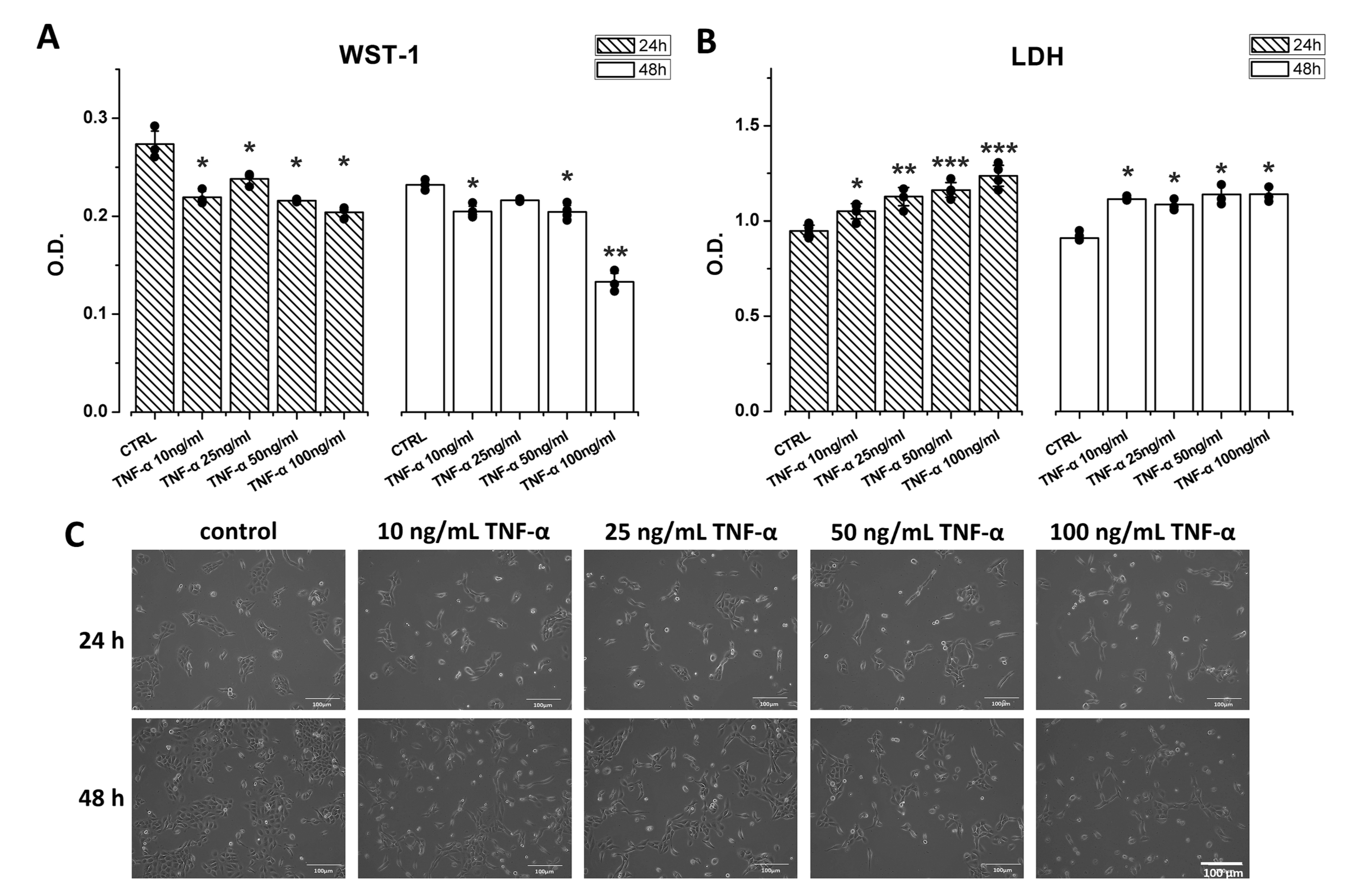
Figure 1: The toxic effects of TNF-α on OECM-1 cells. (A) TNF-α stimulation significantly impaired cell viability (WST-1) in a dose-dependent fashion. (B) TNF-α stimulation induced cell death (LDH). (C) OECM-1 cells exposed to TNF-α (10–50 ng/mL) maintained an epithelial morphology, while a relatively higher concentration (100 ng/mL) caused cell shrinkage. Scale bar: 100 μm. Data are expressed as the mean ± SD (n = 4), *p < 0.05, **p < 0.01, and ***p < 0.001.
3.2 miR Transfection Decreases Cell Activity and Survival in TNF-α-Stimulated OECM-1 Cells
Relative to TNF-α-treated cells, transfection further significantly decreased cell viability (WST-1 assay; Fig. 2A, p < 0.05 for both the 50 nM miR-146a-5p mimic and inhibitor groups, p < 0.05 for the 75 nM miR-146a-5p mimic group, and p < 0.01 for the 75 nM miR-146a-5p inhibitor group) and significantly increased cell death (LDH assay; Fig. 2B, p < 0.01 for both the 50 nM miR-146a-5p mimic and inhibitor groups, p < 0.01 for the 75 nM miR-146a-5p mimic group, and p < 0.001 for the 75 nM miR-146a-5p inhibitor group). Microscopic examination showed that most TNF-α-treated cells had an epithelial morphology; however, we noted changes in cell shape and a reduction in cell number after miRNA transfection (Fig. 2C). TNF-α stimulation decreased the cell survival from 97.09% ± 1.02% to 89.40% ± 3.56% (p < 0.05), while miR-146a-5p mimic or inhibitor transfection further significantly decreased cell survival (71.43% ± 2.50% for the 50 nM miR-146a-5p mimic group, p < 0.01, 68.33% ± 3.26% for the 75 nM miR-146a-5p mimic group, p < 0.01, and 74.88% ± 1.48% for the 75 nM miR-146a-5p inhibitor group, p < 0.01). In addition, the 50 nM miR-146a-5p mimic or inhibitor group had a significantly higher survival rate than the 75 nM miR-146a-5p mimic or inhibitor (p < 0.05 for the 50 nM and 75 nM miR-146a-5p mimic groups and p < 0.01 for the 50 nM and 75 nM miR-146a-5p inhibitor groups; Fig. 2D).
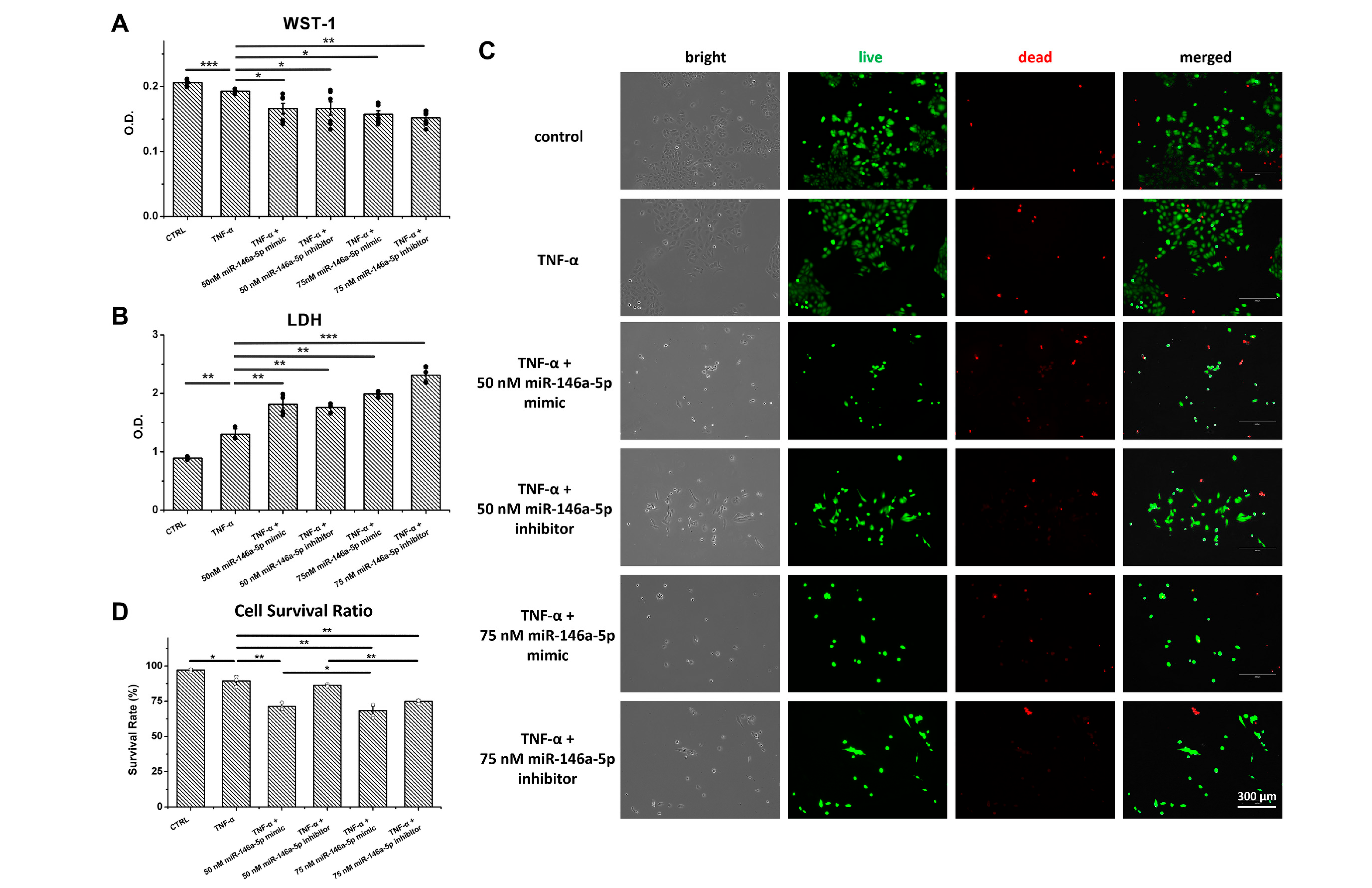
Figure 2: Effects of miR-146a-5p modulation on cell viability, cell death, and cell survival in TNF-α-stimulated OECM-1 cells. OECM-1 cells were stimulated with TNF-α for 48 h and subsequently transfected with a miR-146a-5p mimic or inhibitor for an additional 48 h. (A) Cell viability (WST-1 assay) was assessed following miR-146a-5p mimic or inhibitor transfection. (B) Cell death (LDH assay) was evaluated under the indicated experimental conditions. (C) Representative images and quantitative analysis of live/dead cell staining in TNF-α-stimulated OECM-1 cells following transfection with a miR-146a-5p mimic or inhibitor. Scale bar = 300 μm. (D) Quantitative analysis of cell survival. Data are expressed as the mean ± SD (n = 4). *p < 0.05, **p < 0.01, ***p < 0.001.
3.3 Overexpression of miR-146a-5p Decreases Inflammatory Cytokine mRNA Expression and Protein Production in TNF-α-Stimulated OECM-1 Cells
The miR-146a-5p mimic transfection significantly increased the levels of MIR146A-5p (p < 0.01) compared to TNF-α-stimulated cells (Fig. 3A). In contrast, a miR-146a-5p inhibitor transfection significantly decreased MIR146A-5p levels (p < 0.05), while a scrambled miR-negative control (miR-NC) did not lead to a substantial change. TNF-α stimulation significantly upregulated the mRNA expression of TNFA (p < 0.05), IL1B (p < 0.05), IL6 (p < 0.05), IL6R (p < 0.01), and IL8 (p < 0.05), while IL6ST (p < 0.01) and COL4A1 (p < 0.05) were significantly downregulated. Transfection with a miRNA-146a-5p mimic significantly decreased TNFA (p < 0.05), IL1B (p < 0.01), IL6 (p < 0.01), and IL8 (p < 0.05) and significantly increased IL6R (p < 0.001) and COL4A1 (p < 0.05) mRNA expression in TNF-α-stimulated cells. For the miR-146a-5p inhibitor transfection group, the mRNA levels of TNFA (p < 0.05), IL1B (p < 0.01), IL6 (p < 0.01), IL6R (p < 0.001), IL6ST (p < 0.001), and IL8 (p < 0.01) were significantly upregulated, while COL4A1 (p < 0.05) was significantly downregulated.
The protein array results from a single screening suggested potential changes in cytokine secretion following TNF-α stimulation, including lower levels of IL-6sR, IL-8, PDGF-BB, and TIMP-2 (Fig. 3B). In TNF-α-stimulated OECM-1 cells, transfection with a miR-146a-5p mimic appeared to be associated with lower IL-6, IL-6sR, and IL-8 levels and higher TIMP-2 levels, whereas transfection with a miR-146a-5p inhibitor appeared to be associated with higher levels of IL-6, IL-6sR, IL-8, RANTES, PDGF-BB, and TIMP-2 compared with TNF-α-stimulated cells.
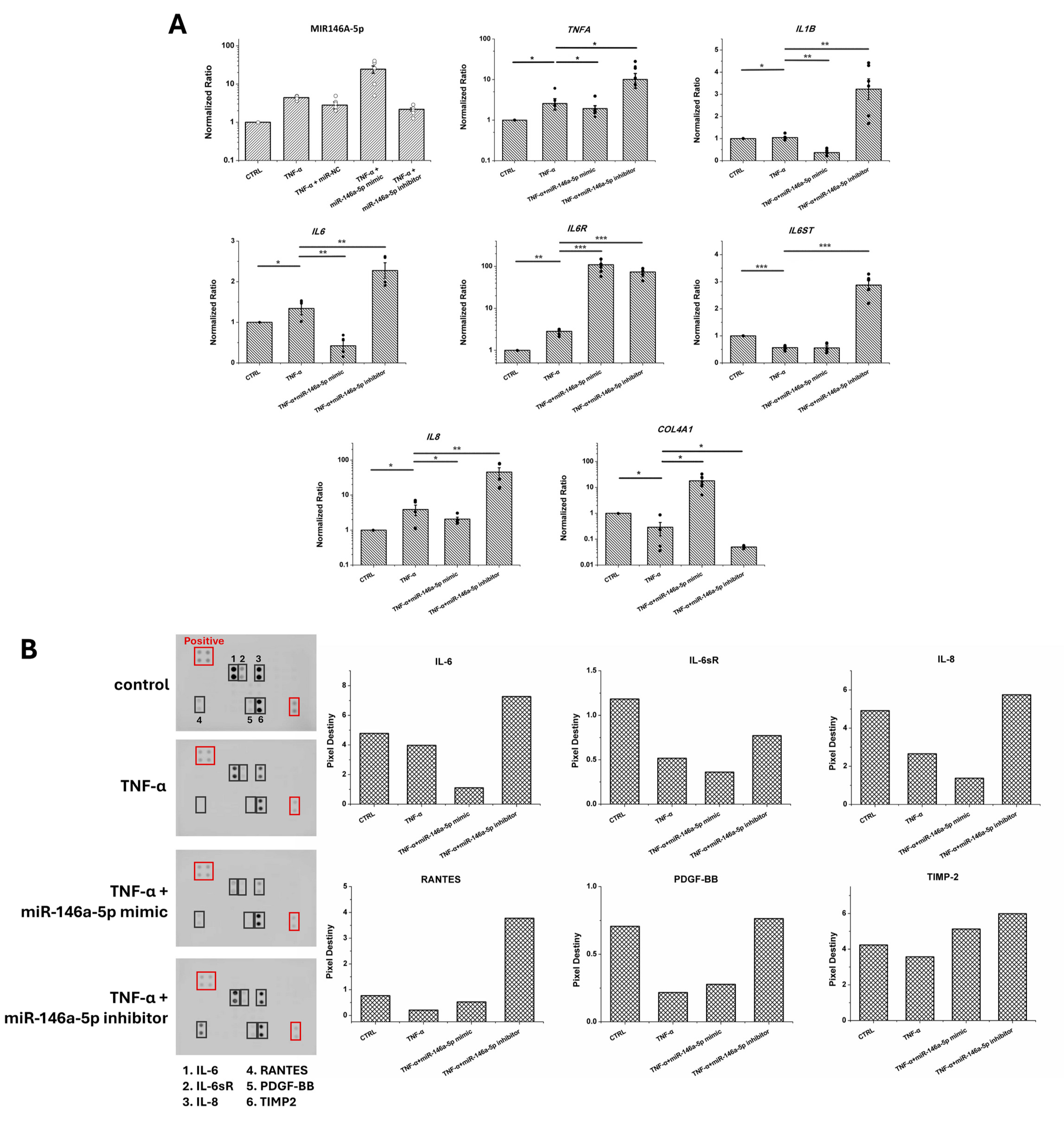
Figure 3: Effects of miR-146a-5p modulation on inflammatory gene expression and cytokine secretion in TNF-α-stimulated OECM-1 cells. (A) Relative mRNA expression of inflammatory cytokines and extracellular matrix-related genes in TNF-α-stimulated OECM-1 cells following transfection with a miR-146a-5p mimic or inhibitor. Data are expressed as the mean ± SD (n = 5). *p < 0.05, **p < 0.01, ***p < 0.001. (B) Cytokine secretion profiles of TNF-α-stimulated OECM-1 cells following transfection with a miR-146a-5p mimic or inhibitor.
3.4 miR-146a-5p Mimic Transfection Impairs Cell Motility, Wound Closure, and Clonogenic Capacity in TNF-α-Treated OECM-1 Cells
TNF-α stimulation significantly upregulated the mRNA levels of CTTN (p < 0.05) and CORO1B (p < 0.01) in OECM-1 cells (Fig. 4A). Transfection with both a miR-146a-5p mimic and an inhibitor significantly decreased these two gene levels (all p < 0.05). Additionally, TNF-α stimulation impaired cell migration, and transfection with either a miR-146a-5p mimic or an inhibitor further reduced cell migration (Fig. 4B). Normal cells showed a significantly smaller wound area than the TNF-α-treated group (p < 0.001 at 12 h). Cells transfected with miR-146a-5p exhibited a significantly larger wound area compared to the TNF-α group (p < 0.01 for the miR-146a-5p mimic group and p < 0.0001 for the miR-146a-5p inhibitor group at 24 h). TNF-α stimulation enhanced cell invasion capacity, but a miR-146a-5p mimic transfection significantly reduced cell invasion (p < 0.05), whereas the miR-146a-5p inhibitor also significantly impaired cell invasion (p < 0.01) in TNF-α-treated cells (Fig. 4C). Lastly, TNF-α stimulation significantly decreased the clonogenic capacity of OECM-1 cells (p < 0.05), and both the miR-146a-5p mimic (p < 0.05) and inhibitor (p < 0.01) further reduced the number of colonies in TNF-α-treated cells (Fig. 4D).
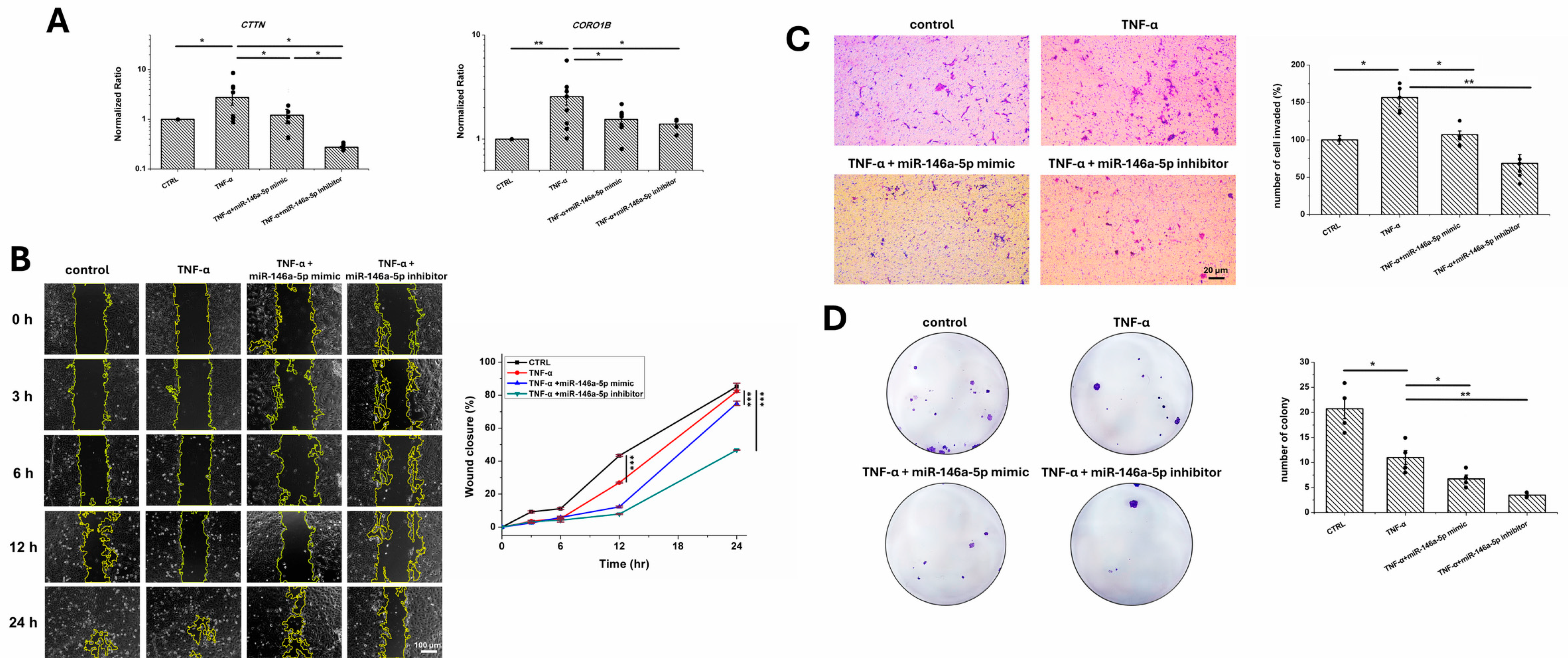
Figure 4: Analysis of cell motility and clonogenic capacity in TNF-α-stimulated OECM-1 cells following miR-146a-5p modulation. (A) Relative mRNA expression of CTTN and CORO1B in TNF-α-stimulated OECM-1 cells following transfection with a miR-146a-5p mimic or inhibitor (n = 8). (B) Representative images and quantitative analysis of wound healing assays performed in TNF-α-stimulated OECM-1 cells following transfection with a miR-146a-5p mimic or inhibitor (n = 4). Scale bar = 100 μm. (C) Representative images and quantitative analysis of Transwell invasion assays performed in TNF-α-stimulated OECM-1 cells following transfection with a miR-146a-5p mimic or inhibitor (n = 5). Scale bar = 20 μm. (D) Representative images and quantification of colony formation assays performed in TNF-α-stimulated OECM-1 cells following transfection with a miR-146a-5p mimic or inhibitor (n = 4). Data are presented as the mean ± SD. *p < 0.05, **p < 0.01, ***p < 0.001.
Oral inflammation can facilitate the invasion and metastasis of OSCC through a TNF-α-dependent mechanism [3,4]. Additionally, miR-146a-5p is recognized as a multifaceted modulator that influences both inflammation and carcinogenesis [8,14]. Furthermore, dysregulation of miR-146a is associated with TNF-α overexpression. Accordingly, this study aims to demonstrate the role of miR-146a-5p in the inflammatory response and cellular mobility in the inflamed OECM-1 cells.
Given that TNF-α is highly expressed in the oral cancer microenvironment [3], we treated OECM-1 cells with TNF-α to mimic the inflammatory conditions found in the oral niche. Exposure to TNF-α demonstrated cytotoxic effects in a dose-dependent manner, particularly leading to cell death (Fig. 1B). A reduction in cell number was observed in the 100 ng/mL TNF-α treatment group (Fig. 1C). Similarly, Basso et al. reported that exposure to 100 ng/mL TNF-α induced apoptosis in gingival fibroblasts and epithelial cells [17]. The cell viability of the 50 ng/mL group was lower than thot of the 25 ng/mL group (Fig. 1A), and the replication rate of cells stimulated with 50 ng/mL was also slower. As a result, we chose to use a TNF-α concentration of 25 ng/mL for the subsequent experiments. Likewise, other studies also treated various OSCC cell lines with TNF-α at concentrations of 10–50 ng/mL for different purposes. For example, Tang et al. used 10 ng/mL TNF-α to treat OSCC cells to study invasion and metastasis [4]. Similarly, Zhou et al. used 10 ng/mL TNF-α to treat OSCC cells to investigate inflammation-induced tumor promotion [18]. On the contrary, Zheng et al. reported that 100 ng/mL TNF-α inhibited the migration of OSCC cell lines, whereas 5, 10, and 50 ng/mL TNF-α did not alter cell proliferation; however, 50 ng/mL showed a higher cytotoxicity than 10 ng/mL [19]. Otherwise, Shu et al. used 50 ng/mL TNF-α to treat OSCC and studied the role of autophagy in OSCC progression [20]. In our study, we noted that a concentration of 25 ng/mL TNF-α elicited effects similar to those observed with 10 ng/mL TNF-α in OECM-1 cells; thus, exposure to 25 ng/mL TNF-α for 48 h was used to ensure the inflammatory response in treated cells.
While lipofection is an efficient approach for delivering nucleic acids to cells, it can also be cytotoxic, inducing apoptosis and cell death [21]. In this study, OECM-1 cells were transfected with 50 or 75 nM of miR-146a-5p using a non-liposomal polymer transfection reagent. The results showed an increase in cell death (Fig. 2B) with both concentrations, while transfection with 75 nM of miR-146a-5p significantly reduced cell survival (Fig. 2C). Nakase et al. reported that cationic liposome reagents influenced cell proliferation activity in oral malignant melanoma and oral osteosarcoma cell lines [22]. Similarly, Kiefer et al. reported that lipofection induced varying levels of cytotoxicity in human aortic smooth muscle cells, human endothelial cells, and rat smooth muscle cells [23]. In contrast, our previous study assessed the cytotoxicity of miRNA transfection and found that lipofection concentrations ranging from 15 to 100 nM did not induce cell death in oral submucosal fibroblasts [24]. Therefore, the toxic effects of transfection reagents depend on the reagent type and dosage, as well as the target cell type.
Exposure to TNF-α triggers cells to overproduce inflammatory mediators such as IL-1β, IL-6, IL-8, and interferon-gamma, which amplify immune responses in oral tissues. After stimulating OECM-1 cells with TNF-α, we observed a significant inflammatory response characterized by an increase in the mRNA levels of proinflammatory cytokines (Fig. 3A). Chan et al. found that CXCL1 treatment increased TNF-α and cyclooxygenase-2 expression in OECM-1 cells, indicating that a heightened inflammatory stimulation can increase proinflammatory mediators [25]. Likewise, our findings align with those of Watanabe et al., which showed that TNF-α stimulation upregulates IL-8 gene expression and increases protein production in human OSCC cell lines [26]. In addition, IL-1β, together with TNF-α, contributes to a pro-tumorigenic microenvironment that promotes tumor invasion, angiogenesis, and metastasis in OSCC [27], emphasizing the intricate interactions and positive feedback loop among cytokines.
Despite some controversial findings, miR-146a-5p is recognized as a microRNA that plays a critical role in immune responses. To address this issue, we transfected a miR-146a-5p mimic into OECM-1 cells, which effectively downregulated the mRNA expression of inflammatory cytokines (Fig. 3A) and protein products (Fig. 3B). Similarly, Pan et al. noted that miR-146a-5p reduced the expression of IL1B and IL6 in cementoblasts [28], which supports our findings. miR-146a-5p has been shown to possess anti-inflammatory effects by targeting IL-1 receptor-associated kinase-1 and TNF receptor-associated factor-6, which are crucial downstream mediators in the cellular response to IL-1β [29]. IL-1β can stimulate the growth of dysplastic oral cells, thereby increasing the invasiveness of OSCC [30]. Additionally, excessive IL-6 not only promotes OSCC progression but also contributes to radiation resistance [31]. The binding of IL-6 to its receptor, IL-6R, leads to homodimer formation and the recruitment of IL6ST, which activates the downstream inflammatory cascade [32]. Interestingly, we found that transfection with a miR-146a-5p mimic significantly downregulated IL6 mRNA expression while upregulating IL6R mRNA expression in TNF-α-stimulated OECM-1 cells, suggesting that miR-146a-5p modulates the IL-6/IL-6R signaling axis during inflammatory responses. Previous studies have demonstrated that miR-146a exerts anti-inflammatory effects by suppressing pro-inflammatory cytokines, including IL-6, through negative regulation of the NF-κB signaling pathway and its downstream mediators such as IRAK1 and TRAF6. For example, Roos et al. reported that miR-146a mimic transfection reduced IL-6 expression in inflamed adipocytes [33], while Zeng et al. showed that miR-146a overexpression suppressed IL-6 production in LPS-induced inflammatory models [34]. These findings are consistent with our observation of reduced IL6 mRNA levels following miR-146a-5p overexpression. In contrast, the observed increase in IL6R mRNA expression may reflect a compensatory regulatory mechanism to maintain IL-6 responsiveness under conditions of suppressed cytokine production, suggesting that miR-146a-5p may fine-tune inflammatory signaling by differentially regulating IL-6 and IL-6R expression. Similarly, Yan et al. found that miR-146a-5p decreased IL-6 and IL-8 levels, thereby alleviating inflammation and airway epithelial cell damage [35], consistent with our experimental results.
In addition to the inflammatory mediators, miR-146a-5p mimic transfection upregulated COL4A1 mRNA levels and TIMP-2 production. The loss of type IV collagen integrity, a major component of the basement membrane, is linked to the invasive nature of OSCC. Therefore, while TIMP-2 functions as either a tumor growth promoter or a suppressor [36], miR-146a-5p modulation not only alleviates the inflammatory response but may also influence the aggressiveness of OSCC. However, the findings of the cytokine array were derived from a single screening experiment; they should be interpreted with caution.
To demonstrate the role of miR-146a-5p in OECM-1 cellular mobility, we further analyzed the mRNA levels of CTTN and CORO1B in transfected cells. CTTN facilitates lamellipodia formation and cell migration, while CORO1B modulates leading-edge dynamics and cell motility; both genes are associated with cell invasion. Additionally, high CTTN expression is associated with nodal metastasis in OSCC, while high CORO1B levels are correlated with a poorer prognosis in OSCC patients [37,38]. TNF-α stimulation upregulated both CTTN and CORO1B mRNA expression; however, transfection with either a miR-146a-5p mimic or an inhibitor decreased these gene levels (Fig. 4A). In order to clarify these findings, we conducted a mock transfection in both normal and inflamed OECM-1 cells, and the results indicated that the blank transfection reagent impaired cell migration in normal cells (Fig. S1A) and invasion in TNF-α-stimulated cells (Fig. S1B). Since TNF-α stimulation upregulated CTTN and CORO1B mRNA levels in OECM-1 cells, our findings raise the possibility that blank transfection reagent may impair cell migration, potentially by suppressing cytoskeletal regulators involved in lamellipodia dynamics. Although mock transfection is widely used as a negative control, several studies indicate that these reagents are not biologically inert. Kleefeldt et al. demonstrated that several commonly used transfection reagents altered endogenous mRNA and protein abundance and reduced cell viability/proliferation even in reagent-only controls [39], emphasizing that mock transfection can significantly perturb cellular physiology. Similarly, Raof et al. reported that the polyethylenimine transfection reagent induced broad gene expression changes in the absence of any siRNA or plasmid cargo [40], supporting the possibility that membrane perturbation and intracellular trafficking associated with cationic transfection reagents are sufficient to trigger cellular stress responses. These findings support that mock transfection may induce membrane/endosomal stress, alter endogenous transcription, and suppress components of the actin-remodeling machinery—including CTTN and CORO1B—thereby impairing migratory capacity.
Inhibition of TNF-α signaling using the anti-TNF-α agent infliximab reduced acute inflammation in oral traumatic ulcers; however, it also delayed cell migration and wound healing in rats [41]. Transfection of a miR-146a-5p mimic reduced cell migration in OECM-1 cells, while transfection of a miR-146a-5p inhibitor further significantly impeded cell migration (Fig. 4B). Transfection of a miR-146a-5p mimic in KATO III gastric cancer cells inhibited cell migration [42], whereas knockdown of miR-146a-5p in Wharton’s jelly-mesenchymal stem cells enhanced their migratory capabilities [43]. Otherwise, miR-146a-5p mimic transfection reduced TNF-α-induced cell invasion (Fig. 4C). Liu et al. showed that miR-146a modulates the aggressive ability of OSCC cells [44]. TNF-α stimulation impaired clonogenic capacity, and both miR-146a-5p mimic and inhibitor transfections further reduced colony counts in OECM-1 cells (Fig. 4D). Since OECM-1 is a tumorigenic OSCC cell line, and foundational studies have shown that it exhibits a less aggressive profile than other established OSCC cell lines, such as SAS [45], the effects of miR-146a-5p on cellular mobility in TNF-α-stimulated OECM-1 cells need further clarification. Furthermore, miR-146a-5p is identified as both an oncomiR and a tumor suppressor in oral cancer [14]. However, the potential influence of the non-liposomal polymer transfection reagent on cell mobility cannot be excluded. Likewise, other OSCC cells should also be verified to clarify the role of miR-146a-5p.
A major limitation of this study is the findings of IL-8 and PDGF-BB. While the IL8 mRNA was increased upon TNF-α exposure, the IL-8 secretion decreased in TNF-α-stimulated cells (Fig. 3). Several articles have reported that miR-146a-5p directly binds to the 3′ UTR of IL-8 mRNA, resulting in degradation or translational repression, which subsequently reduces IL-8 secretion [33,46]. However, we utilized a protein membrane array for cytokine detection and tested only a single representative sample, which may contribute to the contradictory findings. Furthermore, the earlier transcriptional changes may not fully parallel protein secretion measured at the endpoint. A detailed time-course analysis with larger sample numbers, evaluated with relevant ELISA kits, would be valuable for further defining the kinetics of TNF-α-induced cytokine regulation and the temporal effects of miR-146a-5p. In addition, TNF-α stimulation also decreased PDGF-BB production in OECM-1 cells. Although transfection with the miR-146a-5p mimic did not substantially alter the PDGF-BB level, transfection with the miR-146a-5p inhibitor restored PDGF-BB secretion in OECM-1 cells. Overexpression of PDGF-BB has been associated with oral tumorigenesis and poor prognosis in OSCC [47]. Furthermore, once the cytotoxic effects of the transfection reagent on cells can be excluded, the regulatory role of miR-146a-5p in other OSCC cells shall be validated. Despite the above limitations, this study suggests that miR-146a-5p may reduce TNF-α-induced inflammatory responses and modulate ECM remodeling in OECM-1 cells. However, the impacts of miR-146a-5p on cellular mobility require further investigation.
TNF-α stimulation can induce an inflammatory response and enhance cell invasion and migration in OSCC. When OECM-1 cells were transfected with a miR-146a-5p mimic, there was a decrease in the mRNA levels of TNFA, IL1B, IL6, and IL8. Conversely, the levels of IL6R and COL4A1 increased. Despite TNF-α stimulation also increasing CTTN and CORO1B mRNA levels, the blank transfection reagent adversely affected wound closure and reduced cell invasion, thereby compromising the interpretation of miR-146a-5p in TNF-α-stimulated cellular behavior. This study indicates that modulating miR-146a-5p can regulate the inflammatory response, although further research is necessary to understand its impact on cell motility in OSCC cells.
Acknowledgement:
Funding Statement: This work was supported by the National Science and Technology Council, Taiwan (NSTC 113-2813-C-038-109-B and NSTC 114-2221-E-038-017).
Author Contributions: The authors confirm contribution to the paper as follows: Conceptualization, Kai-Chiang Yang; methodology, Kai-Chiang Yang; formal analysis, Pin-Si Yin, Chang-Chin Wu, Shih-Yuan Chiu, Karina Erda Saninggar, Ardianti Maartrina Dewi, and Kai-Chiang Yang; investigation, Pin-Si Yin; data curation, Pin-Si Yin, Shih-Yuan Chiu, and Kai-Chiang Yang; writing—original draft preparation, Pin-Si Yin, Chang-Chin Wu, Karina Erda Saninggar, Ardianti Maartrina Dewi, and Kai-Chiang Yang; writing—review and editing, Kai-Chiang Yang. All authors reviewed and approved the final version of the manuscript.
Availability of Data and Materials: The data that support the findings of this study are available from the corresponding author upon reasonable request.
Ethics Approval: Not applicable.
Conflicts of Interest: The authors declare no conflicts of interest.
Supplementary Materials: The supplementary material is available online at https://www.techscience.com/doi/10.32604/biocell.2026.081934/s1.
References
1. Li R , Hou M , Yu L , Luo W , Liu R , Wang H . Association between periodontal disease and oral squamous cell carcinoma: A systematic review and meta-analysis. Br J Oral Maxillofac Surg. 2023; 61( 6): 394– 402. doi:10.1016/j.bjoms.2023.05.004. [Google Scholar] [CrossRef]
2. Pigossi SC , Oliveira JA , de Medeiros MC , Soares LFF , D’Silva NJ . Demystifying the link between periodontitis and oral cancer: A systematic review integrating clinical, pre-clinical, and in vitro data. Cancer Metastasis Rev. 2025; 44( 3): 67. doi:10.1007/s10555-025-10285-z. [Google Scholar] [CrossRef]
3. Brierly G , Celentano A , Breik O , Moslemivayeghan E , Patini R , McCullough M , et al. Tumour necrosis factor alpha (TNF-α) and oral squamous cell carcinoma. Cancers. 2023; 15( 6): 1841. doi:10.3390/cancers15061841. [Google Scholar] [CrossRef]
4. Tang D , Tao D , Fang Y , Deng C , Xu Q , Zhou J . TNF-alpha promotes invasion and metastasis via NF-kappa B pathway in oral squamous cell carcinoma. Med Sci Monit Basic Res. 2017; 23: 141– 9. doi:10.12659/msmbr.903910. [Google Scholar] [CrossRef]
5. Liu N , Liu GJ , Liu J . Genetic association between TNF-α promoter polymorphism and susceptibility to squamous cell carcinoma, basal cell carcinoma, and melanoma: A meta-analysis. Oncotarget. 2017; 8( 32): 53873– 85. doi:10.18632/oncotarget.17179. [Google Scholar] [CrossRef]
6. Bozgeyik E , Bozgeyik I . Non-coding RNA variations in oral cancers: A comprehensive review. Gene. 2023; 851: 147012. doi:10.1016/j.gene.2022.147012. [Google Scholar] [CrossRef]
7. Danella EB , Costa de Medeiros M , D’Silva NJ . Cytokines secreted by inflamed oral mucosa: Implications for oral cancer progression. Oncogene. 2023; 42( 15): 1159– 65. doi:10.1038/s41388-023-02649-y. [Google Scholar] [CrossRef]
8. Gilyazova I , Asadullina D , Kagirova E , Sikka R , Mustafin A , Ivanova E , et al. MiRNA-146a—A key player in immunity and diseases. Int J Mol Sci. 2023; 24( 16): 12767. doi:10.3390/ijms241612767. [Google Scholar] [CrossRef]
9. Zhou H , Yang RK , Li Q , Li Z , Wang YC , Li SY , et al. microRNA-146a-5p protects retinal ganglion cells through reducing neuroinflammation in experimental glaucoma. Glia. 2024; 72( 11): 2115– 41. doi:10.1002/glia.24600. [Google Scholar] [CrossRef]
10. Chen HR , Huang HS , Zeng SY , Sun YF , Chen L , Lai SY , et al. miR-146a-5p targets IRAK1/TRAF6 to promote Bacillus Calmette-Guérin survival by exosome-mediated autocrine actions. Mol Immunol. 2025; 185: 105– 15. doi:10.1016/j.molimm.2025.07.012. [Google Scholar] [CrossRef]
11. Tang L , Li X , Bai Y , Wang P , Zhao Y . microRNA-146a negatively regulates the inflammatory response to Porphyromonas gingivalis in human periodontal ligament fibroblasts via TRAF6/p38 pathway. J Periodontol. 2019; 90( 4): 391– 9. doi:10.1002/JPER.18-0190. [Google Scholar] [CrossRef]
12. Xie YF , Shu R , Jiang SY , Song ZC , Guo QM , Dong JC , et al. miRNA-146 negatively regulates the production of pro-inflammatory cytokines via NF-κB signalling in human gingival fibroblasts. J Inflamm. 2014; 11( 1): 38. doi:10.1186/s12950-014-0038-z. [Google Scholar] [CrossRef]
13. Xie YF , Shu R , Jiang SY , Liu DL , Ni J , Zhang XL . microRNA-146 inhibits pro-inflammatory cytokine secretion through IL-1 receptor-associated kinase 1 in human gingival fibroblasts. J Inflamm. 2013; 10( 1): 20. doi:10.1186/1476-9255-10-20. [Google Scholar] [CrossRef]
14. Iacona JR , Lutz CS . miR-146a-5p: Expression, regulation, and functions in cancer. Wires RNA. 2019; 10( 4): e1533. doi:10.1002/wrna.1533. [Google Scholar] [CrossRef]
15. Shi Z , Johnson JJ , Jiang R , Liu Y , Stack MS . Decrease of miR-146a is associated with the aggressiveness of human oral squamous cell carcinoma. Arch Oral Biol. 2015; 60( 9): 1416– 27. doi:10.1016/j.archoralbio.2015.06.007. [Google Scholar] [CrossRef]
16. Min SK , Jung SY , Kang HK , Park SA , Lee JH , Kim MJ , et al. Functional diversity of miR-146a-5p and TRAF6 in normal and oral cancer cells. Int J Oncol. 2017; 51( 5): 1541– 52. doi:10.3892/ijo.2017.4124. [Google Scholar] [CrossRef]
17. Basso FG , Pansani TN , Turrioni AP , Soares DG , de Souza Costa CA , Hebling J . Tumor necrosis factor-α and interleukin (IL)-1β, IL-6, and IL-8 impair in vitro migration and induce apoptosis of gingival fibroblasts and epithelial cells, delaying wound healing. J Periodontol. 2016; 87( 8): 990– 6. doi:10.1902/jop.2016.150713. [Google Scholar] [CrossRef]
18. Zhou JP , Gao ZL , Zhou ML , He MY , Xu XH , Tao DT , et al. Snail interacts with Id2 in the regulation of TNF-α-induced cancer cell invasion and migration in OSCC. Am J Cancer Res. 2015; 5( 5): 1680– 91. [Google Scholar]
19. Zheng Z , Luan X , Zha J , Li Z , Wu L , Yan Y , et al. TNF-α inhibits the migration of oral squamous cancer cells mediated by miR-765-EMP3-p66Shc axis. Cell Signal. 2017; 34: 102– 9. doi:10.1016/j.cellsig.2017.03.009. [Google Scholar] [CrossRef]
20. Shu CW , Chen CF , Chang CC , Lee CH , Tseng HH , Hu KF , et al. The reciprocal regulation between TNF-α and autophagy for oral squamous cell carcinoma progression. J Oral Pathol Med. 2026; 55( 3): 355– 67. doi:10.1111/jop.70093. [Google Scholar] [CrossRef]
21. Cui S , Wang Y , Gong Y , Lin X , Zhao Y , Zhi D , et al. Correlation of the cytotoxic effects of cationic lipids with their headgroups. Toxicol Res. 2018; 7( 3): 473– 9. doi:10.1039/c8tx00005k. [Google Scholar] [CrossRef]
22. Nakase M , Okumura K , Kamei T , Nakamura S , Inui M , Tagawa T . The influence of oral tumor cell proliferation activity and membrane potential on the transfection efficiency of a cationic liposome. Oncol Rep. 2005; 14( 6): 1487– 91. doi:10.3892/or.14.6.1487. [Google Scholar] [CrossRef]
23. Kiefer K , Clement J , Garidel P , Peschka-Süss R . Transfection efficiency and cytotoxicity of nonviral gene transfer reagents in human smooth muscle and endothelial cells. Pharm Res. 2004; 21( 6): 1009– 17. doi:10.1023/B:PHAM.0000029291.62615.ec. [Google Scholar] [CrossRef]
24. Fang CY , Chen YJ , Lin KC , Chen YH , Yang KC . Co-transfection with anti-fibrotic microRNAs for treating oral submucous fibrosis. J Oral Pathol Med. 2025; 54( 7): 574– 83. doi:10.1111/jop.70009. [Google Scholar] [CrossRef]
25. Chan LP , Tseng YP , Wang HC , Chien CY , Wu CW , Wang LF , et al. Growth regulated oncogene-α upregulates TNF-α and COX-2 and activates NOD1/RIPK2 mediated-MAPK pathway in head and neck squamous cell carcinoma. J Cancer. 2023; 14( 6): 989– 1000. doi:10.7150/jca.82300. [Google Scholar] [CrossRef]
26. Watanabe H , Iwase M , Ohashi M , Nagumo M . Role of interleukin-8 secreted from human oral squamous cell carcinoma cell lines. Oral Oncol. 2002; 38( 7): 670– 9. doi:10.1016/s1368-8375(02)00006-4. [Google Scholar] [CrossRef]
27. Niklander SE . Inflammatory mediators in oral cancer: Pathogenic mechanisms and diagnostic potential. Front Oral Health. 2021; 2: 642238. doi:10.3389/froh.2021.642238. [Google Scholar] [CrossRef]
28. Pan J , Du M , Cao Z , Zhang C , Hao Y , Zhu J , et al. miR-146a-5p attenuates IL-1β-induced IL-6 and IL-1β expression in a cementoblast-derived cell line. Oral Dis. 2020; 26( 6): 1308– 17. doi:10.1111/odi.13333. [Google Scholar] [CrossRef]
29. Li Y , Tan S , Shen Y , Guo L . miR-146a-5p negatively regulates the IL-1β-stimulated inflammatory response via downregulation of the IRAK1/TRAF6 signaling pathway in human intestinal epithelial cells. Exp Ther Med. 2022; 24( 4): 615. doi:10.3892/etm.2022.11552. [Google Scholar] [CrossRef]
30. Ding Y , Yi J , Wang J , Sun Z . Interleukin-1 receptor antagonist: A promising cytokine against human squamous cell carcinomas. Heliyon. 2023; 9( 4): e14960. doi:10.1016/j.heliyon.2023.e14960. [Google Scholar] [CrossRef]
31. Matsuoka Y , Nakayama H , Yoshida R , Hirosue A , Nagata M , Tanaka T , et al. IL-6 controls resistance to radiation by suppressing oxidative stress via the Nrf2-antioxidant pathway in oral squamous cell carcinoma. Br J Cancer. 2016; 115( 10): 1234– 44. doi:10.1038/bjc.2016.327. [Google Scholar] [CrossRef]
32. Wu CC , Wei Y , Kolmas J , Yang KC . microRNA-146a-3p as an alternative to anti-IL-6 monoclonal antibody therapy: Intra-articular liposome delivery for osteoarthritis treatment. Med Hypotheses. 2025; 200: 111666. doi:10.1016/j.mehy.2025.111666. [Google Scholar] [CrossRef]
33. Roos J , Enlund E , Funcke JB , Tews D , Holzmann K , Debatin KM , et al. miR-146a-mediated suppression of the inflammatory response in human adipocytes. Sci Rep. 2016; 6: 38339. doi:10.1038/srep38339. [Google Scholar] [CrossRef]
34. Zeng Z , Gong H , Li Y , Jie K , Ding C , Shao Q , et al. Upregulation of miR-146a contributes to the suppression of inflammatory responses in LPS-induced acute lung injury. Exp Lung Res. 2013; 39( 7): 275– 82. doi:10.3109/01902148.2013.808285. [Google Scholar] [CrossRef]
35. Yan F , Wufuer D , Ding J , Wang J . microRNA miR-146a-5p inhibits the inflammatory response and injury of airway epithelial cells via targeting TNF receptor-associated factor 6. Bioengineered. 2021; 12( 1): 1916– 26. doi:10.1080/21655979.2021.1927545. [Google Scholar] [CrossRef]
36. Shrestha B , Bajracharya D , Byatnal AA , Kamath A , Radhakrishnan R . May high MMP-2 and TIMP-2 expressions increase or decrease the aggressivity of oral cancer? Pathol Oncol Res. 2017; 23( 1): 197– 206. doi:10.1007/s12253-016-0149-3. [Google Scholar] [CrossRef]
37. Kase-Kato I , Asai S , Minemura C , Tsuneizumi K , Oshima S , Koma A , et al. Molecular pathogenesis of the coronin family: CORO2A facilitates migration and invasion abilities in oral squamous cell carcinoma. Int J Mol Sci. 2021; 22( 23): 12684. doi:10.3390/ijms222312684. [Google Scholar] [CrossRef]
38. Liu HS , Lu HH , Lui MT , Yu EH , Shen W , Chen YP , et al. Detection of copy number amplification of cyclin D1 (CCND1) and cortactin (CTTN) in oral carcinoma and oral brushed samples from Areca chewers. Oral Oncol. 2009; 45( 12): 1032– 6. doi:10.1016/j.oraloncology.2009.06.007. [Google Scholar] [CrossRef]
39. Kleefeldt JM , Pozarska A , Nardiello C , Pfeffer T , Vadász I , Herold S , et al. Commercially available transfection reagents and negative control siRNA are not inert. Anal Biochem. 2020; 606: 113828. doi:10.1016/j.ab.2020.113828. [Google Scholar] [CrossRef]
40. Raof NA , Rajamani D , Chu HC , Gurav A , Johnson JM , LoGerfo FW , et al. The effects of transfection reagent polyethyleneimine (PEI) and non-targeting control siRNAs on global gene expression in human aortic smooth muscle cells. BMC Genomics. 2016; 17: 20. doi:10.1186/s12864-015-2267-9. [Google Scholar] [CrossRef]
41. Freitas MO , Fonseca APR , de Aguiar MT , Dias CC , Avelar RL , Sousa FB , et al. Tumor necrosis factor alpha (TNF-α) blockage reduces acute inflammation and delayed wound healing in oral ulcer of rats. Inflammopharmacology. 2022; 30( 5): 1781– 98. doi:10.1007/s10787-022-01046-3. [Google Scholar] [CrossRef]
42. Baghbanzadeh A , Rahmani S , Eslami S , Ahmadpour Youshanlui M , Shafiee N , Shafiee A , et al. miR-146a-5p suppresses migration and downregulates vimentin and MMP-9 expression in gastric cancer cells. Discov Oncol. 2025; 16( 1): 1075. doi:10.1007/s12672-025-02929-x. [Google Scholar] [CrossRef]
43. Hsieh JY , Huang TS , Cheng SM , Lin WS , Tsai TN , Lee OK , et al. miR-146a-5p circuitry uncouples cell proliferation and migration, but not differentiation, in human mesenchymal stem cells. Nucleic Acids Res. 2013; 41( 21): 9753– 63. doi:10.1093/nar/gkt666. [Google Scholar] [CrossRef]
44. Liu YT , Yu CC , Lu MY , Chao SC , Liao YW , Yu CH , et al. miR-146a participates in the regulation of cancer stemness of oral carcinoma cells. J Dent Sci. 2023; 18( 2): 503– 9. doi:10.1016/j.jds.2022.09.001. [Google Scholar] [CrossRef]
45. Hung PS , Tu HF , Kao SY , Yang CC , Liu CJ , Huang TY , et al. miR-31 is upregulated in oral premalignant epithelium and contributes to the immortalization of normal oral keratinocytes. Carcinogenesis. 2014; 35( 5): 1162– 71. doi:10.1093/carcin/bgu024. [Google Scholar] [CrossRef]
46. Gronau L , Duecker RP , Jerkic SP , Eickmeier O , Trischler J , Chiocchetti AG , et al. Dual role of microRNA-146a in experimental inflammation in human pulmonary epithelial and immune cells and expression in inflammatory lung diseases. Int J Mol Sci. 2024; 25( 14): 7686. doi:10.3390/ijms25147686. [Google Scholar] [CrossRef]
47. Lin LH , Lin JS , Yang CC , Cheng HW , Chang KW , Liu CJ . Overexpression of platelet-derived growth factor and its receptor are correlated with oral tumorigenesis and poor prognosis in oral squamous cell carcinoma. Int J Mol Sci. 2020; 21( 7): 2360. doi:10.3390/ijms21072360. [Google Scholar] [CrossRef]
Cite This Article
 Copyright © 2026 The Author(s). Published by Tech Science Press.
This work is licensed under a Creative Commons Attribution 4.0 International License , which permits unrestricted use, distribution, and reproduction in any medium, provided the original work is properly cited.
 Downloads
 Citation Tools
