
 Login
Login Register
 Submit a Paper
 Propose a Special lssue
 Open Access
REVIEW
Neutrophil Extracellular Traps and Neuroinflammatory Signaling in Brain Ischemic Insults: Mechanisms, Blood-Brain Barrier Dysfunction, and Therapeutic Targeting
1 Asklepios Kliniken Schildautal Seesen, Neurology Clinic, Lower Saxony, Germany
2 Med-International UK Health Agency Ltd., Nuneaton, UK
* Corresponding Author: Nurittin Ardic. Email:
(This article belongs to the Special Issue: Cellular and Molecular Insights into Brain Ischemic Insults)
BIOCELL 2026, 50(10), 2 https://doi.org/10.32604/biocell.2026.082812
Received 23 March 2026; Accepted 21 May 2026; Issue published 22 September 2026
 View Full Text
 Download PDF
Abstract
Neutrophil extracellular traps (NETs) are increasingly recognized as significant contributors to neurovascular damage following ischemic brain injuries. This review examines how NETs link intravascular thrombosis to downstream neuroinflammation via a pathway-centric framework. We synthesize recent preclinical and clinical evidence showing that NET-derived histones, extracellular DNA, and granular enzymes activate convergent inflammatory pathways, including the high mobility group box 1–Toll-like receptor 4 axis, nuclear factor kappa B, Janus kinase 2/signal transducer and transcription activator 3, NOD-like receptor pyrin domain-containing 3 inflammasome, and cyclic GMP–AMP synthase–interferon gene signaling. These mechanisms contribute to disruption of the blood-brain barrier, glial activation, oxidative damage, and amplification of sterile neuroinflammation. We also discuss translational implications, including therapeutic strategies targeting NETs, candidate biomarkers, and current challenges in clinical practice. Overall, NETs appear to be significant enhancers of ischemic brain injury and promising targets for mechanistically directed intervention.Keywords
Brain ischemic insults such as acute ischemic stroke, transient ischemic attack, ischemia-reperfusion injury, and cardiac arrest with return of spontaneous circulation continue to be the leading causes of death and long-term disability worldwide [1]. The Global Burden of Disease Study estimates that ischemic stroke alone causes approximately 7.6 million deaths and 143 million disability-adjusted years of life lost annually [2]. Despite transformative advances in reperfusion therapies, including intravenous thrombolysis with tissue plasminogen activator and mechanical thrombectomy, a significant proportion of patients experience incomplete neurological recovery, and less than half achieve functional independence within 90 days [3,4]. This persistent therapeutic gap is increasingly attributed to secondary damage mechanisms driven by sterile neuroinflammation, which continues to propagate tissue damage even after successful vascular recanalization.
Sterile neuroinflammation is initiated by damage-associated molecular patterns (DAMPs) released from damaged and dying cells in the ischemic zone. These endogenous danger signals, including high-mobility group box 1 protein (HMGB1), extracellular adenosine triphosphate (ATP), heat shock proteins, and mitochondrial DNA, activate innate immune receptors on resident brain cells and infiltrating leukocytes, triggering downstream inflammatory signaling pathways [5,6,7,8]. The resulting cascade involves endothelial activation, leukocyte recruitment, complement activation, oxidative stress, and progressive disruption of the blood-brain barrier (BBB), collectively enhancing tissue damage in the neurovascular unit [5,6,9].
Among the innate immune effectors recruited to the ischemic brain, neutrophils are the earliest and most abundant responders. Neutrophil infiltration into the cerebral parenchyma begins within hours of ischemic onset and peaks within 1–3 days before the arrival of monocytes and lymphocytes [10]. Beyond their classic roles in phagocytosis and degranulation, neutrophils have been shown to contribute to ischemic brain damage through a unique mechanism: the formation and release of neutrophil extracellular traps (NETs). First described by Brinkmann et al. in 2004, NETs are web-like structures composed of decondensed chromatin decorated with histones and granular enzymes such as neutrophil elastase (NE) and myeloperoxidase (MPO) [11]. Initially characterized as antimicrobial defense structures, NETs are now recognized as important mediators of sterile inflammation and immunothrombosis in a wide range of vascular and autoimmune diseases [12,13].
In the context of cerebrovascular disease, NETs have been detected in ischemic thrombi obtained during mechanical thrombectomy, in the circulating blood of stroke patients, and in the periinfarct brain parenchyma, suggesting a multifaceted role in stroke pathophysiology [14,15]. NET-rich thrombi exhibit increased structural stability and resistance to fibrinolysis, reducing the effectiveness of thrombolytic therapies [15,16]. Clinically, these characteristics have been associated with incomplete recanalization after mechanical thrombectomy [17]. Furthermore, circulating markers of NET formation, including cell-free DNA (cfDNA), citrullinated histone H3 (CitH3), and MPO-DNA complexes, have been associated with stroke severity, infarct volume, and adverse functional outcomes as measured by the National Institutes of Health Stroke Scale (NIHSS) [16]. This supports their dual roles as both biomarkers and mediators of disease progression [18,19].
Beyond their structural contributions to thrombosis, emerging evidence suggests that NETs function as active signaling platforms that propagate neuroinflammation deep into the brain parenchyma. Several NET-derived components have been identified, including extracellular histones and DNA. These components function as potent DAMPs capable of activating pattern recognition receptors such as Toll-like receptors (TLR2, TLR4, and TLR9) and initiating downstream inflammatory cascades involving nuclear factor kappa-light-chain-enhancer of activated B cells (NF-κB), Janus kinase/signal transducer and transcription activator 3 (JAK2/STAT3), and NOD-like receptor pyrin domain-containing 3 (NLRP3) inflammasome [20,21]. Recent evidence suggests that the cyclic GMP-AMP synthase (cGAS)-stimulator of interferon genes (STING) pathway also plays a role in mediating inflammatory responses to NET-derived cfDNA, thus adding another layer of complexity to the NET-neuroinflammation axis [22]. Simultaneously, NETs have been shown to mediate BBB dysfunction through direct endothelial cytotoxicity, proteolytic degradation of tight junction proteins, and impaired pericyte function, thereby compromising neurovascular unit integrity and disrupting cerebral autoregulation [23,24].
Despite this rapidly expanding body of evidence, a comprehensive and integrated understanding of how NETs drive neurovascular damage in brain ischemic injuries is still lacking. Previous reviews have largely focused on the thrombotic or general inflammatory aspects of NETs in stroke, failing to fully integrate the signaling cascades linking NET formation to specific downstream effector mechanisms, including BBB disruption, glial activation, and neuronal damage.
In this review, we propose a pathway-centric neurovascular framework in which NETs appear act as important amplifiers within broader sterile neuroinflammation network, linking vascular damage to glial activation and BBB disruption via convergent signaling pathways including NF-κB, NLRP3 inflammasome, STAT3, and HMGB1-TLR4. We synthesize current preclinical and clinical evidence on NET formation in ischemic conditions, identify receptor-mediated and inflammasome-driven signaling cascades initiated by NET components, investigate the mechanisms of NET-mediated neurovascular unit disruption, and critically evaluate the emerging therapeutic landscape targeting NET-driven neuroinflammation. By shifting from a predominantly descriptive cell–cell interaction framework to a pathway-centric systems model, this review aims to provide a mechanistic foundation for the rational development of NET-directed therapies in ischemic stroke and related reperfusion syndromes [25,26].
2 Biology of NET Formation under Ischemic Conditions
2.1 Classical NETosis and Vital NETosis
NET formation, or NETosis, proceeds via at least two mechanistically distinct pathways that differ in their kinetics, signaling requirements, and consequences for neutrophil viability. Classical (suicidal) NETosis is a NADPH oxidase-dependent process that takes 2–4 h to complete and leads to neutrophil cell death. When stimulated by agents such as phorbol 12-myristate 13-acetate (PMA), bacterial lipopolysaccharide, or immune complexes, the Raf-MEK-ERK signaling cascade activates protein kinase C, which in turn stimulates the formation of the NADPH oxidase complex and the production of reactive oxygen species (ROS). ROS production triggers the translocation of neutrophil elastase from azurophilic granules to the nucleus, where it cooperates with myeloperoxidase to promote chromatin decondensation. Simultaneously, peptidylarginine deiminase 4 (PAD4) catalyzes the citrullination of arginine residues on histone H3 and H4, reducing histone-DNA electrostatic interactions and facilitating chromatin unwinding. The nuclear membrane then breaks down, and decondensation allows the chromatin to mix with cytoplasmic and granular contents; subsequently, gasdermin D pore formation enables the release of NETs into the extracellular space [11,12,27].
In contrast, vital NETosis is a rapid, non-lytic process that occurs within minutes and preserves neutrophil viability and phagocytic function. This pathway involves vesicular export of nuclear DNA via blebbing of the nuclear envelope without the need for NADPH oxidase activity or cell lysis. Vital NETosis has been defined as a response to activated platelets and complement-opsonized bacteria and may be particularly important in the early stages of cerebrovascular thromboinflammation, where platelet-neutrophil interactions within the ischemic microvasculature can trigger rapid NET delivery without depleting the neutrophil pool [20,28].
The relative contribution of each NETosis pathway to brain ischemic insults has not yet been fully defined. However, the ischemic and post-reperfusion microenvironment provides strong triggers for both pathways: sustained hypoxia and mitochondrial ROS production may primarily drive classical NETosis, while early platelet activation and HMGB1 release may initiate vital NETosis in the first hours of ischemic onset [15].
2.2 Triggers of NET Formation in Brain Ischemia
The ischemic brain microenvironment provides a range of signals that predispose neutrophils to NETosis. Hypoxia, a defining feature of cerebral ischemia, stabilizes hypoxia-inducible factor-1α (HIF-1α) in circulating neutrophils, which in turn increases the expression of genes involved in NETosis, including components of PAD4 and components of the NADPH oxidase complex [29]. During ischemia-reperfusion, the abrupt restoration of oxygen supply creates a burst of ROS through mitochondrial electron transport chain dysfunction and xanthine oxidase activation, providing a potent stimulus for suicidal NETosis.
DAMPs released from necrotic neurons, astrocytes, and endothelial cells represent another critical class of NETosis triggers. HMGB1, passively released from necrotic cells or actively secreted by activated platelets, binds to the receptor for advanced glycation end-products (RAGE) and TLR4 in neutrophils, initiating intracellular signaling cascades that converge to PAD4 activation and chromatin decondensation [15,30]. Extracellular ATP released from damaged cells via pannexin-1 channels activates the purinergic receptor P2X7R on neutrophils, stimulating NADPH oxidase-dependent ROS production and NETosis [31]. Mitochondrial DNA released from damaged cells also functions as a potent inducer of NETosis via TLR9-mediated signaling.
Platelet-neutrophil interactions constitute a particularly important trigger in the context of ischemic stroke. Activated platelets express P-selectin and secrete HMGB1 and platelet factor 4 (PF4), promoting neutrophil adhesion and NETosis within the thrombus and at sites of vascular damage [32]. Denorme et al. identified the platelet-HMGB1-neutrophil axis as a critical mediator of immunothrombosis in stroke, demonstrating that deactivation of the platelet-specific HMGB1 gene significantly reduced NET formation and improved stroke outcomes in mouse models [15].
2.3 Key Enzymatic and Signaling Pathways
PAD4 plays a central regulatory role in NETosis. By catalyzing the citrullination of histone H3 at arginine residues 2, 8, and 17, PAD4 converts positively charged arginine to neutral citrulline, weakening the electrostatic interaction between histones and DNA and enabling chromatin decondensation [19]. PAD4 expression is significantly increased in the peri-infarct cortex after stroke, and genetic ablation or pharmacological inhibition of PAD4 reduces NET formation, decreases BBB permeability, and enhances functional recovery in middle cerebral artery occlusion (MCAO) models [33]. Both Cl-amidine and GSK484 PAD4 inhibitors have demonstrated efficacy in preclinical stroke models, identifying PAD4 as a drug-treatable therapeutic target [34].
NADPH oxidase (NOX2) produces superoxide anions, which are converted into hydrogen peroxide and other ROS, and these ROS serve a dual function in NETosis: they promote the release of NE and MPO from azurophilic granules and contribute to chromatin decondensation independently of PAD4 [35]. Mitochondrial ROS represent an alternative, NADPH oxidase-independent pathway to NETosis and may be particularly important during ischemia-reperfusion, where mitochondrial dysfunction creates sustained oxidative stress [36]. In addition, gasdermin D, which is cleaved by neutrophil-derived caspases or NE, forms pores in the plasma membrane that facilitate NET release and may link NETosis to the pyroptotic cell death pathway; this link is particularly important in the context of inflammasome activation in the ischemic brain [27].
Two main neutrophil pathways and their ischemia-specific triggers are summarized in Fig. 1; this figure illustrates how hypoxia, ROS, DAMPs, and platelet-derived signaling drive classical and vital neutrophils via PAD4, NADPH oxidase, and gasdermin D-dependent mechanisms.
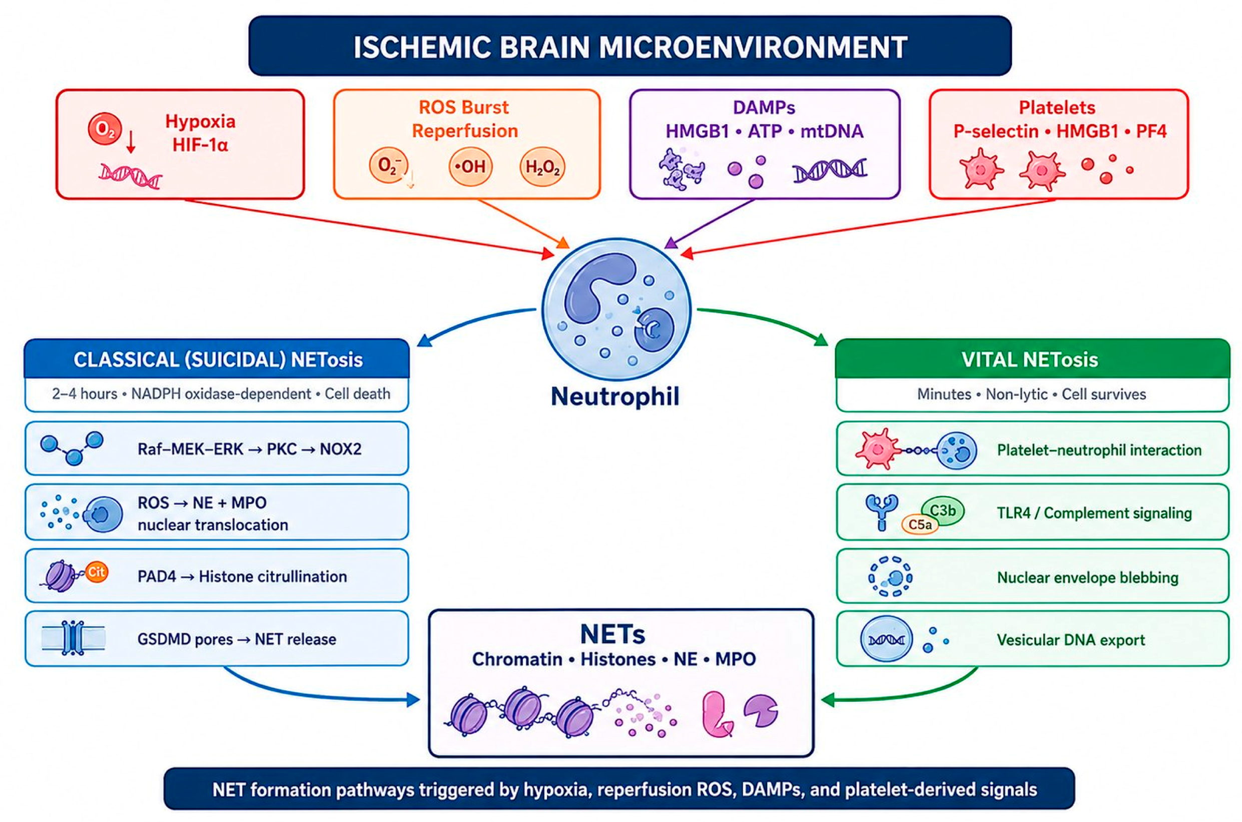
Figure 1: Mechanisms of NET formation in ischemic brain injury. Hypoxia, reperfusion-associated ROS generation, DAMPs release, and platelet-derived signals activate neutrophils and promote NETosis through classical (suicidal) and vital pathways. Classical NETosis involves NADPH oxidase-dependent ROS production, PAD4-mediated histone citrullination, and membrane rupture, whereas vital NETosis occurs via rapid vesicular DNA release without immediate neutrophil death. NETs consist primarily of extracellular chromatin, histones, neutrophil elastase, and myeloperoxidase. This figure was created by the authors for this manuscript using Microsoft PowerPoint (Microsoft Corporation, Redmond, WA, USA).
3 NETs in Cerebral Ischemia: Evidence from Models and Patients
Transient and permanent MCAO models have provided the most detailed characterization of NET dynamics in experimental ischemic stroke. Preclinical studies on stroke have shown that NETs accumulate in cerebral thrombi and periinfarct brain tissue, contributing to ongoing tissue damage beyond the hyperacute phase of ischemia [37,38]. Importantly, NET formation exhibited a spatial gradient, with the highest density observed in the penumbral region, the area most favorable for therapeutic rescue. PAD4, the rate-limiting enzyme for NETosis, was found to be significantly upregulated in the peri-ischemic brain tissue. Overexpression of PAD4 via the adenoviral route exacerbated BBB disruption and reduced neovascularization; In contrast, PAD4 knockout mice and mice treated with Cl-amidine reduced NET formation, decreased BBB permeability, increased vascular repair, and improved functional recovery.
Complementary evidence from the transient MCAO model demonstrates that NETs specifically contribute to ischemia-reperfusion injury. Wang et al. showed that NETs promote tissue plasminogen activator (tPA)-induced hemorrhagic transformation via the cGAS-STING pathway, revealing a clinically significant interaction between NET biology and the most commonly used reperfusion therapy [39]. Similar NET-mediated neurovascular damage has been observed in cardiac arrest/return of spontaneous circulation models, extending the significance of these findings from focal stroke to global cerebral ischemia.
Photothrombotic stroke models further validate the temporal profile of NET accumulation, demonstrating that NETs contribute to the expansion of the ischemic lesion into the penumbra during the subacute phase. Peña-Martínez et al. demonstrated that strategies targeting NETs (DNase I therapy) provide significant neuroprotection even in non-reperfusion settings, suggesting that the harmful effects of NETs extend beyond their role in thrombus stability [37].
Clinical studies have shown consistently elevated NET biomarkers in the peripheral blood of patients with acute ischemic stroke, as summarized in Table 1. Vallés et al. reported that plasma levels of cfDNA, nucleosomes, and citrullinated histone H3 were significantly higher in 243 stroke patients compared to healthy controls, and that CitH3 was independently associated with atrial fibrillation and all-cause mortality at one-year follow-up [18]. A later systematic review and meta-analysis by Wu et al., encompassing 752 patients in seven studies, confirmed that circulating NET markers were significantly associated with stroke severity (measured by NIHSS) and poor functional prognosis [19].
Histological analysis of thrombi removed during mechanical thrombectomy provided direct evidence of NET abundance in cerebral occlusions. Laridan et al. identified citrullinated histone H3 in nearly all thrombi analyzed from 68 ischemic stroke patients, and ex vivo lysis experiments showed that the addition of DNase I to standard tPA significantly enhanced thrombolysis [14]. Ducroux et al. expanded on these findings, demonstrating that high NET content in retrieved thrombi was associated with longer procedure times and a greater number of thrombectomy passes, and noting that NETs contributed to the thrombus’s resistance to mechanical degradation [17]. In a study involving 101 patients, Lapostolle et al. showed that NET abundance in ischemic thrombi was associated with unsuccessful recanalization and worse clinical outcomes, as assessed by the modified Rankin Scale at discharge [40]. Jabrah et al. also demonstrated that the neutrophil and NET composition in thrombi varied according to stroke etiology, and that this may have potential implications for personalized treatment strategies [41].
Table 1: Key preclinical and clinical evidence for NETs in brain ischemia.
| Study | Model/Population | NET Marker(s) | Key Finding |
|---|---|---|---|
| [11] | In vitro (human neutrophils) | DNA, histones, NE, MPO | First description of NETs as network-like chromatin structures with antimicrobial function |
| [30] | tMCAO (mice) | CitH3, cfDNA | HMGB1-induced NETs exacerbate ischemic brain damage; anti-HMGB1 antibody reduces NET formation and infarct size |
| [42] | tMCAO (mice) | CitH3, cfDNA | ATP accumulated after cerebral ischemia induces NETosis via P2X7R activation |
| [39] | tMCAO (mice) + tPA | CitH3, MPO-DNA | NETs promote tPA-induced hemorrhagic transformation via cGAS-STING |
| [15] | tMCAO (mice); platelet HMGB1 KO; human stroke brain tissue | H3Cit, MPO-DNA, cfDNA | Platelet HMGB1 is a critical source of NETs in acute stroke; nNIF treatment reduces infarct volume and improves survival |
| [37] | FeCl3 thrombotic stroke (mice) | CitH3 | DNase I provides neuroprotection even without reperfusion; NETs contribute to damage beyond thrombus stability |
| [43] | tMCAO (mice); BV2 cells | CitH3-DNA, AIM2 | NETs trigger AIM2-dependent microglial pyroptosis; GSK484 and AIM2 suppression improves neurological outcomes |
| [14] | 68 AIS patients (thrombectomy specimens) | CitH3, NE, cfDNA | NETs are found in almost all ischemic thrombi; DNase I + tPA enhances ex vivo thrombolysis compared to tPA alone |
| [18] | 243 AIS patients (plasma) | cfDNA, nucleosomes, CitH3 | NET markers elevated in patients with acute ischemic stroke vs. controls; CitH3 is independently associated with atrial fibrillation and 1-year all-cause mortality |
| [17] | 108 AIS patients (thrombectomy specimens) | NE, CitH3, cfDNA | High NET content is associated with longer procedure times and more thrombectomy passes; NETs impair tPA-induced thrombolysis |
| [40] | 101 AIS patients (thrombectomy specimens) | NE, CitH3, cfDNA | NET abundance is associated with unsuccessful recanalization (mTICI < 2B) and worse clinical outcome (mRS at discharge) |
| [41] | 300 AIS clots (100 AT, 100 CE, 100 cryptogenic) | CitH3, extracellular NETs | Neutrophil and NET composition differ by stroke etiology (AT vs. CE vs. cryptogenic); potential for personalized treatment |
| [19] | Meta-analysis: 7 studies, 752 patients | cfDNA, CitH3, MPO-DNA | Circulating NET markers are significantly associated with stroke severity (NIHSS) and poor functional prognosis |
3.3 NETs in Ischemia-Reperfusion Injury
The temporal dynamics of NET formation differ between the ischemia and reperfusion phases, which have significant implications for therapeutic targeting. During the ischemic phase, NETs are generated primarily within the thrombus and adjacent vascular lumen, where they contribute to thrombus stabilization, fibrinolysis resistance, and vascular occlusion [15]. At this stage, NET-related effects coexist with previous inflammatory events initiated by resident microglia, endothelial activation, platelet activation, and DAMP release from damaged neurons. Therefore, NETs should be viewed not as primary initiators of sterile neuroinflammation, but as amplifiers that reinforce pre-existing ischemia-induced inflammatory signaling.
Following reperfusion, restoration of oxygenated blood produces a burst of ROS in the recruited neutrophils, and resident vascular cells. Together with continued DAMP release from reperfusion-damaged tissue, this environment creates a second wave of NETosis [15,39]. This delayed NET response is particularly relevant to secondary microvascular injury, as NETs accumulating within capillaries can obstruct microvascular flow, act as a scaffold for secondary microthrombosis, and compromise perfusion of the penumbral zone. NET-mediated damage is temporally positioned between early ischemic inflammatory activation and subsequent glial amplification, thereby linking intravascular thromboinflammation with sustained parenchymal neuroinflammation.
This two-phase model creates a potential therapeutic window, but also introduces timing challenges. Early NET targeting may enhance thrombolysis and improve microvascular reperfusion, whereas delayed intervention may be more effective in limiting BBB disruption, glial activation, and sustained inflammatory signaling. However, optimal timing remains uncertain, as excessive or premature NET inhibition can impair host defense and neutrophil-mediated clearance functions. These considerations support a temporally stratified therapeutic approach in which NET-driven interventions are aligned with ischemic, reperfusion, or subacute inflammatory phase, rather than applied as a uniform anti-inflammatory strategy. This phase-dependent variability may partly explain the inconsistent outcomes in experimental NET targeting studies where intervention timing was not standardized.
4 NET-Derived Neuroinflammatory Signaling
This section constitutes the mechanistic core of the review. Rather than treating NET components as general inflammatory mediators, we define the specific receptor-mediated signaling pathways by which individual NET components activate inflammatory cascades that converge in the ischemic brain. This pathway-centric approach distinguishes the current review from previous reviews and provides a framework for rational therapeutic targeting.
Extracellular histones, particularly H3 and H4, are among the most cytotoxic components of NETs. Released in high concentrations from decondensed chromatin, histones exert direct cytotoxicity on endothelial cells, neurons, and glia through mechanisms involving membrane pore formation, calcium influx, and loss of mitochondrial membrane potential. In a cerebrovascular context, histone-mediated endothelial damage directly contributes to the disruption of the BBB. Beyond their cytotoxic effects, histones activate innate immune receptors (TLR2 and TLR4) in microglia, astrocytes, and endothelial cells, initiating downstream NF-κB and MAPK signaling cascades that promote pro-inflammatory cytokine production [44].
The cfDNA released as the structural backbone of NETs represents a distinct class of DAMPs that activate cytoplasmic DNA-sensing pathways. The cGAS-STING pathway, initially identified in the context of antiviral immunity, senses double-stranded DNA in the cytoplasm and activates type I interferon signaling and NF-κB. In ischemic stroke, NET-derived DNA has been shown to activate cGAS-STING signaling in both infiltrating immune cells and resident brain cells, contributing to interferon-β production and persistent neuroinflammation [22,39]. Additionally, NET-derived DNA further potentiates the inflammatory response by activating TLR9, an endosomal receptor that recognizes unmethylated CpG motifs.
Neutrophil elastase and myeloperoxidase, the major granular proteins of NETs, contribute to tissue damage through both enzymatic and signaling mechanisms. While NE directly degrades extracellular matrix components and tight junction proteins, MPO catalyzes the production of hypochlorous acid, a potent oxidant that damages lipids, proteins, and DNA in surrounding tissues. Both enzymes also act as ligands for pattern recognition receptors, further linking NET-derived proteases to inflammatory signaling [12].
4.2 Receptor-Mediated Pathways
The HMGB1–TLR4 axis acts as a critical bridge between NET formation and neuroinflammatory signaling. HMGB1, which is both a NETosis trigger (released from activated platelets) and a NET component (associated with extracellular chromatin), activates the MyD88-dependent pathway by binding to TLR4 in microglia and astrocytes [39,45]. This leads to IκB kinase-mediated phosphorylation and cleavage of IκBα, enabling translocation of NF-κB (p65/p50) to the nucleus and transcriptional activation of pro-inflammatory genes encoding TNF-α, IL-6, IL-1β, CXCL1, CXCL2, and adhesion molecules (ICAM-1, VCAM-1) [5,45]. The TRIF-dependent arm of TLR4 signaling simultaneously activates IRF3 and type I interferon production, linking NET-induced HMGB1 to both acute inflammation and sustained immune activation [20,30].
NF-κB activation represents the central node of NET-driven neuroinflammatory signaling. In the ischemic brain, both canonical (p65/p50-dependent) and non-canonical (RelB/p52-dependent) NF-κB pathways are activated, with the canonical pathway predominantly responsible for acute pro-inflammatory gene transcription. Critically, NF-κB also serves as Signal 1 (priming signal) for NLRP3 inflammasome assembly and transcriptionally upregulates pro-IL-1β, pro-IL-18, and NLRP3 itself. This NF-κB–NLRP3 cross-interaction establishes a molecular bridge between the innate immune recognition of NETs and the effector phase of neuroinflammation, allowing NET-mediated TLR activation to directly prep cells for subsequent inflammation-dependent IL-1β and IL-18 release [21].
The JAK2/STAT3 signaling pathway is activated by NET-derived cytokines (IL-6, IL-10) and DAMPs in microglia and astrocytes. STAT3 phosphorylation directs a transcriptional program involving both pro-inflammatory (iNOS, MMP-9, pro-inflammatory cytokines) and potentially reparative (VEGF, BDNF) gene products, depending on the temporal phase of cellular binding and injury. In the acute phase, STAT3 activation exacerbates neuroinflammation by predominantly promoting microglial M1 polarization and astrocyte A1 reactivity. However, during the recovery phase, STAT3 can facilitate the transition to anti-inflammatory and reparative phenotypes, suggesting a dual role that complicates therapeutic targeting [25].
NET-derived histones and DNA activate distinct pattern recognition receptors, including TLR2 and TLR9, providing additional receptor-mediated pathways that converge on NF-κB and MAPK signaling [12]. TLR9, localized in endosomal compartments, is particularly important for detecting internalized NET-derived DNA fragments and initiating downstream inflammatory responses [46]. These overlapping receptor pathways create a degree of redundancy that may explain the robustness of NET-derived neuroinflammation and the limited effectiveness of targeting any single receptor alone.
The NLRP3 inflammasome has emerged as a central effector of NET-induced neuroinflammation in ischemic stroke. NLRP3 activation requires two sequential signals: Signal 1 (priming) increases NLRP3 and pro-IL-1β expression via NF-κB-dependent transcription, while Signal 2 (activation) triggers NLRP3 oligomerization, ASC speck formation, and caspase-1 activation. NET components provide both signals: histone and DNA-mediated TLR activation provide Signal 1, while histone-induced potassium efflux, ROS production, and lysosomal destabilization following phagocytosis of NET residues provide Signal 2 [21].
Activated caspase-1 cleaves pro-IL-1β and pro-IL-18 into their mature, biologically active forms, which are then released from the cell, enhancing local and systemic inflammation [47]. IL-1β promotes endothelial activation, neutrophil chemotaxis, and microglial pro-inflammatory polarization, while IL-18 increases interferon-γ production and natural killer cell activation. Simultaneously, caspase-1 cleaves gasdermin D, generating N-terminal fragments that oligomerize in the plasma membrane and form pores. These gasdermin D pores act as channels for IL-1β and IL-18 release and, at sufficient concentrations, trigger pyroptosis, an inflammatory form of programmed cell death that releases intracellular DAMPs and further propagates the inflammatory signaling [48].
Beyond NLRP3, the absent in melanoma 2 (AIM2) inflammasome is directly activated by NET-derived double-stranded DNA. Chen et al. recently demonstrated that NETs trigger AIM2-dependent microglial pyroptosis following ischemic stroke and that suppression of AIM2 in brain tissue provides neuroprotective effects similar to pharmacological NET inhibition with GSK484 [43]. This finding is mechanistically important as it identifies a direct molecular link between NET-derived DNA and inflammation-mediated neuronal damage, independent of the NLRP3 pathway.
4.4 Integrated Signaling Network
The signaling pathways activated by NET components do not operate in isolation, but instead form an interconnected network characterized by pathway convergence, redundancy, and positive feedback. NF-κB and NLRP3 are functionally linked through a transcriptional priming step, in which NF-κB-driven upregulation of NLRP3 and pro-IL-1β is required for subsequent inflammasome activation [49]. STAT3 and NF-κB exhibit bidirectional crosstalk, with STAT3 modulating NF-κB activity in a context- and phosphorylation-dependent manner [50]. In parallel, TLR4 activation induces ROS production via the NADPH oxidase pathway, which can promote NET formation and contribute to a self-amplifying inflammatory loop in recruited neutrophils [15,45]. Most current evidence defines these interactions as mechanistic associations rather than strictly causal relationships, highlighting the need for pathway-specific perturbation studies to establish direct causal hierarchies. Taken together, this network structure enables NET-derived signals to propagate to multiple cellular compartments within the neurovascular unit, reinforcing both spatial and temporal expansion of inflammation.
A critical consequence of this network architecture is that NET-derived DAMPs can activate inflammatory pathways both independently and synergistically. Extracellular histones primarily activate TLR2/TLR4-dependent NF-κB and MAPK signaling and exert direct cytotoxic effects on endothelial and nerve cells. NET-derived cfDNA primarily activates DNA sensing pathways such as TLR9 and cGAS-STING, while granular enzymes such as NE and MPO promote proteolytic and oxidative damage. These component-specific mechanisms converge on shared downstream outputs, including cytokine production, inflammasome activation, endothelial dysfunction, and leukocyte recruitment. Therefore, the inflammatory effect of NETs can be attributed not to a single receptor-ligand interaction, but to the combined effect of multiple NET components acting in overlapping innate immune pathways.
This signaling redundancy has important therapeutic implications. Inhibition of a single pathway, such as TLR4 or NLRP3, may attenuate part of the inflammatory response but is unlikely to fully suppress NET-driven injury if parallel pathways, including TLR9, cGAS-STING, or STAT3, remain active. Conversely, upstream interventions that degrade NETs or prevent NET formation may simultaneously reduce multiple downstream signals but carry broader risks related to host defense and immune homeostasis. Therefore, distinguishing causally dominant nodes from parallel or compensatory pathways remains a central challenge for translational targeting.
The concept of feedforward inflammatory loops is central to understanding the reinforcing nature of NET-driven neuroinflammation. NETs activate signaling cascades that produce pro-inflammatory cytokines and chemokines (TNF-α, IL-1β, CXCL1, CXCL2), leading to the recruitment of additional neutrophils to the ischemic brain. These newly recruited neutrophils, primed by the inflammatory environment, undergo NETosis and generate additional NET-derived DAMPs, thereby reinforcing the same signaling circuits. This feedforward loop provides a mechanistic explanation for how a relatively localized initial ischemic injury can evolve into widespread neurovascular inflammation and highlights the importance of early mechanism-guided intervention [26].
The integrated signaling network illustrating how NET-derived DAMPs initiate convergent inflammatory cascades is schematically shown in Fig. 2. This schematic illustrates the flow from NET components to receptor activation (TLR4/HMGB1, TLR2/9, cGAS–STING), downstream signaling (NF-κB, STAT3, MAPK), and inflammasome activation (NLRP3, AIM2), and the critical feedback loop that sustains neuroinflammation.
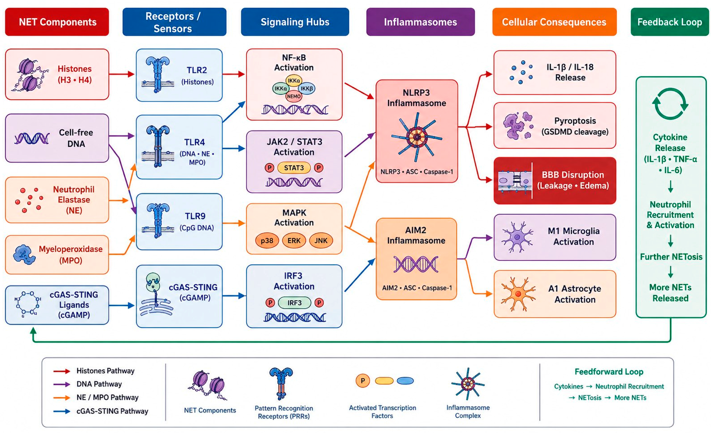
Figure 2: Integrated NET-associated signaling pathways in ischemic neuroinflammation. NET-derived components activate multiple receptor systems, including TLR2, TLR4, TLR9, and cGAS-STING, leading to downstream activation of NF-κB, JAK2/STAT3, MAPK, and IRF3 signaling pathways. These pathways converge on inflammasome activation, cytokine release, pyroptosis, blood-brain barrier disruption, and glial activation. Feedforward inflammatory loops further amplify neutrophil recruitment and NET formation within the neurovascular unit. This figure was created by the authors for this manuscript using Microsoft PowerPoint (Microsoft Corporation, Redmond, WA, USA).
5 NET-Microglia and Astrocyte Interaction
Microglia, resident innate immune cells of the central nervous system, are among the first to respond to NET-induced signals in the ischemic brain. NET components, particularly histones and HMGB1, activate microglia TLR4 and TLR2, driving polarization toward the classically activated (M1) pro-inflammatory phenotype. M1 microglia produce TNF-α, IL-1β, IL-6, inducible nitric oxide synthase (iNOS), and matrix metalloproteinases, collectively exacerbating neuronal damage, BBB disruption, and inflammatory cell recruitment [25].
Conversely, NET-induced signaling suppresses the alternatively activated (M2) phenotype, which is associated with anti-inflammatory cytokine production (IL-10, TGF-β), clearance of remnants via phagocytosis, and support of tissue repair. Both microglia and macrophages can be polarized into pro-inflammatory (M1) or anti-inflammatory (M2) phenotypes depending on microenvironmental signals; this reflects dynamic immune cell plasticity in neuroinflammatory conditions [51]. Experimental evidence suggests that NETs promote pro-inflammatory microglia activation after stroke, whereas interventions targeting NETs reduce microglia-associated inflammatory damage and improve neurological outcomes [43]. This observation is consistent with the concept of monocyte/macrophage phenotypic plasticity in vascular inflammatory diseases [6,51].
An important and not fully resolved question is whether NET remnants cleared via microglial phagocytosis led to inflammatory resolution or amplification. In principle, microglial clearance of NETs may limit the duration of extracellular DAMP exposure. However, internalization of NET-derived DNA can trigger intracellular inflammatory signaling instead of silent clearance by activating endosomal TLR9 and cytoplasmic cGAS-STING. The balance between these outcomes likely depends on the microglial activation status, the magnitude of the NET load, and the temporal phase of ischemic damage.
Astrocytes undergo a marked reactive transformation in response to NET-derived signals and microglial-derived cytokines. The neurotoxic A1 reactive phenotype, characterized by loss of neurotrophic support and complement component C3 upregulation, is induced by a combination of TNF-α, IL-1α, and C1q released from activated microglia. A1 astrocytes lose their capacity to support neuronal survival and synaptogenesis and instead contribute to neuronal death through the release of toxic factors [50,52].
Astrocyte-microglia-NET feedback loops may represent a critical amplification mechanism. Reactive astrocytes produce chemokines such as CXCL1, CXCL2, CCL2 that recruit additional neutrophils, while also simultaneously releasing ATP and HMGB1, further activating both microglia and neutrophils [30,50,52]. This multicellular feedback loop, in which NETs activate microglia, microglia induce astrocyte reactivity, and astrocytes promote further neutrophil recruitment, thereby creating a self-sustaining inflammatory circuit that persists beyond the initial ischemic insult. In the chronic phase, reactive astrocytes contribute to glial scar formation. This limits lesion expansion while simultaneously creating a physical and molecular barrier to axonal regeneration and functional recovery.
The combination of NET, microglial, and astrocyte signaling creates multiple amplification loops that drive progressive neurovascular damage. Cytokine-mediated positive feedback, namely the promotion of neutrophil recruitment and NETosis by microglial TNF-α and IL-1β, is the most direct amplification mechanism. Complement activation provides a second amplification axis: NETs activate the classical complement pathway via histone and DNA components, producing C3a and C5a that promote neutrophil chemotaxis, endothelial activation, and microglial priming. The integrated effect of these overlapping amplification cycles is a progressive increase in neuroinflammation, leading to infarct expansion, hemorrhagic transformation, and irreversible neurological failure if left uninterrupted [51].
The tricellular amplification circuit linking NETs, microglia, and astrocytes is summarized in Fig. 3. This figure demonstrates how NET-derived signals drive M1 microglial polarization, astrocyte conversion from M1 to A1, and chemokine-mediated neutrophil recruitment, creating a self-sustaining inflammatory feedback loop.
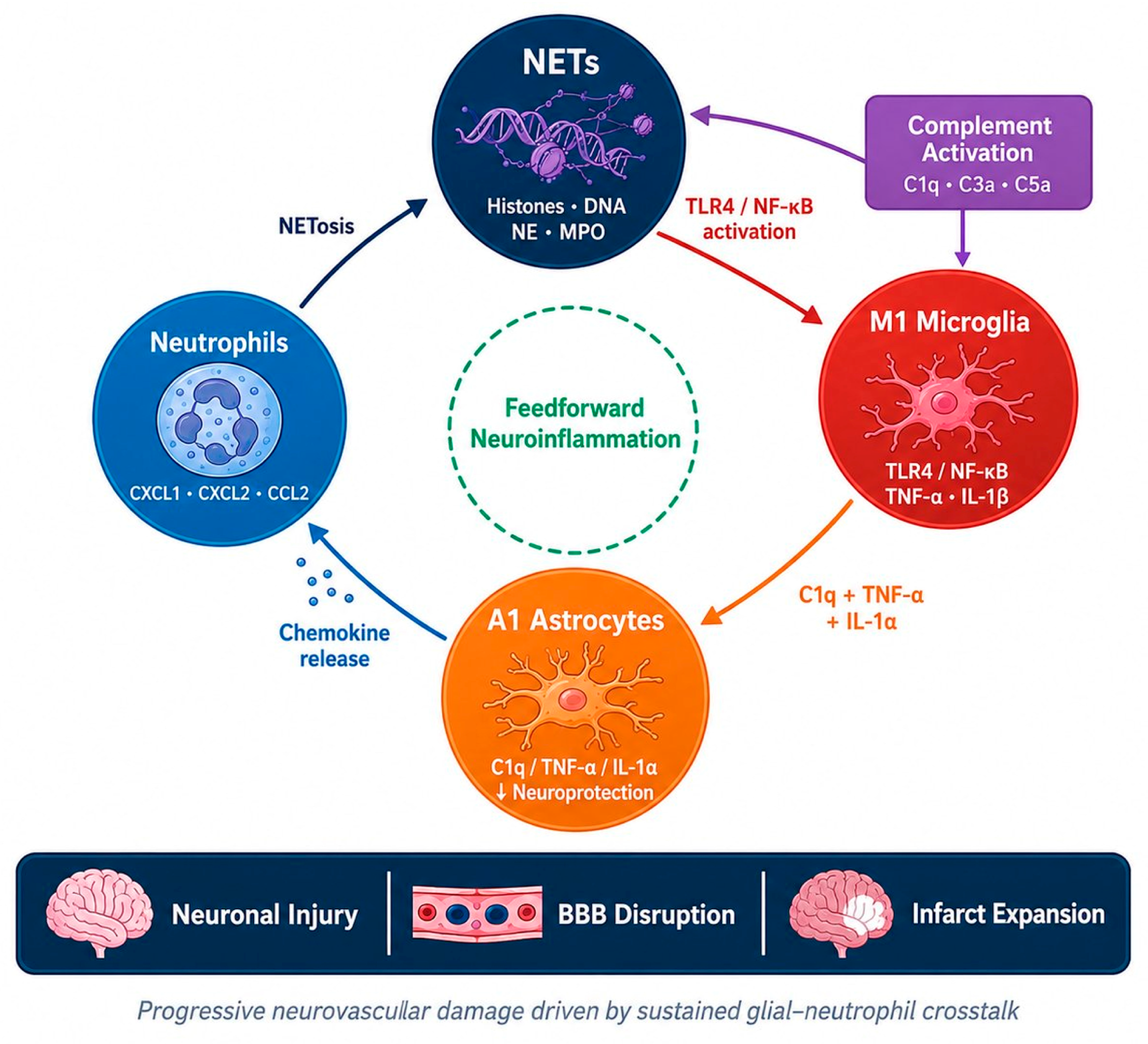
Figure 3: The NET-microglia-astrocyte amplification cycle in ischemic neuroinflammation. NET-derived DAMPs activate microglia via TLR4/NF-κB signaling, promoting the release of pro-inflammatory cytokines and complement-related mediators. Activated M1 microglia induce A1 astrocyte reactivity, while reactive astrocytes release chemokines that attract additional neutrophils and sustain NET formation. This feedforward inflammatory loop contributes to neuronal damage, blood-brain barrier disruption, and infarct expansion. This figure was created by the authors for this manuscript using Microsoft PowerPoint (Microsoft Corporation, Redmond, WA, USA).
6 Blood-Brain Barrier Dysfunction and Neurovascular Unit Destruction
6.1 NET-Mediated Endothelial Damage
Brain microvascular endothelial cells are the primary targets of NET-mediated cytotoxicity. Extracellular histones, particularly H3 and H4 histones, bind to endothelial cell membranes. This binding leads to calcium-dependent cytotoxicity, membrane pore formation, and apoptotic cell death. In addition, histone exposure activates endothelial cells, increasing the expression of adhesion molecules (ICAM-1, VCAM-1, E-selectin) and chemokines (CXCL1, CXCL8) that facilitate leukocyte adhesion and transmigration and drive the inflammatory cascade [12,24].
NET-associated proteases can directly damage the BBB. Neutrophil elastase and matrix metalloproteinases disrupt tight junction and basement membrane components, while myeloperoxidase-induced oxidant production further damages endothelial and extracellular matrix integrity, promoting barrier leakage, vasogenic edema, and hemorrhagic complications [52,53].
The integrity of the BBB is critically dependent on transmembrane tight junction proteins (claudin-5, occludin, and JAM) and their cytoplasmic scaffold partners, zonula occludens proteins (ZO-1, ZO-2, ZO-3). NET-derived proteases achieve proteolytic degradation of these proteins through at least three mechanisms: direct NE-mediated degradation of extracellular domains, MMP-9-mediated degradation of both tight junction proteins and basement membrane anchors, and oxidative modification of reactive species generated by MPO that alters protein conformation and promotes internalization.
The temporal sequence of tight junction degradation follows a biphasic pattern consistent with the ischemia-reperfusion cycle. During ischemia, partial tight junction degradation occurs due to energy deficiency and ATP depletion. Upon reperfusion, the influx of neutrophils and subsequent NET formation trigger a second, more intense phase of tight junction degradation via the mechanisms described above. This biphasic pattern has significant therapeutic implications, as interventions targeting NETs may be most effective in the early reperfusion window when the second wave of BBB disruption is initiated.
6.3 Pericytes and Basement Membrane
Pericytes, which surround the brain capillaries and regulate BBB permeability, capillary diameter, and cerebral blood flow, are susceptible to NET-mediated damage. NET-derived proteases degrade pericyte basement membrane attachment sites (collagen IV, laminin, fibronectin), causing pericytes to detach from the endothelium and migrate. Pericyte loss compromises paracrine signaling between pericytes, endothelial cells, and astrocyte endfoot, which maintains both the structural support of the capillary wall and the integrity of the BBB.
Degradation of the basement membrane by NET-derived MMP-9 and NE eliminates the underlying structural scaffold of endothelial cells that anchors pericytes and astrocyte endfoot. This disruption has consequences beyond acute barrier disruption: loss of basement membrane laminins impairs integrin-mediated survival signaling in endothelial cells, contributing to vascular regression and delayed hemorrhagic transformation.
6.4 NET-Induced Neurovascular Uncoupling
The neurovascular unit (NVU) functions as an integrated system where neuronal activity directs local vasodilation via neuron-astrocyte-vascular coordination; this process is called neurovascular coupling. NET-mediated disruption of any component of this unit (endothelial cells, pericytes, astrocyte endfoot, or basement membrane) impairs the capacity of the local microvasculature to respond to metabolic demands.
NET-driven neurovascular dissociation manifests as impaired perfusion responses in the peri-infarct region, where surviving neurons depend on intact neurovascular coupling for metabolic support [5]. Loss of vasodilator signaling, capillary no-reflow due to NET-associated microthrombosis, and pericyte constriction collectively reduce perfusion to the penumbra, thereby contributing to infarct expansion [15]. The concept of NET-driven neurovascular dissociation provides a mechanistic explanation for the clinical observation that penumbra tissue continues to infarct even after macrovascular recanalization and supports the rationale for adjunctive NET-targeted therapies as well as reperfusion strategies.
In the subacute and chronic phases of ischemic stroke, NETs exert a dual and paradoxical effect on vascular remodeling. On the one hand, NETs impair post-ischemic angiogenesis by damaging endothelial progenitor cells and the growth factor environment, which are essential for neovascularization. Experimental and review evidence suggests that persistent NET formation impairs post-stroke vascular remodeling, while strategies targeting NETs may facilitate revascularization and vascular repair [36,54,55]. On the other hand, VEGF derived from NETs and other angiogenic factors released during NETosis can provide pro-angiogenic signals under certain conditions. In most experimental settings, the net effect is anti-angiogenic and anti-reparative, supporting the therapeutic rationale of early NET inhibition to preserve the regenerative capacity of the cerebral vasculature.
The multiple mechanisms by which NETs disrupt the BBB and the neurovascular unit are integrated in Fig. 4, which shows how NET-derived histones, proteases, and reactive species collectively lead to endothelial damage, loss of tight junctions, pericyte detachment, and neurovascular disintegration, resulting in consequences such as vasogenic edema, capillary no-reflow, and penumbral infarction.
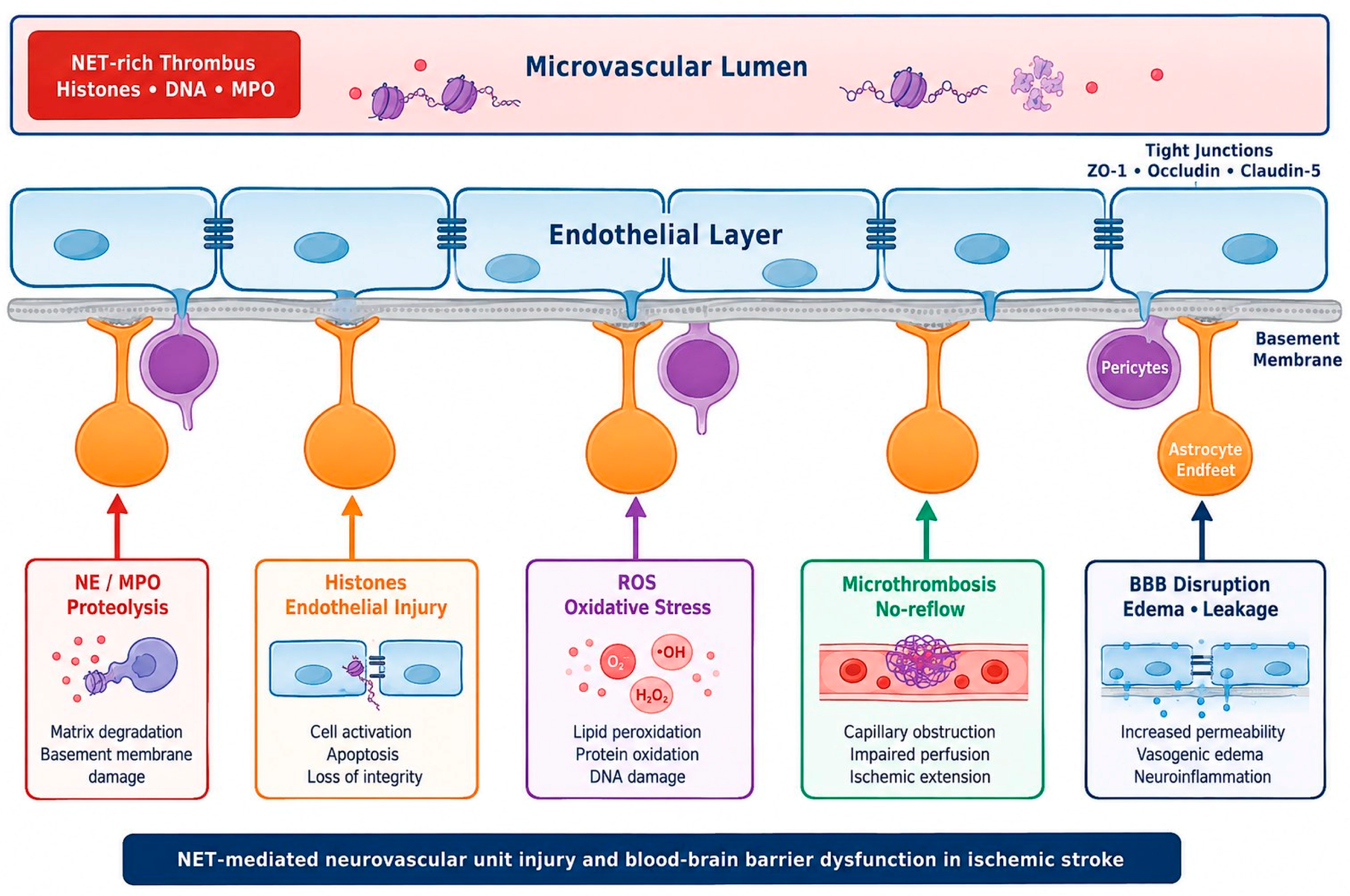
Figure 4: NET-mediated neurovascular unit injury and blood-brain barrier dysfunction. NET-associated histones, proteases, ROS, and microthrombi contribute to endothelial injury, degradation of tight junction proteins, oxidative stress, impaired microvascular perfusion, and blood-brain barrier disruption. Damage to endothelial cells, pericytes, basal membrane components, and astrocyte endfeet collectively promotes edema formation, neurovascular dissociation, and infarct progression. This figure was created by the authors for this manuscript using Microsoft PowerPoint (Microsoft Corporation, Redmond, WA, USA).
7 Oxidative Stress and Mitochondrial Signaling
The relationship between NETs and oxidative stress is bidirectional and mutually reinforcing. ROS produced during ischemia-reperfusion (primarily via mitochondrial electron transport chain dysfunction, xanthine oxidase activation, and NADPH oxidase pathway) act as potent triggers for NETosis. Conversely, NET-induced MPO creates a self-sustaining ROS-NET cycle by producing hypochlorous acid and other reactive species that increase local oxidative stress.
Mitochondrial dysfunction in neurons and glial cells is a hallmark of ischemic injury. Energy deficiency resulting from ATP depletion disrupts ion homeostasis, leading to excitotoxic calcium overload and collapse of the mitochondrial membrane potential. Release of cytochrome c from damaged mitochondria activates the intrinsic apoptotic pathway, while release of mitochondrial DNA into the cytoplasm activates the cGAS-STING pathway, which can also be activated by NET-derived DNA. The convergence of mitochondrial and NET-derived danger signals in common inflammatory pathways (cGAS-STING, TLR9) may partially explain the synergistic neurotoxicity observed when NETs are present in the context of ischemia-reperfusion injury [22].
The nuclear factor erythroid 2-associated factor 2 (Nrf2) pathway represents the major counter-regulatory mechanism against NET-mediated oxidative damage. Nrf2 activation induces the transcription of antioxidant enzymes, including heme oxygenase-1, superoxide dismutase, and glutathione peroxidase, which collectively mitigate ROS-mediated tissue damage. However, the capacity of the Nrf2 response to neutralize the combined oxidative burden of ischemia-reperfusion and NET-derived ROS is limited, particularly in the acute phase where oxidative stress is at its highest. The interaction between Nrf2 and NLRP3 signaling adds further complexity: while Nrf2 activation can suppress NLRP3 inflammatory activity, sustained NLRP3 activation can deplete antioxidant reserves and impair Nrf2 responses, creating a dynamic interaction that influences the course of ischemic damage [26].
8 Therapeutic Targeting of NET-Induced Neuroinflammation
8.1 NET Degradation Strategies
DNase I, an endonuclease that degrades cfDNA, represents the most direct approach to NET clearance. In experimental MCAO models, intravenous DNase I administration reduces NET burden, decreases infarct volume, alleviates BBB disruption, and improves neurological outcomes [15]. Neuroprotection has been demonstrated even in non-reperfusion settings [37]. Importantly, ex vivo studies using patient-derived thrombi have shown that combining DNase I with tPA significantly enhances thrombolysis compared to tPA alone; This finding also points to a potential role for DNase I in addition to standard thrombolytic therapy [14,17].
Dornase alfa (recombinant human DNase I), which is FDA-approved for cystic fibrosis, offers a clinically viable option. However, several challenges remain, including optimal dosing, timing of administration relative to ischemic onset, and the risk of excessive DNA clearance impairing beneficial NET functions in host defense, particularly in stroke patients prone to aspiration pneumonia and urinary tract infections.
PAD4 inhibitors represent the most advanced pharmacological strategy to prevent NET formation. The pan-PAD inhibitor Cl-amidine and the selective PAD4 inhibitor GSK484 have demonstrated efficacy in preclinical stroke models by reducing CitH3 levels, decreasing NET formation, and improving functional outcomes [13,56]. Chen et al. recently showed that GSK484 suppresses NET production, reduces AIM2 inflammasome expression, and improves neurological outcomes in a mouse stroke model [43]. The selectivity of GSK484 for PAD4 compared to other PAD isoforms may reduce off-target effects, but no PAD4 inhibitor has yet entered clinical trials for stroke.
NADPH oxidase inhibitors, including diphenyleneiodonium and apocynin, block ROS production upstream of NET formation [35]. However, broad inhibition of ROS-dependent processes may result in non-specific effects on host defense and cellular signaling, raising concerns about translational applicability. Gasdermin D inhibitors, such as disulfiram, prevent the final membrane pore formation step in NETosis and also suppress pyroptosis in NET-activated cells, offering a dual-action mechanism at the intersection of NETosis and inflammation pathways [56,57].
Given the potent cytotoxicity of NET-derived histones, strategies to neutralize extracellular histones represent a promising therapeutic avenue. Anti-histone antibodies have demonstrated neuroprotective effects in experimental models of sepsis and trauma [58], and their application to ischemic stroke warrants investigation. Histone-neutralizing peptides that bind and sequester extracellular histones represent an additional approach [44]. Activated protein C (APC), which cleaves histone H3 and H4, has shown protective effects in models of histone-mediated organ damage [58]. However, its anticoagulant properties should be carefully evaluated in the context of ischemic stroke and thrombolytic therapy.
8.4 Targeting Upstream Signaling
Targeting signaling pathways downstream of NET recognition offers a complementary strategy to direct NET clearance or NETosis inhibition. TLR4 antagonists, including TAK-242 (resatorvid) and eritoran, block the HMGB1–TLR4–NF-κB axis and have demonstrated neuroprotective effects in experimental stroke models [45,49]. NLRP3 inhibitors, particularly MCC950 (also known as CRID3), have shown efficacy in reducing infarct volume and improving outcomes in MCAO models by suppressing IL-1β and IL-18 release [21,49]. OLT1177 (dapansutrile), an orally bioavailable NLRP3 inhibitor currently in clinical trials for other inflammatory conditions, is a translational candidate for stroke [59].
NF-κB pathway modulators and JAK/STAT3 inhibitors offer additional avenues for therapeutic intervention. However, the pleiotropic roles of these pathways in both inflammatory and reparative processes necessitate careful consideration of timing and dosage to avoid suppressing beneficial immune and repair functions.
8.5 Combination Therapy Concepts
The multifaceted nature of NET-induced neurovascular damage suggests that combination strategies may be more effective than targeting a single component. The combination of NET inhibition with reperfusion therapy (tPA and/or thrombectomy) represents the most clinically urgent combination concept. Co-administration of DNase I with tPA has demonstrated synergistic thrombolytic efficacy in experimental studies [17]. In addition, PAD4 inhibition during reperfusion may limit secondary NET formation and reduce inflammation-related tissue damage [15].
Combining NET targeting with anti-inflammatory modulation (e.g., DNase I with an NLRP3 inhibitor) can address both the proximal cause (NETs) and the distal effector mechanism (inflammasome activation) simultaneously. Sequential targeting strategies, followed by sustained inflammation inhibition after acute NET clearance, may be particularly well-suited to the biphasic temporal profile of NET formation and inflammatory signaling. While preclinical combination data are limited, the strong mechanistic rationale supports prioritizing combination approaches in future translational studies [26].
The current therapeutic landscape targeting NET-induced neuroinflammation is summarized in Fig. 5, which maps intervention strategies (NETosis inhibition, NET degradation, histone neutralization, and signaling modulation) to specific points along the damage cascade and illustrates three proposed combination therapy concepts along with their respective translational statuses.
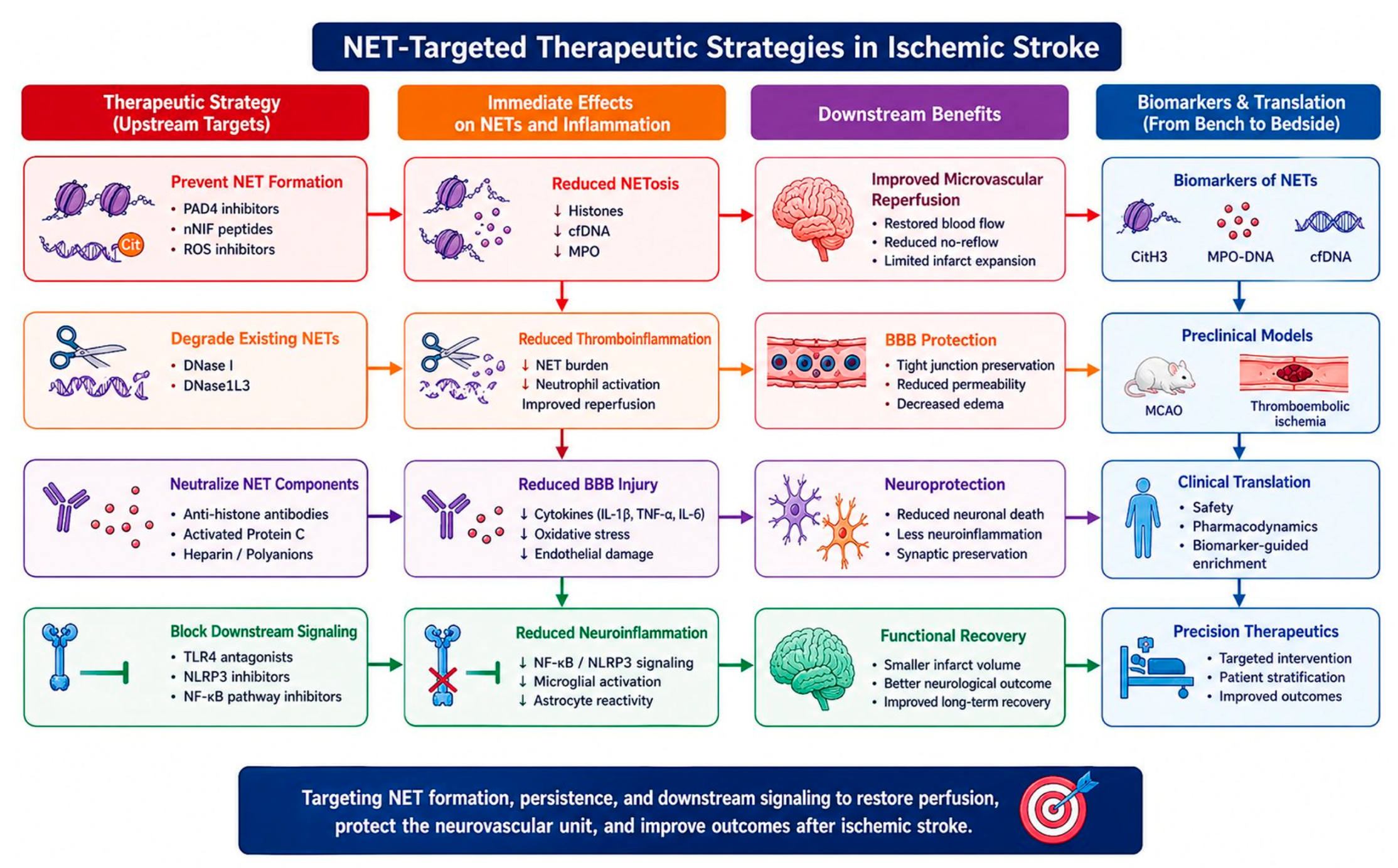
Figure 5: NET-targeted therapy strategies and translational approaches in ischemic stroke.
Therapeutic strategies targeting NET formation, persistence, and downstream inflammatory signaling include PAD4 inhibition, DNase-mediated NET degradation, neutralization of extracellular histones, and blockade of TLR4/NLRP3-associated pathways. These approaches aim to reduce thromboinflammation, maintain blood-brain barrier integrity, improve reperfusion, and enhance neurological recovery. Biomarker-guided translational frameworks may facilitate precise NET-targeted therapies in ischemic stroke. Downward arrows (↓) indicate reduction or suppression of the corresponding pathological process or biomarker level following therapeutic intervention. This figure was created by the authors for this manuscript using Microsoft PowerPoint (Microsoft Corporation, Redmond, WA, USA).
8.6 Translational Gaps and Challenges
Several challenges need to be addressed in order to translate NET-targeted therapies into clinical practice. Timing of intervention is critical: if done too early, NET inhibition can compromise antimicrobial host defense; if done too late, forward inflammatory cascades can become self-sustaining independently of NETs. The dual role of NETs, pathological in the context of sterile inflammation, but protective against infection, means that NET inhibition carries the risk of immunosuppression, especially given the high incidence of infections after stroke.
Biomarker-guided patient selection is a promising strategy to identify patients who will benefit most from NET-targeted therapy. Circulating CitH3, MPO-DNA complexes, and cfDNA can potentially serve as helpful biomarkers to guide treatment decisions, but standardized tests and validated thresholds are needed. Clinical trial design should also consider the heterogeneity of ischemic stroke, narrow therapeutic ranges, and potential interactions between NET-targeted therapies and standard care treatments. More generally, these translational challenges favor selective immunomodulatory approaches rather than non-specific immunosuppression, given the context-dependent protective and noxious functions of innate immune cells [51]. This further highlights the importance of immune cell plasticity in designing targeted therapeutic strategies [6].
Current and emerging treatment strategies targeting NET formation, signaling pathways, and downstream inflammatory effects in ischemic stroke are summarized in Table 2.
Table 2: Therapeutic agents targeting NET-induced neuroinflammation.
| Agent | Class | Mechanism of Action | Target Pathway | Evidence Level | Key Reference |
|---|---|---|---|---|---|
| DNase I (IV) | NET degradation | Cleaves the cfDNA backbone of NETs | NET structure | Preclinical (MCAO) + ex vivo human thrombi | [14] |
| Dornase alpha | NET degradation | Recombinant human DNase I; FDA-approved for cystic fibrosis | NET structure | Preclinical; translational candidate | [17] |
| DNase I + tPA | Combination | Synergistic thrombolysis; DNA clearance + fibrinolysis | NETs + fibrin | Ex vivo human thrombi | [14,17] |
| GSK484 | PAD4 inhibitor (selective) | Selective reversible PAD4 inhibitor (IC50 = 50 nM) | PAD4 → NETosis | Preclinical (MCAO, SAH) | [43] |
| BB-Cl-amidine (BBCA) | PAD4 inhibitor | Cell permeable PAD4 inhibitor; reduces CitH3 and NETosis | PAD4 → NETosis | Preclinical (MCAO) | [60] |
| VAS3947 | NOX2 inhibitor | NADPH oxidase inhibitor; blocks ROS-induced NETosis | NOX2 → ROS → NETosis | In vitro; preclinical | [61] |
| Disulfiram | Gasdermin D inhibitor | Blocks GSDMD pore formation; inhibits NET release and pyroptosis | GSDMD → NET release | Preclinical (repurposed) | [27] |
| Anti-histone antibodies | Histone neutralization | Binds and neutralizes cytotoxic extracellular histones (H3, H4) | Histone → TLR2/4 | Preclinical (sepsis, trauma models) | [58] |
| Activated Protein C | Histone scavenger | Cleaves histone H3 and H4; has anticoagulant properties | Histone proteolysis | Preclinical | [58] |
| nNIF | NET inhibitory factor | Neonatal NET inhibitory factor; blocks NETosis without affecting neutrophil recruitment | NETosis | Preclinical (MCAO; diabetic/elderly mice) | [15] |
| TAK-242 (Resatorvid) | TLR4 antagonist | Blocks the intracellular domain of TLR4; inhibits MyD88/TRIF signaling | TLR4 → NF-κB | Preclinical (MCAO) | [45] |
| Eritoran | TLR4 antagonist | Lipid A analog; competitive TLR4 antagonist | TLR4 → NF-κB | preclinical | [62] |
| MCC950 (CRID3) | NLRP3 inhibitor | Selective NLRP3 inhibitor; blocks ASC oligomerization | NLRP3 → Caspase-1 → IL-1β | Preclinical (MCAO); potent efficacy | [21] |
| OLT1177 (Dapansutrile) | NLRP3 inhibitor | Orally bioavailable NLRP3 inhibitor | NLRP3 → Caspase-1 | Phase II (gout, heart failure); preclinical (stroke) | Clinical trials in progress |
| JAK inhibitors | JAK/STAT3 modulator | Blocks JAK2-mediated STAT3 phosphorylation | JAK2/STAT3 | Preclinical | [20] |
Multiple omics technologies, including single-cell RNA sequencing, spatial transcriptomics, proteomics, and metabolomics, are beginning to reveal the molecular heterogeneity of neutrophils in the ischemic brain and the diversity of NET composition across different vascular regions and stroke etiologies. Single-cell approaches, by identifying different neutrophil subpopulations with different NETosis tendencies, suggest that not all neutrophils contribute equally to NET-mediated damage. These findings may lead to more targeted therapeutic strategies that selectively inhibit pathogenic NETosis while preserving beneficial neutrophil function.
Circulating NET biomarker panels incorporating CitH3, MPO-DNA complexes, cfDNA, and nucleosomes show promise for stroke prognosis and treatment monitoring. The correlation between circulating NET markers and thrombus NET content, although imperfect, can enable non-invasive assessment of thrombus susceptibility to thrombolysis and guide the selection of adjuvant therapies [19,41].
AI-powered biomarker integration and computational modeling of NET dynamics offer pioneering opportunities. Machine learning algorithms trained on multiple omics datasets can identify NET-related molecular signatures predicting stroke outcome and treatment response, potentially enabling a precision medicine approach toward NET-targeted therapy. While these applications are still in their early stages of development, they align with the broader trend toward personalized neurovascular medicine and could accelerate the transfer of NET biology to clinical practice [25,26]. In addition, recent studies have highlighted the interconnected roles of oxidative stress, autophagy, and ferroptosis in shaping neuroinflammatory responses and neuronal injury, suggesting that strategies targeting NETs may need to be integrated with broader cell death and metabolic regulatory pathways to deliver effective neuroprotection [63].
This review has presented a pathway-centric framework for understanding the role of neutrophil extracellular traps (NETs) in brain ischemic insults. The evidence examined supports the central thesis that NETs function as important, context-dependent amplifiers of sterile neuroinflammation, linking intravascular thrombosis to downstream neurovascular injury via convergent signaling pathways including NF-κB, NLRP3 inflammasome, STAT3, and HMGB1-TLR4.
Several important messages emerge from this synthesis. First, NETs are not merely structural components of ischemic thrombi but also active signaling platforms that activate specific receptor-mediated pathways with distinct but overlapping downstream effects through their individual components (histones, DNA, NE, MPO). Second, feedforward inflammatory loops generated by NETs, microglia, and astrocytes explain the progressive and self-sustaining nature of post-ischemic neuroinflammation. Third, NET-induced neurovascular uncoupling and BBB dysfunction offer mechanistic explanations for the clinical observation that successful recanalization does not always translate into proportional neurological recovery. Fourth, treatment options for NET-induced neuroinflammation are expanding; multiple intervention points, such as NET degradation, NETosis inhibition, histone neutralization, and signaling pathway modulation, offer complementary strategies that may be most effective in combination.
Looking ahead, this field requires: (i) more in-depth characterization of NET-mediated signaling at the single-cell level within the ischemic neurovascular unit; (ii) standardized biomarker panels for non-invasive assessment of NET burden and therapeutic response; (iii) preclinical testing of rationally designed combination therapies targeting both NETs and downstream effector pathways; and (iv) clinical trial designs that consider the temporal dynamics of NET formation and the heterogeneity of ischemic stroke. By moving from descriptive biology to a mechanistic, pathway-oriented understanding of NET-driven neuroinflammation, this field could lay the groundwork for NET-focused therapies that significantly improve outcomes for patients with ischemic brain injury. Targeting NET-driven signaling networks represents a promising strategy for bridging the gap between successful recanalization and meaningful neurological recovery.
Acknowledgement:
Funding Statement: The authors received no specific funding for this study.
Author Contributions: The authors confirm their contributions to the article as follows: Study design and structure: Nurittin Ardic; literature review and data collection: Alper Fatih Ardic; analysis and interpretation of results: Alper Fatih Ardic, Nurittin Ardic; drafting of the article: Alper Fatih Ardic; critical revision of the article: Nurittin Ardic. All authors reviewed and approved the final version of the manuscript.
Availability of Data and Materials: This article does not involve data availability and this section is not applicable.
Ethics Approval: Not applicable.
Conflicts of Interest: Alper Fatih Ardic declares no conflict no conflicts of interest to report regarding the present study. Nurittin Ardic is a volunteer consultant for Med-International UK Health Agency Ltd. The authors affirm that they have no additional financial or personal conflicts of interest that could have influenced the work reported in this manuscript.
References
1. Lui F , Khan Suheb MZ , Patti L . Ischemic stroke. Treasure Island, FL, USA: StatPearls Publishing; 2025. [Google Scholar]
2. Feigin VL , Abate MD , Abate YH , Abd ElHafeez S , Abd-Allah F , Abdelalim A , et al. Global, regional, and national burden of stroke and its risk factors, 1990–2021: a systematic analysis for the Global Burden of Disease Study 2021. Lancet Neurol. 2024; 23( 10): 973– 1003. doi:10.1016/S1474-4422(24)00369-7. [Google Scholar] [CrossRef]
3. Campbell BCV , De Silva DA , MacLeod MR , Coutts SB , Schwamm LH , Davis SM , et al. Ischaemic stroke. Nat Rev Dis Primers. 2019; 5: 70. doi:10.1038/s41572-019-0118-8. [Google Scholar] [CrossRef]
4. Powers WJ , Rabinstein AA , Ackerson T , Adeoye OM , Bambakidis NC , Becker K , et al. Guidelines for the early management of patients with acute ischemic stroke: 2019 update to the 2018 guidelines for the early management of acute ischemic stroke: a guideline for healthcare professionals from the American heart association/American stroke association. Stroke. 2019; 50( 12): e344– 418. doi:10.1161/STR.0000000000000211. [Google Scholar] [CrossRef]
5. Anrather J , Iadecola C . Inflammation and stroke: an overview. Neurotherapeutics. 2016; 13( 4): 661– 70. doi:10.1007/s13311-016-0483-x. [Google Scholar] [CrossRef]
6. Beyaz GK , Kirbas A , Tas SK . Monocyte phenotypic plasticity in peripheral artery disease: from pathophysiology to therapeutic targets. Biocell. 2026; 50( 1): 1– 10. doi:10.32604/biocell.2025.072368. [Google Scholar] [CrossRef]
7. Kumari S , Dhapola R , Sharma P , Nagar P , Medhi B , HariKrishnaReddy D . The impact of cytokines in neuroinflammation-mediated stroke. Cytokine Growth Factor Rev. 2024; 78: 105– 19. doi:10.1016/j.cytogfr.2024.06.002. [Google Scholar] [CrossRef]
8. Alsbrook DL , Di Napoli M , Bhatia K , Biller J , Andalib S , Hinduja A , et al. Neuroinflammation in acute ischemic and hemorrhagic stroke. Curr Neurol Neurosci Rep. 2023; 23( 8): 407– 31. doi:10.1007/s11910-023-01282-2. [Google Scholar] [CrossRef]
9. Yakupova E , Bocharnikov A , Fedulova K , Babenko V , Silachev D , Plotnikov E . Changes in bioenergetics and neuroprotective properties of mesenchymal stromal cells after LPS treatment. BIOCELL. 2024; 48( 12): 1827– 34. doi:10.32604/biocell.2024.058496. [Google Scholar] [CrossRef]
10. Jickling GC , Liu D , Ander BP , Stamova B , Zhan X , Sharp FR . Targeting neutrophils in ischemic stroke: translational insights from experimental studies. J Cereb Blood Flow Metab. 2015; 35( 6): 888– 901. doi:10.1038/jcbfm.2015.45. [Google Scholar] [CrossRef]
11. Brinkmann V , Reichard U , Goosmann C , Fauler B , Uhlemann Y , Weiss DS , et al. Neutrophil extracellular traps kill bacteria. Science. 2004; 303( 5663): 1532– 5. doi:10.1126/science.1092385. [Google Scholar] [CrossRef]
12. Papayannopoulos V . Neutrophil extracellular traps in immunity and disease. Nat Rev Immunol. 2018; 18( 2): 134– 47. doi:10.1038/nri.2017.105. [Google Scholar] [CrossRef]
13. Li Y , Liu J , Hu Y , Cong C , Chen Y , Zhou Q . Crossroads: pathogenic role and therapeutic targets of neutrophil extracellular traps in rheumatoid arthritis. BIOCELL. 2024; 48( 1): 9– 19. doi:10.32604/biocell.2023.045862. [Google Scholar] [CrossRef]
14. Laridan E , Denorme F , Desender L , François O , Andersson T , Deckmyn H , et al. Neutrophil extracellular traps in ischemic stroke thrombi. Ann Neurol. 2017; 82( 2): 223– 32. doi:10.1002/ana.24993. [Google Scholar] [CrossRef]
15. Denorme F , Portier I , Rustad JL , Cody MJ , de Araujo CV , Hoki C , et al. Neutrophil extracellular traps regulate ischemic stroke brain injury. J Clin Investig. 2022; 132( 10): e154225. doi:10.1172/JCI154225. [Google Scholar] [CrossRef]
16. Savi M , Su F , Sterchele ED , Bogossian EG , Demailly Z , Baggiani M , et al. Targeting NETosis in acute brain injury: a systematic review of preclinical and clinical evidence. Cells. 2024; 13( 18): 1553. doi:10.3390/cells13181553. [Google Scholar] [CrossRef]
17. Ducroux C , Di Meglio L , Loyau S , Delbosc S , Boisseau W , Deschildre C , et al. Thrombus neutrophil extracellular traps content impair tPA-induced thrombolysis in acute ischemic stroke. Stroke. 2018; 49( 3): 754– 7. doi:10.1161/strokeaha.117.019896. [Google Scholar] [CrossRef]
18. Vallés J , Lago A , Santos MT , Latorre AM , Tembl JI , Salom JB , et al. Neutrophil extracellular traps are increased in patients with acute ischemic stroke: prognostic significance. Thromb Haemost. 2017; 117( 10): 1919– 29. doi:10.1160/TH17-02-0130. [Google Scholar] [CrossRef]
19. Wu ZR , Zhou TQ , Ai SC . Neutrophil extracellular traps correlate with severity and prognosis in patients with ischemic stroke: a systematic review and meta-analysis. Acta Neurol Belg. 2024; 124( 2): 513– 22. doi:10.1007/s13760-023-02409-5. [Google Scholar] [CrossRef]
20. Fang H , Bo Y , Hao Z , Mang G , Jin J , Wang H . A promising frontier: targeting NETs for stroke treatment breakthroughs. Cell Commun Signal. 2024; 22( 1): 238. doi:10.1186/s12964-024-01563-4. [Google Scholar] [CrossRef]
21. Zhao Z , Pan Z , Zhang S , Ma G , Zhang W , Song J , et al. Neutrophil extracellular traps: a novel target for the treatment of stroke. Pharmacol Ther. 2023; 241: 108328. doi:10.1016/j.pharmthera.2022.108328. [Google Scholar] [CrossRef]
22. Chauhan C , Kaundal RK . The role of cGAS-STING signaling in ischemic stroke: from immune response to therapeutic targeting. Drug Discov Today. 2023; 28( 11): 103792. doi:10.1016/j.drudis.2023.103792. [Google Scholar] [CrossRef]
23. Divecha YA , Rampes S , Tromp S , Boyanova ST , Fleckney A , Fidanboylu M , et al. The microcirculation, the blood-brain barrier, and the neurovascular unit in health and Alzheimer disease: the aberrant pericyte is a central player. Pharmacol Rev. 2025; 77( 3): 100052. doi:10.1016/j.pharmr.2025.100052. [Google Scholar] [CrossRef]
24. Xue S , Zhou X , Yang ZH , Si XK , Sun X . Stroke-induced damage on the blood-brain barrier. Front Neurol. 2023; 14: 1248970. doi:10.3389/fneur.2023.1248970. [Google Scholar] [CrossRef]
25. Ardic N , Dinc R . Neutrophil extracellular traps and microglia/macrophages interactions in stroke: from thromboinflammation to immunotherapy. Front Immunol. 2026; 17: 1752471. doi:10.3389/fimmu.2026.1752471. [Google Scholar] [CrossRef]
26. Dinc R , Ardic N . Molecular and cellular mechanisms of neutrophil extracellular traps in cardiovascular diseases: from NET formation to mechanistic therapeutic targeting. BIOCELL. 2026; 50( 1): 1– 10. doi:10.32604/biocell.2025.072337. [Google Scholar] [CrossRef]
27. Sollberger G , Choidas A , Burn GL , Habenberger P , Di Lucrezia R , Kordes S , et al. Gasdermin D plays a vital role in the generation of neutrophil extracellular traps. Sci Immunol. 2018; 3( 26): eaar6689. doi:10.1126/sciimmunol.aar6689. [Google Scholar] [CrossRef]
28. Yipp BG , Petri B , Salina D , Jenne CN , Scott BNV , Zbytnuik LD , et al. Infection-induced NETosis is a dynamic process involving neutrophil multitasking in vivo. Nat Med. 2012; 18( 9): 1386– 93. doi:10.1038/nm.2847. [Google Scholar] [CrossRef]
29. McInturff AM , Cody MJ , Elliott EA , Glenn JW , Rowley JW , Rondina MT , et al. Mammalian target of rapamycin regulates neutrophil extracellular trap formation via induction of hypoxia-inducible factor 1 α. Blood. 2012; 120( 15): 3118– 25. doi:10.1182/blood-2012-01-405993. [Google Scholar] [CrossRef]
30. Kim SW , Lee H , Lee HK , Kim ID , Lee JK . Neutrophil extracellular trap induced by HMGB1 exacerbates damages in the ischemic brain. Acta Neuropathol Commun. 2019; 7( 1): 94. doi:10.1186/s40478-019-0747-x. [Google Scholar] [CrossRef]
31. Bečka E , Hudecová L , Pastorek M . Mitochondria as inducers of neutrophil extracellular traps. Inflammation. 2026; 49( 1): 67. doi:10.1007/s10753-025-02432-z. [Google Scholar] [CrossRef]
32. Fang X , Zhou X , Zhou X , Cheng Z , Hu Y . HMGB1-platelet interactions: mechanisms and targeted therapy strategies. Thromb Haemost. 2026; 126( 4): 346– 58. doi:10.1055/a-2622-0074. [Google Scholar] [CrossRef]
33. Liu C , Chen Q , Chen W , Han X , Qiao X , Guo J , et al. IL-17A promotes NETs formation via the PKCζ-ERK-ROS-PAD4 pathway in a mouse model of ischemic stroke. CNS Neurosci Ther. 2026; 32( 3): e70825. doi:10.1002/cns.70825. [Google Scholar] [CrossRef]
34. Li Z , Wu SH , Zhang X , Li T , Xin M , Liu F . Neutrophil extracellular traps (NETs) in cerebrovascular disease mechanisms, detection, and therapeutic targeting. Exp Neurol. 2026; 400: 115683. doi:10.1016/j.expneurol.2026.115683. [Google Scholar] [CrossRef]
35. Fuchs TA , Abed U , Goosmann C , Hurwitz R , Schulze I , Wahn V , et al. Novel cell death program leads to neutrophil extracellular traps. J Cell Biol. 2007; 176( 2): 231– 41. doi:10.1083/jcb.200606027. [Google Scholar] [CrossRef]
36. Lood C , Blanco LP , Purmalek MM , Carmona-Rivera C , De Ravin SS , Smith CK , et al. Neutrophil extracellular traps enriched in oxidized mitochondrial DNA are interferogenic and contribute to lupus-like disease. Nat Med. 2016; 22( 2): 146– 53. doi:10.1038/nm.4027. [Google Scholar] [CrossRef]
37. Peña-Martínez C , Durán-Laforet V , García-Culebras A , Cuartero MI , Moro MÁ , Lizasoain I . Neutrophil extracellular trap targeting protects against ischemic damage after fibrin-rich thrombotic stroke despite non-reperfusion. Front Immunol. 2022; 13: 790002. doi:10.3389/fimmu.2022.790002. [Google Scholar] [CrossRef]
38. Oh SA , Seol SI , Davaanyam D , Kim SW , Lee JK . Platelet-derived HMGB1 induces NETosis, exacerbating brain damage in the photothrombotic stroke model. Mol Med. 2025; 31( 1): 46. doi:10.1186/s10020-025-01107-7. [Google Scholar] [CrossRef]
39. Wang R , Zhu Y , Liu Z , Chang L , Bai X , Kang L , et al. Neutrophil extracellular traps promote tPA-induced brain hemorrhage via cGAS in mice with stroke. Blood. 2021; 138( 1): 91– 103. doi:10.1182/blood.2020008913. [Google Scholar] [CrossRef]
40. Lapostolle A , Loyer C , Elhorany M , Chaigneau T , Bielle F , Alamowitch S , et al. Neutrophil extracellular traps in ischemic stroke thrombi are associated wth poor clinical outcome. Stroke Vasc Interv Neurol. 2023; 3( 3): e000639. doi:10.1161/SVIN.122.000639. [Google Scholar] [CrossRef]
41. Jabrah D , Rossi R , Molina S , Douglas A , Pandit A , McCarthy R , et al. White blood cell subtypes and neutrophil extracellular traps content as biomarkers for stroke etiology in acute ischemic stroke clots retrieved by mechanical thrombectomy. Thromb Res. 2024; 234: 1– 8. doi:10.1016/j.thromres.2023.12.005. [Google Scholar] [CrossRef]
42. Kim SW , Davaanyam D , Seol SI , Lee HK , Lee H , Lee JK . Adenosine triphosphate accumulated following cerebral ischemia induces neutrophil extracellular trap formation. Int J Mol Sci. 2020; 21( 20): 7668. doi:10.3390/ijms21207668. [Google Scholar] [CrossRef]
43. Chen H , Ni L , Zhang J , Zheng X , Chen Y , Jin X , et al. Neutrophil extracellular traps promote AIM2-dependent microglial pyroptosis following stroke. Aging Dis. 2025; 17( 3): 1616– 33. doi:10.14336/AD.2024.1733. [Google Scholar] [CrossRef]
44. Da Silva DE , Richards CM , McRae SA , Riar I , Yang SS , Zurfluh NE , et al. Extracellular mixed histones are neurotoxic and modulate select neuroimmune responses of glial cells. PLoS One. 2024; 19( 4): e0298748. doi:10.1371/journal.pone.0298748. [Google Scholar] [CrossRef]
45. Jiang L , Xu Z , Li H , Wu M , Wang F , Liu S , et al. TAK-242 exerts a neuroprotective effect via suppression of the TLR4/MyD88/TRIF/NF-κB signaling pathway in a neonatal hypoxic-ischemic encephalopathy rat model. Mol Med Rep. 2020; 22( 2): 1440– 8. doi:10.3892/mmr.2020.11220. [Google Scholar] [CrossRef]
46. Aubé FA , Bidias A , Pépin G . Who and how, DNA sensors in NETs-driven inflammation. Front Immunol. 2023; 14: 1190177. doi:10.3389/fimmu.2023.1190177. [Google Scholar] [CrossRef]
47. Franchi L , Eigenbrod T , Muñoz-Planillo R , Nuñez G . The inflammasome: a caspase-1-activation platform that regulates immune responses and disease pathogenesis. Nat Immunol. 2009; 10( 3): 241– 7. doi:10.1038/ni.1703. [Google Scholar] [CrossRef]
48. Pyrillou K , Burzynski LC , Clarke MCH . Alternative pathways of IL-1 activation, and its role in health and disease. Front Immunol. 2020; 11: 613170. doi:10.3389/fimmu.2020.613170. [Google Scholar] [CrossRef]
49. Liao Y , Kong Y , Chen H , Xia J , Zhao J , Zhou Y . Unraveling the priming phase of NLRP3 inflammasome activation: molecular insights and clinical relevance. Int Immunopharmacol. 2025; 146: 113821. doi:10.1016/j.intimp.2024.113821. [Google Scholar] [CrossRef]
50. He T , Yang GY , Zhang Z . Crosstalk of astrocytes and other cells during ischemic stroke. Life. 2022; 12( 6): 910. doi:10.3390/life12060910. [Google Scholar] [CrossRef]
51. Ardic AF , Ardic N . Role of neutrophils as therapeutic targets in intracerebral hemorrhage. Ther Innov Regul Sci. 2024; 58( 5): 807– 16. doi:10.1007/s43441-024-00668-9. [Google Scholar] [CrossRef]
52. Yuan B , Zhang X , Liu L , Chai Y , Zhang J , Chen X . Neutrophils-astrocyte interactions in central nervous system inflammation. Cell Death Dis. 2025; 16: 643. doi:10.1038/s41419-025-07945-x. [Google Scholar] [CrossRef]
53. Wang H , Zhang Y , Cai J , Guo J . Immune-mediated blood-brain barrier disruption after ischemic stroke: mechanisms and therapeutic targets. Front Immunol. 2025; 16: 1716041. doi:10.3389/fimmu.2025.1716041. [Google Scholar] [CrossRef]
54. Chang S , Zhao Y , Li C , Liu X , Fang Z , Peng Z , et al. Targeting revascularization via tryptophan-indole-NETs axis: the synergistic power of acupuncture and rt-PA thrombolysis in ischemic stroke. Front Neurol. 2025; 16: 1596158. doi:10.3389/fneur.2025.1596158. [Google Scholar] [CrossRef]
55. Di Meglio L , Solo Nomenjanahary M , Bedoucha L , Dupont S , Zemali F , Ollivier V , et al. Neutrophil extracellular traps-targeting therapy with deoxyribonuclease 1 reduces large vessel occlusion-induced downstream microvascular thromboinflammation in a rat model of stroke. Res Pract Thromb Haemost. 2025; 9( 7): 103206. doi:10.1016/j.rpth.2025.103206. [Google Scholar] [CrossRef]
56. Zhu C , Xu S , Jiang R , Yu Y , Bian J , Zou Z . The gasdermin family: emerging therapeutic targets in diseases. Signal Transduct Target Ther. 2024; 9: 87. doi:10.1038/s41392-024-01801-8. [Google Scholar] [CrossRef]
57. Hu R , Liang J , Ding L , Zhang W , Wang Y , Zhang Y , et al. Gasdermin D inhibition ameliorates neutrophil mediated brain damage in acute ischemic stroke. Cell Death Discov. 2023; 9: 50. doi:10.1038/s41420-023-01349-6. [Google Scholar] [CrossRef]
58. Xu J , Zhang X , Pelayo R , Monestier M , Ammollo CT , Semeraro F , et al. Extracellular histones are major mediators of death in sepsis. Nat Med. 2009; 15( 11): 1318– 21. doi:10.1038/nm.2053. [Google Scholar] [CrossRef]
59. Duan M , Sun L , He X , Wang Z , Hou Y , Zhao Y . Medicinal chemistry strategies targeting NLRP3 inflammasome pathway: a recent update from 2019 to mid-2023. Eur J Med Chem. 2023; 260: 115750. doi:10.1016/j.ejmech.2023.115750. [Google Scholar] [CrossRef]
60. Seol SI , Oh SA , Davaanyam D , Lee JK . Blocking peptidyl arginine deiminase 4 confers neuroprotective effect in the post-ischemic brain through both NETosis-dependent and-independent mechanisms. Acta Neuropathol Commun. 2025; 13( 1): 33. doi:10.1186/s40478-025-01951-y. [Google Scholar] [CrossRef]
61. Lu WJ , Li JY , Chen RJ , Huang LT , Lee TY , Lin KH . VAS2870 and VAS3947 attenuate platelet activation and thrombus formation via a NOX-independent pathway downstream of PKC. Sci Rep. 2019; 9: 18852. doi:10.1038/s41598-019-55189-5. [Google Scholar] [CrossRef]
62. Parada E , Casas AI , Palomino-Antolin A , Gómez-Rangel V , Rubio-Navarro A , Farré-Alins V , et al. Early toll-like receptor 4 blockade reduces ROS and inflammation triggered by microglial pro-inflammatory phenotype in rodent and human brain ischaemia models. Br J Pharmacol. 2019; 176( 15): 2764– 79. doi:10.1111/bph.14703. [Google Scholar] [CrossRef]
63. Jindal A , Rai SN . Targeting autophagy, ferroptosis, and neuroinflammation: polyphenol-mediated modulation of emerging signaling pathways in neurodegeneration. Mol Neurobiol. 2026; 63( 1): 470. doi:10.1007/s12035-026-05760-9. [Google Scholar] [CrossRef]
Cite This Article
 Copyright © 2026 The Author(s). Published by Tech Science Press.
This work is licensed under a Creative Commons Attribution 4.0 International License , which permits unrestricted use, distribution, and reproduction in any medium, provided the original work is properly cited.
 Downloads
 Citation Tools
