
 Submit a Paper
 Propose a Special lssue
 Open Access
REVIEW

Smart Nano-Cellulosic-Based Materials as Antiviral Agents: A Brief Insight into Scientific Advances and Functionalization Strategies
1 Cellulose and Paper Department, National Research Centre, Dokki, Giza, 12622, Egypt
2 Photochemistry Department, National Research Centre, Dokki, Giza, 12622, Egypt
* Corresponding Author: Ahmed M. Khalil. Email:
(This article belongs to the Special Issue: Recent Advances on Renewable Materials)
Journal of Renewable Materials 2026, 14(6), 6 https://doi.org/10.32604/jrm.2025.02025-0180
Received 13 September 2025; Accepted 11 November 2025; Issue published 29 June 2026
 View Full Text
 Download PDF
Abstract
The growing threat of viral pandemics necessitates innovative antiviral strategies that are effective, sustainable, and scalable. This review highlights nanocellulose as a renewable, biocompatible nanomaterial and a promising multifunctional antiviral platform. We examine cellulose nanocrystals, nanofibrils, and bacterial nanocellulose, emphasizing their synergistic antiviral mechanisms, including nanoscale viral entrapment and surface-mediated inactivation via sulfation, cationic groups, and metal nanoparticles. Key advances include photothermally active nanocellulose-graphene composites for on-demand viral deactivation, sulfated nanocellulose mimicking heparin’s virus-trapping properties, and engineered biopolymer hybrids for targeted drug delivery and mucosal immunity. Translational applications span antiviral coatings, self-sterilizing filters, and regenerative wound dressings. The review also addresses scalability and regulatory challenges, integrating computational modeling and structure-activity relationships to guide real-world implementation. Nanocellulose-based technologies offer a transformative approach to antiviral defense, merging adaptability, sustainability, and multifunctionality to meet the demands of pandemic preparedness and redefine the future of biomedical materials.Graphic Abstract
Keywords
Cellulose, the most abundant biopolymer on Earth, offers a compelling alternative to synthetic materials in the development of advanced functional systems [1–3], particularly in biomedical [4,5] and environmental [6] applications. Derived from diverse renewable sources, including wood pulp, cotton, algae, bacteria, and agricultural residues such as wheat straw, sugarcane bagasse, and corn husk cellulose is inherently biodegradable, non-toxic, and biocompatible. Its molecular structure, composed of
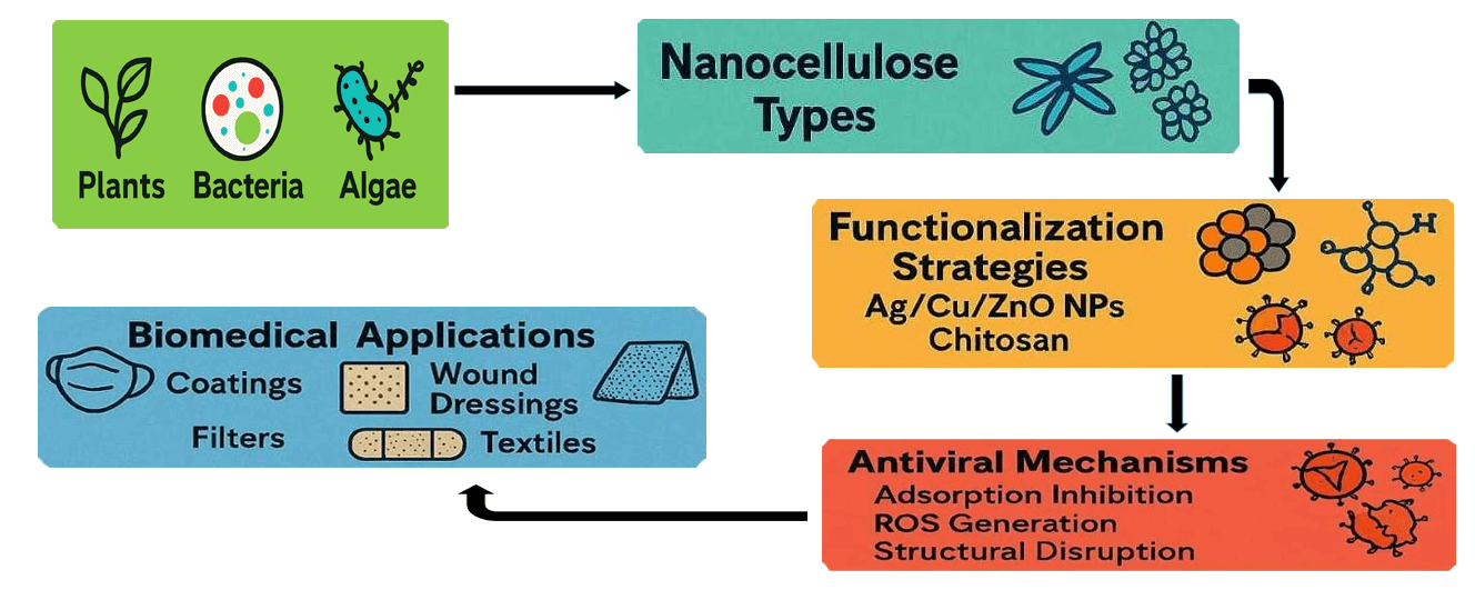
The scope of this concise scientific review encompasses foundational advances in the mechanisms of antiviral action of nano-cellulosic materials, cutting-edge chemical and green functionalization routes, exploration of synergistic nanocomposite architectures, and the broadening landscape of applications in personal protective equipment (PPE), filtration, drug delivery, and wound care [15,16]. Moreover, special emphasis is dedicated to the translational challenges surrounding safety, regulatory, industrial scalability, and the environmental sustainability of these materials. The analysis is guided by the latest peer-reviewed advances, emerging market assessments, and the new frontiers in computational and mechanistic modeling, thereby offering a deep synthesis of state-of-the-art knowledge and strategic directions for the field moving into 2025. Nano-cellulosic-based materials as antiviral agents can be summarized as shown in Fig. 1.

Figure 1: A schematic diagram for nano-cellulosic-based materials as antiviral agents
2 Fundamental Structure, Classes, and Properties of Nanocellulose
2.1 Structural Classes and Sourcing
Nanocellulose is defined by its hierarchical architectures, wherein cellulose chains are organized into high-aspect-ratio nanomaterials with distinct physicochemical profiles [17,18]. Smart nano-cellulosic-based materials as antiviral agents can be classified into several categories [19,20]. The three primary classes used in antiviral material design are cellulose nanocrystals (CNCs), cellulose nanofibrils (CNFs) and bacterial nanocellulose (BNC) as illustrated in Fig. 2. CNCs are rod-shaped, highly crystalline segments produced by acid hydrolysis with typical diameters of 5–20 nm and lengths of 100–500 nm; their high crystallinity and large specific surface area provide superior modulus and abundant reactive sites for surface functionalization [21,22]. CNFs are flexible, entangled fibrils that contain both crystalline and amorphous regions and are typically obtained by mechanical fibrillation or enzymatic/chemical pretreatment; with diameters in the 5–50 nm range and micron-scale lengths, CNFs form extensive networks that offer tunable porosity, high surface area and excellent reinforcement capacity for composite and hydrogel architectures [23]. BNC is biosynthesized by bacteria such as Komagataeibacter species as a pure, three-dimensional nanofibrillar network (fibril diameters typically 10–100 nm) that combines exceptional water-holding capacity, intrinsic purity and an easily modifiable surface, making it particularly attractive for biomedical and wound-care applications [20,24]. Source material (plants, bacteria, algae, tunicates) and the specific extraction or synthesis protocol strongly influence crystallinity, purity and morphology, which in turn determine colloidal stability, modification efficiency and downstream functional performance critical to antiviral material design [25].

Figure 2: Classification of smart nano-cellulosic-based materials as antiviral agents
2.2 Key Physicochemical Properties
Nanocellulose can be tailored for biomedical use and its exceptional, application-enabling traits arise from a combination of mechanical, chemical and structural features [25–28]. It offers very high mechanical strength and stiffness, yielding orders-of-magnitude superior strength-to-weight ratios compared with many conventional polymers and some metals [29,30], while the dense arrays of surface hydroxyl groups provide outstanding reactivity for post-synthetic modification and functionalization [31]. The intrinsic nanoporosity and large specific surface area make nanocellulose well suited for size-exclusion-based virus removal and as a high-loading support for functional nanostructures [32]. Under defined conditions many nanocellulose formulations show favorable biocompatibility and low cytotoxicity in vitro and in vivo, although outcomes are formulation-dependent and must be established case by case [33]. Its biodegradability and renewable feedstocks improve environmental compatibility across the lifecycle and align nanocellulose with circular bioeconomy objectives [34,35]. Recent process and surface-engineering advances have sharpened control over particle dimensions, dispersion quality and zeta potential, providing the critical levers needed to tune material–virus interactions and to develop next-generation antiviral products [36,37].
3 Advanced Antiviral Mechanisms of Nanocellulose
3.1 Intrinsic Physical and Chemical Mechanisms
The antiviral efficacy of nanocellulose-based materials fundamentally arises from two main modes [38,39] as shown in Fig. 3.

Figure 3: Antiviral mechanisms of nanocellulose
3.1.1 Physical Entrapment/Filtration
Nanocellulose membranes, characterized by nanometer-scale pores and high tortuosity, act as mechanical barriers that enable size-exclusion and removal of viral particles from aerosols, liquids, and contaminated surfaces. This filtration mechanism has been validated against a range of viruses including SARS-CoV-2, swine influenza, herpes simplex, and HIV, with studies reporting log reduction values exceeding 6.0 which is equivalent to more than 99.9% removal efficiency [36–40]. Such barrier effects are especially critical for controlling airborne and waterborne virus transmission, as demonstrated in air and water filtration prototypes and personal protective equipment (PPE) applications [41,42]. These findings underscore the potential of nanocellulose membranes in scalable antiviral filtration systems that combine physical entrapment with customizable surface chemistry.
3.1.2 Functionalized Surface Interactions
The dense, readily modifiable surface of nanocellulose enables covalent and electrostatic presentation of antiviral functional groups such as sulfates, carboxylates, amines and immobilized metallic nanoparticles, and these surface chemistries give rise to multiple complementary antiviral modalities [10,42–45]. Electrostatic binding and charge-mediated agglutination of viral glycoproteins can impair receptor engagement and cell entry, while immobilized metal ions contribute direct chemical inactivation through localized reactive oxygen species generation; additionally, designed surface architectures promote encapsulation or aggregation of viral particles, rendering them noninfective [10,42–45]. Several studies report that sulfated nanocellulose acts effectively as a broad-spectrum “virus catcher,” and a landmark investigation of sulfated endospermic nanocellulose crystals (ENC) showed irreversible binding of the SARS-CoV-2 spike protein via cation-mediated crosslinking and prevention of HIV-1 infectivity by nanoencapsulation; that work demonstrated a dual mechanism combining nonspecific charge-based agglutination with targeted molecular capture (for example ACE2 or anti-S IgG immobilized within the matrix) and reported negligible cytotoxicity in cell-based assays [10,43]. Sulfation and phosphorylation increase net negative surface charge and thereby enhance affinity for positively charged viral domains, producing irreversible sequestration in specific designs and supporting development of broad-spectrum antiviral coatings and advanced air-filter materials [41].
The hydroxyl-rich nanocellulose scaffold also supports established functionalization pathways that enable engineered antiviral performance: TEMPO-mediated oxidation introduces reproducible carboxyl densities that facilitate further conjugation and modulate hydrophilicity and surface charge for electrostatic virus capture [46]; amine, guanidine or quaternary-ammonium grafting creates cationic surfaces that increase interactions with negatively charged viruses and have produced >4 log reductions in viral titers in model systems such as porcine parvovirus and Sindbis virus [27]; and grafting of antibacterial or antiviral moieties (for example antibiotics, peptides, or polyelectrolytes such as chitosan) provides dual-action bioactivity useful for wound, textile and coating applications. Modern computational docking, molecular dynamics and SAR studies refine mechanistic understanding and guide the rational selection and spatial arrangement of functional groups (for example guanidine or sulfate) to maximize viral binding while preserving host compatibility [43,47,48], and standardized readouts such as plaque reduction, ISO 21702-style antiviral tests and ELISA-based binding assays quantify deactivation and validate membrane or coating performance [27,28,46,49].
3.1.3 Encapsulation as Physical Entrapment
Encapsulation of viral particles within nanocellulose matrices represents a mechanistic variant of physical entrapment in which the scaffold physically surrounds or isolates virions, preventing diffusion and receptor engagement and promoting removal or inactivation by secondary chemical or enzymatic processes.
Encapsulation can arise from pore-scale confinement in dense CNF/CNC networks [28,36], gel-phase trapping in hydrogels or cryogels [20,36,37], or matrix-mediated nanoencapsulation during composite formation; it therefore functions primarily through steric hindrance, reduced mobility and stabilization of bound virions prior to any surface-chemistry driven inactivation [2,11]. Recognizing encapsulation as a form of mechanical entrapment clarifies the mechanistic continuum between purely size-exclusion filters and hybrid matrices that combine physical capture with chemical antiviral functionalities, as demonstrated in recent composite and inhalable formulations.
3.2 Metal Nanoparticle Integration
Hybridization with metallic nanoparticles, particularly silver (AgNPs), copper (CuO) and zinc oxide (ZnO), has become a central translational avenue because embedding these metal species within nanocellulose matrices yields complementary physical and chemical antiviral mechanisms. Metal nanoparticles immobilized in the cellulose scaffold produce direct virucidal effects by disrupting viral envelopes and membranes, denaturing surface proteins and promoting catalytic generation of reactive oxygen species that can damage viral genomes [44,50]. At the same time, the nanocellulose network functions as a molecular dispersant and stabilizer that prevents nanoparticle aggregation, moderates ion release and thereby reduces the risk of free-particle–induced host toxicity compared with unbound nanoparticle systems [44,50]. These combined effects translate into improved performance when metal-nanocellulose composites are incorporated into filtration media and surface coatings: several prototype air filters and mask coatings demonstrate rapid surface inactivation and high apparent viral log reductions while preserving low pressure-drop and wearer breathability [28,50]. Silver–nanocellulose platforms in particular have been reported to disrupt viral envelopes, bind viral proteins and induce localized oxidative stress, producing broad-spectrum activity against pathogens such as influenza, RSV, herpesviruses and SARS-CoV-2 while leveraging the cellulose scaffold to mitigate cytotoxicity [44,50]. Gold- and copper-based composites expand the functional repertoire by enabling plasmonic photothermal or catalytic deactivation mechanisms useful for on-demand sterilization of air and surfaces [51–53].
3.3 Photodynamic and Photothermal Functionalization
Emerging directions include the incorporation of photothermal agents or photosensitizers onto nanocellulose, enabling light-induced viral inactivation by local heating (destroying viral particles on masks/coatings) or ROS production under specific wavelengths (e.g., visible or UV light) [44,54]. Graphene–nanometal composites offer a promising platform for antiviral filtration technologies by integrating superhydrophobicity with sunlight-activated photodynamic properties. These multifunctional materials leverage the exceptional surface characteristics of graphene and the reactive capabilities of embedded nanometals to repel aqueous contaminants and generate reactive oxygen species under solar irradiation. This dual-action mechanism not only prevents viral adhesion but also facilitates active viral inactivation, making them ideal candidates for next-generation protective textiles and air filtration systems [55,56].
3.4 Green and Enzymatic Modification Approaches
Driving the development of environmentally sustainable nanocellulose platforms, enzymatic tailoring and non-thermal activation methods offer mild, aqueous pathways to install or expose functional groups while minimizing hazardous reagents and waste [57,58]. Enzymes such as cellulases, laccases and esterases enable precise hydrolysis, oxidation, phosphorylation or esterification under benign conditions, supporting eco-friendly functionalization workflows that facilitate in situ polymer conjugation or the creation of “living” catalytic surfaces with reduced cytotoxicity risks [57,58]. Complementary non-thermal plasma treatments effectively prime cellulose surfaces, increasing reactivity and enabling subsequent enzymatic or gentle chemical modifications with improved efficiency and lower environmental footprint, a combination that supports scalable, greener processing for antiviral nanocellulose materials [57].
3.5 Commercial and Healthcare Antiviral Applications of Nanocellulose
3.5.1 Applications in Personal Protective Equipment and Air Filtration
The COVID-19 pandemic accelerated development of biodegradable, high-performance masks that integrate nanocellulose filters as alternatives to polypropylene, driven by a combined desire to reduce environmental impact and to support circular-economy goals [42,59]. In airborne-filtration applications, CNC- and CNF-based materials have enabled reproducible, scalable filter production that can achieve superior removal of viral-sized aerosol nanoparticles (<100 nm) while maintaining high air permeability and wearer comfort comparable to commercial N95 respirators [42,59]. Surface engineering further expands utility: hydrophobic and antimicrobial functionalizations such as silylation, immobilized metallic nanoparticles, chitosan coatings or polyphenolic treatments mitigate moisture-induced filter degradation and provide self-sterilizing properties that reduce cross-contamination and microbial regrowth on mask surfaces [28,42]. Translational implementations have incorporated silver and other metal nanoparticles into PPE and air-filtration media to combine passive contact inactivation with active photothermal or catalytic decontamination mechanisms, improving surface viral inactivation while the nanocellulose scaffold helps retain breathability and structural integrity in both single-use and semi-reusable formats [44,50].
3.5.2 Food Packaging and Water Filters
Nanocellulose-based materials translate the same bioactive mechanisms used in PPE into food packaging and water filtration by combining high surface area, tunable porosity and an engineerable surface chemistry that supports broad-spectrum antimicrobial and antiviral protection [60–62]. In active packaging formats, nanocellulose substrates can be functionalized with agents such as silver nanoparticles, chitosan or quaternary-ammonium compounds to inhibit viral contamination, extend shelf life and simultaneously improve mechanical strength, gas-barrier performance and thermal stability for biodegradable smart-packaging solutions [28,44]. In water treatment, surface-modified nanocellulose membranes exploit electrostatic adsorption together with size-exclusion to achieve high viral removal efficiencies; cationically modified membranes have reported up to 99.9% removal of model nanoscale viruses such as MS2 and can enable regeneration by pH-triggered desorption for sustainable reuse without resorting to toxic chemicals [60–62]. These functionalities mirror antibacterial strategies in which nanocellulose acts either as a carrier for bioactive compounds or as an active interface that directly interacts with pathogens through tailored surface groups.
3.5.3 Nanocellulose-Based Antiviral Hydrogels and Dressings
Nanocellulose hydrogels, particularly those derived from bacterial nanocellulose or from crosslinked cellulose nanofibrils, have emerged as highly effective wound-care materials because their high water content and nanofibrillar network promote moist wound healing while allowing controlled release of incorporated antiviral and bioactive agents; systems loaded with silver nanoparticles, antibiotics, chitosan or sulfated nanocellulose demonstrate enhanced infection control, accelerated tissue regeneration, reduced biofilm formation and minimal cytotoxicity in both in vitro and in vivo wound-healing and mucosal barrier models [20,63]. When functionalized with antiviral peptides, antibodies or other targeting ligands, BNC-based hydrogels serve as bio-adhesive contact-killing barriers that protect epithelial surfaces and support closure in chronic or hard-to-heal wounds, combining local antiviral action with a scaffold that encourages tissue repair [20,63]. Advances in nanoengineering have produced self-healing and 3D-printable nanocellulose hydrogel formulations that are tough, stretchable and cell-instructive, enabling the fabrication of customized scaffolds for regenerative medicine and transdermal antiviral delivery platforms that integrate mechanical resilience with programmable release profiles and biological cues for improved clinical outcomes [20,63].
3.5.4 Case Studies of Anti-Viral Nanocellulose-Based Products in Clinical or Commercial Phases
Sulfated nanocellulose hydrogels developed as intranasal viral-capture barriers exploit densely sulfated cellulose nanofibers to electrostatically bind viral surface proteins, demonstrate spike-protein binding and reduction of infectivity in preclinical assays, and have progressed through GLP toxicology and tolerability work toward early human safety testing; representative experimental reports and mechanistic reviews are cited in the manuscript [41,47]. Photothermally active nanocellulose–graphene composites for reusable self-sterilizing PPE combine a low-pressure-drop cellulose nanofibril scaffold with immobilized photothermal agents to enable rapid light-triggered surface deactivation; pilot validations report filtration performance competitive with certified respirators, rapid viral inactivation under controlled light exposure, and multi-cycle durability after validated cleaning and sterilization protocols [54,55]. Bacterial nanocellulose matrices loaded with biogenic silver nanoparticles have entered small human safety studies and compassionate-use case series as topical mucosal gels for oropharyngeal and nasal prophylaxis, reporting reductions in local viral load and acceptable short-term tolerability when formulations employ matrix-mediated, slow silver-ion release to balance virucidal effect and systemic exposure; translational summaries and safety discussions are included in the manuscript references [44,50]. Industrial-scale CNF/CNC roll goods intended for HVAC and medical filtration are being piloted as biodegradable alternatives to polypropylene media; prototype multilayer architectures with tuned porosity and electrostatic surface functionalization show high capture of virus-sized particles with competitive pressure-drop, and lifecycle assessments indicate reduced end-of-life persistence compared with synthetic media [28,35].
Regulatory classification and entry routes depend on intended use: passive filtration media and PPE normally follow medical-device or consumer-product frameworks and are evaluated against device performance and biocompatibility standards, whereas materials that deliver active antiviral agents, nucleic acids, or make therapeutic or prophylactic claims follow pharmaceutical or combination-product pathways and require drug-style nonclinical and clinical development. Agency expectations for early engagement and nanomaterial characterization are discussed in the manuscript and summarized by regulatory guidance sources [23].
Key standards and test methods to include in regulatory submissions for antiviral nanocellulose products are the antiviral performance protocols and mask/device standards cited in the manuscript: antiviral surface and textile methods (ISO antiviral standards), mask and respirator performance standards, and the ISO 10993 biocompatibility family for medical contact and implantable applications [28]. Nonclinical safety evaluation for antiviral nanocellulose materials should begin with rigorous physicochemical characterization (particle size and morphology, surface area, zeta potential, functional group density, residual reagents, and endotoxin), proceed through standardized in vitro screening (cytotoxicity, epithelial barrier integrity, pro-inflammatory cytokine profiling, oxidative stress assays, hemocompatibility where relevant), and include GLP-compliant in vivo studies matched to the route of administration (inhalation/intranasal deposition studies for respiratory products; mucosal irritation and local tolerance for topical gels; systemic toxicology, biodistribution and clearance for ion-releasing or injectable depots). Nanomaterial-specific characterization and validated endotoxin control requirements are detailed in the regulatory-science literature and guidance cited in the manuscript [23,36]. Antiviral efficacy testing must employ standardized virology end-points (plaque reduction, TCID50, neutralization assays) under application-relevant conditions (including humidity, soiling, mechanical wear and repeated cleaning cycles); the manuscript cites the applicable ISO antiviral frameworks and recommended performance metrics [28]. Environmental and occupational risk assessments are mandatory for metal-doped nanocellulose systems: life-cycle assessment, validated ion-release profiling, OECD-style ecotoxicity testing, and biodegradation studies should be provided to quantify persistence, leaching, and bioaccumulation risk; these environmental considerations and testing frameworks are referenced in the manuscript [35,45]. Manufacturing scale-up must demonstrate reproducible control over nano-dimensions, surface functionalization density, residual reagent removal, and endotoxin mitigation; scalable processing routes such as optimized TEMPO oxidation, enzymatic hydrolysis, and continuous CNF production are discussed in the manuscript alongside lifecycle and process-engineering analyses [25]. For regulatory strategy and dossier preparation, developers should map each antiviral product class to the appropriate pathway and minimal preclinical package, prioritize early pre-submission/scientific advice with regulators, provide a “safe-by-design” rationale (validated endotoxin removal, low-residual functionalization chemistries, controlled ion-release kinetics for metal composites, and batch-to-batch reproducibility metrics for particle size, zeta potential and functional group density), and include a post-market surveillance plan tailored to inhalation, intranasal or implantable use and to environmental monitoring for metal-bearing materials; these regulatory best-practice elements and examples are supported by the references compiled in the manuscript [23,28].
4 Antiviral Drug and Vaccine Delivery System Applications
4.1 Nanocellulose as a Universal Drug and Vaccine Delivery System
Nanocellulose has emerged as a versatile platform for drug delivery, owing to its high surface area, tunable porosity, and modifiable surface chemistry as displayed in Fig. 4. These properties enable the design of sustained and targeted release systems across various administration routes. For oral, injectable, and topical applications, cellulose nanocrystals (CNC) and cellulose nanofibrils (CNF) are formulated into hydrogels, films, and aerogels that encapsulate both hydrophilic and hydrophobic antiviral agents. This enhances bioavailability and facilitates targeted delivery to specific sites such as mucosal surfaces, ocular tissues, or transdermal layers [20,21,64]. Advanced drug delivery systems also incorporate stimulus-responsive functionalities into nanocellulose matrices. For instance, pH-sensitive or near-infrared (NIR)-responsive components allow for site-specific, on-demand release of therapeutics. These features are particularly valuable in applications such as tumor embolization or intelligent implantable devices, where precise control over drug release kinetics is critical [20,63–65]. In the realm of vaccine and adjuvant delivery, nanocellulose plays a pivotal role in enhancing immune protection and responsiveness. Acting as a scaffold for antigen or adjuvant presentation, it safeguards immunogenic components from premature degradation while promoting effective interaction with immune cells. The ability to fine-tune parameters such as pH, surface charge, and particle size further supports targeted delivery to lymphoid tissues and antigen-presenting cells (APCs) [64,66,67]. Moreover, nanocellulose-based systems are revolutionizing mucosal vaccine delivery. Hydrogels and microneedle patches fabricated from nanocellulose offer minimally invasive and patient-friendly administration routes, including nasal, oral, and transdermal pathways. These innovations not only improve patient compliance but also open new avenues for mass immunization strategies [68,69].

Figure 4: Nanocellulose applications, including tissue engineering and delivery applications [68]
Case Studies and Emerging Applications of Nanocellulose as a Drug and Vaccine Delivery System:
Recent studies have highlighted the efficacy of nanocellulose-based systems across diverse therapeutic contexts, showcasing their versatility in drug and vaccine delivery applications. Key examples are shown below (Fig. 4).
i Oral and Transdermal Drug Delivery: Several studies demonstrated that nanocellulose aerogels and cryogels can function as high-performance matrices for both oral and transdermal delivery by combining exceptionally high drug-loading capacity with tunable pore structures and controllable degradation kinetics; their formulations encapsulated anti-inflammatory and anticancer agents within interconnected nanofibrillar networks that preserved drug stability during processing, provided sustained release over multiple days through diffusion-controlled and swelling-mediated mechanisms, and showed minimal cytotoxicity in cellular assays, making these aerogel/cryogel platforms attractive for dose-sparing, prolonged-release regimens and for transdermal routes where mechanical conformity and moisture management are critical [70–74].
ii Intratumoral Delivery: Injectable bacterial nanocellulose hydrogels loaded with doxorubicin that achieved pronounced local retention and therapeutic efficacy in murine tumor models by leveraging BNC’s high water content, nanofibrillar meshwork and mechanical robustness to form a depot that confined the chemotherapeutic at the tumor site, reduced systemic drug exposure and associated toxicities, maintained mechanical integrity under tumor interstitial pressures to sustain release, and promoted improved local pharmacokinetics and tumor growth suppression compared with equivalent systemic dosing [21,64,75].
iii Ocular Applications: Nanocellulose-based ocular formulations have been engineered as mucoadhesive eye drops and thin films that increase residence time on the ocular surface, enhance corneal penetration and reduce dosing frequency by exploiting CNC/CNF-derived viscosity modulation, surface functionalization for improved epithelial interactions, and bioadhesive composite designs that protect labile actives from rapid tear washout, thereby enabling more effective topical treatment options for conditions such as glaucoma and conjunctivitis while preserving corneal biocompatibility and optical clarity [21,64].
iv Nasal Vaccine Delivery: Bacterial nanocellulose scaffolds loaded with recombinant antigens have been advanced for intranasal immunization by exploiting BNC’s inherent mucoadhesive network and high hydration to protect antigens from rapid clearance, promote prolonged antigen contact with nasal-associated lymphoid tissue, and drive robust mucosal IgA and systemic IgG responses in animal studies; these intranasal BNC depots demonstrated enhanced antigen uptake, tolerable local reactogenicity, and clear potential for respiratory pathogen protection through non-invasive administration routes that favor mass immunization campaigns [21,73–76].
v Microneedle Vaccine Patches: Microneedle arrays fabricated from cellulose nanocrystals have been developed as dissolvable and mechanically robust transdermal vaccine platforms that painlessly breach the stratum corneum to deposit influenza antigens into epidermal and dermal antigen-presenting cell niches; these CNC microneedle patches demonstrated reliable insertion mechanics, controlled dissolution kinetics that released antigen payloads over an optimized timeframe, and elicited strong mucosal and systemic antibody responses in preclinical studies while improving user acceptability and compliance relative to hypodermic injections [77–80].
vi Hybrid Nanocomposites: Composite platforms that integrate nanocellulose with complementary biopolymers such as chitosan or alginate have produced synergistic benefits for drug release modulation and immune stimulation by combining cellulose’s mechanical scaffold and mucoadhesion with the antimicrobial, muco-penetrating and immunomodulatory properties of the co-polymers [81–84]. These hybrid nanocomposites enable programmable degradation, adjustable mechanical strength for device integration, and co-delivery of adjuvants or nucleic acids, supporting personalized medicine strategies where tailored release profiles and local immune activation are required [21,85,86].
These examples underscore the adaptability of nanocellulose across multiple delivery routes and therapeutic targets. As research advances, its integration with smart materials and bioresponsive elements will further expand its utility in precision medicine and global immunization strategies [87–89].
Comparative Evaluation of Nanocellulose and Other Nanocarriers for Antiviral Applications
Nanocellulose is particularly well suited to device-integrated, topical and mucosal antiviral roles because its cellulose backbone confers biocompatibility, biodegradability, high mechanical strength, tunable surface chemistry and strong mucoadhesion, which together support sustained local retention, physical sequestration of virions and straightforward integration into filters, wound dressings, microneedle patches and intranasal depots [25,90–94]. Antibody-functionalized lipid nanocarriers exemplify a clinically validated, intracellular-delivery–focused strategy that contrasts with and complements nanocellulose-based scaffolds. Lipid systems are engineered for efficient encapsulation and protection of labile RNA cargos and for promotion of endosomal escape, yielding high transfection efficiencies after systemic or local administration. Surface conjugation with antibodies or antibody fragments provides active, receptor-mediated targeting that increases selective uptake by desired cell populations and enables precise intracellular delivery for gene-therapy and vaccine applications [95–98]. By contrast, nanocellulose carriers offer mechanical robustness, high water retention, mucoadhesion, and depot-like release kinetics that favor localized, sustained antigen presentation at mucosal or wound sites, making them particularly suitable for intranasal gels, microneedle patches, and hydrogel depots where prolonged tissue residence and antigen exposure are desirable [99–101]. Safety and immunogenicity profiles differ: lipid nanoparticles can provoke complement activation and innate immune sensing that must be mitigated through ionizable-lipid chemistry and dosing strategies [72,102,103], whereas nanocellulose platforms generally show low baseline cytotoxicity but require strict control of surface chemistry, residual reagents and any embedded metal ions to avoid local inflammatory responses [23,89]. From a manufacturing and translation viewpoint, lipid carriers benefit from established scalable GMP-compatible processes such as microfluidic mixing and controlled extrusion that have been scaled for mRNA vaccines [73,102,103], while nanocellulose faces challenges in achieving consistent nano-dimensions and functionalization density at industrial scale, although enzymatic and continuous CNF production approaches are closing the gap [25,69]. Targeting capabilities further distinguish the platforms: antibody-conjugated lipids provide active ligand–receptor engagement for intracellular delivery, an asset that nanocellulose can approximate only indirectly via surface grafting of ligands or by hybridization with targeting polymers and lipid particles [11,72]. These complementarities motivate hybrid or co-delivery strategies that pair lipid-mediated cellular entry with cellulose-based sustained presentation; for example, immobilizing antibody-targeted lipid nanoparticles within a mucoadhesive nanocellulose depot to concentrate and prolong exposure at a target mucosal tissue [64,72]. Translationally, a mixed strategy leverages the proven clinical path of lipid systems for systemic RNA therapeutics together with the sustainability, mechanical versatility and device-integration advantages of nanocellulose for mucosal and local vaccine concepts [90,92,102].
Metal nanoparticles such as Ag, Cu and ZnO provide rapid, broad-spectrum contact inactivation through membrane disruption, ion release and ROS generation, making them ideal for short-contact disinfectants and active surface coatings; however, their dose-dependent cytotoxicity, environmental persistence and regulatory challenges favor strategies that immobilize or disperse metals within stabilizing matrices such as nanocellulose to reduce free-ion exposure while retaining antiviral potency [47,103].
Carbon-based nanomaterials (graphene oxide, carbon nanotubes) offer very high surface area and unique physical modalities including adsorption, membrane disruption and photothermal inactivation, but concerns about poor biodegradability, long-term accumulation and toxicity limit their translational appeal for biomedical implants or mucosal devices; integrating such carbon components into a biodegradable nanocellulose matrix can retain functional benefits while mitigating direct exposure risks [64,103]. Polymeric nanoparticles, nanogels and dendrimers deliver tunable release kinetics, high ligand density and the potential for efficient intracellular delivery when carefully designed, which makes them attractive for intracellular antiviral therapeutics and controlled-release vaccines; their more complex synthesis, potential immunogenicity and comparatively lower mechanical robustness for device formats means nanocellulose is often preferable for applications that demand structural integrity and long residence times on mucosal surfaces [11,103]. Viral-like particles and protein cage platforms excel at biomimetic antigen presentation and potent immunogenicity for subunit vaccine design, but their production complexity, stability and cold-chain dependence limit their use in environmental or device contexts where nanocellulose depots, microneedles or patches provide more practical, stable delivery formats [72,102]. Inorganic oxides and silica carriers provide stable, easily modifiable surfaces and high loading capacity for adjuvants or catalytic dopants, yet their limited biodegradability and potential for accumulation make them complementary rather than substitute materials for biodegradable cellulose-based devices; embedding inorganic particles within nanocellulose matrices can combine stability with improved biocompatibility [64,103]. Hybrid composites that combine nanocellulose with active antiviral agents (metals, photothermal agents, polymeric carriers or lipid particles) capture complementary strengths by pairing cellulose’s scaffold, mucoadhesion and form-factor versatility with potent chemical or intracellular antiviral mechanisms, but these composites require careful design to control release kinetics, limit toxicity and preserve the mechanical and safety advantages of the cellulose component [64,72].
4.2 Nanocellulose as a Specifically Antiviral Drug and Vaccine Delivery System
This section extends previous reviews by integrating the latest advances and emerging trends in nanocellulose-based antiviral drug and vaccine delivery. The discussion is aligned with the broader biomedical, safety, and regulatory context and addresses the challenges and outstanding opportunities for translating cutting-edge nanocellulose science into clinical solutions.
4.2.1 Recent Advances in Nanocellulose-Based Antiviral Nanocarriers
Recent progress in nanocellulose platforms has emphasized (a) disease-targeted design, (b) high-efficiency drug/antigen loading, and (c) stimuli-responsive, controlled release. Advanced synthetic methods have enabled the creation of CNCs carrying pH-, redox-, or enzyme-responsive linkers, resulting in precise drug release profiles tailored to infected tissue microenvironments. Notably, nanocellulose hybridization with biopolymers or low-molecular-weight adjuvants enables the co-delivery of multiple antiviral payloads, improving synergistic efficacy and minimizing systemic toxicity [11,27,39]. Studies demonstrate that surface-engineered CNCs functionalized with folate or polyethyleneimine facilitate tumor-targeted antiviral drug delivery with fast release in acidic microenvironments typical of infected tissues. Polycaprolactone/CNC nanofiber composites further exemplify the potential for modulating antiviral release rates by adjusting the CNC content, enabling ‘on-demand’ release in response to environmental stimuli [39]. CNC, CNF, and BNC have all been adapted into film, hydrogel, and nanofiber composites for improved mucoadhesion and retention at mucosal surfaces and wound sites. This versatility broadens their application scope from wound dressings and local antivirals to transmucosal vaccine scaffolds and implantable antiviral depots. Furthermore, nanocellulose particles derived via enzymatic or green chemistry approaches exhibit reduced cytotoxicity and higher biocompatibility compared to those synthesized by harsh acid hydrolysis, supporting safer profiles for in vivo applications. These innovations underscore the unique value of nanocellulose as a customizable carrier platform for next-generation antiviral therapy [9,69].
4.2.2 Stimuli-Responsive Nanocellulose Systems for Controlled Antiviral Release
Smart, stimuli-responsive nanocellulose-based hydrogels and nanocomposites have emerged as the centerpiece of sophisticated antiviral release technologies. These systems respond to physiological stimuli such as pH, ionic strength, temperature, and redox state, allowing for spatially and temporally controlled antiviral drug or antigen delivery. pH-responsive CNF/sodium alginate (SA) hydrogels exhibit minimal swelling in acidic gastric environments but fast expansion and drug release at alkaline or near-neutral pH, ideal for oral or intestinal delivery of acid-sensitive antivirals and mucosal vaccines. Similarly, BNC-grafted poly(acrylic acid) hydrogels are engineered for enteric release of protein antigens (e.g., albumin), with swelling and payload release triggered at intestinal pH, thus protecting against gastric degradation [70,71]. More advanced architectures, such as CNF-based hydrogels loaded with mesoporous polydopamine/graphene oxide nanocomposites, allow for dual pH and near-infrared (NIR) light-triggered release, supporting site-specific, externally controlled dosing within infected or inflamed tissues [64]. The integration of surface-oxidized CNCs (via TEMPO or periodate modification) further enables redox and enzyme-responsive behavior (e.g., in wound healing or local antiviral applications), enhancing drug accessibility and efficacy within diseased microenvironments [27]. The flexibility of nanocellulose hydrogels for embedding, protecting, and responsively releasing nucleic acid payloads (mRNA, DNA, siRNA) represents a major step forward for vaccine applications, especially for platforms requiring controlled depot or slow-release profiles [72,73].
4.2.3 Nanocellulose Scaffolds for Vaccine Antigen Presentation
The 3D architecture and highly tunable surface chemistry of nanocellulose enable it to emulate the natural extracellular matrix, supporting both antigen immobilization and presentation for vaccine engineering. Freeze-dried BNC aerogel scaffolds and injectable CNC/CNF hydrogels have demonstrated sustained, zero-order release kinetics for protein antigens, as well as efficient cellular infiltration. This microenvironment provides persistent stimulation to resident immune cells, including macrophages and dendritic cells, resulting in enhanced antigen recognition and adaptive immune responses in vivo. BNC- and CNF-based cryogels can be functionalized with target antigen moieties, adjuvants, and cell-adhesion peptides (such as RGD), boosting immune cell recruitment and stimulating a robust Th1/Th2 immune response. The prolonged activation of immune cells by nanocellulose scaffolds has been shown to yield a higher frequency of interferon-γ-producing cells, supporting cellular immunity essential for long-term vaccine efficacy. Studies confirm that nanocellulose antigen-release matrices not only function as depots for controlled delivery, but actively enhance immune cell infiltration and activation, making them promising for both injectable and mucosal vaccine platforms. Their biocompatibility and bioinertness allow for repeated administration without significant tissue irritation, a limitation often encountered in synthetic polymer depots [74,75].
4.2.4 Mucosal Delivery of Antiviral Vaccines Using Nanocellulose
Mucosal sites, particularly the nasal, oral, and gastrointestinal tracts, represent first-line points of entry for many viral pathogens. Nanocellulose’s amphiphilic, mucoadhesive properties provide superior retention times and local delivery of antigens, overcoming problems of rapid clearance or low immunogenicity seen with conventional vaccine approaches [76–78]. Nanocellulose-based films, mists, and nanofiber membranes combining bioadhesive polymers (e.g., chitosan, pectin, mucin) and CNCs have demonstrated the ability to adhere to mucosal surfaces and facilitate gradual, targeted antigen release. For instance, chitosan-coated CNC and CNF nanoparticles, when administered orally or intranasally, significantly enhanced the permeability of antigens across epithelial barriers, resulting in elevated mucosal (IgA) and systemic (IgG) antibody responses, and providing broad protection against respiratory and enteric viruses [79,80]. Recent studies have demonstrated that dextran sulfate (a structural analog of nanocellulose) shell-coated nanoparticles can effectively encapsulate mRNA and provide improved compatibility and immunogenicity for intranasal mRNA vaccines, supporting strong antigen-specific CD8+ T cell immune responses in animal models [73]. Also, surface modification strategies such as quaternization and sialylation are being harnessed to achieve site-specific binding and improved cellular uptake at mucosal interfaces [81]. Ongoing clinical trials and in vivo investigations are showing promising results, and tailored nanocellulose architectures may ultimately enable non-invasive, cold-chain-free vaccine delivery for mucosal immunization against rapidly mutating viral threats.
4.2.5 Intranasal Nanocellulose Formulations for Respiratory Virus Immunization
The intranasal route is recognized as especially attractive for immunizing against respiratory viruses, including influenza and coronaviruses, because it engages the extensive immunological network of the nasal-associated lymphoid tissue (NALT) [73,78,80]. Here, nanocellulose-based scaffolds excel thanks to their mucoadhesive nature, ability to entrap and protect labile antigens, and to sustain or trigger antigen release by environmental cues. Recent developments include nanocellulose-based hydrogels, nanocapsules, and nanoemulsions loaded with antiviral agents or nucleic acid vaccines. Dextran sulfate (nanocellulose analog) coated nanocapsules have been demonstrated to enhance the retention and uptake of mRNA cargos by nasal epithelium while minimizing inflammation and toxicity. Polysaccharide (e.g., chitosan, pullulan, starch)-derived nanocellulose carriers, when combined with antigens or mRNA, potentiate mucosal immunity by activating both antibody production (IgA, IgG) and cellular responses upon nasal administration [73,78]. Moreover, the charge and size-modified nanocellulose formulations, e.g., polyethyleneimine (PEI) coated or carboxylated nanocellulose, enable enhanced membrane penetration, targeted antigen delivery, and robust stimulation of tissue-resident memory T cells and mucosal innate immunity. Such innovations are shaping the next generation of nasal sprays, gels, and patches for mass vaccination, where ease of administration, cross-strain protection, and a minimal requirement for cold-chain logistics are of critical importance [80].
4.2.6 Oral Nanocellulose-Based Delivery of Antiviral Therapeutics
Oral administration is desirable for antiviral prophylaxis and therapy, but often hampered by poor drug stability in the GI tract and low permeation. Nanocellulose-based hydrogels, beads, and composite matrices have emerged as viable carriers for oral antiviral agents, protecting labile payloads (including small molecules, proteins, or nucleic acids) from digestive degradation and ensuring controlled release at targeted GI locales [11,21]. Hybrid alginate/CNC beads and films, chitosan-CNC composites, and carboxymethyl cellulose-based hydrogels have shown sustained and pH-dependent release kinetics; for example, limited swelling at stomach pH and burst release in the more alkaline small intestine, thus maximizing bioavailability and therapeutic efficacy. Studies also demonstrate increased solubility and bioactivity of hydrophobic antivirals when encapsulated in surface-modified nanocellulose matrices, including conjugation with surfactants, polymer-grafting, or hydrophobization (e.g., CTAB-coated CNC). CNC- or CNF-based composite structures address the challenge of first-pass hepatic metabolism by providing mucoadhesion and enhanced residence time, enabling absorption over extended periods for drugs or antigens with short half-lives. Their non-toxicity and GRAS (Generally Recognized as Safe) status strengthen the case for clinical translation and regulatory approval for oral antiviral therapeutics [11].
4.2.7 Injectable Nanocellulose-DNA/RNA Vaccine Carriers
The surge in mRNA and DNA vaccines since the COVID-19 pandemic has centered attention on advanced carriers that can stabilize nucleic acids, enable localized or systemic immunization, and sustain their release while minimizing toxicity [72,73]. Nanocellulose-based injectable hydrogels and nanocomposites have been developed as potent platforms for this purpose. Injectable CNC and CNF gels, stabilized through charge screening or ionic cross-linking (e.g., salt-induced gelation, alginate, or dextran-sulfate shelling), can encapsulate labile oligonucleotides, mRNA, or proteins, sustaining their release for days to weeks post-injection [64]. The physicochemical parameters particle size, zeta potential, and crosslinker type can be tuned for organ- and cell-targeted delivery and to optimize immune activation, particularly for antigen-presenting cells (DCs, macrophages). Mannose-modified nanocellulose, for example, enhances DC targeting and ensures robust humoral and cellular immune responses without the need for conventional adjuvants, fulfilling the requirements for next-generation vaccine platforms. Moreover, studies indicate that nanocellulose-lipid hybrid carriers for mRNA can overcome hepatic clearance, allow for extrahepatic targeting (e.g., pulmonary delivery), and minimize off-target effects. Their inherent biocompatibility and modularity signal strong prospects for their adoption in injectable vaccine and nucleic acid therapeutic strategies [72,73].
4.2.8 Topical Antiviral Gel Formulations Incorporating Nanocellulose
Topical application remains a critical route for delivering antivirals against cutaneous viral infections and for wound healing. Nanocellulose’s gel-forming ability, high water content, and robust physicochemical profile facilitate its application in topical creams, films, and microneedle patches. BNC/poloxamer hybrid hydrogels, CNC films incorporating curcumin or silver nanoparticles, and chitosan-nanocellulose patch systems have shown efficacy for controlled, sustained release of active agents against herpes simplex and papillomavirus. Noteworthy is the use of injectable nanocellulose hydrogels for intratumoral injection, allowing for highly localized, sustained delivery of antiviral or chemotherapeutics in solid tumors associated with viral oncogenesis (e.g., HPV-related cervical cancer). Topical nanocellulose formulations not only maintain high local drug concentrations and reduce systemic toxicity but also can be tailored for wound coverage and infection prevention properties, given the material’s high moisture retention and inherent biocompatibility [10,11].
4.2.9 Surface Functionalization of Nanocellulose for Enhanced Antiviral Activity
The abundant surface hydroxyls of nanocellulose fibers provide a gateway for diverse chemical modifications, which are pivotal in tuning antiviral interactions, payload binding, and cell-targeting capabilities [11,27,81]. Covalent attachment of quaternary ammonium (QA) groups, guanidine, aminoalkyl, and carboxyl moieties imparts a permanent or pH-sensitive positive charge, dramatically increasing their electrostatic interaction with the negatively charged viral envelopes of coronaviruses, influenza viruses, and retroviruses. QA-functionalized nanocellulose displays strong and permanent cationic character, making it particularly effective in surface binding and in vaccine or drug formulations requiring mucosal or nasal targeting. Hybridization with organic compounds (benzalkonium chloride, chitosan) or surface coating with cationic surfactants, such as CTAB, enhances cellular uptake, stability, and performance of nanocellulose-based antivirals. Conversely, hydrophobization strategies extend suitability for loading and release of lipophilic antivirals in oral and topical formulations. Surface-initiated atom-transfer radical polymerization and tailored oxidation enable a library of functional nanocellulose structures tuned to interact with select viral epitopes or host cell receptors, expanding the landscape for broad-acting antivirals and rapid adaptation to emerging variants [11,27,81].
4.2.10 Nanocellulose-Metal Nanoparticle Composites for Antiviral Delivery
A growing body of research spotlights nanocellulose-metal nanoparticle composites (notably, Ag, ZnO, TiO2, CuO) for their synergistic antiviral activity [45,50,82,83]. Silver-doped nanocellulose hydrogels and films have achieved broad-spectrum inactivation of HSV, HIV, influenza, and coronaviruses by disrupting viral envelopes, inhibiting adsorption, and generating reactive oxygen species. Intranasal and topical applications of AgNP-nanocellulose systems have demonstrated substantial viral load reduction and improved survival outcomes in animal models of respiratory viral infections, with mucosal adhesion and enhanced retention underlining their value for vaccine and therapeutic prevention strategies [45,50,82,83]. These composites are being engineered for sustained release, dose-controlled systems, where nanocellulose not only stabilizes and disperses metal nanoparticles but also mitigates cytotoxicity through slow, localized ion release. Responsive hydrogels exploit pH- and enzymatic triggers for release kinetics tailored to the mucosal environment. Green chemistry and biogenic synthesis of silver nanoparticles embedded in nanocellulose matrices further lower environmental and human risk, a critical factor in clinical translation.
Beyond silver, ZnO, TiO2, and MgO nanoparticles within NC matrices offer similar antiviral potential with distinct profiles in cytotoxicity, cost, and application-specific suitability. Nanocellulose thus serves as an adaptable scaffold for synergistic combination antiviral platforms [45,50,82,83].
4.2.11 Clinical Trials and Case Studies of Nanocellulose Antiviral Formulations
Clinical innovation around nanocellulose-based systems is accelerating, with commercial products entering the wound healing and topical healthcare markets (e.g., FibDex® BNC wound dressing, UPM Biomedicals; Biofill® BNC wound dressing, Biofil). Though antiviral-specific applications remain primarily at the preclinical or early clinical phases, several case studies are noteworthy. Recent trials using silver-nanocellulose mouthwashes and nasal rinses in healthcare workers resulted in a significant reduction of COVID-19 transmission, underscoring translational potential for mucosal antiviral delivery [84]. Injectable nanocellulose/silver composites for elderly patients with COVID-19 showed improved outcomes and safety in small-scale studies [85]. Single-cell and animal data demonstrate safety, as well as sustained drug or protein release kinetics supporting less frequent dosing attributes that could revolutionize long-acting antiviral and vaccine therapies if validated further. Ongoing validation in diverse populations and expanded post-market surveillance will provide decisive evidence regarding clinical utility [50].
In brief, nanocellulose stands at the forefront of antiviral drug and vaccine delivery science, driving innovation across oral, injectable, mucosal, and topical platforms. The past two years have brought quantum leaps in the understanding and application of its unique properties comprising biocompatibility, modifiable surface chemistry, stimuli-responsiveness, and compatibility with next-generation payloads (including mRNA and combination therapies). While challenges remain regarding scale-up, standardization, and comprehensive safety evaluation, the trajectory is clear: nanocellulose is establishing itself not just as a safe and sustainable excipient, but as a transformative, multifunctional scaffold for intelligent antiviral intervention. The ongoing convergence of nanocellulose science with new synthetic, formulation, and clinical validation techniques promises broader, safer, and more effective antiviral and vaccine solutions for emerging pandemics and chronic viral diseases alike.
5 Synergistic Antiviral Composites of Nanocellulose with Biopolymers
The integration of nanocellulose with other biopolymers, particularly chitosan, has led to significant advancements in antiviral material design. Chitosan imparts polyelectrolyte properties and exhibits intrinsic antiviral and immunomodulatory activity. When combined with nanocellulose, it enables the fabrication of composite membranes, fibers, and coatings with enhanced mechanical strength and bioactivity, making them suitable for a wide range of biomedical applications [67,86]. These chitosan-cellulose hybrids are especially effective in transmucosal and site-specific delivery systems. Nanocellulose-chitosan nanoparticles and hydrogels have demonstrated robust performance in delivering antiviral drugs, small interfering RNAs (siRNAs), and vaccines across mucosal barriers. Such platforms show promise for combating respiratory and enteric viruses, offering targeted therapeutic action and improved patient outcomes. Beyond chitosan, nanocellulose is also blended with other biopolymers such as gelatin, pectin, alginate, and polyvinyl alcohol (PVA). These combinations allow for the creation of materials with tunable porosity and enhanced cellular interactions, which are particularly valuable in tissue regeneration and infection control contexts [63,86]. Additionally, multi-component nanocomposites incorporating metal oxides, antibiotics, and amino acids extend the antimicrobial spectrum of these materials. Such composites are being explored for use in medical devices and environmental remediation, where broad-spectrum efficacy and biocompatibility are essential.
Additional Examples and Case Studies include:
i) Thyme-loaded carrot nanocellulose composites have shown potent antibacterial and antiviral activity, suggesting their utility in wound dressings and topical antiviral formulations [87].
ii) Silver nanoparticle–chitosan–nanocellulose hydrogels have been developed for enhanced antiviral coatings and drug delivery systems, demonstrating efficacy against enveloped viruses such as influenza and herpes simplex virus [87].
iii) Nanocellulose–sulfated polysaccharide composites mimic the structure of heparin and have shown promise in inhibiting viral attachment and replication, particularly for HIV and dengue virus [87].
iv) Textile-based healthcare products incorporating nanocellulose and antiviral natural compounds (e.g., curcumin, quercetin) are being explored for use in surgical masks, wound dressings, and biofunctional garments [88].
v) Bacterial nanocellulose scaffolds functionalized with chitosan and antiviral peptides have been tested in vitro for mucosal delivery of vaccines, showing improved immunogenicity and barrier penetration [38].
These examples underscore the versatility and therapeutic potential of nanocellulose-biopolymer composites in antiviral strategies, ranging from drug delivery and tissue engineering to protective textiles and environmental health.
6 Safety, Biocompatibility, and Environmental Impact of Nanocellulose Antiviral Systems
Nanocellulose materials have demonstrated a favorable safety profile across multiple biological and environmental contexts. Systemic and local tolerance studies consistently report minimal toxicity for both unmodified and enzymatically functionalized nanocellulose, even at high oral and systemic exposure levels in rodent models. Mild histopathological changes such as vacuolation or reversible oxidative stress have only been observed at supra-physiological doses or under prolonged exposure conditions [23,36]. In terms of immunogenicity and genotoxicity, most nanocellulose variants exhibit low levels of TNF-α activation, minimal reactive oxygen species (ROS) generation, and negligible DNA damage. However, these outcomes are highly dependent on surface chemistry, nanoparticle incorporation, and fiber morphology. For instance, high aspect ratios or cationic modifications may elicit stronger immune responses, underscoring the need for rigorous risk assessment protocols tailored to each formulation.
From an environmental standpoint, nanocellulose offers a distinct advantage over synthetic nanomaterials due to its inherent biodegradability. Microbial degradation pathways ensure that cellulose-based materials do not persist in ecosystems, thereby reducing the risk of long-term accumulation and ecological toxicity [28]. Nevertheless, concerns remain regarding the release of metal ions particularly silver, copper, and zinc from functionalized nanocellulose composites. While the cellulose matrix can help stabilize these particles and reduce leaching, chronic exposure scenarios still pose regulatory challenges. Advances in green synthesis and composite engineering are actively addressing these issues [45]. Robust safety and immunogenicity profiling is indispensable for clinical progression. Multiple cell line and animal studies confirm that CNCs, CNFs, and BNCs, especially those prepared enzymatically or via green chemistry, exhibit negligible cytotoxicity at relevant concentrations, low immunogenicity, and minimal genotoxicity or inflammatory response [39,89]. The biocompatibility of pure nanocellulose is well-supported, but functionalization (e.g., with QA groups, metal nanoparticles, or covalent adjuvants) requires careful dose optimization and surface characterization to mitigate risks. Intranasal and pulmonary administration introduces unique risks: particle size, surface charge, and persistence can influence uptake and clearance in lung tissues. Studies indicate that low-dose, well-dispersed CNF and CNC may cause transient, dose-dependent local inflammation, whereas functionalized particles can elicit differential cytokine responses, underlining the need for thorough immunotoxicological evaluation [89]. Meanwhile, in vivo studies report robust biocompatibility for BNC patches and injectable nanocellulose depots over prolonged administration. Endotoxin removal, surface purity, and production consistency are integral to lowering toxicity and immunoreactivity, positioning industrially produced nanocellulose as a safe base for future therapeutic applications [90,91]. Recent longer-term toxicity and ecotoxicology studies indicate that, while pristine CNC, CNF and BNC retain a favorable safety profile in chronic and subchronic animal models and break down readily in many environmental settings, material- and use-specific risks emerge for functionalized antiviral formulations. Chronic and subchronic rodent studies report minimal systemic pathology at exposure levels relevant to biomedical use, with only transient, reversible inflammatory markers or focal histopathology seen at supra-physiological or repeated bolus doses [23,36]. Harmonized life-cycle assessments and aquatic/soil microcosm investigations show rapid microbial degradation of native nanocellulose under aerobic conditions and limited bioaccumulation across food-web models, supporting the view that unmodified nanocellulose is environmentally benign when produced and purified appropriately [28,35]. In contrast, metal-doped, heavily hydrophobized, or carbon-rich composites can retard biodegradation and introduce ecotoxicological stressors: targeted studies of silver- and copper-bearing nanocellulose composites demonstrate potent virucidal activity alongside measurable low-level ion release under worst-case leaching conditions, and these releases can produce sublethal effects on aquatic invertebrates and alter microbial community composition in high-exposure scenarios, underscoring the need for quantitative ion-release profiling and environmental-fate modelling in regulatory dossiers [44,45]. The longer-term literature therefore converges into two actionable conclusions: the cellulose backbone itself is low-risk when manufacturing controls remove endotoxin and residual reagents, and ecotoxicity concerns for antiviral composites are manageable provided that release, persistence and transformation are characterized and limited early in product development.
Mitigation of nanoparticle leaching and environmental release requires coordinated choices across material design, process control, product engineering and end-of-life management [28,35]. At the materials level, anchoring antiviral agents via strong covalent bonds, core–shell encapsulation, or templated in-situ biogenic nanoparticle synthesis within the nanocellulose matrix substantially reduces nanoparticle detachment and slows ion leaching compared with loosely adsorbed blends [44,45,50,82]; selecting lower-solubility metal phases or embedding ion-buffering ligands in the matrix further moderates release kinetics [44,45,50]. Process controls such as validated endotoxin removal, exhaustive washing to eliminate residual acids or salts, strict control of functionalization density, and narrow particle-size specifications reduce unintended mobilization during use and disposal [23,25,36,91]. Green functionalization routes; for example plasma-assisted enzymatic coupling and biogenic deposition techniques, minimize hazardous byproducts and produce composites with more predictable environmental transformation pathways [25,57–59]. Product-level engineering can confine active materials to non-released form factors (immobilized coatings, layered filter architectures with sacrificial outer layers, or sealed hydrogel depots) and incorporate regeneration or capture steps (pH-triggered desorption, electrostatic re-capture) to recover or neutralize active agents before disposal [28,35,64]. Finally, end-of-life strategies informed by life-cycle assessment—validated composting or anaerobic digestion routes for cellulose matrices, wastewater-treatment adaptations to capture residual ions, and manufacturer take-back or recycling programs—close the loop and minimize environmental loading from large-scale deployment [28,35,64]. Combining these mitigation tiers with mandatory ion-release profiling, OECD-style ecotoxicity testing and scenario-based exposure modelling produces a regulatory-ready safety package that balances antiviral performance with environmental stewardship and supports the safe translation of antiviral nanocellulose systems [23,45,92]. Regulatory agencies such as the U.S. Food and Drug Administration (FDA) and the European Food Safety Authority (EFSA) emphasize the importance of comprehensive toxicological evaluation and “safe-by-design” principles. These include ensuring batch-to-batch consistency, traceability, and material reproducibility for applications in food packaging, pharmaceuticals, medical devices, and personal protective equipment [23,92]. Furthermore, the lack of standardized reference materials for nanocellulose remains a critical bottleneck. Establishing universally accepted benchmarks would facilitate cross-comparative studies and streamline regulatory approval across diverse product categories and geographic regions [28].
7 Computational Modeling and Mechanistic Insights
Computational modeling has become an indispensable tool in advancing the design and application of nanocellulose-based antiviral materials. Molecular simulations offer detailed insights into nano–bio interface dynamics, enabling the rational engineering of nanocellulose surfaces for enhanced viral binding and drug delivery. These models have guided the development of nanoparticle-functionalized, polymer-grafted, and charge-tuned nanocellulose systems, optimizing their physicochemical interactions with viral particles and host tissues [47–49]. Modern techniques such as molecular docking and molecular dynamics simulations have further refined the predictive capabilities of antiviral nanomaterials. These approaches have been instrumental in designing aptamer–nanocellulose conjugates for selective capture of SARS-CoV-2 spike proteins, evaluating the antiviral efficacy of surface-functionalized coatings, and screening novel therapeutic agents for integration into nanocellulose scaffolds [47,48]. Such computational frameworks reduce experimental burden and accelerate the translation of lab-scale innovations into clinically viable technologies. The emergence of artificial intelligence (AI) has added a new dimension to this field. AI-driven algorithms are now being employed to predict structure–function relationships, assess toxicity profiles, and optimize processing parameters. These tools enable the customized engineering of nanocellulose composites for biomedical applications, ensuring both safety and performance are tailored to specific therapeutic needs [20].
8 Commercialization and Market Trends
Market analysis indicates robust growth and strong commercial momentum for nanocellulose in biomedical and adjacent sectors, with forecasts projecting the nanocellulose market to expand at a compound annual growth rate above 6 percent toward a multibillion-dollar valuation by 2027 as investment flows into medical, filtration, packaging and sensor platforms [20,28]. Established fiber and pulp producers, particularly in the Nordic EU region, Canada and Japan, are leveraging existing process infrastructure and vertically integrating advanced nanocellulose production to capture higher-value OEM opportunities and to shorten scale-up timelines for medical-grade materials [20,28]. Since 2021 a number of start-ups and industry consortia across Europe and Asia have commercialized certified nanocellulose-based antiviral masks, air filters and surface coatings, translating pandemic-era R&D into market-ready products and stimulating further private and public investment in scalable manufacturing, regulatory validation and supply-chain development [20,28].
9 Challenges and Future Outlook to Regulatory Compliance, Scale-Up, Manufacturing, and Commercialization of Pharmaceutical-Grade Nanocellulose
Despite rapid scientific advances, scalable manufacturing, reproducibility, and regulatory compliance remain significant hurdles for the pharmaceutical deployment of nanocellulose systems [10,90]. BNC production, still largely dependent on microbial fermentation, is limited by cost and slow batch rates, affecting broader and higher-volume applications. Surface modification methods such as TEMPO oxidation and quaternization require optimization for sustainable bulk production while minimizing environmental toxicity. Standardization of testing protocols for antiviral and vaccine systems, including ISO 21702 for antiviral performance and ISO 22196 for antibacterial surfaces, must be broadened to include nanocellulose. Regulatory harmonization, life-cycle safety assessment, and in-depth risk evaluation, especially for inhaled or injectable formulations, are vital to ensure safe clinical translation [10]. On the positive side, the patent landscape is robust, particularly in Europe and Asia, with major advances in plant-based nanocellulose manufacturing and companies like UPM Biomedicals demonstrating industrial-scale production meeting ISO standards. The convergence of GMP/GLP-compliant facilities with advancements in biorefinery and green extraction processes enhances prospects for regulatory approvals in the near future. Energy and chemical inputs remain a major concern: high costs and sustainability impacts arise from energy-intensive mechanical disintegration and chemical oxidation, but innovations such as enzymatic hydrolysis, optimized reaction kinetics, and recycling of reaction media are actively mitigating these bottlenecks [25]. Robustness and uniformity present another challenge; maintaining consistent nano-dimensions, surface chemistry, and functional group density at scale is difficult for medical, filtration, and pharmaceutical applications. Emerging scalable technologies such as Kneader reactors, twin-screw extruders, and plasma–enzyme hybrid processes offer promise for continuous, high-throughput CNF production at high pulp concentrations [25]. The translational “valley of death” persists: while laboratory advances are compelling, conversion to certified, large-scale products is slowed by regulatory uncertainties, lack of harmonized international standards, and misaligned priorities between academia and industry [93]. Regulatory hurdles and standardization remain decisive for adoption in critical applications such as medical devices and drug delivery systems, which hinge on compliance with international manufacturing and product standards, comprehensive toxicological evaluation, and real-world performance data [92]. ISO standards for antiviral materials, for example ISO 21702 and ISO 18184, are used for viral efficacy evaluation, and further harmonization with medical and environmental standards is expected as materials mature beyond COVID-19-focused applications [28]. Demonstrations of compliance with EN14683 for medical masks and ASTM F2100 provide practical precedence; such applications are increasingly feasible, with several commercial filters already meeting or exceeding standards for viral and bacterial filtration efficiency [28].
While precise reproducibility statistics such as standard deviation or coefficient of variation are infrequently reported, published studies consistently report antiviral efficacies above 95% for nanocellulose-based materials, indicating robust and reproducible performance; typical metrics therefore include viral inactivation rates exceeding 95%, with validation commonly performed using standardized test methods such as ISO 21702:2019 and ISO 22196:2011 [70]. Reproducible fabrication of antiviral nanocellulose requires careful control at every stage. This includes managing raw-material variability, monitoring production steps like isolation, fibrillation, and surface functionalization, and standardizing both characterization and antiviral testing. Robust scale-up and quality systems are also essential [25,69]. Consistency in antiviral performance depends on understanding the sources of irreproducibility and how they affect virus interaction. It also relies on applying practical analytical and process-control methods to reduce variation from lab to pilot and production scale [25,28]. A major source of irreproducibility is feedstock variability. Differences in plant species, growth conditions, pulping methods, or bacterial strains can change cellulose crystallinity, polymerization degree, and impurity levels [25]. These factors influence fibril dimensions, surface chemistry, and the ability to load antiviral agents. Isolation and fibrillation methods also vary. Techniques like acid hydrolysis, mechanical fibrillation, TEMPO-oxidation, enzymatic pretreatment, or bacterial synthesis produce materials with different sizes, aspect ratios, and surface groups [25,69]. Even small changes in reagent concentration, temperature, or shear history can shift particle size and surface charge [25].
Surface functionalization and loading introduce further variability. Methods such as covalent grafting, adsorption, or blending with antiviral agents depend heavily on pH, ionic strength, and mixing energy [11,27]. Inconsistent conditions lead to differences in active-site density and release behavior. Aggregation and drying history also matter. Techniques like freeze-drying, oven-drying, or spray-drying can cause irreversible aggregation, reducing surface area and antiviral effectiveness [28]. Measurement inconsistencies between labs add another layer of complexity. Different methods and units for particle size or surface chemistry, along with varied antiviral assay conditions, make comparisons difficult [28,36]. Equipment differences during scale-up can also alter outcomes. Lab-scale tools behave differently from industrial systems, and changes in shear rates or heat transfer can affect material properties [25]. These variations impact antiviral performance in several ways. Changes in particle size and surface area affect how well the material binds to viruses and how quickly it kills them [28,36]. Surface charge and functional-group density influence electrostatic interactions with viral particles [27,41]. Inconsistent loading and release of antiviral agents can shift the balance between short-term and long-term activity [11,64]. Aggregation reduces the active surface area and limits dispersion in applications like coatings or filters [28]. Impurities and contamination levels can also affect cytotoxicity and regulatory approval [23,36]. Improving reproducibility starts with a solid characterization panel. Key measurements include particle size and distribution using TEM, AFM, or DLS; crystallinity via XRD; and polymerization degree through GPC or viscometry [25,36]. Surface chemistry should be analyzed with XPS, FTIR, and conductometric titration [25,27]. Zeta potential helps assess colloidal charge [36]. Purity checks include ICP-MS for metals, elemental analysis for sulfur or nitrogen, and ash content [23]. Surface area and porosity can be measured with BET, while antiviral loading is assessed using TGA, UV–Vis, or HPLC [11,28]. In-process monitoring is also vital. Continuous tracking of pH, temperature, and conductivity helps maintain reaction stability. Torque, power, or pressure readings in homogenizers can indicate fibrillation progress [25]. In-line particle sizing or turbidity measurements offer real-time feedback. Statistical tools enhance control. Design of Experiments helps identify critical process parameters and quality attributes [25]. Statistical Process Control charts and capability analysis detect drift and variation sources.
Standardizing antiviral testing is another key step. Assays should use defined virus panels, consistent inoculum concentrations, and realistic contact times and temperatures [28]. Neutralization and recovery protocols must be clear to avoid false results. Combining infectivity assays with biochemical tests provides deeper insight into antiviral mechanisms [27,41]. Controls are essential. Each test should include known antiviral materials and untreated nanocellulose. Reporting must be thorough, including sample preparation details, virus exposure conditions, and statistical analysis [28]. To ensure reproducibility in the transition from laboratory-scale synthesis to industrial production of antiviral nanocellulose materials, a series of practical measures are typically employed. The process begins with the establishment of stringent feedstock specifications, accompanied by thorough documentation of inherent variability [25]. Process parameters governing isolation and fibrillation are carefully controlled to maintain consistency across batches [25,69]. Functionalization protocols are standardized, and active-site density is routinely verified to ensure uniform antiviral performance [27]. Drying techniques are validated to preserve the high surface area critical to material efficacy [28]. Comprehensive characterization is conducted using a full panel of analytical methods, often supplemented by in-process sensors to monitor key variables in real time [25,36]. During scale-up, Design of Experiments (DoE) methodologies optimize critical process factors [25]. Antiviral assay protocols are harmonized across laboratories to minimize inter-laboratory variability, with emphasis on transparent and consistent reporting [28]. Traceable documentation is maintained throughout the workflow, and statistical controls are implemented to support quality assurance. Lot-release criteria are defined to include both physical properties and antiviral activity metrics [23,28]. Collectively, these practices contribute to enhanced reproducibility and facilitate successful scale-up of antiviral nanocellulose technologies [25,69,92].
10 Emerging Trends and Future Directions
Nanocellulose-based combination of antiviral therapies coincides with emerging trends toward highly multifunctional nanocellulose-based antiviral platforms, and the modularity and plasticity of nanocellulose scaffolds accommodate the co-delivery of small-molecule antivirals, metallic nanoparticles, immunostimulants, nucleic acid payloads, and even diagnostic probes [39,64,72]. Next-generation systems are being engineered for simultaneous delivery of antigen and adjuvant or nucleic acid payloads, for example mRNA/protein co-delivery for vaccines [94], responsive, spatiotemporal, or environmentally triggered drug release [95], synergy between antiviral activities such as AgNP/antiviral drug composites combined with host-directed immune activation [96], and integration with biosensors or theranostic agents for real-time monitoring [97]. Recent work also demonstrates that functionalized nanocellulose with ligand-targeting motifs such as mannose, ACE2-mimics, or sialic acid, or with electrostatic binding domains including quaternary ammonium, guanidine, or polyglutamic acid, can address a broad spectrum of pathogens and facilitate rapid customization for pandemic preparedness. The forecast is optimistic: as scalable production, regulatory harmonization, and clinical safety are incrementally resolved, nanocellulose is poised to underpin intelligent, combination antiviral and vaccine delivery platforms with broad societal impact [98].
Integration of nanocellulose with photothermal agents such as gold nanorods or graphene oxide, or with photosensitizers, promises smart, light-triggered antiviral surfaces and PPE that offer broad-spectrum self-sterilization and environmental resilience [54]. A related “two-in-one” nanomedicine concept inhibition and long-term immune protection, exemplified by photothermal, aptamer-functionalized nanorods that target viral glycan shields (for example SARS-CoV-2, Lassa, or porcine deltacoronavirus), block cell entry, and subsequently induce immune memory, paving the way to nanoengineered quasi-vaccine strategies [54]. Advances in 3D printing and customization including extrusion, direct ink writing, and stereolithography facilitate production of tailored, high-surface-area nanocellulose hydrogels, filters, and tissue scaffolds for targeted antiviral and regenerative therapies, fostering rapid prototyping and bench-to-bedside translational pipelines [63,99–101]. Aptamer-conjugated nanocellulose enables personalized and diagnostic platforms, including biosensors, diagnostic test strips, and antiviral capture devices that leverage SELEX-derived high-affinity DNA aptamers for variant-agnostic, rapid detection and neutralization of viral threats [47,48,67]. Emerging “safe-by-design” protocols, environmental lifecycle analyses, and exploration of biopolymer-based recycling are advancing comprehensive ecotoxicity and sustainability assessments, driving the next phase of regulatory approval and public acceptance for smart nano-cellulosic antivirals [23]. Accelerating clinical translation will benefit from targeted technological innovations that deliver scalable controllable and regulatory-friendly nanocellulose platforms. Investing in continuous TEMPO and enzymatic CNF production together with process intensification can help guarantee reproducible nano-dimensions and surface chemistry at GMP throughput [25,69]. Wider adoption of additive manufacturing and direct-ink-writing toolchains would support rapid prototyping of high-surface-area hydrogels filters and microneedle matrices and thereby simplify device qualification [99–101]. Embedding in-matrix biogenic nanoparticle synthesis alongside core–shell and other controlled encapsulation approaches can provide potent antiviral activity while reducing free-ion release and environmental leaching [44,50,57,58,82]. Developing modular aptamer and ligand conjugation workflows together with validated antigen-presentation scaffolds could enable faster payload swapping for emerging variants and shorten nonclinical comparability testing timelines [47,64,74,75]. Translational success is likely to be strengthened by interdisciplinary collaborations that align discovery with regulatory and manufacturing endpoints [20,93]. Project-level consortia that bring together materials scientists process engineers virologists immunologists analytical chemists ecotoxicologists and regulatory scientists can co-design safety and performance testbeds [23,25,36,47,69]. Shared precompetitive facilities and GLP pipelines for standardized physicochemical characterization ion-release profiling and parallel in vitro and in vivo safety testing would reduce variability and help accelerate acceptance [23,35,36,91]. Early involvement of regulatory scientists and industrial GMP partners can smooth co-design of nonclinical packages clinical protocols and scale-up exercises [25,90,92]. Interlaboratory ring trials and open reference materials are useful mechanisms to lower technical barriers and shorten the translational valley of death [28,35,93].
Policy actions and harmonized standards could catalyze scale and market entry. Requiring ion-release profiling and OECD-style ecotoxicity testing for metal-doped composites and reflecting those endpoints in submission checklists would strengthen environmental safety assessment [35,45,92]. Updating ISO and IEC antiviral and biodegradation test methods to include nanocellulose-specific metrics and life-cycle assessment expectations for environmental fate would improve comparability across studies [28,35,39]. Clarifying regulatory pathways for device-integrated combination products and issuing guidance on suitable nonclinical packages for inhalation mucosal and implantable uses would reduce developer uncertainty [90,92]. Funding shared pilot GMP facilities public-private scale-up programs and procurement incentives tied to open reference materials and inter-laboratory validation would help de-risk industry investment and support post-market environmental monitoring for large-scale deployments [35,64,92,93]. Table 1 summarizes reported specific surface-area ranges for representative antiviral nanocellulose-based materials cited in this review.

The field of nanocellulose antiviral materials has matured beyond conceptual promise into a pragmatic development stage where clear strengths, persistent gaps, and actionable priorities can be identified. Robust preclinical evidence now supports the use of cellulose nanocrystals, nanofibrils and bacterial nanocellulose as effective filtration media, mucoadhesive depots and scaffolds for localized antiviral delivery, and numerous studies demonstrate that tailored surface chemistries and embedded agents markedly enhance viral capture and inactivation; these advances have been summarized in recent comprehensive reviews and exemplified by growing translational efforts to produce medical-grade nanocellulose products. At the same time, enzymatic and greener functionalization pathways are reducing hazardous chemistries and improving biocompatibility, creating viable manufacturing routes that better align with environmental and regulatory expectations.
Critical obstacles that must be resolved before widespread clinical and commercial adoption are well defined and surmountable with coordinated action. Standardized, GLP-grade safety datasets are required for inhalation, intranasal and mucosal applications that systematically pair detailed physicochemical characterization with immunotoxicology, biodistribution and chronic exposure studies; metal-doped and photothermally active composites in particular need quantitative ion-release and ecotoxicity profiling to satisfy regulators and environmental stakeholders. Industrial reproducibility remains a bottleneck: continuous, high-throughput TEMPO and enzymatic CNF production, validated endotoxin removal protocols and narrow product specifications for particle size and surface-group density are necessary to produce pharmaceutical-grade lots. Harmonized interlaboratory antiviral efficacy assays and open reference materials are essential to reduce variability in reported log-reduction metrics and to create a defensible evidence base for regulatory submissions.
To accelerate safe translation, stakeholders should prioritize four parallel actions: implement harmonized, application-specific test packages that include OECD-style environmental endpoints and ISO-aligned antiviral assays; invest in pilot GMP lines and in-process analytical controls that lock in nano-dimensions and surface chemistry; adopt safe-by-design material strategies that favor immobilized or core-shell agent architectures to limit free-ion release; and launch precompetitive consortia to run ring trials and share reference standards. When these elements are combined with early regulatory engagement and targeted clinical studies focused on device classes with the most favorable risk-benefit profiles, nanocellulose can transition from an emerging technical platform into a trusted component of antiviral filtration, topical mucosal prophylaxis and localized therapeutic delivery, delivering sustainable, high-performance solutions aligned with contemporary public health and environmental priorities.
Acknowledgement: The authors appreciate the support of National Research Centre (NRC)—Egypt.
Funding Statement: The authors received no specific funding for this study.
Author Contributions: Authors have the same contribution in conceptualization, draft preparation, writing, and editing. All authors reviewed the results and approved the final version of the manuscript.
Data availability: Not applicable.
Ethics Approval: Not applicable.
Conflicts of Interest: The authors declare no conflicts of interest to report regarding the present study.
References
1. Abdul Karem LK, Al-Farhan BS, Eldin GMG, Kamel S, Khalil AM. Boosting structural and dielectric properties of polyvinyl alcohol/starch/carboxymethyl cellulose films with iron-doped carbon quantum dots for advanced applications. J Renew Mater. 2025;13(7):1459–73. doi:10.32604/jrm.2025.02025-0046. [Google Scholar] [CrossRef]
2. Al Kiey SA, Khalil AM, Kamel S. Microwave-assisted synthesis of N-doped carbon quantum dots embedded in a hierarchically porous PANI/g-CMC hydrogel for enhanced L-arginine sensing. Microchem J. 2025;216(1):114699. doi:10.1016/j.microc.2025.114699. [Google Scholar] [CrossRef]
3. Khalil AM, Kamel S. Microwave-assisted acetylated lignin loaded into cellulose acetate for efficient UV-shielding films. J Renew Mater. 2025;13(2):401–12. doi:10.32604/jrm.2024.057419. [Google Scholar] [CrossRef]
4. Kikuchi K, Fujimoto K, Shimizu A, Kaneko K, Matsuyama T, Ida J. Prediction and elucidation of cellulose dissolution rate in ionic liquids under high pressure using all-atom molecular dynamics simulations. J Mol Liq. 2025;437:128344. doi:10.1016/j.molliq.2025.128344. [Google Scholar] [CrossRef]
5. Chen Q, Zhang M, Liu Y, Liu W, Peng C, Zheng L. Sulfated polysaccharides with anticoagulant potential: a review focusing on structure-activity relationship and action mechanism. Chem Biodivers. 2024;21(6):e202400152. doi:10.1002/cbdv.202400152. [Google Scholar] [PubMed] [CrossRef]
6. Li M, Zhang P, Mao J, Li J, Zhang Y, Xu B, et al. Construction of cellulose-based hybrid hydrogel beads containing carbon dots and their high performance in the adsorption and detection of mercury ions in water. J Environ Manage. 2024;359:121076. doi:10.1016/j.jenvman.2024.121076. [Google Scholar] [PubMed] [CrossRef]
7. Wan X, Li N, Qiu J, Wang Q, Feng Y, Xiong R. Hierarchical nanocellulose-based functional films: a sustainable and high-performance alternative to conventional plastic films. Biomacromolecules. 2025;26(9):5514–46. doi:10.1021/acs.biomac.5c00783. [Google Scholar] [PubMed] [CrossRef]
8. Fahmy Y, Fahmy T, Mobarak F, El-Sakhawy M, Fadl M. Agricultural residues (wastes) for manufacture of paper, board, and miscellaneous products: background overview and future prospects. Int J ChemTech Res. 2017;10(2):424–48. [Google Scholar]
9. Govindarasu M, Palanisamy S, George Joy J, Sharma G, You S, Kim JC. Advances of nanocellulose and cellulose-based derivatives for biomedical applications. Cellulose. 2025;32(10):5735–62. doi:10.1007/s10570-025-06576-x. [Google Scholar] [CrossRef]
10. Kanematsu H, Barry DM, Satoh R, Kawai R, McGrath P. Nanocellulose for antibacterial, anti-biofouling applications: to antiviral development in the future. In: Handbook of nanocelluloses Handbook of nanocelluloses: classification, properties, fabrication, and emerging applications. Cham, Switzerland: Springer International Publishing; 2022. p. 1–14. doi:10.1007/978-3-030-62976-2_20-1. [Google Scholar] [CrossRef]
11. Varghese R, Cherian R, Chirayil C, Antony T, Kargarzadeh H, Thomas S. Nanocellulose as an avenue for drug delivery applications: a mini-review. J Compos Sci. 2023;7(6):210. doi:10.3390/jcs7060210. [Google Scholar] [CrossRef]
12. Lourenço JB, Pasa TS, Bertuol DA, Salau NPG. An approach to assess and identify polymers in the health-care waste of a Brazilian university hospital. J Environ Sci Health A Tox Hazard Subst Environ Eng. 2020;55(7):800–19. doi:10.1080/10934529.2020.1744405. [Google Scholar] [PubMed] [CrossRef]
13. Gussgard AM, Jokstad A. Polymer waste and pollution in oral healthcare clinics: a systematic review. BDJ Open. 2025;11(1):52. doi:10.1038/s41405-025-00342-8. [Google Scholar] [PubMed] [CrossRef]
14. Rumon MMH, Akib AA, Sarkar SD, Khan MAR, Uddin MM, Nasrin D, et al. Polysaccharide-based hydrogels for advanced biomedical engineering applications. ACS Polym Au. 2024;4(6):463–86. doi:10.1021/acspolymersau.4c00028. [Google Scholar] [PubMed] [CrossRef]
15. Das M, Fatma K, Das B, Kundu CN, Yadav M, Tripathy J. TiO2/graphene oxide-filled carboxymethyl cellulose/chitosan blend films. Mater Chem Phys. 2025;332:130233. doi:10.1016/j.matchemphys.2024.130233. [Google Scholar] [CrossRef]
16. Luz EPCG, da Silva TF, Marques LSM, Andrade A, Lorevice MVV, Andrade FK, et al. Bacteria-derived cellulose membranes modified with graphene oxide-silver nanoparticles for accelerating wound healing. ACS Appl Bio Mater. 2024;7(8):5530–40. doi:10.1021/acsabm.4c00650. [Google Scholar] [PubMed] [CrossRef]
17. Rai R, Dhar P. Biomedical engineering aspects of nanocellulose: a review. Nanotechnol. 2022;33(36). doi:10.1088/1361-6528/ac6fef. [Google Scholar] [CrossRef]
18. De France K, Zeng Z, Wu T, Nyström G. Functional materials from nanocellulose: utilizing structure-property relationships in bottom-up fabrication. Adv Mater. 2021;33(28):2000657. doi:10.1002/adma.202000657. [Google Scholar] [PubMed] [CrossRef]
19. Ning L, Jia Y, Zhao X, Tang R, Wang F, You C. Nanocellulose-based drug carriers: functional design, controllable synthesis, and therapeutic applications. Int J Biol Macromol. 2022;222(Pt A):1500–10. doi:10.1016/j.ijbiomac.2022.09.266. [Google Scholar] [PubMed] [CrossRef]
20. Ong XR, Chen AX, Li N, Yang YY, Luo HK. Nanocellulose: recent advances toward biomedical applications. Small Sci. 2022;3(2):2200076. doi:10.1002/smsc.202200076. [Google Scholar] [PubMed] [CrossRef]
21. Hasan N, Rahman L, Kim SH, Cao J, Arjuna A, Lallo S, et al. Recent advances of nanocellulose in drug delivery systems. J Pharm Investig. 2020;50(6):553–72. doi:10.1007/s40005-020-00499-4. [Google Scholar] [CrossRef]
22. George J, Sabapathi SN. Cellulose nanocrystals: synthesis, functional properties, and applications. Nanotechnol Sci Appl. 2015;8:45–54. doi:10.2147/NSA.S64386. [Google Scholar] [PubMed] [CrossRef]
23. Brand W, van Kesteren PCE, Swart E, Oomen AG. Overview of potential adverse health effects of oral exposure to nanocellulose. Nanotoxicology. 2022;16(2):217–46. doi:10.1080/17435390.2022.2069057. [Google Scholar] [PubMed] [CrossRef]
24. Reshmy R, Philip E, Thomas D, Madhavan A, Sindhu R, Binod P, et al. Bacterial nanocellulose: engineering, production, and applications. Bioengineered. 2021;12(2):11463–83. doi:10.1080/21655979.2021.2009753. [Google Scholar] [PubMed] [CrossRef]
25. Luis Sanchez-Salvador J, Xu H, Balea A, Fuente E, Concepcion Monte M, Blanco A, et al. Nanocellulose, a promising raw material: improving the scalability of TEMPO-mediated oxidation process. In: Cellulose—biobased solutions for society. London, UK: IntechOpen; 2025. p. 1–18. doi:10.5772/intechopen.1008755. [Google Scholar] [CrossRef]
26. Mo C, Joo S. The nanofication and functionalization of bacterial cellulose and its applications. Nanomaterials. 2020;10(3):406. doi:10.3390/nano10030406. [Google Scholar] [PubMed] [CrossRef]
27. Norrrahim MNF, Nurazzi NM, Jenol MA, Ahmad Farid MA, Janudin N, Ujang FA, et al. Emerging development of nanocellulose as an antimicrobial material: an overview. Mater Adv. 2021;2(11):3538–51. doi:10.1039/d1ma00116g. [Google Scholar] [CrossRef]
28. Stanislas TT, Bilba K, de Oliveira Santos RP, Onésippe-Potiron C, Savastano Junior H, Arsène MA. Nanocellulose-based membrane as a potential material for high performance biodegradable aerosol respirators for SARS-CoV-2 prevention: a review. Cellulose. 2022;29(15):8001–24. doi:10.1007/s10570-022-04792-3. [Google Scholar] [PubMed] [CrossRef]
29. Seydibeyoğlu MÖ, Dogru A, Wang J, Rencheck M, Han Y, Wang L, et al. Review on hybrid reinforced polymer matrix composites with nanocellulose, nanomaterials, and other fibers. Polymers. 2023;15(4):984. doi:10.3390/polym15040984. [Google Scholar] [PubMed] [CrossRef]
30. Sahu SK, Rama Sreekanth PS, Kota Reddy SV. A brief review on advanced sandwich structures with customized design core and composite face sheet. Polymers. 2022;14(20):4267. doi:10.3390/polym14204267. [Google Scholar] [PubMed] [CrossRef]
31. Yang G, Ullah MW, Shi Z. Nanocellulose: synthesis, structure, properties and applications. London, UK: World Scientific Publishing Europe Ltd.; 2021. 42 p. doi:10.1142/q0278. [Google Scholar] [CrossRef]
32. Subbiah G, Acharya SK, Sudhakara Reddy M, Sharma S, Singh BP, Suchithra R, et al. Roselle plant stem fibers: a sustainable biomaterial for composite reinforcement and antimicrobial applications. Results Eng. 2025;26:104852. doi:10.1016/j.rineng.2025.104852. [Google Scholar] [CrossRef]
33. Nakipoglu M, Özkabadayı Y, Karahan S, Tezcaner A. Bilayer wound dressing composed of asymmetric polycaprolactone membrane and chitosan-carrageenan hydrogel incorporating storax balsam. Int J Biol Macromol. 2024;254:128020. doi:10.1016/j.ijbiomac.2023.128020. [Google Scholar] [PubMed] [CrossRef]
34. Schiros TN, Antrobus R, Farías D, Chiu YT, Joseph CT, Esdaille S, et al. Microbial nanocellulose biotextiles for a circular materials economy. Environ Sci Adv. 2022;1(3):276–84. doi:10.1039/d2va00050d. [Google Scholar] [PubMed] [CrossRef]
35. Kane S, Miller SA, Kurtis KE, Youngblood JP, Landis EN, Weiss WJ. Harmonized life-cycle inventories of nanocellulose and its application in composites. Environ Sci Technol. 2023;57(48):19137–47. doi:10.1021/acs.est.3c04814. [Google Scholar] [PubMed] [CrossRef]
36. Ventura C, Pinto F, Lourenço AF, Ferreira PJT, Louro H, Silva MJ. On the toxicity of cellulose nanocrystals and nanofibrils in animal and cellular models. Cellulose. 2020;27(10):5509–44. doi:10.1007/s10570-020-03176-9. [Google Scholar] [CrossRef]
37. Randhawa A, Dutta SD, Ganguly K, Patil TV, Patel DK, Lim KT. A review of properties of nanocellulose, its synthesis, and potential in biomedical applications. Appl Sci. 2022;12(14):7090. doi:10.3390/app12147090. [Google Scholar] [CrossRef]
38. Omidi AH, Habibi P. Anticancer, antibacterial, antiviral, and antifungal applications of cellulose and its derivatives: novel formulations. Micro Nano Bio Asp. 2024;3(1):29–34. [Google Scholar]
39. Kupnik K, Primožič M, Kokol V, Leitgeb M. Nanocellulose in drug delivery and antimicrobially active materials. Polymers. 2020;12(12):2825. doi:10.3390/polym12122825. [Google Scholar] [PubMed] [CrossRef]
40. Fahma F, Firmanda A, Widiastuti E, Syarif MA, Robbani S, Sarfat MS, et al. Production of cellulose and chitin/chitosan nanofibers based masks for protecting against Corona virus: a review. J Appl Res Technol. 2022;20(5):613–29. doi:10.22201/icat.24486736e.2022.20.5.1374. [Google Scholar] [CrossRef]
41. Carvajal-Barriga EJ, Fitzgerald W, Dimitriadis EK, Margolis L, Fields RD. Sulfated endospermic nanocellulose crystals prevent the transmission of SARS-CoV-2 and HIV-1. Sci Rep. 2023;13(1):6959. doi:10.1038/s41598-023-33686-y. [Google Scholar] [PubMed] [CrossRef]
42. Garcia RA, Stevanovic T, Berthier J, Njamen G, Tolnai B, Achim A. Cellulose, nanocellulose, and antimicrobial materials for the manufacture of disposable face masks: a review. BioResources. 2021;16(2):4321–53. doi:10.15376/biores.16.2.garcia. [Google Scholar] [CrossRef]
43. Akbari A, Bigham A, Rahimkhoei V, Sharifi S, Jabbari E. Antiviral polymers: a review. Polymers. 2022;14(9):1634. doi:10.3390/polym14091634. [Google Scholar] [PubMed] [CrossRef]
44. Jeevanandam J, Krishnan S, Hii YS, Pan S, Chan YS, Acquah C, et al. Synthesis approach-dependent antiviral properties of silver nanoparticles and nanocomposites. J Nanostruct Chem. 2022;12(5):809–31. doi:10.1007/s40097-021-00465-y. [Google Scholar] [PubMed] [CrossRef]
45. Luceri A, Francese R, Lembo D, Ferraris M, Balagna C. Silver nanoparticles: review of antiviral properties, mechanism of action and applications. Microorganisms. 2023;11(3):629. doi:10.3390/microorganisms11030629. [Google Scholar] [PubMed] [CrossRef]
46. Kanimozhi K, Arularasu MV, Babujanarthanam R, Thema FT. Biomedical applications of nanocellulose-based biomaterials: recent advancements. In: New technologies for energy transition based on sustainable development goals: factors contributing to global warming. Singapore: Springer Nature; 2024. p. 95–115. doi:10.1007/978-981-97-2527-4_5. [Google Scholar] [CrossRef]
47. Long Y, Dimde M, Adler JM, Vidal RM, Povolotsky TL, Nickl P, et al. Sulfated cellulose nanofiber hydrogel with mucus-like activities for virus inhibition. ACS Appl Mater Interfaces. 2024;16(49):67504–13. doi:10.1021/acsami.4c17998. [Google Scholar] [PubMed] [CrossRef]
48. Quintela IA, Vasse T, Jian D, Harrington C, Sien W, Wu VCH. Elucidating the molecular docking and binding dynamics of aptamers with spike proteins across SARS-CoV-2 variants of concern. Front Microbiol. 2025;16:1503890. doi:10.3389/fmicb.2025.1503890. [Google Scholar] [PubMed] [CrossRef]
49. Hosseini M, Chen W, Xiao D, Wang C. Computational molecular docking and virtual screening revealed promising SARS-CoV-2 drugs. Precis Clin Med. 2021;4(1):1–16. doi:10.1093/pcmedi/pbab001. [Google Scholar] [PubMed] [CrossRef]
50. Mosidze E, Franci G, Dell’Annunziata F, Capuano N, Colella M, Salzano F, et al. Silver nanoparticle-mediated antiviral efficacy against enveloped viruses: a comprehensive review. Glob Chall. 2025;9(5):2400380. doi:10.1002/gch2.202400380. [Google Scholar] [PubMed] [CrossRef]
51. Ishida T. Antiviral activities of Cu2+ ions in viral prevention, replication, RNA degradation, and for antiviral efficacies of lytic virus, ROS-mediated virus, copper chelation. World Sci News. 2018;99:148–68. [Google Scholar]
52. Aderibigbe BA. Metal-based nanoparticles for the treatment of infectious diseases. Molecules. 2017;22(8):1370. doi:10.3390/molecules22081370. [Google Scholar] [PubMed] [CrossRef]
53. Zakharova OV, Vasyukova IA, Gusev AA. Metal-based nanoparticles for the diagnostics, therapy, and prevention of viral infections. Nanotechnol Russia. 2023;18(2):165–88. doi:10.1134/s2635167623700027. [Google Scholar] [CrossRef]
54. Chen J, Li Y, Xiao L, Guo Z, Lu W, Huang J, et al. An intranasal nanomedicine functions as both potent broad-spectrum viral inhibitor and quasi-vaccine. Adv Funct Mater. 2025;35(28):2501533. doi:10.1002/adfm.202501533. [Google Scholar] [CrossRef]
55. Gungordu Er S, Edirisinghe M, Tabish TA. Graphene-based nanocomposites as antibacterial, antiviral and antifungal agents. Adv Healthc Mater. 2023;12(6):2201523. doi:10.1002/adhm.202201523. [Google Scholar] [PubMed] [CrossRef]
56. Wang JN, Zhang YL, Liu Y, Zheng W, Lee LP, Sun HB. Recent developments in superhydrophobic graphene and graphene-related materials: from preparation to potential applications. Nanoscale. 2015;7(16):7101–14. doi:10.1039/c5nr00719d. [Google Scholar] [PubMed] [CrossRef]
57. Sarafidou M, Forys A, Godzierz M, Kobyliukh A, Trzebicka B, Pispas S, et al. Modification of bacterial nanocellulose using nonthermal plasma-assisted enzymatic hydrolysis. Biomacromolecules. 2025;26(9):5657–69. doi:10.1021/acs.biomac.5c00397. [Google Scholar] [PubMed] [CrossRef]
58. Kaczmarek M, Białkowska AM. Enzymatic functionalization of bacterial nanocellulose: current approaches and future prospects. J Nanobiotechnol. 2025;23(1):82. doi:10.1186/s12951-025-03163-x. [Google Scholar] [PubMed] [CrossRef]
59. Widdowson N. New mask material can remove virus-size nanoparticles [Internet]. 2020[cited 2025 Nov 10]. Available from: file:///C:/Users/Administrator/Downloads/2020-04-mask-material-virus-size-nanoparticles.pdf. [Google Scholar]
60. Oun AA, Sorour MA, El-Mahrouky AS. Cellulose-based nanomaterials in food packaging. In: Functional nanomaterials and nanocomposites for biodegradable food packaging. Singapore: Springer Nature; 2025. p. 129–49. doi:10.1007/978-981-97-9084-5_6. [Google Scholar] [CrossRef]
61. Voisin H, Bergström L, Liu P, Mathew AP. Nanocellulose-based materials for water purification. Nanomaterials. 2017;7(3):57. doi:10.3390/nano7030057. [Google Scholar] [PubMed] [CrossRef]
62. Watts S, Maniura-Weber K, Siqueira G, Salentinig S. Virus pH-dependent interactions with cationically modified cellulose and their application in water filtration. Small. 2021;17(30):e2100307. doi:10.1002/smll.202100307. [Google Scholar] [PubMed] [CrossRef]
63. John A, Zhong W. Nanocellulose-based hydrogels for biomedical applications. Curr Nanosci. 2019;15(4):371–81. doi:10.2174/1573413714666180723145038. [Google Scholar] [CrossRef]
64. Huo Y, Liu Y, Xia M, Du H, Lin Z, Li B, et al. Nanocellulose-based composite materials used in drug delivery systems. Polymers. 2022;14(13):2648. doi:10.3390/polym14132648. [Google Scholar] [PubMed] [CrossRef]
65. El-Sayed NS, Fahmy TYA, Kamel S. Stimuli-responsive hydrogels derived from agricultural residues: an overview. Chem Pap. 2025;79(6):3475–91. doi:10.1007/s11696-025-03994-w. [Google Scholar] [CrossRef]
66. Zhuo Y, Zeng H, Su C, Lv Q, Cheng T, Lei L. Tailoring biomaterials for vaccine delivery. J Nanobiotechnol. 2024;22(1):480. doi:10.1186/s12951-024-02758-0. [Google Scholar] [PubMed] [CrossRef]
67. Boroumand H, Badie F, Mazaheri S, Seyedi ZS, Nahand JS, Nejati M, et al. Chitosan-based nanoparticles against viral infections. Front Cell Infect Microbiol. 2021;11:643953. doi:10.3389/fcimb.2021.643953. [Google Scholar] [PubMed] [CrossRef]
68. Patil TV, Patel DK, Dutta SD, Ganguly K, Santra TS, Lim KT. Nanocellulose, a versatile platform: from the delivery of active molecules to tissue engineering applications. Bioact Mater. 2021;9:566–89. doi:10.1016/j.bioactmat.2021.07.006. [Google Scholar] [PubMed] [CrossRef]
69. Michelin M, Gomes DG, Romaní A, Polizeli MDLTM, Teixeira JA. Nanocellulose production: exploring the enzymatic route and residues of pulp and paper industry. Molecules. 2020;25(15):3411. doi:10.3390/molecules25153411. [Google Scholar] [PubMed] [CrossRef]
70. Wei L, Chen C, Song X, Yuan QY, Shiu BC, Lin JH. Preparation of antibacterial/antiviral and pH responsive nanofiber membranes based on cellulose acetate. J Polym Res. 2025;32(2):52. doi:10.1007/s10965-025-04278-3. [Google Scholar] [CrossRef]
71. Li Y, Wang C, Luan Y, Liu W, Chen T, Liu P, et al. Preparation of pH-responsive cellulose nanofibril/sodium alginate based hydrogels for drug release. J Appl Polym Sci. 2022;139(7):51647. doi:10.1002/app.51647. [Google Scholar] [CrossRef]
72. Yuan M, Han Z, Liang Y, Sun Y, He B, Chen W, et al. mRNA nanodelivery systems: targeting strategies and administration routes. Biomater Res. 2023;27(1):90. doi:10.1186/s40824-023-00425-3. [Google Scholar] [PubMed] [CrossRef]
73. Borrajo ML, Lou G, Anthiya S, Lapuhs P, Álvarez DM, Tobío A, et al. Nanoemulsions and nanocapsules as carriers for the development of intranasal mRNA vaccines. Drug Deliv Transl Res. 2024;14(8):2046–61. doi:10.1007/s13346-024-01635-5. [Google Scholar] [PubMed] [CrossRef]
74. Ponsoni LV, de Almeida MK, Largo-Barrientos A, Lazari LK, Zugno AI, Arcaro S, et al. Advances in nanocellulose scaffolds based on foams and aerogels for cell culture: a review. Cellulose. 2025;32(4):2125–43. doi:10.1007/s10570-025-06425-x. [Google Scholar] [CrossRef]
75. Nishiguchi A, Taguchi T. Sustained-immunostimulatory nanocellulose scaffold to enhance vaccine efficacy. J Biomed Mater Res A. 2020;108(5):1159–70. doi:10.1002/jbm.a.36890. [Google Scholar] [PubMed] [CrossRef]
76. Mangla B, Javed S, Sultan MH, Ahsan W, Aggarwal G, Kohli K. Nanocarriers-assisted needle-free vaccine delivery through oral and intranasal transmucosal routes: a novel therapeutic conduit. Front Pharmacol. 2022;12:757761. doi:10.3389/fphar.2021.757761. [Google Scholar] [PubMed] [CrossRef]
77. Lavanya Agnes Angalene J, Pa P, Seng CY, Rhee JH, Lee SE. Protein nanocages: a new frontier in mucosal vaccine delivery and immune activation. Hum Vaccin Immunother. 2025;21(1):2492906. doi:10.1080/21645515.2025.2492906. [Google Scholar] [PubMed] [CrossRef]
78. Zhang Z, Yang Y, Huang L, Yuan L, Huang S, Zeng Z, et al. Nanotechnology-driven advances in intranasal vaccine delivery systems against infectious diseases. Front Immunol. 2025;16:1573037. doi:10.3389/fimmu.2025.1573037. [Google Scholar] [PubMed] [CrossRef]
79. Cao P, Amiralian N, Wang J, Sun B, Popat A, Xie F, et al. Engineering nano-cellulose bio-composites to improve protein delivery for oral vaccination. Biomater Adv. 2023;149:213400. doi:10.1016/j.bioadv.2023.213400. [Google Scholar] [PubMed] [CrossRef]
80. Bai Z, Wan D, Lan T, Hong W, Dong H, Wei Y, et al. Nanoplatform based intranasal vaccines: current progress and clinical challenges. ACS Nano. 2024;18(36):24650–81. doi:10.1021/acsnano.3c10797. [Google Scholar] [PubMed] [CrossRef]
81. Huang X, Ali Ganie S, Rather LJ, Mir SS, Assiri MA, Mady MF, et al. Synthesis, characterization, and biological activity assessment of quaternary ammonium-functionalized cellulose: enhanced antimicrobial features, free radical scavenging and thermal stability. Cellulose. 2025;32(10):5935–57. doi:10.1007/s10570-025-06601-z. [Google Scholar] [CrossRef]
82. Kokkarachedu V, Cid DC, Jayaramudu T, Sadiku R, Congreve RC, Quezada CP, et al. Silver nanoparticles: a promising antimicrobial and antiviral material in advanced healthcare applications. In: Nanoparticles in modern antimicrobial and antiviral applications. Cham, Switzerland: Springer International Publishing; 2024. p. 1–17. doi:10.1007/978-3-031-50093-0_1. [Google Scholar] [CrossRef]
83. Hussain FS, Abro NQ, Ahmed N, Memon SQ, Memon N. Nano-antivirals: a comprehensive review. Front Nanotechnol. 2022;4:1064615. doi:10.3389/fnano.2022.1064615. [Google Scholar] [CrossRef]
84. Gunathilake TMSU, Ching YC, Uyama H, Hai ND, Chuah CH. Enhanced curcumin loaded nanocellulose: a possible inhalable nanotherapeutic to treat COVID-19. Cellulose. 2022;29(3):1821–40. doi:10.1007/s10570-021-04391-8. [Google Scholar] [PubMed] [CrossRef]
85. Guan F, Xie Y, Wu H, Meng Y, Shi Y, Gao M, et al. Silver nanowire-bacterial cellulose composite fiber-based sensor for highly sensitive detection of pressure and proximity. ACS Nano. 2020;14(11):15428–39. doi:10.1021/acsnano.0c06063. [Google Scholar] [PubMed] [CrossRef]
86. Abdul Khalil HPS, Saurabh CK, Adnan AS, Nurul Fazita MR, Syakir MI, Davoudpour Y, et al. A review on chitosan-cellulose blends and nanocellulose reinforced chitosan biocomposites: properties and their applications. Carbohydr Polym. 2016;150(1):216–26. doi:10.1016/j.carbpol.2016.05.028. [Google Scholar] [PubMed] [CrossRef]
87. Shaikh AA, Datta P, Dastidar P, Majumder A, Das MD, Manna P, et al. Biopolymer-based nanocomposites for application in biomedicine: a review. J Polym Eng. 2024;44(2):83–116. doi:10.1515/polyeng-2023-0166. [Google Scholar] [CrossRef]
88. Tetik GD, Basal G. Biopolymers for encapsulation of antiviral natural compounds: applications for biomaterials and textiles for healthcare products. In: Recent progress in nanobiotechnology. Singapore: Springer Nature; 2024. p. 65–101. doi:10.1007/978-981-97-2843-5_4. [Google Scholar] [CrossRef]
89. Lie E, Ålander E, Lindström T. Possible toxicological effects of nanocellulose: an updated literature study, No 2 [Internet]. 2017[cited 2025 Nov 10]. Available from: https://www.diva-portal.org/smash/get/diva2:1266175/FULLTEXT01.pdf. [Google Scholar]
90. Garcia KR, Beck RCR, Brandalise RN, Dos Santos V, Koester LS. Nanocellulose, the green biopolymer trending in pharmaceuticals: a patent review. Pharmaceutics. 2024;16(1):145. doi:10.3390/pharmaceutics16010145. [Google Scholar] [PubMed] [CrossRef]
91. Carter N, Grant I, Dewey M, Bourque M, Neivandt DJ. Production and characterization of cellulose nanofiber slurries and sheets for biomedical applications. Front Nanotechnol. 2021;3:729743. doi:10.3389/fnano.2021.729743. [Google Scholar] [CrossRef]
92. Guidance D. Guidance for industry considering whether an FDA-regulated product involves the application of nanotechnology. Biotechnol Law Rep. 2011;30(5):613–6. doi:10.1089/blr.2011.9814. [Google Scholar] [CrossRef]
93. Seyhan AA. Lost in translation: the valley of death across preclinical and clinical divide-identification of problems and overcoming obstacles. Transl Med Commun. 2019;4(1):18. doi:10.1186/s41231-019-0050-7. [Google Scholar] [CrossRef]
94. Huang Z. The role of sequence and antigen multiplicity in spherical nucleic acid immunotherapeutic agents [dissertation]. Evanston, IL, USA: Northwestern University; 2022. [Google Scholar]
95. Xue X, Zhao Y, Dai L, Zhang X, Hao X, Zhang C, et al. Spatiotemporal drug release visualized through a drug delivery system with tunable aggregation-induced emission. Adv Mater. 2014;26(5):712–7. doi:10.1002/adma.201302365. [Google Scholar] [PubMed] [CrossRef]
96. Kumar N, Sharma S, Kumar R, Tripathi BN, Barua S, Ly H, et al. Host-directed antiviral therapy. Clin Microbiol Rev. 2020;33(3):e00168–19. doi:10.1128/cmr.00168-19. [Google Scholar] [PubMed] [CrossRef]
97. Singh V. Theranostics: integrated diagnostics and therapy using nanomedicine. In: Nanomedicine: innovations, applications, and breakthroughs in the quest for health and medicine’s future. Cham, Switzerland: Springer Nature; 2024. p. 505–30. doi:10.1007/978-3-031-72467-1_17. [Google Scholar] [CrossRef]
98. Singh A, Kaur S, Thakur H, Rashi, Kashyap S, Lyudmila A, et al. Unveiling the transformative power of smart cellulosic nanomaterials: revisiting potential promises to sustainable future. In: Functionalized cellulose materials: sustainable manufacturing and applications. Cham, Switzerland: Springer Nature; 2025. p. 1–42. doi:10.1007/978-3-031-76953-5_1. [Google Scholar] [CrossRef]
99. Ee LY, Yau Li SF. Recent advances in 3D printing of nanocellulose: structure, preparation, and application prospects. Nanoscale Adv. 2020;3(5):1167–208. doi:10.1039/d0na00408a. [Google Scholar] [PubMed] [CrossRef]
100. Xu C, Zhang Molino B, Wang X, Cheng F, Xu W, Molino P, et al. 3D printing of nanocellulose hydrogel scaffolds with tunable mechanical strength towards wound healing application. J Mater Chem B. 2018;6(43):7066–75. doi:10.1039/c8tb01757c. [Google Scholar] [PubMed] [CrossRef]
101. Sultan S, Siqueira G, Zimmermann T, Mathew AP. 3D printing of nano-cellulosic biomaterials for medical applications. Curr Opin Biomed Eng. 2017;2:29–34. doi:10.1016/j.cobme.2017.06.002. [Google Scholar] [CrossRef]
102. Nabih NW, Hassan HAFM, Preis E, Schaefer J, Babker A, Abbas AM, et al. Antibody-functionalized lipid nanocarriers for RNA-based cancer gene therapy: advances and challenges in targeted delivery. Nanoscale Adv. 2025;7(19):5905–31. doi:10.1039/d5na00323g. [Google Scholar] [PubMed] [CrossRef]
103. Gressler S, Hipfinger C, Part F, Pavlicek A, Zafiu C, Giese B. A systematic review of nanocarriers used in medicine and beyond—definition and categorization framework. J Nanobiotechnol. 2025;23(1):90. doi:10.1186/s12951-025-03113-7. [Google Scholar] [PubMed] [CrossRef]
Cite This Article
 Copyright © 2026 The Author(s). Published by Tech Science Press.
This work is licensed under a Creative Commons Attribution 4.0 International License , which permits unrestricted use, distribution, and reproduction in any medium, provided the original work is properly cited.
 Downloads
 Citation Tools