
 Login
Login Register
 Submit a Paper
 Propose a Special lssue
 Open Access
ARTICLE
Liposomal Doxorubicin Induces PD-L1-High Tumor-Associated Macrophages and Sensitizes Triple-Negative Breast Cancer to PD-L1 Blockade
1 Department of Breast and Thyroid Surgery, Union Hospital, Tongji Medical College, Huazhong University of Science and Technology, Wuhan, China
2 Department of Thyroid Surgery, Fujian Medical University Union Hospital, Fuzhou, China
3 Department of Cardiovascular Surgery, Union Hospital, Tongji Medical College, Huazhong University of Science and Technology, Wuhan, China
* Corresponding Authors: Zihan Xi. Email: ; Tao Huang. Email:
# These authors contributed equally to this work as the first author
(This article belongs to the Special Issue: Advances in Immunotherapy and Tumor Microenvironment Research: From Mechanisms to Clinical Practice)
Oncology Research 2026, 34(10), 28 https://doi.org/10.32604/or.2026.087138
Received 10 June 2026; Accepted 01 September 2026; Issue published 14 September 2026
 View Full Text
 Download PDF
Abstract
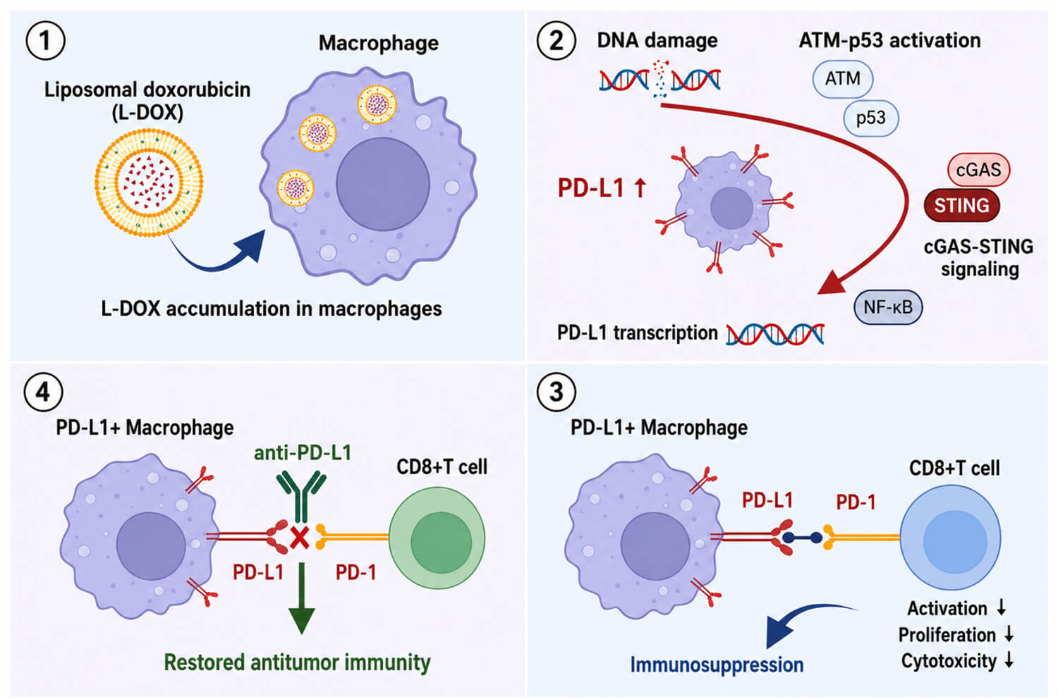
Objective: Liposomal doxorubicin (L-DOX) may alter macrophage-mediated immune regulation in triple-negative breast cancer (TNBC), but its role in programmed death-ligand 1 (PD-L1)-associated immune escape remains unclear. This study aimed to determine whether L-DOX induces a macrophage-centered PD-L1 response and affects the efficacy of PD-L1 blockade in TNBC. Methods: Public bulk and single-cell transcriptomic datasets, bone marrow-derived macrophage models, CD8+ T-cell co-culture assays, promoter-binding analyses, and syngeneic EO771 and 4T1 TNBC mouse models were used to examine PD-L1 regulation and immune function after L-DOX treatment. Results: The principal findings were that L-DOX preferentially induced a PD-L1-high macrophage state and sensitized TNBC tumors to PD-L1 blockade. DOX-containing therapy was associated with PD-L1 upregulation enriched in tumor-associated macrophages, and L-DOX induced stronger macrophage PD-L1 expression than free DOX or taxane-based agents. Mechanistically, L-DOX accumulation triggered DNA damage-associated ATM-p53 and cGAS-STING signaling, leading to RELA/p65-dependent NF-κB activation and PD-L1 transcription. Functionally, L-DOX-conditioned macrophages suppressed CD8+ T-cell activation, proliferation, and tumor-cell killing, whereas PD-L1 blockade restored CD8+ effector function, promoted pro-inflammatory macrophage features, and improved tumor control in EO771 and 4T1 models compared with either monotherapy. Conclusion: L-DOX induces a PD-L1-high macrophage state with immunosuppressive features that constrains antitumor T-cell responses. Combining L-DOX with PD-L1 blockade may provide a rational chemoimmunotherapy strategy for TNBC.Graphic Abstract
Keywords
Supplementary Material
Supplementary Material FileCite This Article
 Copyright © 2026 The Author(s). Published by Tech Science Press.
This work is licensed under a Creative Commons Attribution 4.0 International License , which permits unrestricted use, distribution, and reproduction in any medium, provided the original work is properly cited.
 Downloads
 Citation Tools
