
 Login
Login Register
 Submit a Paper
 Propose a Special lssue
 Open Access
ARTICLE

Exploring the Therapeutic Potential of Horwoodia dicksoniae through Phytochemical Profiling, Antioxidant Assessment, and Biological Activity Evaluation
Department of Zoology, College of Science, King Saud University, Riyadh, Saudi Arabia
* Corresponding Author: Aiman Ammari. Email:
(This article belongs to the Special Issue: Medicinal Plants: Phytochemical and Pharmacological Research)
Phyton-International Journal of Experimental Botany 2026, 95(8), 8 https://doi.org/10.32604/phyton.2026.087258
Received 13 June 2026; Accepted 10 July 2026; Issue published 28 August 2026
 View Full Text
 Download PDF
Abstract
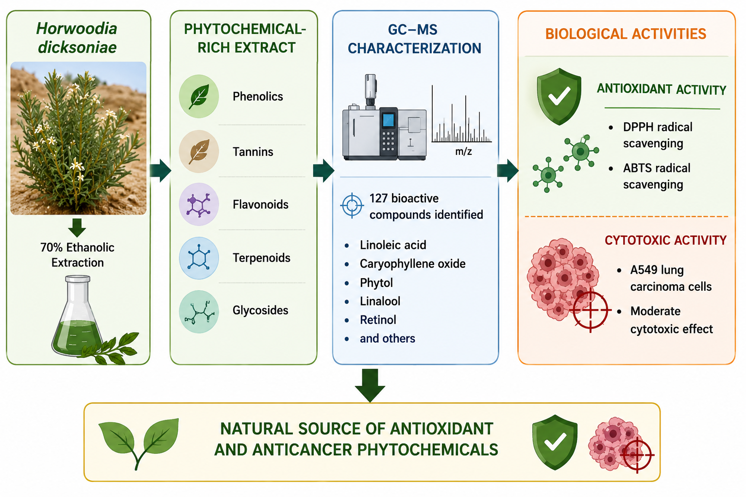
Medicinal plants are valuable sources of bioactive compounds with diverse pharmacological properties. However, the phytochemical composition and biological activities of Horwoodia dicksoniae remain insufficiently investigated. Therefore, this study evaluated the phytochemical composition, antioxidant activity, physicochemical characteristics, and in vitro cytotoxic potential of the ethanolic extract of H. dicksoniae collected from the Thumamah area, Riyadh, Saudi Arabia. The extract was subjected to phytochemical characterization, chemical profiling, physicochemical analysis, antioxidant activity assessment, and cytotoxicity evaluation. Qualitative screening confirmed the presence of phenolics, flavonoids, tannins, terpenoids, and glycosides, while quantitative analysis revealed high levels of phenolic and tannin constituents. The extract exhibited notable antioxidant activity, with half-maximal inhibitory concentration (IC50) values of 500.65 ± 2.54 and 227.52 ± 6.85 μg/mL. Chemical profiling identified 127 phytochemical constituents, predominantly fatty acids, terpenoids, esters, and oxygenated hydrocarbons, with 9,12-octadecadienoic acid (Z,Z)-, cis-(-)-1,2-epoxy-p-menth-8-ene, n-hexadecanoic acid, caryophyllene oxide, phytol, linalool, and retinol as the major compounds. Physicochemical analysis indicated satisfactory dispersion stability, while cytotoxicity testing against A549 cells demonstrated moderate growth inhibition, although the IC50 value was not reached within the tested concentration range. These findings suggest that H. dicksoniae is a promising source of bioactive phytochemicals with antioxidant potential and warrants further investigation for pharmaceutical applications.Graphic Abstract
Keywords
Supplementary Material
Supplementary Material FileMedicinal plants represent an important reservoir of naturally occurring bioactive compounds and have attracted considerable scientific attention because of their diverse pharmacological and therapeutic properties [1]. Plant-derived metabolites continue to contribute significantly to modern drug development owing to their chemical diversity and broad range of biological functions. Numerous secondary metabolites, including phenolics, flavonoids, alkaloids, tannins, terpenoids, and glycosides, are known to exert antioxidant, antimicrobial, anti-inflammatory, antiparasitic, and anticancer effects, making medicinal plants valuable candidates for pharmaceutical and biomedical applications [1,2,3,4]. Natural products continue to represent an important source of lead compounds for drug discovery and clinical development, with many approved drugs being directly derived from or inspired by naturally occurring secondary metabolites [5,6]. Reactive oxygen species (ROS) generated during oxidative stress are strongly implicated in the pathogenesis of many chronic disorders, including cancer, cardiovascular diseases, diabetes, and neurodegenerative conditions. Natural antioxidants derived from plants, particularly phenolic and flavonoid compounds, play an essential role in scavenging free radicals and minimizing oxidative damage to cellular macromolecules. For this reason, medicinal plants enriched with antioxidant phytochemicals have become increasingly important in the search for safer natural therapeutic agents [7,8,9,10,11]. Flavonoids have been extensively reported to regulate oxidative stress and inflammatory pathways associated with several human diseases, further supporting their therapeutic importance as natural bioactive compounds [12,13]. Horwoodia dicksoniae is considered one of the rare medicinal species native to the Arabian Peninsula and belongs to the Brassicaceae family, a group recognized for its richness in biologically active sulfur-containing compounds, glucosinolates, phenolics, and flavonoids. Previous investigations have demonstrated that plants belonging to this family possess significant antioxidant and anticancer potential due to their chemically active constituents. Horwoodia dicksoniae has attracted interest because of its reported phytochemical constituents, potential biological activities, as well as morphological and distribution features [2,14]. Plants belonging to the Brassicaceae family are widely recognized for their richness in glucosinolates, flavonoids, phenolic compounds, and sulfur-containing metabolites that exhibit important biological and pharmacological properties [15,16]. Recent studies have highlighted the chemopreventive and anticancer activities of glucosinolates and their hydrolysis products, particularly isothiocyanates, which contribute significantly to the medicinal value of cruciferous plants [3,15,16,17,18]. Recent investigations on H. dicksoniae have demonstrated promising antimicrobial, antioxidant, and anticancer activities, highlighting its potential as a valuable medicinal resource [1].
Phytochemical screening is considered an essential step in evaluating the medicinal potential of plant extracts because it enables the identification of biologically active secondary metabolites associated with therapeutic activities. Recent advances in analytical technologies have significantly improved the characterization of phytochemicals in medicinal plants. Gas chromatography–mass spectrometry (GC–MS) is among the most powerful analytical techniques used for identifying volatile and semi-volatile compounds in plant extracts. GC–MS profiling provides comprehensive information regarding the chemical composition of medicinal plants and facilitates the identification of compounds associated with biological activities such as antioxidant and anticancer effects [19,20]. Recent studies have further demonstrated the utility of GC–MS in characterizing bioactive metabolites from medicinal plants and correlating their chemical composition with biological activities [21]. Previous studies have identified several biologically active compounds, including fatty acids, esters, terpenoids, alkaloids, and phenolic derivatives, which contribute significantly to the pharmacological properties of medicinal plants. Ref. [7] demonstrated that Rhazya stricta collected from Riyadh, Saudi Arabia, possessed substantial antioxidant activity associated with its phytochemical composition and biological efficacy against Eimeria perforans. Similarly, Ref. [22] reported that aqueous extracts of Trigonella foenum-graecum seeds exhibited notable antioxidant and cytotoxic activities related to their phytochemical constituents. These findings emphasize the importance of phytochemical characterization in understanding the biological and therapeutic properties of medicinal plants. Fourier-transform infrared spectroscopy (FTIR) is another important analytical tool widely used for identifying functional groups and characterizing molecular structures within plant extracts. FTIR analysis enables the detection of alcohols, phenols, aldehydes, ketones, amines, carboxylic acids, and aromatic compounds, thereby providing valuable information regarding the chemical nature of phytochemicals. Recent applications of FTIR spectroscopy have proven valuable for the structural characterization and identification of bioactive phytochemicals in medicinal plants [21]. In addition, physicochemical characterization through zeta potential analysis has become increasingly important in pharmaceutical and biomedical studies. Zeta potential is considered a critical indicator of particle surface charge and colloidal stability, influencing aggregation behavior, dispersion characteristics, and biological interactions. Stable systems with suitable zeta potential values are generally associated with improved colloidal stability, which may facilitate biological performance in pharmaceutical applications [23].
Cytotoxicity assessment using in vitro cancer cell lines remains one of the most important approaches for evaluating the anticancer potential of medicinal plant extracts. Several medicinal plants rich in phenolic and flavonoid compounds have demonstrated selective cytotoxicity against human cancer cell lines through mechanisms involving apoptosis induction and inhibition of cell proliferation [2,4]. Likewise, H. dicksoniae extracts have shown promising cytotoxic activities against different human cancer cell lines, supporting their potential application as natural anticancer agents [1].
Although preliminary studies have reported antimicrobial, antioxidant, and anticancer activities for Horwoodia dicksoniae, comprehensive information integrating its phytochemical composition with physicochemical characterization and biological evaluation remains scarce. In particular, no single study has comprehensively investigated the qualitative and quantitative phytochemical profile together with Gas chromatography–mass spectrometry (GC–MS) analysis, Fourier-transform infrared spectroscopy (FTIR) characterization, zeta potential evaluation, antioxidant activity, and cytotoxic potential of the ethanolic extract of H. dicksoniae. This knowledge gap limits a comprehensive understanding of the relationship between its chemical constituents and biological activities. Therefore, the novelty of the present study lies in integrating these complementary analytical and biological approaches to provide a more comprehensive evaluation of the therapeutic potential of this medicinal plant.
Despite the increasing interest in medicinal plants as sources of natural bioactive compounds, comprehensive information regarding the phytochemical composition and biological properties of Horwoodia dicksoniae remains limited. Therefore, the present study was designed to provide a comprehensive phytochemical characterization of the ethanolic extract of H. dicksoniae collected from the Thumamah region, Riyadh, Saudi Arabia. Specifically, the study aimed to identify its major phytochemical constituents through qualitative screening, quantify total phenolic, flavonoid, and tannin contents, characterize its chemical composition using gas chromatography–mass spectrometry (GC–MS) and Fourier-transform infrared spectroscopy (FTIR), evaluate its physicochemical stability by zeta potential analysis, determine its antioxidant capacity using 2,2-diphenyl-1-picrylhydrazyl (DPPH) and 2,2′-azinobis-(3-ethylbenzothiazoline-6-sulfonic acid) (ABTS) assays, and investigate its cytotoxic activity against the human lung adenocarcinoma cell line (A549). The findings are expected to expand the current knowledge of this native species and provide a scientific basis for its potential pharmaceutical and therapeutic applications.
2.1 Materials and Plant Collection
Dried aerial parts of Horwoodia dicksoniae were obtained from a well-known herbal market (Attar shop) in Riyadh, Saudi Arabia. Ethanol (70%, analytical grade) was used as the extraction solvent. Analytical-grade potassium bromide (KBr) was employed for Fourier-transform infrared spectroscopy (FTIR) analysis. Cell culture and cytotoxicity experiments were performed using Dulbecco’s Modified Eagle’s Medium (DMEM), fetal bovine serum (FBS), penicillin–streptomycin solution, trypsin, phosphate-buffered saline (PBS), dimethyl sulfoxide (DMSO), and 3-(4,5-dimethylthiazol-2-yl)-2,5-diphenyltetrazolium bromide (MTT) reagent. Unless otherwise stated, all chemicals, reagents, and culture media used throughout the study were of analytical grade or cell-culture grade and were obtained from standard commercial suppliers.
2.2 Plant Collection and Preparation
Dried specimens of Horwoodia dicksoniae were purchased from a well-known herbal market (Attar shop) in Riyadh, Saudi Arabia. The plant material was obtained in its dried form as commercially available herbal material. The purchased material was carefully examined and cleaned to remove adhering impurities and foreign matter before being ground into a fine homogeneous powder using a laboratory mill and stored in sterile airtight containers until further extraction and analysis.
Extraction of the powdered plant material was performed using 70% ethanol via a maceration-based extraction approach. The powdered samples were soaked in the solvent and continuously agitated to enhance the diffusion of phytochemical constituents into the extraction medium. The extraction process was conducted at low temperature (4°C) for 24 h to minimize degradation of thermolabile metabolites. After incubation, the mixture was subjected to centrifugation at 5000 rpm for 15 min to remove insoluble plant residues. The resulting liquid phase was carefully separated and filtered through Whatman No. 1 filter paper before being concentrated under reduced pressure at 50°C using a rotary evaporator until complete removal of the solvent. Finally, the concentrated extract was lyophilized to obtain a dry crude extract and preserved at −80°C until subsequent experimental analyses.
2.4 Infrared Spectroscopic Analysis of Horwoodia Dicksoniae
Fourier-transform infrared spectroscopy (FTIR) characterization was carried out to determine the major functional groups present in the plant extract. For spectral preparation, a small quantity of the dried extract was blended thoroughly with analytical-grade potassium bromide (KBr) until a uniform mixture was obtained. The prepared mixture was then compressed into thin pellets suitable for infrared scanning. Spectral acquisition was performed using a Thermo Scientific FTIR spectrometer (NICOLET 6700) across the mid-infrared region between 4000 and 400 cm−1 under controlled laboratory conditions. The obtained spectra were processed to identify characteristic absorption bands corresponding to the chemical functionalities of the detected phytochemicals. Interpretation of the spectral peaks enabled prediction of the major phytochemical classes present within the extract according to their vibrational characteristics.
Qualitative phytochemical evaluation of the ethanolic extract was undertaken to investigate the occurrence of important secondary metabolites. Standard qualitative phytochemical procedures were employed for the detection of alkaloids (Mayer’s test), flavonoids (alkaline reagent test), phenolic compounds (ferric chloride test), tannins (gelatin test), saponins (frothing test), terpenoids (Salkowski test), glycosides (Keller–Killiani test), and steroids (Liebermann–Burchard test). The qualitative analyses were performed according to well-established procedures described in our recent publication [2], and the same methodology was followed in the present study with minor modifications.
2.6 Quantification of Total Phenolic, Flavonoid, and Tannin Contents
Total phenolic content (TPC), total flavonoid content (TFC), and total tannin content (TTC) were quantified using established spectrophotometric methods. TPC was determined using the Folin–Ciocalteu assay and expressed as mg gallic acid equivalents (GAE)/g dry extract. TFC was measured using the aluminum chloride colorimetric assay and expressed as mg quercetin equivalents (QE)/g dry extract, whereas TTC was determined using the Folin–Ciocalteu-based method and expressed as mg tannic acid equivalents (TAE)/g dry extract. Detailed experimental procedures have been described in our recent publication [2], and the same methodology was followed in the present study with minor modifications.
2.7 Gas Chromatography–Mass Spectrometry (GC–MS) Analysis
The chemical constituents present in the extract were characterized using gas chromatography coupled with mass spectrometry (GC–MS). This analytical approach was employed to identify volatile and semi-volatile metabolites present in the extract. GC–MS analysis was performed using an Agilent Technologies GC–MS 7890B gas chromatography system coupled with a mass selective detector (Agilent Technologies, Santa Clara, CA, USA) under optimized analytical conditions. Separation was achieved using a DB-5 MS capillary column (30 m × 0.25 mm i.d., 0.25 μm film thickness) with helium as the carrier gas at a flow rate of 1.0 mL/min. Compound identification was carried out by comparing the obtained mass spectra with the National Institute of Standards and Technology (NIST) Mass Spectral Library, and the identified compounds were considered tentatively identified based on library matching.
The physicochemical properties of the Horwoodia dicksoniae ethanolic extract were evaluated by zeta potential analysis to determine the surface charge and colloidal stability of the dispersed particles. For sample preparation, the lyophilized extract was dispersed in deionized water at a final concentration of 0.1 mg/mL. The suspension was ultrasonicated for 60 min to obtain a homogeneous and stable dispersion while minimizing particle aggregation. The prepared dispersion was allowed to equilibrate at room temperature prior to analysis, and no surfactants or stabilizing agents were added. Zeta potential measurements were subsequently performed using electrophoretic light scattering under ambient laboratory conditions. The obtained zeta potential values were used to evaluate the surface charge characteristics and dispersion stability of the extract particles.
The anticancer potential of the Horwoodia dicksoniae extract was assessed using the human lung adenocarcinoma cell line (A549). Cells were propagated in DMEM enriched with 10% fetal bovine serum and supplemented with penicillin–streptomycin to prevent microbial contamination. The cultured cells were maintained under humidified incubation conditions at 37°C in a 5% CO2 atmosphere. Culture media were routinely refreshed every 48 h to sustain healthy cell growth, while subculturing procedures were carried out once the cells reached near-confluent growth conditions.
The cytotoxic activity of the plant extract against Human lung adenocarcinoma cell line (A549) cells was evaluated using the MTT colorimetric method. Prior to treatment, adherent cells were washed with sterile phosphate-buffered saline (PBS) and detached from the culture surface using trypsin solution. The collected cells were subsequently resuspended in complete growth medium and quantified before experimental seeding. For the assay, approximately 1 × 10⁴ cells in 100 μL of culture medium were transferred into each well of a 96-well plate and maintained under standard incubation conditions for 24 h to permit proper cellular attachment. The extract solutions were freshly prepared in DMEM medium and homogenized by sonication to improve dispersion and reduce particle aggregation. Different concentrations of the extract were then prepared and applied to the cultured cells after replacing the old culture medium with fresh treatment medium. Following exposure to the extract, the treated cells were incubated under controlled conditions to determine the cellular response. Subsequently, MTT reagent prepared in serum-free medium was introduced into each well and incubated to allow viable cells to convert the reagent into insoluble purple formazan crystals through mitochondrial metabolic activity. After completion of the reaction, the medium was carefully discarded, and DMSO was added to solubilize the produced crystals. The absorbance was recorded at 570 nm using a microplate reader, and the percentage of viable cells was calculated relative to untreated control cells.
All measurements were performed in triplicate, and the results are presented as mean ± standard error (SE). Data processing, descriptive statistical calculations, and graphical presentation were carried out using Microsoft Excel 365 (Microsoft Corporation, Redmond, WA, USA).
3.1 Quantification of Bioactive Phytochemical Constituents
In Table 1 and Fig. 1 The quantitative phytochemical evaluation demonstrated that the ethanolic extract of Horwoodia dicksoniae was rich in phenolic and tannin compounds. Among the measured phytochemical groups, total phenolic content (TPC) showed the highest value compared with total tannin content (TTC) and total flavonoid content (TFC). The elevated levels of these phytochemicals suggest their possible contribution to the antioxidant and biological properties exhibited by the extract.
Table 1: Total phenolic content (TPC), total tannin content (TTC), and total flavonoid content (TFC) of Horwoodia dicksoniae extract expressed as mg GAE/g and mg QE/g dry extract.
| Extract | TPC (mg GAE/g) | TTC (mg GAE/g) | TFC (mg QE/g) |
|---|---|---|---|
| Horwoodia dicksoniae | 29.55 ± 1.06 | 21.88 ± 0.44 | 4.21 ± 0.74 |

Figure 1: Calibration curves of total phenolic content (TPC), total tannin content (TTC), and total flavonoid content (TFC) standards used for the quantitative phytochemical analysis of Horwoodia dicksoniae extract. The calibration plots demonstrated excellent linearity with high correlation coefficients (R2), confirming the reliability and accuracy of the spectrophotometric quantification methods.
3.2 Antioxidant Activity of Horwoodia Dicksoniae Extract
The antioxidant potential of the ethanolic extract was evaluated using Diphenyl-1-picrylhydrazyl (DPPH) and 2,2′-azinobis-(3-ethylbenzothiazoline-6-sulfonic acid) (ABTS) radical scavenging assays. The extract exhibited measurable free radical scavenging activity in both assays, with half-maximal inhibitory concentration (IC50) values of 500.65 ± 2.54 μg/mL for DPPH and 227.52 ± 6.85 μg/mL for ABTS. The lower IC50 value obtained in the ABTS assay indicates a stronger antioxidant activity than that observed in the DPPH assay, reflecting the antioxidant potential of the phytochemical constituents present in the extract.
3.3 Gas Chromatography–Mass Spectrometry (GC–MS) Analysis of Horwoodia Dicksoniae Extract
GC–MS analysis identified a total of 127 phytochemical constituents in the ethanolic extract of Horwoodia dicksoniae. As summarized in Table 2 and represented by the major chromatographic peaks in Fig. 2, the predominant compounds included 9,12-octadecadienoic acid (Z,Z)-, cis-(-)-1,2-epoxy-p-menth-8-ene, n-hexadecanoic acid, 2H-1-benzopyran-2-one, 7-methoxy-, caryophyllene oxide, phytol, linalool, retinol, and octadecanoic acid. The complete GC–MS dataset, including all identified compounds and their corresponding analytical data, is provided in the Supplementary Materials.
Table 2: Major bioactive compounds identified by gas chromatography–mass spectrometry (GC–MS) analysis of the ethanolic extract of Horwoodia dicksoniae, together with their retention times (RT), molecular formulas, molecular weights (MW), and relative peak areas.
| RT (min) | Compound | Molecular Formula | MW | Relative Peak Area (%) |
|---|---|---|---|---|
| 42.191 | 9,12-Octadecadienoic acid (Z,Z)- | C18H32O2 | 280 | 9.86 |
| 19.854 | cis-(-)-1,2-Epoxy-p-menth-8-ene | C10H16O | 152 | 6.51 |
| 38.066 | n-Hexadecanoic acid | C16H32O2 | 256 | 5.00 |
| 31.519 | 2H-1-Benzopyran-2-one, 7-methoxy- | C10H8O3 | 176 | 3.17 |
| 15.092 | (E)-2,6-Dimethylocta-3,7-diene-2,6-diol | C10H18O2 | 170 | 2.38 |
| 27.223 | Caryophyllene oxide | C15H24O | 220 | 2.24 |
| 42.589 | Octadecanoic acid | C18H36O2 | 284 | 1.40 |
| 11.498 | Linalool | C10H18O | 154 | 0.92 |
| 40.946 | Phytol | C20H40O | 296 | 0.48 |
| 42.889 | Retinol | C20H30O | 286 | 0.69 |

Figure 2: GC–MS chromatogram of the ethanolic extract of Horwoodia dicksoniae showing the major chromatographic peaks corresponding to the principal phytochemical constituents identified according to their retention times (RT). The chromatographic profile revealed a chemically diverse composition comprising fatty acids, terpenoids, oxygenated hydrocarbons, phenolic derivatives, and other volatile and semi-volatile compounds, highlighting the phytochemical complexity of the extract.
3.4 FTIR Spectral Analysis of Horwoodia Dicksoniae Extract
As presented in Fig. 3 and summarized in Table 3, The FTIR spectrum of the ethanolic extract exhibited several characteristic absorption peaks corresponding to diverse functional groups associated with biologically active phytochemicals. A broad absorption band detected at 3418.16 cm−1 was assigned to O–H stretching vibrations, suggesting the presence of hydroxyl-containing compounds such as phenolics and alcohols. Peaks observed at 2928.76 cm−1 and 2859.58 cm−1 were attributed to aliphatic C–H stretching vibrations related to hydrocarbon and alkane structures.
A strong absorption signal at 1735.91 cm−1 indicated carbonyl (C=O) stretching vibrations commonly associated with aldehydes, ketones, esters, and fatty acid derivatives. Additional bands appearing at 1689.51 cm−1 and 1635.95 cm−1 corresponded to C=C stretching vibrations of aromatic and unsaturated compounds. Furthermore, the absorption peaks detected at 1515.26 cm−1, 1459.88 cm−1, and 1375.33 cm−1 suggested the occurrence of aromatic constituents and methyl bending vibrations.
The spectral region extending from 1321.49 cm−1 to 1072.83 cm−1 was mainly associated with C–O stretching vibrations characteristic of alcohols, ethers, esters, and phenolic compounds. In the lower wavenumber region, peaks ranging between 823.98 cm−1 and 519.11 cm−1 reflected bending vibrations of substituted aromatic structures and other complex metabolites. Collectively, the corresponding functional group assignments for the major absorption bands are summarized in Table 3. the FTIR findings confirmed the occurrence of multiple functional groups corresponding to phenolics, terpenoids, fatty acids, aldehydes, alcohols, and aromatic compounds, emphasizing the chemical diversity and potential biological importance of the extract. The complete FTIR dataset, including all identified compounds and their corresponding analytical data, is provided in the Supplementary Materials.

Figure 3: FTIR spectrum of the ethanolic extract of Horwoodia dicksoniae showing the major absorption peaks and corresponding functional groups.
Table 3: Major FTIR absorption bands and their corresponding functional group assignments for the ethanolic extract of Horwoodia dicksoniae.
| Functional Group | Probable Assignment | Wavenumber (cm−1) |
|---|---|---|
| O–H stretching | Phenols and alcohols | 3418.16 |
| C–H stretching | Aliphatic hydrocarbons | 2928.76 |
| C–H stretching | Alkanes | 2859.58 |
| C=O stretching | Esters and carboxylic acids | 1735.91 |
| C=C/C=O stretching | Conjugated carbonyl compounds | 1689.51 |
| C=C stretching | Aromatic compounds | 1635.95 |
| Aromatic skeletal vibration | Aromatic rings | 1515.26 |
| CH2 bending | Aliphatic compounds | 1459.88 |
| C–H bending | Methyl groups | 1375.33 |
| C–O stretching | Phenolic compounds | 1321.49 |
| C–O stretching | Ethers and esters | 1245.51 |
| C–O stretching | Alcohols | 1158.29 |
| C–O stretching | Secondary alcohols | 1072.83 |
| C–H out-of-plane bending | Aromatic compounds | 823.98 |
| Aromatic C–H bending | Aromatic rings | 775.70 |
| Fingerprint region | Complex skeletal vibrations of organic compounds | 609.31–409.11 |
3.5 Zeta Potential Analysis of Horwoodia Dicksoniae Extract
In Fig. 4 The physicochemical stability and surface charge characteristics of the ethanolic extract of Horwoodia dicksoniae were evaluated using zeta potential analysis. The obtained zeta potential value was −23.9 mV, indicating that the extract particles possessed a moderately negative surface charge. This negative charge contributes to electrostatic repulsion between particles, thereby enhancing colloidal stability and reducing particle aggregation. The zeta potential distribution profile exhibited a single dominant peak with a mean value of −13.0 mV and a standard deviation of 8.01 mV, suggesting a relatively homogeneous particle distribution within the extract system. Additionally, the conductivity value was recorded as 0.662 mS/cm, reflecting the ionic characteristics of the suspension medium. Overall, the obtained zeta potential results indicate that the Horwoodia dicksoniae extract possesses acceptable physicochemical stability, which may positively influence its biological interactions and therapeutic performance.

Figure 4: Zeta potential distribution profile of the ethanolic extract of Horwoodia dicksoniae showing particle surface charge and stability characteristics.
3.6 Cytotoxic Activity of Horwoodia Dicksoniae Extract against Human Lung Adenocarcinoma Cell Line (A549) Cells
In the Table 4 and Fig. 5 The anticancer potential of the ethanolic extract was investigated against A549 human lung carcinoma cells using the MTT assay. The experimental findings indicated that the extract exerted moderate inhibitory effects on cancer cell viability across the tested concentrations. Treatment with the extract resulted in a measurable decline in cell survival, with the strongest inhibitory response observed at 10 μg/mL, where cell viability reached 78.82%. At concentrations above 10 μg/mL, the cytotoxic response did not exhibit a clear dose-dependent pattern, as cell viability gradually increased. This observation may reflect the complex composition of the crude ethanolic extract, in which multiple phytochemicals with different biological activities may interact and influence the overall cellular response. Therefore, additional studies using purified fractions and a broader concentration range are required to clarify this phenomenon.
Although a reduction in cellular viability was detected, none of the tested concentrations reduced cell survival to below 50%; therefore, the Half-maximal inhibitory concentration (IC50) value could not be established within the examined concentration range. The extract exhibited a non-dose-dependent cytotoxic response, with the greatest reduction in cell viability observed at 10 μg/mL. At higher concentrations (25–200 μg/mL), cell viability showed a gradual increase; however, none of the tested concentrations reduced cell viability below 50%, and therefore the IC50 value could not be determined within the investigated concentration range. The observed cytotoxic response may be linked to the presence of several bioactive phytochemicals identified through Gas chromatography–mass spectrometry (GC–MS) analysis, particularly terpenoids, phenolic metabolites, fatty acid derivatives, and oxygenated compounds that are widely recognized for their potential anticancer and growth-inhibitory activities.
Table 4: Effect of Horwoodia dicksoniae extract on the viability of Human lung adenocarcinoma cell line (A549) cells determined by the MTT assay.
| Concentration (μg/mL) | Cell Viability (%) |
|---|---|
| Control | 100 |
| 5 | 97.68 |
| 10 | 78.82 |
| 25 | 83.48 |
| 50 | 87.96 |
| 100 | 88.46 |
| 200 | 96.35 |
The lowest cell viability percentage was observed at the concentration of 10 μg/mL, indicating the highest cytotoxic effect of the extract against A549 cells under the tested experimental conditions. These findings suggest that the phytochemical constituents present in Horwoodia dicksoniae may contribute to its potential anticancer activity.

Figure 5: Effect of the ethanolic extract of Horwoodia dicksoniae on the viability of A549 human lung carcinoma cells as determined by the MTT assay. Data are presented as percentage of viable cells relative to untreated control cells.
The phytochemical characterization of the ethanolic extract of Horwoodia dicksoniae revealed a chemically diverse profile containing multiple classes of bioactive secondary metabolites. Qualitative phytochemical screening confirmed the presence of phenolics, flavonoids, tannins, terpenoids, and glycosides, while Gas chromatography–mass spectrometry (GC–MS) analysis identified 127 compounds belonging mainly to fatty acids, terpenoids, alcohols, aldehydes, esters, and oxygenated hydrocarbons. Such phytochemical diversity is commonly associated with medicinal plants exhibiting important biological and therapeutic properties and may contribute to a wide range of pharmacological activities through the synergistic action of multiple secondary metabolites [1,5,6,21].
The Gas chromatography–mass spectrometry (GC–MS) results demonstrated that the predominant compounds in the extract included 9,12-octadecadienoic acid (Z,Z)-, n-hexadecanoic acid, caryophyllene oxide, phytol, linalool, and retinol. These compounds have previously been reported to possess antioxidant, antimicrobial, anti-inflammatory, and anticancer activities [24]. Fatty acids and oxygenated terpenoids, in particular, are considered major contributors to the biological activity of medicinal plant extracts because of their ability to modulate oxidative stress and cellular membrane integrity. The present results are consistent with those reported by [24], who demonstrated that Gas chromatography–mass spectrometry (GC–MS) analysis of plant-derived oils identified numerous biologically active constituents associated with significant antibacterial and antioxidant activities. Their study emphasized the biological importance of fatty acids, terpenoids, and oxygenated hydrocarbons as dominant phytochemical constituents in medicinal plants. Similarly, the current investigation identified several compounds belonging to these chemical classes, suggesting that the biological activities of H. dicksoniae may result from the synergistic interaction of its phytochemical metabolites. Comparable observations have also been reported in medicinal plants characterized using advanced chromatographic and spectroscopic approaches, where chemically diverse metabolite profiles were associated with multiple biological activities [21]. The antioxidant activity demonstrated by the H. dicksoniae extract in Diphenyl-1-picrylhydrazyl (DPPH) and 2,2′-Azinobis-(3-ethylbenzothiazoline-6-sulfonic acid) (ABTS) assays may be strongly associated with the abundance of phenolic compounds and oxygenated metabolites identified during phytochemical and Gas chromatography–mass spectrometry (GC–MS) analyses. Phenolic compounds are widely recognized for their ability to neutralize reactive oxygen species and reduce oxidative stress, thereby protecting cells from oxidative damage and preventing cellular dysfunction [12,13,25].
The relatively high total phenolic content (TPC), total flavonoid content (TFC), and total tannin content (TTC) determined in Horwoodia dicksoniae may collectively explain the pronounced free radical scavenging activity observed in both the DPPH and ABTS assays. Among these phytochemical groups, phenolic compounds are generally considered the principal contributors to antioxidant capacity because of their ability to donate hydrogen atoms or electrons and neutralize reactive oxygen species. Flavonoids and tannins Similar positive relationships between elevated phenolic and flavonoid contents and enhanced DPPH and ABTS radical scavenging activities have been consistently reported in medicinal plant extracts, supporting the interpretation that the antioxidant activity observed in the present study is largely attributable to these phytochemical constituents [2,7,22], which represent important subclasses of phenolic compounds, further enhance antioxidant activity through complementary mechanisms, including radical scavenging, metal chelation, and inhibition of oxidative chain reactions. Flavonoids, in particular, have been reported to modulate oxidative stress pathways, inhibit free radical generation, and enhance endogenous antioxidant defense systems, which may contribute significantly to the observed antioxidant activity of the extract [12]. Fourier-transform infrared spectroscopy (FTIR) analysis further supported the phytochemical complexity of the extract through the identification of characteristic functional groups corresponding to hydroxyl, carbonyl, aromatic, and ether-containing compounds. The coexistence of these functional groups confirms the presence of phenolics, fatty acids, terpenoids, and related secondary metabolites identified by GC–MS analysis. Similar Fourier-transform infrared spectroscopy (FTIR) profiles have been previously associated with medicinal plants possessing broad biological and pharmacological activities [2,21]. The complementary use of FTIR and GC–MS techniques provides valuable information regarding both functional group composition and individual phytochemical constituents, thereby strengthening the overall characterization of medicinal plant extracts [21]. The zeta potential analysis demonstrated that the extract possessed a moderately negative surface charge, indicating acceptable colloidal stability and dispersion characteristics. Physicochemical stability is considered an important factor influencing the interaction of bioactive compounds with cellular membranes and may contribute to enhanced biological efficacy. Stable dispersions may improve the bioavailability and therapeutic performance of plant-derived phytochemicals [23]. Such physicochemical properties are increasingly recognized as important considerations in the pharmaceutical development of plant-derived products and phytopharmaceutical formulations [26,27]. The cytotoxicity assay against A549 human lung carcinoma cells demonstrated moderate anticancer activity of the extract. Although the Half-maximal inhibitory concentration (IC50) value was not reached within the tested concentration range, the reduction in cell viability indicates that the extract contains compounds capable of interfering with cancer cell growth and metabolism. Several compounds identified in the current study, including phytol, caryophyllene oxide, and unsaturated fatty acids, have previously been associated with apoptosis induction and inhibition of tumor cell proliferation [4,22,28,29,30]. Recent studies demonstrated that phytol exhibits significant antiproliferative activity against A549 lung cancer cells, whereas terpenoid compounds such as β-caryophyllene and its derivatives may exert anticancer effects through apoptosis induction, oxidative stress modulation, and inhibition of angiogenesis [29,30]. Therefore, the observed cytotoxic activity may result from the synergistic effects of multiple phytochemical constituents present in the extract rather than the action of a single compound. The non-dose-dependent cytotoxic response observed at concentrations above 10 μg/mL may be attributed to the complex chemical composition of the crude ethanolic extract, in which multiple phytochemicals with different biological activities may interact and influence the overall cellular response. Furthermore, variations inherent to in vitro MTT assays may also contribute to fluctuations in cell viability at different concentrations. Therefore, additional studies using purified fractions, a broader concentration range, and complementary cytotoxicity assays are warranted to clarify the underlying mechanism.
Overall, the present findings support the growing evidence that medicinal plants native to Saudi Arabia represent promising natural sources of biologically active compounds with antioxidant and anticancer potential. The phytochemical richness and biological activities of Horwoodia dicksoniae suggest that this plant represents a promising source of natural bioactive compounds with potential pharmaceutical and biomedical applications, warranting further investigation to isolate and characterize its active constituents and elucidate their underlying mechanisms of action. Furthermore, future studies focusing on phytopharmaceutical formulation strategies, advanced drug delivery systems, and comprehensive in vitro and in vivo biological evaluations may improve the bioavailability, stability, and therapeutic efficacy of the identified bioactive compounds, thereby facilitating their potential pharmaceutical development [26,27].
The present study demonstrated that the ethanolic extract of Horwoodia dicksoniae is a rich source of bioactive phytochemicals with promising antioxidant and in vitro cytotoxic properties. Phytochemical characterization confirmed the presence of several important secondary metabolites, while GC–MS and FTIR analyses revealed a chemically diverse profile comprising multiple classes of bioactive constituents. The extract exhibited measurable antioxidant activity in both DPPH and ABTS assays and moderate cytotoxic activity against A549 human lung adenocarcinoma cells, although the IC50 value was not reached within the tested concentration range. Collectively, these findings suggest that H. dicksoniae may represent a valuable natural source of biologically active compounds with potential pharmaceutical applications. Further studies are warranted to isolate and characterize the active constituents, investigate their mechanisms of action, and evaluate their efficacy and safety in appropriate in vivo models.
Acknowledgement:
Funding Statement: This work was supported by the Ongoing Research Funding Program (ORF-2026-1490), King Saud University, Riyadh, Saudi Arabia.
Author Contributions: Aiman Ammari: Conceptualization, Methodology, Investigation, Data Curation, Formal Analysis, Writing—Original Draft, Visualization. Ramzi Amran: Methodology, Validation, Resources, Writing—Review & Editing. Ahmad Alhimaidi: Supervision, Project Administration, Validation, Writing—Review & Editing. All authors reviewed and approved the final version of the manuscript.
Availability of Data and Materials: All data generated or analyzed during this study are included in this published article and its figures and tables. In addition, the raw datasets and supporting materials used and/or analyzed during the current study are available from the corresponding author upon reasonable request.
Ethics Approval: Not applicable.
Conflicts of Interest: The authors declare no conflicts of interest.
Supplementary Materials: The supplementary material is available online at https://www.techscience.com/doi/10.32604/phyton.2026.087258/s1.
References
1. Altammar KA . Unveiling therapeutic powers of indigenous flora: Antimicrobial, antioxidant, and anticancer properties of Horwoodia dicksoniae. Pharmaceuticals. 2025; 18( 5): 765. doi:10.3390/ph18050765. [Google Scholar] [CrossRef]
2. Ammari AA , Aljawdah HM , Amran RA , Alhimaidi AR . In vitro bioactivity and phytochemical characterization of a polyherbal extract with antioxidant and anticancer properties. PeerJ. 2026; 14: e20824. doi:10.7717/peerj.20824. [Google Scholar] [CrossRef]
3. Peña M , Guzmán A , Martínez R , Mesas C , Prados J , Porres JM , et al. Preventive effects of Brassicaceae family for colon cancer prevention: A focus on in vitro studies. Biomed Pharmacother. 2022; 151: 113145. doi:10.1016/j.biopha.2022.113145. [Google Scholar] [CrossRef]
4. Mishra P , Sohrab S , Mishra SK . Pioneering insights into Lemon basil: High-resolution phytochemical profiling, antioxidant potential, and in-vitro cytotoxic activity against MCF-7 breast cancer cell lines. Next Res. 2025; 2( 3): 100661. doi:10.1016/j.nexres.2025.100661. [Google Scholar] [CrossRef]
5. Butler MS , Capon RJ , Blaskovich MAT , Henderson IR . Natural product-derived compounds in clinical trials and drug approvals. Nat Prod Rep. 2026; 43( 1): 20– 88. doi:10.1039/d5np00031a. [Google Scholar] [CrossRef]
6. Singh K , Gupta JK , Chanchal DK , Shinde MG , Kumar S , Jain D , et al. Natural products as drug leads: Exploring their potential in drug discovery and development. Naunyn Schmiedeberg’s Arch Pharmacol. 2025; 398( 5): 4673– 87. doi:10.1007/s00210-024-03622-6. [Google Scholar] [CrossRef]
7. Aljawdah HM , Murshed M , Ammari AA , Maodaa SN , Al-Quraishy S . Phytochemical analysis of Rhazya stricta collected from Riyadh, Saudi Arabia as antioxidant and against Eimeria perforans activity. Indian J Anim Res. 2025: 1545– 51. doi:10.18805/ijar.bf-1981. [Google Scholar] [CrossRef]
8. Nicolescu A , Bunea CI , Mocan A . Total flavonoid content revised: An overview of past, present, and future determinations in phytochemical analysis. Anal Biochem. 2025; 700: 115794. doi:10.1016/j.ab.2025.115794. [Google Scholar] [CrossRef]
9. Nagori M , Rajput D , Choudhary G , Khabiya R . Qualitative and quantitative methods of phytochemical analysis. In: Pharmacognosy and phytochemistry: principles, techniques, and clinical applications. New York, NY, USA: John Wiley & Sons, Inc.; 2025. doi:10.1002/9781394203680.ch08. [Google Scholar] [CrossRef]
10. Khan F , Rashan L . Phytochemical analysis and pharmaceutical applications of monoterpenoids present in the essential oil of Boswellia sacra (Omani luban). Adv Pharmacol Pharm Sci. 2025; 2025: 3536898. doi:10.1155/adpp/3536898. [Google Scholar] [CrossRef]
11. Mahmoud NN , Selim MT . Phytochemical analysis and antimicrobial activity of Silybum marianum L. via multisolvent extraction. AMB Express. 2025; 15( 1): 122. doi:10.1186/s13568-025-01925-2. [Google Scholar] [CrossRef]
12. Jomova K , Alomar SY , Valko R , Liska J , Nepovimova E , Kuca K , et al. Flavonoids and their role in oxidative stress, inflammation, and human diseases. Chem Biol Interact. 2025; 413: 111489. doi:10.1016/j.cbi.2025.111489. [Google Scholar] [CrossRef]
13. Parveen B , Rajinikanth V , Narayanan M . Natural plant antioxidants for food preservation and emerging trends in nutraceutical applications. Discover Appl Sci. 2025; 7( 8): 845. doi:10.1007/s42452-025-07464-6. [Google Scholar] [CrossRef]
14. Al-Masoudi RKH . Morphological, anatomical and geographical distribution studies of species Horwoodia dicksoniae (Turrill) in Iraq. Iraqi J Agric Sci. 2019; 50( 6): 1613– 20. doi:10.36103/ijas.v50i6.851. [Google Scholar] [CrossRef]
15. Al-Shehbaz IA . The Brassicaceae then and now: Advancements in the past three decades, a review. Ann Bot. 2025: mcaf055. doi:10.1093/aob/mcaf055. [Google Scholar] [CrossRef]
16. Labudda M , Rybarczyk-Płońska A , Sobieszek KA , Niedziński T , Wurlitzer WB , Muszyńska E , et al. The chemopreventive and anticancer potential of glucosinolates and their hydrolysis products from cruciferous vegetables. Nutrients. 2026; 18( 5): 751. doi:10.3390/nu18050751. [Google Scholar] [CrossRef]
17. Soundararajan P , Kim JS . Anti-carcinogenic glucosinolates in cruciferous vegetables and their antagonistic effects on prevention of cancers. Molecules. 2018; 23( 11): 2983. doi:10.3390/molecules23112983. [Google Scholar] [CrossRef]
18. Karirat T , Saengha W , Matra K , Nakhowong K , Pattanu P , Kitkayun P , et al. Impact of non-thermal plasma seed priming and early development stages of two local Thai Cruciferous plants mustard green and rat-tailed radish on glucosinolates, isothiocyanates, minerals, antioxidant and anticancer activities. Not Bot Horti Agrobo. 2025; 53( 1): 14149. doi:10.15835/nbha53114149. [Google Scholar] [CrossRef]
19. Shalini KS , Ilango KI . Preliminary phytochemical studies, GC-MS analysis and in vitro antioxidant activity of selected medicinal plants and its polyherbal formulation. Pharmacogn J. 2021; 13( 3): 648– 59. doi:10.5530/pj.2021.13.83. [Google Scholar] [CrossRef]
20. Nadeem M , Zarreen F , Rizvi MMA . GC-MS analysis of phytochemical composition and anticancer activities of methanol and aqueous extracts of Bergenia ligulata rhizome. J Food Sci Technol. 2025; 62( 4): 623– 34. doi:10.1007/s13197-024-06047-4. [Google Scholar] [CrossRef]
21. Jain D , Meena M , Singh D , Janmeda P . Structural characterisation of bioactive compounds of Gymnosporia senegalensis (Lam.) Loes. using advanced analytical technique like FT-IR, GC-MS and 1H-NMR spectroscopy. Nat Prod Res. 2025; 39( 2): 351– 61. doi:10.1080/14786419.2023.2269460. [Google Scholar] [CrossRef]
22. Ammari A , Alhimaiidi A , Murshed M , Arman R , Rady A . Phytochemical composition, antioxidant activity, and cytotoxic effects of aqueous extracts from Trigonella foenum-graecum (Fenugreek) seeds. Bol Latinoam Caribe Plant Med Aromat. 2026; 25( 1): 135– 46. doi:10.37360/blacpma.26.25.1.12. [Google Scholar] [CrossRef]
23. Honary S , Zahir F . Effect of Zeta Potential on the Properties of Nano-Drug Delivery Systems—A Review (Part 2). Trop J Pharm Res. 2013; 12( 2): 265– 73. doi:10.4314/tjpr.v12i2.20. [Google Scholar] [CrossRef]
24. Baky MH , El-Taher EM , El Naggar DMY , Abouelela MB . Phytochemical investigation of the n-hexane-extracted oil from four umbelliferous vegetables using GC/MS analysis in the context of antibacterial activity. Sci Rep. 2024; 14: 10592. doi:10.1038/s41598-024-60631-4. [Google Scholar] [CrossRef]
25. El-Naggar SA , Germoush MO , Abdel-Farid IB , Elgebaly HA , Alkhazendar AA . Phytochemical analysis and anticancer screening of some indigenous plants grown in Saudi Arabia. Egypt J Cancer Biomed Res. 2018; 2( 3): 15– 23. doi:10.21608/jcbr.2018.34741. [Google Scholar] [CrossRef]
26. Das U , Mandal S , Panda DK , Singh S . Processed excipients for improving the bioavailability of phytopharmaceuticals. In: Singh S , Prajapati BG , Malviya R , Sundram S , editors. Processed excipients in advanced drug delivery. New York, NY, USA: Apple Academic Press; 2025. p. 319– 48. doi:10.1201/9781779640055-11. [Google Scholar] [CrossRef]
27. Shah S , Chauhan H , Madhu H , Mori D , Soniwala M , Singh S , et al. Lipids fortified nano phytopharmaceuticals: A breakthrough approach in delivering bio-actives for improved therapeutic efficacy. Pharm Nanotechnol. 2025; 13( 1): 70– 89. doi:10.2174/0122117385277686231127050723. [Google Scholar] [CrossRef]
28. Yang CW , Yen CH , Chien TM , Yu SY , Chang FR , Sheu JH , et al. Excavatolide E triggers oxidative stress-associated apoptosis and DNA damage to inhibit bladder cancer cell proliferation. Mol Biol Rep. 2025; 53( 1): 234. doi:10.1007/s11033-025-11399-x. [Google Scholar] [CrossRef]
29. Yu J , Jin F , Tang Y , Huang Y . In vitro anticancer activity of phytol on human non-small cell lung cancer A549 cells. Integr Cancer Ther. 2025; 24: 15347354251344592. doi:10.1177/15347354251344592. [Google Scholar] [CrossRef]
30. Ahmed EA . The potential therapeutic role of beta-caryophyllene as a chemosensitizer and an inhibitor of angiogenesis in cancer. Molecules. 2025; 30( 8): 1751. doi:10.3390/molecules30081751. [Google Scholar] [CrossRef]
Cite This Article
 Copyright © 2026 The Author(s). Published by Tech Science Press.
This work is licensed under a Creative Commons Attribution 4.0 International License , which permits unrestricted use, distribution, and reproduction in any medium, provided the original work is properly cited.
 Downloads
 Citation Tools
