
 Login
Login Register
 Submit a Paper
 Propose a Special lssue
 Open Access
REVIEW
Decoding C:N:S:Fe Stoichiometry in Plant Acclimation to Environmental Stress—Mechanisms, Trade-offs, and Future Challenges
1 Plant Physiology and Morphology Laboratory, Crop Science Department, Agricultural University of Athens, Athens, Greece
2 PlanTerra Institute for Plant Nutrition and Soil Quality, Agricultural University of Athens, Athens, Greece
* Corresponding Author: Dimitris L. Bouranis. Email:
Phyton-International Journal of Experimental Botany 2026, 95(9), 3 https://doi.org/10.32604/phyton.2026.085051
Received 04 May 2026; Accepted 06 July 2026; Issue published 24 September 2026
 View Full Text
 Download PDF
Abstract
Cellular carbon (C) and nitrogen (N) metabolisms are tightly coordinated towards sustaining optimal growth and development for plants. C:N ratio is well-studied. Research results highlight strong interest in C:N dynamics in crops, especially in relation to soil health, crop production, as well as nutrient cycling. However, the C:N:S (sulfur) stoichiometry within plant tissues is under-reviewed. This lack of research persists despite the critical importance of the C:N:S elemental nexus for nutrient use efficiency, stress physiology, yield quality, and sustainable fertilization. Thus, the biochemical integration of C, N, and S and the corresponding homeostases are discussed. Moreover, the C:N:S:Fe (iron) elemental nexus appears to possess strategic contribution, given the close functional relationship between S and Fe. This interaction is increasingly vital as environmental stressors, particularly nutrient imbalances and climate instability become urgent challenges in crop physiology. Moreover, erratic weather patterns alter nutrient availability and uptake, plant metabolism, and stoichiometric homeostasis. Consequently, we highlight the central role of C:N:S:Fe elemental nexus in maintaining crop performance under climate instability and discuss how plants integrate C, N, S, and Fe responses to environmental stressors.Keywords
More than 90% of plant dry matter consists of organic compounds, among them carbohydrates, proteins, organic acids, and lipids [1]. In the organic compounds, carbon (C) is the central element for their construction, and cell wall polymers contribute to a high percentage. The organic compounds are synthesized in primary and secondary metabolism. The production of the dry matter in plants, i.e., the biological yield, is directly related to photosynthesis, the primary process for the synthesis of C compounds. The total dry matter is allocated to various plant organs.
In crops, the dry matter allocated in the harvested plant organs is defined as the economic yield. The proportion of the total dry matter production present in the harvested parts of the crop is the harvest index. Biomass allocation, specifically carbon distribution among plant organs, and its underlying regulatory mechanism are critical for crop production [1].
However, life is also inorganic. In addition to C compounds, plant biomass contains mineral nutrients that are essential for the functionality of the various compounds, and the nutritional quality of harvested organs. After C, nitrogen (N) is the second element in the stoichiometric queue of nutrients. Stoichiometry refers to the molar ratios between the contributing nutrients and their quantitative relationship. Derived from Greek words “element” and “measure”, it is based on laws of mass conservation and definite proportions to determine exact amounts of elements involved and their allocation in the respective compounds. Homeostasis refers to the capacity of plants to maintain stable internal conditions despite fluctuations in the environment [2]. Hence, stoichiometric homeostasis is defined as a plant’s ability to maintain a stable elemental composition regardless of variation in external nutrient supplies [2,3]. According to Elser et al. [4], stoichiometric homeostasis primary refers to the degree to which a plant maintains its C:N ratio around a given species- or stage-specific value, despite variations in the relative availability of elements in its external resource. The regulation of stoichiometric homeostasis reflects underlying biochemical and physiological mechanisms as plants respond to their surrounding environments [5]. Thus, the degree of homeostasis regulation may be highly relevant to plant fitness and to a species’ ecophysiological strategy [6,7].
The elementome, also known as the “ionome” [8], refers to the comprehensive collection of all mineral nutrients and trace elements present whithin an organism, ranging from the subcellular to the whole-plant level. Regulated by multiple physiological processes, its composition dynamically reflects environmental conditions and stresses. While the elementome captures all elements, its nutrient specific subset is defined as the functional ionome or elementome [9]. Integrating the plant ionome and functional elementome—frequently described as plant nutrient signatures [9] or profiles [10]—with genetics [11] and adaptation to environmental factors has positioned plant mineral nutrition as a central theme in ecology, agronomy, and genetics [12,13,14,15].
In plants, the composition of the elementome can be a plastic response to the environment, as well as a genetically determined trait demonstrating considerable interspecific variation that aligns with a distinct phylogenetic signal. Therefore, the elementome is thought to reflect not only an organism’s physiological status [8,14] but also its adaptations to the environment and particularly to the habitat’s soil properties and chemistry. It is also involved in niche partitioning among phylogenetically related plant species [15,16]. A “standard” functional elementome of leaves of an “average” angiosperm, defined as the nutrient composition of leaves when growth is not limited by mineral nutrients, can be used to compare the effects of environment and genetics on plant nutrition. This leaf functional elementome of a plant is influenced by interactions between the environment and genetics. Examples of the effects of the environment on the leaf functional elementome have been analyzed and the consequences of nutrient deficiencies on the leaf functional elementome have been described [9,17]. The shoot functional elementome of a plant represents its mineral nutrient and trace element content and is controlled by multiple physiological processes starting in the rhizosphere and ending with evapotranspiration and phloem recycling. Alterations in any of the processes that transport inorganic ions from the soil solution to the shoot could potentially affect the shoot functional elementome. Consequently, the shoot functional elementome is likely to be very sensitive to the physiological state of the plant, with different signatures reflecting different physiological states [14].
In plants, the mature green leaves are the main producers of photosynthates, i.e., the source, whilst shoots and their apices, flowers, fruits, seeds, as well as all the root axes are the sites of consumption and storage, i.e., the sink [1]. Source and sink organs are separated, hence the long-distance transport of photosynthates from source to sink through the phloem is essential for growth and crop yield, in close cooperation with the long-distance transport of nutrients from the rhizosphere to leaves through the xylem. The stoichiometric relationships between source and sink, and how yield relates to them, are affected by nutrient deficiencies and climate conditions during crop development [1]. Carbon and nutrient acquisition, and their contribution to the source-sink relationship are highly influenced by the climatic conditions. The key climate variables for cropping systems are the maximum and minimum temperatures, rainfall, solar radiation, vapor pressure deficit, and wind speed. Changing climatic conditions (such as temperature and precipitation) influence plant nutrition in a range of ways, comprising mineralization, decomposition, leaching, and nutrients loss in the soil. Changes in extreme temperatures, rainfall, and the duration and frequency of drought conditions will particularly affect agricultural industries. A range of indices may be computed from these climate variables, including chilling requirements and days above a temperature threshold that are critical for horticultural commodities. The instability of the climatic conditions in the short term, during which crops perform their developmental programs, coupled with the long-term climate change produce developmental challenges of high agricultural importance [18,19,20,21].
We will document that sulfur (S) is the third element in the stoichiometric queue of the functional elementome, as regards its contribution to survival and production, in both ecophysiological and agricultural terms. Furthermore, plants require iron (Fe) for photosynthesis, chlorophyll biosynthesis, respiration, and nitrogen metabolism, among other processes. Despite its abundance in the soil, Fe often exists as insoluble Fe(III) oxides or hydroxides, particularly under neutral to alkaline conditions, making it poorly bioavailable for plant uptake. Plants overcome this limitation through a combination of chelation, reduction, and transport processes to maintain Fe homeostasis, while avoiding toxicity.
In this review, we highlight the central role of the functional elementome, specifically the C:N:S in the elemental nexus. We focus on the role of S as a hub within this central non-metallome, regarding stress tolerance, presenting various aspects of its involvement to the stoichiometric homeostasis and regulation, under adverse agricultural conditions. Furthermore, we provide evidence supporting the critical link between this non-metallome with Fe highlighting the C:N:S:Fe nexus in plant acclimation to nutrient perturbations coupled with climate instability, as the C:N:S:Fe nexus, along with associated sensing, metabolic, and regulatory processes, is influenced by climatic instability and nutrient imbalances. Strengthening this elemental nexus through optimized fertilization schemes, supports nutrition-sensitive agriculture, enhancing crop resilience in nutrient-deficient soils amidst increasing climate instability. The conceptual model illustrating the integration of the C:N:S elemental nexus with Fe in handling environmental stressors is depicted in Fig. 1.
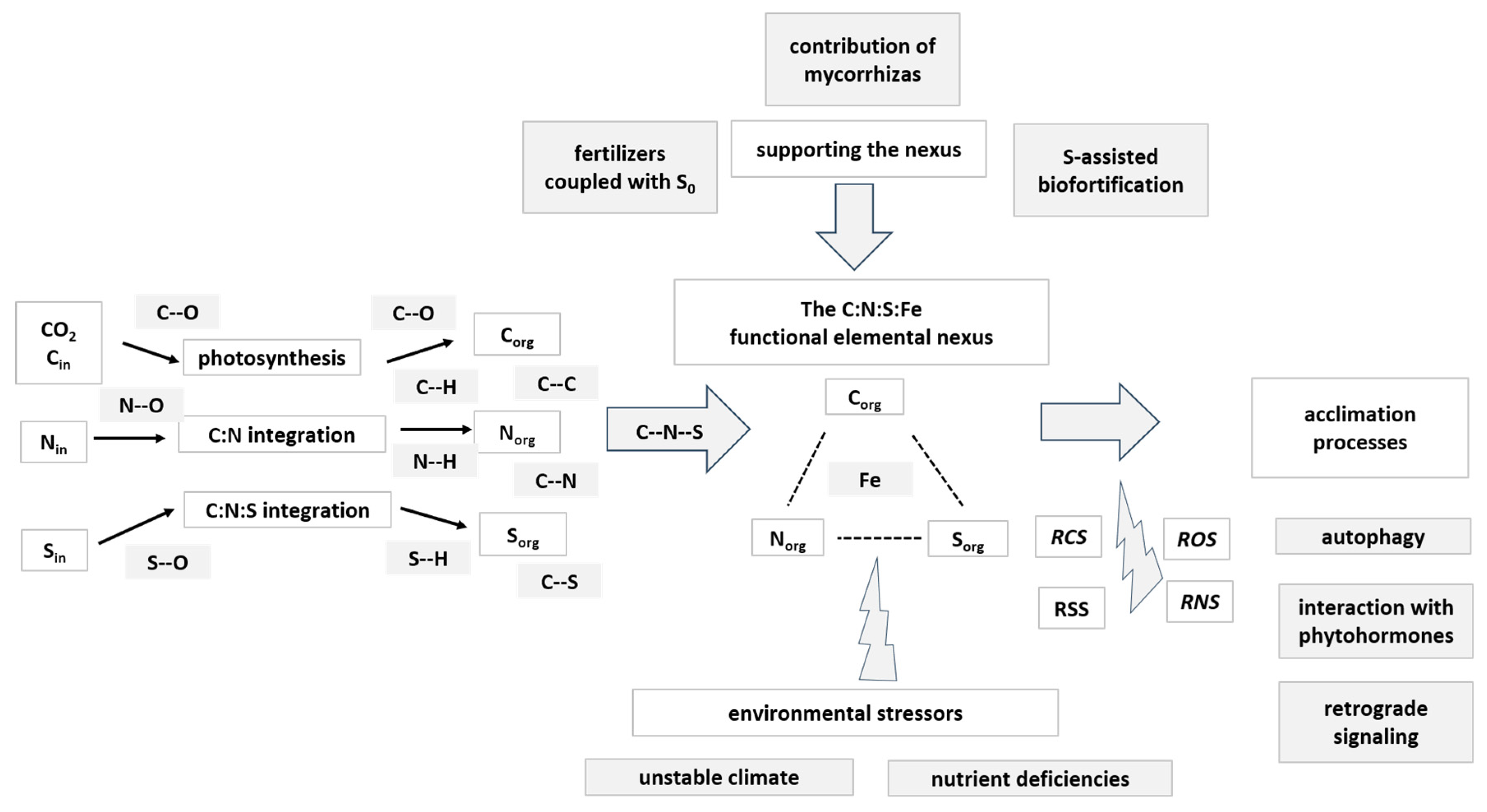
Figure 1: Conceptual model illustrating the integration of the C:N:S elemental nexus with Fe in handling environmental stressors. The model starts with the transformation of inorganic C, N and S forms into the various organic forms and the formation of the C:N:S elemental nexus. Then, Fe is integrated into this nexus. The environmental stressors (among them unstable climate or/and nutrient deficiencies) activate and sustain the formation of various reactive species. Towards handling the environmental stressors, the acclimation processes are based on retrograde signaling, autophagy and interaction with phytohormones. The applied agricultural support actions towards supporting the nexus include the effect of fertilizers coupled with elemental sulfur (S0), the contribution of mycorrhizas, and S-assisted biofortification. Cin: inorganic carbon, Nin: inorganic nitrogen, Sin: inorganic nitrogen, Corg: organic carbon, Norg: organic nitrogen, Sorg: organic sulfur, ROS: reactive oxygen species, RNS: reactive nitrogen species, RSS: reactive sulfur species, RCS: reactive carbon species.
2 The C:N:S:Fe Elemental Nexus
2.1 From Inorganic to Organic Carbon—The CO2 Assimilation
Terrestrial plants capture carbon dioxide (CO2), a form of inorganic C, which is then assimilated through photosynthesis. Assimilation is the incorporation of this inorganic C into existing C skeletons, the organic C. This process is catalyzed by RuBisCO (ribulose-1,5-bisphosphate carboxylase/oxygenase enzyme), where ribulose-1,5-bisphosphate (RuBP) serves as the acceptor molecule, and two molecules of 3-phosphoglycerate (3-PGA) are produced. Inorganic C assimilation occurs in the leaf and 3-PGA is the precursor for all carbon skeletons in plants. Carbon fixation is central to supporting the sustainability of life on Earth and mitigating global climate change. The Calvin–Benson–Bassham (CBB) cycle is the Earth’s primary carbon assimilation pathway, among the seven natural autotrophic C fixation pathways that have been discovered in nature [22]. The resulting C compounds, i.e., the photosynthates, include various carbohydrates, among them glucose (Glc) and sucrose (Suc), photosynthetic products that provide the C-skeletons along with energy. Both Glc and Suc, are converted to pyruvate through glycolysis, and pyruvate is transformed to 2-oxoglutarate (2-OG; or α-ketoglutarate) inside the tricarboxylic acid (TCA) cycle (also called the Krebs cycle or citric acid cycle).
2.2 The Incorporation of Inorganic Nitrogen to Organic Carbon
After C, N is the mineral element required in the largest quantity by plants. Pilbeam [23], and Hawkesford et al. [24] have presented plant N homeostasis in detail, and the assimilation of inorganic N is summarized below. The sources of inorganic N are nitrate (NO3−) and ammonium (NH4+), which are taken up by the roots. Within the plant, for the N in NO3− to be incorporated into organic structures, NO3− must be reduced to NH4+. This reduction is mediated by two enzymes: nitrate reductase (NR), which catalyzes the reduction of nitrate to nitrite (NO2−), and nitrite reductase (NiR), which transforms nitrite to NH4+. NR is a cytosolic enzyme. The NO2− generated by NR is transported to the chloroplast for reduction to NH4+. NiR is a plastidial enzyme localized in the chloroplasts of leaves, as well as in the plastids of roots and other non-green tissues.
Ammonium is a central intermediate in plant N metabolism. The amino acid glutamate (Glu) acts as the existing C, N acceptor molecule for NH4+, and the amide glutamine (Gln) is formed catalyzed by glutamine synthetase (GS). Thus, glutamine is the organic N molecule, that captures the new NH3-N in the form of the amine group (-NH2). The amine group is the organic nitrogen, that is added to the C skeletons. Glutamate synthase (GOGAT; glutamine-oxoglutarate aminotransferase) catalyzes the transfer of the amine group from GLN to 2-OG. Assimilation of most if not all NH4+ derived from NH4+ uptake, N2 fixation, NO3− reduction, and photorespiration is mediated by the GS-GOGAT pathway [23,24].
2-OG provides the C-skeletons and is provided by the respiration, which can be distinguished into three major processes. The first process is glycolysis, whereby Suc and Glc are converted into organic acid anions, such as pyruvate, in the cytosol, which yields a small amount of ATP and reduced nicotianamide dinucleotide (NADH). In the second process, pyruvate is the precursor of 2-OG, an intermediate molecule of the TCA cycle in the mitochondrial matrix, and a considerable amount of reducing power (NADH) and reduced flavin adenine dinucleotide (FADH2) is produced. In the third process, the oxidative phosphorylation, electrons from the donors NADH and FADH2 are transferred along an electron transport chain in the inner mitochondrial membrane to oxygen. The individual electron transport proteins are organized into four multiprotein complexes (CI-CIV). Electrons are transferred from CI (NADH dehydrogenase) and CII (succinate dehydrogenase) via ubiquinone (UQ) to CIII (cytochrome bc1). Cytochrome c transfers the electrons to CIV (cytochrome oxidase), the terminal oxidase that transfers the electrons to molecular oxygen. The production of ATP (Adenosine Triphosphate) is the vital process of generating cellular energy. The vast majority of ATP is produced in mitochondria through cellular respiration [23,24].
Ammonium can be released by organic N compounds by the deamination process. Glutamate dehydrogenase (GDH) catalyzes the reversible amination of 2-OG to form glutamate. GDH was for many years assumed to be involved in NH4+ assimilation. It is now evident that GDH provides carbon skeletons for respiration and oxidative phosphorylation via catalyzing the oxidative deamination of glutamate. GDH in conjunction with NADH-GOGAT, contributes to the control of leaf homeostasis of Gln, the amino acid that plays central role in signaling at the interface of the C and N assimilatory pathways [25]. GDH is involved in the liberation of NH4+ during senescence [23,24].
2.3 The Incorporation of Inorganic Sulfur to Organic Carbon
Sulfur is taken up as sulfate (SO42−) by the root and needs to be reduced in the chloroplasts (plastids in the root) prior to its assimilation into organic sulfur compounds. Sulfate is activated by ATP, the enzyme ATP sulfurylase catalyzes the activation, which leads to the formation of adenosine phosphosulfate (APS). The activated sulfate of APS is then reduced to sulfite (SO32−) by APS reductase. Sulfite is reduced by sulfite reductase (SiR) and sulfide (S22−) is produced. This is the sole reaction of the pathway that occurs only in the chloroplast or plastid. The formed sulfide is transferred to O-acetylserine (OAS) by the enzyme O-acetylserine(thiol)lyase (OASTL), and cysteine (Cys) is formed. The substrate O-acetylserine is synthesized from the amino acid serine (Ser) and acetyl coenzyme A (CoA; an S-containing compound) catalyzed by serine acyltransferase (SAT or SERAT). Cysteine is the first stable product of the assimilatory SO42− reduction and acts as a precursor for the synthesis of most other organic S compounds containing reduced S. Thus, the synthesis of Cys is the key metabolic link between C, N, and S assimilation in plants [24,26,27,28].
2.3.1 Organic C-N and C-N-S Compounds
CO2 assimilation occurs in the leaf. The carbon that plants, along with algae, and cyanobacteria convert into sugars is approximately 120 billion tons [22,29]. Inorganic N uptake occurs in the root system. About 1%–5% of total plant dry matter consists of N as an integral constituent of proteins, nucleic acids, chlorophyll, coenzymes, phytohormones, and secondary metabolites, i.e., the organic N [24]. The availability of inorganic N to roots is therefore a decisive factor for plant growth. Nitrate is readily mobile in the xylem and can also be stored in the vacuoles of roots, shoots, and storage organs. Irrespective of the source of NH4+, or the organ in which it is assimilated (roots, root nodules, and leaves), the key enzymes involved in N assimilation are present in chloroplasts, in roots, and in N2-fixing microorganisms [21,22].
Glutamate, Gln, aspartate (Asp), and asparagine (Asn) occupy a central position in amino acid metabolism and in C:N interactions in plants. Glutamine and Glu are the major entry points of NH3 into organic compounds. The amino groups of glutamate and aspartate and the amide group of Gln are the N source for most plant N compounds, including other amino acids [30]. Aspartate is a metabolically reactive amino acid that serves as a donor in numerous aminotransferase reactions. Asparagine is relatively inert and serves primarily as N storage. Glutamate, Asp, Gln, and Asn participate in long-distance transport of reduced N. Plants can store large amounts of nitrate; however, they cannot re-oxidize organically bound N to nitrate. Amino acids and amides serve also as transient storage. Moreover, plants do not excrete substantial amounts of organically bound N [24]. The synthesis of other N-containing compounds includes other amides as well as amino acids, ureides, amines, peptides, proteins, nucleic acids among them. The low-molecular-weight (LMW) N organic compounds act as intermediates between the assimilation of inorganic N and the synthesis of high-molecular-weight (HMW) N compounds (from amino acids to peptides and proteins). The other amino acids are synthesized by using NH4+ donated from Glu and Gln. Therefore, proteins are synthesized. Proteins are essential for all cellular activities, including C and N metabolism. The LMW N organic compounds are also important for various other reasons, i.e., for the transfer of N from source organs to sink tissues, as well as to build up reserves during periods of high N availability [21,26].
Given the aforementioned metabolic activities, both C and N are essential nutrients for various cellular functions, the first and second in the stoichiometric queue, respectively. It is obvious that coordination of cellular C metabolism with N metabolism is needed for optimal growth and development of plants. Hence, adequate supply of both C and N is critical for plant growth and development, the production of harvestable organs and the completion of life cycle. It is also critical for the response of plants to stress.
Cysteine synthesis is the convergence point of the three major pathways of primary metabolism: CO2, NO3−, and SO42− assimilation; hence, in plants, the newly reduced S passes through cysteine. Cysteine biosynthesis can be distinguished in three processes involving: (a) the photosynthetic assimilation of CO2, that provides the C backbone; (b) the assimilation of N and its incorporation into the C backbone, resulting in the amino acid serine, among others, and (c) the reduction of inorganic SO42− into sulfide and its incorporation into the Ser-derived organic compound OAS. The resulting Cys represents the first form of reduced organic S from primary metabolism in plant cells and serves as the source of organic S for subsequent metabolic reactions [21,22,26].
The convergence of these three essential assimilatory pathways in plant metabolism raises several integration issues, starting with the coordination of S and N mineral nutrition, with the photosynthetic capacity and performance, as the regulatory mechanisms integrating these pathways and their interaction are still partially known. Partial information exists regarding the simultaneous coordination and regulation of these pathways at the whole plant level.
The biosynthesis of OAS is strategic in S metabolism because its availability is coordinated with NO3− reduction, SO42− reduction and carbohydrate metabolism and may control Cys biosynthesis. Very little OAS is found free in solution. The enzymes SAT and OAS-TL form a complex and the OAS produced by SAT is channeled directly to OAS-TL for Cys synthesis. The activity of this complex is controlled jointly by the availability of acetyl CoA and by the availability of Ser providing C and N, along with H2S providing S. Cysteine is reported to inhibit SAT in an allosteric manner. OAS-TL is normally formed in excess of SAT, and under conditions of both N and S deficiency the mRNA that specifies OAS-TL increases. Due to its thiol group, Cys is a very reactive compound. Hence, in most biological tissues, Cys is usually maintained at a very low concentration. For storage or transport, Cys is usually incorporated into glutathione or homoglutathione where the thiol group is less reactive. Glutathione and homoglutathione also contribute to control the concentration of reactive oxygen species (ROS) produced during photolysis of water and participate in the regulation of SO42− uptake and in the detoxification of heavy metals. Cysteine is an important and active component of zinc-finger regulatory proteins, defensins, and other low molecular mass Cys-rich proteins [24,26,28,31,32].
Cysteine is the precursor in the synthesis of methionine (Met), also an important and versatile compound [32]. Cystathionine γ-synthase condenses cysteine and O-phospho-L-homoserine in a trans-sulfuration reaction yielding cystathionine. Cystathionine β-lyase cleaves cystathionine yielding homocystathionine, pyruvate and ammonia, and the S is transferred from Cys to homocysteine. Then, a methyl group is transferred by the Met synthase from N-methyl-tetrahydrofolic acid to homocysteine yielding Met. S-adenosylmethionine (SAM) is formed from methionine and ATP by SAM synthetase. SAM is a hub molecule, the source compound for the hormone ethylene and the polyamines spermidine and spermine. Furthermore, SAM acts as an obligatory methyl donor in over 100 biological reactions, including DNA methylation and the biosynthesis of chlorophyll, heme groups, choline, and lignin, thus underscoring the profound biochemical link between S metabolism and essential plant functions [24,26].
2.3.2 The Contribution of Sulfur in the Acetylation Process
Cysteine formation occurs when OASTL transfers sulfide to OAS, a substrate generated by SAT from serine and acetyl-CoA. The interaction between these enzymes is regulatory: SAT requires complexation with OASTL for activity, while OASTL is inhibited in this state. When sulfide is scarce, accumulating OAS disrupts the complex, halting SAT activity to conserve acetyl-CoA. Beyond its role as a precursor, OAS functions as a signal to stimulate the expression of sulfate transporters and APS reductase, boosting S flux. This OAS-mediated activation balances the feedback inhibition often exerted by reduced S compounds on the assimilation pathway [26].
As a central metabolic intermediate, the compartmentalized abundance of acetyl-CoA reflects the general energy state of the cell. It functions as a key regulatory molecule, influencing enzyme activity and the acetylation of proteins like histones, which impacts mitosis, autophagy, and epigenetic signaling. By operating as both a metabolic substrate and a second messenger, acetyl-CoA plays a decisive role in balancing anabolic and catabolic processes within the cell [33].
The biosynthesis of CoA from pantothenic acid is a critical pathway involving C, N, S, and P. It proceeds through a sequence of enzymatic steps: phosphorylation of vitamin B5, condensation with cysteine, and decarboxylation to form 4′-phosphopantetheine. This intermediate is then converted into dephospho-CoA and undergoes a final phosphorylation to produce the functional CoA molecule. As a central cofactor in carboxylic and fatty acid metabolism, CoA is indispensable for cellular function. Current research has successfully identified the complete set of genes responsible for its synthesis and established the molecular structures of many key enzymes in the pathway [34].
Serving as a vital link between catabolic and anabolic metabolism, acetyl-CoA is a membrane-impermeant molecule characterized by a high-energy thioester bond between an acetyl moiety and CoA. This chemical structure enables the transfer of acetyl groups for processes such as protein acetylation. Acetyl-CoA is compartmentalized within the cell, with independent production and consumption occurring in the plastids, mitochondria, peroxisomes, and cytosol, and it acts as a universal precursor for diverse biosynthetic pathways, including those for lipids, isoprenoids, and flavonoids. Consequently, the metabolic flux of acetyl-CoA is a key determinant in the conversion of C reserves into critical plant chemicals [33,35,36].
2.3.3 The C:N:S Nexus in the Transition from C3 to C4 Photosynthesis
C4 plants minimize photorespiration and maximize carbon fixation in hot, bright environments through two key adaptations: (a) Anatomical adaptation (Kranz Anatomy). Unlike standard C3 plants, C4 leaves feature a specialized ring of cells around the vascular bundle, known as bundle sheath cells. The mesophyll cells, located outside the bundle sheath, are the initial site of CO2 capture. By separating initial CO2 fixation and the Calvin cycle into two different cell types, C4 plants prevent the enzyme Rubisco from interacting with oxygen, eliminating photorespiration. (b) Biochemical adaptation (Two-Step CO2 Pump). Instead of fixing CO2 directly in the Calvin cycle, C4 plants utilize a highly efficient biochemical pump: In Step 1 (Mesophyll), the enzyme PEP carboxylase fixes atmospheric CO2 into a 4-carbon molecule (oxaloacetate, which is converted to malate or aspartate). Because PEP carboxylase is blind to oxygen, it works rapidly even when stomata are partially closed. In Step 2 (Bundle Sheath), these 4-carbon molecules are transported into the bundle sheath cells, where they are broken down to release CO2. This creates a high local concentration of CO2 around Rubisco, forcing the Calvin cycle to run at maximum efficiency [1,37].
The synthesis of Cys is a critical intersection where C, NO3−, and SO42− assimilation pathways meet. As Jobe et al. [37] highlighted, coordinating these pathways is essential for nutrient balance, yet the underlying molecular signals remain largely elusive—especially in C4 plants due to their distinct spatial organization. The shift to C4 photosynthesis alters the traditional coordination of S, N, and C assimilation seen in C3 plants. By evolving carbon-concentrating mechanisms to suppress Rubisco’s oxygenase activity, C4 plants have developed a complex system of modified anatomy and biochemistry. This evolutionary path introduces an extra layer of complexity, requiring highly coordinated, cell-specific compartmentalization to manage the assimilation of soil-derived N and S effectively [38,39,40].
In C4 species, the distinct distribution of CO2 fixation enzymes between mesophyll and bundle sheath cells is mirrored by the segregation of N and S assimilation components. This anatomical division necessitates sophisticated coordination to maintain metabolic balance under varying conditions. While this spatial partitioning of S metabolism has been recognized for over 40 years, modern molecular studies have yet to fully clarify its physiological relevance or the regulatory mechanisms that align it with C assimilation [37,38,39,40].
2.3.4 The Source-Sink Relationship and the C:N:S Nexus
A central focus of crop physiology is the source–sink relationship, a concept vital for optimizing crop yield. Within this framework, ‘source’ organs—predominantly leaves—are responsible for the production and temporary storage of photosynthates. These assimilates, mainly in the form of Suc, are subsequently transported to ‘sink’ organs, such as roots, fruits, or developing leaves, where they are consumed to support growth and development [22].
The vascular system plays a vital role in plant development by facilitating the systemic transport of water, minerals, and organic compounds. The overall source–sink balance of the plant is tightly regulated by its nutrient status, particularly the C:N ratio, which directly links photosynthesis to assimilate distribution. Both C (as Suc) and N (as amino acids) are indispensable for growth; therefore, any imbalance in their allocation can severely compromise plant development and final crop yield. Manipulating the source–sink relationship can significantly alter sink size and strength, as discussed by Qin et al. and references therein [22]. For example, while increasing the capacity of source leaves is a viable strategy to meet rising sink demands, any reduction in source capacity has the opposite effect—diminishing photosynthetic rates and restricting the flow of assimilates. In crops such as cotton, such limitations have been shown to directly reduce fiber yield [41,42,43,44].
An imbalance between source and sink can lead to the excessive accumulation of non-structural carbohydrates (NSC) in leaves, which triggers the downregulation of photosynthesis and accelerates leaf senescence. Research in soybean and common bean demonstrates that NSC buildup reduces maximum photosynthetic capacity and delays photosynthetic induction. In soybean specifically, this process is linked to the downregulation of genes essential for the CBB cycle, Rubisco activase, photochemical reactions, and stomatal opening. Furthermore, such an imbalance can alter the coordination of sugar phosphate translocators within chloroplast membranes, ultimately favoring starch accumulation over active transport [45].
The dynamics between source and sink tissues are significantly influenced by atmospheric conditions and interactions with other organisms. In C3 plants, elevated CO2 levels reduce photorespiration and enhance photosynthetic assimilation, providing more carbohydrates for sinks. This increased substrate availability stimulates sink activity by upregulating genes involved in Suc catabolism, mirroring the regulatory response of leaves to sugar concentrations. Furthermore, plants synchronize photosynthesis with N metabolism by modulating the activities of three key enzymes: phosphoenolpyruvate carboxylase (PEPC), sucrose-phosphate synthase (SPS), and nitrate reductase (NR). Specifically, higher NO3− concentrations promote the phosphorylation of PEPC and SPS, leading to divergent metabolic effects: PEPC activation increases the production of malate and oxaloacetate, facilitating ammonia assimilation and aspartate synthesis. SPS inhibition (via phosphorylation) reduces Suc synthesis. Consequently, increased NO3− availability acts as a metabolic switch, directing the flow of photosynthate toward protein synthesis and away from starch accumulation [1,31].
The N source significantly influences photosynthesis, with N stress leading to lower photosynthetic efficiency and increased thermal energy dissipation. The coordination of C and N metabolism relies heavily on key enzymes: reduced activity in Gln synthetase or glutamate synthase alters photorespiratory C:N metabolism and affects the activation of Rubisco, while Glu dehydrogenase serves as a vital metabolic integrator. To maintain physiological balance, plants have evolved sophisticated mechanisms that synchronize NO3− uptake with organic acid production in the leaves and the abundance of organic N in the phloem. This regulation ensures the alignment of photosynthesis, chlorophyll synthesis, and protein production. Key signaling molecules, including nitrate, glutamine, glutamyl-tRNA, and S-containing amino acids, play a decisive role in coordinating these essential biological processes [1,29].
2.4 The Incorporation of Inorganic Iron to Organic C:N:S Nexus
Iron is the central player in biological electron transfer processes. To sustain these functions, plants must extract inorganic Fe from the rhizosphere and convert it into organic forms that can be assimilated and utilized. In this context, Fe chelators are indispensable, serving three primary roles: (a) solubilizing and mobilizing Fe from the soil, (b) facilitating its internal transport and bioavailability, and (c) maintaining intracellular Fe homeostasis. These chelators operate both extracellularly, for rhizosphere mobilization, and intracellularly, for transport via the xylem and phloem or sequestration in vacuoles. By managing Fe in this way, they protect the plant from both deficiency and toxicity, enabling it to thrive despite fluctuating Fe availability and diverse environmental conditions in the rhizosphere [46,47,48].
2.4.1 The Fe Mobilization Process from the Rhizosphere
Although Fe is abundant in soil, it often exists as insoluble Fe(III) hydroxides, making it chemically unavailable, especially in alkaline or calcareous environments. To overcome this, plants have evolved acquisition strategies. Fe acquisition follows two distinct pathways: the Fe(III)-reduction-based mechanism (strategy I), found primarily in dicots and non-grass monocots, and the Fe(III)-chelation-based mechanism (strategy II), utilized by grasses. In strategy II, chelators secreted in the rhizosphere, solubilize Fe(III) into complexes, preventing precipitation and facilitating diffusion toward the roots. Hence, graminaceous and non-graminaceous plants employ distinct mechanisms for Fe acquisition and vascular transport. While graminaceous plants take up Fe as Fe-phytosiderophore complexes, non-graminaceous species primarily absorb Fe(II) ions following the reduction of Fe(III) at the root cell membrane. Below we discuss the nature and contribution of the various Fe-handling complexes [49,50,51,52,53].
2.4.2 The Central Role of Nicotianamine
Nicotianamine (NA) is a ubiquitous metal-chelator in higher plants, synthesized from three molecules of SAM, by the enzyme nicotianamine synthase (NAS). NA appears to be the universal carrier for Fe(II) across all plants. The role of Met is pivotal in this process, as it serves as the precursor for SAM. Consequently, the availability of S and Met significantly influences plant growth and Fe acquisition [31,47].
2.4.3 The Contribution of Nicotianamine Derivatives in the C:N:S:Fe Nexus
The specialized acquisition strategy of graminaceous plants is based in the release of phytosiderophores (PS) into the rhizosphere. This is because grasses possess the unique ability to convert NA into derivatives, such as mugineic acid (MA) and avenic acid (AvA), which effectively chelate Fe(III). This dual capacity to mobilize both Fe(II) and Fe(III) makes graminaceous plants remarkably resilient to Fe deficiency. These organic acids possess a high affinity for Fe(III), forming stable Fe(III)-MA complexes that solubilize otherwise inaccessible Fe. Once formed, these complexes are actively transported into root cells via YELLOW STRIPE1 (YS1) and YS1-LIKE (YSL) transporters. Within the MA family, deoxymugineic acid (DMA) serves as the primary chelator, while hydroxylated derivatives, such as 3-hydroxymugineic acid, enhance chelation stability across fluctuating soil pH levels [48,49].
While NA binds efficiently to Fe(II), it is primarily used as the essential precursor for PS, a class of LMW, high-affinity Fe(III)-chelating compounds secreted by the roots of graminaceous plants under Fe deficiency. The synthesis of PS follows a specific pathway: NA is first converted to DMA via the sequential action of nicotianamine aminotransferase (NAAT) and 3′-deamino-3′-oxonicotianamine reductase (DMAS). DMA then serves as the immediate precursor for MA, which consists of an azetidine group and three carboxylates. Related PS include AvA, a non-proteinogenic amino acid first isolated from oats (Avena sativa). These PS play a critical role in Fe acquisition, particularly in alkaline and calcareous soils where Fe solubility is otherwise extremely low [48,49].
2.4.4 Iron Transport within the Vascular System
Once inside the root, Fe is transported to the aerial parts via the xylem, where Fe concentrations typically range between 9 and 40 μM. Nicotianamine binds Fe(II) and Fe(III) within the symplastic space to aid translocation, while preventing the generation of free Fe-induced ROS. For vascular transport, Fe moves through the xylem primarily as Fe(III)-citrate, whereas Fe(II)-NA is the predominant form in the phloem. These chelators are essential for maintaining chemical stability across the varying pH levels of different tissues [48,49].
The composition of xylem sap further highlights the aforementioned divergent strategies: NA is present in the xylem of all plants, but reaches higher concentrations in non-graminaceous species. PS, such as DMA, are predominantly found in the xylem of grasses. Citrate (Cit) concentrations vary significantly across species (4–2200 μM). In non-graminaceous plants, Fe in the xylem primarily forms Fe-Cit complexes (e.g., Fe3Cit3 and Fe2Cit2), whereas in grasses, Fe is distributed between Cit complexes and various Fe-PS. The Fe-Cit complex represents a key organic C-Fe compound within plants. As an intermediate of the TCA cycle, Cit acts as an efficient chelating agent in the xylem sap. By binding to Fe(III), Cit plays a fundamental role in both the chelation and the long-distance trafficking of Fe throughout the plant’s vascular system [48,49].
2.4.5 The Storage and Detoxification Processes for Iron
Finally, for storage and detoxification, Fe-chelator complexes are either sequestered in vacuoles via VIT transporters or bound to ferritin, ensuring that Fe remains bioavailable for metabolic needs without causing oxidative damage [48,49].
Heme is an Fe-containing coordination complex characterized by a ring-shaped tetrapyrrole structure, which acts as a tetradentate ligand for an Fe ion. Chemically, it consists of four pyrrole rings featuring two vinyl and two propionic acid side chains, often coordinated with one or two additional axial ligands. As one of the most prevalent metallo-porphyrins used as prosthetic groups, heme defines the essential family of hemoproteins. These proteins include, among others, hemoglobin, cytochromes, catalases, and heme peroxidases, where heme serves as a critical functional component [48,49,54].
2.4.7 The Contribution of Phenolics and Coumarines
Interestingly, the Fe-acquisition strategies of different plant groups often converge; for instance, non-grass species secrete Fe-mobilizing coumarins (FMCs) that function through biochemical mechanisms remarkably similar to those of PS [50,51]. Both plant groups can secrete phenolics and coumarins (such as fraxetin, scopoletin, and sideretin) to chelate Fe(III). This secretion is particularly crucial under high pH conditions, where the standard reductive mechanism becomes inefficient [55,56].
Coumarins are a group of plant-derived phenolic secondary metabolites, synthesized by specific species—most notably Arabidopsis thaliana—as a specialized response to Fe deficiency. This group of molecules is secreted into the rhizosphere to enhance Fe bioavailability through several synergistic pathways. (a) Chelation of Fe(III): Utilizing catechol and hydroxyl functionalities, coumarins bind Fe(III) via bidentate coordination. This process transforms insoluble Fe hydroxides into stable, soluble Fe-coumarin complexes, making the iron accessible for root uptake. (b) Reductive Mobilization: Certain coumarins reduce Fe(III) to Fe(II), a form that is more soluble in neutral to alkaline soils and is preferentially absorbed through IRT1 transporters in root epidermal cells. (c) Microbial Interactions: By modulating the rhizosphere microbiome, coumarins promote the growth of beneficial microbes that produce siderophores, indirectly boosting Fe mobilization. (d) Rhizosphere Modification: The secretion of these compounds can lead to localized soil acidification, further increasing the solubility of Fe(III) [55,56].
Coumarins are crucial for Strategy I Fe acquisition in non-grass plants, serving a biochemical adaptation to Fe-limited soils. Under Fe-deficient conditions, plants increase the secretion of these compounds, which enhance soil Fe availability through three primary mechanisms. (a) Solubilization: chelating insoluble Fe(III) into soluble complexes, (b) Reduction: reducing Fe(III) to Fe(II) for easier uptake, and (c) Rhizosphere optimization: modifying microbial communities and the microenvironment to favor Fe mobilization.
The formation of Fe(III)-coumarin complexes in soil solutions increases root Fe(II) levels and promotes growth recovery upon exogenous application [55,56]. Furthermore, studies on coumarin-deficient Arabidopsis mutants confirm their functional role, as these plants exhibit impaired Fe uptake and chlorosis [55,56,57,58,59,60]. Ultimately, this improved Fe acquisition supports chlorophyll biosynthesis, photosynthesis, and overall plant growth.
2.4.8 The Collaboration of Sulfur and Iron in the C:N:S:Fe Nexus
Sulfur acts as a critical hub within the C:N:S elementome, where nutrient homeostasis depends on dynamic networks. As highlighted by Courbet et al. [61], these interactions occur at every level, from cellular compartments to the whole plant. A deficiency in S limits overall growth and reduces N uptake, while N deficiency similarly impairs S acquisition. Although plant mineral composition is tightly governed by mechanisms such as translocation, storage, and remobilization, the full extent of this homeostatic coordination is an open research topic. This underscores the deep and inseparable integration of the C:N:S elementome.
Iron is fundamental to the coordination of C, N, and S metabolism, primarily through its role in electron transport. In leaves, PSI produces reduced ferredoxin (Fd), which donates electrons to nitrite reductase via Fe-S clusters and siroheme cofactors. In non-photosynthetic tissues, i.e., in root plastids, reduced Fd is instead supplied by the pentose phosphate pathway through NADPH and Fd-NADP+ reductase. This site-specific generation of reducing power underscores the integration of Fe in both light-dependent and heterotrophic nutrient assimilation [62]. Several S-Fe interactions have been extensively discussed by Courbet et al. [61]; Astolfi et al. [63]; Chorianopoulou and Bouranis [64]; Haque et al. [65]; Wang and Zhao [66]. The various close relationships between Fe and S nutrition that have been demonstrated suggest common regulatory mechanisms for the homeostasis of these two elements.
Fe-S clusters function as a central nexus bridging the stoichiometry of Fe with the C:N:S elementome. Composed of Fe and sulfide, these molecular ensembles are indispensable for the activity of various catalytic proteins, necessitating the synchronized regulation of Fe and S homeostasis. The biological significance of Fe stems from the Fe(II)/Fe(III) redox couple, whose interconversion drives vital cellular electron-transfer processes. While Fe typically adopts octahedral geometry in aqueous solutions, Fe-S clusters with different geometry such as [2Fe-2S], [3Fe-4S], and [4Fe-4S] are existing [67]. These clusters are pivotal for energy metabolism, facilitating electron transport in both photosynthesis (thylakoid membranes) and respiration (inner mitochondrial membrane). Their biosynthesis is compartmentalized across mitochondria, plastids, and the cytosol, managed by specialized assembly machineries [68,69,70].
Within chloroplasts, Fe–S clusters are integral components of photosystem I (PSI), the cytochrome b6f complex, Fd, and both sulfite and nitrite reductases. Within PSI, electrons are captured by chlorophyll-a and shuttled through a series of redox centers—specifically phylloquinones and three distinct Fe-S clusters—to Fd. This soluble stromal protein possesses a low redox potential, enabling the reduction of NADP+ to NADPH via Fd NADP+-reductase. Beyond its role in the thylakoid electron transport chain, reduced Fd serves as a vital electron donor for nitrite and sulfite reduction, linking photosynthetic energy to nutrient assimilation. This coupling ensures that the reduction of N and S is synchronized with the light-driven generation of C skeletons, facilitating the synthesis of organic compounds like amino acids [70].
Within mitochondria, Fe-S proteins are central to metabolic energy conversion, notably represented by aconitase in the Krebs cycle and complexes I, II, and III of the respiratory chain. Electron translocation within these complexes is mediated by various Fe-S clusters. Emerging evidence suggests that Cit may function as a critical signaling molecule for sensing Fe and S status. While general nutrient deficiency signals may arise from respiratory chain impairment, specific signals are likely linked to Krebs cycle perturbations. Consequently, mitochondrial dysfunction under Fe and S deprivation is hypothesized to trigger specialized deficiency-induced responses in plants. Given their high demand for metallic co-factors and their primary role in Fe-S cluster biogenesis, mitochondria serve as a fundamental hub for the metabolic interaction between Fe and S [1,70,71,72,73].
Fe-S cluster biogenesis necessitates the simultaneous availability of Fe and S; consequently, a deficiency in either element impairs energy metabolism and elevates oxidative stress. Due to the inherent toxicity of free Fe, this assembly process is strictly regulated and highly sensitive to Fe and S levels. In plants, Fe-S cluster-dependent enzymes constitute the majority of Fe-requiring enzyme categories. Their synthesis involves scaffold proteins that coordinate with Fe and S donors, with S derived from Cys via Cys desulfurases and Fe delivered with the contribution of frataxin. Once assembled, these clusters are distributed to target proteins across the mitochondria, plastids, cytosol, and nucleus by at least nine identified carriers [69,72,73,74,75]. The plant Fe-S assembly machinery is compartmentalized into three distinct systems: the ISC (mitochondrial), SUF (plastidial), and CIA (cytosolic/nuclear) pathways. Notably, the ISC machinery supplies glutathione persulfide to the CIA system through the ATM3-mediated export pathway [76,77,78,79,80].
According to Gomez-Casati et al. [76] frataxin is an evolutionarily conserved protein essential for cellular iron homeostasis, facilitating Fe-S cluster assembly, heme biosynthesis, and oxidative phosphorylation. Distinctly in plants, frataxin exhibits dual localization to both mitochondria and chloroplasts via one or two specialized isoforms. Consequently, frataxin deficiency precipitates a dual-organelle crisis, characteristically impairing mitochondrial respiration while inducing chlorophyll loss and photosynthetic electron transport failure. Furthermore, unique biochemical properties allow plant frataxins to bind Cu(II) ions and dimerize. This interaction actively reduces free Cu pools, potentially serving as a novel defense mechanism to protect vulnerable Fe-S groups from Cu-induced oxidative degradation. These unique features—isoform multiplicity, dual compartmentalization, and copper-binding capabilities—together strongly indicate that plant frataxins have evolved expanded, tissue-specific regulatory roles beyond traditional eukaryotic models [76].
The plant mitochondrial Iron-Sulfur Cluster (ISC) assembly machinery is an essential, highly conserved pathway responsible for synthesizing Fe-S cofactors. It acts as the core hub for the maturation of essential mitochondrial respiratory enzymes and supplies vital signals needed by the cytosol. The ISC machinery consists of several key protein components. (a) Scaffold Protein (ISU): The structural platform where the Fe and S are initially assembled into a labile Fe-S cluster. (b) Cysteine Desulfurase (NFS1): Extracts elemental S from Cys and provides it to the scaffold. (c) Iron Donor and Reductants: Frataxin regulates iron delivery to the scaffold, while Fd provides the necessary electrons. (d) Chaperone System (HSCB/HSPA): Assists in transferring the newly formed Fe-S cluster from the ISU scaffold to target apo-proteins [74,75,76,77,78,79,80].
The plant plastidial Sulfur Utilization (SUF) machinery is an independent, ancient system responsible for synthesizing Fe-S clusters inside chloroplasts. It is heavily adapted to function efficiently under high oxidative stress and light-driven conditions to sustain photosynthesis. The SUF system relies on a specific set of core proteins: (a) Sulfur Mobilization (SufS & SufE): SufS is a cysteine desulfurase that extracts sulfur from Cys. SufE acts as an activator, dramatically accelerating SufS activity. (b) The SUF Scaffold Complex (SufB-SufC-SufD): A unique, stable multi-protein complex where transient Fe-S clusters are built. SufC is an ATPase that drives the conformational changes needed for cluster assembly. (c) Carrier Proteins (SufA, NFUs, GRX): Specialized transfer proteins that accept the cluster from the scaffold and safely deliver it to target plastid proteins [74,75,76,77,78,79,80].
The plant cytosolic Fe-S Cluster Assembly (CIA) machinery is a dedicated eukaryotic pathway responsible for synthesizing and inserting Fe-S clusters into target proteins located within the cytosol and the nucleus. Unlike the semi-autonomous mitochondrial ISC and plastidial SUF systems, the CIA pathway lacks its own S-extracting enzyme and is strictly dependent on the mitochondrial ISC machinery to initiate the process. The CIA pathway operates in two sequential stages: de novo cluster assembly on a scaffold and target-specific delivery [79,80].
2.5 The N2 Fixation in Legumes
Legumes possess the ability to fix atmospheric N2, a process heavily influenced by S nutrition, as discussed by Scherer [81] and Courbet et al. [61]. Adequate S-nutrition positively correlates with total biomass and nodule size [82], stimulating root development and increasing nodule density [83]. These nodules possess high S concentrations to support their functional requirements [84]; for instance, white clover nodules can contain up to 3% S per unit DW, a 6-fold higher concentration than in leaves [85]. Consequently, S deficiency reduces N2 fixation. Beyond affecting nodule number and size, high S demand is critical for synthesizing key N2 fixation compounds, including leghemoglobin, nitrogenase, Fe-S clusters, and Fd [81].
Nitrogenase (Nase) is the primary enzyme responsible for reducing N2 to NH3 during nitrogen fixation. Due to its high enrichment in S-containing amino acids, substantial amounts of S are essential to maintain full enzymatic activity, which depends on S-rich proteins such as nitrogenase and nitrogenase reductase (NifH) [86,87]. Under S-deficient conditions, Nase activity is more severely impacted than photosynthesis, as S deficiency leads to enzyme deactivation and a subsequent reduction in N2 fixation [84,88,89,90,91]. Furthermore, Nase function requires ATP, carbohydrates, and reducing equivalents derived from electron transfer via Fd. Sulfur deficiency compromises these processes by lowering ATP levels in bacteroids and host cell mitochondria, reducing Fd concentrations, and decreasing NifH protein levels, ultimately diminishing the activity of the nitrogenase complex [85,89,91].
Both Fd and Nase are enzymes characterized by their Fe-S clusters. Furthermore, S deficiency has been shown to reduce leghemoglobin content, which is critical for regulating the O2 supply to Nase. Transcriptomic analysis of different nodule zones has identified specific sulfate transporters—namely SULTR3;5, SULTR2;1, and SULTR1;3—as key candidates for allocating sulfate to the N2-fixing zone (Zone III) [92,93,94]. The preferential expression of these SULTR genes in Zone III, the region furthest from the apical meristem where bacterial nitrogen fixation occurs, underscores the complexity of S transport systems in supporting S-dependent metabolic activities. Additionally, both nitrate and sulfate assimilation processes are active within the nodules. O-acetylserine serves as a positive effector for the expression of genes encoding sulfate transporters and assimilation enzymes, such as sulfite reductase. Given the significant transcriptional shifts in S assimilation pathways within nodules, OAS-mediated positive feedback activation also plays a vital role in the nitrogen-fixing zone [95,96,97].
As an electron carrier, Fd provides the reducing power for sulfite reductase, nitrite reductase, and nitrogenase. The Fd-dependent conversion of nitrite to ammonia is vital for protecting nitrogenase from nitrite-induced inhibition. The activity of these enzymes relies on the prosthetic groups heme and siroheme. Specifically, siroheme enables the six-electron reduction of S and N, with its synthesis requiring SAM-dependent methyltransferase activity. Similarly, heme synthesis involves coproporphyrinogen III oxidase and is fundamental for leghemoglobin formation. Furthermore, SAM contributes to the synthesis of NA, which facilitates the Fe supply necessary for bacteroid Nase and leghemoglobin functionality [98,99,100,101,102].
The aforementioned information shows strong relationships of the C:N:S:Fe nexus and N2 fixation in legumes.
2.6 The Integration of the C:N:S Nexus with Fe in the Rhizosphere
Li et al. [103] discussed how Fe interacts with the biogeochemical cycles of C, N, and S in environments with fluctuating redox conditions, such as the rhizosphere. Despite Fe being among the most abundant elements in the Earth’s crust, its availability may be restricted within microsites or micro-ecosystems for reduction or oxidation processes. Additionally, the accessibility of C, S, or N can decrease due to the oxidation or complexation by Fe oxides. The cycle of Fe between its Fe(II) (ferrous) and Fe(III) (ferric) states is referred to as the “Fe(III)–Fe(II) redox wheel” (FeRW) [103].
The complex interactions between FeRW and the biogeochemical cycles of C, N, and S, make it difficult to pinpoint certain dynamic redox environments. FeRW has been shown to interact with each cycle individually in soils or sediments and fluctuating redox conditions in the rhizosphere indicate that FeRW actively influences these cycles together. These interactions underscore the significance of Fe in both individual and combined biogeochemical processes [103].
When Fe is chemically inert—owing either to Fe–S precipitation or Fe–humic complexation—or when Fe(III) (hydr)oxides are reduced through abiotic or biotic nitrification, or their specific surface area decreases because of crystallization, phosphorus (P) levels in the aqueous phase of the micro-ecosystem may rise. This increase occurs due to a lack of sorption surfaces and the co-precipitation of Fe(III) oxide minerals. Both crystalline and non-crystalline Fe(III) (hydr)oxides act as terminal electron acceptors for organic matter and play roles in mineralization and transformations of N and S, while also serving as sorbents for dissolved P compounds [103].
Processes such as electron transfer from organic matter to Fe(III) (hydr)oxides during oxidation, conversion of NH4+ to NO2−, formation and subsequent oxidation of iron sulfide minerals within the sulfur cycle, as well as P transformation, have all been linked to the FeRW [103].
FeRW plays important role in the biogeochemical cycles of C, N, S, and P, and at least partially drives or is involved into their biogeochemical cycles at various scales. Fe(III) (hydr)oxides are essential in biogeochemical cycles of C, N, and S within dynamic redox micro-ecosystems, acting as terminal electron acceptors for organic matter mineralization and N and S transformations. These substances have an exceptionally high specific surface area and serve as sorbents for dissolved P compounds. Considering these characteristics, the C, N, and S, cycles are incorporated into the FeRW system. Fe(III) acts as a terminal electron acceptor during organic matter mineralization. Its reduction to Fe(II) is linked to transformations such as NH4+ to NO2−, S2O32− to S4O62−, and S0 to SO42−. Conversely, Fe(II) oxidation to Fe(III) couples with SO42− to HS− and H2S conversions. These Fe redox processes alter solid surface structures and solution chemistry, influencing NH4+ and P shifts between soluble and solid forms. The FeRW integrates with C, N, and S cycles—especially C and N—in dynamic redox environments [103,104,105].
2.7 The Contribution of Secondary S Metabolites to the C:N:S:Fe Nexus
Organic S exists as both reduced and oxidized forms. Sulfate is converted to APS, then reduced to sulfite and sulfide for Cys production—this is primary sulfate metabolism. Alternatively, APS can be phosphorylated to PAPS, transferring sulfate in its oxidized form to various metabolites. This process is known as secondary sulfate metabolism. Notably, the synthesis of PAPS is vital for plant development, as even a slight deficiency can result in dwarfism. Koprivova and Kopriva [106] have reviewed the enzymes involved in plant sulfation pathways, emphasizing both the similarities and distinctions across different biological kingdoms.
Historically, plant research has predominantly concentrated on primary reductive S metabolism. Nevertheless, significant interest has also been directed toward sulfated compounds and their associated metabolic pathways for several reasons: (a) the established connection between sulfation and phosphoadenosine phosphate (PAP), a crucial plant signaling molecule [107,108], (b) the identification of sulfated peptides as key growth hormones [109], and (c) evidence that PAPS constrains glucosinolate biosynthesis [110,111,112].
The enzymes and genes involved in plant sulfation pathways have been examined, with particular attention given to both the similarities and distinctions across different biological kingdoms. Plants are known to produce a wide array of secondary metabolites, among which sulfation represents one potential modification. In addition to glucosinolates, sulfated flavonoids constitute the most thoroughly documented category of sulfated secondary metabolites [106].
Plant sulfated secondary metabolites (such as glucosinolates and sulfated flavonoids) are specialized, S-rich chemical compounds. While not essential for basic growth, they provide a vital chemical defense system, regulate cellular signaling, and act as potent antioxidants. Sulfated flavonoids act as antioxidants, protect plants against UV stress, and assist in chemical defense and cellular transport. Sulfation dramatically increases the polarity of flavonoids. This improves their water solubility and facilitates easier transport through biological fluids. The primary significance of these metabolites spans ecology, plant physiology, and human health [58,106].
2.7.1 Plant Defense and Survival (Biotic Stress)
Because plants are sessile, they rely on specialized chemicals to survive. Sulfated metabolites act as natural deterrents against herbivores, insects, and pathogens. (a) Activated Defense Systems: In the Brassicaceae (mustard) family, glucosinolates are stored alongside an enzyme called myrosinase. When tissue is damaged by pests, the enzyme breaks the glucosinolates down into toxic, volatile compounds (like isothiocyanates) that act as strong feeding repellents. (b) Phytoalexins: Sulfated compounds (e.g., camalexin) are rapidly synthesized in response to bacterial or fungal infections [106,107,108].
2.7.2 Environmental Adaptation (Abiotic Stress)
Sulfation is a crucial conjugation mechanism that allows plants to regulate their responses to harsh environmental conditions. (a) Stress Tolerance: The sulfation of molecules, particularly flavonoids, helps the plant deal with extreme conditions like heavy metal toxicity, drought, and salinity. (b) Antioxidant Activity: Many sulfated phytochemicals act as antioxidants, scavenging the harmful ROS produced when a plant is subjected to environmental stress. (c) Cellular Signaling and Hormone Regulation: Sulfation is a key post-translational modification used to dictate the biological activity and stability of small molecules. (d) Hormone Modulation: Sulfotransferases (SOTs) add sulfate groups to phytohormones (such as auxins and brassinosteroids) and various intermediates to either activate them, store them, or mark them for deactivation. This ensures a localized and finely tuned balance of growth regulators [106,107,108].
Sulfur content varies significantly among plant families, mainly because of differences in secondary S compounds. These global patterns are strongly tied to phylogeny, with evolutionary history being a key factor in determining S levels. Herbaceous plants, especially those in the Brassicaceae family, generally have more S than woody families. While S often correlates with N and P, its levels can still fluctuate independently among different plant groups. Other factors include rapid metabolic turnover in fast-growing herbs, ecological adaptation to varying soil S content, and genetic control of sulfate transport and assimilation. Sulfur-rich metabolites are unevenly spread across plants, with Brassicaceae showing the highest concentrations, while many woody families have much lower, non-specialized S levels. Phylogeny strongly influences S stoichiometry [26].
2.7.4 Modification and Chelation of Flavonoids
While plants do not covalently attach Fe or S atoms to a flavonoid’s carbon backbone, Fe and S fundamentally modify the structural conformation, chemical properties, and stability of flavonoids. Flavonoids (such as quercetin and catechin) possess specific hydroxyl and carbonyl arrangements (e.g., the 3-hydroxy-4-keto group) that act as powerful chelators for transition metals. By binding to Fe(II) or Fe(III), the flavonoid molecule is structurally altered into an Fe complex. This modification shields the reactive metal, preventing it from undergoing the cytotoxic Fenton reaction and generating ROS. Under Fe deficiency, plants increase S assimilation to manufacture sulfate-modified phenolics and glutathione. At the same time, specialized flavonoids are excreted into the rhizosphere. These flavonoids reduce insoluble Fe(III) to bioavailable Fe(II), allowing the corresponding root transporters to pull both elements into the plant systematically [58,60].
2.8 Amino Acid Recycling and Catabolism
Amino acids serve several important roles in plants. Not only are they involved in protein synthesis, but they also act as key components in other biosynthetic pathways and play significant parts in signaling and plant stress responses. Typically, the amounts of each of the 20 amino acids vary greatly and shift dynamically based on the developmental stage and physiological condition of the plant cell. Amino acid breakdown regulates their levels and, under conditions like C starvation, helps maintain plant cell energy. Hildebrandt et al. [113] have discussed the biological role of amino acid catabolism and presented knowledge on amino acid degradation pathways and their regulation in the context of plant cell physiology.
Cysteine is converted to pyruvate through three distinct pathways. The amino group may be released as ammonium or transferred to glutamate, while the thiol group undergoes oxidation to thiosulfate or sulfate [113]. Additionally, Cys synthesis predominantly occurs in the cytosol, with the cytosolic OASTL enzyme OAS-A1 identified as the primary catalytic agent. DES1 is another member of the OASTL family and functions as a Cys desulfhydrase, breaking down Cys to produce sulfide, which contrasts with the activity of OAS-A1. Both DES1 and OAS-A1 proteins work together to regulate Cys levels and generate cytosolic sulfide for signaling. The concentration of Cys in the cytosol significantly influences plant responses to various types of stress, whereas sulfide formed from Cys breakdown can inhibit autophagy under certain conditions. Therefore, maintaining proper levels of Cys and sulfide is likely crucial for optimal plant functioning [114].
Methionine can be converted to methanethiol and 2-oxobutyrate or to homocysteine via S-adenosylmethionine, and the respective enzymes are known in plants. However, the subsequent steps are still rather speculative. Methionine catabolism in plants is primarily initiated by the enzyme methionine γ-lyase (MGL), which breaks down excess free Met to manage S balance and fuel secondary metabolic pathways. Plants lack this specific pathway and rely heavily on MGL-driven cleavage [113].
3 Plant Stress Driven by Environmental Stressors
3.1 The Environmental Stressors
Environmental stressors for plants are external, non-biological (abiotic) or biological (biotic) conditions that negatively impact their growth, development, and overall survival. Major abiotic stressors include climate extremes like severe drought, flooding, high salinity, heavy metal toxicity, and extreme temperatures (heat or frost), alongside inadequate or excessive light levels. Biotic stressors, on the other hand, involve living organisms such as pests, pathogens (bacteria, fungi), viruses, and competition from weeds, all of which force the plant to redirect its energy from growth and development to defense and survival mechanisms [115].
The action of environmental stressors significantly reduces agricultural crop yields by disrupting fundamental plant processes and forcing crops to redirect energy from growth to survival. For example, drought and extreme heat force plants to close their stomata to prevent water loss, which stops CO2 intake and halts growth. Flooding creates oxygen-depleted soil, destroying root systems and blocking nutrient absorption. High salinity, freezing temperatures, and heavy metals damage plant cell membranes and cause oxidative stress. Pests and pathogens directly destroy leaf tissue, roots, or vascular systems, reducing the plant’s capacity to produce grain or fruit. Stress during critical vegetative phases leads to shorter plants with fewer reproductive branches. Heat or drought during flowering causes pollen sterility, leading to poor grain filling or fruit drop. Weakened plants are highly susceptible to lodging (falling over) and post-harvest diseases [115].
Plants cannot run away from danger, so they have evolved complex internal systems to survive. Their defense mechanisms against environmental stressors are divided into two main categories. Sessile organisms must dynamically modulate their physiological, biochemical, and structural configurations to survive concomitant environmental pressures. There are dual-faceted defense strategies employed by plants to mitigate both biotic and abiotic stresses. Biotic defense mechanisms establish a multi-layered immune response, initiated by structural modifications such as cuticle and cell wall thickening to impede pathogen ingress. This physical barrier is reinforced by the biosynthesis of toxic secondary metabolites—including alkaloids, terpenes, and phenolics—to deter herbivores. Upon localized infection, the induction of a hypersensitive response isolates pathogens via programmed cell death, concurrently triggering systemic acquired resistance mediated by salicylic acid signaling to prime distal tissues. Communication extends extracellularly through volatile organic compound emissions, which alert neighboring flora and recruit predatory arthropods [116].
Parallelly, abiotic stress tolerance involves rapid homeostasis preservation. Under moisture deficit or salinity, immediate stomatal closure regulates transpirational loss, while the intracellular accumulation of compatible osmolytes, such as proline and soluble sugars, maintains turgor pressure. Oxidative stress is countered by the upregulation of antioxidative enzymes, specifically superoxide dismutase and catalase, which neutralize reactive oxygen species. At the macromolecular level, heat shock proteins function as molecular chaperones to prevent thermal denaturation. Finally, developmental plasticity manifests as root architecture alteration, optimizing spatial configuration to access deep water tables or circumvent phytotoxic soil zones. Together, these integrated response networks illustrate the evolutionary complexity of plant adaptation and survival [116].
3.1.1 Abiotic Stress Defenses (Physical & Chemical)
Plants mitigate environmental adversity through five highly coordinated physiological and biochemical adaptations. Initially, under drought conditions, the immediate induction of stomatal closure minimizes transpirational water loss. Concurrently, cellular osmotic adjustment is achieved through the intracellular accumulation of compatible osmolytes, such as proline and soluble sugars, which facilitates water retention under osmotic and saline stress. To counteract oxidative damage, plants upregulate antioxidant production, synthesizing enzymes like superoxide dismutase (SOD) and catalase to neutralize destructive ROS. Furthermore, the expression of heat shock proteins serves a chaperone function, stabilizing and protecting critical macromolecular and cellular structures from thermal degradation or freezing stress. Lastly, dynamic root architecture alteration optimizes the root system’s spatial configuration, promoting deeper soil penetration to access water reserves or structural modification to bypass phytotoxic zones [115,116].
3.1.2 Biotic Stress Defenses (Anti-Pest & Anti-Pathogen)
Plants defend themselves against biological threats through five coordinated mechanisms. First, physical barriers like the thickening of the cuticle and cell walls physically block pests and fungi from penetrating the plant tissue. Second, chemical warfare involves the production of toxic secondary metabolites such as alkaloids, terpenes, and phenolics, which actively deter herbivores. Third, the hypersensitive response triggers the intentional death of cells surrounding an infection site, effectively starving and trapping the pathogen to prevent further spread. Fourth, systemic acquired resistance acts as an internal alarm system, utilizing signaling molecules like salicylic acid to prepare distant leaves for an incoming attack. Finally, volatile emission allows plants to release airborne chemicals that warn neighboring plants of danger or attract predatory insects to consume the attacking pests [115,116].
An unstable climate triggers extreme, unpredictable abiotic stressors like droughts, heatwaves, and flooding. Unstable weather creates extreme environment shifts. An unstable climate may directly amplify nutrient imbalances in soil. Erratic weather patterns may alter the availability and uptake of essential plant nutrients. Some examples are the following: (a) Flooding and Waterlogging: Heavy downpours leach mobile nutrients like N and S deep into the soil, away from the root zone. Submerged, oxygen-deprived roots also lose the energy required for active nutrient uptake. (b) Severe Drought: Without water as a solvent, nutrients cannot move through the soil via mass flow or diffusion. Plants become progressively incapable of absorbing immobile elements like P and K, leading to acute starvation despite nutrients being present in the soil. (c) Extreme Temperatures: Heatwaves and unseasonal cold snaps alter soil microbial activity. This disrupts the natural mineralization of organic matter, locking up nutrients in forms that plants cannot utilize [115,116].
3.2 Acclimation Processes to Climate Change Drivers
3.2.1 High Light-Induced Stress
High light stress occurs when the absorption of light by the photosystems within thylakoid membranes is not effectively coordinated with its utilization for CO2 fixation. In experimental settings, light intensities ranging from 1000 to 2000 μM m−2 s−1 are typically classified as high light exposure for plants. Photosystem II (PSII) is primarily affected by such high light stress [117].
This term describes the condition of water shortage and is often referred to as drought stress. During drought stress, leaf stomata close to limit both CO2 intake and H2O loss, which suppresses photosynthesis. While water stress affects various biochemical pathways, it is well established that PSII is primarily impacted by drought stress. Stress conditions lead to a reduction in both the levels and activities of enzymes involved in the photosynthetic C reduction cycle, notably the essential enzyme Rubisco [117,118,119,120,121]. Such stress may cause a disproportionate decline in the efficiency of photoelectron transport and the C reduction cycle, resulting in an imbalance in the redox state of photosynthetic organelles. This imbalance is further exacerbated if light-driven electron transport continues without regulation. Water stress has also been reported to elevate concentrations of H2O2 and O2•– radicals, while increasing the activities of superoxide dismutase (SOD), ascorbate peroxidase, and glutathione reductase (GR) [122,123,124,125].
PSI is particularly susceptible to stress under conditions of low temperature (4°C), low light intensity (100–200 μmol m−2 s−1), and the presence of oxygen. Chilling stress inhibits PSI activity, while the effect on PSII activity is minimal [117]. Exposure to chilling impairs the Fe-S centers within PSI, likely due to the generation of superoxide anion (O2•−) on the reducing side of PSI. High temperature stress adversely affects PSII. Exposure to temperatures between 45–60°C results in the inactivation of Rubisco, which may be attributed to the thermal denaturation of activase. Elevated temperatures can cause sequestration of Rubisco activase to the thylakoid membrane. The activity of SBPase prevents this sequestration, thereby preserving Rubisco activity [126,127].
UV radiation can impact plant growth, photosynthetic efficiency, and gene expression. It decreases the activity of chloroplast ATPase, Rubisco, and violaxanthin de-epoxidase. Among all thylakoid complexes, PSII is affected most significantly [117].
3.2.5 The Chloroplast under Climate Stress
Chloroplasts react to various abiotic stress factors, such as intense light, water shortages, extreme temperatures, and UV radiation, which lead to oxidative stress and are influenced by sulfur metabolism. While highly sensitive to these environmental challenges, chloroplasts are also key sites for S assimilation. Hence, chloroplast plays a major role in the modulation of stress response. Sulfur metabolism mediates modulation of plant responses to various abiotic stress factors. The photosynthetic organelle orchestrates metabolic pathways involving C, N, S and Fe, supplying critical precursors for the biosynthesis of S compounds. Exposure to abiotic stress factors such as intense or limited light, temperature fluctuations, drought, and ultraviolet radiation induces an oxidative environment within the organelle, resulting in the generation of ROS. Organic S metabolites with thiol groups that can undergo reversible oxidation and reduction actively neutralize ROS through various biochemical processes. Abiotic stress factors lead to changes in the expression of multiple stress-related genes, either increasing or decreasing their activity. The signaling pathways involved in transmitting these stress signals and regulating gene expression are highly complex [117,128,129].
The stress-induced redox signals generated in chloroplast play a major role in different signal transduction systems and expression of stress-responsive genes in plants. A mature chloroplast maintains a nonequilibrium stationary state. It demonstrates resilience against external disturbances, such as fluctuations in temperature, light intensity, CO2 concentration, salinity, and osmotic pressure. Internal modifications within the system generally act to restore its state to the greatest extent possible. External changes are referred to as stress, while the subsequent internal responses within the organelle collectively define the adaptation process. In a mature chloroplast, functions such as photon absorption, electron transport, proton translocation, CO2 fixation, and nutrient assimilation are conducted in a coordinated manner. Accordingly, stress responses also follow an integrated pattern [117,128,129].
Light is a key environmental influence on green plants. It is mainly detected by PSI and PSII, which then send photo signals through various parts of the chloroplast organelle. When other environmental factors change alongside light, the photosystems can also sense these shifts, often resulting in photoinhibition of PSII in the thylakoid [130]. Because PSII is particularly sensitive, the chloroplast serves as an important stress sensor for plants. During stress acclimation, a feedback mechanism based on the redox state of components involved in PSI and PSII’s electron transport system has been observed [131,132].
Stress-induced changes in the chloroplast redox state can cause O2 to become harmful free radicals. Strong oxidants from PSII may damage thylakoid pigments, proteins, or lipids, making the photosynthetic organelle a key stress sensor in green plants. Chloroplasts under abiotic stress do not use energy efficiently due to a disrupted sink, causing redox or excitation pressure on photosystems. This pressure can lead to either stress adaptation or system damage and is known as high light stress syndrome [117].
Most abiotic stress leads to photoinhibitory damage in chloroplasts, causing oxidative stress. Models suggest that adaptation involves components of the electron transport chain, redox-responsive protein kinases, and thiol-regulated enzymes, with chlorophyll precursors possibly mediating signal transduction. In response, chloroplasts use various antioxidants and enzymes as adaptive strategies [117,133].
Retrograde signaling in plants is the mechanism by which chloroplasts and mitochondria communicate their physiological and developmental states to the nucleus. This process allows the plant to adjust nuclear gene expression, coordinate organelle biogenesis, and trigger rapid stress-acclimation responses. Communication from these energy-converting organelles to the nucleus relies on several distinct pathways and signaling molecules [134].
3.3.1 The Biogenic vs. Operational Network
Plant retrograde signaling is broadly divided into two forms: Biogenic control operates during early chloroplast development or biogenesis to synchronize nuclear and plastid genomes. It ensures that the hundreds of nuclear-encoded photosynthetic proteins are produced in balance with plastid-encoded components. On the other hand, operational control functions in mature chloroplasts to continuously monitor environmental changes (e.g., light, drought, oxidative stress) and adapt cell metabolism [134,135].
3.3.2 Key Signaling Molecules and Pathways
Organelles monitor and communicate their functional status to the nucleus via specific chemical messengers that traverse the cytosol. This intracellular communication systematically alters nuclear gene transcription and, under specific conditions, modifies the epigenome. The primary retro-signaling molecules and pathways are categorized into four distinct mechanisms [136,137,138]:
- (a)Tetrapyrroles and the GUN Network: Intermediates within the chlorophyll and heme biosynthetic pathways, most notably magnesium-protoporphyrin IX (Mg-ProtoIX) and heme, serve as potent retrograde signals. This pathway is mediated by the Genomes Uncoupled (GUN) genetic network. Key proteins, including GUN1 and GUN5, integrate these signals to modulate nuclear gene expression in response to chloroplast developmental status.
- (b)Reactive Oxygen Species: Chloroplasts and mitochondria represent major intracellular sites for electron transport activities, inherently generating ROS, such as hydrogen peroxide H2O2 and singlet oxygen 1O2, under environmental stress. Beyond their role in inducing cellular damage, specific ROS species function as highly regulated signal transduction molecules that selectively induce nuclear stress-response genes.
- (c)Metabolic Intermediates: Metabolic flux provides immediate, real-time feedback regarding organellar functional capacity. For instance, the accumulation of methylerythritol cyclodiphosphate (MEcPP) within the plastidial methylerythritol phosphate (MEP) pathway, alongside fluctuations in cellular sugar and adenosine triphosphate (ATP) pools, serves as a metabolic sensor that signals physiological stress or altered metabolic states to the nucleus.
- (d)Phytohormonal Crosstalk: Organelles function as biosynthetic hubs for critical plant hormones, including abscisic acid (ABA), jasmonic acid (JA), and salicylic acid (SA). Consequently, perturbations in organellar homeostasis directly alter the biosynthesis, transport, or perception of these phytohormones, triggering downstream transcriptional reprogramming in the nucleus [136,137,138].
3.3.3 Transcription Factors and Epigenetics
Ultimately, these organelle-derived signals converge on the nucleus to modulate transcription: (a) Direct Binding: Specific transcription factors, such as ABI4 (ABA Insensitive 4) or plastid-associated transcription factors (like PTM), relay signals into the nucleus to repress or activate specific gene networks. (b) Epigenetic Reprogramming: Recent research indicates that retrograde signals can induce dynamic switches in histone modifications (epigenetic remodeling), controlling how genes associated with photosynthesis are accessed and transcribed by the nucleus [139,140].
3.3.4 Retrograde Signaling and S Metabolism
Retrograde signaling (organelle-to-nucleus communication) and S metabolism are tightly linked in plants, forming a crucial sensory loop that monitors the cell’s energetic and stress status. Because primary S assimilation occurs exclusively inside the chloroplasts, the plant relies on specific retrograde signals to inform the nucleus when to adjust gene expression for defense and nutrient management. The critical intersecting mechanisms between retrograde signaling and S in plants are outlined below [141,142]:
- (a)PAP: The Primary Sulfur-Derived Retrograde Signal. The most direct biochemical link between sulfur and retrograde signaling is a nucleotide called 3′-phosphoadenosine 5′-phosphate (PAP). Sulfate assimilation requires energy to create an activated S intermediate known as PAPS. When sulfotransferase enzymes (SOTs) use PAPS to modify secondary S metabolites, PAP is produced as a byproduct. The SAL1 “Reset” Switch: Under normal conditions, a chloroplast-localized phosphatase enzyme called SAL1 (or FRY1) rapidly destroys PAP to prevent it from building up. The Retrograde Cascade: When a plant encounters drought or intense light, the SAL1 enzyme is oxidized and inactivated. PAP quickly accumulates inside the chloroplast, escapes into the cytosol, and moves into the nucleus. Once in the nucleus, PAP alters gene transcription to trigger stress-acclimation responses, such as stomatal closure to conserve water.
- (b)Glutathione and Photo-Oxidative Signaling. Glutathione (GSH) is a S-rich tripeptide synthesized using the S-containing amino acid cysteine. It plays a dominant role in operational retrograde signaling. The ratio of reduced GSH to oxidized glutathione (GSSG) acts as a physical readout of the chloroplast’s current workload. High levels of oxidative stress oxidize the GSH pool in the chloroplast. This shift initiates a retrograde signal that changes the nuclear transcription of defense genes, including the upregulation of Glutathione Reductase (GR) to help restore baseline balance [143,144].
- (c)Metabolic Repurposing Under Sulfur Deficiency. When external S nutrients are scarce, the plant triggers a retrograde emergency response to mobilize hidden S reserves. In plants like Arabidopsis, secondary S-rich defense compounds (such as glucosinolates) are stored away. Under severe starvation, signaling networks initiate a “retrograde flow” where these specialized secondary metabolites are broken down inside the cell. The released S atoms are recycled backward into primary cysteine synthesis, keeping the protein translation machinery alive until the environmental stress passes [137,138].
3.3.5 Oxidative Stress and Reactive Species: The Roles of O, N, S, and C
Oxidative stress happens when there is an imbalance between the creation and removal of oxidant species, with production outweighing elimination. The reactive species that contribute to oxidative stress are grouped according to their O or N content as ROS and reactive nitrogen species (RNS). ROS and RNS can be further divided into two types: free radicals, such as hydroxyl (HO•), peroxyl (ROO•), alkoxyl (RO•), superoxide anions (O2•−) and nitric oxide (NO•); and non-radical oxidizing species, including hydrogen peroxide (H2O2), organic hydroperoxides (ROOH), and peroxynitrous acid (ONOOH). ROS and RNS can interact with and oxidize biological molecules like proteins, lipids, and nucleic acids, leading to changes in both their structure and function. Under normal physiological conditions, these oxidation reactions are blocked by the body’s antioxidant defenses, both enzymatic and non-enzymatic, which compete with susceptible molecules and thus greatly slow down or prevent their oxidation [145].
Due to the ease and reversibility with which thiol groups undergo oxidation and reduction, redox regulation of cellular metabolism has emerged as a prominent area of research interest. Analogous to the ROS and RNS associated with O and N, S also generates reactive sulfur species (RSS), such as when a –SH group is oxidized. Multiple redox reactions proceed through RSS intermediates. Furthermore, various naturally occurring S-containing molecules function as RSS and, owing to their physiological activity, contribute significantly to the plant’s intrinsic defense mechanisms against herbivore and pathogen invasions [146].
Reactive carbonyl species (RCS) from lipid peroxidation, whether free or conjugated, are commonly used as markers of oxidative stress. Interest in protein carbonylation has grown, as protein adducts are now known to contribute to oxidative damage, making their measurement useful not just for assessing lipid peroxidation but also for understanding the molecular mechanisms of oxidative stress [145].
Since ROS are essential in determining cell fate in plants under different physiological conditions, there is significant interest in understanding the biochemical mechanisms behind ROS signal transmission. In connection with ROS, RCS (including α,β-unsaturated aldehydes and ketones generated from lipid peroxides) can transmit ROS signals to proteins by covalently modifying them. Several types of RCS, such as acrolein, 4-hydroxy-(E)-2-nonenal, and malondialdehyde, originate from various cellular membranes, and some of these increase and alter proteins in response to oxidative stress. In the early stages of this response, certain groups of proteins are specifically targeted and modified by RCS [147].
RCS participate in ROS signaling through several steps: (a) when plants experience a stimulus that raises ROS levels, RCS levels also increase; (b) using scavenging enzymes or chemicals to block the rise of RCS reduces the plant’s response to ROS; (c) introducing RCS into plants triggers responses similar to those caused by ROS. These processes show that RCS play roles as both damaging and signaling agents, being involved in root injury, programmed cell death, silique senescence, stomatal reactions to abscisic acid, and root responses to auxin. In various physiological contexts, RCS serve as downstream mediators of damage and signals following ROS activity. Mano et al. [147] provided an overview and future outlook on RCS research.
3.4 Acclimation Processes to Nutrient Imbalances
The stoichiometry of the C:N:S:Fe nexus must adapt dynamically to various factors. Initially, levels within each organ must be optimized to support adequate growth and development. Furthermore, climatic parameters influence the enzymatic activity responsible for integrating C with N and S—a process also shaped by nutrient availability in the rhizosphere. Ultimately, maintaining functional stoichiometry requires the plant to effectively sense individual or combined stressors. Understanding the adaptability of the C:N:S elementome therefore depends on evaluating each organ’s developmental stage and its vulnerability to environmental stress [24].
As already mentioned, S is an essential and quantitatively important element for living organisms. Plants contain on average approximately 1 g S kg−1 DW and 20 g N kg−1 DW). As already discussed, S is a constituent of many organic molecules, among them with central contribution the amino acids such as Cys and Met and the tripeptide glutathione, but S is also essential in the form of Fe-S clusters for the activity of many enzymes, particularly those involved in redox reactions. Sulfur chemistry is therefore important. In particular, S in the form of thiol groups is central to manifold aspects of metabolism [24].
Sulfur and N assimilation are tightly coordinated to maintain a reduced S-to-N ratio. This coordination is driven by a cross-talk mechanism where reduced S compounds stimulate nitrate reductase, while reduced N compounds activate ATP sulfurase and APS reductase. Conversely, both pathways are regulated by feedback inhibition, as reduced compounds inhibit their own biosynthetic enzymes; for instance, S uptake remains unstimulated under N-limiting conditions. While C–N interactions form the basis of most biochemical routes, the cross-talk between C and S metabolism is equally extensive and complex. Such integration among C, N, and S pathways is essential for optimizing S assimilation in chloroplasts, particularly when plants must adapt to fluctuating environmental conditions [148].
3.4.1 Impact of Sulfur Deficiency on C:N:S:Fe Interactions
Visually, S deficiency is often indistinguishable from N deficiency across many plant species. Although S is not a structural component of chlorophyll, deficient plants exhibit yellowing (chlorosis) due to reduced chlorophyll levels. This occurs because S is vital for the integrity of cellular membranes, particularly in higher green plants which rely on polar glycerolipids, such as galactolipids, phospholipids, and sulfolipids. Notably, sulfolipids (sulfoquinovosyldiacylglycerols, SQDG) constitute approximately 10% of total thylakoid lipids. While non-photosynthetic tissues are primarily composed of phospholipids, chloroplast membranes consist chiefly of galactolipids and sulfolipids. Consequently, an S deficiency inhibits the synthesis of thylakoid membranes, leading to chlorophyll loss, despite these membranes containing less total lipid than the envelope membrane. The primary linkage between C and S metabolism is evidenced by the preferential degradation of Rubisco—the most abundant leaf protein, during sulfur deficiency. Beyond its role in protein stability, S is a fundamental constituent of coenzyme A and essential vitamins, including biotin, lipoic acid, and thiamine. Furthermore, S is integral to the structure of signaling compounds, such as phytosulfokine and specific flavonols, as well as specialized metabolites like glucosinolates and thiophenes [24].
3.4.2 Alterations in Root-to-Shoot Biomass Allocation
Sulfur deficiency typically leads to a marked increase in the root-to-shoot ratio. As discussed by Hawkesford and De Kok [26], the plant’s response to S limitation—aimed at maintaining the adaptability of the C:N:S elementome—can be categorized into three strategic levels: (a) Initial Acquisition and Utilization Efficiency: This includes optimizing the assimilatory pathway (e.g., ATP sulfurylase, APS reductase), enhancing uptake via high-affinity sulfate transporters, and mobilizing inorganic reserves through vacuolar efflux. (b) Metabolic Integration: Plants initiate the turnover of organic S, activate antioxidant defense mechanisms (e.g., the glutathione pathway), and down-regulate N uptake and assimilation to maintain balance. (c) Developmental Adjustments: These involve the modification of the root-to-shoot biomass allocation, growth deceleration, induction of senescence, and early initiation of reproductive tissues. Key responses such as the formation of aerenchyma and altered cell death programming serve as adaptive mechanisms to maximize S uptake and utilization efficiency. While these strategies moderate the impact of S limitation, they cannot ultimately prevent an increased susceptibility to both abiotic and biotic stresses [26].
Consistent with observations across various species, S deprivation triggers a strategic shift in biomass allocation toward the root system. In maize, for instance, ten-day-old plants subjected to S deficiency initially show a slight increase in shoot biomass for four days and a progressive rise in root biomass for six days, followed by an overall decline. However, in terms of dry biomass, S deprivation significantly inhibits shoot growth while promoting root proliferation [149,150].
As documented by Bouranis et al. [151], the accumulation rate of dry mass in the shoot decreases by day 6, leading to a 44% reduction by day 18. Conversely, root dry mass accumulation is enhanced, resulting in a 63% increase compared to S-sufficient controls. This shift is reflected in the root-to-shoot ratio, which rises from 0.53 at day 6 to 0.84 by day 18. Furthermore, while the growth rate of crown roots in S-sufficient plants remains stable at 2.2 cm d−1, S-deprived crown roots accelerate their growth significantly after an initial lag, reaching 4.1 cm d−1 by day 18. By the end of this period, the root length of S-deprived plants is 40.2% greater than that of the controls [151,152,153,154,155].
A substantial portion of plant dry mass is allocated to the cell wall, a complex C:N structure primarily composed of carbohydrate polymers—cellulose, hemicelluloses, pectins, and lignins—within which proteins are embedded. The primary cell wall consists of a cellulose network (15%–30% of dry weight) stabilized by crosslinking glycans, while cutin and suberin may also be present depending on the tissue [156,157].
As elaborated by Ogden et al. [158], root cell walls undergo significant adaptation in response to nutrient availability, potentially serving as sensors for nutrient status. Since nutrient depletion severely limits crop yield, plants optimize acquisition by modifying their root architecture—a process fundamentally driven by the synthesis, deposition, and remodeling of the cell wall. These structural changes determine the physical properties of the cell and protect it against environmental pressures. Understanding how nutrient status governs cell wall organization and, consequently, plant morphology, remains a highly active field of research [156,157,158].
The evidence suggests that under S deprivation, C is preferentially allocated toward cell wall reinforcement. A parallel phenomenon is observed with N supply in sorghum (Sorghum bicolor), a crop of increasing importance for biomass energy. While N is known to drive growth and biomass accumulation, its impact on biomass quality—specifically the structure of lignin and other cell wall components—remains less explored. Research by Rivai et al. [159] demonstrated that N availability significantly alters the cell wall composition of sorghum seedlings. Specifically, N limitation led to a decreased syringyl-to-guaiacyl (S/G) lignin ratio and increased levels of various hemicelluloses, such as mixed-linkage β-glucans and arabinoxylan, alongside changes in their tissue distribution. These structural modifications, likely driven by shifts in gene expression, highlight N status as a critical factor in determining the physicochemical properties of the plant cell wall. The response of cell walls under stress is always a dynamic one [160,161,162,163].
3.4.4 Sulfur Deprivation Affects Leaf Lignification
In maize, S deprivation induces significant anatomical modifications in the leaves. By the sixth day of deprivation, the lamina of fully expanded second leaves exhibits a more developed sclerenchyma and intense lignification compared to S-sufficient plants, particularly within the vascular bundles and the epidermal cells adjacent to the sclerenchyma. Similarly, the expanding fourth leaves of S-deprived plants show more advanced vascular development, characterized by an increased number and larger diameter of xylem vessels. While the precise functional significance of S-deprivation-induced lignification remains unclear, S is fundamentally involved in at least two critical steps of the lignification pathway: CoASH is essential for converting p-coumaric acid into p-coumaryl-SCoA, and SAM serves as the primary methyl donor in the biosynthesis of ferulic and sinapic acids [164,165,166].
3.4.5 Root Aerenchyma Formation
Under well-oxygenated conditions, S deprivation was found to induce aerenchyma formation in maize roots, a response comparable to that triggered by N or P deficiency. When the onset of S deprivation coincided with the emergence of a crown root, lysigenous aerenchyma began to develop in the root cortex by the sixth day. The initial aerenchymatous spaces typically formed within the mid-cortex of these S-deprived roots [165,166].
The lytic process involved in aerenchyma formation does not affect the hypodermis or the endodermis; instead, in fully developed aerenchyma, radial chains of cells remain to bridge the hypodermis to the endodermis and the root stele. After 12 days of S deprivation, aerenchyma was found to cover the entire root sector associated with emerging lateral roots. In these plants, expanded lateral roots accounted for 66% of the total root length, with aerenchyma occupying 14% of the cortical area. Developmentally, aerenchyma distribution shifted toward the basal part in S-sufficient roots, whereas in S-deprived roots, it disseminated toward the apical region. Notably, both the extreme basal and apical sectors remained devoid of aerenchyma. The functional significance of this formation under S-deficiency remains to be fully elucidated; however, it is hypothesized that it may serve to redirect scarce resources toward essential sinks or form part of a broader adaptive program involving the disinvestment in non-essential metabolic pathways [167,168,169].
3.4.6 Root System Architecture
Sulfur nutrition significantly influences root system architecture. In Arabidopsis, S limitation has been shown to trigger the proliferation of lateral roots. Similarly, in aeroponically-grown maize, S deprivation leads to both increased lateral root length and a higher density of laterals near the apex of the primary root. Conversely, hydroponic studies have indicated that S deficiency can result in shorter lateral roots in sectors proximal to the root base. This lateral root proliferation is closely associated with the formation of aerenchyma. Specifically, aerenchyma develops within the cortex along the root, particularly in regions where lateral roots emerge or develop. Notably, these aerenchymatous structures are absent in the basal and apical sectors and do not maintain a physical connection with the shoot [167,170,171].
3.4.7 Sulfur Deficiency and Nitrogen
Since proteins require both N and S, a deficiency in either element severely restricts protein synthesis and overall plant growth. An imbalanced supply of these nutrients disrupts the homeostasis of nitrate and sulfate ions, as well as their intermediary metabolites. To mitigate these imbalances, plants employ integrated regulatory mechanisms at the levels of gene expression and enzymatic activity, ensuring the coordination of N and S uptake and assimilation. The induction or de-repression of the sulfate uptake pathway under S-deficient conditions is contingent upon the presence of N. Conversely, S-limiting conditions lead to a reduction in the expression and activity of key enzymes within the nitrate assimilation pathway. Despite this cross-coordination, imbalances often result in the vacuolar accumulation of nitrate or sulfate and significant perturbations in amino acid pools, particularly basic amino acids. Notably, these elevated amino acid levels have been shown to stem from de novo synthesis rather than protein degradation [172,173].
O-acetylserine serves as the fundamental metabolite linking N and S pathways. As the immediate precursor for Cys biosynthesis, OAS combines with sulfide in a reaction catalyzed by OASTL. When N assimilation outpaces sulfate reduction, OAS accumulates, acting as a positive regulator for the expression of sulfate transporters and related genes. Furthermore, a sophisticated regulatory mechanism governs this process: SAT, the enzyme responsible for OAS synthesis, is active only when bound in a complex with OASTL. This Cys synthase complex is stabilized by sulfide but dissociated by an excess of OAS or cysteine. This dynamic interaction ensures that cysteine synthesis proceeds efficiently when sulfate reduction is active and metabolic sinks are available, while simultaneously preventing the over-accumulation of OAS. Consequently, the SAT-OASTL complex functions as a critical sensor of the N:S balance within the plant [174].
3.4.8 Sulfur Deficiency and Iron
Sulfur deficiency severely impairs the assembly of Fe–S clusters, consequently disrupting electron transfer and vital enzymatic activities. To mitigate this, plants under S limitation may downregulate aggressive Fe uptake programs—a strategy that likely prevents Mn/Zn toxicity and sustains photosynthesis until S availability is restored. In grasses, the Met-NA-PS axis renders S availability a direct determinant of Fe mobilization. In dicots, S acts as both a limiting factor and a safeguard, regulating the intensity of reduction-based Fe uptake and its long-distance partitioning [175,176,177,178]. Furthermore, adequate S supply has been shown to enhance Fe use efficiency across various crop species. This synergy is largely attributed to the requirement of S for the synthesis of SAM, a critical intermediate in the Met cycle [179,180,181].
The IRT1 (Iron-Regulated Transporter 1) protein is a member of the ZIP (ZRT/IRT-like Protein) family and serves as the primary root uptake system for Fe(II) from the soil in strategy I plants (like Arabidopsis thaliana). However, IRT1 exhibits a notorious lack of substrate specificity, acting as a broad-spectrum divalent cation transporter. This multi-metal transport capability has major implications for plant nutrition, toxicity, and food safety. When plants face Fe deficiency, they heavily upregulate IRT1 in the root epidermis. Because the transport pore cannot fully discriminate between different divalent cations with similar ionic radii, IRT1 accidentally mediates the uptake of several other metals among the Zn2+, Mn2+, Co2+, Cd2+, and Ni2+. IRT1 is documented as the primary pathway accidental cadmium entry into plants [182].
Across various plant species, S assimilation is highly responsive to Fe status. Under Fe deficiency, both dicots and grasses increase S uptake and assimilation as a proactive measure to maintain the capacity for Fe-S cluster biogenesis. In environments with chronic Fe deficiency, such as calcareous soils, maintaining adequate S levels is crucial; it preserves the release of PS in cereals and protects dicot leaves from photoinhibition triggered by manganese (Mn) imbalance. Interestingly, Fe deficiency partially mimics the molecular responses of S deficiency, notably through the significant upregulation of sulfate transporter genes [73,179,181]. However, Fe deficiency also regulates a specific subset of S-homeostasis genes that remain unaffected by S deficiency itself. This divergence suggests that the intricate interplay between S and Fe is governed by independent and distinct signal transduction cascades [183,184,185].
In Strategy I species, the interaction between S and Fe is often linked to the impairment of ethylene and NA production. Conversely, in Strategy II species, S limitation may disrupt Fe uptake by reducing the synthesis and release of PS. The metabolic interplay between these two nutrients is highly specific, as evidenced by both transcriptomic and metabolomic data. While deficiencies in either Fe or S (individually or combined) lead to mitochondrial dysfunction in roots, they affect the respiratory chain and the Krebs cycle differently; for instance, Fe deficiency results in a more pronounced accumulation of Cit compared to S deficiency [24,71,73]. Given that the mitochondrial metallome comprises essential elements like Fe, Zn, Cu, Mn, Mo, and Co—and that S is critical for mitochondrial function and its interactions with Fe and Mo—mitochondria serve as a central hub for nutrient crosstalk and homeostasis in plants [186,187,188].
4 The C:N:S:Fe Nexus in Stress Signal Perception, Regulation and Stress Acclimation
Plants and their associated microorganisms, including phytopathogens, must constantly adapt to fluctuating environmental conditions. Due to their sessile nature, plants have evolved robust mechanisms to cope with stresses, such as nutrient starvation, oxidative stress, drought, and pathogen invasion throughout their life cycle [189]. As reviewed by Yoshimoto et al. [190], autophagy is a major, conserved pathway responsible for delivering redundant proteins or damaged organelles to the vacuole for degradation and recycling.
Beyond its role as a fundamental housekeeping process for cellular homeostasis under normal conditions, autophagy is significantly induced by stress and senescence. Consequently, it plays a vital role in plant development, stress tolerance, and metabolic regulation. This process can function either through bulk degradation or via highly selective targeting of cargo, depending on specific environmental cues or developmental stages [191,192].
Numerous studies have highlighted the pivotal role of autophagy across various facets of plant life, including seedling establishment, development, stress resistance, metabolism, and reproduction. This significance stems from autophagy’s dual capacity to perform bulk degradation under severe environmental stress while maintaining high selectivity in targeting specific protein complexes and compartments to regulate key cellular processes under favorable conditions. By delivering cellular components to the vacuole for recycling, autophagy significantly reshapes the plant metabolome, particularly during stress. Recent research in Arabidopsis has further elucidated fundamental mechanistic aspects of autophagy that may also be relevant to non-plant systems. In addition to regulating senescence and pathogen-induced cell death, autophagy—alongside pexophagy (when the cell breaks down and recycles damaged or obsolete peroxisomes)—plays a critical role in the differentiation and host-cell invasion processes of phytopathogenic fungi [190,192,193].
Hence, autophagy in plants is an essential survival and recycling mechanism that breaks down and reallocates intracellular components (proteins, carbohydrates, and lipids) during nutrient imbalances. It ensures cellular homeostasis and nutrient use efficiency (NUE) by supplying essential building blocks when external nutrients are scarce. Autophagy operates constantly at basal levels but is rapidly upregulated in response to environmental stressors such as nutrient starvation or micronutrient imbalances. The absence of C, or N, strongly induces autophagy to retrieve critical elements from old, unneeded, or damaged cellular structures for redistribution. Autophagy acts as a rheostat to prevent toxicity and maintain cellular balance. For example, specific autophagy pathways are critical in balancing the “zinc-iron seesaw,” targeting specific proteins to maintain optimal levels of these metals. Below we summarize the various aspects of this mechanism [194,195,196,197].
The plant’s response to nutrient levels is governed by conserved signaling pathways, primarily the Target of Rapamycin (TOR) kinase and SnRK1 (SNF-related kinase 1). Under nutrient-rich conditions, the TOR kinase is active and acts as a negative regulator of autophagy by preventing the assembly of the autophagy initiation complex. During nutrient imbalances, TOR is suppressed and SnRK1 is activated. This removes the inhibition, allowing autophagy to begin.
Once triggered, the autophagy process isolates cytoplasmic materials in double-membrane vesicles called autophagosomes. These fuse with the plant vacuole, where lytic enzymes degrade the cargo. Because a large percentage of leaf N is stored in chloroplasts, plants undergo selective degradation of chloroplasts (often called chlorophagy) to remobilize N to developing seeds and young leaves during starvation or senescence. Degradation of proteins and carbohydrates yields amino acids and sugars, which are then used as metabolic substrates to sustain the plant through nutrient-poor phases.
Unstable climate rapidly ramps up plant autophagy as a primary defense mechanism. Because climate instability inflicts massive, rapid cellular damage on plants, autophagy acts as the ultimate “damage control” and cleanup system to prevent cell death and keep the plant alive. Environmental stressors may induce high autophagic flux. For example, extreme heat denatures vital proteins and destroys cellular structures. Autophagy clears out these toxic, misfolded protein aggregates. Furthermore, during the “recovery phase” after a heatwave, autophagy resets the plant’s metabolism back to its normal growth state. Prolonged dry spells severely disrupt a plant’s water potential and trigger the accumulation of destructive ROS. Autophagy targets and degrades ROS-producing damaged organelles to prevent oxidative poisoning. Sudden downpours submerge fields, depriving plant roots of oxygen. Autophagy helps manage energy deprivation during oxygen deficiency by recycling non-essential cellular components for immediate survival.
Rather than randomly consuming the cell, climate-stressed plants employ selective autophagy via specific receptors to preserve core metabolic machinery: (a) Chlorophagy: Extreme weather damages chloroplasts, crippling the plant’s ability to photosynthesize. Autophagy selectively isolates and digests damaged chloroplast fragments to save the remaining healthy tissue. (b) Mitophagy: Damaged mitochondria leak harmful free radicals. Plants use selective mitophagy to clear out compromised mitochondria, maintaining efficient cellular respiration despite harsh outside conditions.
Unstable climates force plants to constantly alternate between “growth mode” and “defense mode” and autophagy acts as the biochemical bridge. (a) The COST1 Protein Pathway: Plants possess a unique regulatory protein called COST1. Under stable conditions, COST1 suppresses autophagy to allow standard growth. When extreme climate conditions like drought hit, COST1 degrades, instantly unleashing the autophagy machinery to protect the plant. (b) Hormone Crosstalk: Autophagy actively interacts with climate-responsive plant hormones like ABA (which controls stomatal closure during drought) and SA to fine-tune the plant’s physical defenses against fluctuating weather.
Plants use autophagy as a master survival bridge to link environmental stress defense with nutrient management. When erratic weather disrupts the soil ecosystem, autophagy shifts from a standard recycling pathway into an emergency triage system that redistributes critical resources to keep the plant alive.
When climate-driven nutrient starvation strikes, the plant cannot wait for weather conditions to improve. Then, autophagy acts as an internal recycling plant to sustain life: (a) The Energy/Nutrient Trade-off: Extreme heat or drought damages chloroplasts, halting photosynthesis and stopping the production of new sugars. Autophagy breaks down non-essential proteins and old organelles to release amino acids and C skeletons, feeding them directly into central metabolism to maintain basal energy. (b) Remobilization to Reproductive Organs: Unstable climate signals can trigger early plant senescence (aging). Autophagy accelerates the breakdown of older “source” leaves, rapidly mobilizing N, P and micronutrients upward to sustain “sink” tissues like developing seeds and fruits, ensuring the survival of the next generation.
The plant integrates climate stress signals and nutrient availability through a single, centralized molecular pathway: (a) SnRK1 Activation: Severe climate events (like drought or flooding) drop the cell’s energy and nutrient levels. This activates the energy-sensor kinase SnRK1. (b) TOR Suppression: Active SnRK1 directly suppresses the growth-promoting TOR kinase. (c) Autophagy Induction: Turning off TOR acts as a green light for the ATG (Autophagy-Related) protein complex. This molecular switch shifts the plant’s strategy from “growth and expansion” to “defense, recycling, and survival”.
The relationship between the aforementioned factors forms a continuous, compounding loop: (a) Climate Event: A sudden heatwave dries the soil. (b) Nutrient Imbalance: The plant experiences acute N and P deficiencies. (c) Oxidative Stress: The combination of heat and nutrient starvation creates toxic levels of ROS that damage cellular structures. (d) Autophagic Cleansing: Autophagy is heavily upregulated to clear the toxic ROS-damaged components while simultaneously recycling them to supply the missing N and P, allowing the plant to survive until the climate stress passes.
Unstable climates directly threaten global food security because wild-type plants cannot adapt quickly enough to volatile weather patterns. Functional autophagy is critical for plant fitness; mutant plants with defective autophagy genes (known as atg mutants) fail to survive nutrient-limiting conditions, showing premature yellowing (chlorosis) and reduced seed yield. Manipulating autophagy-related genes can improve crop resilience and nutrient-use efficiency. Studies on atg (autophagy-deficient) mutants prove that without functioning autophagy, crops suffer premature yellowing, cell poisoning, and complete crop failure when subjected to climate shifts. It seems that targeting autophagy pathways via CRISPR genome editing will provide next-generation climate-resilient crops that can withstand erratic weather without sacrificing overall yield [194,195,196,197].
4.1.2 The Contribution of TOR Protein Kinase
Cysteine plays a central role in coordinating the availability of N, C, and S with plant growth. Within the regulatory framework of the C:N:S elementome, the TOR protein kinase—traditionally known for balancing C and N metabolism—has been shown to participate in S sensing as well. Interestingly, Cys is not sensed directly; instead, it is monitored via its precursors, OAS and sulfide. Cysteine synthesis relies on the OAS (C:N backbone) provided by SAT and the sulfide generated by sulfite reductase (SiR). The plant distinguishes between nutrient limitations through the synchronized action of two kinases: (a) GCN2 kinase monitors the availability of the C:N backbone. (b) TOR kinase activity decreases under S limitation, alongside a reduction in glucose and sucrose levels. By working in tandem, GCN2 and TOR allow the plant to differentiate between C:N and S scarcities, effectively coordinating nutrient fluxes for optimal S assimilation and growth control [198,199].
According to Dobrenel et al. [198], TOR signaling serves as a master regulator of nutrient sensing. Since all living organisms must calibrate their metabolism and energy production based on resource availability, the TOR protein kinase acts as a central hub. It integrates environmental data regarding nutrient quantity and quality, linking them to metabolic and developmental processes to maintain cellular homeostasis. When C and N metabolites are abundant, TOR is activated to promote energy-intensive activities such as: cell division, mRNA translation, anabolism. Conversely, in times of scarcity, TOR activity is suppressed, leading to the repression of growth and the activation of nutrient remobilization processes, such as autophagy [198].
The TOR kinase plays a fundamental role in S sensing, regulating plant growth through the Glc-TOR signaling pathway. This kinase fine-tunes developmental plasticity—specifically meristem activity and autophagy—to help plants adapt to nutrient stress. A critical survival strategy involves the antagonistic activation of TOR in roots versus shoots. When sulfate is limited, TOR is downregulated in the shoots, which triggers autophagy and enhances the allocation of C to the roots. This shift increases the root-to-shoot ratio, allowing the plant to prioritize root expansion and improve its foraging capacity for essential mineral ions [199].
The allocation of C to the roots is driven by the targeted upregulation of the Suc-transporter genes SWEET11/12 in the shoots. This activation is essential, as it allows Suc to function both as a C source for growth and as a signal that tunes root apical meristem activity via Glc-TOR signaling. In the roots, sugar-stimulated TOR activity suppresses autophagy and maintains meristematic function, supporting root expansion to enhance the ‘mining’ of new sulfate resources from the soil. This organ-specific regulation of autophagy is a key factor in increasing the root-to-shoot ratio under S limitation. Overall, these findings demonstrate how S deficiency controls the central TOR kinase to enable nutrient recycling and facilitate the plant’s stress-induced morphological adaptation [200].
4.1.3 The Contributions of LSU and SDI Proteins
The study by Sirko et al. [201] explores the ‘hidden players’ involved in plant responses to S deficit and other stresses and provides critical insights into the roles of two specific protein families: Sulfur Deficiency Induced (SDI) and Response to Low Sulfur (LSU). When plants encounter S-limiting conditions, they initiate a comprehensive transcriptional reprogramming. This process leads to the activation of numerous genes that encode proteins, many of which still have largely unidentified functions. Among these, the SDI and LSU families have emerged as the most intensively studied, serving as key regulators in how plants adapt to nutrient scarcity.
These proteins play a vital role in S assimilation, stress responses, and the general adaptation of plants to S scarcity. Particular focus is given to the LSU (Response to Low Sulfur) family, as emerging evidence suggests their function extends far beyond S limitation. LSUs appear to have broader roles in plant growth, development, and resilience to various environmental stresses. Functioning as critical hubs within protein interaction networks, LSU proteins are key players in plant stress signaling. Research has shown that they interact with components of the brassinosteroid, JA, and ETH biosynthetic pathways, indicating that they modulate phytohormone signaling to manage stress. This crosstalk is further supported by the presence of cis-regulatory elements responsive to ABA, IAA, and JA within their regulatory regions, reinforcing the link between LSU proteins and complex hormonal pathways [202].
4.1.4 The Connection between Iron and Sulfur Deficiencies towards Triggering Autophagy
The connection between Fe and S deficiencies in plants represents a profound biochemical dilemma because these two nutrients are chemically bound to one another. Their primary metabolic intersection occurs in Fe-S clusters, which are vital cofactors required for photosynthesis, respiration, and redox regulation. When either element is lacking, the plant experiences a cascade of cellular crises that heavily activate selective autophagy to prevent metabolic collapse. (a) The Co-Dependency: Why One Shortage Implies Both—Plants cannot manage Fe and S independently. A deficiency in one directly crippling the utilization of the other: (b) Sulfur’s Role in Iron Uptake: To absorb Fe from the soil, plants rely on S-containing molecules like methionine. If a plant experiences S deficiency, its ability to manufacture Fe-chelating compounds shrinks. As a result, S-deficient plants fail to accumulate Fe, inducing a secondary, functional Fe shortage. (c) Iron’s Impact on Sulfur Demand: Conversely, when Fe is scarce, the plant desperately tries to upregulate its internal machinery to assemble more Fe-S clusters with what little Fe it has. This spikes the plant’s biological demand for S, depleting its internal S reserves [195,196,197,198,199].
4.1.5 Autophagic Triggers under Iron Deficiency
Iron is physically immobilized inside older tissues, mostly trapped within storage proteins like ferritin or tucked inside older chloroplasts. (a) Ferritinophagy: When Fe levels plunge, the plant activates a highly specialized selective autophagy pathway known as ferritinophagy. Autophagy vesicles isolate the Fe-storing ferritin proteins, shuttling them to the vacuole for degradation. This breaks down the protein frame, freeing up mobile Fe ions to feed young growing leaves. (b) Chlorophagy & Young-Leaf Chlorosis: Fe is a core requirement for synthesizing chlorophyll. Because Fe is immobile, symptoms of deficiency—interveinal chlorosis (yellowing)—appear first on young, newly forming leaves. To keep these tissues alive, older leaves undergo accelerated chlorophagy (autophagic digestion of chloroplasts) to mobilize and shift remaining Fe stocks upward [195,196,197,198,199].
4.1.6 Autophagic Triggers under Sulfur Deficiency
Sulfur starvation fundamentally changes the cell’s energetic signaling networks: (a) TOR Kinase Deactivation: A lack of S directly impairs glucose and primary amino acid metabolism. The plant senses this decline in metabolic output, rapidly suppressing the growth-promoting TOR kinase. Turning off TOR immediately kicks macroautophagy into overdrive. (b) NBR1-Mediated Recycling: Plants experiencing S starvation upregulate the selective autophagy cargo receptor NBR1. NBR1 targets specific, non-essential protein complexes and shuttles them to the vacuole. This intensive recycling remobilizes organic sulfur fractions from source leaves toward sink tissues, helping maintain primary sulfate assimilation despite empty soils. (c) The Hydrogen Sulfide Rheostat: Under normal conditions, healthy S metabolism produces H2S, which acts as an endogenous repressor to keep autophagy at a steady, baseline pace. When S disappears, H2S levels plummet. This lifts the molecular brakes, allowing autophagy to surge unchecked to clear out the starving cells. Ultimately, when an unstable climate triggers a dual Fe and S deficiency, the production of Fe-S clusters ceases entirely. The plant loses its ability to photosynthesize or manage ROS. In the extreme scenario, autophagy transitions from a supportive recycling network into a final survival triage system, cannibalizing older leaves to ensure that a minimal amount of Fe-S clusters can be maintained in the plant’s core reproductive structures [195,196,197,198,199,201,202].
4.2 The Connection between the C:N:S:Fe Elemental Nexus and the Phytohormones
The connection between S metabolism and phytohormones functions as a highly integrated bidirectional signaling loop that determines whether a plant prioritizes growth or stress-induced survival. Sulfur serves as a chemical building block for certain hormones, while hormones simultaneously act as master switches to upregulate or suppress S uptake and assimilation channels depending on environmental cues.
4.2.1 The Interaction of Sulfur with Phytohormones
Sulfur is fundamental to core plant processes and the regulation of multiple metabolic pathways, and it also supports protection against adverse conditions. Hasanuzzaman et al. [203] reviewed how S interacts with phytohormones and other signaling molecules (e.g., NO, glutathione, hydrogen sulfide, and polyamines) to enhance tolerance to abiotic stress, often through coordinated crosstalk among these signaling networks. Moreover, Wawrzyńska and Sirko [204] reviewed how plants regulate S nutrition, with emphasis on phytohormone involvement. Sulfur-containing amino acids and metabolites support cellular stress tolerance and interact with signaling networks that include phytohormones, polyamines, NO, and other nutrients. In turn, S-derived compounds can activate signaling cascades that elevate key messengers such as ABA, Ca2+, and NO [205,206,207,208,209].
Growth and developmental programs in plants are modulated by the multifactorial crosstalk between phytohormone signaling pathways [210,211,212,213,214]. Sulfur availability plays a key role in this process, regulating plant development through molecular mechanisms that are deeply intertwined with these hormonal circuits. Conversely, various phytohormones can trigger shifts in S metabolism via interconnected pathways. Extensive research has shown that S metabolism is closely linked to ABA, IAA, brassinosteroids, CYT, ETH, GA, JA, SA, and strigolactones. Indeed, several core signaling components have been identified as vital regulatory nodes at the intersection of S availability and major phytohormone pathways [203,204,215,216].
4.2.2 Structural Dependency: Sulfur as a Hormone Precursor
A plant cannot synthesize several key stress and growth hormones without an adequate internal pool of sulfur-containing metabolites: (a) The SAM Bottleneck: SAM serves as the indispensable structural precursor for ethylene biosynthesis. (b) Abscisic Acid and Cysteine: The biosynthesis of ABA—the primary drought and climate-defense hormone—requires molybdenum-containing enzymes that must be chemically activated by sulfur donor proteins derived from cysteine. (c) The Growth Hormone (Auxin) Link: The synthesis of indole-3-acetic acid (IAA) relies on S-rich pathways. A drop in the primary sulfur antioxidant GSH breaks down the auxin gradient at root tips, halting lateral root expansion [203,204].
4.2.3 Hormonal Switches Controlling Sulfur Uptake
When plants face S fluctuations, they use specific hormones to reprogram their roots and metabolic pathways: (a) Cytokinins as Negative Regulators: Under nutrient-rich conditions, high CYT levels repress the expression of high-affinity sulfate transporters (SULTR genes). This acts as a metabolic brake to prevent the luxury consumption of S when tissue reserves are already full. (b) Auxin as a Root Architect: When external S disappears, the plant upregulates IAA-inducible genes to drastically alter root morphology. Auxin shifts growth resources away from the shoot, increasing the root-to-shoot ratio and extending lateral root hair density to scavenge the soil for trace sulfate. (c) Gibberellins: GA directly interacts with the primary S assimilation enzyme, adenosine 5′-phosphosulfate reductase (APR). GA upregulates APR transcription, accelerating the processing of raw sulfate into organic compounds [203,204].
4.2.4 The Stress Response-Related Hormones: Ethylene, JA, and SA
During adverse conditions like climate shifts, pathogens, or nutrient stress, the defense-related hormones closely collaborate with sulfur pools: (a) Ethylene Signaling Convergence: When S is scarce, ETH production drops due to a lack of SAM. However, applying exogenous ETH signaling can stimulate S assimilation pathways, improving chlorophyll maintenance and photosynthetic efficiency under stress. (b) Jasmonic Acid Accumulation: Sulfur deficiency rapidly triggers transcription factors and genes responsible for JA biosynthesis. Elevated JA acts as a distress signal, inducing the production of specialized S-defense metabolites like glucosinolates to safeguard the starving plant against opportunistic pests. (c) Salicylic Acid Balance: Sulfur metabolism maintains cellular redox states through GSH. When S starvation drops GSH levels and generates oxidative stress, it triggers an accumulation of SA. This spikes premature leaf aging (senescence) to speed up nutrient salvage [203,204,217,218].
4.2.5 Integration with Autophagy
This hormonal-S interaction is the exact mechanism that governs the autophagic recycling discussed earlier: Under optimal S conditions, high auxin and CYT signaling supports the TOR kinase, keeping autophagy completely turned off. When sulfur drops, the loss of active S compounds triggers ABA and JA accumulation while suppressing TOR. This hormonal shift acts as the immediate green light for autophagosomes to begin breaking down old cell tissue [202,205,206,207,208].
4.2.6 The Interaction of Iron with Phytohormones
Interaction between Fe homeostasis and phytohormones—This interaction forms a highly sensitive regulatory network. Because iron is both essential for life and highly toxic in excess (due to the production of free radicals), plants use phytohormones as rapid signaling molecules to control iron uptake from the soil and optimize its internal distribution.
Hormonal Activation of Strategy I Iron Uptake—Three main hormones act as positive switches to turn on Strategy I machinery when Fe levels drop: (a) Ethylene Burst: Fe deficiency rapidly triggers an internal spike in ETH production. Ethylene directly travels to the roots and upregulates master transcription factors (like FIT and bHLH subfamilies). This turns on the genes responsible for soil acidification (AHA2) and Fe reduction (FRO2). (b) Auxin Redistribution: When Fe is scarce, auxin accumulates in the root tips. Auxin promotes the elongation of root hairs and the formation of lateral roots, expanding the physical surface area available to intercept trace iron in the soil. (c) Nitric Oxide: Though a gas, NO closely cooperates with IAA and ETH. It acts as a mandatory downstream signal to stabilize the iron-uptake proteins, ensuring the machinery stays active even under severe starvation.
Hormonal Suppression of Iron Uptake—To prevent Fe toxicity, the plant must have a way to turn off uptake channels when tissue Fe reserves are full or when other growth stresses take priority: (a) Cytokinins as Brakes: When iron levels are sufficient, high CYT levels actively repress Fe-uptake genes (FIT, FRO2, and IRT1). Cytokinins signal to the root that the shoot has enough Fe, halting further energy expenditure on nutrient absorption. (b) Abscisic Acid Dual Role: Under normal conditions, ABA suppresses Fe uptake to prioritize stress survival. However, under extreme Fe starvation, ABA can redirect internal Fe pools, helping to remobilize iron from old root vacuoles and move it upward to young leaves. (c) Brassinosteroids: BR generally act as negative regulators of the Fe deficiency response, balancing growth signals with nutritional reality to prevent the plant from over-extracting iron [203,204].
4.2.7 The Defense Hormones: Jasmonic Acid and Salicylic Acid
Iron is a mandatory cofactor for enzymes that defend against pests and pathogens. Therefore, defense hormones are deeply intertwined with Fe tracking: (a) Jasmonic Acid: When a plant experiences Fe deficiency, it downregulates JA signaling. This is a survival trade-off; the plant suppresses its standard pest defenses to save energy for nutrient scavenging. (b) Salicylic Acid: High SA accumulation under Fe deficiency helps manage the oxidative stress caused by a lack of Fe. SA also influences the production of Fe-chelating compounds (coumarins) secreted by roots to dissolve tightly bound soil Fe [203,204].
4.2.8 Intersection with Selective Autophagy
As discussed previously, when Fe deficiency hits a critical threshold, it triggers ferritinophagy (the autophagic degradation of Fe-storing ferritin). This transition from hormonal signaling to physical organelle recycling is closely coordinated: The drop in CYT and the spike in ETH during severe Fe stress signals a drop in cellular energy. This hormonal shift suppresses the growth-promoting TOR kinase, directly triggering the selective autophagy machinery to harvest internal iron stocks from older tissues [203,204].
4.2.9 The Phytohormone Nexus under Climate Stress
An unstable climate directly disrupts the delicate balance of phytohormones in plants. Because erratic weather fluctuations—such as sudden heatwaves, flash floods, or prolonged droughts—inflict rapid environmental stress, plants use their hormonal network as a primary sensory and signaling system to shift resources away from growth and toward immediate survival [206,207,208,210,211,212,213].
Upregulation of stress phytochormones under climate stress—When the climate becomes volatile, plants instantly elevate specific stress hormones to initiate protective anatomical and biochemical changes: (a) Abscisic Acid—The Climate Shield: ABA is the master responder to drought and heatwaves. In response to sudden water deficits, ABA surges in leaves, forcing stomata (leaf pores) to close within minutes to halt water loss through transpiration. (b) Ethylene—The Emergency Alarm: Extreme temperatures and flooding trigger rapid bursts of ETH. During flash floods, ETH accumulation induces the formation of specialized, gas-filled root tissues (aerenchyma) to prevent root drowning. Under severe heat, ETH accelerates leaf drop (abscission) to reduce the plant’s overall surface area. (c) Jasmonic Acid & Salicylic Acid—The Defense Coordinators: Unstable weather leaves plants highly vulnerable to opportunistic pathogens and pests. Plants elevate JA and SA to produce protective antioxidants and chemical defenses, buffering the cell against oxidative damage caused by extreme weather [219].
Suppression of plant hormones under climate stress—To conserve energy during erratic weather, plants actively downregulate hormones that promote cellular division and elongation: (a) Auxin & Gibberellins: Drought and thermal stress cause a sharp decline in auxin and GA levels. This hormonal drop halts shoot elongation and canopy expansion, shifting metabolic energy downward into the roots to search for deep soil moisture. (b) Cytokinins: Extreme climate events severely suppress cytokinin synthesis in the roots. Because cytokinins delay aging, their absence triggers premature leaf yellowing and aging (senescence), allowing the plant to salvage and relocate nutrients to core reproductive structures [203,204,219].
4.2.10 The Molecular Link to Nutrient Imbalances and Autophagy
Climate-driven hormonal shifts serve as the exact molecular switch that connects weather volatility to the nutrient imbalances and autophagy processes discussed previously: (a) The TOR Kinase Interface: Under stable climates, high IAA and CYT levels keep the TOR kinase active, promoting growth and suppressing autophagy. (b) The Survival Switch: When climate stress hits, the spike in ABA and the drop in CYT directly turn off the TOR kinase. This instantly unleashes the autophagy machinery to digest damaged organelles and remobilize critical, immobilized nutrients like Fe and S.
The greatest danger of an unstable climate is hormonal asynchrony. In a stable environment, hormones fluctuate predictably. In an unstable climate, a plant might experience a flash flood immediately followed by a heatwave. This rapid cycling confuses the hormonal network—for example, the ETH signal to open tissues for oxygen during a flood directly conflicts with the ABA signal to seal tissues during a subsequent drought. This hormonal confusion leads to cellular exhaustion, severe nutrient deficiencies, and eventual crop failure [219].
4.3 The Contribution of Glutathione Reductase
Sulfur nutrition supports plant growth and health and underpins multiple resistance mechanisms against abiotic and biotic stresses. When S supply is limited, plants reallocate available S, often prioritizing root development over S-containing secondary metabolites. Under S limitation, glutathione (GSH) pools decline [220], ROS accumulate, and enhancing GSH biosynthesis can increase oxidative-stress tolerance [221,222]. In maize, tolerance to metal stress and high irradiance depends on adequate S nutrition, and studies in other species similarly indicate that sufficient—sometimes more than sufficient—S fertilization improves resistance to pathogens [218,223,224,225].
Sulfur contributes in several ways to handling climate stress. One way is the response of chloroplasts to abiotic stress and the modulation by sulfur metabolism [117]. Another way is the role of glutathione synthase [226,227].
Reduced glutathione (GSH; γ-Glu-Cys-Gly) is a major cellular thiol and a core component of the antioxidant system that scavenges ROS in plants. Beyond redox buffering, GSH contributes to regulation of enzyme activity and signaling, and supports processes including cell differentiation, senescence and cell death, pathogen defense, phytochelatin formation, xenobiotic detoxification, and the storage and transport of reduced sulfur. Together with its oxidized form GSSG, GSH forms a redox couple that helps maintain cellular homeostasis and regulates diverse signaling pathways. During oxidative stress, GSH is oxidized to GSSG, which in turn stimulates GSH biosynthesis. By acting as a cellular redox sensor, GSH helps coordinate stress responses, and higher GSH levels are associated with greater tolerance to abiotic stresses such as drought, salinity, and temperature extremes. GSH also represents a major reservoir of nonprotein reduced sulfur and contributes to cellular defense through its reversible oxidation to GSSG [227].
Glutathione reductase reduces GSSG back to reduced GSH using NADPH as the electron donor, thereby maintaining high GSH/GSSG and ascorbic acid/dehydroascorbic acid ratios during oxidative stress. Chalapathi Rao and Reddy [227] reviewed GR structure, conserved domains and isoforms, and summarized its roles in sulfur assimilation and in maintaining cellular redox balance under environmental and biotic stresses. Glutathione reductase activity increases in many plant species under diverse stresses (e.g., high light, salinity, chilling, drought, heavy metals, ozone, and paraquat/methyl viologen), supporting tolerance to oxidative damage. Within antioxidant defenses, GR is a key enzyme of the active oxygen-scavenging system, acting alongside SOD and the enzymes of the ascorbate–glutathione cycle [227,228].
In plants, the increased activity of GR is achieved primarily through increased transcription. However, plants also uniquely rely on rapid post-translational enzyme activation as a critical short-term mechanism. The specific regulatory mechanisms in plants are detailed below: (a) Increased Transcription (Primary Long-Term Response): When plants experience environmental stress (e.g., drought, salinity, chilling, or heavy metals), it triggers a massive surge of ROS. This oxidative stress triggers retrograde signaling pathways from the chloroplasts and mitochondria to the nucleus. Phytohormones like ABA and SA are recruited to induce the upregulated transcription of specific GR genes (such as GR1 and GR2). This creates more mRNA transcripts, directly elevating the overall enzyme pool to handle chronic stress. (b) Enzyme Activation (Crucial Short-Term Response): Because plants cannot move away from stress, they require instantaneous biochemical buffers. Under sudden oxidative stress, pre-existing GR proteins in the chloroplasts and stroma undergo post-translational modification and conformational changes. Changes in the local microenvironment (such as stress-induced fluctuations in stromal pH, magnesium ion concentration, or altered availability of the NADPH cofactor) instantly shift the baseline kinetics of the existing GR pool, activating the enzyme pool without waiting for new gene expression. (c) Translation: The translation rate of transcribed GR mRNA into a functional peptide sequence by the ribosomes. While general translation is heavily impacted during severe stress (often suppressed to save energy), there is no evidence that plant GR is selectively regulated by direct acceleration of its translation efficiency. Increased translation is simply a downstream consequence of having more transcripts available via transcription [144,228].
5 Agronomic Strategies to Optimize the C:N:S:Fe Elemental Nexus
As suggested by Hawkesford and De Kok [26], unraveling the underlying mechanisms at the genetic, cellular, and whole-plant levels is essential for developing crops with enhanced quality and stress resilience. From a nutritional management perspective, innovative approaches to S fertilization can optimize overall fertilizer use efficiency. Specifically, to support the C:N:S elementome and its intricate link with Fe, it is crucial to implement precise fertilization and biofortification strategies. In the following sections, we present several such schemes based on our research.
5.1 Fertilizers Coupled with Elemental Sulfur
Elemental sulfur (S0) serves as an ideal slow-release fertilizer and a long-standing soil amendment. By using a binder, S0 can be successfully coated onto commercial fertilizer (F) beads at a rate of 2% w/w (FS0). Durum wheat treated with this FBS0 scheme exhibited denser plantation and more robust growth. Compared to conventional fertilization (F-crop), the FS0-crop accumulated significantly higher amounts of Fe and organic S across all plant parts, leading to a 27.3% increase in commercial yield. Furthermore, the co-application of S0 with sulfate within the same granule proved more efficient than S0 alone. The highest relative yields were achieved when fertilizer mixtures combined the urease inhibitor NBPT with both S0 and ammonium sulfate [229,230,231,232].
FS0 improves rhizosoil quality, particularly under P-limiting conditions, by modulating bacterial communities to enhance microbially mediated nutrient mobilization. Further field trials across various durum wheat varieties revealed a strong positive correlation between soil P-Olsen content and relative yield changes (YF/YFS0), identifying 8 ppm of available soil P as a critical threshold. Above this value, S0 incorporation consistently enhanced yields. To evaluate the impact of FS0 on rhizospheric bacterial dynamics compared to conventional fertilization scheme, the agronomic profile of cultivable bacteria was monitored alongside the dynamics of P, Fe, organic S, and organic N in both the soil and the plant. The incorporation of S0 significantly increased the population of arylsulfatase (ARS)-producing bacteria, primarily belonging to the Pseudomonas genus. This shift suggests an enhanced mobilization of sulfate from the soil’s organic pool, making it readily available for plant uptake. A substantial portion of these isolates also exhibited phosphate solubilization, siderophore production, and ureolytic activities, collectively improving the plant’s P, Fe, S, and N balance. Furthermore, specific ARS-producing strains from the Pseudomonas and Bacillus genera demonstrated additional plant growth-promoting (PGP) traits, such as indole compound production and biocontrol activity. In vitro studies confirmed that these multi-trait isolates increased lateral root abundance and shoot biomass while mitigating salinity stress, highlighting their potential as functional microbial fertilizers and eco-friendly alternatives to chemical inputs [230,231,232,233].
5.2 The Contribution of Mycorrhizas in the C:N:S:Fe Nexus
The uptake of sulfate anions, their compartmentalization into plastids for assimilation, long-distance vascular transport, and vacuolar storage are all mediated by specific sulfate transporter proteins. In a relevant study [234], mycorrhizal and non-mycorrhizal maize plants were cultivated for 60 days under S deprivation, with Fe supplied in the sparingly soluble form of FePO4. Following the reintroduction of sulfate on day 60, the expression patterns of sulfate transporters and assimilatory enzymes were analyzed in both roots and leaves. While prolonged S deprivation induced a relatively uniform gene expression response in the roots of both mycorrhizal and non-mycorrhizal plants, distinct differences emerged in the leaves during deprivation and in both tissues following S resupply. These findings suggest that mycorrhizal symbiosis significantly modifies plant requirements for reduced S, thereby modulating the uptake, distribution, and assimilation of sulfate anions [234].
Arbuscular mycorrhizal symbiosis profoundly reprogrammed S and Fe homeostasis in maize, generating a coordinated transcriptional shift across sulfate transport, sulfate assimilation, and Fe-acquisition pathways. Under prolonged S deprivation, non-mycorrhizal plants exhibited the classical stress signature, with strong induction of the high-affinity root transporter ZmSULTR1.2a, the xylem-loading transporter ZmSULTR2.1, and the vacuolar exporter ZmSULTR4.1, alongside repression of leaf transporters ZmSULTR1.3 and ZmSULTR3.3. In contrast, mycorrhizal plants displayed attenuated or transient transporter activation, including enhanced ZmSULTR1.2a expression at day 60 and early leaf upregulation of ZmSULTR1.3 and ZmSULTR3.3, indicating a more buffered sulfur status. Sulfate assimilation genes followed the same pattern: ZmAPRL1 and ZmAPRL2 were strongly repressed in NM leaves but remained stable in M plants, while both genotypes upregulated these genes in roots, with mycorrhizal roots showing a more pronounced response. After sulfate resupply, NM plants maintained or re-induced transporter and APR expression to sustain methionine and DMA biosynthesis, whereas mycorrhizal plants rapidly downregulated ZmSULTR1.2a, ZmSULTR2.1, ZmAPRL1, and ZmAPRL2, reflecting reduced demand for sulfate-derived precursors. Integration with Fe-homeostasis genes revealed a parallel pattern: NM plants activated the Strategy II machinery, strongly inducing root ZmNAS1 and ZmYS1 and repressing leaf ZmNAS3, whereas mycorrhizal plants suppressed ZmNAS1 and ZmYS1 while upregulating ZmNAS3, consistent with a perception of Fe sufficiency. Following sulfate addition, NM plants reactivated NAS1/NAS3/YS1, while mycorrhizal plants sharply downregulated all three genes. Collectively, these transcriptional patterns demonstrate that mycorrhizal symbiosis mitigates sulfur-induced Fe deficiency, suppresses the canonical Fe-starvation program, and reshapes sulfur uptake and assimilation to support a more efficient and less stress-responsive nutrient homeostasis [234,235].
5.3 Sulfur-Assisted Biofortification Processes
Wheat is characterized by inherently low concentrations and bioavailability of essential micronutrients (EMi) such as Fe, Zn, Mn, and Cu, often failing to meet human nutritional requirements. Agronomic biofortification offers a cost-effective and sustainable strategy to address these deficiencies. Typically, EMi are applied foliarly as sulfate salts, a method known as S-assisted biofortification. The formation of EMi complexes ensures solubility and protects these nutrients during long-distance transport within the plant. Sulfur-containing amino acids, such as Cys and Met, serve as potential ligands, directly linking EMi homeostasis to S metabolism. Dimitriadi et al. [236] explored this strategy by applying sulfate-based EMi coupled with S-amino acids, investigating how different cations (Zn, Fe, Mn, Cu) affect the grain’s metallome and biofortification efficiency. In field trials with durum wheat, these mixtures were applied at the dough stage, often combined with surfactants like organosilicon ethoxylates (SiE) or alcohol ethoxylates (AE) to enhance uptake. Additionally, arginine was included as a metabolic additive; its breakdown by arginase and urease provides a supplementary ammonium source. Whether applied as aqueous solutions or emulsions, these S-assisted treatments successfully produced biofortified grains with Fe and other essential micronutrients [236].
The dialogue between C, N, S, and Fe demonstrates how the C:N:S non-metallome is linked to Fe metal, revealing that plant nutrition relies on an interconnected system rather than separate pipelines. This system adjusts resource availability in coordination with membrane and organelle composition, balances anabolism and catabolism, manages redox states, and integrates cofactors. Within this network, the roles of Cys and Met, SQDG-phospholipid performance, and Fe-S clusters stand out as crucial functional nodes.
Sulfur sits at the intersection of the efficient allocation of C-skeletons, the non-metal-based signaling networks of N and S, and the Fe nutrient homeostasis. Integrating these principles into fertilization strategies allows crops to optimize elementome utilization, mitigate heavy metal elementome, maintain yield quality, and enhance resilience within sustainable and nutrition-sensitive agricultural systems experiencing environmental shifts.
Acknowledgement:
Funding Statement: The authors received no specific funding for this study.
Author Contributions: Conceptualization, Dimitris L. Bouranis; writing—original draft preparation, Georgios P. Stylianidis, Dimitris L. Bouranis; writing—review and editing, Georgios P. Stylianidis, Chrysoula K. Pantazopoulou, Styliani N. Chorianopoulou, Dimitris L. Bouranis. All authors reviewed and approved the final version of the manuscript.
Availability of Data and Materials: Not applicable.
Ethics Approval: Not applicable.
Conflicts of Interest: The authors declare no conflicts of interest.
References
1. Kirkby EA , Nikolic M , White PJ , Xu G . Mineral nutrition, yield, and source–sink relationships. In: Renger Z , Cakmak I , White PJ , editors. Marschner’s mineral nutrition of plants. Amsterdam, The Netherlands: Elsevier; 2023. p. 131– 200. doi:10.1016/b978-0-12-819773-8.00015-0. [Google Scholar] [CrossRef]
2. Yu Q , Elser JJ , He N , Wu H , Chen Q , Zhang G , et al. Stoichiometric homeostasis of vascular plants in the Inner Mongolia grassland. Oecologia. 2011; 166( 1): 1– 10. doi:10.1007/s00442-010-1902-z. [Google Scholar] [CrossRef]
3. Yu Q , Wilcox K , La Pierre K , Knapp AK , Han X , Smith MD . Stoichiometric homeostasis predicts plant species dominance, temporal stability, and responses to global change. Ecology. 2015; 96( 9): 2328– 35. doi:10.1890/14-1897.1. [Google Scholar] [CrossRef]
4. Elser JJ , Fagan WF , Kerkhoff AJ , Swenson NG , Enquist BJ . Biological stoichiometry of plant production: metabolism, scaling and ecological response to global change. New Phytol. 2010; 186( 3): 593– 608. doi:10.1111/j.1469-8137.2010.03214.x. [Google Scholar] [CrossRef]
5. Hessen DO , Ågren GI , Anderson TR , Elser JJ , de Ruiter PC . Carbon sequestration in ecosystems: the role of stoichiometry. Ecology. 2004; 85( 5): 1179– 92. doi:10.1890/02-0251. [Google Scholar] [CrossRef]
6. Frost PC , Evans-White MA , Finkel ZV , Jensen TC , Matzek V . Are you what you eat? Physiological constraints on organismal stoichiometry in an elementally imbalanced world. Oikos. 2005; 109( 1): 18– 28. doi:10.1111/j.0030-1299.2005.14049.x. [Google Scholar] [CrossRef]
7. Jeyasingh PD , Weider LJ , Sterner RW . Genetically-based trade-offs in response to stoichiometric food quality influence competition in a keystone aquatic herbivore. Ecol Lett. 2009; 12( 11): 1229– 37. doi:10.1111/j.1461-0248.2009.01368.x. [Google Scholar] [CrossRef]
8. Salt DE , Baxter I , Lahner B . Ionomics and the study of the plant ionome. Annu Rev Plant Biol. 2008; 59: 709– 33. doi:10.1146/annurev.arplant.59.032607.092942. [Google Scholar] [CrossRef]
9. Neugebauer K , Broadley MR , El-Serehy HA , George TS , McNicol JW , Moraes MF , et al. Variation in the angiosperm ionome. Physiol Plant. 2018; 163( 3): 306– 22. doi:10.1111/ppl.12700. [Google Scholar] [CrossRef]
10. Tennakoon KU , Chak WH , Bolin JF . Nutritional and isotopic relationships of selected Bornean tropical mistletoe-host associations in Brunei Darussalam. Funct Plant Biol. 2011; 38( 6): 505– 13. doi:10.1071/FP10211. [Google Scholar] [CrossRef]
11. Conn S , Gilliham M . Comparative physiology of elemental distributions in plants. Ann Bot. 2010; 105( 7): 1081– 102. doi:10.1093/aob/mcq027. [Google Scholar] [CrossRef]
12. Yan P , He N , Yu K , Sack L , Jiang L , Fernandez-Martínez M . Plant elemental diversity increases ecosystem productivity and temporal stability. Ecol Monogr. 2026; 96: e70061. doi:10.1002/ecm.70061. [Google Scholar] [CrossRef]
13. Pang Z , Nikolic N , Nikolic M , Lux A , Liang Y . Where is plant beneficial element research heading? J Integr Agric. 2026; 25: 829– 46. doi:10.1016/j.jia.2025.05.008. [Google Scholar] [CrossRef]
14. Baxter IR , Vitek O , Lahner B , Muthukumar B , Borghi M , Morrissey J , et al. The leaf ionome as a multivariable system to detect a plant’s physiological status. Proc Natl Acad Sci U S A. 2008; 105( 33): 12081– 6. doi:10.1073/pnas.0804175105. [Google Scholar] [CrossRef]
15. Katz O , Moura RF , Gruntman M , Sternberg M . The plant ionome as a functional trait: variation across bioclimatic regions and functional groups. Physiol Plant. 2025; 177( 1): e70076. doi:10.1111/ppl.70076. [Google Scholar] [CrossRef]
16. Pivato M . Chloroplast ionome characterization provides new avenues to shape organellar iron homeostasis. Plant Cell. 2026; 38( 3): koag027. doi:10.1093/plcell/koag027. [Google Scholar] [CrossRef]
17. Willby NJ , Pulford ID , Flowers TH . Tissue nutrient signatures predict herbaceous-wetland community responses to nutrient availability. New Phytol. 2001; 152( 3): 463– 81. doi:10.1046/j.0028-646X.2001.00274.x. [Google Scholar] [CrossRef]
18. Elbasiouny H , El-Ramady H , Elbehiry F , Rajput VD , Minkina T , Mandzhieva S . Plant nutrition under climate change and soil carbon sequestration. Sustainability. 2022; 14( 2): 914. doi:10.3390/su14020914. [Google Scholar] [CrossRef]
19. Chapman SC , Chakraborty S , Dreccer MF , Howden SM . Plant adaptation to climate change-opportunities and priorities in breeding. Crop Pasture Sci. 2012; 63: 251– 68. doi:10.1071/CP11303. [Google Scholar] [CrossRef]
20. Zuo Z , Wang Z , Zhao H , Zhao P , Qu R , Yu D . Plant elemental homeostasis enhances species performance and community functioning in wetlands: looking beyond nitrogen and phosphorus. Ecol Lett. 2025; 28( 7): e70152. doi:10.1111/ele.70152. [Google Scholar] [CrossRef]
21. Fernández-Martínez M . From atoms to ecosystems: elementome diversity meets ecosystem functioning. New Phytol. 2022; 234( 1): 35– 42. doi:10.1111/nph.17864. [Google Scholar] [CrossRef]
22. Qin K , Ye X , Luo S , Fernie AR , Zhang Y . Engineering carbon assimilation in plants. J Integr Plant Biol. 2025; 67( 4): 926– 48. doi:10.1111/jipb.13825. [Google Scholar] [CrossRef]
23. Pilbeam DJ . Nitrogen. In: Barker AV , Pilbeam DJ , editors. Handbook of plant nutrition. 2nd ed. Boca Raton, FL, USA: CRC Press; 2015. p. 17– 64. [Google Scholar]
24. Hawkesford MJ , Cakmak I , Coskun D , De Kok LJ , Lambers H , Schjoerring JK , et al. Functions of macronutrients. Chapter 6. In: Renger Z , Cakmak I , White PJ , editors. Marschner’s mineral nutrition of plants. Amsterdam, The Netherlands: Elsevier; 2023. p. 201– 81. doi:10.1016/B978-0-12-819773-8.00019-8. [Google Scholar] [CrossRef]
25. Labboun S , Tercé-Laforgue T , Roscher A , Bedu M , Restivo FM , Velanis CN , et al. Resolving the role of plant glutamate dehydrogenase. I. In vivo real time nuclear magnetic resonance spectroscopy experiments. Plant Cell Physiol. 2009; 50( 10): 1761– 73. doi:10.1093/pcp/pcp118. [Google Scholar] [CrossRef]
26. Hawkesford MJ , De Kok LJ . Managing sulphur metabolism in plants. Plant Cell Environ. 2006; 29( 3): 382– 95. doi:10.1111/j.1365-3040.2005.01470.x. [Google Scholar] [CrossRef]
27. Narayan OP , Kumar P , Yadav B , Dua M , Johri AK . Sulfur nutrition and its role in plant growth and development. Plant Signal Behav. 2023; 18: 2030082. doi:10.1080/15592324.2022.2030082. [Google Scholar] [CrossRef]
28. Grant C , Hawkesford MJ . Sulfur (Chapter 7). In: Barker AV , Pilbeam DJ , editors. Handbook of plant nutrition. 2nd ed. Boca Raton, FL, USA: CRC Press; 2015. p. 261– 304. [Google Scholar]
29. Prywes N , Phillips NR , Tuck OT , Valentin-Alvarado LE , Savage DF . Rubisco function, evolution, and engineering. Annu Rev Biochem. 2023; 92: 385– 410. doi:10.1146/annurev-biochem-040320-101244. [Google Scholar] [CrossRef]
30. Morot-Gaudry JF , Job D , Lea PJ . Amino acid metabolism. In: Lea PJ , Morot-Gaudry JF , editors. Plant nitrogen. Berlin/Heidelberg, Germany: Springer; 2001. p. 167– 211. doi:10.1007/978-3-662-04064-5_7. [Google Scholar] [CrossRef]
31. El-Beltagi HS , Abdel-Haleem M , Hanafy M , Ismail AM , Elmenofy W , Khedr EH . The sulfur Saga in plants: from uptake to metabolism, stress response, and microbe interaction. J Plant Growth Regul. 2026: 1– 19. doi:10.1007/s00344-025-12030-w. [Google Scholar] [CrossRef]
32. Nikiforova VJ , Gakière B , Kempa S , Adamik M , Willmitzer L , Hesse H , et al. Towards dissecting nutrient metabolism in plants: a systems biology case study on sulphur metabolism. J Exp Bot. 2004; 55( 404): 1861– 70. doi:10.1093/jxb/erh177. [Google Scholar] [CrossRef]
33. Pietrocola F , Galluzzi L , Bravo-San Pedro JM , Madeo F , Kroemer G . Acetyl coenzyme A: a central metabolite and second messenger. Cell Metab. 2015; 21( 6): 805– 21. doi:10.1016/j.cmet.2015.05.014. [Google Scholar] [CrossRef]
34. Leonardi R , Zhang YM , Rock CO , Jackowski S . Coenzyme A: back in action. Prog Lipid Res. 2005; 44: 125– 53. doi:10.1016/J.PLIPRES.2005.04.001. [Google Scholar] [CrossRef]
35. Shi L , Tu BP . Acetyl-CoA and the regulation of metabolism: mechanisms and consequences. Curr Opin Cell Biol. 2015; 33: 125– 31. doi:10.1016/j.ceb.2015.02.003. [Google Scholar] [CrossRef]
36. Oliver DJ , Nikolau BJ , Wurtele ES . Acetyl-CoA—life at the metabolic nexus. Plant Sci. 2009; 176( 5): 597– 601. doi:10.1016/j.plantsci.2009.02.005. [Google Scholar] [CrossRef]
37. Jobe TO , Zenzen I , Rahimzadeh Karvansara P , Kopriva S . Integration of sulfate assimilation with carbon and nitrogen metabolism in transition from C3 to C4 photosynthesis. J Exp Bot. 2019; 70( 16): 4211– 21. doi:10.1093/jxb/erz250. [Google Scholar] [CrossRef]
38. Hibberd JM , Covshoff S . The regulation of gene expression required for C4 photosynthesis. Annu Rev Plant Biol. 2010; 61: 181– 207. doi:10.1146/annurev-arplant-042809-112238. [Google Scholar] [CrossRef]
39. Kopriva S , Koprivova A . Sulfate assimilation and glutathione synthesis in C4 plants. Photosynth Res. 2005; 86( 3): 363– 72. doi:10.1007/s11120-005-3482-z. [Google Scholar] [CrossRef]
40. Weckopp SC , Kopriva S . Are changes in sulfate assimilation pathway needed for evolution of C4 photosynthesis? Front Plant Sci. 2015; 5: 773. doi:10.3389/fpls.2014.00773. [Google Scholar] [CrossRef]
41. Han X . New crop breeding technique from source-path-sink theory. Biotechnol Bull. 2015; 31: 34. [Google Scholar]
42. Zhang Y , Fernie AR . Dynamically regulating metabolic fluxes with synthetic metabolons. Trends Biotechnol. 2022; 40( 9): 1019– 20. doi:10.1016/j.tibtech.2022.06.005. [Google Scholar] [CrossRef]
43. Mangi N , Nazir MF , Wang X , Iqbal MS , Sarfraz Z , Jatoi GH , et al. Dissecting source-sink relationship of subtending leaf for yield and fiber quality attributes in upland cotton (Gossypium hirsutum L.). Plants. 2021; 10( 6): 1147. doi:10.3390/plants10061147. [Google Scholar] [CrossRef]
44. Ding X , Salles T , Flament N , Rey P . Quantitative stratigraphic analysis in a source-to-sink numerical framework. Geosci Model Dev. 2019; 12( 6): 2571– 85. doi:10.5194/gmd-12-2571-2019. [Google Scholar] [CrossRef]
45. Ozawa Y , Tanaka A , Suzuki T , Sugiura D . Sink–source imbalance triggers delayed photosynthetic induction: transcriptomic and physiological evidence. Physiol Plant. 2023; 175( 5): e14000. doi:10.1111/ppl.14000. [Google Scholar] [CrossRef]
46. Dey S , Regon P , Kar S , Panda SK . Chelators of iron and their role in plant’s iron management. Physiol Mol Biol Plants. 2020; 26( 8): 1541– 9. doi:10.1007/s12298-020-00841-y. [Google Scholar] [CrossRef]
47. Mapuranga J . Iron in plant signaling, sensing, and interactions with abiotic and biotic stresses: A comprehensive review. Physiol Mol Plant Pathol. 2026; 144: 103253. doi:10.1016/j.pmpp.2026.103253. [Google Scholar] [CrossRef]
48. Li M , Watanabe S , Gao F , Dubos C . Iron nutrition in plants: Towards a new paradigm? Plants. 2023; 12: 384. doi:10.3390/plants12020384. [Google Scholar] [CrossRef]
49. Cakmak I , Brown P , Colmenero-Flores JM , Husted S , Kutman BY , Nikolic M , et al. Micronutrients. In: Renger Z , Cakmak I , White PJ , editors. Marschner’s mineral nutrition of plants. Amsterdam, The Netherlands: Elsevier; 2023. p. 283– 385. doi:10.1016/b978-0-12-819773-8.00017-4. [Google Scholar] [CrossRef]
50. Liang G . Iron uptake, signaling, and sensing in plants. Plant Commun. 2022; 3( 5): 100349. doi:10.1016/j.xplc.2022.100349. [Google Scholar] [CrossRef]
51. Kroh GE , Pilon M . Regulation of iron homeostasis and use in chloroplasts. Int J Mol Sci. 2020; 21( 9): 3395. doi:10.3390/ijms21093395. [Google Scholar] [CrossRef]
52. Velez-Bermudez IC , Schmidt W . Iron sensing in plants. Front Plant Sci. 2023; 14: 1145510. doi:10.3389/fpls.2023.1145510. [Google Scholar] [CrossRef]
53. Harish V , Aslam S , Chouhan S , Pratap Y , Lalotra S . Iron toxicity in plants: A review. Int J Environ Clim Change. 2023; 13: 1894– 900. doi:10.9734/IJECC/2023/v13i82145. [Google Scholar] [CrossRef]
54. Hebelstrup KH , Shah JK , Igamberdiev AU . The role of nitric oxide and hemoglobin in plant development and morphogenesis. Physiol Plant. 2013; 148( 4): 457– 69. doi:10.1111/ppl.12062. [Google Scholar] [CrossRef]
55. Schmid NB , Giehl RFH , Döll S , Mock HP , Strehmel N , Scheel D , et al. Feruloyl-CoA 6′-Hydroxylase1-dependent coumarins mediate iron acquisition from alkaline substrates in Arabidopsis. Plant Physiol. 2014; 164( 1): 160– 72. doi:10.1104/pp.113.228544. [Google Scholar] [CrossRef]
56. Tsai HH , Rodríguez-Celma J , Lan P , Wu YC , Vélez-Bermúdez IC , Schmidt W . Scopoletin 8-hydroxylase-mediated fraxetin production is crucial for iron mobilization. Plant Physiol. 2018; 177( 1): 194– 207. doi:10.1104/pp.18.00178. [Google Scholar] [CrossRef]
57. Bashir K , Ahmad Z , Kobayashi T , Seki M , Nishizawa NK . Roles of subcellular metal homeostasis in crop improvement. J Exp Bot. 2021; 72( 6): 2083– 98. doi:10.1093/jxb/erab018. [Google Scholar] [CrossRef]
58. Kejík Z , Kaplánek R , Masařík M , Babula P , Matkowski A , Filipenský P , et al. Iron complexes of flavonoids-antioxidant capacity and beyond. Int J Mol Sci. 2021; 22( 2): 646. doi:10.3390/ijms22020646. [Google Scholar] [CrossRef]
59. Aznar A , Chen NWG , Thomine S , Dellagi A . Immunity to plant pathogens and iron homeostasis. Plant Sci. 2015; 240: 90– 7. doi:10.1016/j.plantsci.2015.08.022. [Google Scholar] [CrossRef]
60. Michalak A . Phenolic compounds and their antioxidant activity in plants growing under heavy metal stress. Pol J Environ Stud. 2006; 15: 523– 30. [Google Scholar]
61. Courbet G , Gallardo K , Vigani G , Brunel-Muguet S , Trouverie J , Salon C , et al. Disentangling the complexity and diversity of crosstalk between sulfur and other mineral nutrients in cultivated plants. J Exp Bot. 2019; 70( 16): 4183– 96. doi:10.1093/jxb/erz214. [Google Scholar] [CrossRef]
62. Bowsher CG , Lacey AE , Hanke GT , Clarkson DT , Saker LR , Stulen I , et al. The effect of Glc6P uptake and its subsequent oxidation within pea root plastids on nitrite reduction and glutamate synthesis. J Exp Bot. 2007; 58( 5): 1109– 18. doi:10.1093/jxb/erl269. [Google Scholar] [CrossRef]
63. Astolfi S , Celletti S , Vigani G , Mimmo T , Cesco S . Interaction between sulfur and iron in plants. Front Plant Sci. 2021; 12: 670308. doi:10.3389/fpls.2021.670308. [Google Scholar] [CrossRef]
64. Chorianopoulou SN , Bouranis DL . The role of sulfur in agronomic biofortification with essential micronutrients. Plants. 2022; 11( 15): 1979. doi:10.3390/plants11151979. [Google Scholar] [CrossRef]
65. Haque AM , Takahashi H , Rouached H . Sulfur in dialogue with phosphorus, nitrogen, and iron: regulatory networks in plant nutrient homeostasis. J Exp Bot. 2026: erag119. doi:10.1093/jxb/erag119. [Google Scholar] [CrossRef]
66. Wang P , Zhao FJ . Bridging sulfur assimilation to trace element homeostasis: mechanisms and potential applications for crop improvement. J Exp Bot. 2026: erag065. doi:10.1093/jxb/erag065. [Google Scholar] [CrossRef]
67. SantaMaria AM , Rouault TA . Regulatory and sensing iron–sulfur clusters: New insights and unanswered questions. Inorganics. 2024; 12: 101. doi:10.3390/inorganics12040101. [Google Scholar] [CrossRef]
68. Balk J , Pilon M . Ancient and essential: the assembly of iron–sulfur clusters in plants. Trends Plant Sci. 2011; 16( 4): 218– 26. doi:10.1016/j.tplants.2010.12.006. [Google Scholar] [CrossRef]
69. Balk J , Schaedler TA . Iron cofactor assembly in plants. Annu Rev Plant Biol. 2014; 65: 125– 53. doi:10.1146/annurev-arplant-050213-035759. [Google Scholar] [CrossRef]
70. Przybyla-Toscano J , Christ L , Keech O , Rouhier N . Iron–sulfur proteins in plant mitochondria: roles and maturation. J Exp Bot. 2021; 72( 6): 2014– 44. doi:10.1093/jxb/eraa578. [Google Scholar] [CrossRef]
71. Spigelmyer SM , Dos Santos PC . Intricacies in iron–sulfur cluster function and biogenesis: functional versatility, sulfur sources, and enzyme specificity. RSC Chem Biol. 2026; 7: 763. doi:10.1039/d5cb00330j. [Google Scholar] [CrossRef]
72. Forieri I , Wirtz M , Hell R . Toward new perspectives on the interaction of iron and sulfur metabolism in plants. Front Plant Sci. 2013; 4: 357. doi:10.3389/fpls.2013.00357. [Google Scholar] [CrossRef]
73. López-López A , Keech O , Rouhier N . Maturation and assembly of iron-sulfur cluster-containing subunits in the mitochondrial complex I from plants. Front Plant Sci. 2022; 13: 916948. doi:10.3389/fpls.2022.916948. [Google Scholar] [CrossRef]
74. Vigani G , Briat JF . Impairment of respiratory chain under nutrient deficiency in plants: does it play a role in the regulation of iron and sulfur responsive genes? Front Plant Sci. 2016; 6: 1185. doi:10.3389/fpls.2015.01185. [Google Scholar] [CrossRef]
75. Vigani G , Murgia I . Iron-requiring enzymes in the spotlight of oxygen. Trends Plant Sci. 2018; 23( 10): 874– 82. doi:10.1016/j.tplants.2018.07.005. [Google Scholar] [CrossRef]
76. Gomez-Casati DF , Busi MV , Pagani MA . Plant frataxin in metal metabolism. Front Plant Sci. 2018; 9: 1706. doi:10.3389/fpls.2018.01706. [Google Scholar] [CrossRef]
77. Lu Y . Assembly and transfer of iron-sulfur clusters in the plastid. Front Plant Sci. 2018; 9: 336. doi:10.3389/fpls.2018.00336. [Google Scholar] [CrossRef]
78. Schaedler TA , Thornton JD , Kruse I , Schwarzländer M , Meyer AJ , van Veen HW , et al. A conserved mitochondrial ATP-binding cassette transporter exports glutathione polysulfide for cytosolic metal cofactor assembly. J Biol Chem. 2014; 289( 34): 23264– 74. doi:10.1074/jbc.M114.553438. [Google Scholar] [CrossRef]
79. Couturier J , Touraine B , Briat JF , Gaymard F , Rouhier N . The iron-sulfur cluster assembly machineries in plants: current knowledge and open questions. Front Plant Sci. 2013; 4: 259. doi:10.3389/fpls.2013.00259. [Google Scholar] [CrossRef]
80. Frigerio C , Galli M , Castelli S , Da Prada A , Clerici M . Control of replication stress response by cytosolic Fe-S cluster assembly (CIA) machinery. Cells. 2025; 14( 6): 442. doi:10.3390/cells14060442. [Google Scholar] [CrossRef]
81. Scherer HW . Impact of sulfur on N2 fixation of legumes. In: Khan NA , Singh S , Umar S , editors. Sulfur assimilation and abiotic stress in plants. Berlin/Heidelberg, Germany: Springer; 2008. p. 43– 54. doi:10.1007/978-3-540-76326-0_3. [Google Scholar] [CrossRef]
82. De Notaris C , Maltais-Landry G . Editorial: nutrient cycling and management with cover crops. Nutr Cycl Agroecosyst. 2026; 133( 1): 13. doi:10.1007/s10705-026-10511-2. [Google Scholar] [CrossRef]
83. Scherer HW , Lange A . N2 fixation and growth of legumes as affected by sulphur fertilization. Biol Fertil Soils. 1996; 23( 4): 449– 53. doi:10.1007/BF00335921. [Google Scholar] [CrossRef]
84. Zhao FJ , Wood AP , McGrath SP . Effects of sulphur nutrition on growth and nitrogen fixation of pea (Pisum sativum L.). Plant Soil. 1999; 212( 2): 207– 17. doi:10.1023/A:1004618303445. [Google Scholar] [CrossRef]
85. Varin S , Cliquet JB , Personeni E , Avice JC , Lemauviel-Lavenant S . How does sulphur availability modify N acquisition of white clover (Trifolium repens L.)? J Exp Bot. 2010; 61( 1): 225– 34. doi:10.1093/jxb/erp303. [Google Scholar] [CrossRef]
86. Ma Y , Xiao C , Liu J , Ren G . Nutrient-dependent regulation of symbiotic nitrogen fixation in legumes. Hortic Res. 2025; 12: uhae321. doi:10.1093/hr/uhae321. [Google Scholar] [CrossRef]
87. Zhang LM , Morrison CN , Kaiser JT , Rees DC . Nitrogenase MoFe protein from Clostridium pasteurianum at 1.08?Å resolution: comparison with the Azotobacter vinelandii MoFe protein. Acta Crystallogr D Biol Crystallogr. 2015; 71( Pt 2): 274– 82. doi:10.1107/S1399004714025243. [Google Scholar] [CrossRef]
88. Pacyna S , Schulz M , Scherer HW . Influence of sulphur supply on glucose and ATP concentrations of inoculated broad beans (Vicia faba minor L.). Biol Fertil Soils. 2006; 42( 4): 324– 9. doi:10.1007/s00374-005-0030-0. [Google Scholar] [CrossRef]
89. Scherer HW , Pacyna S , Spoth KR , Schulz M . Low levels of ferredoxin, ATP and leghemoglobin contribute to limited N2 fixation of peas (Pisum sativum L.) and alfalfa (Medicago sativa L.) under S deficiency conditions. Biol Fertil Soils. 2008; 44( 7): 909– 16. doi:10.1007/s00374-008-0273-7. [Google Scholar] [CrossRef]
90. Khan TA , Mazid M . Nutritional significance of sulphur in pulse cropping system. Biol Med. 2011; 3: 114– 33. [Google Scholar]
91. Krusell L , Krause K , Ott T , Desbrosses G , Krämer U , Sato S , et al. The sulfate transporter SST1 is crucial for symbiotic nitrogen fixation in Lotus japonicus root nodules. Plant Cell. 2005; 17( 5): 1625– 36. doi:10.1105/tpc.104.030106. [Google Scholar] [CrossRef]
92. Hu Y , Chen Y , Yang X , Deng L , Lu X . Enhancing soybean yield: the synergy of sulfur and rhizobia inoculation. Plants. 2023; 12( 22): 3911. doi:10.3390/plants12223911. [Google Scholar] [CrossRef]
93. Gregg CM , Gall B , Wang X , Okada S , Rapson TD , Allen RS , et al. Computationally guided design of a soluble, abundant and functional AnfH variant for the expression of Fe-only nitrogenase in plant mitochondria. Plant Biotechnol J. 2025; 23: 5622– 34. doi:10.1111/PBI.70263. [Google Scholar] [CrossRef]
94. Roux B , Rodde N , Jardinaud MF , Timmers T , Sauviac L , Cottret L , et al. An integrated analysis of plant and bacterial gene expression in symbiotic root nodules using laser-capture microdissection coupled to RNA sequencing. Plant J. 2014; 77( 6): 817– 37. doi:10.1111/tpj.12442. [Google Scholar] [CrossRef]
95. Kataoka T , Hayashi N , Yamaya T , Takahashi H . Root-to-shoot transport of sulfate in Arabidopsis. Evidence for the role of SULTR3;5 as a component of low-affinity sulfate transport system in the root vasculature. Plant Physiol. 2004; 136( 4): 4198– 204. doi:10.1104/pp.104.045625. [Google Scholar] [CrossRef]
96. Kalloniati C , Krompas P , Karalias G , Udvardi MK , Rennenberg H , Herschbach C , et al. Nitrogen-fixing nodules are an important source of reduced sulfur, which triggers global changes in sulfur metabolism in Lotus japonicus. Plant Cell. 2015; 27( 9): 2384– 400. doi:10.1105/tpc.15.00108. [Google Scholar] [CrossRef]
97. Koprivova A , Suter M , den Camp RO , Brunold C , Kopriva S . Regulation of sulfate assimilation by nitrogen in Arabidopsis. Plant Physiol. 2000; 122( 3): 737– 46. doi:10.1104/pp.122.3.737. [Google Scholar] [CrossRef]
98. Dance I . The mechanism of nitrogenase: formation and release of the second NH3 and completion of the cycle. Dalton Trans. 2025; 54( 25): 9877– 900. doi:10.1039/d5dt00658a. [Google Scholar] [CrossRef]
99. Tripathy BC , Sherameti I , Oelmüller R . Siroheme: an essential component for life on earth. Plant Signal Behav. 2010; 5( 1): 14– 20. doi:10.4161/psb.5.1.10173. [Google Scholar] [CrossRef]
100. Layer G , Grage K , Teschner T , Schünemann V , Breckau D , Masoumi A , et al. Radical S-adenosylmethionine enzyme coproporphyrinogen III oxidase HemN: functional features of the [4Fe-4S] cluster and the two bound S-adenosyl-L-methionines. J Biol Chem. 2005; 280( 32): 29038– 46. doi:10.1074/jbc.M501275200. [Google Scholar] [CrossRef]
101. Hakoyama T , Watanabe H , Tomita J , Yamamoto A , Sato S , Mori Y , et al. Nicotianamine synthase specifically expressed in root nodules of Lotus japonicus. Planta. 2009; 230( 2): 309– 17. doi:10.1007/s00425-009-0944-0. [Google Scholar] [CrossRef]
102. Avenhaus U , Cabeza RA , Liese R , Lingner A , Dittert K , Salinas-Riester G , et al. Short-term molecular acclimation processes of legume nodules to increased external oxygen concentration. Front Plant Sci. 2016; 6: 1133. doi:10.3389/fpls.2015.01133. [Google Scholar] [CrossRef]
103. Li Y , Yu S , Strong J , Wang H . Are the biogeochemical cycles of carbon, nitrogen, sulfur, and phosphorus driven by the “FeIII–FeII redox wheel” in dynamic redox environments? J Soils Sediments. 2012; 12( 5): 683– 93. doi:10.1007/s11368-012-0507-z. [Google Scholar] [CrossRef]
104. Davidson EA , Chorover J , Dail DB . A mechanism of abiotic immobilization of nitrate in forest ecosystems: the ferrous wheel hypothesis. Glob Change Biol. 2003; 9( 2): 228– 36. doi:10.1046/j.1365-2486.2003.00592.x. [Google Scholar] [CrossRef]
105. Colman BP , Fierer N , Schimel JP . Abiotic nitrate incorporation in soil: is it real? Biogeochemistry. 2007; 84( 2): 161– 9. doi:10.1007/s10533-007-9111-5. [Google Scholar] [CrossRef]
106. Koprivova A , Kopriva S . Sulfation pathways in plants. Chem Biol Interact. 2016; 259: 23– 30. doi:10.1016/j.cbi.2016.05.021. [Google Scholar] [CrossRef]
107. Jez JM , Ravilious GE , Herrmann J . Structural biology and regulation of the plant sulfation pathway. Chem Biol Interact. 2016; 259: 31– 8. doi:10.1016/j.cbi.2016.02.017. [Google Scholar] [CrossRef]
108. Chan KX , Phua SY , Van Breusegem F . Secondary sulfur metabolism in cellular signalling and oxidative stress responses. J Exp Bot. 2019; 70( 16): 4237– 50. doi:10.1093/jxb/erz119. [Google Scholar] [CrossRef]
109. Amano Y , Tsubouchi H , Shinohara H , Ogawa M , Matsubayashi Y . Tyrosine-sulfated glycopeptide involved in cellular proliferation and expansion in Arabidopsis. Proc Natl Acad Sci U S A. 2007; 104( 46): 18333– 8. doi:10.1073/pnas.0706403104. [Google Scholar] [CrossRef]
110. Møldrup ME , Geu-Flores F , Olsen CE , Halkier BA . Modulation of sulfur metabolism enables efficient glucosinolate engineering. BMC Biotechnol. 2011; 11: 12. doi:10.1186/1472-6750-11-12. [Google Scholar] [CrossRef]
111. Mugford SG , Yoshimoto N , Reichelt M , Wirtz M , Hill L , Mugford ST , et al. Disruption of adenosine-5′-phosphosulfate kinase in Arabidopsis reduces levels of sulfated secondary metabolites. Plant Cell. 2009; 21( 3): 910– 27. doi:10.1105/tpc.109.065581. [Google Scholar] [CrossRef]
112. Estavillo GM , Crisp PA , Pornsiriwong W , Wirtz M , Collinge D , Carrie C , et al. Evidence for a SAL1-PAP chloroplast retrograde pathway that functions in drought and high light signaling in Arabidopsis. Plant Cell. 2011; 23( 11): 3992– 4012. doi:10.1105/tpc.111.091033. [Google Scholar] [CrossRef]
113. Hildebrandt TM , Nunes Nesi A , Araújo WL , Braun HP . Amino acid catabolism in plants. Mol Plant. 2015; 8( 11): 1563– 79. doi:10.1016/j.molp.2015.09.005. [Google Scholar] [CrossRef]
114. Gotor C , Laureano-Marín AM , Moreno I , Aroca Á , García I , Romero LC . Signaling in the plant cytosol: cysteine or sulfide? Amino Acids. 2015; 47( 10): 2155– 64. doi:10.1007/s00726-014-1786-z. [Google Scholar] [CrossRef]
115. Pessarakli M . Plant responses under environmental stress conditions. Adv Plants Agric Res. 2015; 2( 6): 276– 86. doi:10.15406/apar.2015.02.00073. [Google Scholar] [CrossRef]
116. Mareri L , Parrotta L , Cai G . Environmental stress and plants. Int J Mol Sci. 2022; 23( 10): 5416. doi:10.3390/ijms23105416. [Google Scholar] [CrossRef]
117. Biswal B , Raval MK , Biswal UC , Joshi P . Response of photosynthetic organelles to abiotic stress: modulation by sulfur metabolism. In: Sulfur assimilation and abiotic stress in plants. Berlin/Heidelberg, Germany: Springer; 2008. p. 167– 91. doi:10.1007/978-3-540-76326-0_8. [Google Scholar] [CrossRef]
118. Peco JD , Centeno A , Moratiel R , Villena J , Lopez-Perales JA , Moreno MM , et al. Intermittent versus continuous drought: chlorophyll a fluorescence reveals photosystem resilience in tomato. Front Plant Sci. 2025; 16: 1699777. doi:10.3389/fpls.2025.1699777. [Google Scholar] [CrossRef]
119. Zahra N , Bilal Hafeez M , Kausar A , Al Zeidi M , Asekova S , Siddique KHM , et al. Plant photosynthetic responses under drought stress: Effects and management. J Agron Crop Sci. 2023; 209: 651– 72. doi:10.1111/jac.12652. [Google Scholar] [CrossRef]
120. Branch HA , Moxley DR , Angert AL . Regional differences in leaf evolution facilitate photosynthesis following severe drought. New Phytol. 2024; 243: 2457– 69. doi:10.1111/nph.19963. [Google Scholar] [CrossRef]
121. Cornic G . Drought stress and high light effect on photosynthesis. In: Baker NR , Boyer JR , editors. Photoinhibition of photosynthesis. Oxford, UK: Bios Scientific Publishers; 1994. p. 297– 313. [Google Scholar]
122. Qiao M , Hong C , Jiao Y , Hou S , Gao H . Impacts of drought on photosynthesis in major food crops and the related mechanisms of plant responses to drought. Plants. 2024; 13: 1808. doi:10.3390/plants13131808. [Google Scholar] [CrossRef]
123. Jiang M , Zhang J . Water stress-induced abscisic acid accumulation triggers the increased generation of reactive oxygen species and up-regulates the activities of antioxidant enzymes in maize leaves. J Exp Bot. 2002; 53( 379): 2401– 10. doi:10.1093/jxb/erf090. [Google Scholar] [CrossRef]
124. Reddy AR , Chaitanya KV , Vivekanandan M . Drought-induced responses of photosynthesis and antioxidant metabolism in higher plants. J Plant Physiol. 2004; 161( 11): 1189– 202. doi:10.1016/j.jplph.2004.01.013. [Google Scholar] [CrossRef]
125. Abobatta WF . Drought adaptive mechanisms of plants–a review. Aaeoa. 2019; 2( 1): 42– 5. doi:10.30881/aaeoa.00021. [Google Scholar] [CrossRef]
126. Vani B , Saradhi PP , Mohanty P . Characterization of high temperature induced stress impairments in thylakoids of rice seedlings. Indian J Biochem Biophys. 2001; 38( 4): 220– 9. [Google Scholar]
127. Li G , Mao H , Ruan X , Xu Q , Gong Y , Zhang X , et al. Association of heat-induced conformational change with activity loss of rubisco. Biochem Biophys Res Commun. 2002; 290( 3): 1128– 32. doi:10.1006/bbrc.2001.6322. [Google Scholar] [CrossRef]
128. Azizi S , Aliniaeifard S , Zarbakhsh S , Esmaeili S , Baghalian K , Gruda NS . Photobiology, photosynthesis, and plant responses under artificial lighting in controlled environment agriculture. Sci Hortic. 2025; 349: 114248. doi:10.1016/j.scienta.2025.114248. [Google Scholar] [CrossRef]
129. Fallah S , Yang M and Peralta-Videa JR . Editorial: Enhancement of photosynthesis through light utilization in plants and crops. Front Plant Sci. 2025; 16: 1569835. doi:10.3389/fpls.2025.1569835. [Google Scholar] [CrossRef]
130. Biswal B . Chloroplasts, pigments and molecular responses of photosynthesis under stress. In: Pessarakli M , editor. Handbook of photosynthesis. New York, NY, USA: Marcel Dekker Inc.; 1997. p. 877– 85. [Google Scholar]
131. Chorianopoulou SN , Giamouroglou M , Bouranis DL . Differential early fluctuations in superoxide dismutase and catalase activities are included in the responses of young maize organs to S-deprivation. Am J Plant Sci. 2012; 3( 3): 338– 45. doi:10.4236/ajps.2012.33040. [Google Scholar] [CrossRef]
132. Biswal B , Biswal UC . Photosynthesis under stress: stress signals and adaptive response of chloroplasts. In: Pessarakli M , editor. Handbook of plant and crop stress. New York, NY, USA: Marcel Dekker Inc.; 1999. p. 315– 36. doi:10.1201/9780824746728.ch13. [Google Scholar] [CrossRef]
133. Scotti N , Tamburino R . Chloroplasts and plant sustainability: Key roles and emerging insights. Int J Mol Sci. 2026; 27: 4675. doi:10.3390/ijms27114675. [Google Scholar] [CrossRef]
134. Leister D . Retrograde signaling in plants: from simple to complex scenarios. Front Plant Sci. 2012; 3: 135. doi:10.3389/fpls.2012.00135. [Google Scholar] [CrossRef]
135. Zhu Y , Zhao Y , Khan GA , Whelan J . Mitochondrial retrograde signaling: Integrating metabolic, environmental, and hormonal cues to shape plant development and growth. Mol Plant. 2026; 19: 1– 21. doi:10.1016/j.molp.2026.05.016. [Google Scholar] [CrossRef]
136. Sugiyama R , Li R , Kuwahara A , Nakabayashi R , Sotta N , Mori T , et al. Retrograde sulfur flow from glucosinolates to cysteine in Arabidopsis thaliana. Proc Natl Acad Sci U S A. 2021; 118( 22): e2017890118. doi:10.1073/pnas.2017890118. [Google Scholar] [CrossRef]
137. Richter AS , Nägele T , Grimm B , Kaufmann K , Schroda M , Leister D , et al. Retrograde signaling in plants: a critical review focusing on the GUN pathway and beyond. Plant Commun. 2023; 4( 1): 100511. doi:10.1016/j.xplc.2022.100511. [Google Scholar] [CrossRef]
138. Crawford T , Lehotai N , Strand Å . The role of retrograde signals during plant stress responses. J Exp Bot. 2018; 69( 11): 2783– 95. doi:10.1093/jxb/erx481. [Google Scholar] [CrossRef]
139. de Souza A , Wang JZ , Dehesh K . Retrograde signals: integrators of interorganellar communication and orchestrators of plant development. Annu Rev Plant Biol. 2017; 68: 85– 108. doi:10.1146/annurev-arplant-042916-041007. [Google Scholar] [CrossRef]
140. Mielecki J , Gawroński P , Karpiński S . Retrograde signaling: understanding the communication between organelles. Int J Mol Sci. 2020; 21( 17): 6173. doi:10.3390/ijms21176173. [Google Scholar] [CrossRef]
141. Balparda M , Armas AM , Estavillo GM , Roschzttardtz H , Pagani MA , Gomez-Casati DF . The PAP/SAL1 retrograde signaling pathway is involved in iron homeostasis. Plant Mol Biol. 2020; 102( 3): 323– 37. doi:10.1007/s11103-019-00950-7. [Google Scholar] [CrossRef]
142. Balparda M , Armas AM , Gomez-Casati DF , Pagani MA . PAP/SAL1 retrograde signaling pathway modulates iron deficiency response in alkaline soils. Plant Sci. 2021; 304: 110808. doi:10.1016/j.plantsci.2020.110808. [Google Scholar] [CrossRef]
143. Cheng MC , Ko K , Chang WL , Kuo WC , Chen GH , Lin TP . Increased glutathione contributes to stress tolerance and global translational changes in Arabidopsis. Plant J. 2015; 83( 5): 926– 39. doi:10.1111/tpj.12940. [Google Scholar] [CrossRef]
144. Dorion S , Ouellet JC , Rivoal J . Glutathione metabolism in plants under stress: beyond reactive oxygen species detoxification. Metabolites. 2021; 11( 9): 641. doi:10.3390/metabo11090641. [Google Scholar] [CrossRef]
145. Altomare A , Baron G , Gianazza E , Banfi C , Carini M , Aldini G . Lipid peroxidation derived reactive carbonyl species in free and conjugated forms as an index of lipid peroxidation: limits and perspectives. Redox Biol. 2021; 42: 101899. doi:10.1016/j.redox.2021.101899. [Google Scholar] [CrossRef]
146. Gruhlke MCH , Slusarenko AJ . The biology of reactive sulfur species (RSS). Plant Physiol Biochem. 2012; 59: 98– 107. doi:10.1016/j.plaphy.2012.03.016. [Google Scholar] [CrossRef]
147. Mano J , Biswas MS , Sugimoto K . Reactive carbonyl species: a missing link in ROS signaling. Plants. 2019; 8( 10): 391. doi:10.3390/plants8100391. [Google Scholar] [CrossRef]
148. Kopriva S , Rennenberg H . Control of sulphate assimilation and glutathione synthesis: interaction with N and C metabolism. J Exp Bot. 2004; 55( 404): 1831– 42. doi:10.1093/jxb/erh203. [Google Scholar] [CrossRef]
149. Bektas H , Hohn CE , Lukaszewski AJ , Waines JG . On the possible trade-off between shoot and root biomass in wheat. Plants. 2023; 12: 2513. doi:10.3390/plants12132513. [Google Scholar] [CrossRef]
150. Seidel SJ , Ahmadi SH , Weihermüller L , Couedel A , Lopez G , Behrend D , et al. The overlooked effects of environmental impacts on root:shoot ratio in experiments and soil-crop models. Sci Total Environ. 2024; 955: 176738. doi:10.1016/j.scitotenv.2024.176738. [Google Scholar] [CrossRef]
151. Bouranis DL , Chorianopoulou SN , Protonotarios VE , Siyiannis VF , Hopkins L , Hawkesford MJ . Leaf responses of young iron-inefficient maize plants to sulfur deprivation. J Plant Nutr. 2003; 26( 6): 1189– 202. doi:10.1081/PLN-120020364. [Google Scholar] [CrossRef]
152. Bouranis DL , Chorianopoulou SN , Kollias C , Maniou P , Protonotarios VE , Siyiannis VF , et al. Dynamics of aerenchyma distribution in the cortex of sulfate-deprived adventitious roots of maize. Ann Bot. 2006; 97( 5): 695– 704. doi:10.1093/aob/mcl024. [Google Scholar] [CrossRef]
153. Bouranis DL , Buchner P , Chorianopoulou SN , Hopkins L , Protonotarios VE , Siyiannis VF , et al. Responses to sulfur limitation in maize. In: Khan NA , Singh S , Umar S , editors. Sulfur assimilation and abiotic stress in plants. Berlin/Heidelberg, Germany: Springer; 2008. p. 1– 19. doi:10.1007/978-3-540-76326-0_1. [Google Scholar] [CrossRef]
154. Bouranis DL , Chorianopoulou SN , Dionias A , Sofianou G , Thanasoulas A , Liakopoulos G , et al. Comparison of the S-, N- or P-deprivations’ impacts on stomatal conductance, transpiration and photosynthetic rate of young maize leaves. Am J Plant Sci. 2012; 3( 8): 1058– 65. doi:10.4236/ajps.2012.38126. [Google Scholar] [CrossRef]
155. Bouranis DL , Chorianopoulou SN , Siyiannis VF , Protonotarios VE , Koufos C , Maniou P . Changes in nutrient allocation between roots and shoots of young maize plants during sulfate deprivation. J Plant Nutr Soil Sci. 2012; 175( 3): 499– 510. doi:10.1002/jpln.201100154. [Google Scholar] [CrossRef]
156. Burton RA , Fincher GB . Evolution and development of cell walls in cereal grains. Front Plant Sci. 2014; 5: 456. doi:10.3389/fpls.2014.00456. [Google Scholar] [CrossRef]
157. Cosgrove DJ . Diffuse growth of plant cell walls. Plant Physiol. 2018; 176( 1): 16– 27. doi:10.1104/pp.17.01541. [Google Scholar] [CrossRef]
158. Ogden M , Hoefgen R , Roessner U , Persson S , Khan GA . Feeding the walls: how does nutrient availability regulate cell wall composition? Int J Mol Sci. 2018; 19( 9): 2691. doi:10.3390/ijms19092691. [Google Scholar] [CrossRef]
159. Rivai RR , Miyamoto T , Awano T , Takada R , Tobimatsu Y , Umezawa T , et al. Nitrogen deficiency results in changes to cell wall composition of sorghum seedlings. Sci Rep. 2021; 11: 23309. doi:10.1038/s41598-021-02570-y. [Google Scholar] [CrossRef]
160. Shikata H , Yoshinari A , Asaoka M , Takahashi D . Memory in the wall: expanding our understanding of the roles of plant cell walls. New Phytol. 2026; 249: 56– 72. doi:10.1111/NPH.70677. [Google Scholar] [CrossRef]
161. Debnath J , Morton RN , Engelsdorf T , Gigli-Bisceglia N . Plant cell wall remodeling and peptide signaling under abiotic and biotic stress. Plant Comm. 2026; 7: 101741. doi:10.1016/J.XPLC.2026.101741. [Google Scholar] [CrossRef]
162. Tariq F , Ma C , Zhao S . Integrative dynamics of cell wall architecture and plant growth under salt stress. Front Plant Sci. 2025; 16: 1644412. doi:10.3389/fpls.2025.1644412. [Google Scholar] [CrossRef]
163. Lu C , Li W , Feng X , Chen J , Hu S , Tan Y , et al. The dynamic remodeling of plant cell wall in response to heat stress. Genes. 2025; 16: 628. doi:10.3390/genes16060628. [Google Scholar] [CrossRef]
164. Bouranis DL , Chorianopoulou SN , Siyiannis VF , Protonotarios VE , Hawkesford MJ . Lysigenous aerenchyma development in roots-triggers and cross-talks for a cell elimination program. Int J Plant Dev Biol. 2007; 1: 127– 40. [Google Scholar]
165. Bouranis DL , Chorianopoulou SN , Siyiannis VF , Protonotarios VE , Hawkesford MJ . Aerenchyma formation in roots of maize during sulphate starvation. Planta. 2003; 217( 3): 382– 91. doi:10.1007/s00425-003-1007-6. [Google Scholar] [CrossRef]
166. Bouranis DL , Chorianopoulou SN . Advances in understanding plant root anatomy and nutrient acquisition. In: Gregory P , editor. Understanding and improving crop root function. Cambridge, UK: Burleigh Dodds Science Publishing; 2021. p. 99– 126. doi:10.19103/as.2020.0075.05. [Google Scholar] [CrossRef]
167. Bouranis DL , Chorianopoulou SN , Protonotarios VE , Siyiannis VF , Hawkesford MJ . Localization of reactive oxygen species and lignification in leaves of young sulphate-deprived maize plants. Funct Plant Sci Biotechnol. 2007; 1: 347– 54. [Google Scholar]
168. Siyiannis VF , Protonotarios VE , Zechmann B , Chorianopoulou SN , Müller M , Hawkesford MJ , et al. Comparative spatiotemporal analysis of root aerenchyma formation processes in maize due to sulphate, nitrate or phosphate deprivation. Protoplasma. 2012; 249( 3): 671– 86. doi:10.1007/s00709-011-0309-y. [Google Scholar] [CrossRef]
169. Maniou F , Chorianopoulou SN , Bouranis DL . New insights into trophic aerenchyma formation strategy in maize (Zea mays L.) organs during sulfate deprivation. Front Plant Sci. 2014; 5: 581. doi:10.3389/fpls.2014.00581. [Google Scholar] [CrossRef]
170. Hopkins L , Parmar S , Bouranis DL , Howarth JR , Hawkesford MJ . Coordinated expression of sulfate uptake and components of the sulfate assimilatory pathway in maize. Plant Biol. 2004; 6( 4): 408– 14. doi:10.1055/s-2004-820872. [Google Scholar] [CrossRef]
171. Kutz A , Müller A , Hennig P , Kaiser WM , Piotrowski M , Weiler EW . A role for nitrilase 3 in the regulation of root morphology in sulphur-starving Arabidopsis thaliana. Plant J. 2002; 30( 1): 95– 106. doi:10.1046/j.1365-313x.2002.01271.x. [Google Scholar] [CrossRef]
172. Migge A , Bork C , Hell R , Becker TW . Negative regulation of nitrate reductase gene expression by glutamine or asparagine accumulating in leaves of sulfur-deprived tobacco. Planta. 2000; 211( 4): 587– 95. doi:10.1007/s004250000322. [Google Scholar] [CrossRef]
173. Prosser IM , Purves JV , Saker LR , Clarkson DT . Rapid disruption of nitrogen metabolism and nitrate transport in spinach plants deprived of sulphate. J Exp Bot. 2001; 52( 354): 113– 21. doi:10.1093/JEXBOT/52.354.113. [Google Scholar] [CrossRef]
174. Hawkesford MJ , Howarth JR , Buchner P . Control of sulfur uptake, assimilation and metabolism. In: Plaxton WC , McManus MT , editors. Control of primary metabolism in plants. Oxford, UK: Blackwell Publishing; 2006. p. 348– 72. doi:10.1002/9780470988640.ch13. [Google Scholar] [CrossRef]
175. Vert G , Grotz N , Dédaldéchamp F , Gaymard F , Guerinot ML , Briat JF , et al. IRT1, an Arabidopsis transporter essential for iron uptake from the soil and for plant growth. Plant Cell. 2002; 14( 6): 1223– 33. doi:10.1105/tpc.001388. [Google Scholar] [CrossRef]
176. Dubeaux G , Neveu J , Zelazny E , Vert G . Metal sensing by the IRT1 transporter-receptor orchestrates its own degradation and plant metal nutrition. Mol Cell. 2018; 69( 6): 953– 64.e5. doi:10.1016/j.molcel.2018.02.009. [Google Scholar] [CrossRef]
177. Long L , Persson DP , Duan F , Jørgensen K , Yuan L , Schjoerring JK , et al. The iron-regulated transporter 1 plays an essential role in uptake, translocation and grain-loading of manganese, but not iron, in barley. New Phytol. 2018; 217( 4): 1640– 53. doi:10.1111/nph.14930. [Google Scholar] [CrossRef]
178. Kobayashi T , Nishizawa NK . Iron uptake, translocation, and regulation in higher plants. Annu Rev Plant Biol. 2012; 63: 131– 52. doi:10.1146/annurev-arplant-042811-105522. [Google Scholar] [CrossRef]
179. Robe K , Gao F , Bonillo P , Tissot N , Gaymard F , Fourcroy P , et al. Sulphur availability modulates Arabidopsis thaliana responses to iron deficiency. PLoS One. 2020; 15( 8): e0237998. doi:10.1371/journal.pone.0237998. [Google Scholar] [CrossRef]
180. Zuchi S , Cesco S , Varanini Z , Pinton R , Astolfi S . Sulphur deprivation limits Fe-deficiency responses in tomato plants. Planta. 2009; 230( 1): 85– 94. doi:10.1007/s00425-009-0919-1. [Google Scholar] [CrossRef]
181. Ciaffi M , Paolacci AR , Celletti S , Catarcione G , Kopriva S , Astolfi S . Transcriptional and physiological changes in the S assimilation pathway due to single or combined S and Fe deprivation in durum wheat (Triticum durum L.) seedlings. J Exp Bot. 2013; 64( 6): 1663– 75. doi:10.1093/jxb/ert027. [Google Scholar] [CrossRef]
182. Cointry V , Vert G . The bifunctional transporter-receptor IRT1 at the heart of metal sensing and signalling. New Phytol. 2019; 223( 3): 1173– 8. doi:10.1111/nph.15826. [Google Scholar] [CrossRef]
183. Astolfi S , Cesco S , Zuchi S , Neumann G , Roemheld V . Sulfur starvation reduces phytosiderophores release by iron-deficient barley plants. Soil Sci Plant Nutr. 2006; 52( 1): 43– 8. doi:10.1111/j.1747-0765.2006.00010.x. [Google Scholar] [CrossRef]
184. Paolacci AR , Celletti S , Catarcione G , Hawkesford MJ , Astolfi S , Ciaffi M . Iron deprivation results in a rapid but not sustained increase of the expression of genes involved in iron metabolism and sulfate uptake in tomato (Solanum lycopersicum L.) seedlings. J Integr Plant Biol. 2014; 56( 1): 88– 100. doi:10.1111/jipb.12110. [Google Scholar] [CrossRef]
185. Zuchi S , Watanabe M , Hubberten HM , Bromke M , Osorio S , Fernie AR , et al. The interplay between sulfur and iron nutrition in tomato. Plant Physiol. 2015; 169( 4): 2624– 39. doi:10.1104/pp.15.00995. [Google Scholar] [CrossRef]
186. Vigani G , Pii Y , Celletti S , Maver M , Mimmo T , Cesco S , et al. Mitochondria dysfunctions under Fe and S deficiency: is citric acid involved in the regulation of adaptive responses? Plant Physiol Biochem. 2018; 126: 86– 96. doi:10.1016/j.plaphy.2018.02.022. [Google Scholar] [CrossRef]
187. Tan YF , O’Toole N , Taylor NL , Millar AH . Divalent metal ions in plant mitochondria and their role in interactions with proteins and oxidative stress-induced damage to respiratory function. Plant Physiol. 2010; 152( 2): 747– 61. doi:10.1104/pp.109.147942. [Google Scholar] [CrossRef]
188. Nouet C , Motte P , Hanikenne M . Chloroplastic and mitochondrial metal homeostasis. Trends Plant Sci. 2011; 16( 7): 395– 404. doi:10.1016/j.tplants.2011.03.005. [Google Scholar] [CrossRef]
189. Jeon HS , Jang E , Park OK . The multifaceted roles of autophagy in plant immunity. Curr Opin Plant Biol. 2025; 87: 102781. doi:10.1016/j.pbi.2025.102781. [Google Scholar] [CrossRef]
190. Yoshimoto K , Takano Y , Sakai Y . Autophagy in plants and phytopathogens. FEBS Lett. 2010; 584( 7): 1350– 8. doi:10.1016/j.febslet.2010.01.007. [Google Scholar] [CrossRef]
191. Wang P , Mugume Y , Bassham DC . New advances in autophagy in plants: regulation, selectivity and function. Semin Cell Dev Biol. 2018; 80: 113– 22. doi:10.1016/j.semcdb.2017.07.018. [Google Scholar] [CrossRef]
192. Michaeli S , Galili G , Genschik P , Fernie AR , Avin-Wittenberg T . Autophagy in plants—what’s new on the menu? Trends Plant Sci. 2016; 21( 2): 134– 44. doi:10.1016/j.tplants.2015.10.008. [Google Scholar] [CrossRef]
193. Kwon SI , Park OK . Autophagy in plants. J Plant Biol. 2008; 51( 5): 313– 20. doi:10.1007/BF03036132. [Google Scholar] [CrossRef]
194. Shinozaki D , Yoshimoto K . Autophagy balances the zinc–iron seesaw caused by Zn-stress. Trends Plant Sci. 2021; 26( 9): 882– 4. doi:10.1016/j.tplants.2021.06.014. [Google Scholar] [CrossRef]
195. Magen S , Seybold H , Laloum D , Avin-Wittenberg T . Metabolism and autophagy in plants—a perfect match. FEBS Lett. 2022; 596( 17): 2133– 51. doi:10.1002/1873-3468.14359. [Google Scholar] [CrossRef]
196. Agbemafle W , Jayasinghe V , Bassham DC . Can autophagy enhance crop resilience to environmental stress? Philos Trans R Soc Lond B Biol Sci. 2025; 380( 1927): 20240245. doi:10.1098/rstb.2024.0245. [Google Scholar] [CrossRef]
197. Azmat MA , Zaheer M , Shaban M , Arshad S , Hasan M , Ashraf A , et al. Autophagy: a new avenue and biochemical mechanisms to mitigate the climate change. Scientifica. 2024; 2024( 1): 9908323. doi:10.1155/2024/9908323. [Google Scholar] [CrossRef]
198. Dobrenel T , Caldana C , Hanson J , Robaglia C , Vincentz M , Veit B , et al. TOR signaling and nutrient sensing. Annu Rev Plant Biol. 2016; 67: 261– 85. doi:10.1146/annurev-arplant-043014-114648. [Google Scholar] [CrossRef]
199. O’Leary BM , Oh GGK , Lee CP , Millar AH . Metabolite regulatory interactions control plant respiratory metabolism via target of rapamycin (TOR) kinase activation. Plant Cell. 2020; 32: 666– 82. doi:10.1105/tpc.19.00157. [Google Scholar] [CrossRef]
200. Dong Y , Aref R , Forieri I , Schiel D , Leemhuis W , Meyer C , et al. The plant TOR kinase tunes autophagy and meristem activity for nutrient stress-induced developmental plasticity. Plant Cell. 2022; 34( 10): 3814– 29. doi:10.1093/plcell/koac201. [Google Scholar] [CrossRef]
201. Sirko A , Wawrzyńska A , Olszak M , Piotrowska J , Sieńko M . Hidden players in plant response to sulfur deficit and beyond: insights into the function of the proteins Sulfur Deficiency Induced and Response to Low Sulfur. J Exp Bot. 2026: erag083. doi:10.1093/jxb/erag083. [Google Scholar] [CrossRef]
202. Canales J , Arenas-M A , Medina J , Vidal EA . A revised view of the LSU gene family: new functions in plant stress responses and phytohormone signaling. Int J Mol Sci. 2023; 24( 3): 2819. doi:10.3390/ijms24032819. [Google Scholar] [CrossRef]
203. Hasanuzzaman M , Bhuyan MHMB , Mahmud JA , Nahar K , Mohsin SM , Parvin K , et al. Interaction of sulfur with phytohormones and signaling molecules in conferring abiotic stress tolerance to plants. Plant Signal Behav. 2018; 13( 5): e1477905. doi:10.1080/15592324.2018.1477905. [Google Scholar] [CrossRef]
204. Wawrzyńska A , Sirko A . Sulfate availability and hormonal signaling in the coordination of plant growth and development. Int J Mol Sci. 2024; 25( 7): 3978. doi:10.3390/ijms25073978. [Google Scholar] [CrossRef]
205. Liao C-Y , Wang P , Yin Y , Bassham DC . Interactions between autophagy and phytohormone signaling pathways in plants. FEBS Lett. 2022; 596: 2198– 214. doi:10.1002/1873-3468.14355. [Google Scholar] [CrossRef]
206. Qureshi H , Ahmed W . Role of plant hormones under abiotic stress conditions. J Adv Nutr Sci Technol. 2022; 2: 14– 24. doi:10.15228/ANST.2022.v02.i01.p03. [Google Scholar] [CrossRef]
207. Zheng Y , Wang X , Cui X , Wang K , Wang Y , He Y . Phytohormones regulate the abiotic stress: An overview of physiological, biochemical, and molecular responses in horticultural crops. Front Plant Sci. 2023; 13: 1095363. doi:10.3389/fpls.2022.1095363. [Google Scholar] [CrossRef]
208. Ali J , Mukarram M , Ojo J , Dawam N , Riyazuddin R , Ghramh HA , et al. Harnessing phytohormones: Advancing plant growth and defence strategies for sustainable agriculture. Physiol Plantarum. 2024; 176: e14307. doi:10.1111/ppl.14307. [Google Scholar] [CrossRef]
209. Kumari S , Nazir F , Maheshwari C , Kaur H , Gupta R , Siddique KHM , et al. Plant hormones and secondary metabolites under environmental stresses: Enlightening defense molecules. Plant Physiol Biochem. 2024; 206: 108238. doi:10.1016/j.plaphy.2023.108238. [Google Scholar] [CrossRef]
210. Mahendhiran M , Beleri PS , Kotyal K , Chowhan LB , Khokale SK . The role of phytohormones in regulating plant growth and development under climate change scenarios. Plant Sci Rev. 2024; 5: 8– 14. doi:10.51470/PSR.2024.05.01.08. [Google Scholar] [CrossRef]
211. Das S , Shil S , Rime J , Alice AK , Yumkhaibam T , Mounika V , et al. Phytohormonal signaling in plant resilience: advances and strategies for enhancing abiotic stress tolerance. Plant Growth Reg. 2025; 105: 329– 60. doi:10.1007/s10725-025-01279-6. [Google Scholar] [CrossRef]
212. Das D , Kashtoh H , Panda J , Rustagi S , Mohanta YK , Singh N , et al. From hormones to harvests: A pathway to strengthening plant resilience for achieving sustainable development goals. Plants. 2025; 14: 2322. doi:10.3390/plants14152322. [Google Scholar] [CrossRef]
213. Liao Z , Chen B , Boubakri H , Farooq M , Mur LAJ , Urano D , et al. The regulatory role of phytohormones in plant drought tolerance. Planta. 2025; 261: 98. doi:10.1007/s00425-025-04671-8. [Google Scholar] [CrossRef]
214. Panozzo A , Bolla PK , Barion G , Botton A , Vamerali T . Phytohormonal regulation of abiotic stress tolerance, leaf senescence and yield response in field crops: A comprehensive review. BioTech. 2025; 14: 14. doi:10.3390/biotech14010014. [Google Scholar] [CrossRef]
215. Prisa D , Matsoukis A , Jamal A , Spagnuolo D . Environmental impacts of plant growth regulators in modern agriculture: Advances, risks, and sustainable rerspectives. Agrochemicals. 2026; 5: 14. doi:10.3390/agrochemicals5010014. [Google Scholar] [CrossRef]
216. Sreekanth D , Singh C , Yadav MK , Singh T , Kuwardadra SI , Pawar DV . Phytohormones and plant growth regulators as sustainable modulators of salinity stress tolerance in major crops: Mechanistic insights and environmental perspectives. Plant Stress. 2026; 21: 101421. doi:10.1016/j.stress.2026.101421. [Google Scholar] [CrossRef]
217. Shan C , Wang T , Zhou Y , Wang W . Hydrogen sulfide is involved in the regulation of ascorbate and glutathione metabolism by jasmonic acid in Arabidopsis thaliana. Biol Plant. 2018; 62: 188– 93. doi:10.1007/s10535-017-0740-9. [Google Scholar] [CrossRef]
218. Deng G , Zhou L , Wang Y , Zhang G , Chen X . Hydrogen sulfide acts downstream of jasmonic acid to inhibit stomatal development in Arabidopsis. Planta. 2020; 251: 42. doi:10.1007/s00425-019-03334-9. [Google Scholar] [CrossRef]
219. Li J , Forghieri G , Geelen D , du Jardin P , Brown PH . The optimization of crop response to climatic stress through modulation of plant stress response mechanisms. Opportunities for biostimulants and plant hormones to meet climate challenges. New Phytol. 2026; 249( 1): 130– 51. doi:10.1111/nph.70701. [Google Scholar] [CrossRef]
220. Angon PB , Das A , Roy AR , Khan JJ , Ahmad I , Biswas A , et al. Plant development and heat stress: role of exogenous nutrients and phytohormones in thermotolerance. Discov Plants. 2024; 1( 1): 17. doi:10.1007/s44372-024-00020-3. [Google Scholar] [CrossRef]
221. Blake-Kalff MMA , Hawkesford MJ , Zhao FJ , McGrath SP . Diagnosing sulfur deficiency in field-grown oilseed rape (Brassica napus L.) and wheat (Triticum aestivum L.). Plant Soil. 2000; 225( 1): 95– 107. doi:10.1023/A:1026503812267. [Google Scholar] [CrossRef]
222. Sirko A , Błaszczyk A , Liszewska F . Overproduction of SAT and/or OASTL in transgenic plants: a survey of effects. J Exp Bot. 2004; 55( 404): 1881– 8. doi:10.1093/jxb/erh151. [Google Scholar] [CrossRef]
223. Youssefian S , Nakamura M , Orudgev E , Kondo N . Increased cysteine biosynthesis capacity of transgenic tobacco overexpressing an O-acetylserine(thiol) lyase modifies plant responses to oxidative stress. Plant Physiol. 2001; 126( 3): 1001– 11. doi:10.1104/pp.126.3.1001. [Google Scholar] [CrossRef]
224. Cooper RM , Williams JS . Elemental sulphur as an induced antifungal substance in plant defence. J Exp Bot. 2004; 55( 404): 1947– 53. doi:10.1093/jxb/erh179. [Google Scholar] [CrossRef]
225. Rausch T , Wachter A . Sulfur metabolism: a versatile platform for launching defence operations. Trends Plant Sci. 2005; 10( 10): 503– 9. doi:10.1016/j.tplants.2005.08.006. [Google Scholar] [CrossRef]
226. Williams JS , Hall SA , Hawkesford MJ , Beale MH , Cooper RM . Elemental sulfur and thiol accumulation in tomato and defense against a fungal vascular pathogen. Plant Physiol. 2002; 128( 1): 150– 9. doi:10.1104/pp.010687. [Google Scholar] [CrossRef]
227. Rao ASVC , Reddy AR . Glutathione reductase: a putative redox regulatory system in plant cells. In: Sulfur assimilation and abiotic stress in plants. Berlin/Heidelberg, Germany: Springer; 2008. p. 111– 47. doi:10.1007/978-3-540-76326-0_6. [Google Scholar] [CrossRef]
228. Srivalli S , Khanna-Chopra R . Role of glutathione in abiotic stress tolerance. In: Sulfur assimilation and abiotic stress in plants. Berlin/Heidelberg, Germany: Springer; 2008. p. 207– 25. doi:10.1007/978-3-540-76326-0_10. [Google Scholar] [CrossRef]
229. Chorianopoulou SN , Nikologiannis S , Gasparatos D , Bouranis DL . Relationships between iron, sulfur, nitrogen, and phosphorus in lawns grown on a calcareous soil irrigated by slightly saline water. Fresenius Environ Bull. 2017; 26: 1240– 6. [Google Scholar]
230. Bouranis DL , Chorianopoulou SN , Margetis M , Saridis GI , Sigalas PP . Effect of elemental sulfur as fertilizer ingredient on the mobilization of iron from the iron pools of a calcareous soil cultivated with durum wheat and the crop’s iron and sulfur nutrition. Agriculture. 2018; 8( 2): 20. doi:10.3390/agriculture8020020. [Google Scholar] [CrossRef]
231. Bouranis DL , Gasparatos D , Zechmann B , Bouranis LD , Chorianopoulou SN . The effect of granular commercial fertilizers containing elemental sulfur on wheat yield under Mediterranean conditions. Plants. 2019; 8( 1): 2. doi:10.3390/plants8010002. [Google Scholar] [CrossRef]
232. Bouranis DL , Venieraki A , Chorianopoulou SN , Katinakis P . Impact of elemental sulfur on the rhizospheric bacteria of durum wheat crop cultivated on a calcareous soil. Plants. 2019; 8( 10): 379. doi:10.3390/plants8100379. [Google Scholar] [CrossRef]
233. Venieraki A , Chorianopoulou SN , Katinakis P , Bouranis DL . Multi-trait wheat rhizobacteria from calcareous soil with biocontrol activity promote plant growth and mitigate salinity stress. Microorganisms. 2021; 9( 8): 1588. doi:10.3390/microorganisms9081588. [Google Scholar] [CrossRef]
234. Chorianopoulou SN , Sigalas PP , Tsoutsoura N , Apodiakou A , Saridis G , Ventouris YE , et al. Regulation of sulfur homeostasis in mycorrhizal maize plants grown in a Fe-limited environment. Int J Mol Sci. 2020; 21( 9): 3249. doi:10.3390/ijms21093249. [Google Scholar] [CrossRef]
235. Chorianopoulou SN , Saridis YI , Dimou M , Katinakis P , Bouranis DL . Arbuscular mycorrhizal symbiosis alters the expression patterns of three key iron homeostasis genes, ZmNAS1, ZmNAS3, and ZmYS1, in S deprived maize plants. Front Plant Sci. 2015; 6: 257. doi:10.3389/fpls.2015.00257. [Google Scholar] [CrossRef]
236. Dimitriadi D , Stylianidis GP , Tsirogiannis I , Chorianopoulou SN , Bouranis DL . Biofortification of durum wheat grain: interactions between micronutrients as affected by potential biofortification enhancers and surfactants. Plants. 2025; 14( 24): 3759. doi:10.3390/plants14243759. [Google Scholar] [CrossRef]
Cite This Article
 Copyright © 2026 The Author(s). Published by Tech Science Press.
This work is licensed under a Creative Commons Attribution 4.0 International License , which permits unrestricted use, distribution, and reproduction in any medium, provided the original work is properly cited.
 Downloads
 Citation Tools
