
 Login
Login Register
 Submit a Paper
 Propose a Special lssue
 Open Access
REVIEW
SCFA Depletion Secondary to Gut Dysbiosis May Drive Endocannabinoid Imbalance and Oxidative Stress in Type 1 Diabetes
CARE FOR T1D, Bydgoszcz, Poland
* Corresponding Authors: Wojciech Łukowski. Email: ,
(This article belongs to the Special Issue: Cellular and Molecular Mechanisms of Gut Microbiota, Oxidative Stress, and Inflammation in Health and Disease)
BIOCELL 2026, 50(10), 6 https://doi.org/10.32604/biocell.2026.081112
Received 24 February 2026; Accepted 29 June 2026; Issue published 22 September 2026
 View Full Text
 Download PDF
Abstract
Type 1 diabetes (T1D) is traditionally described as a T cell–mediated autoimmune disease, yet accumulating longitudinal evidence indicates that metabolic and environmental perturbations—including depletion of short-chain fatty acid (SCFA)–producing gut microbiota—precede seroconversion and overt autoimmunity. We propose that SCFA loss represents an upstream trigger of endocannabinoid system (ECS) imbalance in T1D. Integrating evidence from microbiome, lipid signaling, mitochondrial biology, and immunometabolic research, we construct a mechanistic model in which reduced SCFA availability impairs lipid homeostasis and promotes overproduction of 2-arachidonoylglycerol (2-AG), potentially driving cannabinoid receptor 1 (CB1) dominance and receptor asymmetry. The resulting arachidonic acid turnover may destabilize endoplasmic reticulum (ER) membranes and dysregulate transient receptor potential vanilloid 1 (TRPV1)–mediated calcium influx, promoting ER stress, mitochondrial calcium overload, and reactive oxygen species (ROS) amplification. Within this framework, oxidative stress emerges as a downstream consequence of microbiota-driven ECS imbalance rather than a primary trigger. Phytocannabinoids (Δ9-THC, CBD) are discussed as experimental probes for testing this axis in T1D-specific preclinical models, not as therapeutic recommendations; their clinical use remains unvalidated and constrained by safety, ethical, and regulatory considerations.Keywords
The global incidence of type 1 diabetes (T1D) has been rising over the past four decades, with the steepest increases observed among children under five years of age—a trajectory incompatible with stable underlying genetic susceptibility and consistent with growing environmental, metabolic, and microbial contributions to disease initiation [1,2]. Despite decades of immunological research, clinical management of T1D remains anchored in lifelong insulin replacement, and recent disease-modifying approaches such as anti-CD3 monoclonal antibody therapy delay rather than abrogate clinical onset [1]. This therapeutic gap, together with the recognition of a prolonged pre-clinical phase during which islet autoantibodies become detectable months to years before symptomatic hyperglycemia, has shifted attention toward the biology of the period preceding seroconversion. Within that window, immune tolerance is still partly intact and environmental, metabolic, and microbial perturbations act on a host system that has not yet committed to overt autoimmunity, offering a biologically tractable opportunity to identify upstream drivers rather than manage downstream endpoints [2,3].
Over the past decade, longitudinal cohort studies have demonstrated that alterations in gut microbiota composition precede the development of islet autoantibodies and clinical diabetes. Children who later progress to T1D display reduced microbial diversity and a consistent depletion of short-chain fatty acid (SCFA)–producing taxa months to years before diagnosis [2,4]. Functional metagenomic analyses further reveal that microbial pathways involved in butyrate biosynthesis are diminished prior to seroconversion, suggesting that metabolic output rather than taxonomy alone may be critical in early disease programming [5,6].
SCFAs, particularly butyrate, exert broad regulatory effects on host physiology. Beyond maintaining epithelial barrier integrity and promoting regulatory T cell differentiation [7,8], SCFAs influence mitochondrial function, lipid oxidation, and AMP-activated protein kinase (AMPK) signaling, thereby linking microbial metabolism directly to host energy homeostasis [7,9]. These pathways intersect with lipid remodeling and membrane phospholipid turnover, processes that are tightly coupled to the regulation of the endocannabinoid system (ECS) [10].
The ECS has emerged as a critical modulator of metabolic inflammation, mitochondrial respiration, and cellular redox balance. Activation of cannabinoid receptor 1 (CB1), particularly in peripheral tissues and mitochondria, has been shown to impair oxidative phosphorylation efficiency and increase susceptibility to oxidative stress [11,12]. The principal endocannabinoid 2-arachidonoylglycerol (2-AG) is derived from membrane phospholipids and reflects cellular lipid flux, positioning the ECS at the interface between metabolic state and inflammatory signaling [13].
In parallel, calcium signaling and endoplasmic reticulum–mitochondrial coupling have gained recognition as key determinants of β-cell survival and immune activation. Dysregulated calcium flux contributes to mitochondrial overload, reactive oxygen species (ROS) generation, and redox-sensitive inflammatory amplification, all of which are implicated in T1D pathogenesis [14,15].
Despite these advances, the mechanistic links connecting early microbial metabolite depletion, endocannabinoid imbalance, calcium instability, and downstream oxidative stress have not been integrated into a unified model in T1D [10,12]. As depicted in Fig. 1, in this review, we propose that SCFA depletion may destabilize lipid handling and endocannabinoid tone, potentially leading to sustained 2-AG elevation, receptor asymmetry, and dysregulation of calcium-permeable channels such as TRPV1. In this framework, oxidative stress emerges not as the initiating insult but as a downstream consequence of microbiota-driven ECS–calcium destabilization [16]. This integrative perspective aims to bridge gut microbiota alterations with intracellular bioenergetic collapse, offering a mechanistically coherent axis that links environmental exposures to autoimmune escalation in type 1 diabetes.
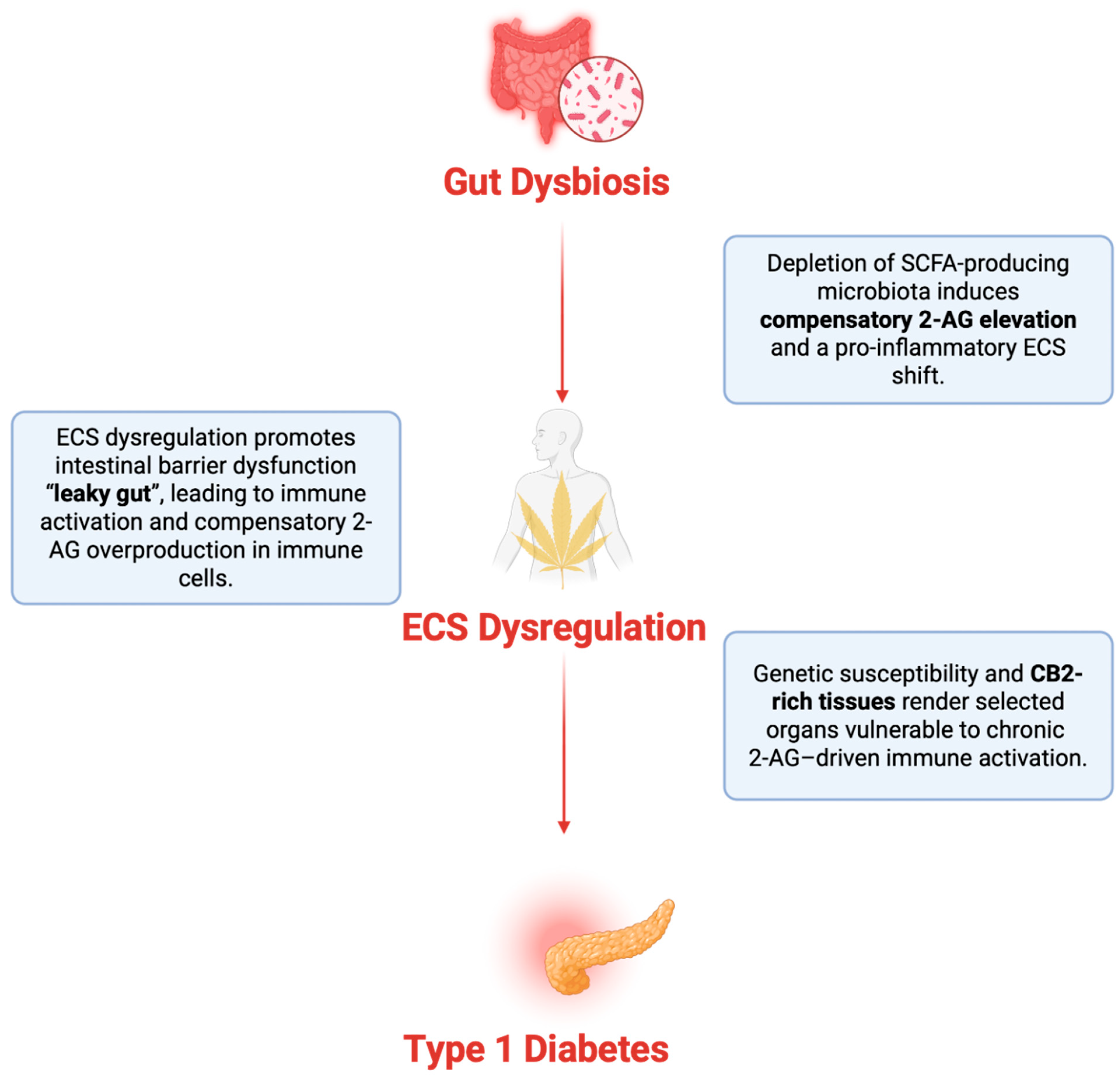
Figure 1: Gut dysbiosis–driven dysregulation of the endocannabinoid system as an upstream mechanism in autoimmune disease. This schematic illustrates how intestinal dysbiosis, characterized by depletion of short-chain fatty acid (SCFA)–producing bacteria, acts as an upstream driver of endocannabinoid system (ECS) imbalance [15,17,18,19,20]. Figure created by the author using BioRender.com (BioRender, Toronto, ON, Canada; subscription license). The figure is an original schematic and was not adapted from any previously published figure.
1.1 Relationship to Prior Work
This review builds on the previously published ECBoM systems-biology perspective on type 1 diabetes, but it has a narrower mechanistic focus [9]. Whereas the earlier article introduced the endocannabinoidome–microbiota axis as a broad framework, the present review examines one specific branch of that model: the proposed lipid–calcium–redox pathway linking SCFA depletion, altered 2-AG/CB1 signaling, TRPV1-sensitive calcium handling, ER–mitochondrial stress, and ROS amplification. This manuscript is therefore intended as a focused mechanistic extension, not as a repetition of the previous perspective.
1.2 Literature Search Strategy
This manuscript is a narrative, mechanistic review and conceptual hypothesis paper. It does not report original experimental data and was not designed as a systematic review or meta-analysis. Relevant literature was identified through searches of PubMed/MEDLINE, Scopus, Web of Science, and Google Scholar, supplemented by reference-list screening. Searches combined terms related to type 1 diabetes, gut microbiota, SCFAs, the endocannabinoid system, 2-AG, CB1, CB2, TRPV1, ER stress, mitochondrial calcium, oxidative stress, and phytocannabinoids. Peer-reviewed primary studies, longitudinal T1D cohort studies, mechanistic animal and cellular studies, and relevant reviews were included. Evidence from non-T1D models was used only when it addressed candidate mechanisms of the proposed axis and is treated as hypothesis-generating rather than direct evidence of T1D pathogenesis. The search covered the period from database inception to May 2026, was restricted to English-language publications, and was last updated in May 2026. In prioritizing evidence, the highest weight was given to direct human T1D studies and longitudinal pre-seroconversion cohorts, followed by T1D-specific preclinical models; non-T1D mechanistic studies (including type 2 diabetes, neurodegeneration, and other metabolic-disease systems) were used only to illustrate candidate mechanisms. Throughout the manuscript, claims derived from preclinical, indirect, or non-T1D sources are identified as such, so that established evidence is distinguished from extrapolation, hypothesis, and author interpretation.
1.3 Non-ECS Upstream Hubs and Rationale for Focusing on the ECS
SCFAs, particularly butyrate, influence β-cell biology and immune tolerance through several well-characterized non-ECS pathways operating in parallel with ECS signaling. GPR43/FFAR2 and GPR41/FFAR3 mediate SCFA-driven peripheral regulatory T-cell (Treg) induction, GLP-1–dependent β-cell secretory effects, and attenuation of pro-inflammatory cytokine release [5,17], while butyrate’s class-I/II histone deacetylase (HDAC)-inhibitory activity reshapes chromatin in epithelial, immune, and β-cell compartments, with downstream consequences for Foxp3 stabilization and cellular stress-response programmes [5,21,22]. These routes contribute directly to SCFA-mediated tolerogenic effects and are not displaced by the ECS-centred axis proposed here. The ECS is emphasized in this review not as the sole mediator of SCFA loss, but as a candidate primary integrator of lipid, calcium, immune, and mitochondrial stress signals: 2-AG biosynthesis is strictly lipid-coupled (sn-2 arachidonoyl-DAG → sn-1-selective DAGLα/β) [13], CB1, CB2, and TRPV1 are co-distributed across immune, epithelial, metabolic, mitochondrial, and ER-membrane compartments [23], and downstream amplifiers—TRPV1-mediated calcium flux, mtCB1-driven respiratory suppression, and ROS–NF-κB activation—are continuous outputs of sustained ECS tone rather than the transcriptional and immunoregulatory outputs typical of GPR41/43 and HDAC pathways [11]. Direct evidence that ECS imbalance precedes—rather than follows—metabolic stress in pre-seroconversion T1D is not yet available, and this framing therefore remains hypothesis-generating [10].
2 Subclinical Dysbiosis and Early SCFA Depletion in Type 1 Diabetes
Mounting longitudinal evidence indicates that gut microbial perturbations precede the onset of islet autoimmunity and clinical type 1 diabetes. Prospective cohort studies following genetically at-risk children have consistently demonstrated reduced microbial diversity and altered community structure prior to seroconversion. In particular, children who later develop T1D show a decline in butyrate-producing taxa and a shift toward a more inflammatory microbial profile months to years before diagnosis [2,24]. Functional metagenomic analyses further reveal diminished expression of genes involved in short-chain fatty acid biosynthesis during the preclinical phase, indicating that metabolic output is disrupted even when taxonomic changes are subtle [5,8].
As depicted in Fig. 2, among microbial metabolites, short-chain fatty acids—particularly butyrate—have emerged as central regulators of intestinal and systemic homeostasis. Butyrate supports epithelial barrier integrity by enhancing tight junction assembly and mucin production, thereby limiting microbial translocation and low-grade endotoxemia [4,8]. In parallel, SCFAs promote immune tolerance through induction of regulatory T cells and suppression of pro-inflammatory cytokine production via histone deacetylase inhibition and G-protein–coupled receptor signaling [7,25]. Although these immunological effects are well characterized, increasing attention has turned toward the metabolic consequences of SCFA deficiency.
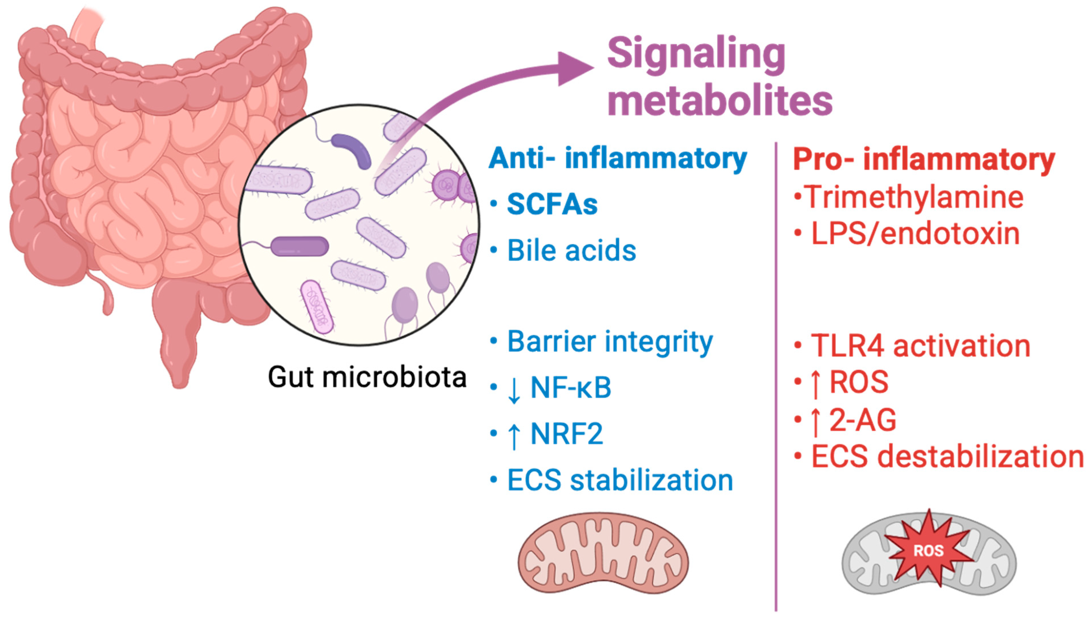
Figure 2: Gut microbiota-derived metabolites as regulators of redox, inflammatory, and endocannabinoid signaling. Gut microbiota communicate with host tissues through bioactive metabolites that exert divergent immunometabolic effects [3,26]. Anti-inflammatory metabolites, including short-chain fatty acids (SCFAs) and selected bile acids, promote epithelial barrier integrity, suppress NF-κB signaling, enhance NRF2-dependent antioxidant pathways, and stabilize endocannabinoid tone [10,24]. In contrast, pro-inflammatory metabolites such as lipopolysaccharide (LPS) and trimethylamine activate TLR4 signaling, increase reactive oxygen species (ROS) production, elevate 2-arachidonoylglycerol (2-AG) levels, and contribute to ECS destabilization. Upward arrows indicate increased pathway activity or molecular abundance, whereas downward arrows indicate reduced activity or suppression. Figure created by the author using BioRender.com (BioRender, Toronto, ON, Canada; subscription license). The figure is an original schematic and was not adapted from any previously published figure. Any visual resemblance to previously published schematics reflects the use of standard, licensed BioRender library elements rather than reproduction of published data or figures.
SCFAs influence host energy metabolism by activating AMP-activated protein kinase (AMPK), enhancing mitochondrial oxidative capacity, and stimulating peroxisome proliferator–activated receptor gamma (PPARγ), thereby supporting coordinated lipid oxidation and metabolic flexibility [21,27]. Through these pathways, SCFAs contribute to the stabilization of intracellular lipid flux and membrane phospholipid homeostasis. A decline in butyrate availability may therefore extend beyond barrier dysfunction, potentially altering cellular lipid handling and phospholipid remodeling processes that regulate downstream signaling networks [28,29].
Environmental exposures associated with increased T1D risk—including low-fiber dietary patterns, early-life antibiotic use, cesarean delivery, and pesticide-related metabolic disturbance—converge on reduced microbial SCFA production and altered early-life microbial maturation [5,30,31]. As pictured in Fig. 3: These converging influences support the concept that SCFA depletion represents a biologically coherent upstream event linking environmental pressure to metabolic and immune instability [28].
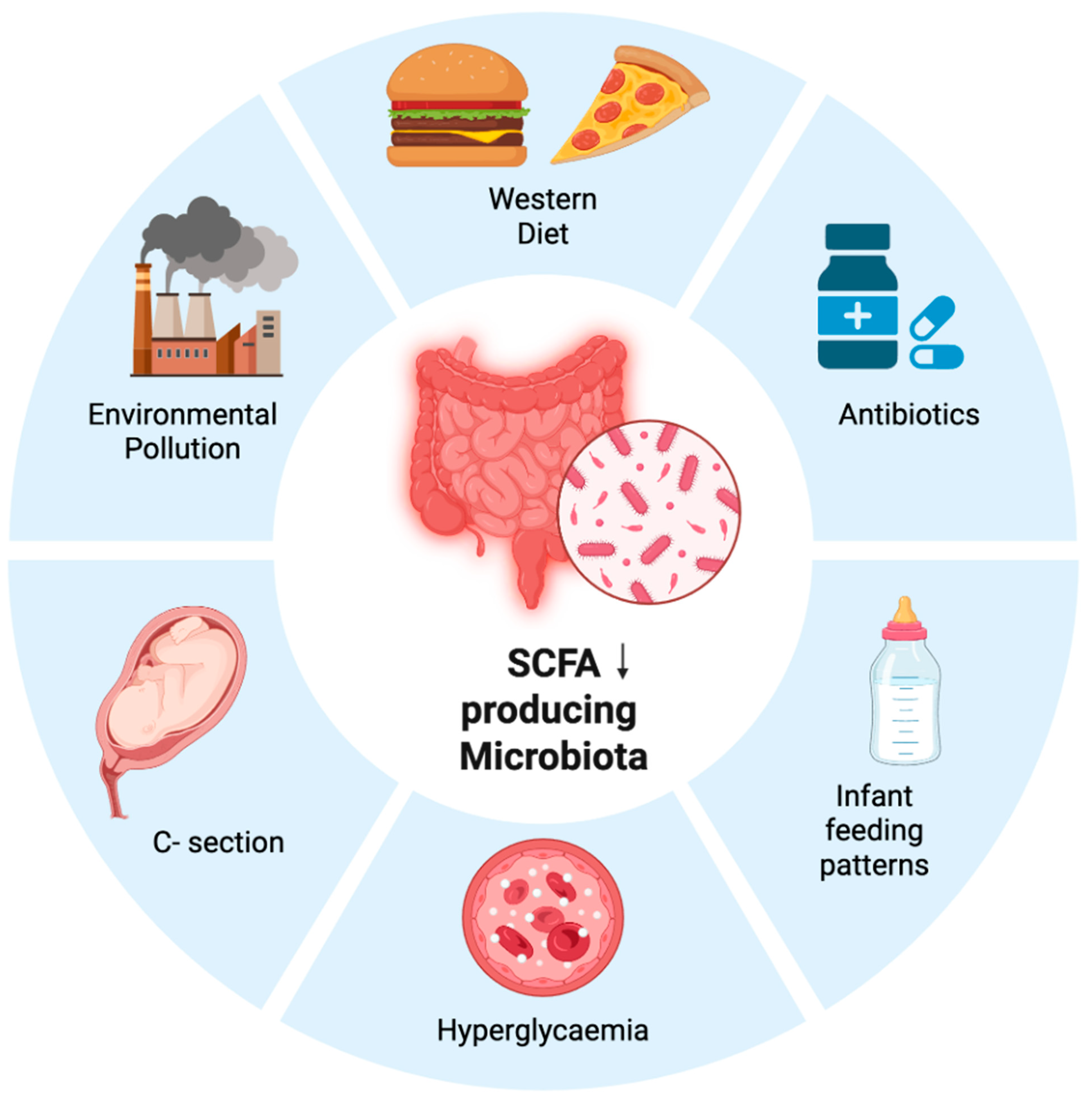
Figure 3: Environmental convergence on SCFA-producing microbiota depletion in type 1 diabetes. Diverse environmental and metabolic exposures—including Western diet, environmental pollution, antibiotic use, cesarean delivery, infant feeding patterns, and chronic hyperglycemia—converge on depletion of SCFA-producing microbiota in T1D [12,30,32]. Reduced abundance of butyrate-producing taxa and diminished SCFA availability represent a central upstream disturbance linking modern environmental pressures to intestinal dysbiosis and immunometabolic instability. The downward arrow indicates reduced abundance or depletion of SCFA-producing microbiota [10,29,31]. Figure created by the author using BioRender.com (BioRender, Toronto, ON, Canada; subscription license). The figure is an original schematic and was not adapted from any previously published figure.
Taken together, current evidence supports the view that subclinical dysbiosis in T1D is characterized not merely by compositional shifts but by functional impairment of SCFA biosynthesis [8,9]. This early decline in microbial-derived metabolic signaling may create a permissive environment for lipid remodeling and downstream perturbation of signaling systems tightly coupled to membrane phospholipid turnover, including the endocannabinoid system [10,15]. The mechanistic consequences of this lipid–endocannabinoid destabilization are addressed in the following section.
3 SCFA Loss and Lipid–Endocannabinoid Destabilization
Short-chain fatty acids are not only immunomodulatory metabolites but also key regulators of host lipid handling and intracellular energy balance. Through activation of AMP-activated protein kinase (AMPK) and peroxisome proliferator–activated receptor gamma (PPARγ), SCFAs promote coordinated lipid oxidation, limit ectopic lipid accumulation, and maintain metabolic flexibility [9,27,28,33]. Butyrate has also been shown to enhance mitochondrial oxidative capacity and improve coupling efficiency, thereby stabilizing redox balance under metabolic stress [8,34]. Although much of this work has been performed in metabolic disease models, the same regulatory nodes—AMPK, PPARγ, and mitochondrial respiration—are central to β-cell survival and immune cell activation [27,32].
Loss of SCFAs signaling, therefore, implies more than reduced barrier integrity; it suggests a shift in intracellular lipid flux and phospholipid remodeling. Membrane phospholipids serve as substrates for diacylglycerol (DAG) and 2-arachidonoylglycerol (2-AG) synthesis, placing lipid turnover at the core of endocannabinoid regulation. The endocannabinoid 2-AG is generated from DAG subpool via sn-1-selective diacylglycerol lipase α/β (DAGLα/β) and hydrolyzed primarily by monoacylglycerol lipase (MAGL), linking its abundance directly to membrane lipid dynamics and inflammatory phospholipid remodeling [35,36]. Perturbations in lipid handling can therefore shift the balance of endocannabinoid tone.
Under inflammatory or metabolic stress, 2-AG levels increase in multiple tissues, including immune and metabolic compartments [13]. Elevated 2-AG has been associated with enhanced cannabinoid receptor 1 (CB1) signaling, lipogenic bias, and altered mitochondrial respiration [37,38]. Importantly, CB1 receptors are localized not only at the plasma membrane but also at the mitochondrial membrane (mtCB1), where their activation directly modulates oxidative phosphorylation efficiency [11]. Sustained CB1 activation has been shown to reduce respiratory chain activity and increase susceptibility to oxidative stress, particularly under conditions of metabolic strain.
In the context of SCFA depletion, as pictured in Fig. 4, impaired AMPK and PPARγ signaling may promote increased availability of sn-2-arachidonoyl-enriched DAG substrates and favor 2-AG synthesis. Although direct studies linking SCFAs loss to 2-AG elevation in T1D are limited, mechanistic intersections between lipid flux, this specific DAG subpool, and endocannabinoid production support this possibility. SCFAs deficiency may thus destabilize the finely tuned DAGLα/β–MAGL equilibrium, shifting the system toward sustained 2-AG elevation [35,36,39].
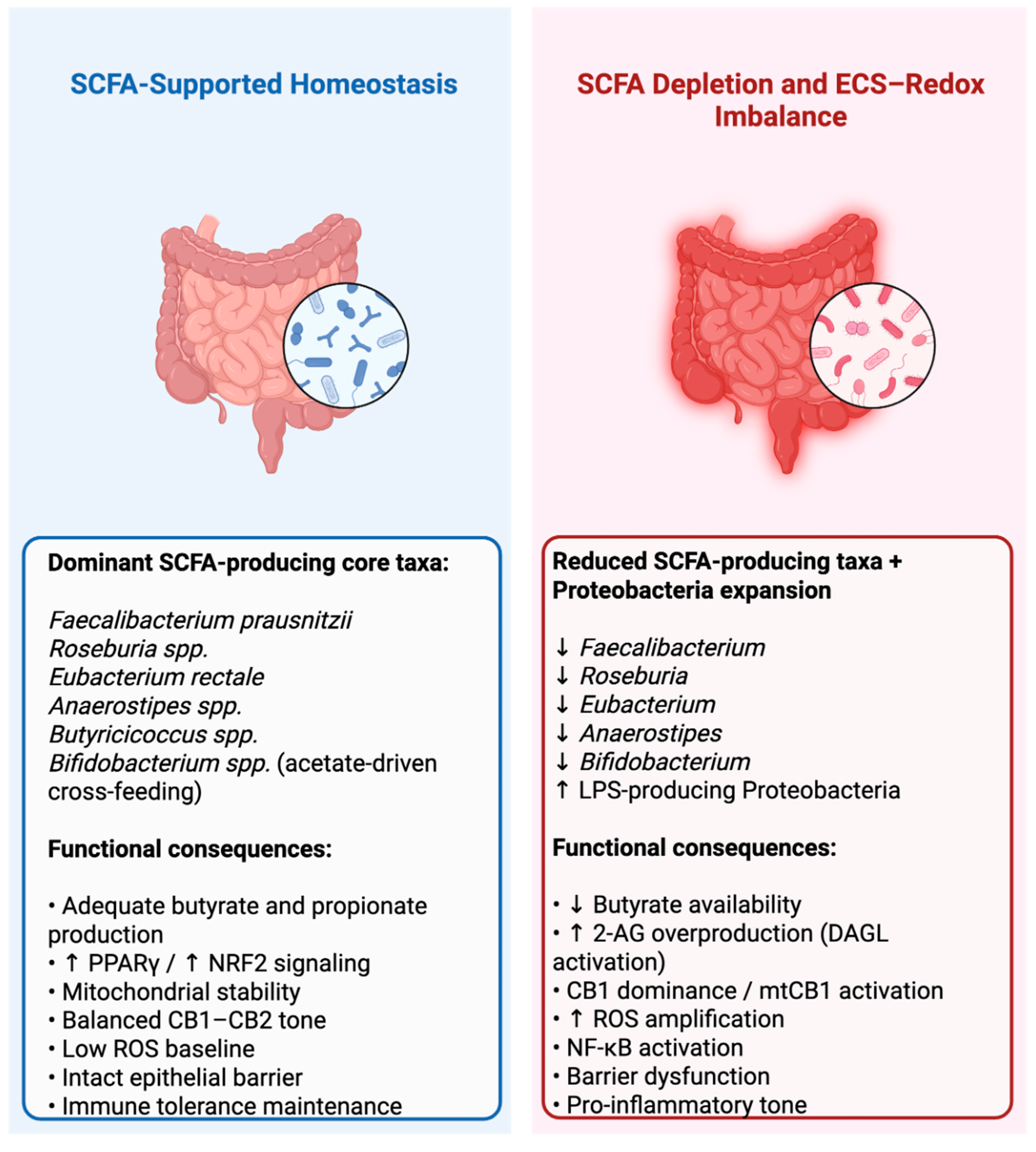
Figure 4: Eubiosis versus dysbiosis-driven redox–endocannabinoid imbalance in type 1 diabetes. The left panel depicts an SCFA-supported homeostatic state, in which dominant SCFA-producing core taxa—including Faecalibacterium prausnitzii, Roseburia spp., Eubacterium rectale, Anaerostipes spp., Butyricicoccus spp., and Bifidobacterium spp.—are associated with adequate butyrate and propionate production, epithelial mitochondrial stability, intact barrier function, immune tolerance maintenance, low reactive oxygen species (ROS) baseline, and balanced CB1–CB2 signaling. In this panel, upward arrows next to PPARγ and NRF2 indicate increased activation of antioxidant and metabolic regulatory pathways [10,12,40]. The right panel depicts a dysbiosis-associated state, characterized by reduced SCFA-producing taxa and expansion of LPS-producing Proteobacteria. Downward arrows next to Faecalibacterium, Roseburia, Eubacterium, Anaerostipes, and Bifidobacterium indicate reduced abundance of these taxa, whereas the upward arrow next to LPS-producing Proteobacteria indicates their increased abundance. Functionally, this state is proposed to involve reduced butyrate availability, increased 2-arachidonoylglycerol (2-AG) overproduction through DAGL activation, CB1 dominance, including mitochondrial CB1 (mtCB1) activation, increased ROS amplification, NF-κB activation, epithelial barrier dysfunction, and a pro-inflammatory tone [41,42,43,44]. Figure created by the author using BioRender.com (BioRender, Toronto, ON, Canada; subscription license). The figure is an original schematic and was not previously published. Any visual resemblance to previously published schematics reflects the use of standard, licensed BioRender library elements rather than reproduction of published data or figures.
Initially, increased 2-AG may represent a compensatory response aimed at buffering inflammatory or metabolic stress. However, chronic elevation can induce receptor asymmetry characterized by CB1 dominance and functional attenuation of CB2-mediated resolution pathways [10,45]. This altered endocannabinoid tone not only biases metabolic signaling but may also influence calcium-permeable channels such as TRPV1, which are sensitive to endocannabinoid modulation and membrane lipid composition. In this framework, SCFAs depletion becomes a metabolic destabilizer that perturbs lipid remodeling and shifts endocannabinoid tone toward sustained CB1 activation.
Proposed mechanistic model (Section 4, Section 5, Section 6 and Section 7). The four sections that follow develop the lipid–calcium–redox axis introduced above as a proposed mechanistic model, not as an established pathobiological sequence in human type 1 diabetes. Each step—chronic 2-AG elevation, CB1-dominant receptor asymmetry, TRPV1-mediated calcium dysregulation, ER–mitochondrial calcium overload, and mitochondrial CB1-driven ROS amplification—is grounded in evidence from chronic inflammation, neurodegeneration, metabolic disease, and immunometabolic models, but has not been directly demonstrated during the pre-seroconversion phase of human T1D. The mechanistic statements presented below should therefore be read as a coherent set of testable hypotheses anchored in T1D-relevant biology, rather than as confirmed steps in human disease pathogenesis. The model is intentionally constructed in a way that allows each node to be falsified or refined independently in T1D-specific preclinical and longitudinal human studies [10].
4 Chronic 2-AG Elevation and Receptor Asymmetry
Sustained elevation of 2-arachidonoylglycerol may reshape endocannabinoid signaling architecture. The immediate biosynthetic precursor of 2-AG is a specific subpool of diacylglycerol (DAG) carrying arachidonic acid esterified at the sn-2 stereospecific position of the glycerol backbone, hydrolyzed by sn-1-selective diacylglycerol lipase α/β (DAGLα/β); this stereospecificity, rather than DAG availability in general, may define the lipid bottleneck through which membrane remodeling translates into 2-AG production [10,36,46]. While acute increases in 2-AG may serve adaptive roles in limiting excessive inflammation, chronic elevation could promote persistent cannabinoid receptor 1 (CB1) activation and alter the balance between CB1- and CB2-mediated pathways. The proposed shift toward CB1-dominant tone is inferred largely from chronic inflammation, neurodegeneration, and metabolic-disease models; it has not been directly demonstrated during the pre-seroconversion phase of human T1D, and any metabolic or redox implications discussed below should be read accordingly.
CB1 activation in peripheral tissues can promote lipogenesis, suppress fatty acid oxidation, and reduce mitochondrial respiratory efficiency. Experimental studies have demonstrated that mitochondrial CB1 (mtCB1) signaling can impair oxidative phosphorylation by decreasing complex I activity and lowering ATP production [11,23]. In metabolic disease models, enhanced CB1 activity has been associated with increased oxidative stress and pro-inflammatory signaling [13] These findings suggest that sustained 2-AG elevation may progressively sensitize mitochondria to bioenergetic instability [36,47].
In parallel, chronic endocannabinoid stimulation by elevated 2-AG levels may induce regulatory mechanisms that attenuate CB2 signaling. Receptor phosphorylation by G protein–coupled receptor kinases (GRKs), followed by β-arrestin recruitment and internalization, can reduce CB2 surface expression and functional responsiveness under conditions of persistent activation. Because CB2 signaling is generally associated with anti-inflammatory and pro-resolution pathways in immune cells, its functional attenuation may further skew the system toward a pro-inflammatory bias.
The proposed receptor asymmetry—characterized by CB1 dominance and relative CB2 attenuation, a pattern inferred from chronic inflammation, immune-cell, and metabolic-disease models and not yet directly demonstrated in the pre-seroconversion phase of human type 1 diabetes [48] may have broader consequences beyond metabolic rewiring. Endocannabinoid receptors are functionally linked to intracellular calcium dynamics. CB1 activation can modulate voltage-gated calcium channels and intracellular calcium stores, while endocannabinoids can also interact with transient receptor potential channels, particularly TRPV1. Elevated 2-AG levels may therefore influence calcium-permeable channel activity both directly and indirectly.
Importantly, sustained CB1 activation in immune and metabolic tissues has been shown to promote NF-κB signaling and enhance inflammatory gene expression under stress conditions [49,50]. When combined with mitochondrial respiratory inefficiency, this may create a permissive environment for redox amplification.
As depicted in Fig. 5, within this framework, chronic 2-AG elevation does not simply enhance cannabinoid signaling; it may reorganize receptor hierarchy and prime intracellular calcium machinery for instability. The next step in this cascade involves the calcium-permeable channel TRPV1, proposed here as a candidate node at which lipid composition and endocannabinoid signaling may transition metabolic adaptation toward pathological calcium overload [10].
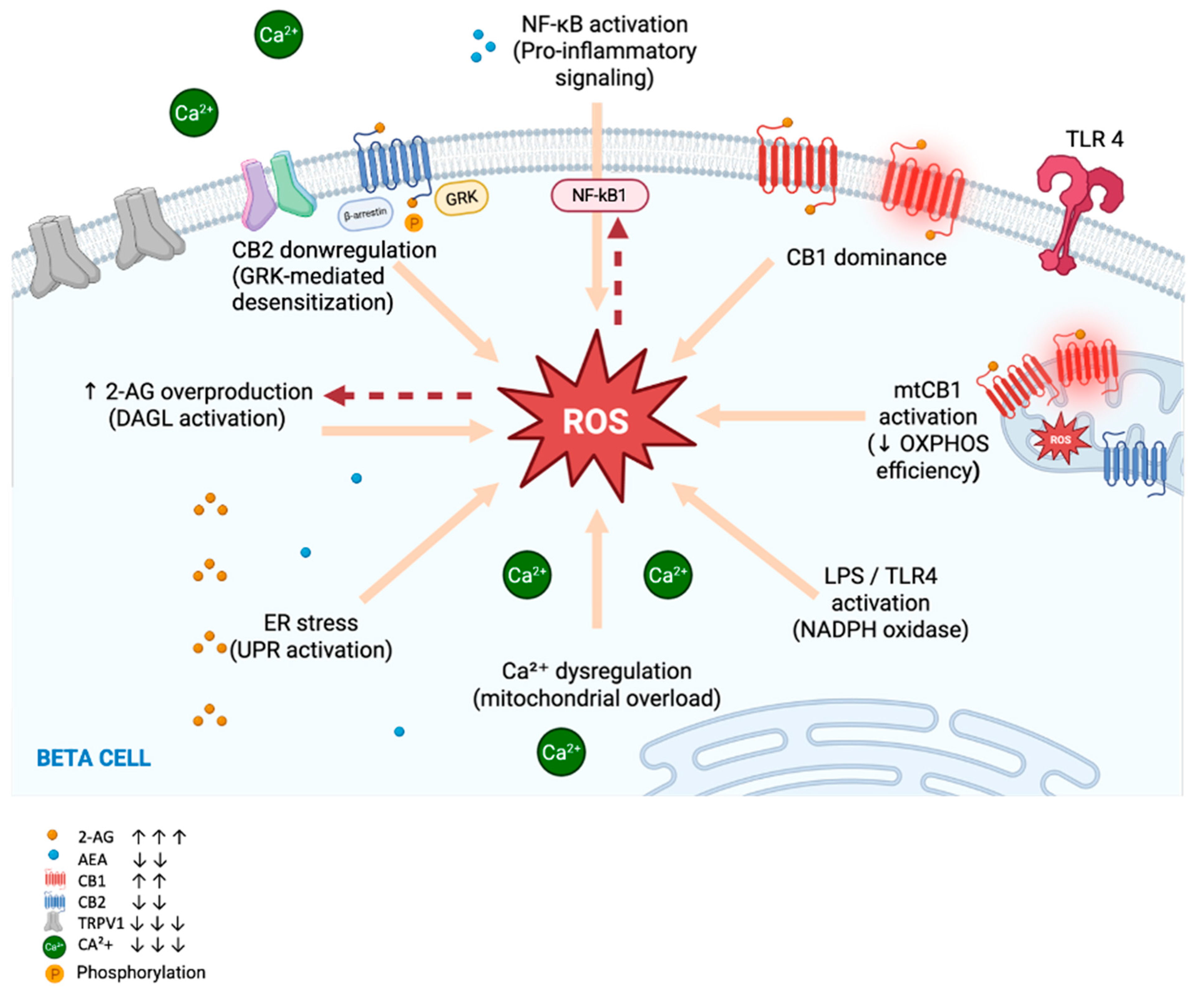
Figure 5: Endocannabinoid-driven redox amplification in T1D. In this proposed model, increased 2-arachidonoylglycerol (2-AG) production constitutes an upstream event that may promote CB1 dominance and mitochondrial CB1 (mtCB1) activation. Enhanced mtCB1 signaling can impair oxidative phosphorylation and increase mitochondrial ROS generation. Elevated ROS may further amplify NF-κB activation and sustain endocannabinoid turnover, establishing a self-reinforcing redox–ECS loop. Although ROS can also modulate ECS signaling, this framework positions 2-AG overproduction as a candidate upstream driver of redox amplification in T1D [10,40,41,43]. This schematic distinguishes established mitochondrial and inflammatory mechanisms from the ECS-specific hypothesis proposed in this review. The 2-AG–CB1/mtCB1–ROS feedback loop is presented as a hypothesis-generating model derived from T1D-relevant β-cell biology, endocannabinoid studies, and non-T1D inflammatory/metabolic models, and requires direct validation in T1D-specific systems. In the inset legend, upward arrows denote increased abundance or activity and downward arrows denote decreased abundance or activity of the indicated mediators (increased 2-AG and CB1; decreased anandamide [AEA], CB2, TRPV1, and Ca2+), with the number of arrows reflecting the relative magnitude of change; the encircled symbol denotes receptor phosphorylation. The red, dashed arrow from ROS to NF-κB denotes that elevated reactive oxygen species promote activation of the NF-κB signaling pathway. Figure created by the author using BioRender.com (BioRender, Toronto, ON, Canada; subscription license). The figure is an original schematic and was not adapted from any previously published figure.
5 TRPV1 Destabilization and Early Calcium Dysregulation
The transient receptor potential vanilloid 1 (TRPV1) channel is positioned at the intersection of lipid signaling and calcium homeostasis and is therefore proposed here as a candidate node for further mechanistic investigation, not as an established mediator of early T1D pathogenesis. Originally characterized as a capsaicin-sensitive nociceptive receptor, TRPV1 is now recognized as a broadly expressed, calcium-permeable channel present in intestinal epithelium, immune cells, and pancreatic β-cells. Direct evidence for TRPV1 dysregulation during the pre-seroconversion phase of human T1D remains limited; the rationale for considering this channel here derives mainly from its lipid sensitivity and from the convergence between endocannabinoid biology, calcium handling, and immune activation [51,52,53].
TRPV1 is highly sensitive to membrane lipid composition and is functionally coupled to the endocannabinoid system. Endocannabinoids, including anandamide and 2-arachidonoylglycerol, can modulate TRPV1 activity either directly or indirectly, and the channel itself is influenced by phospholipid remodeling and arachidonic acid availability [53]. Under conditions of sustained 2-AG elevation and increased arachidonic acid turnover, TRPV1 gating properties may shift toward enhanced activation or altered desensitization kinetics [33,52].
Acute TRPV1 activation induces transient calcium influx that participates in physiological signaling. However, chronic or excessive stimulation can disrupt calcium oscillatory control. Sustained calcium entry through TRPV1 can increase cytosolic Ca2+ load and can sensitize intracellular calcium release channels, including inositol 1,4,5-trisphosphate receptors (IP3Rs), thereby amplifying endoplasmic reticulum (ER) calcium release [54]. In immune and epithelial cells, dysregulated TRPV1 activity has been associated with enhanced pro-inflammatory signaling and increased cellular stress [55].
As depicted in Fig. 6. In pancreatic β-cells, calcium oscillations are tightly linked to insulin secretion and mitochondrial coupling efficiency. Perturbation of calcium dynamics can impair ATP production and promote oxidative stress [56]. Although direct evidence linking TRPV1 dysregulation to β-cell failure in T1D remains limited, the channel’s presence in both metabolic and immune compartments suggests that its destabilization may represent a convergent mechanism [7].
Importantly, prolonged activation of TRPV1 can trigger calcium-dependent desensitization mechanisms involving calmodulin binding and channel phosphorylation, which can paradoxically result in unstable or dysregulated calcium signaling rather than simple attenuation. In the context of chronic endocannabinoid elevation and altered membrane lipid composition, TRPV1 may therefore transition from a transient signaling mediator to a source of calcium instability [55].
Within the proposed model, sustained 2-AG elevation and CB1 dominance create a permissive lipid environment that enhances TRPV1 sensitivity. Early increases in calcium influx through TRPV1 may precede overt mitochondrial dysfunction, priming the ER–mitochondrial axis for overload [10].
6 ER Lipid Stress and ER–Mitochondrial Calcium Dysregulation
Sustained perturbation of lipid remodeling and calcium influx does not remain confined to the plasma membrane. As 2-AG turnover increases and monoacylglycerol lipase activity liberates arachidonic acid, membrane phospholipid composition progressively shifts. Enrichment of arachidonic acid within endoplasmic reticulum (ER) membranes may alter membrane fluidity and sensitize unfolded protein response (UPR) pathways, even in the absence of overt protein misfolding [57]. Lipid bilayer stress is now recognized as a distinct trigger of ER stress signaling, linking phospholipid remodeling directly to cellular stress responses. Notably, recent work in non-obese diabetic mice has shown that mesenchymal stromal cell infusion can attenuate UPR activation and preserve β-cell mass in T1D, indicating that the β-cell ER stress arm of this axis is experimentally modifiable [58].
It is important here to distinguish between canonical ER and mitochondrial calcium biology, which is well established and independent of the ECS, and the ECS-specific hypotheses proposed in this review. Canonically, the ER functions as the principal intracellular calcium reservoir, regulating cytosolic oscillations through SERCA-mediated uptake and IP3R/RyR-mediated release; calcium is then transferred to mitochondria across MAM contacts through the IP3R–GRP75–VDAC1–MCU axis. The ECS-specific element proposed here is that 2-AG/CB1-driven membrane remodeling and TRPV1 sensitization may bias these otherwise canonical calcium-handling mechanisms toward overload in the context of T1D, not that ECS itself constitutes the calcium-handling apparatus [14,59,60,61].
Building on this canonical framework, the ECS-driven addition proposed here is that sustained 2-AG turnover and arachidonic-acid enrichment of ER membranes may impair SERCA efficiency and promote enhanced IP3R-mediated calcium release [57,59], leading to gradual depletion of ER calcium stores and increased cytosolic calcium load [58]. When combined with sustained TRPV1-mediated calcium influx, this may generate a dual-source calcium burden that, within the proposed model, links ECS-driven lipid remodeling to otherwise canonical ER calcium-handling machinery [52].
The structural and functional interface between the ER and mitochondria, known as mitochondria-associated membranes (MAMs) [61], plays a central role in calcium transfer and metabolic coordination-a canonical observation that is independent of the ECS hypothesis advanced here [14,62]. Under physiological conditions, transient calcium transfer from the ER to mitochondria stimulates oxidative phosphorylation and supports ATP production. However, excessive or sustained calcium flux can lead to mitochondrial calcium overload, destabilization of the mitochondrial membrane potential (ΔΨm), and impairment of respiratory chain function. MAM hyperactivation under stress conditions has been linked to both metabolic disease and inflammatory signaling [63].
In β-cells, where mitochondrial ATP production is tightly coupled to calcium oscillations and insulin secretion, sustained mitochondrial calcium overload is particularly deleterious. Excessive calcium entry into the mitochondrial matrix enhances electron transport chain instability, increases electron leak, and promotes reactive oxygen species generation [33,54]. In immune cells, similar calcium-dependent mitochondrial stress can amplify pro-inflammatory signaling and cytokine production.
In addition to altering membrane fluidity, sustained liberation of arachidonic acid (AA) from phospholipid pools may create a pro-oxidative intracellular milieu. Excess free AA can undergo enzymatic and non-enzymatic oxidation, generating bioactive eicosanoids and lipid peroxidation products that further amplify inflammatory signaling and oxidative burden [36,37]. Accumulation of AA within ER and mitochondrial membranes increases susceptibility to lipid peroxidation and destabilizes membrane-associated protein complexes, thereby sensitizing both compartments to stress-induced dysfunction [37]. Thus, in this proposed model, chronic 2-AG turnover does not merely reflect altered endocannabinoid tone; it could actively reshape intracellular lipid architecture, promoting ER stress, mitochondrial vulnerability, and redox amplification.
Within the proposed framework, ER lipid stress and MAM hyperactivation represent the transition point at which early ECS–TRPV1 destabilization evolves into bioenergetic collapse. Calcium overload is no longer a transient signaling fluctuation but becomes structurally embedded within the ER–mitochondrial axis [10,37,54].
7 Mitochondrial CB1 Signaling, ROS Amplification and NF-κB Activation
As calcium overload becomes embedded within the ER–mitochondrial axis, mitochondrial signaling is further reshaped by sustained endocannabinoid activation. Cannabinoid receptor 1 is not confined to the plasma membrane; a functionally active pool localizes to the outer mitochondrial membrane, where it directly modulates respiratory chain activity [11,14]. Activation of mitochondrial CB1 (mtCB1) has been shown to reduce complex I–dependent respiration, decrease ATP production, and alter mitochondrial membrane potential [11,14]. Under physiological conditions, this modulation may fine-tune metabolic output. Under chronic stimulation, however, it can predispose mitochondria to bioenergetic instability. Disturbed mitochondrial dynamics and impaired mitophagy quality control may further sensitize this axis to chronic stress [64]. In parallel, gut–mitochondria stress models from other disease contexts, including neurodegeneration, suggest that microbial metabolite scarcity may contribute to mitochondrial bioenergetic failure [14,32,65].
When mitochondrial CB1 activation coincides with sustained calcium influx, the combined stress may become particularly deleterious. Excess matrix calcium enhances the activity of dehydrogenases but, beyond a threshold, destabilizes electron transport chain integrity, increases electron leak from complexes I and III, and promotes superoxide formation. This process may transform mitochondria from adaptive energy hubs into sources of reactive oxygen species (ROS).
Elevated ROS levels activate redox-sensitive transcription factors, most notably nuclear factor kappa B (NF-κB), which orchestrates inflammatory gene expression. NF-κB activation enhances cytokine production, promotes further phospholipid turnover, and increases inflammatory lipid mediator synthesis [26]. Because phospholipid remodeling feeds directly into 2-arachidonoylglycerol synthesis, this may create a feedback loop linking oxidative stress to further endocannabinoid elevation.
As depicted in Fig. 6, in immune cells, ROS-dependent NF-κB activation may amplify antigen presentation and pro-inflammatory signaling, reinforcing autoimmune progression. In β-cells, which possess relatively limited antioxidant capacity, sustained ROS exposure exacerbates mitochondrial dysfunction and sensitizes cells to apoptosis [66]. Thus, mitochondrial redox amplification contributes both to immune escalation and to β-cell vulnerability. In type 2 diabetes models, endocannabinoid-driven activation of the NLRP3 inflammasome in infiltrating macrophages has been linked to β-cell loss [67]; whether an analogous mechanism operates in T1D remains untested.
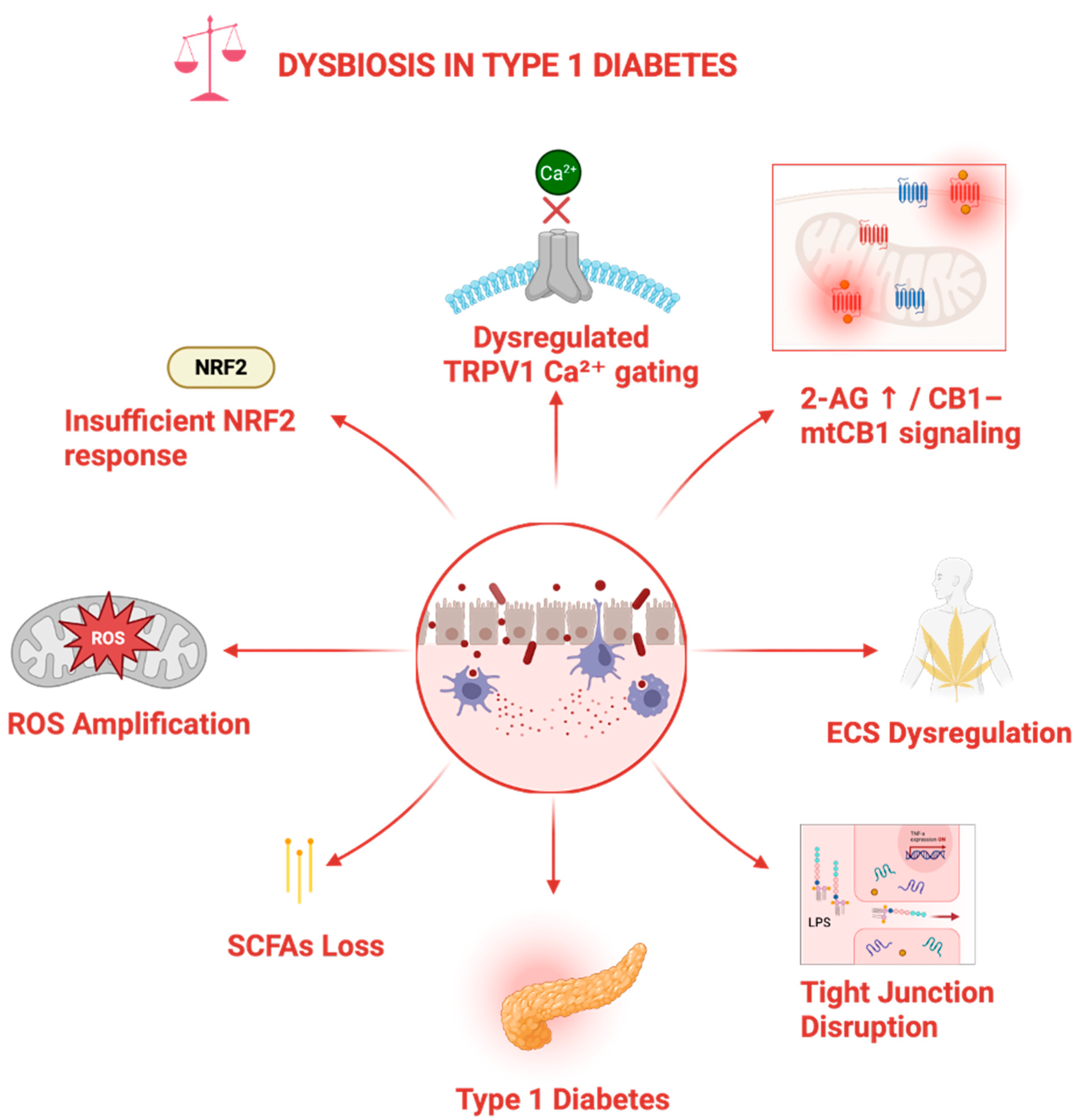
Figure 6: Dysbiosis-driven redox and endocannabinoid imbalance in T1D. Intestinal dysbiosis, characterized by loss of short-chain fatty acid (SCFAs)–producing microbiota, is proposed to promote epithelial tight junction disruption, inflammatory activation, endocannabinoid system (ECS) dysregulation, and mitochondrial reactive oxygen species (ROS) amplification. The upper pathway modules summarize three candidate mechanistic links emphasized in this review: insufficient NRF2-mediated antioxidant compensation, dysregulated TRPV1-mediated Ca2+ handling, and 2-AG–CB1/mtCB1 signaling [10,11,26,38,68]. This figure is a hypothesis-generating schematic and does not indicate that the complete sequence has been directly demonstrated in human T1D. The upward arrow next to 2-AG denotes increased 2-arachidonoylglycerol abundance and signaling. Figure created by the author using BioRender.com (BioRender, Toronto, ON, Canada; subscription license). The figure is an original schematic and was not adapted from any previously published figure.
Within this integrative model, oxidative stress is repositioned as a downstream amplifier rather than a primary driver of disease initiation [37,54]. Once established, however, redox signaling may reinforce phospholipid turnover and sustain 2-AG elevation, embedding the system in a self-perpetuating destabilized state.
8 Phytocannabinoids as Experimental Probes for Testing the Proposed ECS–Calcium–Redox Model
The lipid–calcium–redox axis developed in Section 4, Section 5, Section 6 and Section 7 generates testable predictions at several proposed nodes, including altered 2-AG signaling, CB1/CB2 asymmetry, TRPV1-sensitive calcium dysregulation, ER–mitochondrial overload, and ROS/NF-κB amplification. As depicted in Fig. 7, these nodes are presented as experimentally testable points within a hypothesis-generating model, not as validated therapeutic targets [10].
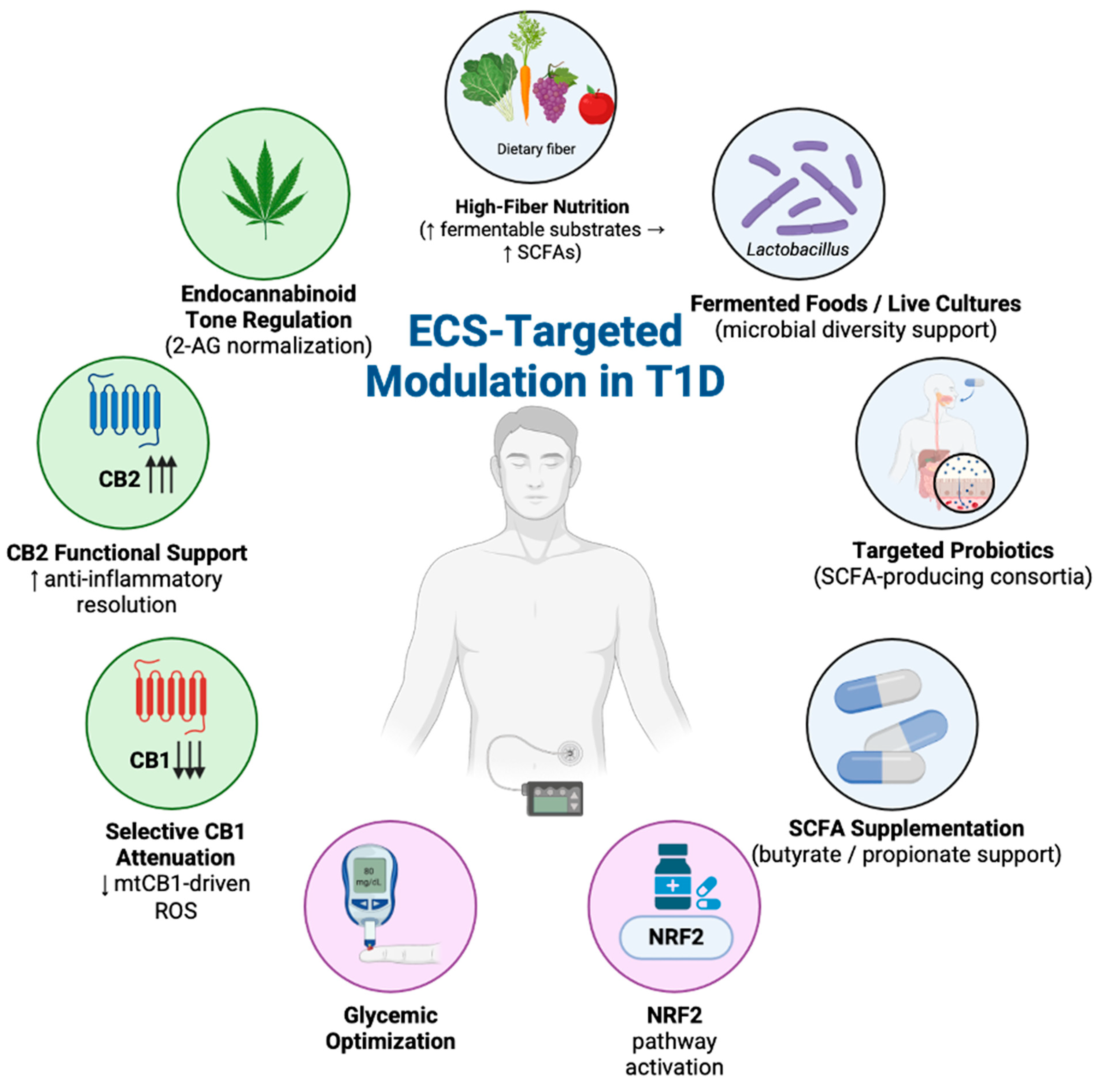
Figure 7: ECS-targeted modulation within a multilevel microbiota–redox experimental framework in type 1 diabetes. The schematic summarizes proposed microbiota-, metabolic-, redox-, and ECS-directed intervention nodes in T1D. High-fiber nutrition, fermented foods/live cultures, targeted probiotics, and SCFAs supplementation are shown as candidate, experimentally testable nodes for supporting microbial short-chain fatty acid (SCFAs) production and epithelial stability. Glycemic optimization and NRF2 pathway activation are shown as metabolic and antioxidant support mechanisms. Endocannabinoid tone regulation, CB2 functional support, and selective CB1 attenuation are shown as candidate ECS-directed nodes that might reduce mitochondrial CB1 (mtCB1)-driven reactive oxygen species (ROS) amplification. Upward arrows indicate increased substrate availability, pathway activation, or receptor support, whereas downward arrows indicate reduced receptor activity or downstream oxidative output [10,40,68] The nodes shown represent research gaps rather than validated interventions: their influence on endocannabinoid signaling in T1D has not been established and requires dedicated study.
The role of phytocannabinoid-sensitive signaling in T1D remains poorly defined. Given this limited disease-specific evidence, the present discussion, together with the candidate nodes illustrated in Fig. 7 and Fig. 8, is intentionally confined to conceptual, experimentally testable elements and is not intended to indicate translational, therapeutic, or clinical applicability in T1D. Accordingly, phytocannabinoids are discussed here only as mechanistic probes for testing selected nodes of the proposed ECS–calcium–redox axis, not as candidate therapeutic agents. No clinical use of phytocannabinoids in T1D is proposed or implied; the comparatively large experimental literature on Δ9-THC and CBD reflects their value as pharmacological tools and should not be read as evidence of disease-specific therapeutic benefit or safety in T1D. Δ9-THC, as a partial CB1/mtCB1 agonist, may help perturb mitochondrial CB1-dependent oxidative phosphorylation and ROS output [11,38], whereas CBD, through modulation of CB2, TRPV1, and indirect CB1-related effects, may help interrogate receptor asymmetry, calcium influx, ER stress, and NF-κB signaling [40,54,55,69,70]. In particular, it remains unknown whether cannabinoid-sensitive pathways can modulate TRPV1-dependent calcium handling, ER stress, mitochondrial redox output, or immune activation in T1D-specific experimental models. A recent systematic review of high-dose oral Δ9-THC studies is cited only to indicate that controlled laboratory exposure has been examined in cannabis-experienced individuals; these data are not T1D-specific and should not be interpreted as evidence of safety or therapeutic relevance in T1D [71]. As depicted in Fig. 8, abnormal cannabidiol findings in NOD mice provide preclinical proof-of-principle that cannabinoid-sensitive immune and β-cell stress pathways can be experimentally perturbed in autoimmune diabetes models, but they do not constitute clinical evidence for phytocannabinoid therapy in humans [72]. More fundamentally, Fig. 7 and Fig. 8 are intended as a map of current research gaps rather than a therapeutic roadmap: the microbiota-, metabolic-, redox-, and ECS-directed factors they depict have not been systematically studied in relation to endocannabinoid signaling in T1D. The inputs highlighted in Fig. 7 are proposed to modulate the ECS components shown in Fig. 8, yet whether and how each of these inputs influences endocannabinoid tone in the T1D context remains unestablished. Likewise, the cannabidiol (CBD) and Δ9-THC effects summarized in Fig. 8 are derived from other diseases and non-T1D models, and their actions on the ECS in T1D have not been characterized. These open questions—alongside the still-undemonstrated influence of SCFA depletion and gut microbiota on the ECS in human T1D—are the central evidence gaps that this review seeks to make explicit. Future work should prioritize T1D-specific preclinical validation before any clinical or translational interpretation is considered.
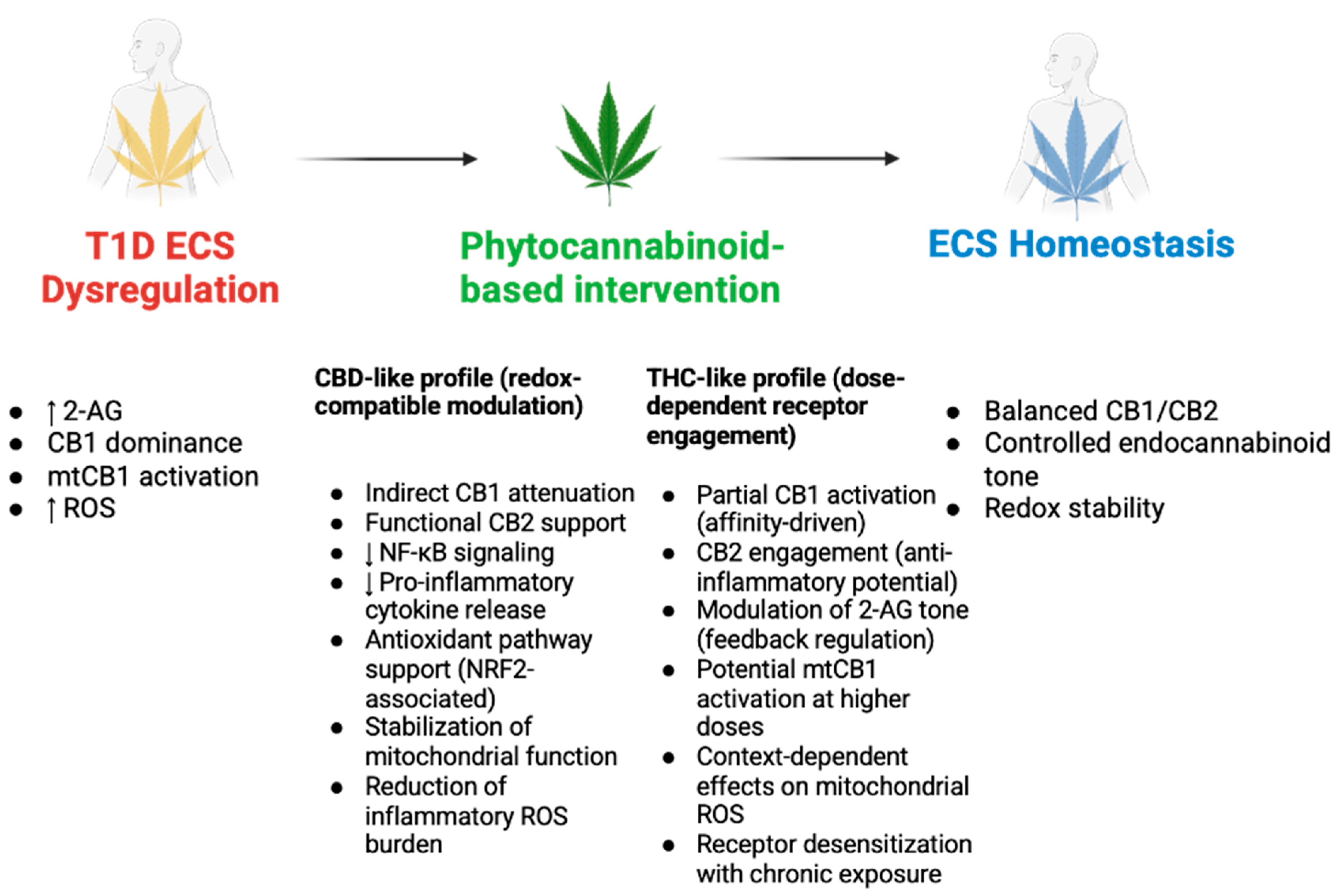
Figure 8: Conceptual framework for phytocannabinoid-guided modulation of endocannabinoid dysregulation in type 1 diabetes. This schematic presents a proposed experimental framework in which phytocannabinoid-based modulation is used to interrogate endocannabinoid system (ECS) dysregulation in type 1 diabetes. The left panel summarizes the proposed dysregulated state, in which upward arrows denote increased 2-arachidonoylglycerol (2-AG) tone, cannabinoid receptor 1 (CB1) dominance, mitochondrial CB1 (mtCB1) activation, and reactive oxygen species (ROS) amplification [10,68,73]. The middle panel distinguishes CBD-like profiles, proposed to reduce NF-κB signaling, pro-inflammatory cytokine release, and inflammatory ROS burden, while supporting CB2-mediated immunoregulation, antioxidant pathway activity, and mitochondrial stabilization [70,72]. THC-like profiles are presented as dose-dependent experimental modulators that may engage CB1/CB2 receptors, influence 2-AG feedback regulation, alter mitochondrial ROS generation in a context-dependent manner, and induce receptor desensitization during chronic exposure [71,73]. The right panel represents the hypothesized target state of ECS homeostasis, characterized by balanced CB1/CB2 signaling, controlled endocannabinoid tone, and improved redox stability [15,40]. Figure created by the author using BioRender.com (BioRender, Toronto, ON, Canada; subscription license). The figure is an original schematic and was not adapted from any previously published figure. The CBD- and THC-associated effects shown derive from non-T1D models; their actions on the ECS in T1D are unverified and represent research gaps requiring T1D-specific investigation.
Safety, ethical, and regulatory considerations. To close this research gap in an ethically and legally acceptable manner, future work should proceed through a stepwise validation pathway. First, observational ECBoM profiling in individuals with T1D and at-risk groups could determine whether endocannabinoid tone, SCFA availability, gut microbiota composition, inflammatory markers, and β-cell stress markers form a reproducible disease-associated signature [5,10,15,74,75]. Second, T1D-specific preclinical studies, particularly in NOD mice, should test whether defined cannabinoid-related probes can modify insulitis, β-cell stress, endocannabinoid signaling, and mitochondrial redox outcomes under controlled experimental conditions [72]. Complementary diabetic or prediabetic rodent/islet studies of Δ9-THC may help define vascular, metabolic, and β-cell-related endpoints before any clinical translation is considered [73,76]. Human tolerability data may inform laboratory study design but cannot substitute for disease-specific safety validation in T1D [71].
Several limitations of this review should be acknowledged. First, the proposed microbiota–endocannabinoid system (ECS)–calcium–redox axis is presented as a hypothesis-generating framework rather than an established pathogenic sequence, and no single human study has yet demonstrated the complete cascade in type 1 diabetes (T1D). Second, and most importantly, there is currently no direct longitudinal evidence linking short-chain fatty acid (SCFA) depletion to ECS dysregulation and oxidative stress in human T1D; the temporal and causal ordering proposed here therefore remains inferential. Third, several mechanistic transitions—including SCFA-driven 2-AG overproduction, CB1 dominance, TRPV1 destabilization, and ER–mitochondrial calcium collapse—are supported largely by indirect, preclinical, or non-T1D evidence (including type 2 diabetes, neurodegeneration, and other metabolic-disease models), and their extrapolation to T1D requires caution. Fourth, as a narrative review, the literature search was not systematic and is therefore subject to selection bias. Finally, the discussion of phytocannabinoids is intended only to identify experimental probes for testing the proposed model and should not be interpreted as evidence of therapeutic efficacy or safety in T1D. Collectively, these limitations define the principal directions for the T1D-specific preclinical and longitudinal human studies required to test, refine, or refute the proposed model.
10 Conclusion: Microbiota–ECS–Oxidative Stress Axis in Type 1 Diabetes
This review proposes a microbiota–endocannabinoid system (ECS)–calcium–redox framework in type 1 diabetes (T1D) in which oxidative stress represents a downstream consequence rather than a primary trigger. In this framework, the ECS functions as the central integrator, translating microbial perturbation into intracellular signaling instability.
Chronic 2-AG turnover may liberate arachidonic acid, reshaping ER and mitochondrial membranes and sensitizing them to lipid bilayer stress. Thus, oxidative stress in T1D can be understood as the bioenergetic endpoint of microbiota–ECS destabilization.
Positioning the ECS at the center of this axis carries mechanistic implications. In this hierarchy, mitigation of oxidative stress would be explored upstream through restoration of lipid and calcium stability rather than through antioxidant intervention alone, however this remains a hypothesis requiring disease-specific validation.
Future research should directly investigate microbiota–ECS interplay in preclinical T1D and determine whether targeted modulation of ECS signaling can stabilize mitochondrial resilience before autoimmune escalation. Within this context, phytocannabinoids represent mechanistically informative tools, rather than therapeutic recommendations. Δ9-tetrahydrocannabinol (THC), through partial CB1 agonism and engagement of mitochondrial CB1 receptors, may help to interrogate CB1-dependent bioenergetic regulation, while cannabidiol (CBD) may inform receptor asymmetry and TRPV1-mediated calcium dynamics. Carefully designed translational studies are warranted to assess whether modulation of the microbiota–ECS–calcium–redox axis can limit oxidative and inflammatory progression in early T1D.
Acknowledgement:
Funding Statement: The author received no specific funding for this study.
Availability of Data and Materials: Not applicable.
Ethics Approval: Not applicable.
Conflicts of Interest: The author declares no conflicts of interest.
References
1. Aamodt KI , Powers AC . The pathophysiology, presentation and classification of Type 1 diabetes. Diabetes Obes Metab. 2025; 27( S6): 15– 27. doi:10.1111/dom.16628. [Google Scholar] [CrossRef]
2. Kostic AD , Gevers D , Siljander H , Vatanen T , Hyötyläinen T , Hämäläinen AM , et al. The dynamics of the human infant gut microbiome in development and in progression toward type 1 diabetes. Cell Host Microbe. 2015; 17( 2): 260– 73. doi:10.1016/j.chom.2015.01.001. [Google Scholar] [CrossRef]
3. Lau WL , Tran T , Rhee CM , Kalantar-Zadeh K , Vaziri ND . Diabetes and the gut microbiome. Semin Nephrol. 2021; 41( 2): 104– 13. doi:10.1016/j.semnephrol.2021.03.005. [Google Scholar] [CrossRef]
4. Palmnäs-Bédard MS , Costabile G , Vetrani C , Åberg S , Hjalmarsson Y , Dicksved J , et al. The human gut microbiota and glucose metabolism: A scoping review of key bacteria and the potential role of SCFAs. Am J Clin Nutr. 2022; 116( 4): 862– 74. doi:10.1093/ajcn/nqac217. [Google Scholar] [CrossRef]
5. Vatanen T , Franzosa EA , Schwager R , Tripathi S , Arthur TD , Vehik K , et al. The human gut microbiome in early-onset type 1 diabetes from the TEDDY study. Nature. 2018; 562( 7728): 589– 94. doi:10.1038/s41586-018-0620-2. [Google Scholar] [CrossRef]
6. Ratajczak W , Rył A , Mizerski A , Walczakiewicz K , Sipak O , Laszczyńska M . Immunomodulatory potential of gut microbiome-derived short-chain fatty acids (SCFAs). Acta Biochim Pol. 2019; 66( 1): 1– 12. doi:10.18388/abp.2018_2648. [Google Scholar] [CrossRef]
7. Zhou H , Sun L , Zhang S , Zhao X , Gang X , Wang G . Evaluating the causal role of gut microbiota in type 1 diabetes and its possible pathogenic mechanisms. Front Endocrinol. 2020; 11: 125. doi:10.3389/fendo.2020.00125. [Google Scholar] [CrossRef]
8. Fu X , Ren X , Zhao M , Li L , Zhou Y , Lu Y , et al. Disruption of intestinal barrier and dysbiosis of gut microbiota in an experimental rhesus macaque model with 6-year diabetes mellitus. Exp Anim. 2025; 74( 4): 384– 98. doi:10.1538/expanim.24-0125. [Google Scholar] [CrossRef]
9. Garcia-Gutierrez E , O’Mahony AK , dos Santos RS , Marroquí L , Cotter PD . Gut microbial metabolic signatures in diabetes mellitus and potential preventive and therapeutic applications. Gut Microbes. 2024; 16( 1): 2401654. doi:10.1080/19490976.2024.2401654. [Google Scholar] [CrossRef]
10. Łukowski W . Reframing type 1 diabetes through the endocannabinoidome-microbiota axis: A systems biology perspective. Front Endocrinol. 2025; 16: 1576419. doi:10.3389/fendo.2025.1576419. [Google Scholar] [CrossRef]
11. Hebert-Chatelain E , Desprez T , Serrat R , Bellocchio L , Soria-Gomez E , Busquets-Garcia A , et al. A cannabinoid link between mitochondria and memory. Nature. 2016; 539( 7630): 555– 9. doi:10.1038/nature20127. [Google Scholar] [CrossRef]
12. Siljander H , Jason E , Ruohtula T , Selvenius J , Koivusaari K , Salonen M , et al. Effect of early feeding on intestinal permeability and inflammation markers in infants with genetic susceptibility to type 1 diabetes: A randomized clinical trial. J Pediatr. 2021; 238: 305– 11. doi:10.1016/j.jpeds.2021.07.042. [Google Scholar] [CrossRef]
13. di Marzo V , Silvestri C . Lifestyle and metabolic syndrome: Contribution of the endocannabinoidome. Nutrients. 2019; 11( 8): 1956. doi:10.3390/nu11081956. [Google Scholar] [CrossRef]
14. Giorgi V , Marotto D , Batticciotto A , Atzeni F , Bongiovanni S , Sarzi-Puttini P . Cannabis and autoimmunity: Possible mechanisms of action. Immunotargets Ther. 2021; 10: 261– 71. doi:10.2147/ITT.S267905. [Google Scholar] [CrossRef]
15. Cani PD , Plovier H , Van Hul M , Geurts L , Delzenne NM , Druart C , et al. Endocannabinoids—At the crossroads between the gut microbiota and host metabolism. Nat Rev Endocrinol. 2016; 12( 3): 133– 43. doi:10.1038/nrendo.2015.211. [Google Scholar] [CrossRef]
16. Aboutorabi RB , Mohebbi M , Ali Yaghoubi M , Rezaee AR , Sahebkar A . TRPV1 gene polymorphisms in patients with diabetes compared with healthy individuals. Comp Clin Pathol. 2017; 26( 4): 971– 4. doi:10.1007/s00580-017-2481-8. [Google Scholar] [CrossRef]
17. Arpaia N , Campbell C , Fan X , Dikiy S , van der Veeken J , DeRoos P , et al. Metabolites produced by commensal bacteria promote peripheral regulatory T-cell generation. Nature. 2013; 504( 7480): 451– 5. doi:10.1038/nature12726. [Google Scholar] [CrossRef]
18. Malenczyk K , Keimpema E , Piscitelli F , Calvigioni D , Björklund P , Mackie K , et al. Fetal endocannabinoids orchestrate the organization of pancreatic islet microarchitecture. Proc Natl Acad Sci U S A. 2015; 112( 45): E6185– 94. doi:10.1073/pnas.1519040112. [Google Scholar] [CrossRef]
19. Chiurchiù V , Battistini L , Maccarrone M . Endocannabinoid signalling in innate and adaptive immunity. Immunology. 2015; 144( 3): 352– 64. doi:10.1111/imm.12441. [Google Scholar] [CrossRef]
20. Richardson P , Ortori C , Barrett D , O’Sullivan S , Idris I . Endocannabinoids in aqueous humour of patients with or without diabetes. BMJ Open Ophthalmol. 2020; 5( 1): e000425. doi:10.1136/bmjophth-2019-000425. [Google Scholar] [CrossRef]
21. Takeda S , Ikeda E , Su S , Harada M , Okazaki H , Yoshioka Y , et al. Δ9-THC modulation of fatty acid 2-hydroxylase (FA2H) gene expression: Possible involvement of induced levels of PPARα in MDA-MB-231 breast cancer cells. Toxicology. 2014; 326: 18– 24. doi:10.1016/j.tox.2014.09.011. [Google Scholar] [CrossRef]
22. Furusawa Y , Obata Y , Fukuda S , Endo TA , Nakato G , Takahashi D , et al. Commensal microbe-derived butyrate induces the differentiation of colonic regulatory T cells. Nature. 2013; 504( 7480): 446– 50. doi:10.1038/nature12721. [Google Scholar] [CrossRef]
23. Aseer KR , Egan JM . An autonomous cannabinoid system in islets of Langerhans. Front Endocrinol. 2021; 12: 699661. doi:10.3389/fendo.2021.699661. [Google Scholar] [CrossRef]
24. Suriano F , Manca C , Flamand N , Depommier C , Van Hul M , Delzenne NM , et al. Exploring the endocannabinoidome in genetically obese (ob/ob) and diabetic (db/db) mice: Links with inflammation and gut microbiota. Biochim Biophys Acta Mol Cell Biol Lipids. 2022; 1867( 1): 159056. doi:10.1016/j.bbalip.2021.159056. [Google Scholar] [CrossRef]
25. Abdellatif AM , Sarvetnick NE . Current understanding of the role of gut dysbiosis in type 1 diabetes. J Diabetes. 2019; 11( 8): 632– 44. doi:10.1111/1753-0407.12915. [Google Scholar] [CrossRef]
26. Bielka W , Przezak A , Pawlik A . The role of the gut microbiota in the pathogenesis of diabetes. Int J Mol Sci. 2022; 23( 1): 480. doi:10.3390/ijms23010480. [Google Scholar] [CrossRef]
27. Grabacka M , Płonka PM , Pierzchalska M . The PPARα regulation of the gut physiology in regard to interaction with microbiota, intestinal immunity, metabolism, and permeability. Int J Mol Sci. 2022; 23( 22): 14156. doi:10.3390/ijms232214156. [Google Scholar] [CrossRef]
28. Yap YA , Mariño E . Dietary SCFAs immunotherapy: Reshaping the gut microbiota in diabetes in diabetes. In: Islam MdS , editor. Diabetes: From research to clinical practice. Cham, Switzerland: Springer; 2020. p. 499– 519. doi:10.1007/5584_2020_515. [Google Scholar] [CrossRef]
29. Yuan S , Han G . Research advances and therapeutic potential of gut microbiota in metabolic diseases [Internet]. 2026 [cited 2026 Feb 21]. Available from: https://www.techscience.com/biocell/online/detail/25653. [Google Scholar]
30. Fliegerová KO , Mahayri TM , Sechovcová H , Mekadim C , Mrázek J , Jarošíková R , et al. Diabetes and gut microbiome. Front Microbiol. 2025; 15: 1451054. doi:10.3389/fmicb.2024.1451054. [Google Scholar] [CrossRef]
31. Flores-Gutiérrez C , Torres-Sánchez E , Reyes-Uribe E , Torres-Jasso J , Salazar-Flores J . The role of pesticides in the pathogenesis of diabetes: A review of possible mechanisms. Biocell. 2025; 49( 5): 767– 87. doi:10.32604/biocell.2025.062225. [Google Scholar] [CrossRef]
32. Luppi S , Aldegheri L , Azzalini E , Pacetti E , Barucca Sebastiani G , Fabiani C , et al. Unravelling the role of gut and oral microbiota in the pediatric population with type 1 diabetes mellitus. Int J Mol Sci. 2024; 25( 19): 10611. doi:10.3390/ijms251910611. [Google Scholar] [CrossRef]
33. Gao N , Li M , Wang W , Liu Z , Guo Y . The dual role of TRPV1 in peripheral neuropathic pain: Pain switches caused by its sensitization or desensitization. Front Mol Neurosci. 2024; 17: 1400118. doi:10.3389/fnmol.2024.1400118. [Google Scholar] [CrossRef]
34. Tillett BJ , Dwiyanto J , Secombe KR , George T , Zhang V , Anderson D , et al. SCFA biotherapy delays diabetes in humanized gnotobiotic mice by remodeling mucosal homeostasis and metabolome. Nat Commun. 2025; 16( 1): 2893. doi:10.1038/s41467-025-58319-y. [Google Scholar] [CrossRef]
35. Hukuda D , de Lima H , Pirro NT , Yamaleyeva LM , Grabenauer MA , Thomas BT , et al. Abstract 501: Enhanced expression of the endocannabinoid 2-AG and CB-1 receptor in the kidney of the diabetic hypertensive female rat. Hypertension. 2013; 62: A501. doi:10.1161/hyp.62.suppl_1.A501. [Google Scholar] [CrossRef]
36. Di Marzo V . New approaches and challenges to targeting the endocannabinoid system. Nat Rev Drug Discov. 2018; 17( 9): 623– 39. doi:10.1038/nrd.2018.115. [Google Scholar] [CrossRef]
37. Blagov AV , Summerhill VI , Sukhorukov VN , Popov MA , Grechko AV , Orekhov AN . Type 1 diabetes mellitus: Inflammation, mitophagy, and mitochondrial function. Mitochondrion. 2023; 72: 11– 21. doi:10.1016/j.mito.2023.07.002. [Google Scholar] [CrossRef]
38. Bénard G , Massa F , Puente N , Lourenço J , Bellocchio L , Soria-Gómez E , et al. Mitochondrial CB1 receptors regulate neuronal energy metabolism. Nat Neurosci. 2012; 15( 4): 558– 64. doi:10.1038/nn.3053. [Google Scholar] [CrossRef]
39. Bisogno T . Assay of DAGLα/β activity. Methods Mol Biol. 2016; 1412: 149– 56. doi:10.1007/978-1-4939-3539-0_16. [Google Scholar] [CrossRef]
40. Gojani EG , Wang B , Li DP , Kovalchuk O , Kovalchuk I . Effect of THC, CBD, THCV, CBC and CBN cannabinoids on β-cells exposed to high glucose-high lipids. Preprints. 2023. doi:10.20944/preprints202309.0973.v1. [Google Scholar] [CrossRef]
41. Jimenez-Blasco D , Busquets-Garcia A , Hebert-Chatelain E , Serrat R , Vicente-Gutierrez C , Ioannidou C , et al. Glucose metabolism links astroglial mitochondria to cannabinoid effects. Nature. 2020; 583( 7817): 603– 8. doi:10.1038/s41586-020-2470-y. [Google Scholar] [CrossRef]
42. Mosterd CM , Kanbay M , van den Born BJH , van Raalte DH , Rampanelli E . Intestinal microbiota and diabetic kidney diseases: The Role of microbiota and derived metabolites inmodulation of renal inflammation and disease progression. Best Pract Res Clin Endocrinol Metab. 2021; 35( 3): 101484. doi:10.1016/j.beem.2021.101484. [Google Scholar] [CrossRef]
43. Liang J , Chen X , Chu Y , Leng Y . The gut–adipose/pancreas axis: A novel perspective on glycolipid metabolism dysregulation in MAFLD and T2DM pathogenesis. Front Endocrinol. 2025; 16: 1664233. doi:10.3389/fendo.2025.1664233. [Google Scholar] [CrossRef]
44. Siljander H , Honkanen J , Knip M . Microbiome and type 1 diabetes. EBioMedicine. 2019; 46: 512– 21. doi:10.1016/j.ebiom.2019.06.031. [Google Scholar] [CrossRef]
45. Abate A , Santiago M , Garcia-Bennett A , Connor M . Polyunsaturated fatty acids and their endocannabinoid-related metabolites activity at human TRPV1 and TRPA1 ion channels expressed in HEK-293 cells. PeerJ. 2025; 13: e19125. doi:10.7717/peerj.19125. [Google Scholar] [CrossRef]
46. Ahmed A , Abdel-Rahman A . Endocannabinoid GPR18 receptor activation confers cardiovascular protection in diabetic rats. FASEB J. 2015; 29( S1): 623.1. doi:10.1096/fasebj.29.1_supplement.623.1. [Google Scholar] [CrossRef]
47. Acharya N , Penukonda S , Shcheglova T , Hagymasi AT , Basu S , Srivastava PK . Endocannabinoid system acts as a regulator of immune homeostasis in the gut. Proc Natl Acad Sci U S A. 2017; 114( 19): 5005– 10. doi:10.1073/pnas.1612177114. [Google Scholar] [CrossRef]
48. Veilleux A , di Marzo V , Silvestri C . The expanded endocannabinoid system/endocannabinoidome as a potential target for treating diabetes mellitus. Curr Diab Rep. 2019; 19( 11): 117. doi:10.1007/s11892-019-1248-9. [Google Scholar] [CrossRef]
49. Jourdan T , Godlewski G , Kunos G . Endocannabinoid regulation of β-cell functions: Implications for glycaemic control and diabetes. Diabetes Obes Metab. 2016; 18( 6): 549– 57. doi:10.1111/dom.12646. [Google Scholar] [CrossRef]
50. Liu Z , Iyer MR , Godlewski G , Jourdan T , Liu J , Coffey NJ , et al. Functional selectivity of a biased cannabinoid-1 receptor (CB1R) antagonist. ACS Pharmacol Transl Sci. 2021; 4( 3): 1175– 87. doi:10.1021/acsptsci.1c00048. [Google Scholar] [CrossRef]
51. Fernández-Moncada I , Rodrigues RS , Fundazuri UB , Bellocchio L , Marsicano G . Type-1 cannabinoid receptors and their ever-expanding roles in brain energy processes. J Neurochem. 2024; 168( 5): 693– 703. doi:10.1111/jnc.15922. [Google Scholar] [CrossRef]
52. Chen J , Sun W , Zhu Y , Zhao F , Deng S , Tian M , et al. TRPV1: The key bridge in neuroimmune interactions. J Intensive Med. 2024; 4( 4): 442– 52. doi:10.1016/j.jointm.2024.01.008. [Google Scholar] [CrossRef]
53. De Petrocellis L , Di Marzo V . An introduction to the endocannabinoid system: From the early to the latest concepts. Best Pract Res Clin Endocrinol Metab. 2009; 23( 1): 1– 15. doi:10.1016/j.beem.2008.10.013. [Google Scholar] [CrossRef]
54. Zhou Y , Sun P , Wang T , Chen K , Zhu W , Wang H . Inhibition of calcium influx reduces dysfunction and apoptosis in lipotoxic pancreatic β-cells via regulation of endoplasmic reticulum stress. PLoS One. 2015; 10( 7): e0132411. doi:10.1371/journal.pone.0132411. [Google Scholar] [CrossRef]
55. Bertin S , Aoki-Nonaka Y , de Jong PR , Nohara LL , Xu H , Stanwood SR , et al. The ion channel TRPV1 regulates the activation and proinflammatory properties of CD4+ T cells. Nat Immunol. 2014; 15( 11): 1055– 63. doi:10.1038/ni.3009. [Google Scholar] [CrossRef]
56. Thompson PJ , Pipella J , Rutter GA , Gaisano HY , Santamaria P . Islet autoimmunity in human type 1 diabetes: Initiation and progression from the perspective of the beta cell. Diabetologia. 2023; 66( 11): 1971– 82. doi:10.1007/s00125-023-05970-z. [Google Scholar] [CrossRef]
57. Volmer R , Ron D . Lipid-dependent regulation of the unfolded protein response. Curr Opin Cell Biol. 2015; 33: 67– 73. doi:10.1016/j.ceb.2014.12.002. [Google Scholar] [CrossRef]
58. Liu S , Zhao Y , Yu Y , Ye D , Wang Q , Wang Z , et al. Mesenchymal stromal cells modulate unfolded protein response and preserve β-cell mass in type 1 diabetes. Biocell. 2024; 48( 7): 1115– 26. doi:10.32604/biocell.2024.050493. [Google Scholar] [CrossRef]
59. Carreras-Sureda A , Pihán P , Hetz C . Calcium signaling at the endoplasmic reticulum: Fine-tuning stress responses. Cell Calcium. 2018; 70: 24– 31. doi:10.1016/j.ceca.2017.08.004. [Google Scholar] [CrossRef]
60. Yang J , Zhang J , Zhang L , Yang Z . Mitochondrial oxidative stress-associated mechanisms in the development of metabolic dysfunction-associated steatotic liver disease. Biocell. 2025; 49( 3): 399– 417. doi:10.32604/biocell.2025.059908. [Google Scholar] [CrossRef]
61. Zeng Y , Fu BM . Subcellular organelles and cellular molecules: Localization, detection, prediction, and diseases. Biocell. 2025; 49( 6): 925– 30. doi:10.32604/biocell.2025.065879. [Google Scholar] [CrossRef]
62. Hegde M , Daimary UD , Jose S , Sajeev A , Chinnathambi A , Alharbi SA , et al. Differential expression of genes regulating store-operated calcium entry in conjunction with mitochondrial dynamics as potential biomarkers for cancer: A single-cell RNA analysis. Front Genet. 2022; 13: 866473. doi:10.3389/fgene.2022.866473. [Google Scholar] [CrossRef]
63. Arruda AP , Pers BM , Parlakgül G , Güney E , Inouye K , Hotamisligil GS . Chronic enrichment of hepatic endoplasmic reticulum-mitochondria contact leads to mitochondrial dysfunction in obesity. Nat Med. 2014; 20( 12): 1427– 35. doi:10.1038/nm.3735. [Google Scholar] [CrossRef]
64. Peace MR , Madide T , Sibiya N . Drug-induced insulin sensitivity impairments: Potential involvement of disturbed mitochondrial dynamics and mitophagy pathways. Biocell. 2025; 49( 11): 2069– 91. doi:10.32604/biocell.2025.068017. [Google Scholar] [CrossRef]
65. Seneff S , Nigh G , Kyriakopoulos AM . Mitochondrial dysfunction in Parkinson’s disease: Is impaired deuterium depleted nutrient supply by gut microbes a primary factor? Biocell. 2025; 49( 9): 1545– 72. doi:10.32604/biocell.2025.066687. [Google Scholar] [CrossRef]
66. Bermudez-Silva FJ , Romero-Zerbo SY , Haissaguerre M , Ruz-Maldonado I , Lhamyani S , El Bekay R , et al. The cannabinoid CB1 receptor and mTORC1 signalling pathways interact to modulate glucose homeostasis in mice. Dis Model Mech. 2016; 9( 1): 51– 61. doi:10.1242/dmm.020750. [Google Scholar] [CrossRef]
67. Jourdan T , Godlewski G , Cinar R , Bertola A , Szanda G , Liu J , et al. Activation of the Nlrp3 inflammasome in infiltrating macrophages by endocannabinoids mediates beta cell loss in type 2 diabetes. Nat Med. 2013; 19( 9): 1132– 40. doi:10.1038/nm.3265. [Google Scholar] [CrossRef]
68. Ruz-Maldonado I , Liu B , Atanes P , Pingitore A , Huang GC , Choudhary P , et al. The cannabinoid ligands SR141716A and AM251 enhance human and mouse islet function via GPR55-independent signalling. Cell Mol Life Sci. 2020; 77( 22): 4709– 23. doi:10.1007/s00018-019-03433-6. [Google Scholar] [CrossRef]
69. De Petrocellis L , Marini P , Matias I , Moriello AS , Starowicz K , Cristino L , et al. Mechanisms for the coupling of cannabinoid receptors to intracellular calcium mobilization in rat insulinoma beta-cells. Exp Cell Res. 2007; 313( 14): 2993– 3004. doi:10.1016/j.yexcr.2007.05.012. [Google Scholar] [CrossRef]
70. Mujahid K , Rasheed MS , Sabir A , Nam J , Ramzan T , Ashraf W , et al. Cannabidiol as an immune modulator: A comprehensive review. Saudi Pharm J. 2025; 33( 3): 11. doi:10.1007/s44446-025-00005-7. [Google Scholar] [CrossRef]
71. Rozanc J , Klumpers LE , Huestis MA , Tagen M . Tolerability of high-dose oral Δ9-THC: Implications for human laboratory study design. Cannabis Cannabinoid Res. 2024; 9( 2): 437– 48. doi:10.1089/can.2023.0209. [Google Scholar] [CrossRef]
72. González-Mariscal I , Pozo-Morales M , Romero-Zerbo SY , Espinosa-Jimenez V , Escamilla-Sánchez A , Sánchez-Salido L , et al. Abnormal cannabidiol ameliorates inflammation preserving pancreatic beta cells in mouse models of experimental type 1 diabetes and beta cell damage. Biomed Pharmacother. 2022; 145: 112361. doi:10.1016/j.biopha.2021.112361. [Google Scholar] [CrossRef]
73. Garcia-Luna GM , Bermudes-Contreras JD , Hernández-Correa S , Suarez-Ortiz JO , Diaz-Urbina D , Garfias-Ramirez SH , et al. Δ9-tetrahydrocannabinol treatment modifies insulin secretion in pancreatic islets from prediabetic mice under hypercaloric diet. Cannabis Cannabinoid Res. 2024; 9( 5): 1277– 90. doi:10.1089/can.2023.0017. [Google Scholar] [CrossRef]
74. Yuan X , Wang R , Han B , Sun C , Chen R , Wei H , et al. Functional and metabolic alterations of gut microbiota in children with new-onset type 1 diabetes. Nat Commun. 2022; 13( 1): 6356. doi:10.1038/s41467-022-33656-4. [Google Scholar] [CrossRef]
75. SIMãO VA , De Almeida Chuffa LG , Ferder L , Inserra F , Manucha W . Mitochondrial-epigenetic crosstalk as an integrative standpoint into gut microbiome dysbiosis and related diseases. Biocell. 2024; 48( 10): 1429– 42. doi:10.32604/biocell.2024.053478. [Google Scholar] [CrossRef]
76. Altınok A , Coşkun ZM , Karaoğlu K , Bolkent S , Akkan AG , Özyazgan S . Δ9-tetrahydrocannabinol treatment improved endothelium-dependent relaxation on streptozotocin/nicotinamide-induced diabetic rat aorta. Acta Physiol Hung. 2015; 102( 1): 51– 9. doi:10.1556/APhysiol.102.2015.1.5. [Google Scholar] [CrossRef]
Cite This Article
 Copyright © 2026 The Author(s). Published by Tech Science Press.
This work is licensed under a Creative Commons Attribution 4.0 International License , which permits unrestricted use, distribution, and reproduction in any medium, provided the original work is properly cited.
 Downloads
 Citation Tools
