
 Login
Login Register
 Submit a Paper
 Propose a Special lssue
 Open Access
ARTICLE
The Role of Hypoxia-Induced Tumor-Associated Macrophages in Regulating miR-320/SOX4 Axis and ER Stress in Colorectal Cancer Cells
Department of Anesthesiology, Jinshan Branch of Shanghai Sixth People’s Hospital, Shanghai, China
* Corresponding Author: Xing Xue. Email:
# These authors contributed equally to this work
(This article belongs to the Special Issue: Advanced Cell Signaling Pathways in Health and Disease)
BIOCELL 2026, 50(10), 9 https://doi.org/10.32604/biocell.2026.081318
Received 27 February 2026; Accepted 16 June 2026; Issue published 22 September 2026
 View Full Text
 Download PDF
Abstract
Objectives: Tumor-associated macrophages (TAMs) within the hypoxic tumor microenvironment critically drive colorectal cancer (CRC) progression, yet their specific regulatory mechanisms on endoplasmic reticulum (ER) homeostasis via microRNAs remain understood. This study aims to assess the impact of hypoxia-induced TAMs on CRC cells through the microRNA-320 (miR-320)/SRY-box transcription factor 4 (SOX4) axis, focusing on ER homeostasis and malignant behaviors. Methods: Human CRC cell lines HCT116 and SW480 were co-cultured with hypoxia-induced macrophages. Cells were treated with miR-320 mimics and inhibitors to evaluate proliferation and apoptosis using Cell-counting kit-8 (CCK8) assays and flow cytometry. Western blotting was performed to detect ER stress-related proteins. Dual-luciferase assays were conducted to confirm SOX4 as a target of miR-320. Results: Hypoxia-induced macrophages significantly downregulated miR-320 expression in CRC cells (by up to 45%). miR-320 mimics decreased CRC cell proliferation and increased apoptosis (with an absolute rate increase of 6.0–8.5%), while inhibitors had the opposite effect. Western blot analysis showed that miR-320 mimics promoted ER stress markers, upregulating the levels of phosphorylated eukaryotic translation initiation factor 2 subunit alpha (p-EIF2α), DNA damage inducible transcript 3 (DDIT3), and activating transcription factor 6 (ATF6) by 2- to 3-fold. MiR-320 directly targeted SOX4, suppressing its mRNA expression by over 70%, to inhibit tumor cell malignant behavior. The miR-320/SOX4 axis modulated proliferation and ER stress, with SOX4 overexpression significantly reversing the apoptotic and stress-inducing effects of miR-320 mimics on CRC cells. Conclusion: Hypoxia-induced macrophages in the tumor microenvironment modulate CRC progression via the miR-320/SOX4 axis, influencing ER homeostasis and cell proliferation. These findings highlight potential therapeutic targets for CRC treatment strategies, aiming to improve clinical outcomes.Keywords
Colorectal cancer (CRC) is one of the most common and lethal malignancies worldwide [1]. In recent years, both its incidence and mortality rates have been on the rise [2,3]. According to global cancer statistics from 2018, CRC ranks third in incidence and second in mortality among all cancer types globally [4]. Currently, the primary treatments for CRC include surgery, radiotherapy, chemotherapy, and anticancer drugs, but these methods have significant limitations in effectively addressing the needs of patients with advanced CRC [5,6]. Therefore, it is crucial to conduct in-depth research into the pathogenesis of CRC to identify new therapeutic targets and strategies.
The tumor immune microenvironment (TIM) comprises a complex network of tumor cells, immune cells, cytokines, and matrix components [7]. Macrophages differentiate into tumor-associated macrophages (TAMs), forming a major part of immune infiltration in solid tumors, with TAMs accumulating significantly in hypoxic tumor regions [8]. TAMs contribute to cancer progression by creating an immunosuppressive environment, promoting angiogenesis, extracellular matrix degradation and remodeling, and facilitating tumor cell migration, invasion, and metastasis [9]. Tumor infiltration by TAMs is associated with poor prognosis in patients with gastric cancer, urogenital system cancers, and head and neck cancers [10]. Hypoxia creates an oxygen gradient within tumors, contributing to their plasticity and heterogeneity and promoting tumor cell invasion and metastasis [11]. Since tumor hypoxia plays a critical role in polarizing macrophages into pro-tumoral TAMs (M2 type), TAMs and hypoxia form a lethal combination [12]. Thus, researching the protective mechanisms of hypoxia-induced macrophages on tumor cells is important for understanding how TAMs within the tumor microenvironment act as critical targets in cancer therapy by promoting tumor progression and immune suppression.
Endoplasmic Reticulum (ER) homeostasis is essential for maintaining proper protein folding and processing within cells, crucial for cell survival and function [13]. Disruption of ER homeostasis can lead to ER stress [14]. Recent studies indicate that miRNAs can influence ER stress by regulating genes associated with the ER, affecting tumor cell proliferation, apoptosis, or autophagy [15,16]. These studies highlight the significant research value of miRNAs in regulating ER homeostasis and their role in cancer development. miR-320 is known to be abnormally expressed in various cancer types and is a potential tumor suppressor gene [17,18]. SRY-box transcription factor 4 (SOX4), a potential target gene of miR-320, is upregulated in many types of cancers and promotes the proliferation, migration, and invasion of cancer cells [19,20,21]. For instance, Sinner et al. [19] report that SOX4 promotes CRC cell proliferation by activating the Wnt signaling pathway. However, the exact mechanism of miR-320 in CRC and whether it targets SOX4 remains unclear. Moreover, studies on whether macrophages in the tumor microenvironment can influence tumor cell ER homeostasis through miRNA regulation are relatively limited. This study aims to investigate whether macrophages in the tumor microenvironment affect ER homeostasis in CRC cells by regulating the miR-320/SOX4 axis, thereby impacting CRC progression. This research hopes to provide new molecular targets and strategies for the clinical treatment of colorectal cancer, improving patient prognosis.
2.1 Cell Culture and Hypoxia Treatment
HCT-116 (MSI-H) and SW480 (MSS) cell lines were selected to represent distinct genetic backgrounds of colorectal cancer, ensuring that the observed regulation of the miR-320/SOX4 axis is a general mechanism in CRC rather than a cell-line-specific effect. THP-1 and colorectal cancer cell lines (HCT-116 and SW480) were obtained from the China Cell Culture Center (Shanghai, China). Prior to the experiments, all cell lines were authenticated by short tandem repeat (STR) DNA profiling and routinely verified to be free of mycoplasma contamination. THP-1 cells and CRC cell lines were maintained in RPMI-1640 (ServiceBio, G4531, Wuhan, China) and high-glucose DMEM (ServiceBio, G4524, Wuhan, China), respectively, in a 37°C incubator with 5% CO2. The media included 10% fetal bovine serum (FBS, ServiceBio, G8003, Wuhan, China) and 1% penicillin-streptomycin (Solarbio, P1400, Beijing, China). The media were replaced every 2–3 days. When HCT-116 and SW480 cells reached 70%–80% confluence, they were digested with 0.25% trypsin for subculturing. THP-1 monocytes were treated with 100 ng/mL phorbol 12-myristate 13-acetate (PMA, S1819, Beyotime, Shanghai, China) for 48 h to induce differentiation into macrophages. After treatment, cells were washed twice with PMA-free media and cultured for another 24 h in PMA-free media to obtain fully differentiated THP-1 macrophages.
THP-1 macrophages were co-cultured with CRC cells (HCT-116 and SW480) in a Transwell setup (JetBioFil, Guangzhou, China). Macrophages were placed in the upper chamber and CRC cells in the lower chamber, and co-cultured for 48 h. Hypoxia treatment was conducted using a hypoxia incubator chamber (1% O2, 5% CO2, 94% N2). Co-cultured cells were treated in the hypoxia chamber for 24 h to simulate the tumor microenvironment.
miR-320 mimics, inhibitors (chemically modified single-stranded antisense oligonucleotides), and their corresponding negative controls were synthesized by GenePharma (Shanghai, China). The transfection was performed at a final concentration of [50 nM] for both mimics and inhibitors. Empty vector (pcDNA4.0) and SOX4 overexpression plasmid (pcDNA4.0-SOX4) were obtained from Synbio Technologies Co., Ltd. (Suzhou, China). HCT-116 and SW480 cells were seeded in six-well plates at a density of 1 × 10⁵ cells/mL. When cells reached 70%–80% confluence, transfection was performed using Lipofectamine 2000 (11668027, Thermo Fisher Scientific, Waltham, USA) according to the manufacturer’s protocol. Cells were collected 24 h post-transfection for further experiments.
Cell proliferation was assessed using the Cell Counting Kit-8 (CCK-8, C0037, Beyotime, Shanghai, China). HCT-116 and SW480 cells were seeded in 96-well plates at a density of 5 × 10⁴ cells/mL. CCK-8 reagent (10 μL) was added to each well at 0 h, 24 h, 48 h, and 72 h time points. After gentle mixing, the plates were incubated at 37°C with 5% CO2 for 2 h. Optical density (OD) was measured at 450 nm using a microplate reader (Bio-Rad 680, Hercules, CA, USA).
Apoptosis was assessed using the Annexin V-FITC/PI double staining apoptosis detection kit (BD 556547, BD Pharmingen, San Diego, CA, USA) according to the manufacturer’s protocol. After transfection, HCT116 and SW480 cells were collected by centrifugation at 1000× g for 5 min. Cell pellets were resuspended in 500 μL binding buffer, then treated with 5 μL Annexin V-FITC and 5 μL PI solution. After a 15-min incubation at room temperature, data were collected using a flow cytometer (BD FACSCalibur; BD Biosciences, San Jose, CA, USA) and analyzed with FlowJo software (version 10; FlowJo LLC, Ashland, OR, USA).
2.5 Real-Time Quantitative Reverse Transcription Polymerase Chain Reaction (qRT-PCR)
The expression levels of miR-320 and SOX4 in HCT116 and SW480 cells were measured by real-time qRT-PCR. Total RNA was extracted using TRIzol Reagent (15596026; Invitrogen, Carlsbad, CA, USA), and 2 μg of total RNA was used for reverse transcription. A stem-loop miRNA reverse transcription kit (NucleoTech Scientific, Beijing, China) was used for miR-320, and a Prime Script RT reagent kit (RR037A; Takara Bio, Kusatsu, Japan) was used for mRNA. qRT-PCR was performed using SYBR Premix Ex Taq II reagent (RR820A, Takara Bio, Kusatsu, Japan). Gene expression was calculated using the 2−ΔΔCt method with Ct values obtained using an ABI 7500 Fast Detection System (Applied Biosystems, Foster City, CA, USA). GAPDH and U6 served as internal controls for mRNA and miRNA, respectively. Primer sequences are listed in Table A1.
2.6 Dual-Luciferase Reporter Assay
The SOX4 mRNA 3′UTR containing the miR-320 binding site (wild-type (WT) or mutant (MUT)) was cloned into a luciferase reporter plasmid (Promega, Madison, WI, USA) to construct WT or MUT luciferase reporter plasmids. Using Lipofectamine 2000, HCT116 and SW480 cells were co-transfected with WT or MUT vector and miR-320 mimics or control (NC) according to the manufacturer’s protocol. Following 48-h transfection, cell lysates were prepared, and luciferase activity was measured using the Dual-Luciferase Reporter Assay System (E1910; Promega, Madison, WI, USA). The binding site between the miR-320 and SOX4 was predicted on TargetScan (https://www.targetscan.org/vert_80/).
Total protein was extracted from treated HCT-116 and SW480 cells using ice-cold RIPA lysis buffer with 1% phosphatase inhibitor and protease inhibitor (P0013B, Beyotime, Shanghai, China), and lysed on ice for 30 min. After centrifugation (12,000× g, 4°C, 15 min), supernatants were collected, and protein concentration was determined using the BCA protein assay kit (P0009, Beyotime, Shanghai, China). Proteins were separated by SDS-PAGE and transferred onto PVDF membranes (EMD Millipore, Burlington, MA, USA). Membranes were blocked with 5% non-fat milk in TBST buffer at room temperature for 1 h, then incubated with primary antibodies overnight at 4°C: SOX4 antibody (1:1000, Abcam, Cambridge, UK, ab243739), p-EIF2α antibody (1:1000, Abcam, Cambridge, UK, ab32157), EIF2α antibody (1:1000, Abcam, Cambridge, UK, ab242148), DDIT3 antibody (1:500, Abcam, Cambridge, UK, ab11419), ATF6 antibody (1:500, Abcam, Cambridge, UK, ab122897), GAPDH antibody (1:5000, Abcam, Cambridge, UK, ab8245). After washing, membranes were incubated with HRP-conjugated secondary antibodies (anti-rabbit or anti-mouse, 1:2000, Cell Signaling Technology, Danvers, MA, USA) for 1 h at room temperature. Detection was performed using the ECL detection system (Bio-Rad, Hercules, CA, USA).
All experiments were performed in triplicate to ensure reliability. Results were expressed as mean ± standard deviation (SD). Data were analyzed using GraphPad Prism 9.0 (GraphPad Software, San Diego, CA, USA). Differences between two independent groups were compared using an unpaired, two-tailed Student’s t-test. The luciferase reporter assay results were compared using a two-way ANOVA. In accordance with standard reporting guidelines, exact p-values are indicated directly in the bar charts, replacing traditional asterisks. Statistical significance was defined at p < 0.05.
3.1 Hypoxia-Induced Macrophages Downregulate miR-320 Expression in CRC Cells
To explore the influence of macrophages on tumor cells in the tumor microenvironment, macrophages were exposed to hypoxic conditions and co-cultured with colorectal cancer cell lines HCT116 and SW480. As shown in Fig. 1, miR-320 expression was significantly downregulated in CRC cells after co-culture with macrophages. Moreover, when co-cultured with hypoxia-induced macrophages, miR-320 expression in CRC cells was further downregulated compared to the normoxic group (Fig. 1A,B). These results suggest that the hypoxia-induced alteration of miR-320 by macrophages might play a key role in tumors.
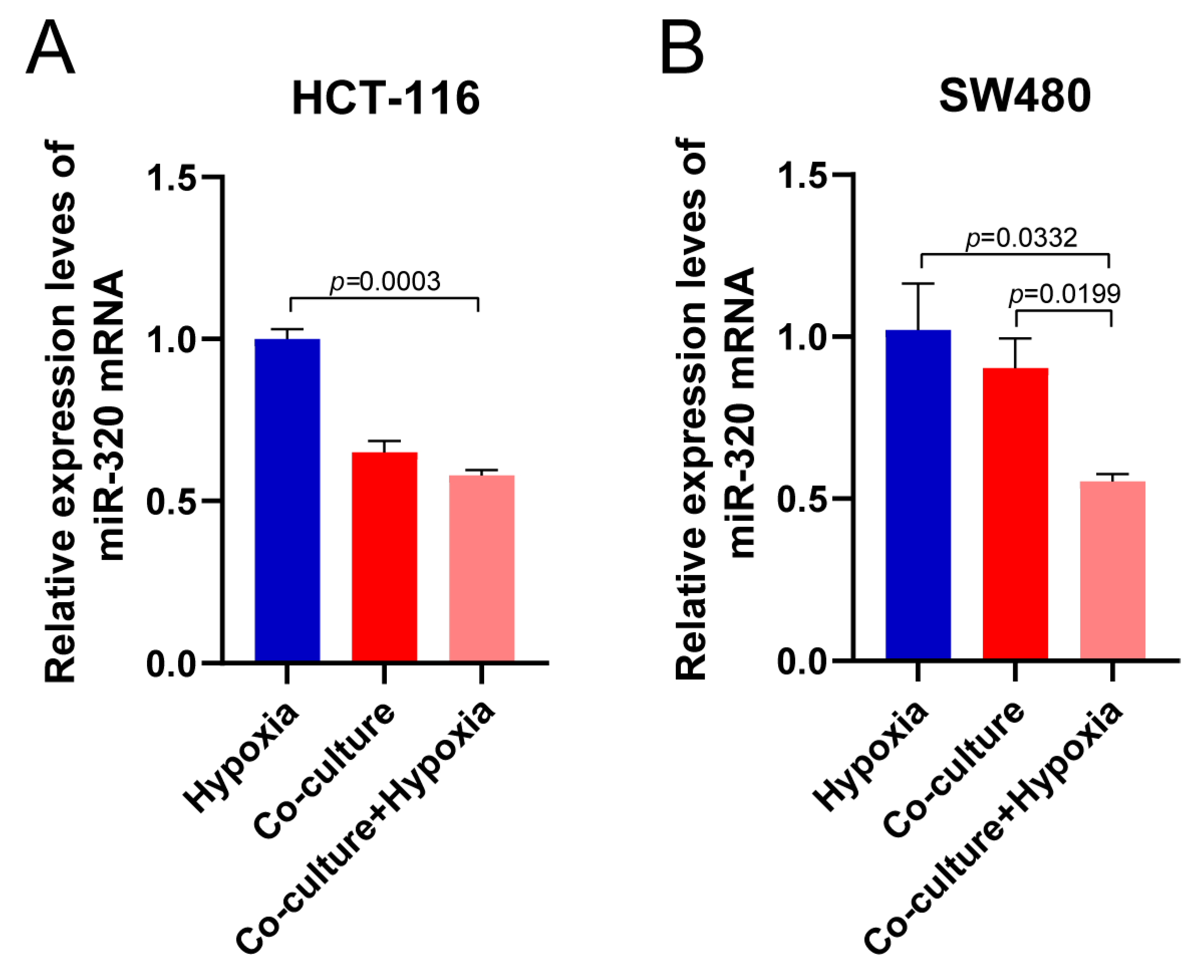
Figure 1: Hypoxia-induced TAMs impact miR-320 expression in CRC cells. (A,B) Relative expression levels of miR-320 in HCT116 (A) and SW480 (B) cells co-cultured with macrophages under normoxic and hypoxic conditions.
3.2 miR-320 Regulates CRC Cell Proliferation and Apoptosis under Hypoxia
To further elucidate the role of miR-320 in CRC, miR-320 inhibitors or mimics were transfected into CRC cells. As shown in Fig. 2, miR-320 inhibitors and mimics caused significant downregulation and upregulation of miR-320 expression, respectively, in CRC cell lines (Fig. 2A). Co-culturing the different groups with hypoxia-induced macrophages, the CCK8 assay revealed that miR-320 mimics significantly decreased CRC cell proliferation compared to the NC group, while miR-320 inhibitors significantly enhanced proliferation (Fig. 2B). Flow cytometry analysis showed that miR-320 mimics significantly increased apoptosis levels, whereas miR-320 inhibitors decreased apoptosis (Fig. 2C).
To assess the effect of miR-320 on ER homeostasis in CRC cells, a western blot was employed to detect ER stress-related proteins. As shown in Fig. 2D, miR-320 inhibitors suppressed the expression of EIF2α, DDIT3, and p-ATF6 (total ATF6 expression level was not altered), whereas miR-320 mimics promoted the expression of these proteins in CRC cells, indicating that miR-320 may induce ER stress in CRC cells. Altogether, the downregulation of miR-320 in colorectal cancer cells might contribute to cell survival, potentially by promoting ER homeostasis maintenance.
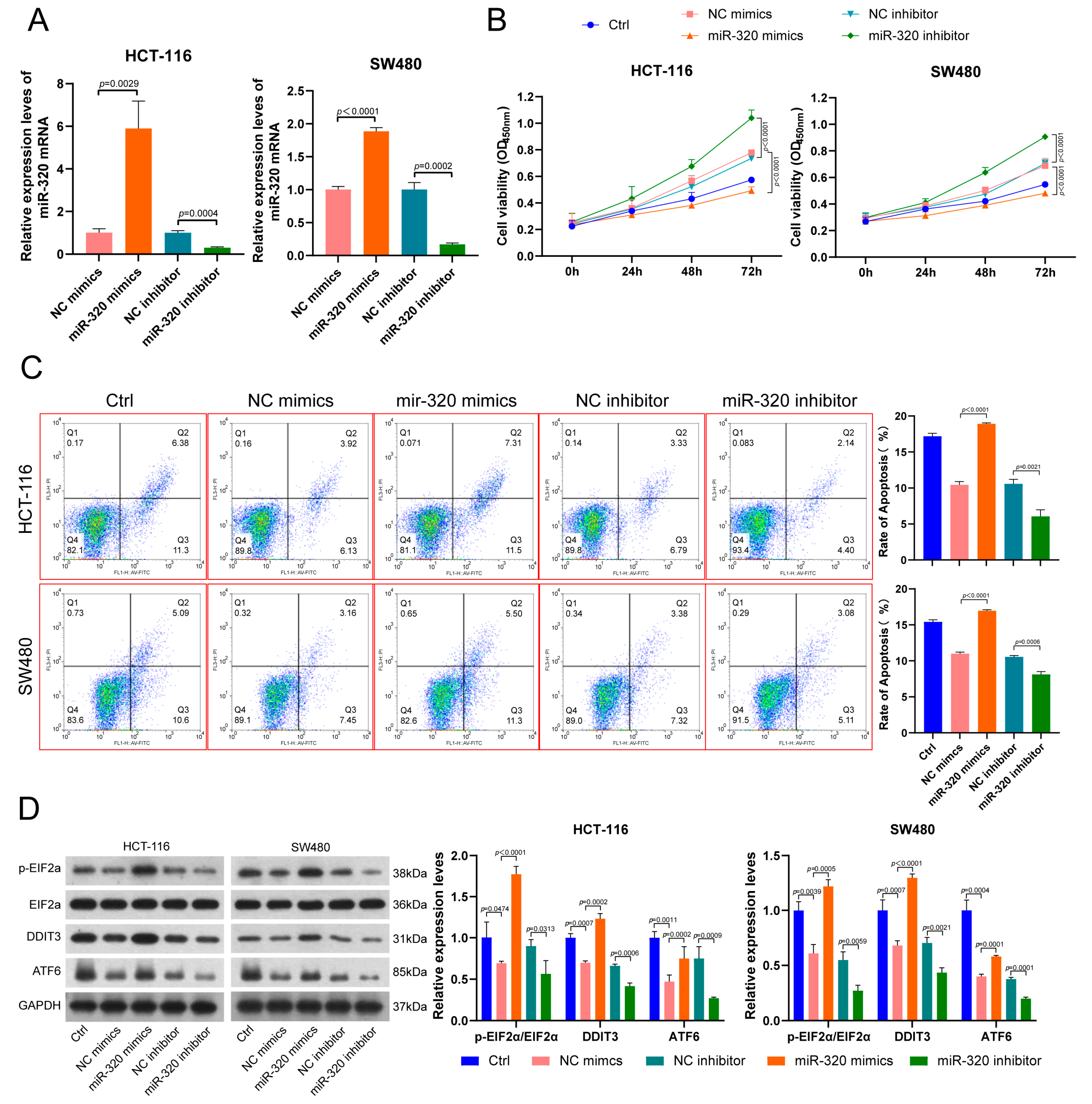
Figure 2: The role of miR-320 in CRC cell proliferation, apoptosis, and ER stress regulation. (A) miR-320 expression in HCT116 and SW480 cells transfected with miR-320 mimics or inhibitors. (B) Cell proliferation analysis using CCK8 assay in HCT116 and SW480 cells transfected with miR-320 mimics or inhibitors, and co-cultured with hypoxia-induced TAMs. (C) Flow cytometry analysis of apoptosis levels in CRC HCT116 and SW480 cells after transfection with miR-320 mimics or inhibitors. (D) Western blot analysis of ER stress markers (EIF2α, DDIT3, ATF6, p-EIF2α) in HCT116 and SW480 cells transfected with miR-320 mimics or inhibitors.
3.3 miR-320 Targets SOX4 to Inhibit Malignant Behavior in CRC Cells
To elucidate the mechanism of miR-320 action in CRC, TargetScan was used to predict miR-320 targets. As shown in Fig. 3A, miR-320 has binding sites with SOX4. The binding sites were mutated, and dual-luciferase assays were employed for validation (Fig. 3A). Fig. 3B shows that transfection of miR-320 mimics significantly reduced the relative luciferase activity in HCT116 and SW480 cells. Furthermore, after transfecting miR-320 mimics and co-culturing with hypoxia-induced macrophages, miR-320 mimics markedly inhibited SOX4 expression in tumor cells, whereas transfection with inhibitors substantially increased SOX4 expression (Fig. 3C). The findings suggest that miR-320 may inhibit malignant behavior in CRC by targeting SOX4.
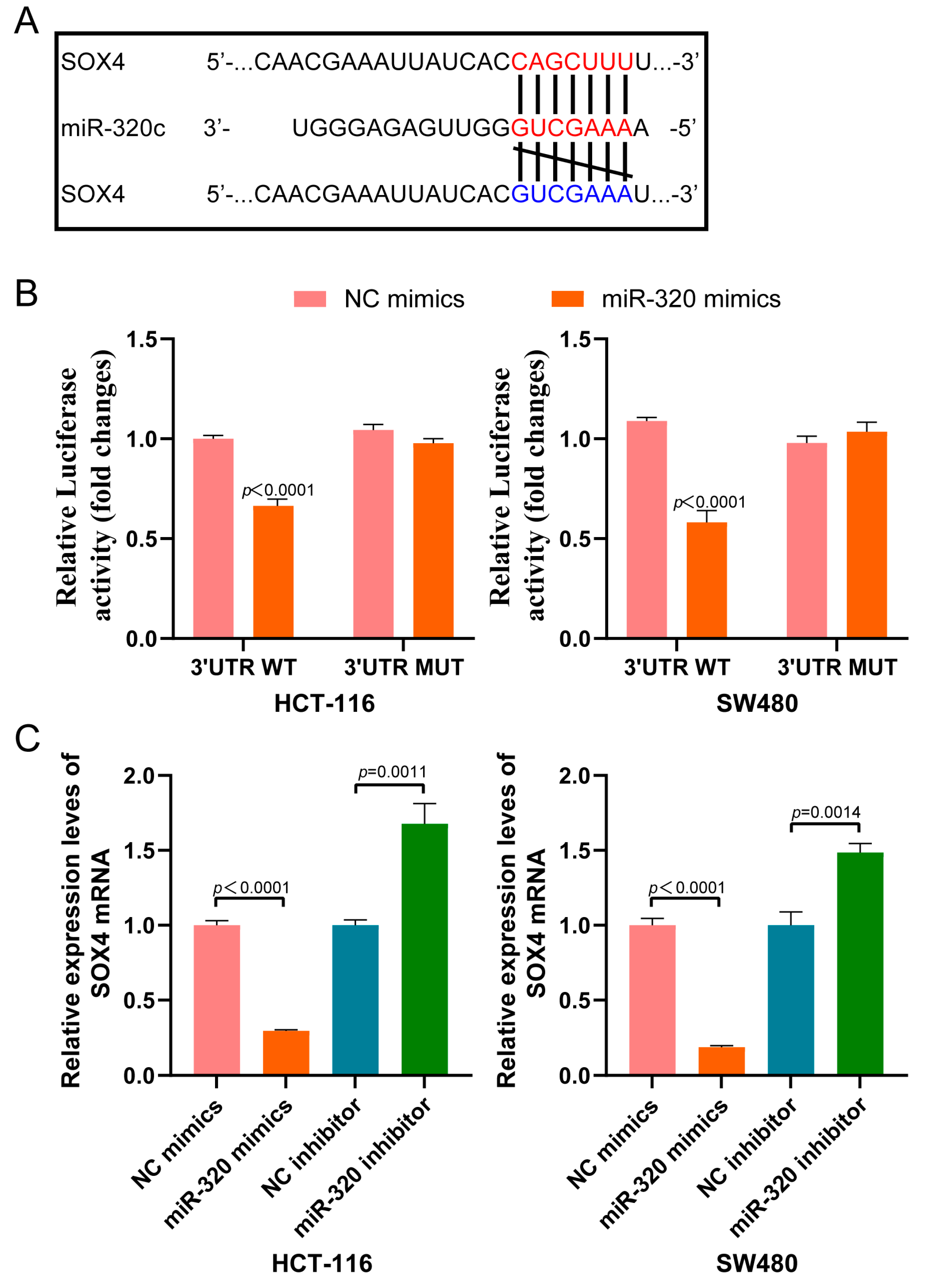
Figure 3: miR-320 targets SOX4 in CRC cells. (A) Predicted binding sites between miR-320 and SOX4 using TargetScan. Schematic representation of wild-type and mutated binding sites. (B) Relative luciferase activity in HCT116 and SW480 cells transfected with wild-type or mutated SOX4 3′UTR luciferase reporter constructs and miR-320 mimics. (C) qRT-PCR to examine SOX4 mRNA expression in HCT116 and SW480 cells transfected with miR-320 mimics or inhibitors, and co-cultured with hypoxia-induced TAMs.
3.4 The miR-320/SOX4 Axis Modulates CRC Cell Proliferation and ER Stress
To further clarify miR-320’s role in CRC malignant behavior regulation through targeting SOX4, SOX4 was overexpressed in CRC cells transfected with miR-320 mimics and co-cultured with hypoxia-induced macrophages. This rescue group aimed to elucidate the miR-320/SOX4 axis’s role in the tumor microenvironment. Results showed that the low SOX4 expression in HCT116 and SW480 cells due to miR-320 mimics was significantly reversed after transfecting the SOX4 overexpression vector (Fig. 4A). CCK8 assays demonstrated that the reduced proliferation and increased apoptosis in HCT116 and SW480 due to miR-320 mimics were significantly reversed following SOX4 overexpression (Fig. 4B,C). Hence, the miR-320/SOX4 axis might play a regulatory role in CRC malignant proliferation.
Western blot results indicated that miR-320 mimics increased the expression of EIF2α, DDIT3, and p-ATF6 (total ATF6 expression was not changed in various treatments) in HCT116 and SW480 cells. However, SOX4 overexpression markedly decreased the expression levels of these proteins (Fig. 4D). In conclusion, miR-320 might obstruct ER homeostasis and inhibit malignant proliferation events in CRC cells by targeting SOX4.
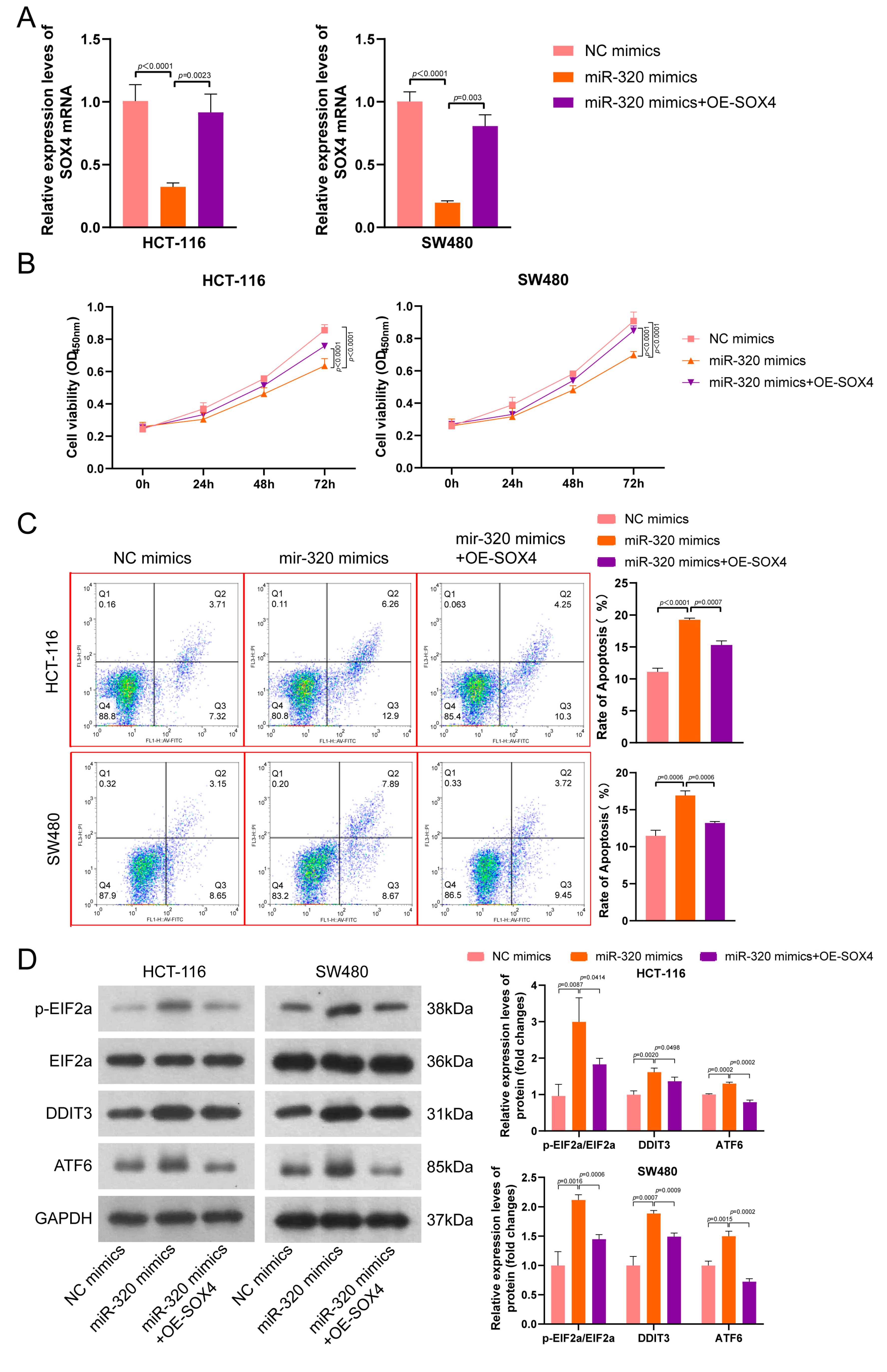
Figure 4: miR-320/SOX4 axis regulates CRC cell proliferation and ER stress. HCT116 and SW480 cells were transfected with miR-320 mimics, miR-320 mimics + SOX4 overexpression vectors, or control vectors, and then co-cultured with hypoxia-induced macrophages. (A) qRT-PCR analysis of SOX4 mRNA expression in CRC cells. (B) CCK8 assay to analyze cell proliferation in CRC cells. (C) Flow cytometry analysis of apoptosis levels in CRC cells. (D) Western blot analysis of ER stress markers (EIF2α, DDIT3, ATF6, p-EIF2α) in CRC cells.
Tumor-associated macrophages (TAMs) are a crucial component of the tumor microenvironment, playing a significant role in tumor development and drug resistance by creating an immunosuppressive milieu [22]. Due to uncontrolled growth and insufficient angiogenesis, hypoxia within the tumor microenvironment (TME) is an intrinsic characteristic of all solid malignancies [11]. In solid tumors, macrophages often accumulate in hypoxic regions. Numerous studies indicate that TAMs, under hypoxic conditions, significantly promote tumor development. For instance, Guo et al. [23] demonstrate that TAMs promote EMT, invasion, and migration in gastric cancer cells via FOXQ1. In renal cell carcinoma, TAMs enhance inflammation, immunosuppression, and malignant progression by increasing 15-LOX2 activity [24]. Moreover, TAMs facilitate CRC proliferation, invasion, and metastasis through the secretion of various cytokines or exosomes. For example, hypoxic TAMs promote renal cancer progression by exosomal transfer of miR-155-5p, activating the IGF1R/PI3K/AKT cascade [25]. Additionally, exosomal miR-223-3p from TAMs can be delivered to breast cancer 4T1 cells, facilitating lung metastasis via the miR-223-3p/Cbx5 axis [26]. Macrophages, by secreting cytokines such as TGF-β1, IL-10, and TNF-α, can significantly alter the tumor microenvironment, thus affecting the proliferation and invasion of tumor cells. In this study, hypoxia-induced TAMs inhibited miR-320 expression in CRC cells, promoting tumor growth and migration. These results further confirm the critical role of TAMs in the progression of colorectal cancer within the tumor microenvironment, though the mechanism by which TAMs affect miR-320 expression in CRC cells requires further investigation.
miR-320 is downregulated in various cancers and acts as a tumor suppressor by regulating target gene expression, inhibiting tumor cell proliferation, apoptosis, and migration [27]. miR-320a suppresses cell proliferation and migration, and induces p53-dependent apoptosis in prostate cancer by negatively regulating the long non-coding RNA TP73-AS1 [28]. Both in vitro and in vivo experiments show that miR-320 inhibits the occurrence, development, and metastasis of cholangiocarcinoma by targeting NRP-1 [29]. Bai et al. [20] find that miR-320 inhibits breast cancer cell proliferation and invasion by targeting SOX4. This is consistent with our findings; in this study, miR-320 expression in CRC cells was significantly downregulated after co-culture with hypoxia-induced macrophages, promoting the expression of its target gene SOX4 and thereby enhancing CRC cell proliferation and apoptosis. These results further validate the tumor-suppressing role of miR-320 in colorectal cancer and its importance in regulating SOX4 expression.
SOX4, as an important developmental transcription factor, regulates stemness, differentiation, progenitor cell development, and various developmental pathways, including PI3K, Wnt, and TGFβ signaling [30]. Several studies indicate that SOX4 is highly expressed in CRC and serves as a key oncogene. Sinner et al. [19] report that SOX4 enhances the activity and stability of β-catenin/TCF, activating the Wnt pathway to promote SW480 colon cancer cell proliferation. High expression of SOX4 mRNA is closely related to colorectal cancer recurrence [31]. Similarly, our rescue experiments demonstrate that SOX4 promotes CRC cell proliferation and migration, suggesting SOX4 could serve as a potential biomarker and therapeutic target for CRC, though this certainly requires further validation [32].
When cells experience environmental stress such as hypoxia, low glucose, infection, and chemotherapy, unfolded proteins accumulate in the endoplasmic reticulum (ER), causing ER stress (ERS) and activating the unfolded protein response (UPR) to restore cellular homeostasis [33]. While ERS can protect cells from some harmful stimuli, it also plays a crucial role in tumors. In various cancer types, overexpression of ERS markers is associated with poor prognosis and clinical outcomes [34,35]. However, some studies find that ERS can also promote tumor cell apoptosis. For example, Flightless-1 promotes ERS by regulating intracellular Ca2+ concentrations, inducing colon cancer cell apoptosis [36]. It is reported that ERS facilitates tumor angiogenesis by accelerating the synthesis and secretion of angiogenic factors like angiopoietin, VEGF, and PD-ECGF [37]. Additionally, some miRNAs are found to promote tumor cell apoptosis by regulating ERS. miR-451a promotes ERS and induces apoptosis in HCT116 and SW620 colorectal cancer cells by increasing the expression of ERS-related proteins [38]. miR-26 participates in ERS signaling pathways, inducing apoptosis and inhibiting autophagy in human NSCLC cells [39]. In this study, miR-320 promotes ERS in CRC cells by targeting and inhibiting SOX4 expression, thus interfering with tumor progression. This suggests that SOX4 may play an important role in inhibiting ERS in CRC cells, which is similar to previous findings by Kang et al. [40] who report that TAMs inhibit ERS through activating SOX4/TMEM2 signaling in CRC cells. This finding highlights the critical role of SOX4 in ER stress, providing new directions for future research.
Despite this study revealing the significant roles of miR-320 and SOX4 in colorectal cancer progression, there are limitations that require further investigation. Firstly, this study focused primarily on the effects of macrophages and miR-320 regulation on tumor cells, but the exact mechanisms remain unclear. In 2022, Kang et al. [40] demonstrated that hypoxia-induced macrophages stimulate the secretion of TGF-β1, inhibiting ERS in CRC cells, thereby promoting proliferation and restraining apoptosis. Moreover, elevated TGF-β1 levels enhance SOX4 and TMEM2 expression in CRC cells. It can be speculated that macrophages might also induce changes in tumor cells through TGF-β1 secretion in this study, which needs further verification. Additionally, research indicates that crosstalk between CRC cells and TAMs mutually facilitates colorectal cancer progression. For example, TAMs promote CRC growth and migration by releasing immunosuppressive cytokines like IL-8, and cancer cells secrete significant levels of IL-10 to drive M2 macrophage differentiation [41]. It has been demonstrated that exosomal miR-1246 drives the polarization of TAMs by modulating NLRP3 at the post-transcriptional level, which subsequently disrupts CD8+ T cell infiltration and function within the tumor immune microenvironment [42]. Whether CRC cells in this study also secreted cytokines to promote TAM recruitment requires further research.
This study reveals that hypoxia-induced macrophages influence endoplasmic reticulum homeostasis and malignant behavior in colorectal cancer cells by regulating the miR-320/SOX4 axis, providing new targets and strategies for colorectal cancer treatment.
Acknowledgement:
Funding Statement: This project was supported by Shanghai University of Medicine & Health Sciences Foundation of China (No. SSF-23-25-005, Yuqing Kang) and the Natural Science Foundation of Gansu Province of China (No. 23JRRA1592, Xing Xue).
Author Contributions: The authors confirm contribution to the paper as follows: study conception and design: Yuqing Kang, Chunyan Mou; data collection: Jiangbo Zheng, Ranxu Lv; analysis and interpretation of results: Yuqing Kang, Jiangbo Zheng, Ranxu Lv; draft manuscript preparation: Yuqing Kang, Xing Xue. All authors reviewed and approved the final version of the manuscript.
Availability of Data and Materials: The datasets generated and analysed during the current study are available from the corresponding author on reasonable request.
Ethics Approval: Not applicable.
Conflicts of Interest: The authors declare no conflict of interest.
Abbreviations
| TAMs | Tumor-Associated Macrophages |
| CRC | Colorectal Cancer |
| ER | Endoplasmic Reticulum |
Table A1: The sequences of all primers.
| Gene | Sequence (5′→3′) |
|---|---|
| SOX4-Forward | GCACTAGGACGTCTGCCTTT |
| SOX4-Reverse | ACACGGCATATTGCACAGGA |
| miR-320-RT | GTCGTATCCAGTGCAGGGTCCGAGGTATTCGCACTGGATACGACTCGCCC |
| miR-320-Forward | AAAAGCUGGGUUGAGAGGGCGA |
| miR-320-Reverse | GTGCAGGGTCCGAGGT |
| GAPDH-Forward | GAAGGTGAAGGTCGGAGTC |
| GAPDH-Reverse | GAAGATGGTGATGGGATTTC |
| U6-RT | AAAATATGGAACGCTTCACGAATTTG |
| U6-Forward | CTCGCTTCGGCAGCACATATACT |
| U6-Reverse | ACGCTTCACGAATTTGCGTGTC |
References
1. Wu M , Wu X , Han J . KIF20A promotes CRC progression and the Warburg effect through the C-myc/HIF-1α axis. Protein Pept Lett. 2024; 31( 2): 107– 15. doi:10.2174/0109298665256238231120093150. [Google Scholar] [CrossRef]
2. Zhao J , Xue E , Zhou S , Zhang M , Sun J , Tan Y , et al. Allostatic load, genetic susceptibility, incidence risk, and all-cause mortality of colorectal cancer. J Natl Cancer Inst. 2025; 117( 1): 134– 43. doi:10.1093/jnci/djae223. [Google Scholar] [CrossRef]
3. Ge H , Yan Y , Wang H , Bian J , Deng Z , Su X , et al. Circular RNA hsa_circ_0005939 regulates UHRF1BP1L expression by targeting miR-4693-3p to promote colorectal cancer progression. Protein Pept Lett. 2024; 31( 6): 437– 46. doi:10.2174/0109298665297110240611115010. [Google Scholar] [CrossRef]
4. Matsuda T , Fujimoto A , Igarashi Y . Colorectal cancer: epidemiology, risk factors, and public health strategies. Digestion. 2025; 106( 2): 91– 9. doi:10.1159/000543921. [Google Scholar] [CrossRef]
5. Kumar B , Akhtar MJ , Paul J , Singh K , Pannu S , Pal R , et al. An update on recently developed analytical and bio-analytical methods for some anticancer drugs. Curr Pharm Anal. 2023; 19( 2): 117– 35. doi:10.2174/1573412919666221123110420. [Google Scholar] [CrossRef]
6. Pathak S , Meng WJ , Sriramulu S , Jothimani G , Jangamreddy JR , Banerjee A , et al. Association of microRNA-652 expression with radiation response of colorectal cancer: a study from rectal cancer patients in a Swedish trial of preoperative radiotherapy. Curr Gene Ther. 2023; 23( 5): 356– 67. doi:10.2174/1566523223666230418111613. [Google Scholar] [CrossRef]
7. Pang F , Yang P , Wang T , Li X , Wu X , Yue R , et al. Comprehensive analysis of alternative polyadenylation events associated with the tumor immune microenvironment in colon adenocarcinoma. Curr Genomics. 2023; 24( 1): 48– 61. doi:10.2174/1389202924666230503122134. [Google Scholar] [CrossRef]
8. Liu Y , Xun Z , Ma K , Liang S , Li X , Zhou S , et al. Identification of a tumour immune barrier in the HCC microenvironment that determines the efficacy of immunotherapy. J Hepatol. 2023; 78( 4): 770– 82. doi:10.1016/j.jhep.2023.01.011. [Google Scholar] [CrossRef]
9. Mantovani A , Marchesi F , Di Mitri D , Garlanda C . Macrophage diversity in cancer dissemination and metastasis. Cell Mol Immunol. 2024; 21( 11): 1201– 14. doi:10.1038/s41423-024-01216-z. [Google Scholar] [CrossRef]
10. Qin R , Ren W , Ya G , Wang B , He J , Ren S , et al. Role of chemokines in the crosstalk between tumor and tumor-associated macrophages. Clin Exp Med. 2023; 23( 5): 1359– 73. doi:10.1007/s10238-022-00888-z. [Google Scholar] [CrossRef]
11. Wang C , Xu S , Yang X . Hypoxia-driven changes in tumor microenvironment: insights into exosome-mediated cell interactions. Int J Nanomed. 2024; 19: 8211– 36. doi:10.2147/IJN.S479533. [Google Scholar] [CrossRef]
12. Cao Y , Wo M , Xu C , Fei X , Jin J , Shan Z . An AMPK agonist suppresses the progress of colorectal cancer by regulating the polarization of TAM to M1 through inhibition of HIF-1α and mTOR signal pathway. J Cancer Res Ther. 2023; 19( 6): 1560– 7. doi:10.4103/jcrt.jcrt_2670_22. [Google Scholar] [CrossRef]
13. Ramkumar KM , Vijayalalitha R , Archita T , Juanitaa GR , Jayasuriya R , Amin KN . Role of long non-coding RNA in regulating ER stress response to the progression of diabetic complications. Curr Gene Ther. 2023; 23( 2): 96– 110. doi:10.2174/1566523222666220801141450. [Google Scholar] [CrossRef]
14. Liu Z , Liu Q , Zeng A , Song L . Regulatory function of endoplasmic reticulum stress in colorectal cancer: mechanism, facts, and perspectives. Int Immunopharmacol. 2025; 147: 114024. doi:10.1016/j.intimp.2025.114024. [Google Scholar] [CrossRef]
15. Barez SR , Attar AM , Aghaei M . microRNA-30c-2-3p regulates ER stress and induces apoptosis in ovarian cancer cells underlying ER stress. EXCLI J. 2021; 20: 922– 34. doi:10.17179/excli2020-2970. [Google Scholar] [CrossRef]
16. Cui Y , Xu H , Yang Y , Zhao D , Wen Y , Lv C , et al. The regulation of miR-320a/XBP1 axis through LINC00963 for endoplasmic reticulum stress and autophagy in diffuse large B-cell lymphoma. Cancer Cell Int. 2021; 21( 1): 305. doi:10.1186/s12935-021-01992-y. [Google Scholar] [CrossRef]
17. Zhang Z , Zhang J , Li J , Geng H , Zhou B , Zhang B , et al. miR-320/ELF3 axis inhibits the progression of breast cancer via the PI3K/AKT pathway. Oncol Lett. 2020; 19( 4): 3239– 48. doi:10.3892/ol.2020.11440. [Google Scholar] [CrossRef]
18. Mohanta A , Kumar RR , Singh RK , Mandal S , Yadav R , Khatkar R , et al. Emerging role of miR-320a in lung cancer: a comprehensive review. Biomark Med. 2023; 17( 18): 767– 81. doi:10.2217/bmm-2023-0215. [Google Scholar] [CrossRef]
19. Sinner D , Kordich JJ , Spence JR , Opoka R , Rankin S , Lin SC , et al. Sox17 and Sox4 differentially regulate beta-catenin/T-cell factor activity and proliferation of colon carcinoma cells. Mol Cell Biol. 2007; 27( 22): 7802– 15. doi:10.1128/MCB.02179-06. [Google Scholar] [CrossRef]
20. Bai JW , Wang X , Zhang YF , Yao GD , Liu H . microRNA-320 inhibits cell proliferation and invasion in breast cancer cells by targeting SOX4. Oncol Lett. 2017; 14( 6): 7145– 52. doi:10.3892/ol.2017.7087. [Google Scholar] [CrossRef]
21. Zhang J , Liang Q , Lei Y , Yao M , Li L , Gao X , et al. SOX4 induces epithelial-mesenchymal transition and contributes to breast cancer progression. Cancer Res. 2012; 72( 17): 4597– 608. doi:10.1158/0008-5472.CAN-12-1045. [Google Scholar] [CrossRef]
22. Li M , Yang Y , Xiong L , Jiang P , Wang J , Li C . Metabolism, metabolites, and macrophages in cancer. J Hematol Oncol. 2023; 16( 1): 80. doi:10.1186/s13045-023-01478-6. [Google Scholar] [CrossRef]
23. Guo J , Yan Y , Yan Y , Guo Q , Zhang M , Zhang J , et al. Tumor-associated macrophages induce the expression of FOXQ1 to promote epithelial-mesenchymal transition and metastasis in gastric cancer cells. Oncol Rep. 2017; 38( 4): 2003– 10. doi:10.3892/or.2017.5877. [Google Scholar] [CrossRef]
24. Daurkin I , Eruslanov E , Stoffs T , Perrin GQ , Algood C , Gilbert SM , et al. Tumor-associated macrophages mediate immunosuppression in the renal cancer microenvironment by activating the 15-lipoxygenase-2 pathway. Cancer Res. 2011; 71( 20): 6400– 9. doi:10.1158/0008-5472.CAN-11-1261. [Google Scholar] [CrossRef]
25. Gu W , Gong L , Wu X , Yao X . Hypoxic TAM-derived exosomal miR-155-5p promotes RCC progression through HuR-dependent IGF1R/AKT/PI3K pathway. Cell Death Discov. 2021; 7( 1): 147. doi:10.1038/s41420-021-00525-w. [Google Scholar] [CrossRef]
26. Wang Z , Zhang C , Guo J , Wang W , Si Q , Chen C , et al. Exosomal miRNA-223-3p derived from tumor associated macrophages promotes pulmonary metastasis of breast cancer 4T1 cells. Transl Oncol. 2023; 35: 101715. doi:10.1016/j.tranon.2023.101715. [Google Scholar] [CrossRef]
27. Liang Y , Li S , Tang L . microRNA 320, an anti-oncogene target miRNA for cancer therapy. Biomedicines. 2021; 9( 6): 591. doi:10.3390/biomedicines9060591. [Google Scholar] [CrossRef]
28. Bozgeyik E , Arslan A , Temiz E , Batar B , Koyuncu I , Tozkir H . miR-320a promotes p53-dependent apoptosis of prostate cancer cells by negatively regulating TP73-AS1 invitro. Biochem Biophys Res Commun. 2022; 619: 130– 6. doi:10.1016/j.bbrc.2022.06.034. [Google Scholar] [CrossRef]
29. Zhu H , Jiang X , Zhou X , Dong X , Xie K , Yang C , et al. Neuropilin-1 regulated by miR-320 contributes to the growth and metastasis of cholangiocarcinoma cells. Liver Int. 2018; 38( 1): 125– 35. doi:10.1111/liv.13495. [Google Scholar] [CrossRef]
30. Nie J , Tang J , Zhang Z , Sun H , Wang X , Wang L , et al. SOX4 as a key oncogene driving tumor invasion in retinoblastoma. Investig Ophthalmol Vis Sci. 2025; 66( 5): 24. doi:10.1167/iovs.66.5.24. [Google Scholar] [CrossRef]
31. Andersen CL , Christensen LL , Thorsen K , Schepeler T , Sørensen FB , Verspaget HW , et al. Dysregulation of the transcription factors SOX4, CBFB and SMARCC1 correlates with outcome of colorectal cancer. Br J Cancer. 2009; 100( 3): 511– 23. doi:10.1038/sj.bjc.6604884. [Google Scholar] [CrossRef]
32. Seyfinejad B , Jouyban A . Importance of method validation in the analysis of biomarker. Curr Pharm Anal. 2022; 18( 6): 567– 9. doi:10.2174/1573412918666211213142638. [Google Scholar] [CrossRef]
33. Yilmaz E . Endoplasmic reticulum stress and obesity. In: Obesity and Lipotoxicity. Cham, Switzerland: Springer; 2024. p. 373– 90. doi:10.1007/978-3-031-63657-8_13. [Google Scholar] [CrossRef]
34. Xu Z , Xu J , Sun S , Lin W , Li Y , Lu Q , et al. Mecheliolide elicits ROS-mediated ERS driven immunogenic cell death in hepatocellular carcinoma. Redox Biol. 2022; 54: 102351. doi:10.1016/j.redox.2022.102351. [Google Scholar] [CrossRef]
35. Yang L , Xue R , Yang C , Lv Y , Li S , Xiang W , et al. Endoplasmic reticulum stress on glioblastoma: tumor growth promotion and immunosuppression. Int Immunopharmacol. 2025; 157: 114806. doi:10.1016/j.intimp.2025.114806. [Google Scholar] [CrossRef]
36. Choi SS , Lee SK , Kim JK , Park HK , Lee E , Jang J , et al. Flightless-1 inhibits ER stress-induced apoptosis in colorectal cancer cells by regulating Ca2+ homeostasis. Exp Mol Med. 2020; 52( 6): 940– 50. doi:10.1038/s12276-020-0448-3. [Google Scholar] [CrossRef]
37. Paridaens A , Laukens D , Vandewynckel YP , Coulon S , Van Vlierberghe H , Geerts A , et al. Endoplasmic reticulum stress and angiogenesis: is there an interaction between them? Liver Int. 2014; 34( 6): e10– 8. doi:10.1111/liv.12457. [Google Scholar] [CrossRef]
38. Xu K , Han B , Bai Y , Ma XY , Ji ZN , Xiong Y , et al. miR-451a suppressing BAP31 can inhibit proliferation and increase apoptosis through inducing ER stress in colorectal cancer. Cell Death Dis. 2019; 10( 3): 152. doi:10.1038/s41419-019-1403-x. [Google Scholar] [CrossRef]
39. He Y , Liu H , Jiang L , Rui B , Mei J , Xiao H . miR-26 induces apoptosis and inhibits autophagy in non-small cell lung cancer cells by suppressing TGF-β1-JNK signaling pathway. Front Pharmacol. 2018; 9: 1509. doi:10.3389/fphar.2018.01509. [Google Scholar] [CrossRef]
40. Kang Y , Lv R , Feng Z , Zhu J . Tumor-associated macrophages improve hypoxia-induced endoplasmic reticulum stress response in colorectal cancer cells by regulating TGF-β1/SOX4. Cell Signal. 2022; 99: 110430. doi:10.1016/j.cellsig.2022.110430. [Google Scholar] [CrossRef]
41. Zhang Y , Sime W , Juhas M , Sjölander A . Crosstalk between colon cancer cells and macrophages via inflammatory mediators and CD47 promotes tumour cell migration. Eur J Cancer. 2013; 49( 15): 3320– 34. doi:10.1016/j.ejca.2013.06.005. [Google Scholar] [CrossRef]
42. Feng Y , Jin C , Wang T , Chen Z , Ji D , Zhang Y , et al. The uridylyl transferase TUT7-mediated accumulation of exosomal miR-1246 reprograms TAMs to support CRC progression. Adv Sci. 2024; 11( 15): 2304222. doi:10.1002/advs.202304222. [Google Scholar] [CrossRef]
Cite This Article
 Copyright © 2026 The Author(s). Published by Tech Science Press.
This work is licensed under a Creative Commons Attribution 4.0 International License , which permits unrestricted use, distribution, and reproduction in any medium, provided the original work is properly cited.
 Downloads
 Citation Tools
