
 Login
Login Register
 Submit a Paper
 Propose a Special lssue
 Open Access
REVIEW
Narrative Review on the Role of Oxidative Stress in the Pathomechanism of Ocular Diseases: Update 2025
1 Department of Ophthalmology, University of Warmia and Mazury, Olsztyn, Poland
2 Institute for Research in Ophthalmology, Foundation for Ophthalmology Development, Poznan, Poland
3 Department of Ophthalmology, Medical Academy, Lithuanian University of Health Sciences, Kaunas, Lithuania
* Corresponding Author: Andrzej Grzybowski. Email:
(This article belongs to the Special Issue: Modulation of Inflammation, Oxidative Stress, and Mitochondrial Function: Therapeutic Perspectives Across Diseases)
BIOCELL 2026, 50(10), 4 https://doi.org/10.32604/biocell.2026.083756
Received 09 April 2026; Accepted 22 July 2026; Issue published 22 September 2026
 View Full Text
 Download PDF
Abstract
Oxidative stress results from an imbalance between reactive oxygen species (ROS) production and antioxidant defense mechanisms and contributes to cellular damage and the pathogenesis of various diseases. This review summarizes recent advances in the molecular and biochemical mechanisms associated with oxidative stress in ocular diseases, including dry eye disease, diabetic retinopathy, age-related macular degeneration (AMD), glaucoma, and cataract. A literature search was conducted using PubMed and Google Scholar databases to identify relevant studies published up to 31 December 2025. Current evidence suggests that oxidative stress plays an important role in inflammation, mitochondrial dysfunction, and cellular damage associated with ocular diseases. A better understanding of oxidative stress-related mechanisms may provide new insights into the pathogenesis and progression of ocular diseases.Keywords
Oxidative stress has a recognized role in the pathogenesis of various pathological processes and diseases. Oxidative stress refers to an imbalance between the production of reactive oxygen species (ROS) and the ability of the body to detoxify these harmful molecules. An imbalance between oxidant and antioxidant results in both intercellular and intracellular homeostasis dysregulation, impairs normal functions of tissue cells, and contributes to the development of various health problems, ranging from cardiovascular, neurodegenerative, immune, and autoimmune diseases, cancer, to eye disorders [1,2,3]. Due to their exposure to higher oxygen levels compared to other tissues, the eyes are particularly sensitive to oxidative stress. Multiple studies have emphasized the significant association between oxidative stress and the onset and progression of ocular diseases. Despite the extensive publication of data on the pathomechanisms of ocular diseases and the significance of oxidative stress in their development, the mechanisms underlying disease progression remain inadequately understood. Therefore, this review aims to summarize current evidence on the molecular and biochemical mechanisms of oxidative stress across major ocular diseases and to highlight shared pathogenic pathways, disease-specific features, and emerging therapeutic strategies.
A comprehensive literature search was conducted using the electronic databases PubMed and Google Scholar to identify relevant studies published up to 31 December 2025. The following search terms and Boolean combinations were used: (“reactive oxygen species” OR “oxidative stress”) AND (“ocular diseases” OR “cataract” OR “dry eye disease” OR “diabetic retinopathy” OR “age-related macular degeneration” OR “glaucoma”). Articles were selected based on their relevance to the topic, with inclusion criteria focusing on publications addressing oxidative stress mechanisms in ocular diseases. Both original research articles and systematic or narrative reviews published in English were included. Exclusion criteria comprised studies not directly related to oxidative stress or ocular pathology, non-English publications, conference abstracts, and articles with insufficient methodological detail. As this study represents a narrative review, no formal quality assessment or risk-of-bias analysis was performed. However, priority was given to recent studies and those providing mechanistic or clinical insights. Potential limitations of the search strategy include restriction to selected databases and language bias.
3 The Basic Mechanism of Oxidative Stress
The term oxidative stress refers to the disturbance in the pro-oxidant-antioxidant balance in favor of the former, leading to potential damage. Reduced antioxidant levels and/or increased production of reactive species result in oxidative damage to cellular biomolecules during oxidative stress. Multiple cellular components, including lipids, DNA, proteins, and carbohydrates, may be damaged by oxidative stress. Oxidative damage, especially to DNA, may trigger cell death by apoptosis or necrosis [4] (Fig. 1 [5]). ROS: superoxide, O2−; hydroxyl, OH; hydroperoxyl, HO2; and reactive nitrogen species (RNS): nitric oxide, NO; nitrogen dioxide, NO2, formed under normal physiological conditions and may have both beneficial and harmful roles. Free radicals and other reactive species of oxygen/nitrogen/chlorine play an important role in the development of various diseases by inducing oxidative stress and oxidative damage. ROS are generated as byproducts of incomplete oxygen reduction during mitochondrial oxidative phosphorylation or through the activity of the nicotinamide adenine dinucleotide phosphate (NADPH) oxidase complex (NOX). This complex comprises seven isoforms (NOX1, NOX2, NOX3, NOX4, NOX5, DUOX1, DUOX2) that share structural similarities but differ according to tissue distribution and regulatory systems. In addition to mitochondrial ROS production, enzymatic and environmental sources substantially contribute to oxidative stress in ocular tissues, highlighting the multifactorial nature of ROS-mediated damage. The human eye is constantly exposed to both natural sunlight and artificial lighting. This exposure includes exogenous sources of ROS such as UV light, visible light, and ionizing radiation, which may contribute to oxidative damage in ocular tissues [6]. Moreover, the aging eye is particularly vulnerable to oxidative stress, a critical factor in the development of various chronic ocular diseases [7]. These diseases include dry eye disease (DED), glaucoma, diabetic retinopathy (DR), age-related macular degeneration (AMD), and cataract.
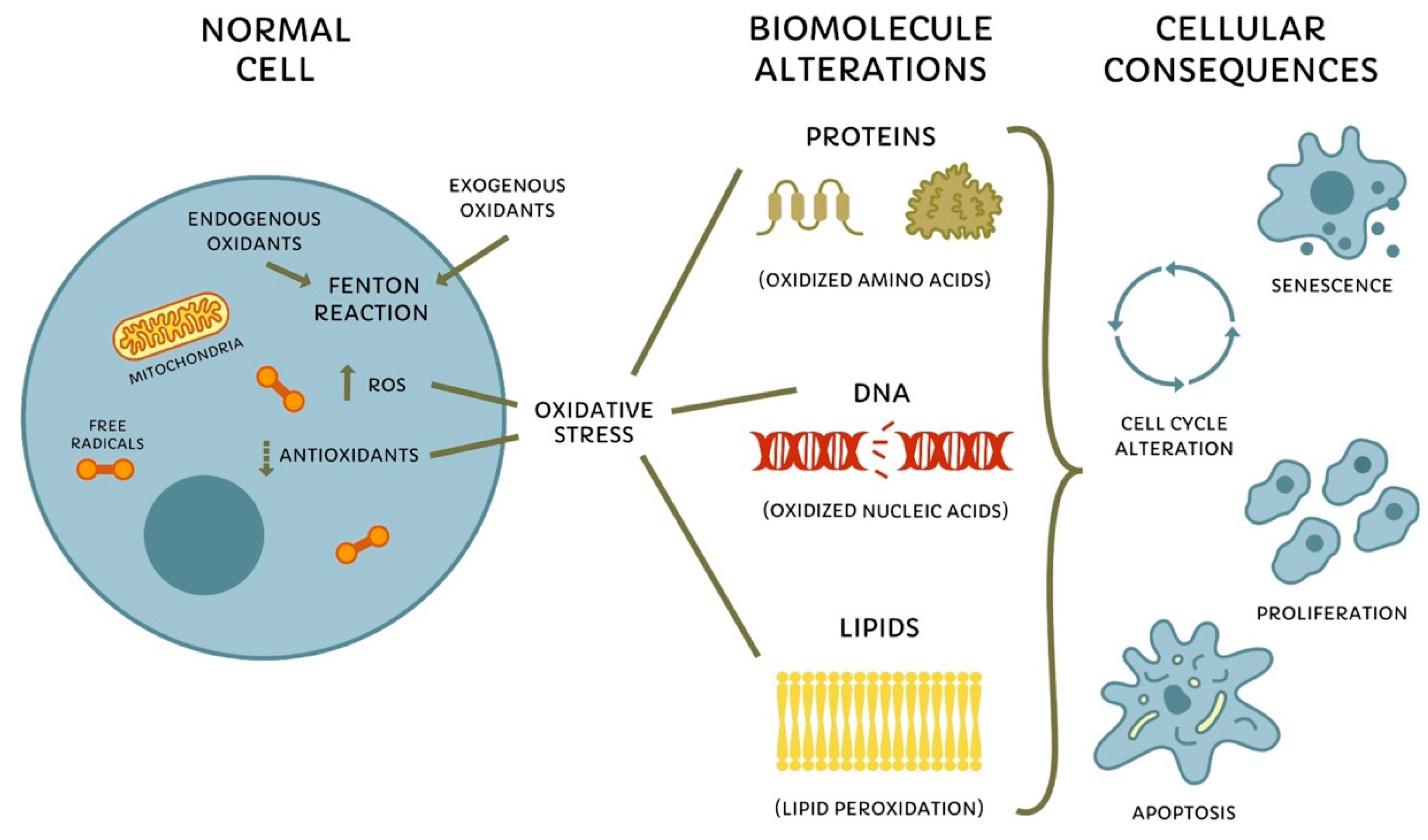
Figure 1: The Basic Mechanism of Oxidative Stress. Adapted from [5] under CC BY 4.0. ROS are generated by endogenous and exogenous sources and are normally balanced by antioxidant defenses. When ROS production exceeds the capacity of antioxidant systems, oxidative stress develops, leading to protein oxidation, DNA damage, and lipid peroxidation. These biomolecular alterations disrupt cellular homeostasis and contribute to changes in cell cycle regulation, cellular senescence, proliferation, and apoptosis. ROS, reactive oxygen species; DNA, deoxyribonucleic acid. The upward arrow (↑) indicates increased ROS production, whereas the downward arrow (↓) indicates reduced antioxidant defense.
4 The Role of Oxidative Stress in the Pathogenesis of Dry Eye Disease
Dry eye is a multifactorial disease that affects the ocular surface. It is characterized by a loss of tear film homeostasis and is accompanied by ocular symptoms. Key etiological factors include tear film instability and hyperosmolarity, ocular surface inflammation and damage, and neurosensory abnormalities [8]. DED induces a large panel of ocular symptoms depending on the stage of tissue damage, such as superficial pain, foreign body sensation, ocular discomfort, and visual disturbance. Tear film hyperosmolarity seems to be the core mechanism of dry eye disease. The ocular surface, including the tear film, contributes to maintaining redox homeostasis through antioxidant defense mechanisms. Disruption of this balance has been associated with oxidative stress, inflammation, and cellular damage in DED [9]. It is suggested that an imbalance between oxidative and antioxidative processes plays a key role in the pathology of DED. Some studies indicate that oxidative stress markers, including lipid peroxide (LPO), hexanoyl-lysine (HEL), 4-hydroxy-2-nonenal (4HNE), myeloperoxidase (MPO), nitric oxide synthase 3 (NOS3), xanthine oxidase/oxidoreductase, malondialdehyde (MDA), and ROS, are elevated in the tears, conjunctival cells, and conjunctival biopsies of patients with DED compared to controls [10]. ROS are implicated in the pathogenesis of autoimmunity, yet they also exhibit protective roles in autoimmune diseases. For instance, a missense variant in the NCF1 gene, which leads to decreased ROS production, increases susceptibility to systemic lupus erythematosus and other autoimmune disorders such as primary Sjögren’s syndrome [11]. Additionally, reduced NOX2 function leads to decreased cytosolic ROS during neutrophil activation, impairing the clearance of apoptotic and necrotic bodies. T regulatory (Treg) cells, which are a type of T cell characterized by the expression of Forkhead box P3 (FoxP3), help suppress the immune response and prevent autoimmunity [12]. Evidence suggests that ROS are vital for the stability and function of Treg cells, with a notable reduction in the immunosuppressive capacity of Treg cells in Ncf1−/− mice due to the absence of cytoplasmic ROS [13]. On the other hand, ROS also contributes pathogenically to autoimmune diseases. Cytoplasmic ROS, produced by NOX during T cell activation, influences their differentiation. This results in T cells favoring a T helper 17 phenotype over T helper 1, accompanied by reduced production of interferon-γ, interleukin (IL)-2, and tumor necrosis factor (TNF)-α. Additionally, there is increased production of IL-6 and IL-17. Furthermore, mitochondrial (mt-) ROS-deficient T cells do not properly activate or expand in response to antigens [14]. Oxidative stress and inflammation are two closely interrelated and interdependent pathophysiological processes: ROS trigger intracellular signaling cascades that increase the expression of proinflammatory genes. Inflammatory cells release ROS along with immune mediators such as cytokines and chemokines, which together exacerbate oxidative stress and cause tissue damage at the inflammation site (Fig. 2) [15].
Several antioxidant and mitochondria-targeted therapies, including melatonin, α-lipoic acid, and SkQ1, a mitochondria-targeted plastoquinone derivative, have shown potential for reducing oxidative stress and inflammation in DED. Melatonin reduces ROS generation and suppresses inflammatory signaling [16,17]; α-lipoic acid enhances the antioxidant capacity of ocular surface cells [18], whereas SkQ1 directly scavenges mitochondrial ROS and helps preserve endogenous antioxidant defenses [19].
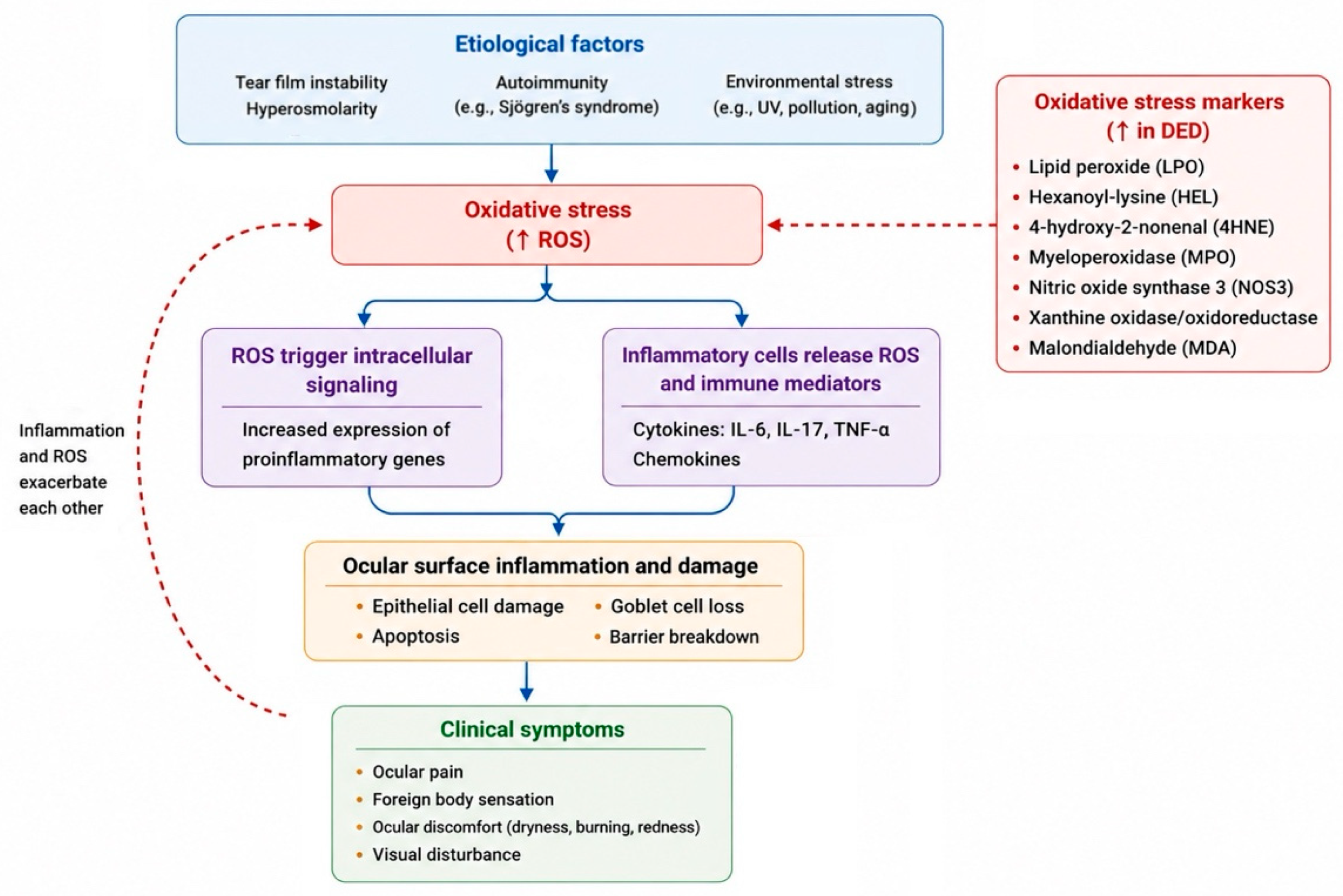
Figure 2: The Mechanism of Oxidative Stress in the Pathogenesis of Dry Eye Disease. Etiological factors induce oxidative stress, which activates inflammatory signaling cascades and promotes the release of inflammatory mediators, resulting in ocular surface inflammation, damage, and clinical manifestations of dry eye disease. UV, ultraviolet radiation; DED, dry eye disease; ROS, reactive oxygen species. The upward arrow (↑) indicates increased oxidative stress markers, increased ROS levels.
5 The Role of Oxidative Stress in the Pathogenesis of Diabetic Retinopathy
Hyperglycemia-Induced Metabolic Abnormalities
DR is a progressive microvascular complication of diabetes that leads to irreversible retinal damage and is a leading cause of acquired blindness in diabetic patients. The pathogenesis of DR involves numerous factors that operate through integrated molecular signaling pathways and cellular processes. Oxidative stress is a critical contributor to the pathogenesis of DR. It can both contribute to and result from metabolic abnormalities in the retina induced by hyperglycemia [20]. ROS are primarily generated through mitochondrial oxidative phosphorylation and the NADPH oxidase (NOX) system. Hyperglycemic conditions activate the NOX complex, which intensifies oxidative stress in the retina. This increased oxidative stress, in turn, directly impairs mitochondrial function [21]. Retinal mitochondria exposed to hyperglycemia exhibit altered membrane potential and increased membrane permeability, contributing to mitochondrial swelling [22]. Additionally, classical metabolic abnormalities are involved in causing hyperglycemia-induced oxidative damage in the retina. Protein kinase C (PKC) plays a crucial role in various cellular responses associated with diabetes. Under conditions of elevated glucose levels, such as hyperglycemia, the activation of PKC is significantly increased. This increase in glucose levels triggers the glycolysis pathway, which in turn enhances the synthesis of diacylglycerol (DAG). DAG then activates PKC, which contributes to the activation of NADPH oxidase. The activation of NADPH oxidase initiates a redox reaction, leading to the accumulation of oxidative stress. As a consequence, ROS and advanced glycation end products (AGEs) accumulate in cells, leading to cell death and apoptosis [21]. The polyol pathway is a two-step metabolic pathway in which glucose is reduced to sorbitol by a rate-limiting enzyme known as aldose reductase using NADPH. Then, the enzyme sorbitol dehydrogenase converts the sorbitol to fructose. The increased activity of the polyol pathway depletes intracellular NADPH, thereby limiting the regeneration of reduced glutathione, weakening antioxidant defense mechanisms, and contributing to oxidative stress [23]. Hyperglycemia-mediated mitochondrial superoxide overproduction can suppress the activity of glyceraldehyde-3-phosphate dehydrogenase (GAPDH) and lead to the activation of the hexosamine pathway with the increasing influx of phosphorylated glucose [24]. Rather than acting independently, the PKC, polyol, and hexosamine pathways are highly interconnected and collectively establish a self-reinforcing cycle that amplifies oxidative stress, promoting retinal injury and disease progression [3]. All of these factors increase retinal oxidative stress and trigger hypoxia, endothelial tissue damage, hemodynamic and structural changes, basement membrane thickening, and greater perfusion and angiogenesis [25].
Mitochondrial Dysfunction and Epigenetic Alterations
In mitochondrial deoxyribonucleic acid (mtDNA), there is a large non-coding sequence, a highly vulnerable region, containing the necessary elements for transcription and control regions for mtDNA replication. Hyperglycemia-induced hypermethylation of mtDNA disrupts its transcription, leading to mitochondrial dysfunction and promoting apoptosis [26]. This epigenetic modification impairs mtDNA transcription and protein synthesis, which compromises normal functions of the electron transport chain (ETC) and mitochondrial homeostasis [27]. In diabetic animal models, retinal microvessels exhibit mtDNA damage with reduced mtDNA copy number and decreased mitochondrial superoxide dismutase (MnSOD), a key mitochondrial antioxidant enzyme that detoxifies superoxide, consistent with heightened oxidative stress [28,29]. These changes disrupt the mitochondrial fusion-fission balance in the retina, leading to mitochondrial fragmentation and triggering intrinsic apoptotic pathways, thereby contributing to retinal microvascular cell loss [30,31,32]. Hyperglycemic conditions elevate ROS generation in RPE cells while reducing PINK1/Parkin expression, key regulators of mitophagy, thereby suppressing mitochondrial quality control. Impaired mitophagy in DR promotes the accumulation of dysfunctional mitochondria and sustains oxidative stress, contributing to ongoing retinal injury [33]. Moreover, “metabolic memory” describes how ROS-driven epigenetic changes can maintain oxidative damage even after glycemic control has improved [34]. Clinically, “metabolic memory” suggests that retinal damage may continue to progress despite improved glycemic control, emphasizing the importance of early and sustained glycemic management to prevent irreversible retinal injury [35].
Inflammatory Pathways and Angiogenesis
In addition, nuclear factor kappa-B (NF-ĸB) and other pathways are activated, producing downstream cytokines, tumor necrosis factor-α, interleukin-6, and others. Activated NOX complex in diabetes increases intracellular ROS, leading to oxidative DNA damage and activation of inflammation and extracellular matrix (ECM)-remodeling proteins, including matrix metalloproteinases (MMPs). Higher levels of angiogenic factors like soluble MMP-2 and MMP-9 are also observed in DR [36]. The retina, vulnerable due to its structure and continuous exposure to ROS-generating light, suffers from these biochemical changes. Photoreceptors, rich in polyunsaturated fatty acids (PUFAs) like docosahexaenoic acid (DHA), are particularly sensitive to oxidative stress, with their high energy demands met by mitochondria located in their inner segments. This chronic oxidative environment activates glial cells, increasing proinflammatory cytokine levels and contributing to a persistent inflammatory response that leads to neuropathy [37].
Several antioxidant-based therapeutic strategies have been investigated for DR, including vitamins C and E, N-acetylcysteine (NAC), mitoquinone (MitoQ), and Skulachev Ion-1 (SkQ1). Vitamins C and E scavenge ROS and inhibit lipid peroxidation, whereas NAC enhances endogenous antioxidant capacity through glutathione replenishment [38,39]. MitoQ and SkQ1 selectively accumulate in mitochondria, suppress mitochondrial ROS production, and contribute to retinal protection [40].
6 The Role of Oxidative Stress in the Pathogenesis of Age-Related Macular Degeneration
Oxidative stress plays a crucial role in the development of aging-related eye diseases, including AMD [41]. It is a multifactorial progressive neurodegenerative disease that primarily affects the central region of the retina (macula) [42]. The retina is among the tissues with the highest oxygen consumption in the human body. The cumulative oxidative damage to the retinal pigment epithelium (RPE) primarily results from an imbalance between the production and elimination of ROS [43]. This imbalance contributes to anatomical and physiological changes in the photoreceptors, the RPE, Bruch’s membrane, and the choriocapillaris. Excessive oxidative stress plays a detrimental role in causing cellular damage across various ocular cell types, thereby contributing to the onset and progression of disease (Fig. 3) [44]. Many exogenous factors, such as lifestyle, unhealthy diet, environmental pollution, UV, chemicals, as well as endogenous factors like cellular metabolism, genetic alterations, infection, and aging, impact the production of ROS. Healthy cells have the endogenous antioxidative capacity for self-protection, including antioxidant defenses such as superoxide dismutase (SOD), catalase, and glutathione peroxidase, which neutralize free radicals and reduce the resulting oxidative damage [43]. In RPE cells, SOD is a key antioxidant enzyme within the mitochondrial defense system against oxidative stress. SOD deficiency results in structural mitochondrial alterations, including swelling and cristae disruption, accompanied by reduced ATP production and increased oxidative mtDNA damage, ultimately impairing RPE function [45]. There is strong evidence that mitochondrial dysfunction is involved in AMD, with mitochondria being the major source of intracellular ROS in most cells [46]. Due to their high reactivity and local production within mitochondria, mitochondrial components are likely the first to be exposed to and damaged by ROS. Oxidative reactions within the mitochondria contribute to mtDNA damage. The vulnerability of mtDNA to oxidative damage is influenced by several factors: its location, the lack of histones (which could otherwise act as a physical barrier against ROS), its intron-less structure, and its high transcription rate, which increases the likelihood of mutations and/or deletions. Additionally, mtDNA repair mechanisms can be less efficient under certain circumstances [47]. Such mitochondrial damage is closely linked to age-related RPE dysfunction, including cellular senescence, and is influenced by changes in mitochondrial dynamics. Mitochondrial function and RPE cell senescence are increasingly recognized as important contributors to AMD pathogenesis [48]. In aging RPE cells, mitochondria exhibit structural and morphological alterations. Mitochondrial dynamics play a crucial role in modulating oxidative stress by regulating intracellular ROS levels and preserving mitochondrial function. Experimental models show that aging shifts mitochondrial dynamics in the RPE—choroid complex toward increased fission, consistent with mitochondrial fragmentation and impaired function [49].
Mitochondria in the eye and skin face constant exposure to visible light, which activates mitochondrial photosensitizers such as cytochrome c oxidase, leading to the production of ROS and mtDNA damage. In the retina, specifically the RPE complex, numerous chromophores like retinoids, lipofuscin, and melanosomes can generate various ROS. Additionally, the retina’s high levels of polyunsaturated fatty acids (PUFAs) are particularly vulnerable to oxidative damage [50]. With age, the accumulation of lipofuscin in the RPE leads to an increase in ROS, exacerbating oxidative stress in the retina. Excessive ROS accumulation within RPE cells disrupts intracellular degradative pathways, promoting the retention of cellular debris. Enhanced lipid peroxidation further accelerates lipofuscin formation derived from incompletely processed photoreceptor outer segments. As a consequence of impaired clearance and oxidative modification of lipids and proteins, extracellular deposits known as drusen form between the RPE and Bruch’s membrane during early AMD [51]. Moreover, epidemiological evidence suggests a possible association between long-term sunlight exposure and AMD risk, although findings across studies and meta-analyses remain inconsistent [52,53].
The vital role of RPE lies in its participation in metabolic and supportive functions that supply oxygen and remove waste from the retina. It is generally accepted that ROS induce autophagy and that autophagy, in turn, serves to reduce oxidative damage. Autophagy dysfunction results in decreased clearance of cellular waste in RPE cells and increased intracellular residual corpuscles, which interfere with cell metabolism [54]. Mitophagy contributes to the maintenance of oxidative balance by selectively eliminating damaged mitochondria, thereby preventing excessive ROS accumulation and oxidative stress. In AMD, impaired mitophagy in RPE cells compromises mitochondrial clearance, increases ROS levels, and can promote epithelial–mesenchymal transition [55].
ROS stimulate cells’ death via the apoptosis process, participate in the activation of proinflammatory and proangiogenic pathways, and are associated with the autophagy process [54]. In advanced dry AMD, progressive oxidative injury is closely linked to RPE degeneration and the development of geographic atrophy, whereas in wet AMD, it enhances VEGF-mediated signaling pathways that promote choroidal neovascularization. Elevated levels of oxidative damage markers, including 8-OHdG and carboxyethylpyrrole adducts, have been reported in AMD eyes, accompanied by a decline in macular antioxidant defenses [34,56,57,58]. Furthermore, complement factor H polymorphisms may amplify oxidative inflammatory responses and contribute to more rapid disease progression [58,59].
Several mitochondria-targeted therapeutic strategies have been proposed for AMD, including melatonin, metformin, lipoic acid, astaxanthin (ATX), and elamipretide (SS-31), a mitochondria-targeted tetrapeptide. Melatonin has demonstrated retinal protective effects in AMD models, and metformin has been associated with a lower risk of AMD in clinical observational studies [60,61,62]. Lipoic acid and ATX enhance antioxidant defenses, whereas elamipretide improves mitochondrial function by stabilizing mitochondrial cristae and reducing ROS production [63,64], and clinical trials have demonstrated improvements in visual acuity and slower progression of geographic atrophy [65].
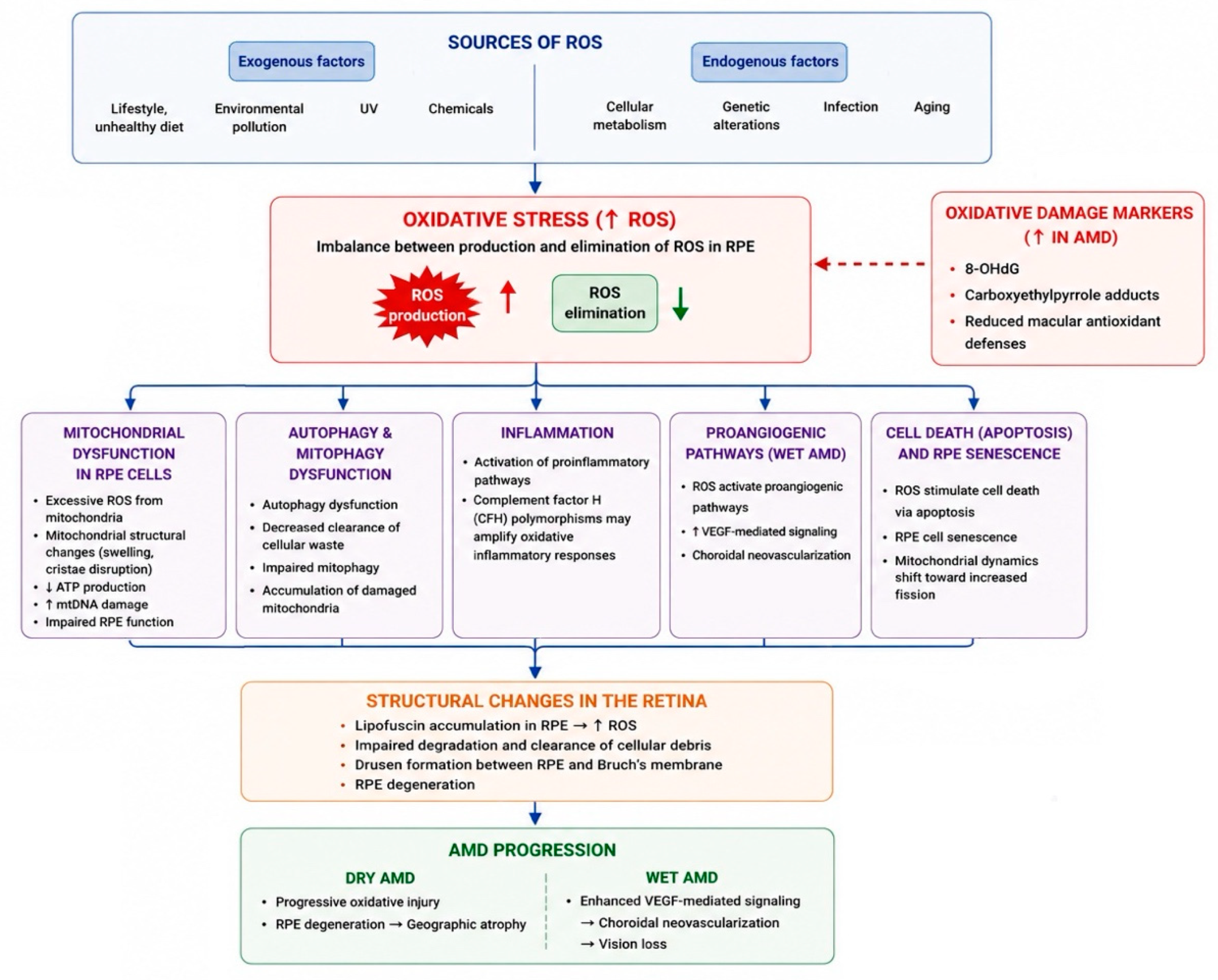
Figure 3: The Mechanism of Oxidative Stress in the Pathogenesis of Age-Related Macular Degeneration. Endogenous and exogenous factors, together with aging, promote oxidative stress through increased ROS production and impaired ROS elimination in retinal pigment epithelium (RPE) cells. Oxidative stress contributes to mitochondrial dysfunction, autophagy and mitophagy impairment, inflammation, proangiogenic signaling, apoptosis, and RPE senescence, leading to structural retinal changes and the progression of dry and wet AMD. ROS, reactive oxygen species; RPE, retinal pigment epithelium; AMD, age-related macular degeneration. The upward arrow (↑) indicates increased ROS production, increased oxidative stress markers and VEGF-mediated signaling, whereas the downward arrow (↓) indicates reduced ROS elimination.
7 The Role of Oxidative Stress in the Pathogenesis of Glaucoma
Glaucoma is a group of ocular disorders with multifactorial etiology characterized by progressive optic neuropathy that ultimately leads to irreversible visual impairment [66]. Increasing age, a positive family history of glaucoma, and elevated intraocular pressure (IOP) are among a number of well-recognized risk factors for glaucoma. In experimental glaucoma models, where intraocular pressure is increased, the majority have been shown to induce oxidative stress, characterized by a significant decrease in retinal antioxidants and an increase in retinal lipid peroxidation [67]. Other factors in glaucoma pathogenesis, like the elevation of glutamate levels, dysregulated nitric oxide (NO) metabolism, and oxidative damage caused by ROS overproduction, may significantly contribute to the neurodegeneration [68]. During scientific research, elevation of IOP in the aging mouse eye was found to induce reversible functional impairment of the inner retina along with significant increases in oxidative stress [69]. Therefore, elevated IOP, regardless of the mechanism or duration of the insult, appears capable of inducing oxidative stress in the retina and optic nerve. One mechanism that explains the etiology of this disease suggests that oxidative stress-induced vascular alterations lead to impaired autoregulation of blood flow to the optic nerve. Despite normal IOP in normal-tension glaucoma, vascular dysregulation and ischemia-reperfusion-related oxidative stress can still contribute to optic nerve head damage [51,59,70].
Additionally, oxidative injury to the trabecular meshwork may impair aqueous humor outflow, resulting in elevated IOP. This may be driven by oxidative agents in the aqueous humor acting on the trabecular meshwork (TM). In the anterior segment, the TM, a sieve-like tissue regulating aqueous humor outflow, is directly exposed to ROS in the aqueous humor, including hydrogen peroxide and superoxide. Chronic oxidative insult induces TM cell loss, cytoskeletal reorganization, and extracellular matrix stiffening, thereby increasing outflow resistance and elevating IOP [3,51,59].
Oxidative stress can also directly damage retinal ganglion cells (RGCs). In the posterior segment, RGCs are particularly prone to mitochondrial oxidative injury due to their high metabolic demand. Mitochondrial dysfunction affecting RGCs as well as supporting glial cells can directly promote apoptosis and contribute to optic nerve degeneration [3,71]. ROS serve as critical signaling molecules that activate apoptosis pathways. Research has demonstrated that reducing ROS generation can temporarily protect RGCs from apoptosis, both through caspase-independent pathways and by directly inhibiting RGC apoptosis in vitro [72]. Furthermore, oxidative stress disrupts retinal glutamine cycling, leading to the accumulation of neurotoxic levels of glutamate. A reduction in the activity of glutamine synthetase—the enzyme that converts retinal glutamate into non-toxic forms—has also been observed. Glutamate neurotoxicity typically damages cellular components, including mitochondria, which results in the depolarization of these organelles and the excessive generation of ROS. Moreover, the apoptotic process itself produces ROS. Oxidative stress indirectly damages RGCs through aberrant immune responses and glial cell dysfunction. ROS also triggers autophagy and apoptosis in retinal RGCs, eventually leading to optic nerve atrophy and blindness [73,74].
Several mitochondria-targeted therapeutic strategies have been proposed for glaucoma, including nicotinamide (vitamin B3), Mdivi-1 (mitochondrial division inhibitor 1), lithium chloride, and apolipoprotein A-I binding protein (AIBP). Nicotinamide enhances cellular nicotinamide adenine dinucleotide (NAD+) levels and has demonstrated neuroprotective effects in preclinical models, while its potential to slow glaucoma progression is currently being evaluated in clinical trials [75,76]. Mdivi-1 and lithium chloride exert neuroprotective effects by reducing excessive mitochondrial fission [64,77], whereas AIBP attenuates mitochondrial dysfunction and neuroinflammation, thereby reducing retinal ganglion cell loss [78,79].
8 The Role of Oxidative Stress in the Pathogenesis of Cataract
The lens is an avascular and transparent tissue that is composed of lens epithelial cells (LECs), lens fiber cells, and mature lens fiber cells (LFCs) [80]. Cataracts mainly result from crystallin protein aggregation within lens fiber cells, which are crucial for refraction and transparency. The lens is comprised of three principal classes of crystallins: α, β, and γ crystallins, essential proteins for preserving lens clarity [81].
Protein Oxidation and Crystallin Aggregation
The development of cataracts is influenced by various factors, among which oxidative stress and ROS play crucial roles [82]. Under physiological conditions, lens transparency is preserved by a strong antioxidant system that includes glutathione (GSH), ascorbate, and enzymatic defenses [51]. The encapsulated and avascular nature of the lens protects it from oxidative damage, as there is very low oxygen tension inside the lens core. Although ROS serve physiological functions at low concentrations, they become toxic at elevated levels, leading to lens opacification when there is either an increase in oxidative stress or a diminished capacity to eliminate ROS [74]. ROS are mostly generated within the mitochondria in lens epithelium cells and the superficial fiber cells, which are highly reactive and can damage macromolecules, such as lipids, proteins, and nucleic acids, causing mutagenesis and cell death [83]. The lens is exposed to multiple oxidative stressors: endogenous, such as altered mitochondrial respiration, phagocytic respiratory bursts, viral infections, and exogenous: UV light, metals, drugs, and cigarette smoke. All of these can lead to the production of ROS. Lens epithelial cells (LECs) utilize ROS-scavenging enzymes to maintain low ROS levels, thereby protecting the lens’s structural components and ensuring its clarity. Oxidative modification of lens epithelial cells and crystallin proteins is promoted by redox imbalance, which becomes more pronounced when ROS production rises and antioxidant reserves decline. This occurs commonly with aging and at-risk conditions such as prolonged UV exposure, diabetes, and hypertension. In particular, oxidation of methionine and cysteine residues can lead to disulfide bond formation, S-glutathionylation, and crystallin aggregation into high-molecular-weight complexes that scatter light within the lens [51,84].
Lipid Peroxidation and Membrane Damage
Moreover, lipid peroxidation is initiated by free-radical attack on membrane lipids, generating large amounts of reactive products, which have been strongly implicated in the mechanisms of cataractogenesis [85]. These peroxidized lipids alter the permeability of cell membranes, changing the internal composition and structure of cells, which ultimately disrupts protein function and contributes to the development of cataracts.
Oxidative stress may lead to several protein modifications, resulting in the formation of high-molecular-weight insoluble aggregates commonly found in cataractous lenses [86]. Clinical studies of human cataractous lenses consistently show increased protein carbonyl levels together with reduced glutathione (GSH) content, and these changes correlate with cataract severity. A decline in GSH, considered a biochemical hallmark of cataract formation, leaves crystallin proteins more vulnerable to irreversible cross-linking. At the same time, lipid peroxidation in lens fiber cell membranes compromises membrane integrity, promoting fiber disorganization and resulting in optical aberrations [84].
Cellular Alterations and Signaling Dysfunction
Oxidative stress can activate factors such as transforming growth factor beta (TGF-β), which prompts the transformation of lens epithelial cells (LECs) into mesenchymal cells. These cells then migrate and form fibrotic plaques, significantly impairing vision. Additionally, oxidative stress can induce defects in Na+/K+-ATPase on the membranes of lens epithelial cells. This disruption can lead to the accumulation of sodium and water within the cells, ultimately causing lens opacification (Fig. 4) [81].
As an example, in diabetes, hyperglycemia-induced ROS accelerates oxidation of lens epithelial cells and crystallin proteins, an effect further amplified by protein glycation and osmotic stress. More broadly, oxidative injury to lens proteins and membranes can precede clinically visible opacities, supporting the view that ROS-mediated damage is an initiating rather than a secondary event in cataract development [51].
Several antioxidant-based approaches have been proposed for cataract management, including ATX and biliverdin reductase A (BVRA). ATX inhibits ferroptosis through modulation of glutathione peroxidase 4 (GPX4) [87], whereas BVRA reduces intracellular ROS by promoting the formation of the antioxidant bilirubin, thereby helping to restore redox balance [88].
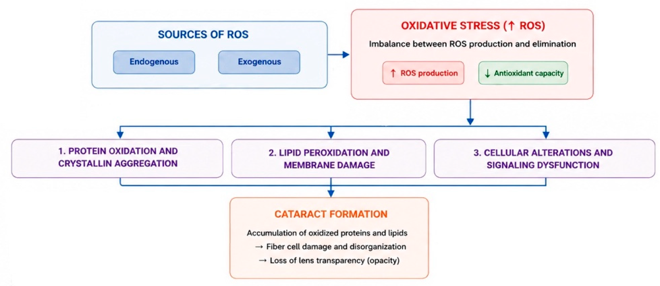
Figure 4: The Mechanism of Oxidative Stress in the Pathogenesis of Cataract. Endogenous and exogenous factors promote oxidative stress through increased ROS production and reduced antioxidant capacity. Oxidative stress induces protein oxidation and crystallin aggregation, lipid peroxidation and membrane damage, as well as cellular alterations and signaling dysfunction, ultimately leading to cataract formation. The upward arrow (↑) indicates increased ROS production, whereas the downward arrow (↓) indicates reduced antioxidant capacity. ROS, reactive oxygen species.
Oxidative stress and the overproduction of ROS are crucial pathomechanisms implicated in a variety of ocular diseases. An in-depth understanding of the underlying molecular and biochemical mechanisms offers substantial prospects for preventing these disorders, achieving early diagnosis, and implementing novel therapeutic strategies. Continued exploration in this field is essential for enhancing treatment protocols and ultimately improving patient outcomes in ophthalmic care. Future research should focus on further evaluating promising antioxidant and mitochondria-targeted therapies in well-designed clinical trials. In addition, a deeper understanding of oxidative stress-related molecular mechanisms may support the development of more effective and personalized therapeutic strategies for ocular diseases.
Acknowledgement:
Funding Statement: The authors received no specific funding for this study.
Author Contributions: Study conception and design: Andrzej Grzybowski; draft manuscript preparation: Aušrinė Bajoriūnaitė; review and editing: Andrzej Grzybowski, Reda Žemaitienė; visualization: Aušrinė Bajoriūnaitė; supervision: Andrzej Grzybowski, Reda Žemaitienė. All authors reviewed and approved the final version of the manuscript.
Availability of Data and Materials: Data sharing is not applicable to this article as no datasets were generated or analyzed during the current study.
Ethics Approval: This article is based on previously conducted studies and does not contain any new studies with human participants or animals performed by any of the authors.
Conflicts of Interest: The authors declare no conflicts of interest.
References
1. Jomova K , Raptova R , Alomar SY , Alwasel SH , Nepovimova E , Kuca K , et al. Reactive oxygen species, toxicity, oxidative stress, and antioxidants: Chronic diseases and aging. Arch Toxicol. 2023; 97( 10): 2499– 574. doi:10.1007/s00204-023-03562-9. [Google Scholar] [CrossRef]
2. Qin Y , Qian C , Li W , Wang Q , Sheng Q , Chen Z , et al. Oxidative stress: Molecular mechanisms, diseases, and therapeutic targets. MedComm. 2026; 7( 2): e70600. doi:10.1002/mco2.70600. [Google Scholar] [CrossRef]
3. Abukhaled Y . Reactive oxygen species and oxidative stress in ocular disease: From molecular mechanisms to targeted therapies. Med Hypothesis Discov Innov Ophthalmol. 2025; 14( 3): 136– 45. doi:10.51329/mehdiophthal1527. [Google Scholar] [CrossRef]
4. Halliwell B , Whiteman M . Measuring reactive species and oxidative damage in vivoand in cell culture: How should you do it and what do the results mean? Br J Pharmacol. 2004; 142( 2): 231– 55. doi:10.1038/sj.bjp.0705776. [Google Scholar] [CrossRef]
5. Santibáñez-Andrade M , Quezada-Maldonado EM , Rivera-Pineda A , Chirino YI , García-Cuellar CM , Sánchez-Pérez Y . The road to malignant cell transformation after particulate matter exposure: From oxidative stress to genotoxicity. Int J Mol Sci. 2023; 24( 2): 1782. doi:10.3390/ijms24021782. [Google Scholar] [CrossRef]
6. Saccà SC , Roszkowska AM , Izzotti A . Environmental light and endogenous antioxidants as the main determinants of non-cancer ocular diseases. Mutat Res Rev Mutat Res. 2013; 752( 2): 153– 71. doi:10.1016/j.mrrev.2013.01.001. [Google Scholar] [CrossRef]
7. Giorgi C , Marchi S , Simoes ICM , Ren Z , Morciano G , Perrone M , et al. Mitochondria and reactive oxygen species in aging and age-related diseases. Int Rev Cell Mol Biol. 2018; 340: 209– 344. doi:10.1016/bs.ircmb.2018.05.006. [Google Scholar] [CrossRef]
8. Craig JP , Nichols KK , Akpek EK , Caffery B , Dua HS , Joo CK , et al. TFOS DEWS II definition and classification report. Ocul Surf. 2017; 15( 3): 276– 83. doi:10.1016/j.jtos.2017.05.008. [Google Scholar] [CrossRef]
9. Bui HL , Su YH , Yang CJ , Huang CJ , Lai JY . Mucoadhesive, antioxidant, and lubricant catechol-functionalized poly(phosphobetaine) as biomaterial nanotherapeutics for treating ocular dryness. J Nanobiotechnol. 2024; 22( 1): 160. doi:10.1186/s12951-024-02448-x. [Google Scholar] [CrossRef]
10. Navel V , Sapin V , Henrioux F , Blanchon L , Labbé A , Chiambaretta F , et al. Oxidative and antioxidative stress markers in dry eye disease: A systematic review and meta-analysis. Acta Ophthalmol. 2022; 100( 1): 45– 57. doi:10.1111/aos.14892. [Google Scholar] [CrossRef]
11. Zhao J , Ma J , Deng Y , Kelly JA , Kim K , Bang SY , et al. A missense variant in NCF1 is associated with susceptibility to multiple autoimmune diseases. Nat Genet. 2017; 49( 3): 433– 7. doi:10.1038/ng.3782. [Google Scholar] [CrossRef]
12. Mohr A , Atif M , Balderas R , Gorochov G , Miyara M . The role of FOXP3+ regulatory T cells in human autoimmune and inflammatory diseases. Clin Exp Immunol. 2019; 197( 1): 24– 35. doi:10.1111/cei.13288. [Google Scholar] [CrossRef]
13. Efimova O , Szankasi P , Kelley TW . Ncf1 (p47phox) is essential for direct regulatory T cell mediated suppression of CD4+ effector T cells. PLoS One. 2011; 6( 1): e16013. doi:10.1371/journal.pone.0016013. [Google Scholar] [CrossRef]
14. Scherlinger M , Tsokos GC . Reactive oxygen species: The Yin and Yang in (auto-) immunity. Autoimmun Rev. 2021; 20( 8): 102869. doi:10.1016/j.autrev.2021.102869. [Google Scholar] [CrossRef]
15. Mittal M , Siddiqui MR , Tran K , Reddy SP , Malik AB . Reactive oxygen species in inflammation and tissue injury. Antioxid Redox Signal. 2014; 20( 7): 1126– 67. doi:10.1089/ars.2012.5149. [Google Scholar] [CrossRef]
16. Meng J , Lin B , Huang S , Li Y , Wu P , Zhang F , et al. Melatonin exerts anti-angiogenic and anti-inflammatory effects in alkali-burned corneas. Ann Transl Med. 2022; 10( 8): 432. doi:10.21037/atm-21-4927. [Google Scholar] [CrossRef]
17. Wang B , Zuo X , Peng L , Wang X , Zeng H , Zhong J , et al. Melatonin ameliorates oxidative stress-mediated injuries through induction of HO-1 and restores autophagic flux in dry eye. Exp Eye Res. 2021; 205: 108491. doi:10.1016/j.exer.2021.108491. [Google Scholar] [CrossRef]
18. Ajith TA . Alpha-lipoic acid: A possible pharmacological agent for treating dry eye disease and retinopathy in diabetes. Clin Exp Pharmacol Physiol. 2020; 47( 12): 1883– 90. doi:10.1111/1440-1681.13373. [Google Scholar] [CrossRef]
19. Zernii EY , Gancharova OS , Baksheeva VE , Golovastova MO , Kabanova EI , Savchenko MS , et al. Mitochondria-targeted antioxidant SkQ1 prevents anesthesia-induced dry eye syndrome. Oxid Med Cell Longev. 2017; 2017: 9281519. doi:10.1155/2017/9281519. [Google Scholar] [CrossRef]
20. Luo W , Zou Y , Wu H , Yang Z , You Z . Blueberry anthocyanins extract attenuates oxidative stress and angiogenesis on an in vitro high glucose-induced retinopathy model through the miR-33/GLCCI1 axis. BIOCELL. 2024; 48( 8): 1275– 84. doi:10.32604/biocell.2024.051045. [Google Scholar] [CrossRef]
21. Pan D , Xu L , Guo M . The role of protein kinase C in diabetic microvascular complications. Front Endocrinol. 2022; 13: 973058. doi:10.3389/fendo.2022.973058. [Google Scholar] [CrossRef]
22. Kanwar M , Chan PS , Kern TS , Kowluru RA . Oxidative damage in the retinal mitochondria of diabetic mice: Possible protection by superoxide dismutase. Investig Ophthalmol Vis Sci. 2007; 48( 8): 3805– 11. doi:10.1167/iovs.06-1280. [Google Scholar] [CrossRef]
23. Brownlee M . Biochemistry and molecular cell biology of diabetic complications. Nature. 2001; 414( 6865): 813– 20. doi:10.1038/414813a. [Google Scholar] [CrossRef]
24. Kaneto H , Xu G , Song KH , Suzuma K , Bonner-Weir S , Sharma A , et al. Activation of the hexosamine pathway leads to deterioration of pancreatic beta-cell function through the induction of oxidative stress. J Biol Chem. 2001; 276( 33): 31099– 104. doi:10.1074/jbc.M104115200. [Google Scholar] [CrossRef]
25. ValdezGuerrero AS , Quintana-Pérez JC , Arellano-Mendoza MG , Castañeda-Ibarra FJ , Tamay-Cach F , Alemán-González-Duhart D . Diabetic retinopathy: Important biochemical alterations and the main treatment strategies. Can J Diabetes. 2021; 45( 6): 504– 11. doi:10.1016/j.jcjd.2020.10.009. [Google Scholar] [CrossRef]
26. Pinti MV , Fink GK , Hathaway QA , Durr AJ , Kunovac A , Hollander JM . Mitochondrial dysfunction in type 2 diabetes mellitus: An organ-based analysis. Am J Physiol Endocrinol Metab. 2019; 316( 2): E268– 85. doi:10.1152/ajpendo.00314.2018. [Google Scholar] [CrossRef]
27. Kowluru RA , Kowluru A , Mishra M , Kumar B . Oxidative stress and epigenetic modifications in the pathogenesis of diabetic retinopathy. Prog Retin Eye Res. 2015; 48: 40– 61. doi:10.1016/j.preteyeres.2015.05.001. [Google Scholar] [CrossRef]
28. Kowluru RA . Retinopathy in a diet-induced type 2 diabetic rat model and role of epigenetic modifications. Diabetes. 2020; 69( 4): 689– 98. doi:10.2337/db19-1009. [Google Scholar] [CrossRef]
29. Kowluru RA , Kowluru V , Xiong Y , Ho YS . Overexpression of mitochondrial superoxide dismutase in mice protects the retina from diabetes-induced oxidative stress. Free Radic Biol Med. 2006; 41( 8): 1191– 6. doi:10.1016/j.freeradbiomed.2006.01.012. [Google Scholar] [CrossRef]
30. Kim D , Sesaki H , Roy S . Reduced levels of Drp1 protect against development of retinal vascular lesions in diabetic retinopathy. Cells. 2021; 10( 6): 1379. doi:10.3390/cells10061379. [Google Scholar] [CrossRef]
31. Kim D , Votruba M , Roy S . Opa1 deficiency promotes development of retinal vascular lesions in diabetic retinopathy. Int J Mol Sci. 2021; 22( 11): 5928. doi:10.3390/ijms22115928. [Google Scholar] [CrossRef]
32. Kim D , Roy S . Effects of diabetes on mitochondrial morphology and its implications in diabetic retinopathy. Investig Ophthalmol Vis Sci. 2020; 61( 10): 10. doi:10.1167/iovs.61.10.10. [Google Scholar] [CrossRef]
33. Li M , Yang L , Zhai H , Qiao L , Wang Z , An X , et al. A new perspective on protecting the blood-retinal barrier against injury in diabetic retinopathy: Mitophagy. Front Endocrinol. 2025; 16: 1617797. doi:10.3389/fendo.2025.1617797. [Google Scholar] [CrossRef]
34. Liu X , Chang Y , Li Y , Liu Y , Song W , Lu J , et al. Oxidative stress and retinopathy: Evidence from epidemiological studies. J Transl Med. 2025; 23( 1): 94. doi:10.1186/s12967-025-06110-4. [Google Scholar] [CrossRef]
35. Sheemar A , Bellala K , Sharma SV , Sharma S , Kaur I , Rani P , et al. Metabolic memory and diabetic retinopathy: Legacy of glycemia and possible steps into future. Indian J Ophthalmol. 2024; 72( 6): 796– 808. doi:10.4103/ijo.ijo_2563_23. [Google Scholar] [CrossRef]
36. Loukovaara S , Robciuc A , Holopainen JM , Lehti K , Pessi T , Liinamaa J , et al. Ang-2 upregulation correlates with increased levels of MMP-9, VEGF, EPO and TGFβ1 in diabetic eyes undergoing vitrectomy. Acta Ophthalmol. 2013; 91( 6): 531– 9. doi:10.1111/j.1755-3768.2012.02473.x. [Google Scholar] [CrossRef]
37. Azuma K , Koumura T , Iwamoto R , Matsuoka M , Terauchi R , Yasuda S , et al. Mitochondrial glutathione peroxidase 4 is indispensable for photoreceptor development and survival in mice. J Biol Chem. 2022; 298( 4): 101824. doi:10.1016/j.jbc.2022.101824. [Google Scholar] [CrossRef]
38. Tsai GY , Cui JZ , Syed H , Xia Z , Ozerdem U , McNeill JH , et al. Effect of N-acetylcysteine on the early expression of inflammatory markers in the retina and plasma of diabetic rats. Clin Exper Ophthalmol. 2009; 37( 2): 223– 31. doi:10.1111/j.1442-9071.2009.02000.x. [Google Scholar] [CrossRef]
39. Garcia-Medina JJ , Rubio-Velazquez E , Foulquie-Moreno E , Casaroli-Marano RP , Pinazo-Duran MD , Zanon-Moreno V , et al. Update on the effects of antioxidants on diabetic retinopathy: In vitro experiments, animal studies and clinical trials. Antioxidants. 2020; 9( 6): 561. doi:10.3390/antiox9060561. [Google Scholar] [CrossRef]
40. Jiang Q , Yin J , Chen J , Ma X , Wu M , Liu G , et al. Mitochondria-targeted antioxidants: A step towards disease treatment. Oxid Med Cell Longev. 2020; 2020: 8837893. doi:10.1155/2020/8837893. [Google Scholar] [CrossRef]
41. van Lookeren Campagne M , LeCouter J , Yaspan BL , Ye W . Mechanisms of age-related macular degeneration and therapeutic opportunities. J Pathol. 2014; 232( 2): 151– 64. doi:10.1002/path.4266. [Google Scholar] [CrossRef]
42. Fine SL , Berger JW , Maguire MG , Ho AC . Age-related macular degeneration. N Engl J Med. 2000; 342( 7): 483– 92. doi:10.1056/nejm200002173420707. [Google Scholar] [CrossRef]
43. Maurya M , Bora K , Blomfield AK , Pavlovich MC , Huang S , Liu CH , et al. Oxidative stress in retinal pigment epithelium degeneration: From pathogenesis to therapeutic targets in dry age-related macular degeneration. Neural Regen Res. 2023; 18( 10): 2173– 81. doi:10.4103/1673-5374.369098. [Google Scholar] [CrossRef]
44. Barot M , Gokulgandhi MR , Mitra AK . Mitochondrial dysfunction in retinal diseases. Curr Eye Res. 2011; 36( 12): 1069– 77. doi:10.3109/02713683.2011.607536. [Google Scholar] [CrossRef]
45. Brown EE , DeWeerd AJ , Ildefonso CJ , Lewin AS , Ash JD . Mitochondrial oxidative stress in the retinal pigment epithelium (RPE) led to metabolic dysfunction in both the RPE and retinal photoreceptors. Redox Biol. 2019; 24: 101201. doi:10.1016/j.redox.2019.101201. [Google Scholar] [CrossRef]
46. Bugger H , Pfeil K . Mitochondrial ROS in myocardial ischemia reperfusion and remodeling. Biochim Biophys Acta Mol Basis Dis. 2020; 1866( 7): 165768. doi:10.1016/j.bbadis.2020.165768. [Google Scholar] [CrossRef]
47. Kaarniranta K , Uusitalo H , Blasiak J , Felszeghy S , Kannan R , Kauppinen A , et al. Mechanisms of mitochondrial dysfunction and their impact on age-related macular degeneration. Prog Retin Eye Res. 2020; 79: 100858. doi:10.1016/j.preteyeres.2020.100858. [Google Scholar] [CrossRef]
48. Tong Y , Zhang Z , Wang S . Role of mitochondria in retinal pigment epithelial aging and degeneration. Front Aging. 2022; 3: 926627. doi:10.3389/fragi.2022.926627. [Google Scholar] [CrossRef]
49. Yako T , Nakamura M , Otsu W , Nakamura S , Shimazawa M , Hara H . Mitochondria dynamics in the aged mice eye and the role in the RPE phagocytosis. Exp Eye Res. 2021; 213: 108800. doi:10.1016/j.exer.2021.108800. [Google Scholar] [CrossRef]
50. Skowronska-Krawczyk D , Chao DL . Long-chain polyunsaturated fatty acids and age-related macular degeneration. Adv Exp Med Biol. 2019; 1185: 39– 43. doi:10.1007/978-3-030-27378-1_7. [Google Scholar] [CrossRef]
51. Bissell M , Fetian I , Mozaffarieh M . The role of oxidative stress in the pathogenesis of eye diseases. hb TIMES Schw Aerztej. 2022; 6( 3–4): 72– 7. doi:10.36000/hbT.2022.06.002. [Google Scholar] [CrossRef]
52. Zhou H , Zhang H , Yu A , Xie J . Association between sunlight exposure and risk of age-related macular degeneration: A meta-analysis. BMC Ophthalmol. 2018; 18( 1): 331. doi:10.1186/s12886-018-1004-y. [Google Scholar] [CrossRef]
53. Sui GY , Liu GC , Liu GY , Gao YY , Deng Y , Wang WY , et al. Is sunlight exposure a risk factor for age-related macular degeneration? A systematic review and meta-analysis. Br J Ophthalmol. 2013; 97( 4): 389– 94. doi:10.1136/bjophthalmol-2012-302281. [Google Scholar] [CrossRef]
54. Nita M , Grzybowski A . The role of the reactive oxygen species and oxidative stress in the pathomechanism of the age-related ocular diseases and other pathologies of the anterior and posterior eye segments in adults. Oxid Med Cell Longev. 2016; 2016: 3164734. doi:10.1155/2016/3164734. [Google Scholar] [CrossRef]
55. Datta S , Cano M , Satyanarayana G , Liu T , Wang L , Wang J , et al. Mitophagy initiates retrograde mitochondrial-nuclear signaling to guide retinal pigment cell heterogeneity. Autophagy. 2023; 19( 3): 966– 83. doi:10.1080/15548627.2022.2109286. [Google Scholar] [CrossRef]
56. Xiong R , Yuan Y , Zhu Z , Wu Y , Ha J , Han X , et al. Micronutrients and diabetic retinopathy: Evidence from the national health and nutrition examination survey and a meta-analysis. Am J Ophthalmol. 2022; 238: 141– 56. doi:10.1016/j.ajo.2022.01.005. [Google Scholar] [CrossRef]
57. Zhou FL , Zhang WG , Wei YC , Meng S , Bai GG , Wang BY , et al. Involvement of oxidative stress in the relapse of acute myeloid leukemia. J Biol Chem. 2010; 285( 20): 15010– 5. doi:10.1074/jbc.M110.103713. [Google Scholar] [CrossRef]
58. Kulbay M , Wu KY , Nirwal GK , Bélanger P , Tran SD . The role of reactive oxygen species in age-related macular degeneration: A comprehensive review of antioxidant therapies. Biomedicines. 2024; 12( 7): 1579. doi:10.3390/biomedicines12071579. [Google Scholar] [CrossRef]
59. Feilchenfeld Z , Yücel YH , Gupta N . Oxidative injury to blood vessels and glia of the pre-laminar optic nerve head in human glaucoma. Exp Eye Res. 2008; 87( 5): 409– 14. doi:10.1016/j.exer.2008.07.011. [Google Scholar] [CrossRef]
60. Brown EE , Ball JD , Chen Z , Khurshid GS , Prosperi M , Ash JD . The common antidiabetic drug metformin reduces odds of developing age-related macular degeneration. Investig Ophthalmol Vis Sci. 2019; 60( 5): 1470– 7. doi:10.1167/iovs.18-26422. [Google Scholar] [CrossRef]
61. Blitzer AL , Ham SA , Colby KA , Skondra D . Association of metformin use with age-related macular degeneration: A case-control study. JAMA Ophthalmol. 2021; 139( 3): 302– 9. doi:10.1001/jamaophthalmol.2020.6331. [Google Scholar] [CrossRef]
62. Yu L , Sun Y , Cheng L , Jin Z , Yang Y , Zhai M , et al. Melatonin receptor-mediated protection against myocardial ischemia/reperfusion injury: Role of SIRT1. J Pineal Res. 2014; 57( 2): 228– 38. doi:10.1111/jpi.12161. [Google Scholar] [CrossRef]
63. Krestinina O , Baburina Y , Krestinin R , Odinokova I , Fadeeva I , Sotnikova L . Astaxanthin prevents mitochondrial impairment induced by isoproterenol in isolated rat heart mitochondria. Antioxidants. 2020; 9( 3): 262. doi:10.3390/antiox9030262. [Google Scholar] [CrossRef]
64. Sun H , Li B , Gu Y , Li F , Di G , Chen P . Imbalanced mitochondrial homeostasis in ocular diseases: Unique pathogenesis and targeted therapy. Exp Eye Res. 2025; 260: 110632. doi:10.1016/j.exer.2025.110632. [Google Scholar] [CrossRef]
65. Nashine S . Potential therapeutic candidates for age-related macular degeneration (AMD). Cells. 2021; 10( 9): 2483. doi:10.3390/cells10092483. [Google Scholar] [CrossRef]
66. Jayaram H , Kolko M , Friedman DS , Gazzard G . Glaucoma: Now and beyond. Lancet. 2023; 402( 10414): 1788– 801. doi:10.1016/s0140-6736(23)01289-8. [Google Scholar] [CrossRef]
67. Moreno MC , Saenz DA , Campanelli JL , Sarmiento MIK , Benozzi JL , Rosenstein RE . Retinal oxidative stress induced by high intraocular pressure. Investig Ophthalmol Vis Sci. 2003; 44( 13): 468. [Google Scholar]
68. Vishwaraj CR , Kavitha S , Venkatesh R , Shukla AG , Chandran P , Tripathi S . Neuroprotection in glaucoma. Indian J Ophthalmol. 2022; 70( 2): 380– 5. doi:10.4103/ijo.ijo_1158_21. [Google Scholar] [CrossRef]
69. Kong YXG , van Bergen N , Bui BV , Chrysostomou V , Vingrys AJ , Trounce IA , et al. Impact of aging and diet restriction on retinal function during and after acute intraocular pressure injury. Neurobiol Aging. 2012; 33( 6): 1126.e15– 25. doi:10.1016/j.neurobiolaging.2011.11.026. [Google Scholar] [CrossRef]
70. Konieczka K , Ritch R , Traverso CE , Kim DM , Kook MS , Gallino A , et al. Flammer syndrome. EPMA J. 2014; 5( 1): 11. doi:10.1186/1878-5085-5-11. [Google Scholar] [CrossRef]
71. Nucci C , Tartaglione R , Cerulli A , Mancino R , Spanò A , Cavaliere F , et al. Retinal damage caused by high intraocular pressure-induced transient ischemia is prevented by coenzyme Q10 in rat. Int Rev Neurobiol. 2007; 82: 397– 406. doi:10.1016/S0074-7742(07)82022-8. [Google Scholar] [CrossRef]
72. Li GY , Osborne NN . Oxidative-induced apoptosis to an immortalized ganglion cell line is caspase independent but involves the activation of poly(ADP-ribose)polymerase and apoptosis-inducing factor. Brain Res. 2008; 1188: 35– 43. doi:10.1016/j.brainres.2007.10.073. [Google Scholar] [CrossRef]
73. Ishikawa M . Abnormalities in glutamate metabolism and excitotoxicity in the retinal diseases. Scientifica. 2013; 2013: 528940. doi:10.1155/2013/528940. [Google Scholar] [CrossRef]
74. Hsueh YJ , Chen YN , Tsao YT , Cheng CM , Wu WC , Chen HC . The pathomechanism, antioxidant biomarkers, and treatment of oxidative stress-related eye diseases. Int J Mol Sci. 2022; 23( 3): 1255. doi:10.3390/ijms23031255. [Google Scholar] [CrossRef]
75. Williams PA , Harder JM , Foxworth NE , Cochran KE , Philip VM , Porciatti V , et al. Vitamin B3 modulates mitochondrial vulnerability and prevents glaucoma in aged mice. Science. 2017; 355( 6326): 756– 60. doi:10.1126/science.aal0092. [Google Scholar] [CrossRef]
76. Petriti B , Rabiolo A , Chau KY , Williams PA , Montesano G , Lascaratos G , et al. Peripheral blood mononuclear cell respiratory function is associated with progressive glaucomatous vision loss. Nat Med. 2024; 30( 8): 2362– 70. doi:10.1038/s41591-024-03068-6. [Google Scholar] [CrossRef]
77. Zeng Z , You M , Fan C , Rong R , Li H , Xia X . Pathologically high intraocular pressure induces mitochondrial dysfunction through Drp1 and leads to retinal ganglion cell PANoptosis in glaucoma. Redox Biol. 2023; 62: 102687. doi:10.1016/j.redox.2023.102687. [Google Scholar] [CrossRef]
78. Zuo Y , Yin L , Cheng X , Li J , Wu H , Liu X , et al. Elamipretide attenuates pyroptosis and perioperative neurocognitive disorders in aged mice. Front Cell Neurosci. 2020; 14: 251. doi:10.3389/fncel.2020.00251. [Google Scholar] [CrossRef]
79. Liu Y , Bu Q , Hu D , Chen C , Zhu J , Zhou Q , et al. NAD+ supplementation improves mitochondrial functions and normalizes glaucomatous trabecular meshwork features. Exp Cell Res. 2024; 440( 1): 114137. doi:10.1016/j.yexcr.2024.114137. [Google Scholar] [CrossRef]
80. Shu DY , Chaudhary S , Cho KS , Lennikov A , Miller WP , Thorn DC , et al. Role of oxidative stress in ocular diseases: A balancing act. Metabolites. 2023; 13( 2): 187. doi:10.3390/metabo13020187. [Google Scholar] [CrossRef]
81. Lee B , Afshari NA , Shaw PX . Oxidative stress and antioxidants in cataract development. Curr Opin Ophthalmol. 2024; 35( 1): 57– 63. doi:10.1097/ICU.0000000000001009. [Google Scholar] [CrossRef]
82. Kulbay M , Wu KY , Nirwal GK , Bélanger P , Tran SD . Oxidative stress and cataract formation: Evaluating the efficacy of antioxidant therapies. Biomolecules. 2024; 14( 9): 1055. doi:10.3390/biom14091055. [Google Scholar] [CrossRef]
83. Zhou YF , Guo B , Ye MJ , Liao RF , Li SL . Protective effect of rutin against H2O2-induced oxidative stress and apoptosis in human lens epithelial cells. Curr Eye Res. 2016; 41( 7): 933– 42. doi:10.3109/02713683.2015.1082186. [Google Scholar] [CrossRef]
84. Li J , Buonfiglio F , Zeng Y , Pfeiffer N , Gericke A . Oxidative stress in cataract formation: Is there a treatment approach on the horizon? Antioxidants. 2024; 13( 10): 1249. doi:10.3390/antiox13101249. [Google Scholar] [CrossRef]
85. Sunkireddy P , Jha SN , Kanwar JR , Yadav SC . Natural antioxidant biomolecules promises future nanomedicine based therapy for cataract. Colloids Surf B Biointerfaces. 2013; 112: 554– 62. doi:10.1016/j.colsurfb.2013.07.068. [Google Scholar] [CrossRef]
86. Michael R , Bron AJ . The ageing lens and cataract: A model of normal and pathological ageing. Philos Trans Biol Sci. 2011; 366( 1568): 1278– 92. doi:10.1098/rstb.2010.0300. [Google Scholar] [CrossRef]
87. Kong D , Liu Y , Li L , Wang H , Li K , Zheng G . Astaxanthin ameliorates oxidative stress in lens epithelial cells by regulating GPX4 and ferroptosis. Chem Biol Interact. 2023; 383: 110684. doi:10.1016/j.cbi.2023.110684. [Google Scholar] [CrossRef]
88. Huang Y , Liu Y , Yu S , Li W , Li J , Zhao B , et al. Biliverdin reductase a protects lens epithelial cells against oxidative damage and cellular senescence in age-related cataract. Oxid Med Cell Longev. 2022; 2022: 5628946. doi:10.1155/2022/5628946. [Google Scholar] [CrossRef]
Cite This Article
 Copyright © 2026 The Author(s). Published by Tech Science Press.
This work is licensed under a Creative Commons Attribution 4.0 International License , which permits unrestricted use, distribution, and reproduction in any medium, provided the original work is properly cited.
 Downloads
 Citation Tools
