
 Login
Login Register
 Submit a Paper
 Propose a Special lssue
 Open Access
MINI REVIEW
The Impact of COVID-19 on Breast Cancer and the Role of Neutrophil Extracellular Traps
1 School of Health Professions, D’Youville University, 320 Porter Ave, Buffalo, NY, USA
2 College of Science and Technology, Fairmont State University, 1201 Locust Ave, Fairmont, WV, USA
* Corresponding Author: Amitabha Ray. Email:
BIOCELL 2026, 50(9), 3 https://doi.org/10.32604/biocell.2026.076530
Received 22 November 2025; Accepted 28 April 2026; Issue published 26 August 2026
 View Full Text
 Download PDF
Abstract
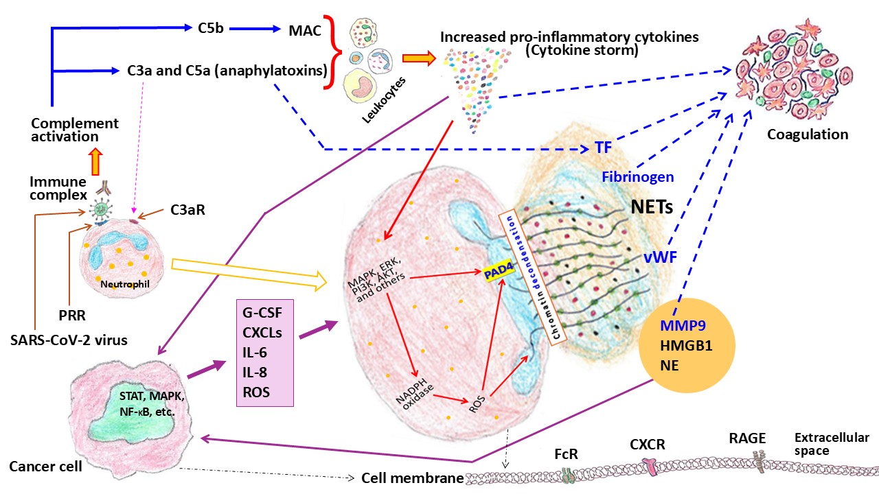
Severe acute respiratory syndrome coronavirus 2 (SARS-CoV-2)-related critical illness, i.e., severe form of coronavirus disease 2019 (COVID-19), is associated with a hyperinflammatory state. In COVID-19 disease, several components of the body, including the complement system, different cells such as endothelial cells, platelets, monocytes, and neutrophils, and various pro-inflammatory cytokines such as interleukin-6 and tumor necrosis factor α, can contribute to a state of coagulopathy, and ultimately, all these factors cause extensive tissue damage. This pathological process may contribute to increased aggressiveness in cancer cells or to the reawakening of dormant cancer cells. Studies have documented an increase in the incidence of aggressive breast cancer, such as triple-negative breast cancer, tumors with overexpression of human epidermal growth factor receptor 2 (HER2), and lymph node involvement during the COVID-19 pandemic. One etiological mechanism is perhaps the activation of neutrophils and the formation of neutrophil extracellular traps, which are thought to be linked to both severe COVID-19 disease and aggressive tumors.Graphic Abstract
Keywords
The critical phase of the coronavirus disease 2019 (COVID-19) pandemic, caused by the severe acute respiratory syndrome coronavirus 2 (SARS-CoV-2) RNA virus, occurred from December 2019 to roughly the middle of 2021. Several studies have shown an association between COVID-19 and different molecules/signaling pathways that are also involved in cancer, e.g., epidermal growth factor (EGF) and related receptor family members (such as EGFR and HER2) [1,2,3], interleukin-6 (IL-6), tumor necrosis factor α (TNF-α), mitogen-activated protein kinase (MAPK), the phosphatidylinositol 3-kinase/protein kinase B (PI3K/AKT) pathway, Janus kinase/signal transducer and activator of transcription (JAK/STAT), and nuclear factor kappa-light-chain-enhancer of activated B cells (NF-κB) [3,4]. Many of these are associated with the inflammatory process. For example, in a study in Cuba, Pérez & Crombet observed a relationship between serum EGF and the inflammatory condition of COVID-19 patients, as measured by the neutrophil-lymphocyte ratio and platelet-lymphocyte ratio [1]. Furthermore, the researchers of a Korean study categorized 444 COVID-19 patients according to disease progression into two groups: deterioration phase (moderate cases-52, severe-28) and recovery phase (moderate-72, severe-27) [3]. Differences were observed between the deteriorating and recovering groups in various cytokines, including CXCL10, PTX3, and TNFSF10. Interestingly, these cytokines are associated with signaling pathways such as JAK-STAT, NF-κB, and MAPK, which are involved in the inflammatory response. Notably, severely infected COVID-19 patients experience a hyperinflammatory state (or cytokine storm), which is characterized by the release of an excessive amount of pro-inflammatory cytokines like IL-6 and TNF-α, as well as the recruitment of immune cells such as neutrophils and lymphocytes into the affected organs, like the lungs.
Besides the higher risks of severe COVID-19 disease, bacterial infections with increased antibiotic resistance, and mortality in cancer patients with COVID-19, the pandemic generally showed an increase in advanced malignant features, including elevated risk of lung metastasis and cancer-related death [5,6,7]. A similar situation has also been considered in the case of breast cancer [8]. In COVID-19, inflammatory mediators like IL-6 can initially activate dormant cancer cells in the lung (or other tissues), and subsequently, immune cells such as T-cells are unable to control the activity of these cancer cells.
Overall, the diagnosis of breast cancer decreased during the pandemic phase compared to the pre-pandemic period—this feature was commonly recorded throughout the world, probably due to a hindrance in the healthcare system, including cancer screening. On the other hand, during the pandemic and post-pandemic times, many investigators noticed an upsurge in aggressive breast tumors, such as triple-negative tumors (TNBC) and increased lymph node involvement, including distant metastasis [9,10,11]. However, these variations might occur depending on cancer type, geographical location, and sociodemographic factors. For instance, a study on Utah women (United States) observed an increase in late-stage breast cancer incidence among Hispanic women, while late-stage cervical cancer incidence was increased in non-Hispanic White women during the pandemic compared to the pre-pandemic period [12]. Moreover, an Italian study documented a higher risk of hospitalization and death in SARS-CoV-2-positive individuals with cancer (current cases) or a history of cancer, particularly in neoplasms of the lung and breast and hematological malignancies [13]. It is worth noting that dormant cancer cells may be present in individuals with a previous history of malignant disease [14]. Furthermore, patients with different cancers, including breast cancer, who underwent surgical procedures within two weeks of COVID-19 diagnosis, had a higher incidence of post-operative complications, along with mortality, compared to those without a COVID-19 infection [15,16]. Nevertheless, one of the critical factors for the adverse clinicopathological features of breast cancer in the pandemic period was perhaps the delay in diagnosis and treatment.
The entry of the SARS-CoV-2 virus occurs generally through the binding of its spike (S) glycoprotein to the angiotensin-converting enzyme 2 (ACE-2) of the host’s target cells. However, the virus can also utilize other transmembrane proteins, such as extracellular matrix metalloproteinase inducer (EMMPRIN) or CD147, transmembrane serine protease 2 (TMPRSS2), and ADAM metallopeptidase domain 17 (ADAM17) [17]. Interestingly, these cell-surface proteins are also associated with cancer-related pathological processes in different ways. Physiologically, the conversion of angiotensin I to angiotensin II is catalyzed by ACE, a crucial step in the renin-angiotensin-aldosterone system, which regulates blood pressure. Conversely, the ACE homolog ACE-2 converts angiotensin II to angiotensin 1-7, counterbalancing the activity of ACE; thus, ACE and ACE-2 are functionally antagonistic [17]. Apart from supporting viral entry, ACE-2 is overexpressed in various tumor tissues, including carcinomas of the lung, pancreas, kidney, and colon, but exhibits a tumor-suppressive character in some cancers, like breast cancer, by inhibiting angiogenesis [18]. In addition, it is believed that SARS-CoV-2 entry decreases the expression of ACE-2, resulting in an increase in angiotensin II, which interacts to a greater extent with its receptors (AT1R and AT2R). This interaction successively promotes angiogenesis via the PI3K/AKT/NF-κB signaling pathway and eventually by vascular endothelial growth factor (VEGF). In a study using MDA-MB-231 breast cancer cells and female CD1 nu/nu mice, Tang et al. demonstrated that EMMPRIN also promoted tumor angiogenesis by stimulating VEGF [19]. On the other hand, McGowan et al., using breast cancer tissue samples and primarily MCF-7 cells, observed that ADAM-17 (alternatively referred to as TNF-α converting enzyme) was involved in tumor progression [20].
In an interesting study, Sommariva et al. documented the impact of SARS-CoV-2 infection in MCF-7, MDA-MB-231, and HCC1937 breast cancer cells [21]. Of note, the estrogen receptor (ER) and progesterone receptor (PR)-positive MCF-7 cell line represents the luminal A subtype, whereas the MDA-MB-231 and HCC1937 cell lines are negative for ER, PR, and HER2 and serve as models for TNBC. However, the investigators of this study found that, for viral replication, the most permissive cell line was MCF-7, and tamoxifen treatment reduced the viral replication rate, indicating ER involvement. In another study, the SARS-CoV-2 membrane (M) protein induced proliferation, mobility, stemness (i.e., stem cell-like characteristics), and in vivo metastatic ability of MDA-MB-231 cells in female C57BL/6J mice, as compared to MCF-7 cells that displayed a lesser response to the M protein [22]. The abovementioned changes in MDA-MB-231 cells were associated with upregulation of the NF-κB and STAT-3 pathways. Interestingly, the investigators observed a similar aggressive behavior in MCF-7 cells, with upregulation of genes linked to the epithelial–mesenchymal transition (EMT) and inflammatory cytokines, when cocultured with M protein-treated MDA-MB-231 cells [22]. Therefore, the SARS-CoV-2 virus might influence the aggressive behavior of breast cancer and particularly poor prognosis in TNBC patients. On the other hand, in a study conducted by Brichenko and colleagues, incubation with the SARS-CoV-2 S protein for up to 72 h did not produce significant changes in the proliferative activity of either MDA-MB-231 or MCF-7 cells [23]. However, viral S protein administration in both breast cancer cell lines increased the cell cycle regulator p53 levels, which might be mutant p53, at least in MDA-MB-231 cells. Intriguingly, unlike these breast cancer cells, the SARS-CoV-2 S protein has been shown to inhibit the proliferation and growth of the SiHa cell line [24], which is derived from HPV-16-positive cervical squamous cell carcinoma. Additionally, in SiHa cells, the S protein induced apoptosis and upregulation of p53.
Clearly, in vitro, cells show distinct responses to actual viral infection compared with exposure to viral proteins. Viral infections are associated with viral entry into host cells, completion of their life cycle, the production of active virions, and cytopathic effects. In contrast, exposure to viral proteins leads to phenomena such as binding to cell-surface receptors, endocytosis, effects on intracellular signaling, and immune responses in appropriate settings. The aforementioned study by Sommariva et al. on three breast cancer cell lines displayed that SARS-CoV-2 infection led to a continuous increase in viral RNA copy number in all cell lines for the first 72 h after infection, along with a reduction of about 20% in cell proliferation during that time [21]. Furthermore, SARS-CoV-2 infection had the strongest effect on MDA-MB-231 cells (293 genes affected), followed by MCF-7 cells (218 genes) and HCC1937 cells (69 genes). Regarding exposure to viral proteins, Nguyen et al. observed that, compared with the SARS-CoV-2 S and nucleocapsid (N) proteins, the M protein induced a greater migration ability in MDA-MB-231 and MCF-7 cells [22]. In addition to enhancing stemness and EMT-related genes, the M protein induced upregulation of mesenchymal markers, such as N-cadherin and vimentin, and of the SARS-CoV-2 binding receptors ACE-2 and TMPRSS2, as well as increased expression of TNF-α, IL-6, and IL-8 in MDA-MB-231 cells, but not in MCF-7 cells. On the other hand, Brichenko et al. documented that treatment with the S protein altered the cell phenotype, particularly in MDA-MB-231 cells—for example, a decrease in CD105+CD90+ and CD105+CD90− subpopulations [23]. Of note, CD105+CD90+ surface marker denotes mesenchymal stem cells (MSCs), and CD105+CD90− shows the characteristics of a less mature/modified subset of MSCs.
3 The Role of Neutrophils and NETs in Breast Cancer
Around seven million people worldwide have died due to COVID-19, which has shown an increased risk for individuals with comorbidities such as obesity and metabolic disorders like type 2 diabetes mellitus and cancers, including breast cancer [17,25]. Apart from problems in the healthcare system during the pandemic period, poor outcomes in cancer patients with SARS-CoV-2 infection could be due to several other factors, e.g., immunosuppressive effects of COVID-19, cancer, and treatment procedures like chemotherapy or radiotherapy, hypoxia-related impacts on tumor cells, and consequences of excessive release of pro-inflammatory cytokines. In fact, the underlying pathology of disease severity in COVID-19 results from interactions among various factors, e.g., complement activation, immune cell responses, pro-inflammatory cytokines, and hyperactivity of coagulation proteases [26]. In patients with COVID-19, complement activation triggers cells like endothelial cells, monocytes, and neutrophils, leading to the release of pro-inflammatory cytokines, as well as potentiating the formation of neutrophil extracellular traps (NETs) or NETosis, and the thrombotic pathway. Of note, NETs are mesh-like structures derived from activated neutrophils and consist of DNA, histone proteins, proteolytic enzymes, and other cytosolic proteins to entrap and kill pathogens (Fig. 1). Aside from the beneficial role of NETs against pathogens, excessive or imbalanced NETosis may contribute to coagulopathy or thrombotic disorders and specifically cancer-associated thrombosis [27]. Therefore, while NETs in COVID-19 patients are responsible for immunothrombosis, on the other hand, NETs construct a physical barrier for the movement of immune cells, and along with other pro-inflammatory factors, NETs could induce activation of dormant cancer cells [28], cause harmful changes in the tumor microenvironment, induce EMT, and promote metastasis [14]. In this context, it is worth mentioning that many investigators have documented leukocytosis as an important risk factor for mortality in cancer patients with COVID-19 [29,30]. In general, roughly 40% to 60% of total white blood cells (WBCs) or leukocytes are neutrophils, and thus the most common cause of leukocytosis (i.e., a WBC count exceeding 11,000 cells/μL) is neutrophilia, defined as a neutrophil count greater than 7700/μL [31].

Figure 1: Schematic diagram of neutrophil extracellular traps (NETs) and another comparable phenomenon, neutrophil-derived extracellular vesicles. A, Neutrophil extracellular traps; B, Neutrophil; C, Neutrophil-derived extracellular vesicles. The micrograph in the upper left is a human neutrophil in its early stage (which belongs to a category of less mature band neutrophils, with a typical diameter of 10–18 μm) selected from a peripheral blood smear (original micrograph captured by the authors). The yellow arrow indicates its progression to a mature segmented neutrophil, as illustrated in sketch B. Footnote: Neutrophil-derived extracellular vesicles are generated in response to inflammatory stimuli, although they also mediate intercellular communication. In addition, these vesicles extraordinarily perform both pro-inflammatory and anti-inflammatory functions.
It is pertinent to note that several NET-inducers have been suggested, for instance, cholesterol crystals, inflammatory cytokines, immune complexes, tumor cells, microbes, including viruses, and activated platelets [27]. Alternatively, NETs promote platelet activation, i.e., the initiation of primary hemostasis, thereby activating the coagulation cascade. Hence, NET-associated thrombus formation provides a scaffold for platelets, erythrocytes, extracellular vesicles, and other pro-coagulant molecules, such as von Willebrand factor (vWF), fibrinogen, factor XII, and tissue factor, and can cause vessel occlusion [27]. Clearly, the development of thrombus due to excessive NET formation could be harmful and might hinder the movement of various blood cells, including immune cells. In addition, NETs play a key role in immunothrombosis, in which innate immune cells can exhibit prothrombotic activity. Notably, NET formation in cancer could be induced by tumor-educated platelets (which are modified by the tumor) and cytokines, apart from tumor cells; and interestingly, cancer is involved with both arterial and venous thrombosis [27]. On the other hand, a recent study has shown that NETs promoted pro-metastatic signaling and thus metastatic dissemination [32]. In this connection, it is noteworthy that Albrengues et al., in an in vivo study using MCF-7 and murine D2.0R breast cancer cells and syngeneic BALB/c and nude mice, documented that NETs formed during inflammation induced the awakening of dormant cancer cells [28]. In particular, NET-associated proteases, such as neutrophil elastase (NE) and matrix metalloproteinase-9 (MMP-9), were involved in remodeling the extracellular matrix (precisely, laminins).
An important role of NETs in COVID-19 was initially hypothesized based on the disease’s link with a number of health conditions that are also associated with NET dysregulation, for example, obesity, hypertension, cardiovascular disease, chronic inflammatory disease, and rheumatoid arthritis [33]. Apart from triggering thrombosis by high blood levels of NETs in COVID-19, the components/by-products of NETs, such as released histones and high concentrations of proteases, are toxic to cells, particularly endothelium and epithelium, as well as stimulation of the inflammatory process by extracellular DNA [33,34]. Interestingly, NETs can be a source of circulating DNA in blood, in addition to apoptosis. In healthy individuals, the turnover of leukocytes, i.e., apoptosis and replacement by new cells, is a common source of circulating cell-free DNA, which is generally present as mononucleosome-associated (precisely, DNA around a histone octamer). In a study conducted by Pisareva et al., a significant increase in circulating DNA levels was observed in the plasma of COVID-19 patients, which correlated positively with NE and myeloperoxidase (MPO) levels compared with those in healthy individuals [35].
In a study in Sweden, blood samples were analyzed from 30 healthy individuals (controls) and 106 patients with COVID-19 (high respiratory support–19 cases, deceased–11) [36]. Patients were followed for 4 months. Compared with controls, patients had elevated plasma levels of nucleosomal citrullinated histone H3, cell-free DNA, and NE, which were correlated with inflammatory state (TNF-α and IL-6) and endothelial damage (vWF). These findings verified a link between an immunothrombotic state and NET markers that also revealed a relation to disease severity/clinical outcome. Similarly, in a case-control study on hospitalized patients with COVID-19, which included 11 patients who developed thrombosis and 33 patients without a thrombotic event (i.e., matched controls) [37]. In comparison with controls, the thrombosis group displayed higher levels of calprotectin, which is present mainly in neutrophils, and markers of NETs, such as cell-free DNA, MPO-DNA complexes, and citrullinated histone H3; these parameters demonstrated a strong correlation with D-dimer, a product of blood clot/fibrin degradation. On the other hand, Kapoor et al. examined a large cohort of subjects: infection with the Delta variant–351 patients, Omicron–53, and healthy individuals–140 [38]. In this study, plasma levels of different NET markers, such as NE, MPO, and circulating nuclear DNA, showed a significant and gradual increase with the disease severity. Intriguingly, even asymptomatic cases of COVID-19 exhibited higher levels of NETs than healthy individuals. Taken together, the findings from the abovementioned studies illustrated an association between the formation of NETs and COVID-19, as well as related phenomena such as inflammation, cellular injury, and hemostatic disorders, culminating in disease severity.
The negative impact of SARS-CoV-2 and other respiratory viruses, like influenza A virus, on breast cancer, particularly HER2-positive tumors, might be primarily mediated by dysregulated immune responses [39,40,41]. Interestingly, infections with different viruses, including COVID-19, could play an important role in autoimmune diseases such as systemic lupus erythematosus (SLE) [42]. One common pathological feature of these diseases, i.e., infections with SARS-CoV-2 and influenza A virus as well as SLE, is the formation of NETs [43,44,45] (Table 1). Moreover, in SLE, NET activation has a fundamental role in the disease process, including the promotion of EMT and lung injury [43]. It may be worth noting that the EMT enables cancer cells to acquire invasive/metastatic characteristics.
Table 1: Influences of COVID-19 on breast cancer and autoimmune diseases like systemic lupus erythematosus (SLE), in relation to the formation of NETs, as well as probable comparable effects of the recent influenza pandemic.
| Disease Pathologies | Investigators, Study Types, and Methods | Findings | Biological Mechanisms |
|---|---|---|---|
| Virus-related biological effects in breast cancer | Chen et al. 2025 [39]. The data obtained from the COVID-19 Host Genetics Initiative genome-wide association study (GWAS). | The investigators isolated two immune cell subsets, specifically IgD-CD27-AC (atypical memory B cells) and CD27 on IgD+CD38-unsw mem (memory B cells), as mediators linking critically ill COVID-19 to HER2+ breast cancer. | The study indicated that critically ill COVID-19 cases affected HER2+ breast cancer, probably through the dysregulation of these immune cell subsets. |
| Wang et al. 2025 [40]. In the retrospective and prospective cohort study, the responses to the neoadjuvant chemotherapy (i.e., medication prior to the main treatment, particularly surgery) were evaluated between breast cancer patients with or without COVID-19. | Breast cancer patients with COVID-19 displayed poor responses to neoadjuvant chemotherapy, including a poor prognosis in HER+ cases. | The study identified ABLIM1 and GZMM as potential genes responsible for hindering therapeutic efficacy (ABLIM1 encodes a protein that binds to actin, and GZMM encodes Granzyme M, which is associated with inflammation and the immune response). | |
| Chia et al. 2025 [41]; in vivo study. MMTV-Her2 mice were infected with the influenza A virus (sublethal dose). HER2+ cancer cells may exist as dormant cells in various organs, including the lungs. | In the lung, viral infection disrupted the dormancy of cancer cells, leading to rapid cell proliferation and expansion. | The phenomena were dependent on IL-6 and impairment of T cell activation, including inhibition of CD8+ T cells. | |
| SLE, COVID-19, and NETs | Rajalingam et al. 2025 [42]. The information was obtained from the GEO database for rhinovirus, influenza virus, respiratory syncytial virus, and COVID-19. | The aim was to determine the extent of autoimmune activation against antigens associated with these respiratory viruses. The observation led to the identification of three genes: TRIM21, ELANE, and CTSG. | These three genes are thought to be involved in various pathways, including NETosis, apoptosis, the RAS, and the lysosome, which are commonly activated in the SLE pathway in genetically susceptible SLE patients. |
| Lin et al. 2023 [43]. Lung biopsy samples were analyzed using histopathology and RNA sequencing from patients with SLE, COVID-19-induced pulmonary fibrosis, and idiopathic pulmonary fibrosis. | There were higher levels of NETs in the pulmonary tissues of SLE and COVID-19 than in those with idiopathic pulmonary fibrosis. NETosis-associated genes were significantly enriched in SLE and COVID-19. | The pathway of NET formation promoted EMT and expression of α-SMA, Twist, and Snail, but decreased E-cadherin expression. SLE and COVID-19 possibly activate the NETs/EMT axis in pulmonary fibrosis. | |
| Knopf et al. 2022 [44]. The study assessed NET formation in a cohort of SLE patients using blood samples collected before COVID-19 and during the pandemic. | NE activity and the presence of NE-DNA complexes differed significantly between the pre-pandemic and pandemic serum samples. | In COVID-19-positive samples, a significant positive correlation was observed between NE activity and WBC/neutrophils; however, in COVID-19-negative samples, NET markers did not correlate significantly with other clinical parameters. | |
| Influenza virus, anaphylatoxin, and NETs | Garcia et al. 2013 [45]; in vivo study. C57Bl/6j mice were infected intranasally with the mouse-adapted influenza A virus (H1N1). | Complement C5a (anaphylatoxin) was increased in bronchoalveolar fluid due to influenza A virus infection. | C5a activation was involved in inflammatory lung injury. Similarly, increased levels of C5a were detected in severe cases during the most recent H1N1 influenza pandemic. During influenza A virus infection, NETs are produced and associated with alveolar damage. |
It is thought that through the formation of NETs, neutrophils play a significant role in the pathology of TNBC [46,47]. In a study conducted in China, investigators examined circulating levels of the markers of NETs, such as MPO-DNA complexes, nucleosomes, and NE, as well as hypercoagulability markers such as thrombin-antithrombin III, fibrinogen, and D-dimer, in 112 metastatic breast cancer patients and 55 healthy controls [48]. Among patients, NET markers were significantly elevated and positively correlated with hypercoagulability markers. Perhaps, in certain aggressive breast cancer subtypes, such as TNBC and HER2-positive tumors, NETs promote both inflammation and a hypercoagulable state, thus facilitating cancer-associated thrombosis [49,50]. Of note, cancer-associated thrombosis can occur as a complication of cancer or its therapy, although the risk is low in breast cancer in comparison to some other cancers, such as pancreatic and ovarian cancers [51]. Nevertheless, in breast cancer, obesity (a poor prognostic factor) and de novo cholesterol biosynthesis have also been shown to be linked with NETs formation and metastasis [52,53].
In both the aggressive forms of breast cancer and the critical illness of COVID-19, there are some commonalities between these two different pathological processes. In cancer patients with SARS-CoV-2 infection, these two disease processes may act synergistically, increasing the magnitude of adverse effects. One of these pathological characteristics is the imbalanced activation of neutrophils, leading to the overproduction of NETs. Although certain therapeutic strategies targeting NETs have been studied, such as PAD4 inhibitors (e.g., Cl-amidine and GSK484), DNase I (e.g., recombinant human deoxyribonuclease I/dornase alfa), and elastase inhibitors (e.g., Sivelestat), these therapeutic compounds are usually in the preclinical stage, and obviously, there is a need to address their adverse effects. Interestingly, some phytochemicals, such as curcumin, resveratrol, and kaempferol, have also been studied for their efficacy in preventing NETosis. HER2-positive breast cancer or TNBC commonly develops, and suitable approaches to inhibit the formation of NETs could effectively improve the strategies for cancer therapy.
Acknowledgement:
Funding Statement: The authors received no specific funding for this study.
Author Contributions: The authors confirm contribution to the paper as follows: conceptualization, data collection, and writing—original draft preparation, Amitabha Ray; writing—review and editing, Thomas F. Moore. All authors reviewed and approved the final version of the manuscript.
Availability of Data and Materials: Not applicable.
Ethics Approval: Not applicable.
Conflicts of Interest: The authors declare no conflicts of interest.
Abbreviations
| AKT | a serine/threonine protein kinase/protein kinase B |
| AT1R | Angiotensin II type 1 receptor |
| AT2R | Angiotensin II type 2 receptor |
| C3a | A fragment of the complement component C3 |
| C3aR | C3a receptor |
| C5a | A fragment of the complement C5 |
| C5b | A fragment of the complement C5 (the first component in the MAC formation) |
| COVID-19 | Coronavirus disease 2019 |
| CXCL | C-X-C motif chemokine ligand |
| CXCR | CXC chemokine receptor |
| EGFR | Epidermal growth factor receptor |
| ERK | Extracellular signal-regulated kinase (a member of the MAPK family) |
| FcR | Fc receptor, binds the Fc portion of the immunoglobulin molecule |
| G-CSF | Granulocyte-colony stimulating factor |
| HER2 | Human epidermal growth factor receptor 2 |
| HMGB1 | High mobility group box 1, a nuclear protein, binds with RAGE and toll-like receptors |
| HPV | Human papillomavirus |
| IL | Interleukin |
| JAK | Janus kinase |
| M protein | Viral membrane protein |
| MAC | Membrane attack complex |
| MAPK | Mitogen-activated protein kinase |
| MMP9 | Matrix metalloproteinase-9 |
| NADPH | Nicotinamide adenine dinucleotide phosphate (reduced form) |
| NADPH oxidase | an enzyme complex, generates ROS |
| NE | Neutrophil elastase |
| NETs | Neutrophil extracellular traps |
| NF-κB | Nuclear factor kappa-light-chain-enhancer of activated B cells |
| PAD4 | Peptidylarginine deiminase type 4 |
| PI3K | Phosphatidylinositol 3-kinase |
| PRR | Pattern recognition receptor that also includes toll-like receptors |
| PTX3 | Pentraxin 3 |
| RAGE | Receptor of advanced glycation end products |
| ROS | Reactive oxygen species |
| S protein | Viral spike protein |
| SARS-CoV-2 | Severe acute respiratory syndrome coronavirus 2 |
| STAT | Signal transducer and activator of transcription |
| TF | Tissue factor |
| TNBC | Triple-negative breast cancer |
| TNFSF10 | Tumor necrosis factor (ligand) superfamily, member 10 |
| vWF | von Willebrand factor |
References
1. Pérez HJ , Crombet T . Notable correlation between serum epidermal growth factor values and inflammatory status in patients with COVID-19. Immun Inflamm Dis. 2024; 12( 8): e1355. doi:10.1002/iid3.1355. [Google Scholar] [CrossRef]
2. Razzaq A , Disoma C , Zhou Y , Tao S , Chen Z , Liu S , et al. Targeting epidermal growth factor receptor signalling pathway: a promising therapeutic option for COVID-19. Rev Med Virol. 2024; 34( 1): e2500. doi:10.1002/rmv.2500. [Google Scholar] [CrossRef]
3. Han E , Youn S , Kwon KT , Kim SC , Jo HY , Jung I . Disease progression associated cytokines in COVID-19 patients with deteriorating and recovering health conditions. Sci Rep. 2024; 14( 1): 24712. doi:10.1038/s41598-024-75924-x. [Google Scholar] [CrossRef]
4. Ahmad F , Keshri V , Singh SK . ORF3a of SARS-CoV-2 modulates PI3K/AKT signaling in human lung epithelial cells via hsa-miR-155-5p. Int J Biol Macromol. 2024; 268( Pt 1): 131734. doi:10.1016/j.ijbiomac.2024.131734. [Google Scholar] [CrossRef]
5. Abdel-Hamid RM , Bayoumi A , Abdellateif MS , Nooh HA , Refaat L , Kandeel EZ , et al. Bacterial co-infections in cancer patients with COVID-19: predictors and antimicrobial resistance trends. J Infect Dev Ctries. 2024; 18( 8): 1185– 95. doi:10.3855/jidc.19731. [Google Scholar] [CrossRef]
6. Tang Y , Lin L , Han Z , Yi X , Li X , Meng L , et al. Impact of the COVID-19 pandemic in 2020 on the diagnosis, treatment, and prognosis of major cancers. Int J Surg. 2025; 111( 11): 7576– 82. doi:10.1097/JS9.0000000000003266. [Google Scholar] [CrossRef]
7. Parums DV . Editorial: dormant cancer cells, cancer progression, and post-acute sequelae of COVID-19 and influenza. Med Sci Monit. 2025; 31: e951178. doi:10.12659/MSM.951178. [Google Scholar] [CrossRef]
8. Anderer S . COVID-19, flu may reawaken dormant cancer cells. JAMA. 2025; 334( 12): 1047– 8. doi:10.1001/jama.2025.14772. [Google Scholar] [CrossRef]
9. Pintican R , Duma MM , Spada AM , Szep M , Dan E , Chiorean A . COVID-19 pandemic resulted in more metastatic breast cancer cases at diagnosis. Sci Rep. 2025; 15: 29296. doi:10.1038/s41598-025-14582-z. [Google Scholar] [CrossRef]
10. Howdle G , Stephanou A , Barrett J , Roushdy S , Ngui N , Hughes M , et al. Has the COVID-19 pandemic resulted in more advanced breast cancer? A hospital-based retrospective study. ANZ J Surg. 2024; 94( 9): 1539– 44. doi:10.1111/ans.19028. [Google Scholar] [CrossRef]
11. Resende CAA , Fernandes Cruz HM , Costa E Silva M , Paes RD , Dienstmann R , Barrios CHE , et al. Impact of the COVID-19 pandemic on cancer staging: an analysis of patients with breast cancer from a community practice in Brazil. JCO Glob Oncol. 2022; 8: e2200289. doi:10.1200/GO.22.00289. [Google Scholar] [CrossRef]
12. Mumper M , Nolen L , Herget KA , Codden RR , Carter ME , Nagata M , et al. Changes in breast and cervical cancer incidence by stage at diagnosis during the COVID-19 pandemic in Utah. Cancer Med. 2025; 14( 10): e70952. doi:10.1002/cam4.70952. [Google Scholar] [CrossRef]
13. Rugge M , Zorzi M , Guzzinati S , Stocco C , Avossa F , Del Zotto S , et al. Outcomes of SARS-CoV-2 infection in cancer versus non-cancer-patients: a population-based study in northeastern Italy. Tumori. 2023; 109( 1): 38– 46. doi:10.1177/03008916211073771. [Google Scholar] [CrossRef]
14. Francescangeli F , De Angelis ML , Zeuner A . COVID-19: a potential driver of immune-mediated breast cancer recurrence? Breast Cancer Res. 2020; 22( 1): 117. doi:10.1186/s13058-020-01360-0. [Google Scholar] [CrossRef]
15. Ju JW , Yoon SH , Oh TK , Lee HJ . Preoperative COVID-19 and postoperative mortality in cancer surgery: a South Korean nationwide study. Ann Surg Oncol. 2024; 31( 10): 6394– 404. doi:10.1245/s10434-024-15594-1. [Google Scholar] [CrossRef]
16. Bi Z , Cheng WH , Zheng WH , Ren TY , Chen P , Liu YB , et al. The optimal timing of breast cancer surgery after COVID-19 infection: an observational study. BMC Cancer. 2024; 24( 1): 1348. doi:10.1186/s12885-024-13080-1. [Google Scholar] [CrossRef]
17. Ray A , Bonorden MJL , Pandit R , Nkhata KJ , Bishayee A . Infections and immunity: associations with obesity and related metabolic disorders. J Pathol Transl Med. 2023; 57( 1): 28– 42. doi:10.4132/jptm.2022.11.14. [Google Scholar] [CrossRef]
18. Tang X , Lu L , Li X , Huang P . Bridging cancer and COVID-19: the complex interplay of ACE2 and TMPRSS2. Cancer Med. 2025; 14( 7): e70829. doi:10.1002/cam4.70829. [Google Scholar] [CrossRef]
19. Tang Y , Nakada MT , Kesavan P , McCabe F , Millar H , Rafferty P , et al. Extracellular matrix metalloproteinase inducer stimulates tumor angiogenesis by elevating vascular endothelial cell growth factor and matrix metalloproteinases. Cancer Res. 2005; 65( 8): 3193– 9. doi:10.1158/0008-5472.CAN-04-3605. [Google Scholar] [CrossRef]
20. McGowan PM , Ryan BM , Hill ADK , McDermott E , O’Higgins N , Duffy MJ . ADAM-17 expression in breast cancer correlates with variables of tumor progression. Clin Cancer Res. 2007; 13( 8): 2335– 43. doi:10.1158/1078-0432.CCR-06-2092. [Google Scholar] [CrossRef]
21. Sommariva M , Dolci M , Triulzi T , Ambrogi F , Dugo M , De Cecco L , et al. Impact of in vitro SARS-CoV-2 infection on breast cancer cells. Sci Rep. 2024; 14: 13134. doi:10.1038/s41598-024-63804-3. [Google Scholar] [CrossRef]
22. Nguyen HT , Kawahara M , Vuong CK , Fukushige M , Yamashita T , Ohneda O . SARS-CoV-2 M protein facilitates malignant transformation of breast cancer cells. Front Oncol. 2022; 12: 923467. doi:10.3389/fonc.2022.923467. [Google Scholar] [CrossRef]
23. Brichenko V , Shlapatska L , Zavelevich M , Zvarych L , Panchenko V , Lyaskivska O , et al. Effects of SARS-COV-2 spike protein on the growth and phenotype of MDA-MB-231 and MCF-7 breast cancer cells and their sensitivity to radiation-induced apoptosis. Exp Oncol. 2025; 47( 1): 24– 33. doi:10.15407/exp-oncology.2025.01.024. [Google Scholar] [CrossRef]
24. Willson CM , Lequio M , Zhu Z , Wakefield MR , Bai Q , Fajardo E , et al. The role of SARS-CoV-2 spike protein in the growth of cervical cancer cells. Anticancer Res. 2024; 44( 5): 1807– 15. doi:10.21873/anticanres.16982. [Google Scholar] [CrossRef]
25. Case SJ , Sabik L , Grant H . Factors associated with long COVID among cancer survivors: a population-based analysis. Cancer Epidemiol. 2026; 100: 102984. doi:10.1016/j.canep.2025.102984. [Google Scholar] [CrossRef]
26. Ray A , Winter KAK , Naik DSL , Okorie C . Prognostic significance of the coagulation and complement systems in critical COVID-19 infection. Prague Med Rep. 2023; 124( 2): 77– 93. doi:10.14712/23362936.2023.7. [Google Scholar] [CrossRef]
27. Thålin C , Hisada Y , Lundström S , Mackman N , Wallén H . Neutrophil extracellular traps: villains and targets in arterial, venous, and cancer-associated thrombosis. Arterioscler Thromb Vasc Biol. 2019; 39( 9): 1724– 38. doi:10.1161/ATVBAHA.119.312463. [Google Scholar] [CrossRef]
28. Albrengues J , Shields MA , Ng D , Park CG , Ambrico A , Poindexter ME , et al. Neutrophil extracellular traps produced during inflammation awaken dormant cancer cells in mice. Science. 2018; 361( 6409): eaao4227. doi:10.1126/science.aao4227. [Google Scholar] [CrossRef]
29. Al-Rabi K , Al-Qadi F , Al-Ibraheem A , Halahleh K , Salah S , Ababneh H , et al. The impact COVID-19 infection on cancer patients: a tertiary cancer center experience in Jordan. Cureus. 2023; 15( 12): e51310. doi:10.7759/cureus.51310. [Google Scholar] [CrossRef]
30. Özdemir N , Dizdar Ö , Yazıcı O , Aksoy S , Dede DS , Budakoğlu B , et al. Clinical features and outcomes of COVID-19 in patients with solid tumors: Turkish National Registry Data. Int J Cancer. 2021; 148( 10): 2407– 15. doi:10.1002/ijc.33426. [Google Scholar] [CrossRef]
31. Mank V , Azhar W , Brown K . Leukocytosis. In: StatPearls. Treasure Island, FL, USA: StatPearls Publishing; 2024. [Google Scholar]
32. Quan Y , Yuan Y , Chen K , Li T , Li J , Zhang M , et al. A dual pH/ROS-sensitive nanoplatform blocking NETs formation and co-delivering paclitaxel for potent therapeutic efficacy against triple-negative breast cancer and lung metastasis. Nanoscale. 2026; 18( 6): 3312– 30. doi:10.1039/D5NR04757A. [Google Scholar] [CrossRef]
33. Thierry AR , Roch B . SARS-CoV2 may evade innate immune response, causing uncontrolled neutrophil extracellular traps formation and multi-organ failure. Clin Sci. 2020; 134( 12): 1295– 300. doi:10.1042/CS20200531. [Google Scholar] [CrossRef]
34. Barnes BJ , Adrover JM , Baxter-Stoltzfus A , Borczuk A , Cools-Lartigue J , Crawford JM , et al. Targeting potential drivers of COVID-19: neutrophil extracellular traps. J Exp Med. 2020; 217( 6): e20200652. doi:10.1084/jem.20200652. [Google Scholar] [CrossRef]
35. Pisareva E , Mihalovičová L , Pastor B , Kudriavtsev A , Mirandola A , Mazard T , et al. Neutrophil extracellular traps have auto-catabolic activity and produce mononucleosome-associated circulating DNA. Genome Med. 2022; 14( 1): 135. doi:10.1186/s13073-022-01125-8. [Google Scholar] [CrossRef]
36. Ng H , Havervall S , Rosell A , Aguilera K , Parv K , von Meijenfeldt FA , et al. Circulating markers of neutrophil extracellular traps are of prognostic value in patients with COVID-19. Arterioscler Thromb Vasc Biol. 2021; 41( 2): 988– 94. doi:10.1161/ATVBAHA.120.315267. [Google Scholar] [CrossRef]
37. Zuo Y , Zuo M , Yalavarthi S , Gockman K , Madison JA , Shi H , et al. Neutrophil extracellular traps and thrombosis in COVID-19. J Thromb Thrombolysis. 2021; 51( 2): 446– 53. doi:10.1007/s11239-020-02324-z. [Google Scholar] [CrossRef]
38. Kapoor S , Mihalovičová L , Pisareva E , Pastor B , Mirandola A , Roch B , et al. Association of vascular netosis with COVID-19 severity in asymptomatic and symptomatic patients. iScience. 2024; 27( 5): 109573. doi:10.1016/j.isci.2024.109573. [Google Scholar] [CrossRef]
39. Chen J , Xin B , Tan W , Chen Z , Zhang L , Zhu X . Genetic liability to critically ill COVID-19 increased risk of HER2-positive breast cancer through the immune pathway: a Mendelian randomization study. Medicine. 2025; 104( 19): e42372. doi:10.1097/MD.0000000000042372. [Google Scholar] [CrossRef]
40. Wang Y , Zhan J , Liu S , Chen M , Chen L , Lin Y , et al. Impact of COVID-19 on neoadjuvant chemotherapy efficacy in patients with breast cancer: an ambidirectional cohort study and Mendelian randomization analysis. Eur J Surg Oncol. 2025; 51( 12): 110496. doi:10.1016/j.ejso.2025.110496. [Google Scholar] [CrossRef]
41. Chia SB , Johnson BJ , Hu J , Valença-Pereira F , Chadeau-Hyam M , Guntoro F , et al. Respiratory viral infections awaken metastatic breast cancer cells in lungs. Nature. 2025; 645( 8080): 496– 506. doi:10.1038/s41586-025-09332-0. [Google Scholar] [CrossRef]
42. Rajalingam A , Kanagaraj S , Ganjiwale A . Respiratory viruses and systemic lupus erythematosus: biomarkers and mechanisms leading to autoimmunity. J Biosci. 2025; 50( 3): 60. doi:10.1007/s12038-025-00522-6. [Google Scholar] [CrossRef]
43. Lin H , Liu J , Li N , Zhang B , Nguyen VD , Yao P , et al. NETosis promotes chronic inflammation and fibrosis in systemic lupus erythematosus and COVID-19. Clin Immunol. 2023; 254: 109687. doi:10.1016/j.clim.2023.109687. [Google Scholar] [CrossRef]
44. Knopf J , Sjöwall J , Frodlund M , Hinkula J , Herrmann M , Sjöwall C . NET formation in systemic lupus erythematosus: changes during the COVID-19 pandemic. Cells. 2022; 11( 17): 2619. doi:10.3390/cells11172619. [Google Scholar] [CrossRef]
45. Garcia CC , Weston-Davies W , Russo RC , Tavares LP , Rachid MA , Alves-Filho JC , et al. Complement C5 activation during influenza A infection in mice contributes to neutrophil recruitment and lung injury. PLoS One. 2013; 8( 5): e64443. doi:10.1371/journal.pone.0064443. [Google Scholar] [CrossRef]
46. Yao L , Sheng X , Dong X , Zhou W , Li Y , Ma X , et al. Neutrophil extracellular traps mediate TLR9/Merlin axis to resist ferroptosis and promote triple negative breast cancer progression. Apoptosis. 2023; 28( 9): 1484– 95. doi:10.1007/s10495-023-01866-w. [Google Scholar] [CrossRef]
47. Zheng C , Xu X , Wu M , Xue L , Zhu J , Xia H , et al. Neutrophils in triple-negative breast cancer: an underestimated player with increasingly recognized importance. Breast Cancer Res. 2023; 25( 1): 88. doi:10.1186/s13058-023-01676-7. [Google Scholar] [CrossRef]
48. Gong Y , Chen B , Tan Q , Wei W . Neutrophil extracellular traps enhance procoagulant activity and predict poor prognosis in patients with metastatic breast cancer. Int J Gen Med. 2025; 18: 1247– 59. doi:10.2147/IJGM.S511024. [Google Scholar] [CrossRef]
49. Obeagu EI . Thromboinflammatory pathways in breast cancer: clinical and molecular insights into venous thromboembolism risk—a narrative review. Ann Med Surg. 2025; 87( 9): 5822– 8. doi:10.1097/MS9.0000000000003644. [Google Scholar] [CrossRef]
50. Xu X , Wang X , Zheng Z , Guo Y , He G , Wang Y , et al. Neutrophil extracellular traps in breast cancer: roles in metastasis and beyond. J Cancer. 2024; 15( 11): 3272– 83. doi:10.7150/jca.94669. [Google Scholar] [CrossRef]
51. Tatsumi K . The pathogenesis of cancer-associated thrombosis. Int J Hematol. 2024; 119( 5): 495– 504. doi:10.1007/s12185-024-03735-x. [Google Scholar] [CrossRef]
52. McDowell SAC , Milette S , Doré S , Yu MW , Sorin M , Wilson L , et al. Obesity alters monocyte developmental trajectories to enhance metastasis. J Exp Med. 2023; 220( 8): e20220509. doi:10.1084/jem.20220509. [Google Scholar] [CrossRef]
53. Tang Q , Liang B , Zhang L , Li X , Li H , Jing W , et al. Enhanced CHOLESTEROL biosynthesis promotes breast cancer metastasis via modulating CCDC25 expression and neutrophil extracellular traps formation. Sci Rep. 2022; 12( 1): 17350. doi:10.1038/s41598-022-22410-x. [Google Scholar] [CrossRef]
Cite This Article
 Copyright © 2026 The Author(s). Published by Tech Science Press.
This work is licensed under a Creative Commons Attribution 4.0 International License , which permits unrestricted use, distribution, and reproduction in any medium, provided the original work is properly cited.
 Downloads
 Citation Tools
