
 Login
Login Register
 Submit a Paper
 Propose a Special lssue
 Open Access
ARTICLE
Phytochemical Profiling and Pharmacological Evaluation of Astrantia major L. Herb Extracts with Systemic Anti-Inflammatory and Topical Hemostatic, Wound-Healing, and Antimicrobial Activities
1 Department of Pharmaceutical Management, Drug Technology and Pharmacognosy, Ivano-Frankivsk National Medical University, Ivano-Frankivsk, Ukraine
2 Department of Chemistry, Pharmaceutical Analysis and Postgraduate Education, Ivano-Frankivsk National Medical University, Ivano-Frankivsk, Ukraine
3 Institute of Pharmacy, Faculty of Medicine, University of Tartu, Tartu, Estonia
4 Departments of Clinical Pharmacy, Pharmacotherapy, Pharmacognosy, and Pharmaceutical Chemistry, Zaporizhzhia State Medical and Pharmaceutical University, Zaporizhzhia, Ukraine
* Corresponding Author: Oleh Koshovyi. Email:
(This article belongs to the Special Issue: Medicinal Plants: Phytochemical and Pharmacological Research)
Phyton-International Journal of Experimental Botany 2026, 95(9), 9 https://doi.org/10.32604/phyton.2026.085064
Received 07 May 2026; Accepted 10 August 2026; Issue published 24 September 2026
 View Full Text
 Download PDF
Abstract
Astrantia major L. (Apiaceae) is a non-official medicinal plant with a long history of use in the traditional medicine of many European countries; however, its phytochemical composition and pharmacological properties remain insufficiently studied. This study aimed to investigate the phenolic profile of A. major herb extracts and evaluate their biological activities. A total of 17 phenolic compounds were identified, including hydroxycinnamic acids, flavonoids, catechins and tannin-related metabolites. Chlorogenic acid was the predominant hydroxycinnamic acid, while rutin was the major flavonoid. Among the tannin-related metabolites, gallocathechin predominated in the aqueous extract (AM0), whereas epigallocatechin was the major constituent in the hydroethanolic extract (AM70). The total phenolic content ranged from 14.42 ± 0.13% to 16.91 ± 0.09%. Pharmacological evaluation of aqueous and 70% hydroethanolic extracts demonstrated statistically significant anti-inflammatory activity compared to the pathological control group, reduced bleeding time, and accelerated wound healing. The extracts also exhibited selective antimicrobial activity against Staphylococcus epidermidis and Escherichia coli. The selectivity pattern differed between the extracts. The observed biological activities are likely correlated with the phenolic profile of the plant. A. major represents a promising source of biologically active compounds with hemostatic, wound-healing, and anti-inflammatory potential.Keywords
The genus Astrantia L. (Apiaceae) comprises ten species distributed in Central and Southern Europe, the Caucasus, and Asia: A. maxima, A. colchica, A. pontica, A. major, A. trifida, A. minor, A. bavarica, A. carniolica, A. biebersteinii, and A. montana [1]. Among these, only A. major occurs in Ukraine [2].
A. major L. is a Central European species predominantly distributed in mountainous regions, while it is rare in lowland areas. In Ukraine, it is relatively abundant in the Carpathians and occurs sporadically in Podillia and the Right-Bank Polissia, typically in forested areas, moist forest edges, and shrub communities [2]. This plant is a perennial herbaceous plant characterized by solitary, erect stems reaching 50–70 cm in height, which may be simple or sparsely branched. The basal leaves are long-petiolate, with blades deeply divided into 3–7 large, lanceolate lobes bearing serrated or incised margins ending in bristle-like tips, while the cauline leaves are smaller and sessile. The plant produces numerous flowers arranged on thin glandular pedicels, with narrow, lanceolate sepals measuring 1.5–3 mm in length. These morphological features are consistent with the species’ taxonomic characteristics and support its identification in the plant material studied.
The chemical composition of A. major is characterized by a wide range of biologically active compounds, including flavonoids [3,4], organic acids, saponins, and essential oil [5,6]. According to the literature, the plants of the Saniculeae genera contain saponins as well as a number of flavonoids, such as isoorientin, isovitexin, carlinoside, isocarlinoside, schaftoside, astragalin, isoquercitrin, kaempferitrin, nicotiflorin, rutin, quercetin, and kaempferol [3,7]. Among other groups of compounds, phytosterols have been identified, including β-sitosterol-3-O-glucoside, as well as volatile sesquiterpenes (β-sinesal, β-sinesol, β-sinesyl acetate), and simple sugars [8]. Additionally, the presence of the characteristic saponin has been reported [9,10,11]. Phytochemical studies of A. major raw material are frequently performed within the framework of investigations on Saniculoideae constituents.
A. major is a non-official medicinal plant with a long history of use in the traditional medicine of many European countries; however, there is a lack of scientific data and research confirming these applications. In Ukraine, infusions and aqueous extracts of the herb and rhizomes are traditionally used as hemostatic, diuretic, and diaphoretic agents, as well as in the management of hypertension, chronic coronary insufficiency, and early stages of acute and chronic glomerulonephritis. They are also applied to reduce edema and are used in cases of neonatal jaundice and rheumatic joint pain [12]. In Central and Western European countries, preparations derived from the herb of A. major are used as gastrotonic and appetite-stimulating agents that enhance gastric secretion and improve gastrointestinal motility. In the traditional medicine of these regions, decoctions of the plant are employed as anti-inflammatory and antipyretic remedies for colds, fever, and rheumatic conditions [13,14]. Root powder has been reported to be effective in treating fever [7]. According to data reported by American researchers, the roots of A. major are used as astringents, expectorants, and sedative agents. They are also employed for gargling in cases of irritation and ulceration of the oral cavity and throat. Data on the lactogenic activity of A. major have been reported [15]. It is also included in diuretic herbal formulations [7].
Despite its widespread use in traditional medicine across various European regions, A. major remains absent from national pharmacopoeias, and no standardized pharmaceutical products based on this species are currently available on the modern market. Nevertheless, its rich phytochemical composition and the broad spectrum of pharmacological effects reported in ethnomedical practices highlight its potential as a valuable yet underexplored medicinal resource. These considerations underscore the need for systematic pharmacognostic investigations to support its possible integration into evidence-based medicine.
The present study aimed to characterize the major phenolics of A. major herb extracts and to evaluate their antimicrobial, anti-inflammatory, hemostatic, and wound-healing activities, which have not been previously studied in line with its traditional use. By combining detailed phenolic profiling with in vitro and in vivo biological evaluation, this work provides novel data on tannin-related metabolites, a class of compounds not previously characterized in detail in this species. The results contribute to a better understanding of the phytochemical composition of A. major and its potential functional significance. Collectively, these findings support the traditional use of A. major and indicate its potential for further exploration in phytopharmaceutical development.
The experimental work was carried out using reagents of analytical or HPLC purity. Ultrapure water with a resistivity of 18.2 MΩ·cm, produced by a Milli-Q purification system (MilliporeSigma, Burlington, MA, USA), served as the solvent for all analytical procedures. Organic solvents employed in this study included ethanol (≥96.0%), acetonitrile (HPLC grade, ≥99.9%), and ethyl acetate (≥99.5%). Formic acid (≥98%) together with glacial acetic acid (≥99.7%) were used as analytical-grade reagents. All of these chemicals were supplied by VWR Chemicals (Radnor, PA, USA). Trifluoroacetic acid (HPLC grade, ≥99%) and orthophosphoric acid (analytical grade, ≥85%) were obtained from Sigma-Aldrich (St. Louis, MO, USA). Identification of phenolic constituents was performed using commercially available reference standards. Isoquercitrin, rutin, hyperoside, apigenin, luteolin, as well as chlorogenic, caffeic, ferulic, and rosmarinic acids (purity ranging from 95 to 98%) were purchased from Carl Roth GmbH (Karlsruhe, Germany). The HPLC calibration set was further complemented with gallocatechin, catechin, epicatechin, catechin gallate, epicatechin gallate, epigallocatechin, gallic acid, ellagic acid, and aluminium chloride, all sourced from Sigma-Aldrich (St. Louis, MO, USA). Food-grade purified carrageenan powder and microbiological-grade agar were supplied by Merck KGaA (Darmstadt, Germany).
The aboveground parts of A. major were harvested in June 2024, during the peak flowering period, from a natural population located near Bodnariv village, Kalush district, Ivano-Frankivsk region, Ukraine (49.025° N, 24.551° E) (Fig. 1). A total of 0.5 kg of fresh plant material was collected. Collection procedures complied with established guidelines for harvesting medicinal plants while minimizing disturbance to the surrounding vegetation and preserving the natural habitat [16]. Before sampling, the botanical identity of the species was established using an authoritative taxonomic reference [17] and subsequently verified by Prof. A. R. Hrytsyk (Ivano-Frankivsk National Medical University, IFNMU). Representative voucher specimens (Nos. 682–683) were deposited in the herbarium collection of the Department of Pharmaceutical Management, Drug Technology and Pharmacognosy, IFNMU. Only the aerial portions of the plants, approximately 20 cm in length, were collected using a clean cutting tool. Harvesting was performed under dry weather conditions after complete evaporation of morning dew and following at least three to five rain-free days to ensure optimal quality of the raw material. After collection, the plant material was dried naturally in a shaded, well-ventilated environment. The samples were arranged in a thin layer on paper and regularly turned to promote uniform moisture removal throughout the drying process. Once completely dried, the material was packed into paper bags and stored under appropriate conditions until use in the experimental work, which was conducted within 12 months after harvesting.
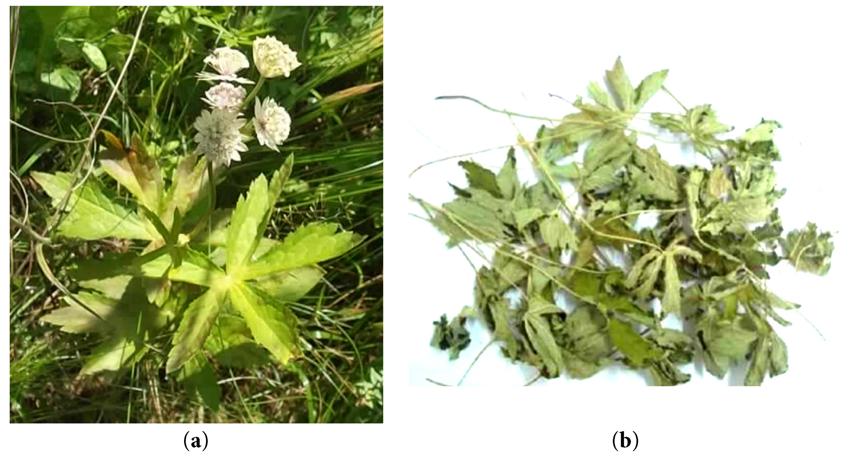
Figure 1: Astrantia major L. in natural growth conditions (a) and its plant raw materials (b).
Aqueous (AM0) and hydroethanolic (AM70) extracts were prepared from A. major herb. The starting material for the preparation of dry extracts was comminuted aerial parts of A. major (particle size 0.5–2.5 mm).
For the preparation of the aqueous extract, 100.0 g of the comminuted plant material was extracted with 1500 mL of purified water in a round-bottom flask equipped with a reflux condenser using a water bath at 50°C for 30 min. The extract was filtered, and the residual plant material was re-extracted twice under identical conditions. The resulting extracts were filtered and combined.
70% ethanol was selected as an extraction solvent alongside purified water, as hydroethanolic preparations (tinctures) are widely employed in traditional medicine, reflecting the practical relevance of the extraction conditions. For the preparation of the hydroethanolic extract, 100.0 g of the comminuted plant material was extracted with 1000 mL of 70% ethanol under reflux at 50°C for 30 min. The extracts were filtered, and the plant residues were re-extracted twice under the same conditions. The resulting extracts were combined and the solvent was removed under reduced pressure with a Buchi B-300 rotary evaporator (Buchi AG, Flawil, Switzerland). Concentration was carried out at a vacuum of 150 mbar, a flask rotation speed of 50 rpm, and a bath temperature maintained at 85°C until a viscous residue was obtained.
Following concentration, both aqueous and hydroethanolic extracts were dispensed into sterile 400 mL containers and freeze-dried using a SCANVAC COOLSAFE 55-4 Pro lyophilizer (LaboGene ApS, Lillerød, Denmark). Before lyophilization, the samples underwent a two-step freezing procedure consisting of rapid cooling to temperatures below −40°C for 30 min, followed by storage at temperatures below −30°C for 12 h to ensure complete solidification. The lyophilization process was carried out for 28–32 h, resulting in dry powdered extracts. The obtained lyophilizates were subsequently weighed in the required amounts and used in all subsequent analytical investigations.
Phenolic constituents were characterized by HPLC using an Agilent 1200 chromatographic platform (Agilent Technologies, USA). The system was configured with a diode-array UV–Vis detector (G1315), an online vacuum degasser (G1322A), an autosampler (G1329A), and a thermostatically controlled column compartment (G1316A). Instrument operation, data acquisition, and chromatographic processing were carried out using Agilent ChemStation software according to previously described procedures [18,19].
Hydroxycinnamic acid derivatives were separated by reversed-phase HPLC employing a Discovery C18 analytical column (250 × 4.6 mm, 5 μm; Supelco). Elution was achieved with a binary solvent system composed of 0.005 N orthophosphoric acid in water (mobile phase A) and acetonitrile (mobile phase B). The chromatographic analysis was conducted at a constant flow rate of 0.7 mL/min while maintaining the column at 25°C. Sample aliquots of 5–10 μL were injected, and the operating pressure ranged between 10,000 and 12,000 kPa throughout the 50-min analytical run. Gradient elution was programmed as follows: the initial concentration of mobile phase B was 5%, increasing to 8% after 8 min, 10% at 15 min, 20% at 30 min, and 40% at 40 min. The proportion of acetonitrile was then raised to 75% between 41 and 42 min before returning to the initial composition (5% B), which was maintained until completion of the run at 50 min. Ultraviolet spectra were continuously recorded over the wavelength range of 190–400 nm. Quantitative evaluation was performed at 320 and 330 nm, with spectral acquisition at 0.6-s intervals.
Flavonoid profiling was carried out under reversed-phase HPLC conditions using the same Discovery C18 analytical column. Separation was achieved with a binary mobile phase comprising 0.005 N orthophosphoric acid (solvent A) and acetonitrile (solvent B). Chromatographic analysis was performed at a constant column temperature of 25°C and a flow rate of 0.8 mL/min. Injection volumes ranged from 5 to 10 μL, while the system pressure did not exceed 15,600 kPa during the 60-min chromatographic run. The gradient elution program was initiated with 12% solvent B and increased to 25% at 30 min, remaining unchanged until 33 min. The proportion of acetonitrile was then raised to 30% at 38 min, 40% at 40 min, and 80% at 41 min, which was maintained until 48 min. Subsequently, the initial mobile-phase composition (12% solvent B) was restored at 49 min and held constant until the end of the analysis at 60 min. Spectral data were collected continuously within the ultraviolet range of 190–400 nm. Quantification of flavonoids was performed at wavelengths of 255 and 340 nm, with a detector scan interval of 0.6 s.
Tannin-related metabolites were analyzed by reversed-phase HPLC using the previously described Discovery C18 analytical column (250 × 4.6 mm, 5 μm). Separation was performed with a binary elution system consisting of 0.1% trifluoroacetic acid containing 5% acetonitrile in deionized water (mobile phase A) and 0.1% trifluoroacetic acid in acetonitrile (mobile phase B). The chromatographic procedure was conducted at 25°C with an injection volume of 10 μL. A flow rate of 0.1 mL/min was selected to improve the resolution and selectivity of tannin metabolites, while the operating pressure remained below 40,000 kPa during the 40-min analysis. The gradient profile began with 100% mobile phase A (0% B), followed by an increase of mobile phase B to 12% at 8 min, which was maintained until 10 min. The organic phase was subsequently increased to 25% at 15 min and held until 20 min, after which it was raised to 75% at 25 min and maintained through 28 min. The initial elution conditions were restored at 29 min and preserved until the completion of the chromatographic run at 40 min. Detection of tannin metabolites was carried out at 280 nm, while ultraviolet spectra were simultaneously recorded across the wavelength range of 190–400 nm. Spectral acquisition was performed using a scan interval of 0.6 s [20,21].
The amounts of individual bioactive constituents were quantified using the external calibration approach. Calibration curves were generated from authentic reference compounds, and the concentrations of the detected analytes were subsequently calculated by comparing their chromatographic responses with those of the corresponding standards.
The overall concentration of phenolic compounds in the prepared extracts was evaluated by spectrophotometric analysis following the methodology prescribed by the State Pharmacopoeia of Ukraine [22]. The obtained results were expressed as pyrogallol equivalents (PGE). The total flavonoid content was determined using the corresponding pharmacopoeial spectrophotometric procedure, with hyperoside serving as the calibration reference standard [23,24]. Each sample was analyzed in three independent replicates, and the average values were used for subsequent statistical evaluation to ensure the reproducibility and reliability of the data.
The assessment of the acute toxicity and pharmacological activity of A. major herb extracts was conducted with the consultative support of Prof. H. M. Erstenyuk, Head of the Department of Biological and Medical Chemistry named after Academician H. O. Babenko, IFNMU.
Experimental studies were performed using adult outbred white mice (Mus musculus) weighing 20–25 g and outbred white rats (Rattus norvegicus) weighing 180–220 g. The animals were bred and maintained in the vivarium of IFNMU under standard laboratory conditions. The animals were standardized according to physiological and biochemical parameters and were kept under controlled environmental conditions with a standard diet, in accordance with sanitary and hygienic requirements and principles of humane treatment of laboratory animals. Animal housing and experimental procedures were carried out in compliance with the fundamental principles of bioethics and ethical review, with the aim of protecting human health and biological diversity. All procedures were conducted in accordance with the European Convention for the Protection of Vertebrate Animals Used for Experimental and Other Scientific Purposes (Strasbourg, 1985), Council Directive 86/609/EEC (1986), the Law of Ukraine No. 3447-IV “On Protection of Animals from Cruelty”, and the general ethical principles of animal experimentation adopted by the First National Congress on Bioethics (Ukraine, 2001) [25,26,27] and approved by the Bioethics Commission of IFNMU (protocol 158/26 dated 25 March 2026).
The acute toxicity of the investigated extracts was evaluated in outbred adult male white mice weighing 18–22 g, bred in the IFNMU vivarium. Acute toxicity was assessed following the preclinical safety evaluation methodology [28,29]. The study was conducted in a reduced design using a single animal species. Animals were randomly divided into three groups (n = 6 per group). The extracts were administered intragastrically. Animals in groups 1 and 2 received the aqueous and hydroethanolic extracts of A. major herb, respectively, while the control group received the vehicle (purified water). Animals were observed for 14 days. Toxicity assessment was based on monitoring general condition, food intake, body weight changes, coat condition, skin and mucous membrane coloration, motor activity, respiratory function, occurrence of convulsions, and mortality. The toxicity class of the extracts was determined according to standard classification criteria [28].
The anti-inflammatory activity of the investigated extracts was assessed using a carrageenan-induced paw edema model in white rats [28,30]. The study was performed on outbred male white rats weighing 180–250 g, which were randomly divided into five groups (n = 6 per group). Inflammatory edema was induced under aseptic conditions by subplantar injection of 0.1 mL of 1% carrageenan solution into the aponeurosis of the hind paw. The development of inflammation was evaluated by measuring paw volume using the plethysmometric (oncometric) method. The investigated extracts and reference compounds were administered intragastrically in appropriate doses twice: 1 h before and immediately after carrageenan injection. The investigated extracts were administered intragastrically at a dose of 100 mg/kg. Diclofenac sodium, a synthetic nonsteroidal anti-inflammatory drug, was used as a reference compound at a dose of 8 mg/kg [31]. Quercetin, a plant-derived compound with established anti-inflammatory activity, was used as an additional reference at an effective dose of 5 mg/kg [18,20]. Paw volume was measured at baseline and at 1, 3, and 5 h after carrageenan administration. The anti-exudative effect of the extracts was evaluated based on their ability to inhibit paw edema formation over time compared with the control group.
The bacteriostatic activity of A. major extracts was evaluated using the agar diffusion method with paper disks [32]. The extracts were initially dissolved in purified water or in a 70% hydroethanolic solution. Sterile paper discs (6 mm in diameter) were impregnated with the resulting solutions in an amount corresponding to 5 mg of extract per disc. A 5% blood agar medium was prepared by adding 5% defibrinated sheep blood to a sterile agar base cooled to approximately 45–50°C, followed by thorough mixing and dispensing into Petri dishes. Bacterial suspensions were prepared from 24 h cultures in broth at a density of approximately 1 × 10⁹ CFU/mL using 1% sugar broth. Inoculation was performed by evenly spreading 1 mL of the bacterial suspension onto the surfaces of the 5% blood agar plates. The plates were incubated at 37°C for 24–72 h, depending on the characteristics of the tested microorganisms. Antimicrobial activity was assessed by measuring the diameter of growth inhibition zones around the paper disks. The following test microorganisms were used: Gram-positive bacteria (Staphylococcus aureus, Staphylococcus epidermidis), Gram-negative bacteria (Pseudomonas aeruginosa, Escherichia coli and Proteus vulgaris). For comparison, paper disks impregnated with antibiotics were used as reference standards: ampicillin (30 μg/disk) and oleandomycin (30 μg/disk) [18].
The hemostatic and wound-healing activities of A. major herb extracts were evaluated in guinea pigs bred in the vivarium of IFNMU and standardized according to physiological and biochemical parameters. Animals were maintained under standard laboratory conditions at 22–23°C with free access to food and water under a 12 h light/dark cycle. They were housed in plastic cages with bedding, with males and females kept separately. Prior to experimentation, animals were acclimatized to laboratory conditions for 14 days. The hemostatic activity of the aqueous and hydroethanolic extracts was assessed by measuring bleeding time following induction of a standardized incisional wound [33,34]. A linear incised wound model was created by making a surgical incision with a scalpel through all layers of the previously depilated skin and underlying muscle on the lateral abdominal surface. The standardized wound dimensions were 2.5 × 0.3 cm. Bleeding time was measured as the interval from wound induction to spontaneous cessation of bleeding. No additional mechanical pressure or topical hemostatic intervention was applied during the measurement period. Animals were randomly divided into four groups (n = 6 per group): (1) intact control; (2) animals treated with a gauze pad impregnated with Polygonum hydropiper (water pepper) extract applied immediately after wound induction; (3) animals treated with a gauze pad impregnated with the aqueous extract of A. major; and (4) animals treated with a gauze pad impregnated with the hydroethanolic extract of A. major. Immediately after wound induction, the animals were treated with a four-layer sterile gauze dressing impregnated with either the aqueous or hydroethanolic extract of A. major herb. The dressing was applied directly to the wound surface without exerting any additional mechanical pressure [18]. The reference preparation was a liquid extract of Persicaria hydropiper herb (“Water pepper extract”, batch No. 20924, Ternopharm, Ukraine) [35].
The wound-healing activity of the investigated preparations was evaluated in clinically healthy guinea pigs aged 3–5 months with body weights between 250 and 400 g. Before surgery, the hair on the lateral thigh was carefully removed. Following local anesthesia, a standardized full-thickness skin incision measuring approximately 25 mm in length and 3 mm in depth was produced using a sterile scalpel. After wound induction, the animals were assigned at random to four experimental groups comprising six animals each. The first group served as the untreated control, whereas the remaining animals received topical administration of one of the tested preparations: the aqueous extract of A. major, the hydroethanolic extract of A. major, or the herbal medicinal product «Rekutan®» (liquid extract of Flores Chamomillae, drug-to-extract ratio 1:1; marketing authorization No. UA/5120/01/01; LLC “Pharmaceutical Company Zdorovya”, Ukraine) as the reference treatment. Regeneration of the injured tissue was followed for 16 days. At scheduled observation points, wound size was determined by direct measurement of its maximum length and width with a millimetre scale. The wound surface area was estimated by multiplying these dimensions, and the sequential reduction in area was used as an indicator of tissue repair and contraction. The progression of healing was additionally characterized by evaluating the intensity of the inflammatory response together with the dynamics of wound closure according to the published methodology [18,28].
Experimental data were statistically evaluated using both descriptive and comparative analytical methods in accordance with the recommendations of the State Pharmacopoeia of Ukraine [22]. The results are expressed as the mean value accompanied by the standard error of the mean (mean ± SEM). Comparisons between the experimental and corresponding control groups were performed using Student’s t-test. A probability value below 0.05 was accepted as indicative of statistical significance. Depending on the experimental procedure, each analysis was carried out using three to nine independent replicates (n = 3–9) [36,37].
The obtained extracts of A. major herb were amorphous powders of yellow color (aqueous extract) and yellow-green color (70% hydroethanolic extract), with a bitter taste and a characteristic odor. The extraction yield ranged from 22.96% to 24.16%, depending on the extraction solvent. The loss on drying ranged from 2.80% to 4.15%, which complies with the established requirements for dry extracts (not exceeding 5%).
Phenolic compounds in the dry extracts of A. major herb were determined by HPLC and spectrophotometric methods (Table 1).
Table 1: Quantitative content of phenolic compounds in A. major dry extracts.
| Compound | Content of Biologically Active Compounds (mg/kg) | |
|---|---|---|
| AM0 | AM70 | |
| Tannin metabolites | ||
| Gallic acid | 246 ± 16 | 234 ±11 |
| Gallocatechin | 3582 ± 98 | 4125 ± 205 |
| Epigallocatechin | 3272 ±143 | 6216 ± 352 |
| Catechin | 487 ± 32 | 1233 ± 86 |
| Epicatechin | 704 ± 28 | 1783 ± 128 |
| Epicatechin gallate | 53 ±3 | 1554 ± 69 |
| Catechin gallate | 152 ± 9 | 445 ± 24 |
| Ellagic acid | 27 ± 4 | 31 ± 5 |
| Flavonoids | ||
| Apigenin | 20 ± 3 | 92 ± 3 |
| Luteolin | 4 ± 1 | 16 ± 1 |
| Rutin | 197 ± 13 | 575 ± 31 |
| Hyperoside | 13 ± 2 | 15 ± 2 |
| Isoquercitrin | 74 ± 4 | 85 ± 4 |
| Hydroxycinnamic acids | ||
| Chlorogenic acid | 1867 ± 76 | 1773 ± 128 |
| Rosmarinic acid | 209 ± 12 | 397 ± 27 |
| Caffeic acid | 157 ± 9 | 179 ± 12 |
| Ferulic acid | 30 ± 2 | 88 ± 7 |
| Content of compound groups in the extract, % (spectrophotometry) | ||
| Total polyphenols | 14.42 ± 0.13 | 16.91 ± 0.09 |
| Flavonoids | 4.88 ± 0.08 | 7.02 ± 0.07 |
A total of 17 phenolic compounds were identified and quantified in A. major herb extracts by HPLC. Chlorogenic acid was the predominant hydroxycinnamic acid, while rutin was the major flavonoid, and gallocatechin (AM0) and epigallocatechin (AM70) were the predominant tannin-related metabolites among the tannin metabolites. It was established that 70% ethanol extract of A. major contained significantly higher levels of phenolic compounds and flavonoids compared to the aqueous extract. This trend can be attributed to the ability of 70% ethanol to provide an optimal balance between hydrophilicity and lipophilicity, thereby enhancing its capacity to solubilize phenolic compounds of varying polarity [38,39]. Consequently, both polar hydroxycinnamic acids and less polar flavonoids and catechins are extracted more efficiently.
3.2 Evaluation of Acute Toxicity
The assessment of acute toxicity represents the initial stage aimed at obtaining information on the safety of dry extracts of A. major herb under short-term exposure. The results of the acute toxicity study show that intragastric administration of aqueous and hydroethanolic extracts of A. major at a dose of 6000 mg/kg did not result in animal mortality. No changes in animal behavior were observed. These findings indicate the absence of acute toxic effects at the tested dose and allow classification of the extracts as toxicity class V substances (LD50 > 5000 mg/kg), i.e., practically non-toxic. In addition, no abnormalities in food intake, body weight gain, coat appearance, locomotor activity, respiration, or mucosal coloration were observed during the 14-day observation period. In this study, only male mice were used in order to minimize the influence of endocrine and hormonal fluctuations associated with the estrous cycle in females, which may significantly affect outcomes in pharmacological studies. The use of a single sex reduced biological variability and improved the reproducibility of the results during the initial screening of the pharmacological activity of the plant extracts.
3.3 Evaluation of Anti-Inflammatory Activity
The anti-exudative activity of the aqueous and hydroethanolic extracts of A. major herb was assessed using a carrageenan-induced paw edema model in white rats [18,28]. Paw volume was measured at baseline and at 1, 3, and 5 h after administration of the phlogogenic agent. The efficacy of the extracts was evaluated based on their ability to inhibit the development of paw edema over time compared with the control group and reference drugs. The increase in paw volume and the anti-exudative activity of A. major extracts are presented in Table 2 and Fig. 2.
Table 2: Effect of test extracts of A. major on rat paw edema volume.
| Animal Group | Dose (mg/kg) | Increase in Rat Paw Volume, a.u., (n = 6) | ||
|---|---|---|---|---|
| 1 h | 3 h | 5 h | ||
| Pathological control | - | 78.58± 0.63 | 105.8 ± 0.92 | 119.05 ± 1.01 |
| АМ0 | 100 | 75.72 ± 0.18* | 87.85 ± 0.18* | 97.43 ± 0.24* |
| АМ70 | 100 | 72.60 ± 0.24* | 87.78 ± 0.34* | 96.28 ± 0.34 |
| Diclofenac sodium | 8 | 63.5 ± 1.08* | 69.9 ± 1.06* | 73.0 ± 1.26* |
| Quercetin | 5 | 72.98 ± 0.21* | 96.9 ± 1.14* | 103.3 ± 1.14* |
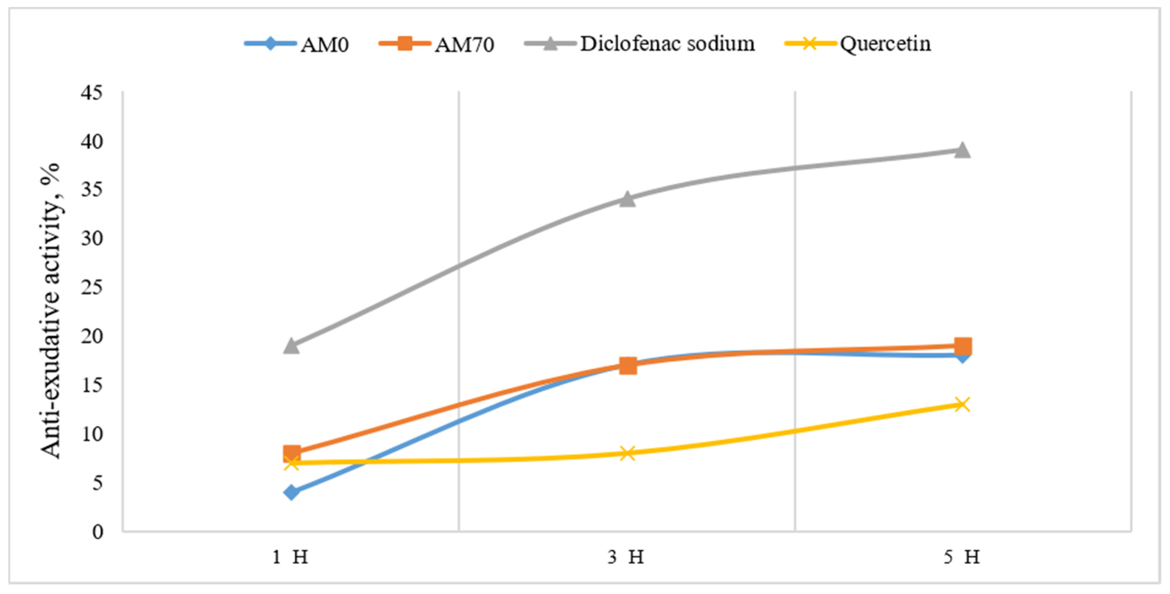
Figure 2: Anti-exudative activity of A. major herb extracts.
The results indicate that A. major extracts exhibit anti-exudative activity, affecting the exudative phase of inflammation, and demonstrate efficacy comparable to that of the plant-derived reference compound quercetin. At 1, 3, and 5 h after the initiation of the experiment, the highest anti-exudative activity was observed for the hydroethanolic extract (AM70) of A. major herb.
3.4 Evaluation of Antimicrobial Activity
The bacteriostatic activity of A. major extracts was assessed using the agar disk diffusion technique employing paper disks. The results of the study are presented in Table 3.
Table 3: Bacteriostatic activity of the investigated extracts of A. major herb.
| Test Object | Inhibition Zone Diameter (mm) | ||||
|---|---|---|---|---|---|
| Pseudomonas aeruginosa | Escherichia coli | Proteus vulgaris | Staphylococcus aureus | Staphylococcus epidermidis | |
| АM70 | 0 | 0 | 7.3 ± 0.5 | 0 | 6.7± 0.5 |
| АМ0 | 0 | 8.7± 1.2 | 6.7± 0.5 | 0 | 14.3 ± 0.5 |
| Ampicillin | 14.3 ± 0.5 | 10.0 ± 0.8 | 0 | 25.3 ± 0.5 | 8.3 ± 0.5 |
| Oleandomycin | 5 ± 0.8 | 5.3 ± 0.5 | 0 | 25 ± 0.8 | 22.3 ± 0.5 |
The results of the antimicrobial activity study demonstrated selective susceptibility of the tested microorganisms to the A. major herb extracts. The hydroethanolic extract (AM70) exhibited weak inhibitory activity against Proteus vulgaris (7.3 ± 0.5 mm) and Staphylococcus epidermidis (6.7 ± 0.5 mm), while no activity was observed against Pseudomonas aeruginosa, Escherichia coli, or Staphylococcus aureus. The aqueous extract (AM0) showed antimicrobial activity against Escherichia coli (8.7 ± 1.2 mm), Proteus vulgaris (6.7 ± 0.5 mm), and the most pronounced effect against Staphylococcus epidermidis (14.3 ± 0.5 mm), but did not inhibit the growth of Pseudomonas aeruginosa or Staphylococcus aureus. Overall, the aqueous extract demonstrated a broader spectrum and higher level of antimicrobial activity than the hydroethanolic extract, particularly against Staphylococcus epidermidis.
3.5 Evaluation of Hemostatic and Wound-Healing Activities
Hemostatic activity was evaluated based on bleeding time measured in a standardized incisional wound model [33]. The results of bleeding time duration (M ± SEM, s) in guinea pigs with incised wounds following local application of A. major extracts and Polygonum hydropiper extract are presented in Table 4.
Table 4: Hemostatic activity of aqueous and hydroethanolic extracts of A. major herb based on bleeding time duration.
| Animal Group n = 6 | Control (Untreated Animal) | Polygonum hydropiper Extract | АМ0 | АМ70 |
|---|---|---|---|---|
| Bleeding time (s) | 224.67 ± 4.35 | 172.67 ± 0.96* | 129.17 ± 0.24* | 126.83 ± 4.99* |
| Reduction in bleeding time (%) | - | 23.15 | 42.51 | 43.54 |
The obtained results (Table 4) demonstrate that local application of the aqueous and hydroethanolic extracts of A. major (via gauze pads impregnated with the test extracts and applied directly to the wound surface immediately after injury) significantly reduced bleeding time compared with the control group. Thus, both A. major extracts showed comparable efficacy and reduced bleeding time by approximately 1.8-fold compared with the control group. These findings indicate a local hemostatic activity of A. major extracts.
The wound-healing activity of A. major herb extracts is presented in Table 5. Wound healing was monitored over a 16-day period using planimetric analysis, along with assessment of the inflammatory response and healing rate.
Table 5: Wound healing dynamics under treatment with plant-derived preparations.
| Experimental Groups n = 6 | Wound Healing Area (% of Initial Wound Area) | |||||||
|---|---|---|---|---|---|---|---|---|
| Day | ||||||||
| 2 | 4 | 6 | 8 | 10 | 12 | 14 | 16 | |
| Pathological control | 3.40 ± 0.75 | 8.25 ± 0.78 | 30.90 ± 1.56 | 51.80 ± 2.42 | 68.91 ± 1.93 | 79.35 ± 2.51 | 95.72 ± 1.49 | 100 |
| АМ0 | 4.73 ± 0.35 | 13.93 ± 0.46* | 38.87 ± 1.21* | 70.55 ± 3.68* | 96.41 ± 1.17* | 100* | 100* | 100 |
| АМ70 | 4.03 ± 0.39 | 10.75 ± 0.56* | 37.44 ± 1.05* | 69.63 ± 2.57* | 96.14 ± 0.73* | 100* | 100* | 100 |
| Rekutan | 4.42 ± 0.42 | 11.12 ± 0.74* | 37.61 ± 1.46* | 69.77 ± 4.28* | 96.27 ± 1.11* | 100* | 100* | 100 |
The results indicate that treatment with aqueous and hydroethanolic extracts of A. major accelerates wound healing, with a marked effect observed on days 8 and 10, respectively. Complete wound closure was achieved by day 14. Both aqueous and hydroethanolic extracts of A. major significantly accelerated wound closure and promoted tissue repair in the incisional wound model, demonstrating efficacy comparable to that of the reference herbal preparation, Rekutan. Notably, the AM0 extract tended to show higher wound-healing values than “Rekutan” during the early healing phase (days 2–8). Overall, these findings suggest that all tested extracts are effective in promoting wound healing and possess significant wound-healing activity.
The growing interest in plant-derived bioactive compounds has stimulated extensive research into medicinal plants as potential sources of novel therapeutic agents. A comprehensive assessment of their phytochemical composition, safety, and biological activities is a prerequisite for their rational pharmaceutical application. Therefore, the present study was undertaken to evaluate the phytochemical profile, toxicity, and pharmacological potential of A. major herb extracts.
Phytochemical analysis revealed the presence of 17 phenolic metabolites, among which chlorogenic acid (hydroxycinnamic acids), rutin (flavonoids), and epigallocatechin (catechins) were predominant. It was established that 70% ethanol is a significantly more efficient extraction solvent for phenolic compounds compared to purified water. Numerous studies have reported the presence of oleanane-type saponins in A. major [7,9]. The fruits of A. major are also known to be rich in essential oil, comprising up to seventy-six constituents [5]. Additionally, diethyl ether extracts of A. major fruits have been shown to contain seven diaryltetrahydrofurofurano lignans [5,7]. Previous phytochemical investigations of A. major raw material, often conducted within studies on Saniculoideae constituents, have consistently identified flavones and flavonols such as quercetin, kaempferol, and their glycosides. More broadly, within the Saniculeae genus, flavonols (isorhamnetin, kaempferol, myricetin, quercetin) and flavones (apigenin, luteolin) represent the dominant flavonoid classes [7]. Our findings are consistent with previous reports on the flavonoid content of Astrantia species. For example, a recent spectrophotometric study of the flowering aerial parts of Astrantia maxima Pall. collected in Azerbaijan also confirmed the presence of substantial amounts of flavonoids [40]. In the present study, similar flavonoid compounds were identified in A. major extracts, consistent with previously reported phytochemical profiles. The available phytochemical data for the genus Astrantia remain limited, and direct comparisons between species are often complicated by differences in plant organs, extraction procedures, and analytical methodologies. Nevertheless, the existing evidence suggests that phenolic compounds are among the major groups of bioactive constituents across the genus. To the best of our knowledge, tannin-related metabolites and hydroxycinnamic acids have not previously been comprehensively characterized and quantified in A. major herb extracts.
This finding significantly expands the current understanding of the phytochemical composition of A. major and provides new insights into the potential contribution of these compound classes to the observed biological activities. In particular, catechin-derived tannins and hydroxycinnamic acids may contribute to the anti-inflammatory, antimicrobial, and wound-healing activities observed in this study, indicating their potential involvement in the pharmacological effects of A. major extracts.
In the carrageenan-induced paw edema model, A. major extracts exhibited anti-exudative activity, as evidenced by a reduction in edema progression over time. Both the hydroethanolic extract (AM70) and the aqueous extract (AM0) demonstrated comparable anti-exudative activity, with slightly lower paw edema values observed for AM70. Compounds such as rutin, apigenin, luteolin, and epigallocatechin are known to modulate inflammatory pathways and oxidative stress, which may contribute to the anti-exudative activity observed for A. major extracts [41,42,43]. In addition, flavonoids such as rutin, apigenin, and luteolin have been reported to inhibit cyclooxygenase-mediated pathways and modulate the production of pro-inflammatory cytokines, including TNF-α, IL-1β, and IL-6, which are involved in the development of carrageenan-induced inflammation [41,42].
The antimicrobial activity of A. major extracts was selective and generally moderate (Table 3), with the aqueous extract showing broader effects against Escherichia coli and Staphylococcus epidermidis. The absence of activity against Pseudomonas aeruginosa and Staphylococcus aureus indicates a limited antibacterial spectrum. Such selectivity is consistent with the known behavior of plant polyphenols, whose antimicrobial effects depend on compound structure, concentration, and interactions with microbial cell components [44,45].
The hemostatic activity study demonstrated that local application of the extracts significantly reduced bleeding time in an incised wound model. The efficacy of both aqueous and hydroethanolic extracts was comparable and exceeded that of Polygonum hydropiper extract, a known herbal hemostatic agent. The hemostatic activity of A. major extracts is likely associated with their high polyphenolic content, particularly catechins and tannins. These compounds are known to exert astringent effects by precipitating proteins, leading to the formation of a protective coagulum on damaged tissues and a reduction in capillary permeability. In addition, flavonoids, particularly rutin, contribute to the strengthening and stabilization of the vascular wall and reduce capillary permeability and fragility. These effects may further support the hemostatic response and help reduce the risk of bleeding [46]. Polyphenols may also enhance platelet aggregation and accelerate primary clot formation. The observed reduction in bleeding time following local application of the extracts suggests a synergistic interaction of these mechanisms, resulting in effective local hemostasis [47,48]. However, these proposed mechanisms remain hypothetical, as they were not directly investigated in the present study.
In the wound-healing model, A. major extracts accelerated tissue repair, promoted faster wound contraction, and contributed to normalization of the inflammatory response. Complete wound closure was observed by day 14, with outcomes comparable to or slightly superior to those of the reference drug “Rekutan”. These findings indicate a pronounced wound-healing effect, which may be associated with the presence of phenolic compounds possessing antioxidant and anti-inflammatory properties [49,50,51,52].
Structure–activity considerations
The biological activities observed for A. major herb extracts may be related, at least in part, to their phenolic composition (Table 1), particularly flavonoids, catechin-derived tannins, and hydroxycinnamic acids. Flavonoids such as rutin, apigenin, luteolin, and isoquercitrin—present at higher levels in the hydroethanolic extract (AM70)—are known to modulate inflammatory pathways, which is consistent with the slightly greater anti-exudative activity of AM70 in the carrageenan-induced edema model (Table 2) [41,42,53].
Catechins and other tannin-related metabolites (e.g., epigallocatechin, gallocatechin; Table 1) may contribute substantially to the hemostatic and wound-healing effects (Table 4 and Table 5) due to their astringent properties and their role in supporting tissue repair processes [46,47,48,51,53]. Hydroxycinnamic acids, including chlorogenic, rosmarinic, and caffeic acids (Table 1), may further enhance anti-inflammatory and regenerative responses [54,55,56,57].
The observed effects may be associated with the presence of multiple phenolic compound classes in the extracts. Differences between aqueous (AM0) and hydroethanolic (AM70) extracts may contribute to the differences observed in the biological activities of the extracts, whereas differences were observed in the antimicrobial activity profiles of the two extracts (Table 3).
Overall, these findings highlight the importance of extraction conditions in determining both phytochemical composition and biological activity. Collectively, the results demonstrate that the biological effects of A. major extracts are closely associated with their phenolic profile and support the relevance of this species as a source of biologically active plant constituents.
The present study demonstrates that the dry extracts of A. major herb exhibit anti-inflammatory, antimicrobial, hemostatic, and wound-healing properties. Phytochemical analysis confirmed a high content of phenolic compounds and led to the identification of 17 phenolics, among which chlorogenic acid and rutin were the predominant hydroxycinnamic acid and flavonoid, respectively, whereas gallocatechin (AM0) and epigallocatechin (AM70) predominated among the tannin-related metabolites.
The aqueous and 70% hydroethanolic extracts were found to possess significant anti-inflammatory activity compared with the pathological control, while also reducing bleeding duration and significantly accelerating wound closure and tissue repair.
Furthermore, selective antimicrobial effects against Staphylococcus epidermidis and Escherichia coli were observed. The combination of pharmacological activity and low toxicity highlights the potential of A. major extracts as a source of biologically active plant constituents. Further studies are warranted to identify the active constituents responsible for the observed biological effects and to further evaluate their pharmacological and pharmacokinetic properties.
Acknowledgement:
Funding Statement: The authors received no specific funding for this study.
Author Contributions: The authors confirm contribution to the paper as follows: Conceptualization, Andriy Grytsyk, Lubov Grytsyk, Ain Raal and Oleh Koshovyi; methodology, Andriy Grytsyk, Lubov Grytsyk and Oleh Koshovyi; software, Taras Koliadzhyn and Nadiia Lehin; validation, Nadiia Lehin, Lubov Grytsyk, Ain Raal and Oleh Koshovyi; formal analysis, Taras Koliadzhyn and Nadiia Lehin; investigation, Taras Koliadzhyn, Andriy Grytsyk, Nadiia Lehin and Oleh Koshovyi; resources, Andriy Grytsyk; data curation, Taras Koliadzhyn, Nadiia Lehin, Ain Raal and Oleh Koshovyi; writing—original draft preparation, Taras Koliadzhyn, Lubov Grytsyk, Ain Raal and Oleh Koshovyi; writing—review and editing, Andriy Grytsyk, Ain Raal and Oleh Koshovyi; visualization, Taras Koliadzhyn and Nadiia Lehin; supervision, Andriy Grytsyk; project administration, Andriy Grytsyk. All authors reviewed and approved the final version of the manuscript.
Availability of Data and Materials: Data available within the article.
Ethics Approval: The study was conducted in accordance with the Declaration of Helsinki, and approved by the Bioethics Commission of the Ivano-Frankivsk National Medical University (protocol 158/26 dated 25 March 2026).
Conflicts of Interest: The authors declare no conflicts of interest.
References
1. Heywood VH . Flowering plants of the world. Oxford, UK: Oxford University Press; 1993. 335 p. [Google Scholar]
2. Klokova MV , Visyulina OD . Flora of the URSR. VII. Kyiv, Ukraine: Institute of the Academy of Sciences of the URSR; 1955. p. 460– 75. [Google Scholar]
3. Hiller K , Gründemann E , Habisch D . Isolation of iso-orientin from Astrantia major L. 24. The constituents of Saniculoideae. Pharmazie. 1975; 30( 12): 809. [Google Scholar]
4. Hiller K , Leska M , Rahn I . Große sterndolde und sanikel. Sci Pharm. 1988; 56: 169. [Google Scholar]
5. Radulović NS , Mladenović MZ , Ethorđević ND . Chemotypification of Astrantia major L. (Apiaceae): essential-oil and lignan profiles of fruits. Chem Biodivers. 2012; 9( 7): 1320– 37. doi:10.1002/cbdv.201100430. [Google Scholar] [CrossRef]
6. Buurma H , Bos R , Tattje DHE , Zwaving JH . Sesquiterpenes from the essential oil of Astrantia major. Phytochemistry. 1978; 17( 12): 2129– 30. doi:10.1016/S0031-9422(00)89296-1. [Google Scholar] [CrossRef]
7. Kikowska M , Hermosaningtyas AA , Chanaj-Kaczmarek J . Little-known Saniculeae genera: phytochemical studies and pharmaceutical activities. Phytochem Rev. 2025; 24( 5): 3941– 4011. doi:10.1007/s11101-024-10027-z. [Google Scholar] [CrossRef]
8. Masan C , Woitke HD , Hiller K , Franke P . Isolation of beta-sitosterol-O-D-glucoside from Astrantia major L. 31. The contents of some Saniculoideae. Pharmazie. 1978; 33( 6): 382. [Google Scholar]
9. Hiller K , Leska M , Gründemann E , Dube G , Karwatzki A , Franke P . Structure of bisdesmosidic saponins from Astrantia major L. Pharm Zentralhalle. 1990; 45: 615– 7. [Google Scholar]
10. Hiller K , Woitke HD , Lehmann G . Saponins in Astrantia major L. 17. Information on components of various saniculoideae. Die Pharm. 1973; 28( 6): 391– 7. (In German). [Google Scholar]
11. Hiller K , Adler C , Galetzka G , Franke P , Gründemann E , Hille J . Zur Struktur der Saponine von Astrantia major L. Pharmazie. 1985; 40( 5): 343– 5. [Google Scholar]
12. Grodzinsky AM . Medicinal plants: encyclopedic guide. Kyiv, Ukraine: Ukrainian Encyclopedia; 1990. 544 p. (In Ukrainian). [Google Scholar]
13. Zabka M . Medicinal and culinary herbs as environmentally safe inhibitors of dangerous toxinogenic plant and human fungal pathogens. Afr J Microbiol Res. 2012; 6( 35): 6468– 75. doi:10.5897/ajmr12.1060. [Google Scholar] [CrossRef]
14. Panda H . Handbook on medicinal herbs with uses. Delhi, India: Asia Pacific Business Press; 2004. 550 p. [Google Scholar]
15. Bakun OV , Kupchanko VG , Berbets AM . Application of phytopreparations for hypogalactia. Tavricheskiy Med-Biol Vestn. 2011; 14( 3): 245– 7. (In Ukrainian). [Google Scholar]
16. Guideline on Good Agricultural and Collection Practice (GACP) for Starting Materials of Herbal Origin. [Internet]. European Medicines Agency; 2006 [cited 2026 Aug 9]. Available from: https://www.ema.europa.eu/en/good-agricultural-collection-practice-starting-materials-herbal-origin-scientific-guideline. [Google Scholar]
17. Dobrochaeva DN , Kotov MI , Prokudin YN , Barbarich AI . Key to higher plants of Ukraine. Kyiv, Ukraine: Naukova Dumka; 1999. [Google Scholar]
18. Lehin N , Grytsyk L , Koshovyi O , Koliadzhyn T , Grytsyk A , Kaplaushenko A , et al. Sanicula europaea L. herb and rhizomes with root extracts with hemostatic, wound healing, anti-inflammatory and antimicrobial activity: phytochemical and pharmacological research. Appl Sci. 2025; 15( 22): 12293. doi:10.3390/app152212293. [Google Scholar] [CrossRef]
19. Maliuvanchuk S , Grytsyk A , Popadynets O , Kotyk T , Raal A , Koshovyi O . Ajuga reptans L. herb extracts: phytochemical composition and pharmacological activity screening. Plants. 2025; 14( 2): 219. doi:10.3390/plants14020219. [Google Scholar] [CrossRef]
20. Hrytsyk Y , Koshovyi O , Hrytsyk R , Raal A . Extracts of the Canadian goldenrod (Solidago Canadensis L.)–promising agents with antimicrobial, anti-inflammatory and hepatoprotective activity. ScienceRise Pharm Sci. 2024; 4( 50): 78– 87. doi:10.15587/2519-4852.2024.310837. [Google Scholar] [CrossRef]
21. Starchenko G , Hrytsyk A , Raal A , Koshovyi О . Phytochemical profile and pharmacological activities of water and hydroethanolic dry extracts of Calluna vulgaris (L.) hull. herb. Plants. 2020; 9( 6): 751. doi:10.3390/plants9060751. [Google Scholar] [CrossRef]
22. Ukrainian Scientific Pharmacopoeial Center for Quality of Medicines . State pharmacopoeia of Ukraine. 2nd ed, Supplement 8. Kharkiv, Ukraine: Ukrainian Scientific Pharmacopoeial Center for Quality of Medicines; 2025. p. 356– 7. [Google Scholar]
23. Council of Europe . European pharmacopoeia. 11th ed. Strasbourg, France: Council of Europe; 2022. [Google Scholar]
24. Vlasova I , Gontova T , Grytsyk L , Zhumashova G , Sayakova G , Boshkayeva A , et al. Determination of standardization parameters of Oxycoccus Macrocarpus (Ait.) Pursh and Oxycoccus palustris Pers. Leaves. ScienceRise Pharm Sci. 2022; 3( 37): 48– 57. doi:10.15587/2519-4852.2022.260352. [Google Scholar] [CrossRef]
25. European Union. European Convention for the protection of vertebrate animals used for experimental and other scientific purposes. Off J Eur Commun. 1999; 42: L222. [Google Scholar]
26. Law of Ukraine No. 3447-IV of 21.02.2006 “On the Protection of Animals from Cruelty” (as amended and supplemented). [Internet]. 2006 [cited 2026 Aug 9]. Available from: https://zakon.rada.gov.ua/laws/show/3447-15#Text. [Google Scholar]
27. Reznikov OH , Soloviov AI , Dobrelya NV , Stefanov OV . Bioethical expertise of preclinical and other scientific studies conducted on animals: methodical recommendations. Kyiv, Ukraine: The National Academy of Medical Sciences of Ukraine; 2006. 28 p. [Google Scholar]
28. Stefanov OV . Preclinical studies of drugs. Kyiv, Ukraine: Avicenna; 2001. [Google Scholar]
29. Hrytsyk NY , Ersteniuk HM . Studies on the acute toxicity of the extracts of Gentiana asclepiadea L. Sci Pract J. 2023; 26( 2): 29– 34. doi:10.21802/artm.2023.2.26.29. [Google Scholar] [CrossRef]
30. Hrytsyk Y , Koshovyi O , Lepiku M , Jakštas V , Žvikas V , Matus T , et al. Phytochemical and pharmacological research in Galenic remedies of Solidago canadensis L. herb. Phyton. 2024; 93( 9): 2303– 15. doi:10.32604/phyton.2024.055117. [Google Scholar] [CrossRef]
31. Tandoh A , Danquah CA , Benneh CK , Adongo DW , Boakye-Gyasi E , Woode E . Effect of diclofenac and andrographolide combination on carrageenan-induced paw edema and hyperalgesia in rats. Dose Response. 2022; 20( 2): 15593258221103846. doi:10.1177/15593258221103846. [Google Scholar] [CrossRef]
32. Kovalenko VL , Napnenko OO , Chornyi I , Zagrebelnyi AV . The study of essential oil on antimicrobial activity and their composition towards microorganism. Sci Messenger Lviv Natl Univ Vet Med Biotechnol. 2018; 20( 87): 38– 41. doi:10.15421/nvlvet8707. [Google Scholar] [CrossRef]
33. Brinkhous KM . W. W. Duke and his bleeding time test. A commentary on platelet function. JAMA. 1983; 250( 9): 1210– 14. doi:10.1001/JAMA.1983.03340090066032. [Google Scholar] [CrossRef]
34. Kickler TS . Dr William W. Duke: pioneer in platelet research. JAMA. 2009; 301( 21): 2267. doi:10.1001/jama.2009.787. [Google Scholar] [CrossRef]
35. Kovalenko VN . Compendium 2024. Medicines. Kyiv, Ukraine: MORION; 2024. 2700 p. [Google Scholar]
36. Lapach SN , Chubenko AV , Babich PN . Statistical methods in biomedical research using Excel. Kyiv, Ukraine: MORION; 2000. 320 p. [Google Scholar]
37. Goyko OV , Mokhnachev SI . Analysis of modern software for statistical processing and analysis of biomedical research. Med Inform Eng. 2012;( 4): 49– 52. [Google Scholar]
38. Do QD , Angkawijaya AE , Tran-Nguyen PL , Huynh LH , Soetaredjo FE , Ismadji S , et al. Effect of extraction solvent on total phenol content, total flavonoid content, and antioxidant activity of Limnophila aromatica. J Food Drug Anal. 2014; 22( 3): 296– 302. doi:10.1016/j.jfda.2013.11.001. [Google Scholar] [CrossRef]
39. Dai J , Mumper RJ . Plant phenolics: extraction, analysis and their antioxidant and anticancer properties. Molecules. 2010; 15( 10): 7313– 52. doi:10.3390/molecules15107313. [Google Scholar] [CrossRef]
40. Center AS , Nagiyeva M , Mammadova A , Jafarova S , Shirinova I , Kazimzada H . Quantification and validation of flavonoids in Astrantia maxima Pall. using spectrophotometry. Sci Work. 2026; 20( 3): 269– 76. doi:10.36719/2663-4619/127/269-276. [Google Scholar] [CrossRef]
41. Kim HP , Son KH , Chang HW , Kang SS . Anti-inflammatory plant flavonoids and cellular action mechanisms. J Pharmacol Sci. 2004; 96( 3): 229– 45. doi:10.1254/jphs.crj04003x. [Google Scholar] [CrossRef]
42. Al-Khayri JM , Sahana GR , Nagella P , Joseph BV , Alessa FM , Al-Mssallem MQ . Flavonoids as potential anti-inflammatory molecules: a review. Molecules. 2022; 27( 9): 2901. doi:10.3390/molecules27092901. [Google Scholar] [CrossRef]
43. Wang S , Chang Y , Xu J , Zhao C , Song Y , Liu M , et al. Comparative study of anti-inflammatory, antioxidant and antibacterial activities of epigallocatechin gallate, epigallocatechin, epicatechin gallate and epicatechin in vitro. Pharmacol Res Mod Chin Med. 2025; 15: 100599. doi:10.1016/j.prmcm.2025.100599. [Google Scholar] [CrossRef]
44. Cushnie TPT , Lamb AJ . Antimicrobial activity of flavonoids. Int J Antimicrob Agents. 2005; 26( 5): 343– 56. doi:10.1016/j.ijantimicag.2005.09.002. [Google Scholar] [CrossRef]
45. De Rossi L , Rocchetti G , Lucini L , Rebecchi A . Antimicrobial potential of polyphenols: mechanisms of action and microbial responses—a narrative review. Antioxidants. 2025; 14( 2): 200. doi:10.3390/antiox14020200. [Google Scholar] [CrossRef]
46. Faisal S , Badshah SL , Kubra B , Muhammad A , Emwas AH , Jaremko M . Flavonoids as hemostasis modulators. In: Natural products: phytochemistry, botany, metabolism of alkaloids, phenolics and terpenes. Berlin/Heidelberg, Germany: Springer; 2025. p. 1– 57. doi:10.1007/978-3-642-36202-6_235-1. [Google Scholar] [CrossRef]
47. Ebrahimi F , Torbati M , Mahmoudi J , Valizadeh H . Medicinal plants as potential hemostatic agents. J Pharm Pharm Sci. 2020; 23( 1): 10– 23. doi:10.18433/jpps30446. [Google Scholar] [CrossRef]
48. Ghosh S , Sarkhel S , Ghosh K , Dhar S , Karar S , Roychowdhury V . Plant-derived hemostats. Rev Bras Farmacogn. 2023; 33( 2): 259– 71. doi:10.1007/s43450-022-00346-7. [Google Scholar] [CrossRef]
49. Zulkefli N , Che Zahari CNM , Sayuti NH , Kamarudin AA , Saad N , Hamezah HS , et al. Flavonoids as potential wound-healing molecules: emphasis on pathways perspective. Int J Mol Sci. 2023; 24( 5): 4607. doi:10.3390/ijms24054607. [Google Scholar] [CrossRef]
50. Carvalho MTB , Araújo-Filho HG , Barreto AS , Quintans-Júnior LJ , Quintans JSS , Barreto RSS . Wound healing properties of flavonoids: a systematic review highlighting the mechanisms of action. Phytomedicine. 2021; 90: 153636. doi:10.1016/j.phymed.2021.153636. [Google Scholar] [CrossRef]
51. Sriram N , Kalayarasan S , Manikandan R , Arumugam M , Sudhandiran G . Epigallocatechin gallate attenuates fibroblast proliferation and excessive collagen production by effectively intervening TGF-β1 signalling. Clin Exp Pharmacol Physiol. 2015; 42( 8): 849– 59. doi:10.1111/1440-1681.12428. [Google Scholar] [CrossRef]
52. Kim H , Kawazoe T , Han DW , Matsumara K , Suzuki S , Tsutsumi S , et al. Enhanced wound healing by an epigallocatechin gallate-incorporated collagen sponge in diabetic mice. Wound Repair Regen. 2008; 16( 5): 714– 20. doi:10.1111/j.1524-475x.2008.00422.x. [Google Scholar] [CrossRef]
53. Chagas MDSS , Behrens MD , Moragas-Tellis CJ , Penedo GXM , Silva AR , Gonçalves-de-Albuquerque CF . Flavonols and flavones as potential anti-inflammatory, antioxidant, and antibacterial compounds. Oxid Med Cell Longev. 2022; 2022: 9966750. doi:10.1155/2022/9966750. [Google Scholar] [CrossRef]
54. Petersen M , Simmonds MSJ . Rosmarinic acid. Phytochemistry. 2003; 62( 2): 121– 5. doi:10.1016/S0031-9422(02)00513-7. [Google Scholar] [CrossRef]
55. Nguyen V , Taine EG , Meng D , Cui T , Tan W . Chlorogenic acid: a systematic review on the biological functions, mechanistic actions, and therapeutic potentials. Nutrients. 2024; 16( 7): 924. doi:10.3390/nu16070924. [Google Scholar] [CrossRef]
56. Contardi M , Lenzuni M , Fiorentini F , Summa M , Bertorelli R , Suarato G , et al. Hydroxycinnamic acids and derivatives formulations for skin damages and disorders: a review. Pharmaceutics. 2021; 13( 7): 999. doi:10.3390/pharmaceutics13070999. [Google Scholar] [CrossRef]
57. Ibrahim N , Wong SK , Mohamed IN , Mohamed N , Chin KY , Ima-Nirwana S , et al. Wound healing properties of selected natural products. Int J Environ Res Public Health. 2018; 15( 11): 2360. doi:10.3390/ijerph15112360. [Google Scholar] [CrossRef]
Cite This Article
 Copyright © 2026 The Author(s). Published by Tech Science Press.
This work is licensed under a Creative Commons Attribution 4.0 International License , which permits unrestricted use, distribution, and reproduction in any medium, provided the original work is properly cited.
 Downloads
 Citation Tools
