
 Login
Login Register
 Submit a Paper
 Propose a Special lssue
 Open Access
REVIEW
Gut Microbiota, Oxidative Stress, and Inflammation: Pathophysiological Crosstalk in MASLD, MASH, and Hepatocellular Carcinoma
1 Department of Advanced Medical and Surgical Sciences, University of Campania Luigi Vanvitelli, Naples, Italy
2 Liver Unit, AORN S.G. Moscati, “A. Landolfi” Hospital, Solofra, Avellino, Italy
3 Department of Human Sciences and Promotion of the Quality of Life, San Raffaele Roma University, Rome, Italy
* Corresponding Author: Davide Nilo. Email:
(This article belongs to the Special Issue: Cellular and Molecular Mechanisms of Gut Microbiota, Oxidative Stress, and Inflammation in Health and Disease)
BIOCELL 2026, 50(10), 7 https://doi.org/10.32604/biocell.2026.081324
Received 28 February 2026; Accepted 08 May 2026; Issue published 22 September 2026
 View Full Text
 Download PDF
Abstract
Metabolically–dysfunction–associated steatotic liver disease (MASLD) is the most prevalent chronic liver disease worldwide and is increasingly recognized as a systemic disorder at the intersection of metabolic dysregulation, inflammation, and carcinogenesis. Progression from simple steatosis to metabolic dysfunction–associated steatohepatitis (MASH), fibrosis, and hepatocellular carcinoma (HCC) reflects the interplay between metabolic overload, oxidative stress, immune activation, and gut microbiota dysbiosis. In this review, we propose the Redox–Microbiota–Inflammatory Axis as an integrative framework linking metabolic stress to fibrogenesis and hepatocarcinogenesis. Nutrient excess and insulin resistance promote mitochondrial dysfunction and reactive oxygen species (ROS) generation, which activate redox-sensitive inflammatory pathways. Concurrently, gut-derived microbial products and metabolites enhance hepatic immune signaling through the gut–liver axis, creating a self-amplifying pathogenic loop. Understanding this interconnected network highlights biologically actionable pathways and supports emerging therapeutic strategies targeting oxidative stress, inflammation, and microbiota-driven mechanisms across the MASLD spectrum.Keywords
Metabolically-dysfunction-associated steatotic liver disease (MASLD) has emerged as a central clinical and pathophysiological challenge at the intersection of hepatology, metabolism, and cardiovascular medicine [1]. Once regarded primarily as a liver-specific condition, MASLD is now increasingly understood as a systemic disease that reflects and amplifies underlying cardiometabolic dysfunction. This conceptual reframing has profound implications for disease definition, risk stratification, clinical management, and the interpretation of mechanistic pathways linking metabolic stress, inflammation, fibrosis, and cancer development.
1.1 Definition and Terminology: From “Non-Alcoholic Fatty Liver Disease” to MASLD
MASLD is currently recognized as the most prevalent chronic liver disease worldwide and is increasingly conceptualized as the hepatic manifestation of systemic cardiometabolic dysfunction rather than an isolated liver disorder. The transition from the term “non-alcoholic fatty liver disease” (NAFLD) to MASLD represents more than a semantic change: it formalizes a paradigm shift from an exclusion-based diagnosis toward a positive, mechanism-oriented definition centered on the coexistence of hepatic steatosis and cardiometabolic risk factors [2]. According to the 2024 EASL–EASD–EASO clinical practice guideline, MASLD is defined as steatotic liver disease in the presence of at least one cardiometabolic risk factor and the absence of harmful alcohol intake [3]. Importantly, evidence generated in NAFLD cohorts remains largely applicable, given the near-complete overlap between NAFLD and MASLD populations demonstrated in re-analyses of large datasets [1,2].
MASLD encompasses a continuous disease spectrum ranging from isolated steatosis (MASL) to metabolic dysfunction-associated steatohepatitis (MASH), progressive fibrosis, cirrhosis, and hepatocellular carcinoma (HCC) [1,3]. Contemporary guidelines emphasize that the fibrosis stage, rather than the severity of steatosis per se, is the most consistent predictor of liver-related outcomes and mortality. This prognostic focus provides the rationale for case-finding strategies aimed at detecting advanced fibrosis in high-risk populations, particularly individuals with type 2 diabetes and obesity [4,5,6].
1.2 Global Burden and Clinical Relevance
From a public health perspective, the clinical relevance of MASLD reflects both its high and rising prevalence and its systemic consequences. Global prevalence estimates now exceed 30% in the general population, while among individuals with type 2 diabetes, the burden of steatosis, MASH, and advanced fibrosis is substantially higher [7,8]. This epidemiological weight explains why population-wide screening for steatotic liver disease is generally discouraged, whereas structured care pathways using non-invasive tests (NITs) are recommended in individuals enriched for metabolic risk [2].
Beyond liver-related outcomes, MASLD is consistently associated with an increased risk of cardiovascular disease, chronic kidney disease, and extrahepatic malignancies. These associations underscore that MASLD functions as a multisystem disease driven by shared mechanisms of metabolic overload, immune activation, and tissue remodeling, rather than as a condition confined to the liver [2,9,10].
1.3 The Metabolic–Inflammatory–Neoplastic Continuum
At a mechanistic level, MASLD is best understood as the hepatic node of a metabolic–inflammatory–neoplastic continuum. Long-standing nutrient excess, insulin resistance, and adipose tissue dysfunction initiate hepatic lipid accumulation and progressively destabilize hepatocellular homeostasis [11,12]. While early triglyceride deposition may serve as an adaptive buffer against lipid toxicity, chronic overnutrition promotes the accumulation of lipotoxic intermediates, including ceramides, diacylglycerols, and free cholesterol, that impair insulin signaling and induce organelle stress [13,14].
Hepatocyte injury is amplified by mitochondrial dysfunction and endoplasmic reticulum stress, leading to reactive oxygen species (ROS) generation, unfolded protein responses, and pro-death signaling. These processes converge on sterile inflammation, whereby injured hepatocytes and activated Kupffer cells drive cytokine production (including TNF-α, IL-1β, and IL-6), chemokine-mediated immune cell recruitment, and hepatic stellate cell activation, thereby establishing the biological substrate for fibrosis progression [12,15,16].
The field has moved beyond the classical “two-hit” hypothesis toward a multiple-parallel-hit model, in which metabolic stress interacts with host genetics, adipose tissue inflammation, dietary factors, and the gut–liver axis [17,18]. In this framework, gut dysbiosis and impaired intestinal barrier integrity increase portal exposure to microbial products and metabolites that exacerbate hepatic inflammation and oxidative stress. In parallel, alterations in bile acid signaling and microbial biotransformation modulate systemic glucose–lipid metabolism and immune tone [19,20,21]. Reflecting this biology, the 2024 EASL–EASD–EASO guideline highlights that most individuals with MASLD are managed outside hepatology settings, reinforcing the need for pragmatic pathways focused on early identification of patients at risk of fibrotic progression and liver-related outcomes [2].
The MASLD–MASH–HCC axis is of increasing clinical importance, as hepatocellular carcinoma may arise not only in cirrhotic disease but also in non-cirrhotic MASLD. This observation challenges the traditional paradigm in which cirrhosis is considered a prerequisite for hepatocarcinogenesis, underscoring the independent oncogenic potential of chronic metabolic and redox stress [22,23]. Chronic oxidative stress promotes DNA damage, lipid peroxidation, and mutagenic microenvironments; persistent inflammation sustains proliferative signaling, immune editing, and fibrotic remodeling; and dysregulated bile acid and microbiota-derived metabolites further modulate oncogenic pathways [24,25]. Collectively, these mechanisms support the concept that MASLD progression reflects a failure of chronic stress adaptation across hepatocytes, immune cells, and metabolic circuits, rather than a simple consequence of fat accumulation.
1.4 Aim and Scope of the Review
This review focuses on the mechanistic crosstalk among gut microbiota, oxidative stress, and inflammation across the MASLD spectrum. Particular attention is given to how these interconnected processes shape hepatic vulnerability, drive fibrosis progression, and facilitate hepatocarcinogenesis. We integrate guideline-relevant clinical framing [11,12], including definitions, risk stratification, and prognostic priorities, with emerging molecular insights, highlighting actionable biological nodes that may inform preventive strategies and therapeutic targeting in MASLD, MASH, and HCC.
Although metabolic stress, oxidative injury, gut dysbiosis, and immune activation have each been extensively investigated in MASLD, these mechanisms are often discussed in parallel rather than within a unified, systems-level framework. In particular, the transition from metabolic dysfunction to progressive fibrosis and hepatocellular carcinoma is frequently conceptualized as a sequence of partially independent events, rather than as the result of self-reinforcing biological circuits.
In this context, we propose the concept of the Redox–Microbiota–Inflammatory Axis as an integrative model of MASLD progression. This framework positions oxidative stress, microbial-derived signals, and inflammatory pathways as components of a tightly interconnected amplification network operating across the disease spectrum. By integrating molecular, immunological, and microbiome-derived mechanisms within a single pathophysiological continuum, from steatosis to carcinogenesis, we aim to provide a cohesive interpretative structure that clarifies disease heterogeneity and highlights biologically actionable nodes for therapeutic intervention.
2 Metabolic Stress and Hepatic Vulnerability in MASLD
MASLD development and progression are rooted in metabolic stress biology, where chronic caloric surplus and insulin resistance impose a persistent substrate burden on hepatocytes. In insulin-resistant states, adipose tissue fails to suppress lipolysis, increasing systemic flux of non-esterified fatty acids to the liver, while hepatic insulin resistance paradoxically preserves or enhances lipogenic signaling through SREBP-1c and ChREBP-driven pathways, promoting de novo lipogenesis even in the context of hyperinsulinemia [11,26]. The net result is hepatocellular triglyceride accumulation, coupled with altered lipid partitioning, impaired metabolic flexibility, and heightened susceptibility to cellular injury.
Although hepatic triglyceride storage can be adaptive, prolonged lipid overload promotes the formation of lipotoxic lipid species, including ceramides, diacylglycerols, lysophosphatidylcholines, and free cholesterol, which directly perturb insulin signaling, mitochondrial function, and membrane integrity [13,27,28]. Ceramides, in particular, have been linked to inhibition of Akt signaling, promotion of mitochondrial outer membrane permeabilization, and activation of stress kinases, providing a plausible molecular bridge between metabolic overload and hepatocellular apoptosis or necroinflammation [13,28]. Clinically, type 2 diabetes and central obesity are repeatedly identified as the metabolic states with the strongest impact on natural history, including progression to advanced fibrosis, cirrhosis, and HCC [2,29].
Within the framework of the Redox–Microbiota–Inflammatory Axis, metabolic overload represents the primary destabilizing force that initiates hepatocellular vulnerability. Chronic nutrient excess and insulin resistance not only promote lipid accumulation but also trigger early redox imbalance and inflammatory priming, thereby creating the permissive environment in which microbial-derived signals and immune amplification mechanisms exert pathogenic effects.
2.1 Hepatic Lipid Overload and Insulin Resistance
Hepatic insulin resistance not only drives steatosis but also alters hepatic glucose production, increases VLDL secretion pressures, and promotes systemic dysmetabolism. The liver becomes both a victim and amplifier of metabolic dysfunction: hepatokines and inflammatory mediators produced in steatotic liver can worsen peripheral insulin resistance, forming a feed-forward loop [11,30,31]. Importantly, guideline-driven management emphasizes that optimal treatment of cardiometabolic comorbidities (including weight reduction strategies and diabetes therapies) is foundational because it targets the upstream metabolic drivers of hepatic injury [2].
2.2 Mitochondrial Dysfunction and Impaired β-Oxidation
Mitochondria are central to hepatocyte resilience because they integrate β-oxidation, tricarboxylic acid flux, oxidative phosphorylation, and ROS handling [27]. In MASLD, excessive fatty acid delivery overwhelms β-oxidation capacity, leading to incomplete oxidation, accumulation of acylcarnitines, and increased electron leakage from the respiratory chain, thereby raising ROS production [32,33,34]. ROS are not merely by-products; they are signaling mediators that can activate redox-sensitive transcription factors and inflammasome pathways. Persistent mitochondrial ROS promotes lipid peroxidation, mtDNA damage, and respiratory chain impairment, establishing a self-reinforcing cycle of oxidative injury and energy failure [34,35]. Impaired mitophagy and altered mitochondrial dynamics further reduce quality control, allowing dysfunctional organelles to accumulate and sensitize hepatocytes to injury [36]. In this context, mitochondrial ROS production functions as a biological threshold mechanism, converting metabolic substrate overload into redox signaling events that prime inflammatory pathways and sensitize hepatocytes to secondary insults, including gut-derived inflammatory mediators. Experimental studies have also demonstrated that ROS activate the NLRP3 inflammasome, leading to caspase-1 activation and maturation of IL-1β and IL-18. Inflammasome activation amplifies inflammatory signaling and contributes to the chronic inflammatory microenvironment characteristic of progressive liver disease.
2.3 Lipotoxicity and Endoplasmic Reticulum Stress
The endoplasmic reticulum (ER) is stressed by altered lipid composition, increased protein synthesis demands, and disruption of membrane homeostasis. The unfolded protein response (UPR) initially aims to restore ER function by reducing translational load and increasing chaperone activity; however, persistent ER stress triggers pro-apoptotic signaling (e.g., CHOP induction), activates JNK pathways, and intersects with mitochondrial dysfunction [37]. ER stress can also impair VLDL assembly and export and promote further lipid retention, thereby intensifying steatosis and lipotoxic injury [37,38]. The integrated consequence is a hepatocyte state characterized by impaired proteostasis, compromised bioenergetics, and heightened oxidative stress.
2.4 Inflammation as the Amplifier of Metabolic Injury
Metabolic and organelle stress translates into tissue-level inflammation through multiple routes. In hepatocytes exposed to lipotoxicity, endoplasmic reticulum stress and activation of the unfolded protein response (UPR) represent key adaptive mechanisms linking intracellular stress to inflammatory signaling pathways. Persistent UPR activation may shift from adaptive responses to pro-inflammatory and pro-apoptotic signaling, thereby contributing to hepatocellular injury and immune activation [39]. Injured hepatocytes release damage-associated molecular patterns (DAMPs), oxidized lipids, and extracellular vesicles that activate Kupffer cells and recruit monocyte-derived macrophages, promoting cytokine release and chemotaxis [15,16]. Rather than representing a downstream consequence of lipid accumulation, inflammatory activation in MASLD constitutes a feed-forward amplification loop in which cytokine signaling impairs mitochondrial efficiency, activates NADPH oxidases and redox-sensitive transcription factors, and enhances ROS generation. In turn, ROS further stimulates inflammatory cascades and inflammasome activation, sustaining hepatocellular stress and promoting fibrogenic remodeling [34,40]. Stellate cell activation is stimulated by inflammatory mediators and oxidative products, promoting extracellular matrix deposition and progressive fibrosis [16]. From a clinical perspective, this biology supports the guideline emphasis that detection and staging of fibrosis are central, because fibrosis is the key determinant of liver-related outcomes and a major therapeutic target [2,5].
2.5 The “Multiple-Hit” Hypothesis Revisited
The multiple-parallel-hit model emphasizes that MASLD progression reflects concurrent insults: metabolic overload, oxidative injury, immune activation, gut-derived inflammatory signals, and genetic susceptibility [12,17]. This framework helps explain heterogeneous trajectories-why many individuals remain stable with steatosis while a subset progresses to MASH with fibrotic remodeling and increased HCC risk [2,11]. It also aligns with guideline-endorsed risk stratification approaches that focus on populations with metabolic enrichment and use stepwise NIT pathways (e.g., FIB-4 followed by elastography) to identify individuals with advanced fibrosis who are most likely to benefit from intensified management [2,6,41].
Overall, hepatic vulnerability is produced by the convergence of lipid overload, mitochondrial and ER dysfunction, oxidative stress, and immune activation. These processes are mechanistically interdependent and provide the biological rationale for therapies targeting weight loss, insulin resistance, inflammatory pathways, and, increasingly, disease-modifying antifibrotic strategies in MASH [2,11,12].
Within this framework, gut-derived microbial signals act not as independent insults but as modulators that interact with metabolically primed and redox-sensitive hepatocytes, amplifying inflammatory and fibrogenic responses.
3 The Gut-Liver Axis as a Metabolic Interface
The gut-liver axis represents the anatomical and functional conduit linking intestinal ecology to hepatic immune and metabolic regulation [42]. Through portal circulation, microbial products, metabolites, and bile acids directly influence hepatocellular signaling and innate immune activation [18]. In the context of metabolic priming, this bidirectional interface becomes a key amplifier of inflammatory and redox pathways [Fig. 1].
The gut-liver axis is a bidirectional network linking intestinal ecology, barrier function, portal circulation, bile acid signaling, and hepatic immune surveillance. Anatomically, the portal vein delivers gut-derived nutrients, microbial products, and metabolites directly to the liver, positioning the liver as a first-line immunometabolic filter. Physiologically, this arrangement enables efficient nutrient processing and detoxification, but it also creates vulnerability: disturbances in microbiota composition or barrier integrity can increase portal influx of inflammatory triggers and metabolic modulators that exacerbate hepatic injury [18,19].
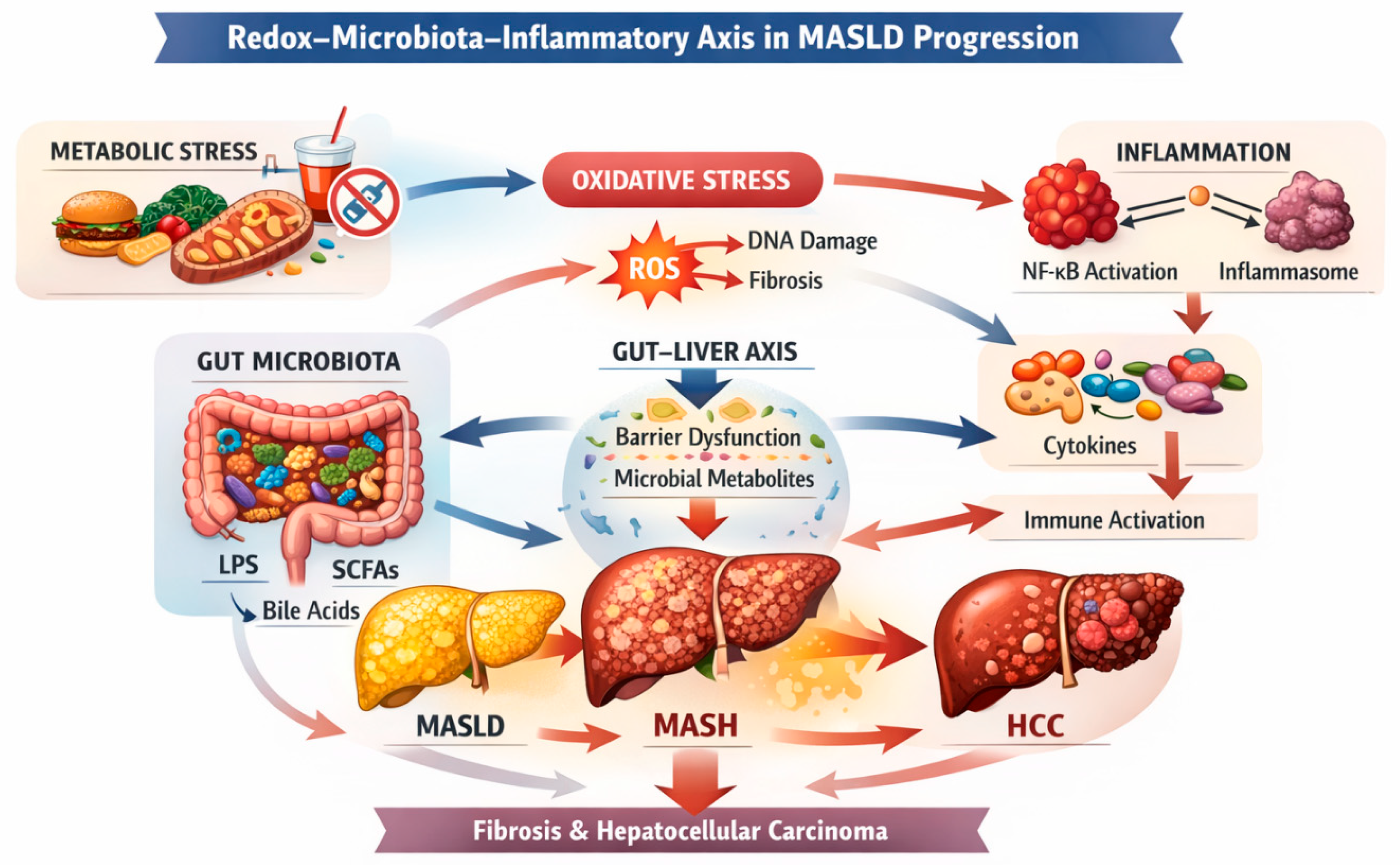
Figure 1: Redox–Microbiota–Inflammatory Axis in MASLD progression.
Diagram illustrating the molecular interactions connecting metabolic stress, gut microbiota dysbiosis, oxidative stress, and immune activation during the progression from MASLD to MASH, fibrosis, and hepatocellular carcinoma. Microbial products such as lipopolysaccharide (LPS) stimulate Toll-like receptor 4 (TLR4) and activate NF-κB–mediated inflammatory pathways, while mitochondrial dysfunction increases reactive oxygen species (ROS) production. These mechanisms interact along the gut–liver axis, amplifying inflammation and fibrogenesis through a self-sustaining pathogenic loop.
3.1 Anatomy and Physiology of the Gut-Liver Connection
The gut and liver communicate through (i) portal transport of microbial components and metabolites, (ii) biliary secretion of bile acids and antimicrobial factors, and (iii) immune signaling networks that shape tolerance and inflammation [18,19]. The liver influences microbiota by regulating bile acid pools and composition, which exert antimicrobial activity and provide substrates for microbial biotransformation; in turn, microbiota shape the bile acid pool by deconjugation and conversion into secondary bile acids, which signal through hepatic and intestinal receptors such as farnesoid X receptor (FXR) and TGR5, influencing glucose and lipid metabolism as well as inflammatory tone [20,21].
3.2 Intestinal Barrier Function and Permeability
Barrier integrity is maintained by tight junction proteins, mucus layers, antimicrobial peptides, and immune mechanisms that enforce compartmentalization between luminal microbes and host tissue. In MASLD, dysbiosis and metabolic inflammation can reduce tight junction integrity and alter mucus architecture, increasing intestinal permeability (“leaky gut”) [18,43]. This permits greater translocation of microbial-associated molecular patterns (MAMPs) such as lipopolysaccharide (LPS) and bacterial DNA into portal blood, resulting in hepatic innate immune activation. Low-grade endotoxemia is a plausible mechanistic bridge between diet-induced dysbiosis and hepatic inflammation, particularly in metabolically inflamed states where immune thresholds are altered [18,44].
3.3 Portal Transport of Microbial Products and Metabolites
Once microbial products reach the liver, pattern recognition receptors (PRRs) on Kupffer cells, hepatocytes, and hepatic stellate cells can activate inflammatory cascades. The stimulation of Toll-like receptor 4 (TLR4) by lipopolysaccharide (LPS) drives Nuclear Factor kappa-light-chain-enhancer of activated B cells (NF-κB) mediated cytokine production and can promote inflammasome activation, while also inducing oxidative stress by stimulating ROS-generating enzymes and amplifying mitochondrial dysfunction [19,34,45]. Hepatocytes exposed to chronic lipotoxic and redox stress exhibit lowered inflammatory thresholds, such that exposure to MAMPs elicits exaggerated PRR signaling and oxidative responses. In this way, gut-derived signals and hepatic metabolic stress synergize, consistent with multiple-hit models of MASH development [12,17].
Microbial metabolites can have beneficial or harmful effects depending on context. Short-chain fatty acids (SCFAs), produced through fermentation of dietary fiber, may promote metabolic homeostasis by improving insulin sensitivity, modulating intestinal barrier function, and influencing immune regulation; however, effects vary with SCFA type, receptor signaling, and host metabolic state [46]. Conversely, microbial conversion of choline and carnitine into trimethylamine (subsequently oxidized to Trimethylamine N-oxide (TMAO) has been linked to cardiometabolic risk pathways and may intersect with MASLD systemic risk, while endogenous ethanol production by dysbiotic microbiota has been proposed as a contributor to hepatic oxidative stress and inflammation in susceptible individuals [47,48]. Bile acid signaling represents a major axis of microbiota-host interaction: altered bile acid pools can influence hepatic lipid handling and inflammatory signaling, and bile acid receptor agonism/antagonism has become a therapeutic focus in metabolic liver disease [20,21].
Human and experimental data support a role for gut microbiota in MASLD heterogeneity. Microbiome signatures associated with NAFLD/MASLD have been described, but disentangling causality from confounding metabolic factors remains challenging; nevertheless, multi-omics approaches increasingly point to microbial functions (e.g., bile acid metabolism, endotoxin synthesis, ethanol production) that could be mechanistically relevant [49]. In parallel, mechanistic reviews emphasize that intestinal microbiota can “fuel” metabolic inflammation through barrier disruption and metabolite translocation, influencing metabolic organs such as the liver and adipose tissue [43].
Clinically, the gut-liver axis has two major implications. First, it provides a mechanistic justification for emphasizing lifestyle interventions (dietary composition, weight reduction, physical activity) because diet is a primary driver of microbial ecology, barrier function, and systemic inflammation, central elements of MASLD management emphasized in guidelines [2]. Second, it highlights potential adjunctive strategies (prebiotics/probiotics, bile acid-targeted approaches, microbiome-informed strategies may modulate inflammatory signaling and oxidative stress pathways; however, robust outcome-driven evidence is still evolving [18,49,50].
In summary, the gut–liver axis functions as a regulatory interface in which microbial composition, barrier integrity, and metabolite flux dynamically modulate hepatic redox balance and inflammatory tone. When metabolic priming and dysbiosis coexist, this interface transforms into a disease-amplifying conduit that facilitates fibrogenesis and oncogenic risk [19,22,23].
4 Gut Microbiota Dysbiosis in MASLD and MASH
MASLD is a multifactorial disease, which includes multiple risk factors: genetic polymorphisms, epigenetic modifications, cardiovascular risk factors, and microbiome alterations contribute to causing a liver disease attributable to a systemic dysfunction [51]. In recent years, many studies have been highlighting the crucial role of the intestinal microbiota in the delicate balance between functionality and dysfunction in the digestive system and its significant contribution to health. This aspect, combined with the recognized pathogenic effect supported by oxidative stress, is the center of a complex interplay responsible for many conditions attributable to pathologies on which only recently attention has begun to be paid [51,52,53]. Within the Redox–Microbiota–Inflammatory Axis framework, gut dysbiosis represents not merely a compositional alteration but a functional shift that modifies microbial metabolite production, barrier integrity, and immune signaling. The pathogenic relevance of dysbiosis in MASLD depends less on specific taxa and more on altered microbial functions that interact with redox-sensitive and inflammatory pathways in metabolically primed hepatocytes [54].
4.1 Composition and Diversity Changes in MASLD
Gut microbiota is the name used to identify the community of microorganisms residing in the digestive system and its dysfunction is called dysbiosis: generally, in a healthy individual, there is a population of microorganisms whose main species are represented by strains of Actinobacteria, Bacterioides, Firmicutes and Proteobacteria, which interact and are influenced in a non-unidirectional manner by genetic alterations (DNA methylation phenomena and modifications of RNA, non-coding RNA and histones) and epigenetic alterations (mainly affecting patatin-like phospholipase domain-containing protein 3 [PNPLA3], glucokinase regulatory protein [GCRK] and hydroxysteroid 17-beta dehydrogenase 13 [HSD17B13]) and by compounds derived from microbial metabolism (SCFAs such as butyric acid, propionic acid and acetic acid, bile acids, LPS), in addition to the exotoxic action mediated by ethanol [51]. Although multiple studies report alterations in Firmicutes, Bacteroidetes, and Proteobacteria abundance in MASLD, taxonomic findings remain partially inconsistent across cohorts [55,56]. Variability in diet, metabolic phenotype, geography, and sequencing methodology suggests that functional microbial signatures may be more reproducible than isolated compositional changes.
4.2 Functional Shifts: SCFAs, Bile Acids, Ethanol, LPS
In addition to the microbiological evidence (modification of the gut microbiota), bile acids and SCFAs are also significantly involved in the functional physiopathology of MASLD: the former, synthesized by the liver and metabolized by the intestinal microbiota, convert primary bile acids into secondary ones and maintain intestinal homeostasis unaltered, interacting in lipid metabolism, insulin resistance, and innate immunity [57,58,59]. Furthermore, the production of SCFAs and the conversion of bile acids from primary to secondary cannot be completed without the contribution of the gut microbiota due to its fundamental importance in glucose metabolism and in maintaining splanchnic energy balance. Intestinal microorganisms, in fact, are capable of influencing iron metabolism, and serum ferritin levels correlate directly with lipid accumulation at the hepatocellular level and, conversely, inversely correlate with the increase in colonies of Pasteurellaceae, Leuconostocaceae, and Micrococcaea strains, germs not normally represented in a healthy intestine [60,61]. The role of bile acids in intestinal health follows a bidirectional pathway, primarily influencing the microbiota and being modulated by it in their composition [58]. SCFAs are molecules produced by the intestinal microbiota starting from the fermentation of dietary fibres and are involved in the modulation of hepatic lipid metabolism through the inhibition of lipogenesis and the facilitation of fatty acid oxidation. Furthermore, SCFAs act as intracellular signaling factors by interacting with G-protein-coupled receptors (GPCRs) such as GPR43 and GPR41, which are expressed to a greater extent on the cell surface of patients with MASLD; finally, the activation of these receptors by SCFAs also causes an integrity modulation in the intestinal barrier and immune responses. In fact, SCFA-producing bacteria such as Bifidobacterium and Prevotella are more abundant in MASLD patients [58,62], and in these patients, the fecal levels of SCFAs are higher.
The recent nosographic classification of MASLD has introduced the category Metabolic and Alcohol-related Liver Disease (MetALD) which identifies individuals with hepatic steatosis and metabolic dysfunction with a daily alcohol consumption above the threshold (≥30 g/day for men and ≥20 g/day for women) [62], overcoming the well-known dichotomy between metabolic and toxic etiology and recognizing the synergistic nature of the interactions existing between alcohol consumption and metabolic dysfunction [63]. In fact, the metabolism of ethyl alcohol substantially modifies the hepatic metabolic profile by inhibiting mitochondrial β-oxidation, activating lipogenesis with concurrent suppression of gluconeogenesis and compromising oxidative phosphorylation at the level of the electron transport chain and the Krebs cycle [64,65,66]. Precisely for this reason, even alcohol consumption traditionally considered moderate is now seen as a fundamental pathogenic cofactor (ethanol-induced injury) for the progression towards steatohepatitis, advanced fibrosis, and HCC [67]. Several of these microbial-derived metabolites converge on redox-sensitive hepatic pathways [68]. Endogenous ethanol production, LPS-mediated TLR4 activation, and altered bile acid signaling enhance CYP2E1 expression and NADPH oxidase activity, thereby increasing hepatic ROS generation and lipid peroxidation [69,70]. In metabolically primed hepatocytes, this redox amplification lowers inflammatory activation thresholds and promotes fibrogenic signaling [70,71,72].
4.3 Mechanisms of Gut Barrier Disruption and Endotoxemia
The intestinal barrier can be damaged when all the previously described factors are altered: in particular, the mucous layer, which acts as the outermost protection of this barrier, can become thinner due to the deficiency of LPS and peptidoglycan, which stimulate its secretion [73]. Furthermore, endotoxinemia in MASLD, associated with translocation of LPS from the enteric to the systemic circulation due to the increased permeability of the vascular bed, promotes a state of constitutive inflammation through the activation of Toll-like receptor 4 (TLR4), which induces insulin resistance and an increased risk of fibrosis [73]. In hepatocytes already exposed to lipotoxic and mitochondrial stress, LPS signaling further disrupts redox homeostasis and enhances ROS production, establishing a feed-forward interaction between endotoxemia and oxidative imbalance that sustains chronic inflammation [74].
4.4 Interplay between Diet, Obesity, and Dysbiosis
Dysbiosis in subjects affected by MASLD is characterized by a disbalance in the resident population, both in terms of resident microbial species and in their representation, highlighting an increase in colonies of Gram-negative bacteria, which strongly correlates with the progression towards obesity [51,58]. Dietary composition is a major determinant of this microbial shift [75]. High-fructose intake promotes de novo lipogenesis and increases intestinal permeability [76,77], while diets rich in saturated fats favor expansion of LPS-producing Gram-negative bacteria and TLR activation [77]. Conversely, dietary fiber supports SCFA-producing taxa that enhance barrier integrity and modulate immune tone [78,79]. Thus, dietary patterns shape microbial ecology in ways that directly influence hepatic redox balance and inflammatory signaling. The principal microbial-derived mediators and their hepatic effects are summarized in Table 1 [58].
Table 1: Microbial-Derived Metabolites and Their Hepatic Impact.
| Microbial Product | Source | Hepatic Target | Mechanistic Effect |
|---|---|---|---|
| LPS | Gram-negative bacteria | TLR4 | NF-κB activation |
| SCFAs | Fiber fermentation | GPCRs | Anti-inflammatory |
| DCA | Secondary bile acid | Senescence pathways | Pro-oncogenic |
| Ethanol | Dysbiosis | CYP2E1 | ROS increase |
5 Oxidative Stress and ROS in Liver Injury
Oxidative stress and ROS play a major role in the pathogenesis of liver damage and in the progression of chronic liver diseases: the liver is a central organ for ROS clearance due to both its great detoxifying capacity on the action of these molecules and its marked mitochondrial activity [80]. Cellular modifications induced by oxidative stress are mainly attributable to two pathways, fibrogenesis and vascular damage, which correlate with an increased mortality in MASLD patients, especially if they belong to the MetALD phenotype [64,81]. Within the Redox–Microbiota–Inflammatory Axis, oxidative stress represents the central amplification node through which metabolic overload and microbial-derived signals converge. In MASLD, ROS are not merely by-products of hepatocellular dysfunction but active drivers of inflammatory signaling, fibrogenesis, and genomic instability.
ROS are highly reactive oxygen-containing molecules, natural by-products of aerobic cellular metabolism [81]. ROS include free radicals (superoxide and hydroxyl) and non-radical species (hydrogen peroxide) [82]. These molecules act differently depending on their concentration: at low levels, they act as second messengers and regulate processes of proliferation, differentiation, apoptosis, and immune response, while at high levels, they cause oxidative stress due to their damaging effect on lipids, proteins, and DNA [83].
5.1 Sources of ROS in Hepatocytes (Mitochondria, CYP2E1, NADPH Oxidases)
The main biological sources of ROS are mitochondria, cytochrome P-450 2E1 (CYP2E1), and Nicotinamide-Adenine Dinucleotide Phosphate (NADPH) oxidase (NOX) [83,84]. In a healthy organism, the production of ROS is a necessary process for cell signaling and, therefore, is finely regulated. The main source is the mitochondria, which, during oxidative phosphorylation, when a portion of electrons (about 1–2%) physiologically escapes the electron transport chain and reacts with unpaired electrons, generating superoxide radicals [81,85]. CYP2E1 is a highly inducible microsomal enzyme that, unlike other cytochromes, has a high activity on NOX even in the absence of substrate. During its catalytic cycle, oxygen is not completely reduced, and it releases mainly hydroxyl and hydrogen peroxide [84]. NOX is an enzymatic complex exclusively responsible for the production of ROS: they are localized on the plasma membrane and in hepatocyte endosomes as well as in Kupffer cells (KC). In hepatocytes, these enzymes are activated by cytokines, metabolic stress, or growth factors and act by recruiting electrons from the cytosol and converting them into ROS [82]. In MASLD and MASH, these ROS-generating systems are synergistically activated. Mitochondrial electron leakage increases under lipid overload, CYP2E1 expression is upregulated in the presence of fatty acids and ethanol exposure, and NADPH oxidases are stimulated by cytokines and TLR4 signaling. The simultaneous activation of these sources amplifies oxidative burden beyond physiological signaling thresholds, promoting sustained hepatocellular injury [86].
5.2 Lipid Peroxidation and Oxidative Damage to Proteins and DNA
Lipid peroxidation is triggered by oxidative stress, affecting polyunsaturated fatty acids (PUFAs), damaging the structure of cell membranes, and generating toxic byproducts that amplify this process. When a free radical removes a hydrogen atom from a fatty acid, it forms a lipid radical, which subsequently reacts with oxygen and forms a peroxyl radical [82,87]: at this point, the reaction is amplified because the peroxyl radical anchors itself to nearby lipids and ends only when two peroxyl radicals interact with each other. The products of PUFAs peroxidation are mainly reactive aldehydes, which bind to amino acids and alter the structure of proteins or promote protein aggregation by forming complexes resistant to degradation [87]. In steatotic livers, lipid peroxidation products such as malondialdehyde and 4-hydroxynonenal form adducts with cellular proteins and DNA, altering signaling pathways and contributing to hepatocyte ballooning, stellate cell activation, and fibrogenesis [88]. Persistent oxidative DNA damage, particularly accumulation of 8-oxo-deoxyguanosine, further increases mutational pressure in the progression toward hepatocellular carcinoma [89]. A growing body of evidence implicates ferroptosis, a regulated, iron-dependent form of cell death driven by lipid peroxidation, as a relevant mechanism in MASH progression and hepatocarcinogenesis [90]. Ferroptosis is characterized by the accumulation of oxidized phospholipids and depletion of glutathione-dependent defenses, particularly glutathione peroxidase 4 (GPX4). In steatotic livers with altered iron metabolism and enhanced ROS production, ferroptotic signaling may contribute to hepatocyte loss, inflammatory amplification, and selection of apoptosis-resistant clones during HCC development [90]. All these products have a great pathophysiological impact and are responsible for the genesis and oncological progression of HCC, dysimmunity, and neurological conditions such as Alzheimer’s and Parkinson’s disease [91].
5.3 Antioxidant Defenses and Redox Imbalance
The development of all these reactive species induces a redox imbalance, which can exist as an excess of oxidizing agents or as an excess of reducing agents, although the latter is a less frequent condition. To buffer the excess of harmful radicals, the organism has developed enzymatic and non-enzymatic defense mechanisms: the first line is represented by enzymatic systems, such as superoxide dismutase (SOD) which converts superoxide radicals into hydrogen peroxide, catalases which decompose hydrogen peroxide into water and hydrogen, glutathione peroxidase which reduces hydrogen and lipid peroxides using glutathione and, finally, the thioredoxin system which exchanges and reduces thiol groups [92]. Non-enzymatic mechanisms are glutathione, vitamins E and C, polyphenols, flavonoids, and proteins such as ceruloplasmin, ferritin, and transferrin, which, by binding specific metals, prevent the genesis of free radicals [92]. The transcription factor Nrf2 (nuclear factor erythroid 2-related factor 2) represents a master regulator of antioxidant defense [93]. Under oxidative stress, Nrf2 dissociates from Keap1 and translocates to the nucleus, promoting transcription of genes encoding glutathione synthesis enzymes, detoxifying proteins, and antioxidant systems [94]. Impaired Nrf2 signaling has been observed in progressive MASLD, contributing to inadequate compensation of chronic oxidative burden [95]. Generally, the redox balance favors antioxidant agents, but age and comorbidities can damage this balance: the first measures to be taken to maintain a correct balance and slow down oxidative stress are the respect of a healthy and balanced lifestyle and the integration of missing factors (especially vitamins) [96].
5.4 Oxidative Stress as Mediator between Dysbiosis and Inflammation
Oxidative stress promoted by ROS increases the levels of circulating pro-inflammatory cytokines through the JAK/STAT, MAPK, and NF-kB pathways [97]. Furthermore, in the presence of dysbiosis, a vicious circle is triggered that causes an increase in ROS which, by raising an inflammatory state, dysregulates the intestinal microbiological habitat, amplifying the dysbiosis itself: in fact, oxidative stress damages the tight junctions and induces a condition of the intestinal mucosa known as “leaky gut”, which, ultimately, allows for greater bacterial translocation [62,98]. In this context, oxidative stress operates bidirectionally: it both results from dysbiosis-driven endotoxemia and further exacerbates intestinal barrier dysfunction, reinforcing microbial translocation. ROS-mediated activation of NF-κB, JNK, and inflammasome pathways establishes a feed-forward inflammatory circuit that lowers immune activation thresholds [99]. Thus, oxidative stress functions as the molecular hinge linking microbial signals to sustained hepatic immune activation.
6 Inflammatory Pathways and Immune Activation
Inflammation is a protective response of the organism that is composed of four phases: inflammatory inducers, inflammatory sensors (like mast cells and macrophages), inflammatory mediators (like cytokines and chemokines), and affected tissues [100]. Each phase has many options that are triggered by the type of pathogen involved in the inflammation [101]. Inflammatory pathways and the immune response are linked by a complex network of finely regulated and interconnected responses, so widespread that, in case of dysregulation, it can induce a state of chronic inflammation and autoimmune diseases. The process begins with the recognition of potentially harmful molecules by the cells of the innate immunity (macrophages, mast cells, and dendritic cells), and this information is transported through specific intracellular signaling pathways in order to activate the necessary inflammatory mediators [102]. In MASLD and MASH, inflammatory activation does not represent a transient defensive response but a sustained, low-grade, metabolically driven process that intersects with oxidative stress and microbial-derived signals [103]. Within the Redox–Microbiota–Inflammatory Axis, immune activation functions as the effector arm that translates redox imbalance and endotoxemia into fibrogenic remodeling and progressive tissue damage.
6.1 Activation of Pattern Recognition Receptors (TLR4, NLRP3 Inflammasome)
In the activation phase, TLR4, the inflammasome, and the NOD-like receptor family pyrin domain-containing 3 (NLRP3) are fundamental: TLR4 has been studied for a long time, and its ability to recognize the LPS of Gram-negative bacteria, an essential component of their external membrane, is currently well known [104]. The inflammasome is a cytosolic complex crucial for the immune response due to its great utility and flexibility in adaptively adjusting to the recognition of antigens expressed during pathological phenomena. Its activation requires receptor signaling (generally mediated by TLR4) and the presence of cellular signals of metabolic stress, such as potassium efflux, rupture of liposomal vesicles, accumulation of cardiolipin, which is an indicator of mitochondrial dysfunction, and the increase in ROS levels [105,106]. NLRP3 is a cytosolic multiprotein complex essential in the innate immune system that acts both as an intracellular sensor for the detection of danger signals and as an effector of a rapid inflammatory response: its triggering is mediated by TLR4 and, once activated, it operates by activating NF-kB, which is necessary for the activation of pro-IL-1β [107]. In steatotic livers, NLRP3 activation is potentiated by mitochondrial ROS, lipid peroxidation products, and cholesterol crystals, creating a convergence between metabolic stress and innate immune sensing. This redox-sensitive inflammasome activation amplifies IL-1β and IL-18 secretion, further promoting hepatocyte injury and stellate cell activation in MASH [108].
6.2 Kupffer Cells, Macrophages, and Hepatic Stellate Cell Crosstalk
The pathophysiological pivot that leads to the development of MASLD is represented by the crosstalk between KC, macrophages, and hepatic stellate cells (HSC), which constitutes the central axis of hepatic fibrogenesis [96,109,110]. KC are macrophages resident in the hepatic sinusoids capable of secreting pro-inflammatory and pro-fibrogenic factors: in MASLD, the number of extracellular vesicles increases, which mediate reciprocal activation with HSC [109]. Macrophages derive from the intra-hepatic activation of monocytes and cross-react with KC and HSC to amplify fibrosis and perpetuate inflammation [109]. HSCs (also known as Ito cells) occupy the spaces of Disse in the liver and are responsible for the storage of vitamin A, the deposition of extracellular matrix, and the chemotaxis of some cells involved in the inflammatory response (especially natural killer cells) [111]. The interaction between these three cell lines causes an increase in circulating iron and ROS, which causes a condition of secondary hemochromatosis associated with oxidative stress [98]. Activated hepatic stellate cells respond to cytokines and oxidative stress by transdifferentiating into myofibroblast-like cells that secrete type I collagen and extracellular matrix components [112]. Persistent immune–redox crosstalk therefore sustains fibrogenesis, establishing the structural substrate for progression from MASH to advanced fibrosis.
6.3 Cytokine Network (TNF-α, IL-6, IL-1β) and Chronic Inflammation
The activation of the crosstalk between KCs, macrophages, and HSCs promotes the production of inflammatory mediators due to the primitive activation of TLR4 and uses signaling pathways involving mediators such as TNF-α, IL-6, and IL-1β to sustain the continuation of the inflammatory stimulus, which, with the deposition of considerable quantities of myofibroblasts and extracellular matrix, becomes chronic and increases the rate of liver fibrosis [83,111]. Chronic exposure to TNF-α, IL-6, and IL-1β not only perpetuates local inflammation but also alters hepatocyte survival signaling and promotes compensatory proliferation [113]. In the presence of oxidative DNA damage, this proliferative pressure increases the likelihood of mutational fixation, providing a mechanistic bridge toward hepatocarcinogenesis.
6.4 Interrelation between Oxidative and Inflammatory Signaling
Oxidative stress and inflammatory signaling operate within a tightly coupled amplification circuit rather than a linear cascade. Redox-sensitive transcriptional programs sustain cytokine production, while inflammatory mediators further impair mitochondrial efficiency and enhance ROS-generating systems, thereby stabilizing a state of chronic oxidative imbalance. Within this framework, modulation of upstream metabolic pathways, such as those influenced by SGLT2 inhibitors, may indirectly attenuate inflammatory signaling, although the precise intrahepatic mechanisms remain to be fully elucidated [111]. The feed-forward architecture of redox and inflammatory signaling exemplifies the Redox–Microbiota–Inflammatory Axis, in which metabolic overload, dysbiosis, and immune activation converge to drive progressive liver injury [Fig. 2].
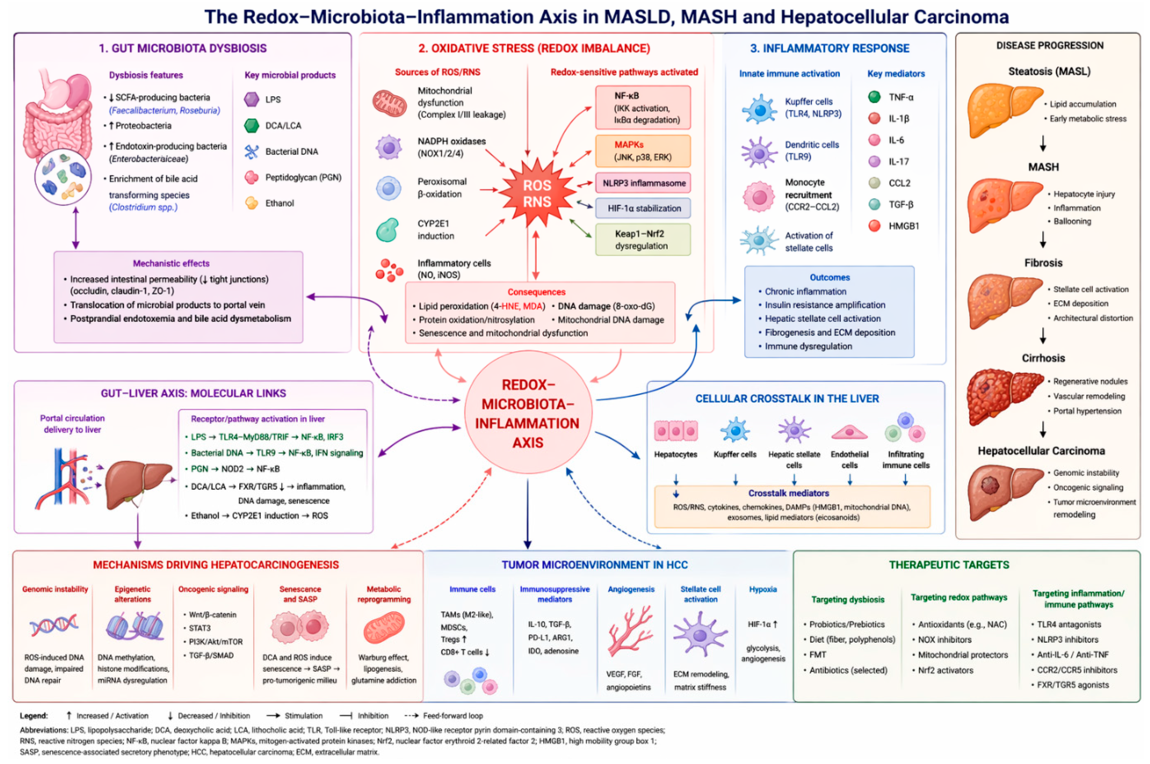
Figure 2: The redox–microbiota–inflammation axis in MASLD, MASH, and Hepatocellular Carcinoma.
Schematic overview of the interplay between gut microbiota dysbiosis, oxidative stress, and inflammatory signaling in the progression of MASLD to MASH and hepatocellular carcinoma. Microbial products reaching the liver through the gut–liver axis activate immune pathways and promote ROS generation. Oxidative stress amplifies inflammatory responses and fibrogenesis, creating a feed-forward loop that drives hepatocellular injury, fibrosis, and tumor development.
7 From MASH to HCC: The Oxidative-Inflammatory-Oncogenic Continuum
Chronic inflammation is a fundamental driver in the progression from MASH to HCC. Persistent immune activation in the liver promotes continuous release of pro-inflammatory cytokines and the generation of reactive oxygen and nitrogen species, which induce cumulative DNA damage in hepatocytes [114,115]. Oxidative lesions, strand breaks, and DNA adducts, when insufficiently repaired, become fixed mutations during repeated cycles of cell death and compensatory proliferation [116,117].
Inflammatory mediators also interfere with DNA repair pathways, including base excision and mismatch repair systems, thereby amplifying genomic vulnerability [115,118]. In parallel, sustained activation of signaling cascades such as NF-κB and STAT3 enhances survival of genetically altered cells, allowing clonal expansion [118,119]. Metabolic stress in MASH further exacerbates mitochondrial dysfunction and lipid peroxidation, reinforcing oxidative injury and mutational burden [116,120]. Within the Redox–Microbiota–Inflammatory Axis framework, hepatocarcinogenesis represents the long-term consequence of sustained redox imbalance, immune dysregulation, and microbiota-derived signaling. Rather than a discrete late event, HCC emerges from chronic amplification circuits that progressively destabilize genomic integrity and reshape the hepatic microenvironment.
7.1 Chronic Inflammation and Genomic Instability
Together, persistent oxidative stress, defective repair mechanisms, and proliferative signaling establish a self-perpetuating cycle that fosters genomic instability, an essential step in the transition from chronic steatohepatitis to malignant transformation [121]. Chronic redox imbalance promotes accumulation of DNA strand breaks, telomere shortening, and chromosomal instability, while inflammatory cytokines sustain compensatory hepatocyte proliferation in a microenvironment permissive to mutational fixation [122,123,124]. Repeated cycles of cell death and regeneration increase replicative stress, favoring clonal selection of genetically altered hepatocytes. Over time, this combination of oxidative genotoxic pressure and proliferative drive establishes the molecular substrate for oncogenic transformation [125].
7.2 ROS-Driven DNA Damage and Oncogenic Mutations
Excessive production of ROS is a central driver of genomic injury in MASH and contributes directly to hepatocarcinogenesis [120]. Mitochondrial dysfunction, impaired β-oxidation, and endoplasmic reticulum stress in steatotic hepatocytes markedly enhance ROS generation, while inflammatory cells further amplify oxidative pressure through NADPH oxidase activation [115,116]. Persistent oxidative stress induces DNA base modifications, most notably 8-oxo-deoxyguanosine, together with single- and double-strand breaks. If not accurately repaired by base excision repair or homologous recombination pathways, these lesions become fixed mutations during hepatocyte replication [116,118].
Oxidative stress also disrupts genomic surveillance mechanisms by altering p53 signaling and reducing the efficiency of DNA repair enzymes, thereby increasing mutational burden [118,120]. Lipid peroxidation products such as 4-hydroxynonenal generate mutagenic DNA adducts that expand the oncogenic mutation spectrum [116,120]. Genomic profiling of MASH-related HCC has revealed recurrent alterations in TP53, CTNNB1, and chromatin remodeling genes, consistent with chronic oxidative and inflammatory genotoxic pressure [119,121].
In addition to direct mutagenic effects, chronic lipid peroxidation may promote ferroptotic cell death in subsets of hepatocytes, creating selective pressure within the regenerating liver. Repeated ferroptosis-associated injury may favor survival of apoptosis-resistant or genetically unstable clones, thereby contributing to clonal evolution during MASH-related hepatocarcinogenesis [126].
7.3 Pro-Tumorigenic Microenvironment and Immune Evasion
Persistent hepatocellular injury promotes recruitment of monocyte-derived macrophages, neutrophils, and dysfunctional T lymphocytes, which sustain cytokine production, including IL-6, TNF-α, and TGF-β [114,127]. These mediators activate oncogenic signaling pathways such as STAT3 and NF-κB, enhancing hepatocyte survival and resistance to apoptosis.
Chronic inflammatory signaling also drives immune exhaustion and tolerance. In MASH-related HCC, CD8+ T cells frequently display an exhausted phenotype with increased expression of immune checkpoint molecules such as PD-1, while regulatory T cells and immunosuppressive macrophages accumulate within the tumor microenvironment [119,128]. Metabolic reprogramming and lipid accumulation further impair antitumor immunity by altering T-cell mitochondrial fitness and cytokine production [114,121].
Sustained inflammatory signaling combined with immune suppression creates a permissive niche that facilitates clonal expansion of transformed hepatocytes and enables immune escape, thereby promoting malignant progression.
Recent experimental studies have also shown that microbiota-derived metabolites modulate immune surveillance. For example, alterations in bile acid composition can regulate hepatic natural killer T cell activity through FXR-dependent signaling pathways. These immune alterations contribute to tumor immune evasion and facilitate HCC development.
7.4 Microbiota-Derived Metabolites in Hepatocarcinogenesis
The gut–liver axis plays a crucial role in the progression from MASH to HCC. Dysbiosis increases intestinal permeability, facilitating translocation of microbial products such as LPS into the portal circulation. LPS activates TLR4 signaling in hepatocytes and Kupffer cells, promoting NF-κB–mediated inflammatory cascades and fibrogenesis [129,130].
Microbiota-derived metabolites further influence hepatocarcinogenesis. Secondary bile acids, particularly deoxycholic acid (DCA), induce DNA damage and a pro-inflammatory senescence-associated secretory phenotype that fosters malignant transformation [131]. Dysregulated bile acid signaling through the FXR contributes to metabolic imbalance and carcinogenic risk [132]. Moreover, reduced production of short-chain fatty acids may impair immune surveillance and epithelial integrity [129].
Microbiota-derived metabolites therefore act as modulators of inflammation, genomic instability, and immune function, reinforcing the oncogenic continuum linking MASH to HCC. These signals do not operate in isolation but intersect with redox-sensitive pathways and inflammatory circuits, amplifying the oxidative–inflammatory–oncogenic cascade that characterizes MASLD progression.
Overall, the transition from MASH to HCC reflects cumulative failure of redox homeostasis, immune surveillance, and metabolic adaptation. Persistent oxidative DNA damage, inflammatory signaling, microbiota-derived metabolites, and proliferative pressure converge to lower the threshold for malignant transformation. In this integrated model, hepatocarcinogenesis emerges not as a stochastic complication but as the biological endpoint of sustained redox–immune amplification in a metabolically primed liver. The progressive shift from steatosis to hepatocarcinogenesis reflects stage-specific yet interconnected molecular events, summarized in Table 2 [133].
Table 2: Key Molecular Events Across MASLD Progression.
| Disease Stage | Dominant Mechanisms | Key Molecular Players | Clinical Correlates |
|---|---|---|---|
| MASL | Lipid overload | SREBP-1c, ChREBP | Steatosis |
| MASH | ROS + Inflammation | NLRP3, TNF-α, IL-6 | Ballooning |
| Fibrosis | Stellate activation | TGF-β, Collagen I | Fibrosis stage |
| HCC | Genomic instability | TP53, CTNNB1 | Tumor |
8 Nutritional and Nutraceutical Modulation of the Gut-Liver Axis
Within this interconnected network, nutritional and nutraceutical interventions represent potential modulators of upstream metabolic stress, redox imbalance, and microbial dysbiosis. Rather than acting on isolated pathways, dietary strategies may influence multiple nodes of the amplification network that drives MASLD progression. Oxidative stress is a central driver of hepatocellular injury, and bioactive compounds such as polyphenols, vitamins, and omega-3 PUFAs exert protective effects through antioxidant, anti-inflammatory, and microbiota-modulating properties [134,135].
8.1 Role of Dietary Antioxidants (Polyphenols, Vitamins, Omega-3)
Polyphenols, including resveratrol, catechins, and curcumin, attenuate hepatic lipid accumulation and inflammation by activating AMPK and inhibiting NF-κB signaling pathways. These compounds also influence gut microbial composition, promoting beneficial taxa and enhancing intestinal barrier integrity [136,137]. Vitamin E has demonstrated efficacy in improving histological features of MASH, primarily by reducing lipid peroxidation and hepatocellular ballooning, although long-term safety remains under evaluation [138]. Omega-3 PUFAs, particularly eicosapentaenoic (EPA) and docosahexaenoic acid (DHA), reduce hepatic triglyceride accumulation and modulate inflammatory signaling, partly through alterations in gut microbiota composition and bile acid metabolism [139,140]. Despite promising mechanistic data, clinical benefits remain heterogeneous, and most nutraceutical interventions demonstrate modest histological effects compared with structured weight loss interventions.
8.2 Probiotics, Prebiotics, and Postbiotics in Restoring Eubiosis
Gut dysbiosis contributes significantly to MASLD progression by increasing intestinal permeability and endotoxin translocation. Probiotics, prebiotics, and postbiotics have emerged as promising interventions to restore microbial balance and reinforce gut barrier function [141,142].
Probiotics such as Lactobacillus and Bifidobacterium species reduce circulating LPS levels, attenuate hepatic inflammation, and improve aminotransferase levels in clinical studies [143]. Prebiotics, including inulin and fructooligosaccharides, selectively stimulate beneficial microbial growth and enhance SCFA production, particularly butyrate, which strengthens epithelial integrity and exerts anti-inflammatory effects [142,144]. Postbiotics, defined as non-viable microbial products or metabolites, represent an emerging therapeutic concept; SCFAs and microbial-derived indoles have demonstrated hepatoprotective properties through modulation of immune and metabolic signaling pathways [145].
Although heterogeneity among studies persists, cumulative evidence suggests that microbiota-targeted interventions can reduce hepatic steatosis, inflammation, and metabolic dysregulation, reinforcing the therapeutic relevance of restoring eubiosis along the gut–liver axis [146]. Most available trials are small, short-term, and rely on surrogate endpoints such as aminotransferases or steatosis indices rather than fibrosis regression, limiting definitive conclusions regarding long-term disease modification.
8.3 Dietary Patterns (Mediterranean, Plant-Based, Low-Fructose)
Beyond single nutrients, overall dietary patterns critically influence MASLD risk and progression. The Mediterranean diet, characterized by high intake of vegetables, fruits, whole grains, legumes, olive oil, and fish, has consistently been associated with reduced hepatic steatosis and improved insulin sensitivity [145,147]. Its benefits derive from the combined effects of monounsaturated fats, polyphenols, fiber, and omega-3 PUFAs, which collectively modulate inflammation, oxidative stress, and gut microbiota composition.
Plant-based dietary patterns further enhance microbial diversity and SCFA production while reducing intake of saturated fats and heme iron, factors implicated in hepatic inflammation [148]. Conversely, excessive fructose consumption promotes de novo lipogenesis, gut permeability, and endotoxemia, exacerbating hepatic steatosis and inflammatory signaling [149]. Low-fructose dietary approaches have therefore gained attention as preventive strategies in MASLD.
Adherence to balanced dietary patterns that prioritize whole, minimally processed foods appears more effective than isolated nutrient supplementation, underscoring the integrative nature of gut–liver axis modulation. The benefits of structured dietary patterns likely derive from simultaneous modulation of substrate flux, microbial ecology, bile acid signaling, and inflammatory tone, supporting the concept that multi-target strategies are more effective than isolated supplementation.
8.4 Emerging Nutraceutical Strategies in MASLD/MASH Prevention
Novel nutraceutical strategies are being explored to complement lifestyle interventions in MASLD and MASH. Compounds targeting bile acid signaling, such as natural FXR modulators, demonstrate potential in regulating lipid metabolism and inflammation [150]. Berberine has shown promising effects in reducing hepatic steatosis and improving insulin resistance, partly through microbiota modulation and AMPK activation [151].
Other emerging agents include silymarin, quercetin, and curcumin derivatives, which exert antifibrotic and anti-inflammatory effects via modulation of TGF-β and NF-κB pathways [137,151]. Synbiotic formulations combining probiotics and prebiotics may provide synergistic benefits by simultaneously enhancing microbial diversity and metabolite production [143,144].
While large-scale randomized trials remain limited, accumulating mechanistic and clinical evidence supports the integration of nutraceutical approaches into comprehensive MASLD/MASH prevention strategies, particularly when combined with structured dietary modification and physical activity. Most emerging nutraceutical strategies should be considered adjunctive and not substitutes for evidence-based metabolic management centered on weight reduction, glycemic control, and cardiovascular risk mitigation [152].
Rather than functioning as stand-alone therapies, nutritional and microbiota-directed approaches are best conceptualized as modulators of an interconnected metabolic–redox–inflammatory network. Their greatest potential likely resides in early disease stages or in combination with pharmacological agents targeting metabolic and fibrotic pathways. Future studies integrating dietary interventions with molecular biomarkers may help identify patients most likely to benefit from axis-targeted modulation. The principal axis-modulating interventions and their current level of evidence are summarized in Table 3 [153].
Table 3: Nutritional and Microbiota-Targeted Interventions.
| Intervention | Target Node | Evidence Level | Limitations |
|---|---|---|---|
| Mediterranean diet | Substrate flux | Strong | Adherence |
| Plant-based diet | Microbial diversity, SCFA production | Moderate | Long-term adherence |
| Low-fructose diet | De novo lipogenesis, endotoxemia | Moderate | Dietary sustainability |
| Vitamin E | Lipid peroxidation | Moderate | Long-term safety |
| Probiotics | Barrier function | Small RCTs | Heterogeneity |
| FMT | Microbiota reshaping | Early trials | Durability |
9 Future Prospects and Therapeutic Outlook
Therapeutic modulation of the gut microbiota represents a promising frontier in MASLD/MASH management. Fecal microbiota transplantation (FMT) has demonstrated the capacity to reshape microbial composition, enhance short-chain fatty acid production, and improve intestinal barrier integrity in early-phase clinical studies. Recent trials indicate that FMT from lean donors can transiently improve insulin sensitivity and reduce hepatic inflammation markers, although durability and donor-recipient compatibility remain critical challenges [154,155]. However, variability in donor composition, engraftment efficiency, and long-term safety currently limit routine clinical implementation, and large, fibrosis-oriented trials are still lacking [156].
9.1 Microbiota-Targeted Pharmacotherapy (FMT, Precision Probiotics)
Precision probiotics, engineered or selectively formulated microbial consortia, are emerging as a more targeted alternative. Unlike conventional probiotics, these strategies aim to restore specific metabolic functions, such as bile acid transformation or butyrate synthesis, thereby modulating host lipid metabolism and immune responses [156]. Advances in metagenomics and metabolomics are enabling the identification of strain-specific effects, paving the way for next-generation live biotherapeutic products tailored to individual microbial signatures [157].
9.2 Biomarkers of Dysbiosis and Oxidative Imbalance
The development of reliable biomarkers reflecting gut dysbiosis and oxidative stress is essential for risk stratification and therapeutic monitoring in MASLD/MASH. Multi-omics approaches integrating metagenomic, transcriptomic, and metabolomic data have identified microbial signatures associated with fibrosis progression and hepatocellular carcinoma risk [158]. Alterations in bile acid profiles, reduced short-chain fatty acids, and increased endotoxin levels are increasingly recognized as functional indicators of gut–liver axis disruption [159].
Parallel assessment of oxidative imbalance, including circulating 8-oxo-deoxyguanosine, malondialdehyde, and antioxidant enzyme activity, may provide insight into redox-driven disease progression [160]. The integration of microbial and oxidative biomarkers with clinical and imaging parameters holds promise for constructing predictive algorithms capable of identifying high-risk patients and guiding personalized interventions [161]. Integration of microbial, redox, inflammatory, and imaging biomarkers may allow construction of composite risk algorithms capable of identifying patients with high oncogenic potential even in non-cirrhotic MASLD.
9.3 Integrative and Personalized Approaches
Future therapeutic strategies are likely to shift toward integrative, precision-based models combining dietary modulation, microbiota-targeted therapies, pharmacological agents, and lifestyle interventions. Artificial intelligence–driven analytics applied to multi-omics datasets may enable individualized prediction of treatment response and disease trajectory [157,162].
Personalized nutrition based on microbiome composition is gaining attention, as inter-individual variability in microbial metabolism influences dietary responsiveness and metabolic outcomes [163]. Furthermore, combination strategies, such as synbiotics with FXR agonists or antioxidant nutraceuticals, may produce synergistic benefits by simultaneously targeting inflammation, metabolic dysfunction, and dysbiosis.
Ultimately, the future of MASLD/MASH management lies in stratified intervention based on individual metabolic phenotype, microbial configuration, and redox-inflammatory burden [164]. Combining structured lifestyle modification, microbiota-targeted therapies, metabolic pharmacotherapy, and biomarker-guided monitoring may enable interruption of the amplification circuits that drive fibrosis and hepatocarcinogenesis. In this perspective, therapeutic success will depend less on single agents and more on integrated modulation of interconnected biological systems.
MASLD progression from steatosis to MASH, advanced fibrosis, and hepatocellular carcinoma reflects a failure of coordinated metabolic, redox, and immune homeostasis rather than the isolated consequence of lipid accumulation [165,166]. Chronic metabolic overload primes hepatocytes for mitochondrial dysfunction and excessive ROS generation, while gut dysbiosis increases exposure to microbial-derived inflammatory signals. Oxidative stress and immune activation do not operate independently; instead, they form a bidirectional amplification loop that sustains hepatocellular injury, promotes fibrogenesis, and progressively destabilizes genomic integrity.
Within this context, the Redox–Microbiota–Inflammatory Axis provides a coherent systems-level model explaining disease heterogeneity and the occurrence of HCC even in non-cirrhotic MASLD [23]. Persistent oxidative DNA damage, inflammatory signaling, ferroptotic injury, and immune exhaustion converge to create a pro-tumorigenic microenvironment in a metabolically primed liver. Hepatocarcinogenesis thus emerges not as a stochastic complication, but as the predictable biological endpoint of sustained redox–immune amplification.
Therapeutic success will likely depend on integrated strategies capable of simultaneously modulating metabolic substrate overload, gut microbial ecology, redox imbalance, and inflammatory signaling. Nutritional patterns, microbiota-directed therapies, metabolic pharmacotherapy, and biomarker-guided precision approaches should be viewed as complementary components of a multidimensional intervention model [12]. Future research integrating multi-omics profiling with longitudinal clinical data will be essential to identify high-risk phenotypes and to interrupt amplification circuits before irreversible fibrosis and malignant transformation occur.
Acknowledgement:
Funding Statement: The authors received no specific funding for this study.
Author Contributions: Conceptualization, Davide Nilo; writing-original draft preparation, Davide Nilo, Giovanni di Lorenzo, Marco La Montagna; writing-review and editing, Riccardo Nevola, Aldo Marrone, and Ferdinando Carlo Sasso, Alfredo Caturano; supervision: Alfredo Caturano. All authors reviewed and approved the final version of the manuscript.
Availability of Data and Materials: No dataset was generated for the publication of this article.
Ethics Approval: Not applicable.
Conflicts of Interest: The authors declare no conflicts of interest.
References
1. Rinella ME , Lazarus JV , Ratziu V , Francque SM , Sanyal AJ , Kanwal F , et al. A multisociety Delphi consensus statement on new fatty liver disease nomenclature. J Hepatol. 2023; 79( 6): 1542– 56. doi:10.1016/j.jhep.2023.06.003. [Google Scholar] [CrossRef]
2. European Association for the Study of the Liver (EASL); European Association for the Study of Diabetes (EASD); European Association for the Study of Obesity (EASO) . EASL-EASD-EASO Clinical Practice Guidelines on the management of metabolic dysfunction-associated steatotic liver disease (MASLD). Obes Facts. 2024; 17( 4): 374– 444. doi:10.1159/000539371. [Google Scholar] [CrossRef]
3. Targher G , Corey KE , Byrne CD , Roden M . The complex link between NAFLD and type 2 diabetes mellitus—mechanisms and treatments. Nat Rev Gastroenterol Hepatol. 2021; 18( 9): 599– 612. doi:10.1038/s41575-021-00448-y. [Google Scholar] [CrossRef]
4. Allen AM , Arab JP , Wong VW . MASLD: a disease in flux. Nat Rev Gastroenterol Hepatol. 2024; 21( 11): 747– 50. doi:10.1038/s41575-024-00990-5. [Google Scholar] [CrossRef]
5. Lazarus JV , Mark HE , Anstee QM , Arab JP , Batterham RC , Castera L , et al. Advancing the global public health agenda for NAFLD: a consensus statement. Nat Rev Gastroenterol Hepatol. 2022; 19( 1): 60– 78. doi:10.1038/s41575-021-00523-4. [Google Scholar] [CrossRef]
6. Vali Y , Lee J , Boursier J , Petta S , Wonders K , Tiniakos D , et al. Biomarkers for staging fibrosis and non-alcoholic steatohepatitis in non-alcoholic fatty liver disease (the LITMUS project): a comparative diagnostic accuracy study. Lancet Gastroenterol Hepatol. 2023; 8( 8): 714– 25. doi:10.1016/S2468-1253(23)00017-1. [Google Scholar] [CrossRef]
7. European Association for the Study of the Liver . EASL Clinical Practice Guidelines on non-invasive tests for evaluation of liver disease severity and prognosis—2021 update. J Hepatol. 2021; 75( 3): 659– 89. doi:10.1016/j.jhep.2021.05.025. [Google Scholar] [CrossRef]
8. Younossi ZM , Golabi P , Paik JM , Henry A , Van Dongen C , Henry L . The global epidemiology of nonalcoholic fatty liver disease (NAFLD) and nonalcoholic steatohepatitis (NASH): a systematic review. Hepatology. 2023; 77( 4): 1335– 47. doi:10.1097/HEP.0000000000000004. [Google Scholar] [CrossRef]
9. Amini-Salehi E , Letafatkar N , Norouzi N , Joukar F , Habibi A , Javid M , et al. Global Prevalence of Nonalcoholic Fatty Liver Disease: An Updated Review Meta-Analysis comprising a Population of 78 million from 38 Countries. Arch Med Res. 2024; 55( 6): 103043. doi:10.1016/j.arcmed.2024.103043. [Google Scholar] [CrossRef]
10. Targher G , Tilg H , Byrne CD . Non-alcoholic fatty liver disease: a multisystem disease requiring a multidisciplinary and holistic approach. Lancet Gastroenterol Hepatol. 2021; 6( 7): 578– 88. doi:10.1016/S2468-1253(21)00020-0. [Google Scholar] [CrossRef]
11. Stefan N , Yki-Järvinen H , Neuschwander-Tetri BA . Metabolic dysfunction-associated steatotic liver disease: heterogeneous pathomechanisms and effectiveness of metabolism-based treatment. Lancet Diabetes Endocrinol. 2025; 13( 2): 134– 48. doi:10.1016/S2213-8587(24)00318-8. [Google Scholar] [CrossRef]
12. Loomba R , Friedman SL , Shulman GI . Mechanisms and disease consequences of nonalcoholic fatty liver disease. Cell. 2021; 184( 10): 2537– 64. doi:10.1016/j.cell.2021.04.015. [Google Scholar] [CrossRef]
13. Friedman SL , Neuschwander-Tetri BA , Rinella M , Sanyal AJ . Mechanisms of NAFLD development and therapeutic strategies. Nat Med. 2018; 24( 7): 908– 22. doi:10.1038/s41591-018-0104-9. [Google Scholar] [CrossRef]
14. Chaurasia B , Summers SA . Ceramides in metabolism: key lipotoxic players. Annu Rev Physiol. 2021; 83( 1): 303– 30. doi:10.1146/annurev-physiol-031620-093815. [Google Scholar] [CrossRef]
15. Samuel VT , Shulman GI . The pathogenesis of insulin resistance: integrating signaling pathways and substrate flux. J Clin Investig. 2016; 126( 1): 12– 22. doi:10.1172/JCI77812. [Google Scholar] [CrossRef]
16. Tilg H , Adolph TE , Dudek M , Knolle P . Non-alcoholic fatty liver disease: the interplay between metabolism, microbes and immunity. Nat Metab. 2021; 3( 12): 1596– 607. doi:10.1038/s42255-021-00501-9. [Google Scholar] [CrossRef]
17. Koyama Y , Brenner DA . Liver inflammation and fibrosis. J Clin Investig. 2017; 127( 1): 55– 64. doi:10.1172/JCI88881. [Google Scholar] [CrossRef]
18. Buzzetti E , Pinzani M , Tsochatzis EA . The multiple-hit pathogenesis of non-alcoholic fatty liver disease (NAFLD). Metabolism. 2016; 65( 8): 1038– 48. doi:10.1016/j.metabol.2015.12.012. [Google Scholar] [CrossRef]
19. Albillos A , de Gottardi A , Rescigno M . The gut-liver axis in liver disease: Pathophysiological basis for therapy. J Hepatol. 2020; 72( 3): 558– 77. doi:10.1016/j.jhep.2019.10.003. [Google Scholar] [CrossRef]
20. Effenberger M , Grander C , Grabherr F , Tilg H . Nonalcoholic fatty liver disease and the intestinal microbiome: an inseparable link. J Clin Transl Hepatol. 2023; 11( 7): 1498– 507. doi:10.14218/JCTH.2023.00069. [Google Scholar] [CrossRef]
21. Arab JP , Karpen SJ , Dawson PA , Arrese M , Trauner M . Bile acids and nonalcoholic fatty liver disease: molecular insights and therapeutic perspectives. Hepatology. 2017; 65( 1): 350– 62. doi:10.1002/hep.28709. [Google Scholar] [CrossRef]
22. Wahlström A , Sayin SI , Marschall HU , Bäckhed F . Intestinal crosstalk between bile acids and microbiota and its impact on host metabolism. Cell Metab. 2016; 24( 1): 41– 50. doi:10.1016/j.cmet.2016.05.005. [Google Scholar] [CrossRef]
23. Llovet JM , Kelley RK , Villanueva A , Singal AG , Pikarsky E , Roayaie S , et al. Hepatocellular carcinoma. Nat Rev Dis Primers. 2021; 7( 1): 6. doi:10.1038/s41572-020-00240-3. [Google Scholar] [CrossRef]
24. Geh D , Anstee QM , Reeves HL . NAFLD-Associated HCC: progress and opportunities. J Hepatocell Carcinoma. 2021; 8: 223– 39. doi:10.2147/JHC.S272213. [Google Scholar] [CrossRef]
25. Lichtenberg D , Pinchuk I , Yonassi E , Weber D , Grune T . Oxidative stress is a concept, not an indication for selective antioxidant treatment. Antioxidants. 2023; 12( 6): 1188. doi:10.3390/antiox12061188. [Google Scholar] [CrossRef]
26. Behary J , Amorim N , Jiang XT , Raposo A , Gong L , McGovern E , et al. Gut microbiota impact on the peripheral immune response in non-alcoholic fatty liver disease related hepatocellular carcinoma. Nat Commun. 2021; 12( 1): 187. doi:10.1038/s41467-020-20422-7. [Google Scholar] [CrossRef]
27. Sunny NE , Bril F , Cusi K . Mitochondrial adaptation in nonalcoholic fatty liver disease: novel mechanisms and treatment strategies. Trends Endocrinol Metab. 2017; 28( 4): 250– 60. doi:10.1016/j.tem.2016.11.006. [Google Scholar] [CrossRef]
28. Softic S , Cohen DE , Kahn CR . Role of dietary fructose and hepatic de novo lipogenesis in fatty liver disease. Dig Dis Sci. 2016; 61( 5): 1282– 93. doi:10.1007/s10620-016-4054-0. [Google Scholar] [CrossRef]
29. Summers SA , Chaurasia B , Holland WL . Metabolic messengers: ceramides. Nat Metab. 2019; 1( 11): 1051– 8. doi:10.1038/s42255-019-0134-8. [Google Scholar] [CrossRef]
30. Cusi K . Time to Include Nonalcoholic Steatohepatitis in the management of patients with type 2 diabetes. Diabetes Care. 2020; 43( 2): 275– 9. doi:10.2337/dci19-0064. [Google Scholar] [CrossRef]
31. Meex RCR , Watt MJ . Hepatokines: linking nonalcoholic fatty liver disease and insulin resistance. Nat Rev Endocrinol. 2017; 13( 9): 509– 20. doi:10.1038/nrendo.2017.56. [Google Scholar] [CrossRef]
32. Hwang JS , Lai TH , Kim DR . Targeting lipophagy in liver diseases: impact on oxidative stress and steatohepatitis. Antioxidants. 2025; 14( 8): 908. doi:10.3390/antiox14080908. [Google Scholar] [CrossRef]
33. Yang J , Zhang J , Zhang L , Yang Z . Mitochondrial oxidative stress-associated mechanisms in the development of metabolic dysfunction-associated steatotic liver disease. Biocell. 2025; 49( 3): 399– 417. doi:10.32604/biocell.2025.059908. [Google Scholar] [CrossRef]
34. Amorim R , Magalhães CC , Borges F , Oliveira PJ , Teixeira J . From non-alcoholic fatty liver to hepatocellular carcinoma: a story of (mal)adapted mitochondria. Biology. 2023; 12( 4): 595. doi:10.3390/biology12040595. [Google Scholar] [CrossRef]
35. Chen Z , Tian R , She Z , Cai J , Li H . Role of oxidative stress in the pathogenesis of nonalcoholic fatty liver disease. Free Radic Biol Med. 2020; 152: 116– 41. doi:10.1016/j.freeradbiomed.2020.02.025. [Google Scholar] [CrossRef]
36. Rives C , Fougerat A , Ellero-Simatos S , Loiseau N , Guillou H , Gamet-Payrastre L , et al. Oxidative Stress in NAFLD: Role of Nutrients and Food Contaminants. Biomolecules. 2020; 10( 12): 1702. doi:10.3390/biom10121702. [Google Scholar] [CrossRef]
37. Fan Y , Pedersen O . Gut microbiota in human metabolic health and disease. Nat Rev Microbiol. 2021; 19( 1): 55– 71. doi:10.1038/s41579-020-0433-9. [Google Scholar] [CrossRef]
38. Maiers JL , Malhi H . Endoplasmic reticulum stress in metabolic liver diseases and hepatic fibrosis. Semin Liver Dis. 2019; 39( 02): 235– 48. doi:10.1055/s-0039-1681032. [Google Scholar] [CrossRef]
39. Hetz C , Papa FR . The Unfolded protein response and cell fate control. Mol Cell. 2018; 69( 2): 169– 81. doi:10.1016/j.molcel.2017.06.017. [Google Scholar] [CrossRef]
40. Oates JR , McKell MC , Moreno-Fernandez ME , Damen MSMA , Deepe GS Jr , Qualls JE , et al. Macrophage function in the pathogenesis of non-alcoholic fatty liver disease: the mac attack. Front Immunol. 2019; 10: 2893. doi:10.3389/fimmu.2019.02893. [Google Scholar] [CrossRef]
41. Wang J , Wang X , Zhuo E , Chen B , Chan S . Gut-liver axis in liver disease: From basic science to clinical treatment (Review). Mol Med Rep. 2025; 31( 1): 10. doi:10.3892/mmr.2024.13375. [Google Scholar] [CrossRef]
42. Serra-Burriel M , Juanola A , Serra-Burriel F , Thiele M , Graupera I , Pose E , et al. Development, validation, and prognostic evaluation of a risk score for long-term liver-related outcomes in the general population: a multicohort study. Lancet. 2023; 402( 10406): 988– 96. doi:10.1016/S0140-6736(23)01174-1. [Google Scholar] [CrossRef]
43. Gu Y , Wu C . The gut-liver axis modulates intestinal immune homeostasis. Mucosal Immunol. 2026; 19( 1): 1470– 80. doi:10.1016/j.mucimm.2025.12.001. [Google Scholar] [CrossRef]
44. Tilg H , Zmora N , Adolph TE , Elinav E . The intestinal microbiota fuelling metabolic inflammation. Nat Rev Immunol. 2020; 20( 1): 40– 54. doi:10.1038/s41577-019-0198-4. [Google Scholar] [CrossRef]
45. Masenga SK , Kabwe LS , Chakulya M , Kirabo A . Mechanisms of oxidative stress in metabolic syndrome. Int J Mol Sci. 2023; 24( 9): 7898. doi:10.3390/ijms24097898. [Google Scholar] [CrossRef]
46. Zheng Z , Wang B . The gut-liver axis in health and disease: the role of gut microbiota-derived signals in liver injury and regeneration. Front Immunol. 2021; 12: 775526. doi:10.3389/fimmu.2021.775526. [Google Scholar] [CrossRef]
47. Mann ER , Lam YK , Uhlig HH . Short-chain fatty acids: linking diet, the microbiome and immunity. Nat Rev Immunol. 2024; 24( 8): 577– 95. doi:10.1038/s41577-024-01014-8. [Google Scholar] [CrossRef]
48. Higashi T , Friedman SL , Hoshida Y . Hepatic stellate cells as key target in liver fibrosis. Adv Drug Deliv Rev. 2017; 121: 27– 42. doi:10.1016/j.addr.2017.05.007. [Google Scholar] [CrossRef]
49. Meijnikman AS , Davids M , Herrema H , Aydin O , Tremaroli V , Rios-Morales M , et al. Microbiome-derived ethanol in nonalcoholic fatty liver disease. Nat Med. 2022; 28( 10): 2100– 6. doi:10.1038/s41591-022-02016-6. [Google Scholar] [CrossRef]
50. Aron-Wisnewsky J , Vigliotti C , Witjes J , Le P , Holleboom AG , Verheij J , et al. Gut microbiota and human NAFLD: disentangling microbial signatures from metabolic disorders. Nat Rev Gastroenterol Hepatol. 2020; 17( 5): 279– 97. doi:10.1038/s41575-020-0269-9. [Google Scholar] [CrossRef]
51. Quigley EMM . Microbiome modulation in liver disease. Clin Liver Dis. 2019; 14( 4): 149– 51. doi:10.1002/cld.862. [Google Scholar] [CrossRef]
52. Ha S , Wong VW , Zhang X , Yu J . Interplay between gut microbiome, host genetic and epigenetic modifications in MASLD and MASLD-related hepatocellular carcinoma. Gut. 2025; 74( 1): 141– 52. doi:10.1136/gutjnl-2024-332398. [Google Scholar] [CrossRef]
53. Ciardullo S , Muraca E , Vergani M , Invernizzi P , Perseghin G . Advancements in pharmacological treatment of NAFLD/MASLD: a focus on metabolic and liver-targeted interventions. Gastroenterol Rep. 2024; 12: goae029. doi:10.1093/gastro/goae029. [Google Scholar] [CrossRef]
54. Termite F , Archilei S , D’Ambrosio F , Petrucci L , Viceconti N , Iaccarino R , et al. Gut microbiota at the crossroad of hepatic oxidative stress and MASLD. Antioxidants. 2025; 14( 1): 56. doi:10.3390/antiox14010056. [Google Scholar] [CrossRef]
55. Acierno C , Nevola R , Rinaldi L , Sasso FC , Adinolfi LE , Caturano A . The intestinal thread of fate: how the microbiota shapes the story of liver disease. Livers. 2025; 5( 2): 17. doi:10.3390/livers5020017. [Google Scholar] [CrossRef]
56. Scarpellini E , Scarcella M , Tack JF , Scarlata GGM , Zanetti M , Abenavoli L . Gut microbiota and metabolic dysfunction-associated steatotic liver disease. Antioxidants. 2024; 13( 11): 1386. doi:10.3390/antiox13111386. [Google Scholar] [CrossRef]
57. Xin Z , Wang Z , Chu M . Insights into intestinal flora in metabolic dysfunction-associated steatotic liver disease. FASEB J. 2025; 39( 16): e70932. doi:10.1096/fj.202403293RR. [Google Scholar] [CrossRef]
58. Zhang R , Yan Z , Zhong H , Luo R , Liu W , Xiong S , et al. Gut microbial metabolites in MASLD: Implications of mitochondrial dysfunction in the pathogenesis and treatment. Hepatol Commun. 2024; 8( 7): e0484. doi:10.1097/HC9.0000000000000484. [Google Scholar] [CrossRef]
59. Yang K , Song M . New Insights into the pathogenesis of metabolic-associated fatty liver disease (MAFLD): gut-liver-heart crosstalk. Nutrients. 2023; 15( 18): 3970. doi:10.3390/nu15183970. [Google Scholar] [CrossRef]
60. Pezzino S , Sofia M , Faletra G , Mazzone C , Litrico G , La Greca G , et al. Gut–liver axis and non-alcoholic fatty liver disease: a vicious circle of dysfunctions orchestrated by the gut microbiome. Biology. 2022; 11: 1622. doi:10.3390/biology11111622. [Google Scholar] [CrossRef]
61. Jiao N , Baker SS , Chapa-Rodriguez A , Liu W , Nugent CA , Tsompana M , et al. Suppressed hepatic bile acid signalling despite elevated production of primary and secondary bile acids in NAFLD. Gut. 2018; 67( 10): 1881– 91. doi:10.1136/gutjnl-2017-314307. [Google Scholar] [CrossRef]
62. Mayneris-Perxachs J , Cardellini M , Hoyles L , Latorre J , Davato F , Moreno-Navarrete JM , et al. Iron status influences nonalcoholic fatty liver disease in obesity through the gut microbiome. Microbiome. 2021; 9( 1): 104. doi:10.1186/s40168-021-01052-7. [Google Scholar] [CrossRef]
63. Jennison E , Byrne CD . The role of the gut microbiome and diet in the pathogenesis of non-alcoholic fatty liver disease. Clin Mol Hepatol. 2021; 27( 1): 22– 43. doi:10.3350/cmh.2020.0129. [Google Scholar] [CrossRef]
64. Hsu CL , Loomba R . From NAFLD to MASLD: implications of the new nomenclature for preclinical and clinical research. Nat Metab. 2024; 6( 4): 600– 2. doi:10.1038/s42255-024-00985-1. [Google Scholar] [CrossRef]
65. Nguyen VH , Le MH , Cheung RC , Nguyen MH . Differential clinical characteristics and mortality outcomes in persons with NAFLD and/or MAFLD. Clin Gastroenterol Hepatol. 2021; 19( 10): 2172– 81.e6. doi:10.1016/j.cgh.2021.05.029. [Google Scholar] [CrossRef]
66. You M , Arteel GE . Effect of ethanol on lipid metabolism. J Hepatol. 2019; 70( 2): 237– 48. doi:10.1016/j.jhep.2018.10.037. [Google Scholar] [CrossRef]
67. Ferré P , Phan F , Foufelle F . SREBP-1c and lipogenesis in the liver: an update. Biochem J. 2021; 478( 20): 3723– 39. doi:10.1042/BCJ20210071. [Google Scholar] [CrossRef]
68. Acierno C , Barletta F , Caturano A , Nevola R , Sasso FC , Adinolfi LE , et al. Alcohol consumption and liver metabolism in the era of MASLD: integrating nutritional and pathophysiological insights. Nutrients. 2025; 17( 13): 2229. doi:10.3390/nu17132229. [Google Scholar] [CrossRef]
69. Martin-Grau M , Monleón D . The Role of microbiota-related co-metabolites in MASLD progression: a narrative review. Curr Issues Mol Biol. 2024; 46( 7): 6377– 89. doi:10.3390/cimb46070381. [Google Scholar] [CrossRef]
70. Farràs Solé N , Wydh S , Alizadeh Bahmani AH , Bui TPN , Nieuwdorp M . Endogenous ethanol metabolism and development of MASLD-MASH. Int J Mol Sci. 2025; 26( 17): 8609. doi:10.3390/ijms26178609. [Google Scholar] [CrossRef]
71. Mercado-Gómez M , Goikoetxea-Usandizaga N , Kerbert AJC , Gracianteparaluceta LU , Serrano-Maciá M , Lachiondo-Ortega S , et al. The lipopolysaccharide-TLR4 axis regulates hepatic glutaminase 1 expression promoting liver ammonia build-up as steatotic liver disease progresses to steatohepatitis. Metabolism. 2024; 158: 155952. doi:10.1016/j.metabol.2024.155952. [Google Scholar] [CrossRef]
72. Li J , Chen X , Song S , Jiang W , Geng T , Wang T , et al. Hexokinase 2-mediated metabolic stress and inflammation burden of liver macrophages via histone lactylation in MASLD. Cell Rep. 2025; 44( 3): 115350. doi:10.1016/j.celrep.2025.115350. [Google Scholar] [CrossRef]
73. Caturano A , Erul E , Nilo R , Nilo D , Russo V , Rinaldi L , et al. Metabolic dysfunction-associated steatotic liver disease, insulin resistance and hepatocellular carcinoma: A deadly triad. Eur J Clin Investig. 2026; 56( 1): e70132. doi:10.1111/eci.70132. [Google Scholar] [CrossRef]
74. Benedé-Ubieto R , Cubero FJ , Nevzorova YA . Breaking the barriers: the role of gut homeostasis in Metabolic-Associated Steatotic Liver Disease (MASLD). Gut Microbes. 2024; 16( 1): 2331460. doi:10.1080/19490976.2024.2331460. [Google Scholar] [CrossRef]
75. LeFort KR , Rungratanawanich W , Song BJ . Contributing roles of mitochondrial dysfunction and hepatocyte apoptosis in liver diseases through oxidative stress, post-translational modifications, inflammation, and intestinal barrier dysfunction. Cell Mol Life Sci. 2024; 81( 1): 34. doi:10.1007/s00018-023-05061-7. [Google Scholar] [CrossRef]
76. Jiménez-González C , Alonso-Peña M , Argos Vélez P , Crespo J , Iruzubieta P . Unraveling MASLD: the role of gut microbiota, dietary modulation, and AI-driven lifestyle interventions. Nutrients. 2025; 17( 9): 1580. doi:10.3390/nu17091580. [Google Scholar] [CrossRef]
77. Guney C , Bal NB , Akar F . The impact of dietary fructose on gut permeability, microbiota, abdominal adiposity, insulin signaling and reproductive function. Heliyon. 2023; 9( 8): e18896. doi:10.1016/j.heliyon.2023.e18896. [Google Scholar] [CrossRef]
78. Burger K , Trauner M , Bergheim I . Pathogenic aspects of fructose consumption in metabolic dysfunction-associated steatotic liver disease (MASLD): A narrative review. Cell Stress. 2025; 9: 49– 64. doi:10.15698/cst2025.06.305. [Google Scholar] [CrossRef]
79. Jawamis A , Al-Domi H , Al Sarayreh N . Effect of dietary fat intake on metabolic endotoxemia: Mechanisms and clinical insights. Clin Nutr ESPEN. 2025; 69: 415– 20. doi:10.1016/j.clnesp.2025.07.1124. [Google Scholar] [CrossRef]
80. Shimomura Y , Takaki A , Wada N , Yasunaka T , Ikeda F , Maruyama T , et al. The serum oxidative/anti-oxidative stress balance becomes dysregulated in patients with non-alcoholic steatohepatitis associated with hepatocellular carcinoma. Intern Med. 2017; 56( 3): 243– 51. doi:10.2169/internalmedicine.56.7002. [Google Scholar] [CrossRef]
81. Allameh A , Niayesh-Mehr R , Aliarab A , Sebastiani G , Pantopoulos K . Oxidative stress in liver pathophysiology and disease. Antioxidants. 2023; 12: 1653. doi:10.3390/antiox12091653. [Google Scholar] [CrossRef]
82. Celsa C , Pennisi G , Tulone A , Ciancimino G , Vaccaro M , Pecorella F , et al. Risk of hepatic and extrahepatic outcomes associated with metabolic dysfunction-associated steatotic liver disease and metabolic dysfunction and alcohol-associated steatotic liver disease: a systematic review and meta-analysis. Lancet Gastroenterol Hepatol. 2025; 10( 11): 998– 1012. doi:10.1016/S2468-1253(25)00188-8. [Google Scholar] [CrossRef]
83. Hong Y , Boiti A , Vallone D , Foulkes NS . Reactive oxygen species signaling and oxidative stress: transcriptional regulation and evolution. Antioxidants. 2024; 13: 312. doi:10.3390/antiox13030312. [Google Scholar] [CrossRef]
84. Sikder MM , Li X , Akumwami S , Labony SA . Reactive oxygen species: role in pathophysiology, and mechanism of endogenous and dietary antioxidants during oxidative stress. Chonnam Med J. 2025; 61( 1): 32– 45. doi:10.4068/cmj.2025.61.1.32. [Google Scholar] [CrossRef]
85. Reczek CR , Chandel NS . The two faces of reactive oxygen species in cancer. Annu Rev Cancer Biol. 2017; 1: 79– 98. doi:10.1146/annurev-cancerbio-041916-065808. [Google Scholar] [CrossRef]
86. Jaara HS , Torres S . Mitochondrial ROS, a trigger for mitochondrial dysfunction and inflammasome activation and a therapeutic target in liver diseases. Explor Dig Dis. 2024; 3( 6): 474– 503. doi:10.37349/edd.2024.00062. [Google Scholar] [CrossRef]
87. Radosavljevic T , Brankovic M , Samardzic J , Djuretić J , Vukicevic D , Vucevic D , et al. Altered mitochondrial function in MASLD: Key features and promising therapeutic approaches. Antioxidants. 2024; 13( 8): 906. doi:10.3390/antiox13080906. [Google Scholar] [CrossRef]
88. Su LJ , Zhang JH , Gomez H , Murugan R , Hong X , Xu D , et al. Reactive oxygen species-induced lipid peroxidation in apoptosis, autophagy, and ferroptosis. Oxid Med Cell Longev. 2019; 2019: 5080843. doi:10.1155/2019/5080843. [Google Scholar] [CrossRef]
89. Qiu Y , Laguna JC , Alegret M , Vilà L . A global perspective on metabolic dysfunction-associated steatotic liver disease: from molecular mechanisms to therapeutic strategy innovation. Nutrients. 2026; 18( 4): 679. doi:10.3390/nu18040679. [Google Scholar] [CrossRef]
90. Wang J , Li C , Han J , Xue Y , Zheng X , Wang R , et al. Reassessing the roles of oxidative DNA base lesion 8-oxoGua and repair enzyme OGG1 in tumorigenesis. J Biomed Sci. 2025; 32( 1): 1. doi:10.1186/s12929-024-01093-8. [Google Scholar] [CrossRef]
91. Peleman C , Francque S , Berghe TV . Emerging role of ferroptosis in metabolic dysfunction-associated steatotic liver disease: revisiting hepatic lipid peroxidation. EBioMedicine. 2024; 102: 105088. doi:10.1016/j.ebiom.2024.105088. [Google Scholar] [CrossRef]
92. Mridha AR , Wree A , Robertson AAB , Yeh MM , Johnson CD , Van Rooyen DM , et al. NLRP3 inflammasome blockade reduces liver inflammation and fibrosis in experimental NASH in mice. J Hepatol. 2017; 66( 5): 1037– 46. doi:10.1016/j.jhep.2017.01.022. [Google Scholar] [CrossRef]
93. Cossu V , Bertola N , Fresia C , Sabatini F , Ravera S . Redox imbalance and antioxidant defenses dysfunction: key contributors to early aging in childhood cancer survivors. Antioxidants. 2024; 13( 11): 1397. doi:10.3390/antiox13111397. [Google Scholar] [CrossRef]
94. He F , Ru X , Wen T . NRF2, a transcription factor for stress response and beyond. Int J Mol Sci. 2020; 21( 13): 4777. doi:10.3390/ijms21134777. [Google Scholar] [CrossRef]
95. Baird L , Yamamoto M . The molecular mechanisms regulating the KEAP1-NRF2 pathway. Mol Cell Biol. 2020; 40( 13): e00099– 20. doi:10.1128/MCB.00099-20. [Google Scholar] [CrossRef]
96. Li N , Hao L , Li S , Deng J , Yu F , Zhang J , et al. The NRF-2/HO-1 signaling pathway: a promising therapeutic target for metabolic dysfunction-associated steatotic liver disease. J Inflamm Res. 2024; 17: 8061– 83. doi:10.2147/JIR.S490418. [Google Scholar] [CrossRef]
97. Jena AB , Samal RR , Bhol NK , Duttaroy AK . Cellular Red-Ox system in health and disease: the latest update. Biomed Pharmacother. 2023; 162: 114606. doi:10.1016/j.biopha.2023.114606. [Google Scholar] [CrossRef]
98. Li L , Peng P , Ding N , Jia W , Huang C , Tang Y . Oxidative stress, inflammation, gut dysbiosis: what can polyphenols do in inflammatory bowel disease? Antioxidants. 2023; 12( 4): 967. doi:10.3390/antiox12040967. [Google Scholar] [CrossRef]
99. Mostafavi Abdolmaleky H , Zhou JR . Gut microbiota dysbiosis, oxidative stress, inflammation, and epigenetic alterations in metabolic diseases. Antioxidants. 2024; 13( 8): 985. doi:10.3390/antiox13080985. [Google Scholar] [CrossRef]
100. Sies H , Jones DP . Reactive oxygen species (ROS) as pleiotropic physiological signalling agents. Nat Rev Mol Cell Biol. 2020; 21( 7): 363– 83. doi:10.1038/s41580-020-0230-3. [Google Scholar] [CrossRef]
101. Hotamisligil GS . Inflammation, metaflammation and immunometabolic disorders. Nature. 2017; 542( 7640): 177– 85. doi:10.1038/nature21363. [Google Scholar] [CrossRef]
102. Donath MY , Dinarello CA , Mandrup-Poulsen T . Targeting innate immune mediators in type 1 and type 2 diabetes. Nat Rev Immunol. 2019; 19( 12): 734– 46. doi:10.1038/s41577-019-0213-9. [Google Scholar] [CrossRef]
103. Hornung V , Gaidt MM . Friendly fire: recognition of self by the innate immune system. Curr Opin Immunol. 2024; 90: 102457. doi:10.1016/j.coi.2024.102457. [Google Scholar] [CrossRef]
104. Steinberg GR , Valvano CM , De Nardo W , Watt MJ . Integrative metabolism in MASLD and MASH: pathophysiology and emerging mechanisms. J Hepatol. 2025; 83( 2): 584– 95. doi:10.1016/j.jhep.2025.02.033. [Google Scholar] [CrossRef]
105. Zamyatina A , Heine H . Lipopolysaccharide recognition in the crossroads of TLR4 and Caspase-4/11 mediated inflammatory pathways. Front Immunol. 2020; 11: 585146. doi:10.3389/fimmu.2020.585146. [Google Scholar] [CrossRef]
106. Paik S , Kim JK , Shin HJ , Park EJ , Kim IS , Jo EK . Updated insights into the molecular networks for NLRP3 inflammasome activation. Cell Mol Immunol. 2025; 22: 563– 96. doi:10.1038/s41423-025-01284-9. [Google Scholar] [CrossRef]
107. Ren W , Sun Y , Zhao L , Shi X . NLRP3 inflammasome and its role in autoimmune diseases: A promising therapeutic target. Biomed Pharmacother. 2024; 175: 116679. doi:10.1016/j.biopha.2024.116679. [Google Scholar] [CrossRef]
108. Que X , Zheng S , Song Q , Pei H , Zhang P . Fantastic voyage: the journey of NLRP3 inflammasome activation. Genes Dis. 2024; 11( 2): 819– 29. doi:10.1016/j.gendis.2023.01.009. [Google Scholar] [CrossRef]
109. Roy S , Saha P , Bose D , Trivedi A , More M , Xiao S , et al. Hepatic NLRP3-derived Hsp70 binding to TLR4 mediates MASLD to MASH progression upon inhibition of PP2A by harmful algal bloom toxin microcystin, a second hit. Int J Mol Sci. 2023; 24( 22): 16354. doi:10.3390/ijms242216354. [Google Scholar] [CrossRef]
110. Tsomidis I , Tsakou A , Voumvouraki A , Kouroumalis E . The role of kupffer cells and liver macrophages in the pathogenesis of metabolic dysfunction-associated steatotic liver disease. Biomedicines. 2026; 14( 1): 151. doi:10.3390/biomedicines14010151. [Google Scholar] [CrossRef]
111. Wang J , Li J , Buist-Homan M , Harmsen MC , Moshage H . Extracellular vesicle-dependent crosstalk between hepatic stellate cells and Kupffer cells promotes their mutual activation. Biochim Biophys Acta Mol Basis Dis. 2025; 1871( 7): 167914. doi:10.1016/j.bbadis.2025.167914. [Google Scholar] [CrossRef]
112. Tsuchida T , Friedman SL . Mechanisms of hepatic stellate cell activation. Nat Rev Gastroenterol Hepatol. 2017; 14( 7): 397– 411. doi:10.1038/nrgastro.2017.38. [Google Scholar] [CrossRef]
113. Kamm DR , McCommis KS . Hepatic stellate cells in physiology and pathology. J Physiol. 2022; 600( 8): 1825– 37. doi:10.1113/JP281061. [Google Scholar] [CrossRef]
114. Taru V , Szabo G , Mehal W , Reiberger T . Inflammasomes in chronic liver disease: Hepatic injury, fibrosis progression and systemic inflammation. J Hepatol. 2024; 81( 5): 895– 910. doi:10.1016/j.jhep.2024.06.016. [Google Scholar] [CrossRef]
115. Wang H , Tsung A , Mishra L , Huang H . Regulatory T cell: a double-edged sword from metabolic-dysfunction-associated steatohepatitis to hepatocellular carcinoma. EBioMedicine. 2024; 101: 105031. doi:10.1016/j.ebiom.2024.105031. [Google Scholar] [CrossRef]
116. Liu L , Duan X , Ju B . Recent advances in non-alcoholic steatohepatitis-associated hepatocellular carcinoma: immune cells, metabolic dysregulation, and therapeutic strategies. Front Oncol. 2026; 15: 1744299. doi:10.3389/fonc.2025.1744299. [Google Scholar] [CrossRef]
117. Chen Y , Yu H , Kai X , Yin R , Ouyang Z , Wei Y , et al. Oxidative stress in metabolic dysfunction-associated steatohepatitis: Mechanisms and emerging therapeutic strategies. Biochem Pharmacol. 2026; 246: 117702. doi:10.1016/j.bcp.2026.117702. [Google Scholar] [CrossRef]
118. Kakehashi A , Suzuki S , Wanibuchi H . Recent insights into the biomarkers, molecular targets and mechanisms of non-alcoholic steatohepatitis-driven hepatocarcinogenesis. Cancers. 2023; 15( 18): 4566. doi:10.3390/cancers15184566. [Google Scholar] [CrossRef]
119. Peiseler M , Tacke F . Inflammatory mechanisms underlying nonalcoholic steatohepatitis and the transition to hepatocellular carcinoma. Cancers. 2021; 13( 4): 730. doi:10.3390/cancers13040730. [Google Scholar] [CrossRef]
120. Anastasopoulos NA , Charchanti AV , Barbouti A , Mastoridou EM , Goussia AC , Karampa AD , et al. The role of oxidative stress and cellular senescence in the pathogenesis of metabolic associated fatty liver disease and related hepatocellular carcinoma. Antioxidants. 2023; 12( 6): 1269. doi:10.3390/antiox12061269. [Google Scholar] [CrossRef]
121. Papadopoulos G , Giannousi E , Avdi AP , Velliou RI , Nikolakopoulou P , Chatzigeorgiou A . T cell-mediated adaptive immunity in the transition from metabolic dysfunction-associated steatohepatitis to hepatocellular carcinoma. Front Cell Dev Biol. 2024; 12: 1343806. doi:10.3389/fcell.2024.1343806. [Google Scholar] [CrossRef]
122. Fujiwara N , Nakagawa H . Clinico-histological and molecular features of hepatocellular carcinoma from nonalcoholic fatty liver disease. Cancer Sci. 2023; 114( 10): 3825– 33. doi:10.1111/cas.15925. [Google Scholar] [CrossRef]
123. Jayaprasad AG , Chandrasekharan A , Arun Jyothi SP , John Sam SM , Santhoshkumar TR , Pillai MR . Telomerase inhibitors induce mitochondrial oxidation and DNA damage-dependent cell death rescued by Bcl-2/Bcl-xL. Int J Biol Macromol. 2024; 264( Pt 1): 130151. doi:10.1016/j.ijbiomac.2024.130151. [Google Scholar] [CrossRef]
124. Yun HR , Jo YH , Kim J , Shin Y , Kim SS , Choi TG . Roles of autophagy in oxidative stress. Int J Mol Sci. 2020; 21( 9): 3289. doi:10.3390/ijms21093289. [Google Scholar] [CrossRef]
125. Kostallari E , Schwabe RF , Guillot A . Inflammation and immunity in liver homeostasis and disease: a nexus of hepatocytes, nonparenchymal cells and immune cells. Cell Mol Immunol. 2025; 22( 10): 1205– 25. doi:10.1038/s41423-025-01313-7. [Google Scholar] [CrossRef]
126. Niu ZS , Niu XJ , Wang WH . Genetic alterations in hepatocellular carcinoma: an update. World J Gastroenterol. 2016; 22( 41): 9069– 95. doi:10.3748/wjg.v22.i41.9069. [Google Scholar] [CrossRef]
127. Peleman C , Hellemans S , Veeckmans G , Arras W , Zheng H , Koeken I , et al. Ferroptosis is a targetable detrimental factor in metabolic dysfunction-associated steatotic liver disease. Cell Death Differ. 2024; 31( 9): 1113– 26. doi:10.1038/s41418-024-01348-9. [Google Scholar] [CrossRef]
128. Zhong X , Lv M , Ma M , Huang Q , Hu R , Li J , et al. State of CD8+ T cells in progression from nonalcoholic steatohepatitis to hepatocellular carcinoma: From pathogenesis to immunotherapy. Biomed Pharmacother. 2023; 165: 115131. doi:10.1016/j.biopha.2023.115131. [Google Scholar] [CrossRef]
129. Ringelhan M , Pfister D , O’Connor T , Pikarsky E , Heikenwalder M . The immunology of hepatocellular carcinoma. Nat Immunol. 2018; 19( 3): 222– 32. doi:10.1038/s41590-018-0044-z. [Google Scholar] [CrossRef]
130. Sandireddy R , Sakthivel S , Gupta P , Behari J , Tripathi M , Singh BK . Systemic impacts of metabolic dysfunction-associated steatotic liver disease (MASLD) and metabolic dysfunction-associated steatohepatitis (MASH) on heart, muscle, and kidney related diseases. Front Cell Dev Biol. 2024; 12: 1433857. doi:10.3389/fcell.2024.1433857. [Google Scholar] [CrossRef]
131. Song Q , Zhang X . The role of gut-liver axis in gut microbiome dysbiosis associated NAFLD and NAFLD-HCC. Biomedicines. 2022; 10( 3): 524. doi:10.3390/biomedicines10030524. [Google Scholar] [CrossRef]
132. Tripathi A , Debelius J , Brenner DA , Karin M , Loomba R , Schnabl B , et al. The gut-liver axis and the intersection with the microbiome. Nat Rev Gastroenterol Hepatol. 2018; 15( 7): 397– 411. doi:10.1038/s41575-018-0011-z. [Google Scholar] [CrossRef]
133. Lekakis V , Papatheodoridis GV . Natural history of metabolic dysfunction-associated steatotic liver disease. Eur J Intern Med. 2024; 122: 3– 10. doi:10.1016/j.ejim.2023.11.005. [Google Scholar] [CrossRef]
134. Wu L , Feng J , Li J , Yu Q , Ji J , Wu J , et al. The gut microbiome-bile acid axis in hepatocarcinogenesis. Biomed Pharmacother. 2021; 133: 111036. doi:10.1016/j.biopha.2020.111036. [Google Scholar] [CrossRef]
135. Barritt AS 4th , Gitlin N , Klein S , Lok AS , Loomba R , Malahias L , et al. Design and rationale for a real-world observational cohort of patients with nonalcoholic fatty liver disease: the TARGET-NASH study. Contemp Clin Trials. 2017; 61: 33– 8. doi:10.1016/j.cct.2017.07.015. [Google Scholar] [CrossRef]
136. Masarone M , Rosato V , Dallio M , Gravina AG , Aglitti A , Loguercio C , et al. Role of oxidative stress in pathophysiology of nonalcoholic fatty liver disease. Oxid Med Cell Longev. 2018; 2018: 9547613. doi:10.1155/2018/9547613. [Google Scholar] [CrossRef]
137. Mohammadhasani K , Vahedi Fard M , Mottaghi Moghaddam Shahri A , Khorasanchi Z . Polyphenols improve non-alcoholic fatty liver disease via gut microbiota: A comprehensive review. Food Sci Nutr. 2024; 12( 8): 5341– 56. doi:10.1002/fsn3.4178. [Google Scholar] [CrossRef]
138. Zhao M , Chen S , Ji X , Shen X , You J , Liang X , et al. Current innovations in nutraceuticals and functional foods for intervention of non-alcoholic fatty liver disease. Pharmacol Res. 2021; 166: 105517. doi:10.1016/j.phrs.2021.105517. [Google Scholar] [CrossRef]
139. Sanyal AJ , Chalasani N , Kowdley KV , McCullough A , Diehl AM , Bass NM , et al. Pioglitazone, vitamin E, or placebo for nonalcoholic steatohepatitis. N Engl J Med. 2010; 362( 18): 1675– 85. doi:10.1056/NEJMoa0907929. [Google Scholar] [CrossRef]
140. Nobili V , Alisi A , Musso G , Scorletti E , Calder PC , Byrne CD . Omega-3 fatty acids: Mechanisms of benefit and therapeutic effects in pediatric and adult NAFLD. Crit Rev Clin Lab Sci. 2016; 53( 2): 106– 20. doi:10.3109/10408363.2015.1092106. [Google Scholar] [CrossRef]
141. Scorletti E , Byrne CD . Omega-3 fatty acids, hepatic lipid metabolism, and nonalcoholic fatty liver disease. Annu Rev Nutr. 2013; 33: 231– 48. doi:10.1146/annurev-nutr-071812-161230. [Google Scholar] [CrossRef]
142. Boursier J , Mueller O , Barret M , Machado M , Fizanne L , Araujo-Perez F , et al. The severity of nonalcoholic fatty liver disease is associated with gut dysbiosis and shift in the metabolic function of the gut microbiota. Hepatology. 2016; 63( 3): 764– 75. doi:10.1002/hep.28356. [Google Scholar] [CrossRef]
143. Leung C , Rivera L , Furness JB , Angus PW . The role of the gut microbiota in NAFLD. Nat Rev Gastroenterol Hepatol. 2016; 13( 7): 412– 25. doi:10.1038/nrgastro.2016.85. [Google Scholar] [CrossRef]
144. Sharpton SR , Maraj B , Harding-Theobald E , Vittinghoff E , Terrault NA . Gut microbiome-targeted therapies in nonalcoholic fatty liver disease: a systematic review, meta-analysis, and meta-regression. Am J Clin Nutr. 2019; 110( 1): 139– 49. doi:10.1093/ajcn/nqz042. [Google Scholar] [CrossRef]
145. Vinderola G , Sanders ME , Salminen S . The concept of postbiotics. Foods. 2022; 11( 8): 1077. doi:10.3390/foods11081077. [Google Scholar] [CrossRef]
146. Amini-Salehi E , Hassanipour S , Keivanlou MH , Shahdkar M , Goorabzarmakhi MO , Vakilpour A , et al. The impact of gut microbiome-targeted therapy on liver enzymes in patients with nonalcoholic fatty liver disease: An umbrella meta-analysis. Nutr Rev. 2024; 82( 6): 815– 30. doi:10.1093/nutrit/nuad086. [Google Scholar] [CrossRef]
147. Romero-Gómez M , Zelber-Sagi S , Trenell M . Treatment of NAFLD with diet, physical activity and exercise. J Hepatol. 2017; 67( 4): 829– 46. doi:10.1016/j.jhep.2017.05.016. [Google Scholar] [CrossRef]
148. Sidhu SRK , Kok CW , Kunasegaran T , Ramadas A . Effect of plant-based diets on gut microbiota: a systematic review of interventional studies. Nutrients. 2023; 15( 6): 1510. doi:10.3390/nu15061510. [Google Scholar] [CrossRef]
149. Lim JS , Mietus-Snyder M , Valente A , Schwarz JM , Lustig RH . The role of fructose in the pathogenesis of NAFLD and the metabolic syndrome. Nat Rev Gastroenterol Hepatol. 2010; 7( 5): 251– 64. doi:10.1038/nrgastro.2010.41. [Google Scholar] [CrossRef]
150. Clifford BL , Sedgeman LR , Williams KJ , Morand P , Cheng A , Jarrett KE , et al. FXR activation protects against NAFLD via bile-acid-dependent reductions in lipid absorption. Cell Metab. 2021; 33( 8): 1671– 84.e4. doi:10.1016/j.cmet.2021.06.012. [Google Scholar] [CrossRef]
151. Zhang Y , Gu Y , Ren H , Wang S , Zhong H , Zhao X , et al. Gut microbiome-related effects of berberine and probiotics on type 2 diabetes (the PREMOTE study). Nat Commun. 2020; 11( 1): 5015. doi:10.1038/s41467-020-18414-8. [Google Scholar] [CrossRef]
152. Tilg H , Petta S , Stefan N , Targher G . Metabolic dysfunction-associated steatotic liver disease in adults: a review. JAMA. 2026; 335( 2): 163– 74. doi:10.1001/jama.2025.19615. [Google Scholar] [CrossRef]
153. Xiao YL , Gong Y , Qi YJ , Shao ZM , Jiang YZ . Effects of dietary intervention on human diseases: molecular mechanisms and therapeutic potential. Signal Transduct Target Ther. 2024; 9( 1): 59. doi:10.1038/s41392-024-01771-x. [Google Scholar] [CrossRef]
154. Salvoza N , Giraudi PJ , Tiribelli C , Rosso N . Natural compounds for counteracting nonalcoholic fatty liver disease (NAFLD): advantages and limitations of the suggested candidates. Int J Mol Sci. 2022; 23( 5): 2764. doi:10.3390/ijms23052764. [Google Scholar] [CrossRef]
155. Merenda T , Juszczak F , Ferier E , Duez P , Patris S , Declèves AE , et al. Natural compounds proposed for the management of non-alcoholic fatty liver disease. Nat Prod Bioprospect. 2024; 14( 1): 24. doi:10.1007/s13659-024-00445-z. [Google Scholar] [CrossRef]
156. Craven L , Rahman A , Nair Parvathy S , Beaton M , Silverman J , Qumosani K , et al. Allogenic fecal microbiota transplantation in patients with nonalcoholic fatty liver disease improves abnormal small Intestinal permeability: a Randomized control trial. Am J Gastroenterol. 2020; 115( 7): 1055– 65. doi:10.14309/ajg.0000000000000661. [Google Scholar] [CrossRef]
157. Veiga P , Suez J , Derrien M , Elinav E . Moving from probiotics to precision probiotics. Nat Microbiol. 2020; 5( 7): 878– 80. doi:10.1038/s41564-020-0721-1. [Google Scholar] [CrossRef]
158. Saeed H , Díaz LA , Gil-Gómez A , Burton J , Bajaj JS , Romero-Gomez M , et al. Microbiome-centered therapies for the management of metabolic dysfunction-associated steatotic liver disease. Clin Mol Hepatol. 2025; 31 Suppl: S94– 111. doi:10.3350/cmh.2024.0811. [Google Scholar] [CrossRef]
159. Loomba R , Seguritan V , Li W , Long T , Klitgord N , Bhatt A , et al. gut microbiome-based metagenomic signature for non-invasive detection of advanced fibrosis in human nonalcoholic fatty liver disease. Cell Metab. 2017; 25( 5): 1054– 62.e5. doi:10.1016/j.cmet.2017.04.001. [Google Scholar] [CrossRef]
160. Hrncir T , Hrncirova L , Kverka M , Hromadka R , Machova V , Trckova E , et al. Gut microbiota and NAFLD: pathogenetic mechanisms, microbiota signatures, and therapeutic interventions. Microorganisms. 2021; 9( 5): 957. doi:10.3390/microorganisms9050957. [Google Scholar] [CrossRef]
161. Arroyave-Ospina JC , Wu Z , Geng Y , Moshage H . Role of oxidative stress in the pathogenesis of non-alcoholic fatty liver disease: implications for prevention and therapy. Antioxidants. 2021; 10( 2): 174. doi:10.3390/antiox10020174. [Google Scholar] [CrossRef]
162. Caussy C , Chuang JC , Billin A , Hu T , Wang Y , Subramanian GM , et al. Plasma eicosanoids as noninvasive biomarkers of liver fibrosis in patients with nonalcoholic steatohepatitis. Ther Adv Gastroenterol. 2020; 13: 1756284820923904. doi:10.1177/1756284820923904. [Google Scholar] [CrossRef]
163. Lynch SV , Pedersen O . The human intestinal microbiome in health and disease. N Engl J Med. 2016; 375( 24): 2369– 79. doi:10.1056/NEJMra1600266. [Google Scholar] [CrossRef]
164. Priego-Parra BA , Gallego-Durán R , Román-Calleja BM , Velarde-Ruiz Velasco JA , Romero-Gómez M , Gracia-Sancho J . Advancing precision medicine in metabolic dysfunction-associated steatotic liver disease. Trends Endocrinol Metab. 2025; 36( 11): 1000– 13. doi:10.1016/j.tem.2025.03.006. [Google Scholar] [CrossRef]
165. Powell EE , Wong VW , Rinella M . Non-alcoholic fatty liver disease. Lancet. 2021; 397( 10290): 2212– 24. doi:10.1016/S0140-6736(20)32511-3. [Google Scholar] [CrossRef]
166. Sanyal AJ . Past, present and future perspectives in nonalcoholic fatty liver disease. Nat Rev Gastroenterol Hepatol. 2019; 16( 6): 377– 86. doi:10.1038/s41575-019-0144-8. [Google Scholar] [CrossRef]
Cite This Article
 Copyright © 2026 The Author(s). Published by Tech Science Press.
This work is licensed under a Creative Commons Attribution 4.0 International License , which permits unrestricted use, distribution, and reproduction in any medium, provided the original work is properly cited.
 Downloads
 Citation Tools
