
 Login
Login Register
 Submit a Paper
 Propose a Special lssue
 Open Access
REVIEW

Mitochondrial Dynamics and Oxidative Stress in Periodontitis
1 State Key Laboratory of Oral Diseases, National Clinical Research Center for Oral Diseases, West China Hospital of Stomatology, Department of Prosthodontics, Sichuan University, Chengdu, China
2 Faculty of Dentistry, The University of Hong Kong, Hong Kong, China
3 School of Dentistry, Shenzhen University Medical School, Shenzhen, China
* Corresponding Author: Xueqi Gan. Email:
(This article belongs to the Special Issue: Modulation of Inflammation, Oxidative Stress, and Mitochondrial Function: Therapeutic Perspectives Across Diseases)
BIOCELL 2026, 50(8), 5 https://doi.org/10.32604/biocell.2026.079089
Received 14 January 2026; Accepted 11 March 2026; Issue published 27 July 2026
 View Full Text
 Download PDF
Abstract
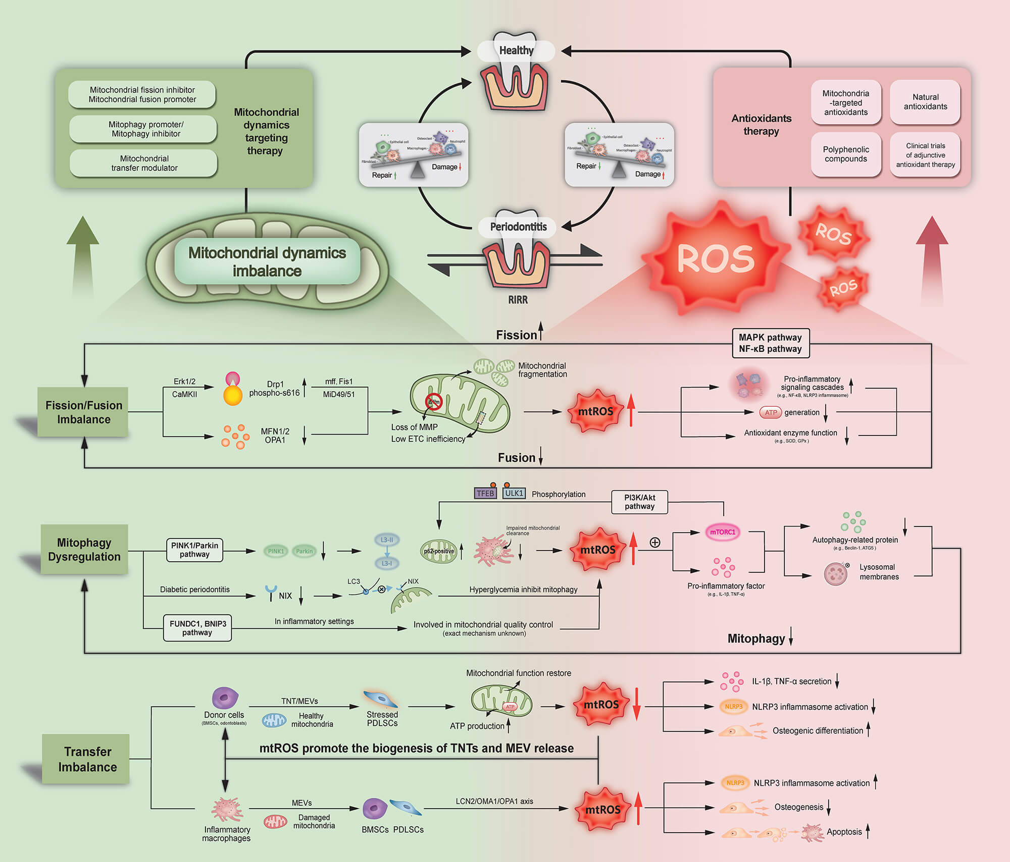
Periodontitis is a prevalent inflammatory disease characterized by the progressive destruction of tooth-supporting tissues. Its pathogenesis remains incompletely understood, but accumulating evidence highlights mitochondrial dynamics imbalance and oxidative stress as interconnected drivers. However, comprehensive reviews elucidating the molecular basis of this interaction are lacking. Therefore, this review aims to clarify the crosstalk between mitochondrial dynamics dysregulation and oxidative stress, and how this bidirectional interaction contributes to periodontal tissue destruction. This review first provides an overview of mitochondrial dynamics and the mechanisms of oxidative stress. We then contextualize these processes within periodontitis, detailing the dual role of reactive oxygen species (ROS), alongside imbalances in mitochondrial dynamics. Crucially, the review demonstrates how dysregulated mitochondrial dynamics, including excessive mitochondrial fission, impaired fusion, and defective mitophagy, lead to mitochondrial fragmentation and mitochondrial ROS (mtROS) overproduction, thereby amplifying inflammatory signaling. Conversely, sustained oxidative stress directly damages periodontal tissues and impairs mitochondrial quality control mechanisms, establishing a vicious cycle of mitochondrial dysfunction and ROS accumulation. Finally, we summarize emerging therapeutic strategies targeting mitochondrial dynamics and oxidative stress pathways. This review provides a deeper understanding of periodontitis pathogenesis and highlights this approach as a promising therapeutic paradigm.Graphic Abstract
Keywords
Periodontitis is a prevalent inflammatory disease affecting the supporting structures of the teeth, including the periodontal ligament and alveolar bone. It is primarily driven by the accumulation of pathogenic biofilms, commonly known as dental plaque, which triggers an inflammatory response in susceptible individuals [1,2]. However, increasing evidence indicates that the severity of tissue destruction depends not only on bacterial burden but also on the magnitude of host inflammatory and oxidative responses. This response leads to the progressive destruction of the periodontal tissues, resulting in tooth loss if left untreated. In addition, periodontitis is associated with many chronic diseases and conditions that affect overall health, including cardiovascular disease, diabetes, cognitive impairment, etc. [2,3]. However, the pathogenesis of periodontitis has not yet been fully elucidated and has become a current research hotspot.
Moreover, oxidative stress and mitochondrial dynamic abnormalities are crucial in periodontitis development. In periodontitis patients, the levels of oxidative stress biomarkers are elevated in both periodontal tissues and gingival crevicular fluid, signifying a close correlation between oxidative stress and periodontal tissue destruction [4]. Oxidative stress exacerbates the pathogenesis of periodontitis via multiple mechanisms. Excessive reactive oxygen species (ROS) directly damage periodontal cells and extracellular matrix, leading to tissue destruction, while mitochondria have been identified as a major intracellular source of pathological ROS under inflammatory conditions. ROS also act as signaling molecules to activate transcription factors like nuclear factor-κB (NF-κB), boosting inflammatory cytokine expression, worsening inflammation, and aggravating periodontal tissue damage [5]. Furthermore, reduced antioxidant enzyme activity in periodontitis patients compromises antioxidant capacity, intensifying oxidative stress [6].
Mitochondria are organelles with remarkable plasticity and dynamics, adapting to and responding to various intracellular stressors and metabolic demands, thereby playing a central role in cellular redox homeostasis. These characteristics enable them to effectively coordinate diverse cellular functions. Among them, mitochondrial dynamics, including fission, fusion, mitophagy, and transfer, are crucial for optimal signal transduction and metabolic function [7]. Imbalances in mitochondrial dynamics can disrupt mitochondrial function, leading to abnormal cell fate and a range of diseases [7,8]. In periodontitis, the levels of mitochondrial biogenesis-related proteins like peroxisome proliferator-activated receptor gamma coactivator 1α (PGC-1α) in periodontal ligament stem cells (PDLSCs) are downregulated, causing a reduction in mitochondrial numbers and significant mitochondrial dysfunction [9], which may further enhance mtROS production. Moreover, mitochondrial dysfunction can also impair the function of gingival fibroblasts and macrophages, thereby exacerbating periodontitis [10].
Such a connection was discovered in the in-depth research: oxidative stress and inflammatory response induced by mitochondrial dysfunction are one of the important mechanisms of periodontitis, and oxidative stress will further aggravate mitochondrial dysfunction in periodontitis [11]. This bidirectional reinforcement suggests the existence of a self-amplifying pathogenic loop between mitochondrial dysfunction and oxidative stress. At present, there is a certain research foundation on the connection among these three, but the specific mechanism still needs to be explored in depth and lacks an integrated mechanistic synthesis [12]. In addition, adjunctive periodontal therapies, including chlorhexidine-based antimicrobial regimens, have recently been reported to influence oxidative stress status in periodontal tissues, further emphasizing the clinical relevance of host-microenvironment interactions in periodontitis [13]. These findings further support the importance of adjunctive antimicrobial strategies in modulating the periodontal microenvironment beyond conventional mechanical debridement.
Although previous studies have addressed oxidative stress or mitochondrial dysfunction separately, a systematic integration of mitochondrial dynamics with oxidative stress in periodontitis is still lacking. This review aims to explore the interaction mechanisms between mitochondrial dynamics and oxidative stress in periodontitis, gain a deeper understanding of the pathogenesis of periodontitis, and provide new ideas for the diagnosis, condition assessment, prognosis judgment, and treatment of periodontitis. Specifically, we aim to provide an integrated mechanistic framework linking mitochondrial dynamics and oxidative stress and to clarify their bidirectional molecular crosstalk as a potential therapeutic target. Unlike previous reviews that have discussed oxidative stress or mitochondrial dysfunction independently, this review integrates mitochondrial dynamics and oxidative stress into a unified interaction mechanism in periodontitis, highlighting their bidirectional regulatory network and potential translational relevance.
This narrative review was based on a focused literature search to identify relevant studies addressing mitochondrial dynamics and oxidative stress in periodontitis. Electronic databases, including PubMed, Web of Science, and Scopus, were searched for articles published up to December 2025. Search terms included combinations of the following keywords: “periodontitis”, “mitochondrial dynamics”, “mitochondrial fission”, “mitochondrial fusion”, “mitophagy”, “mitochondrial transfer”, “oxidative stress”, “reactive oxygen species” and “ROS”.
Priority was given to original research articles and high-quality review articles that provided mechanistic insights into mitochondrial function and redox regulation in periodontal disease. Both in vitro, in vivo, and clinical studies were included. Additional relevant publications were identified through manual screening of the reference lists of selected articles.
Inclusion criteria comprised studies focusing on the molecular mechanisms of mitochondrial dynamics, oxidative stress pathways, or their interaction in the context of periodontitis or related inflammatory conditions. Studies published in English and those providing clear mechanistic or therapeutic relevance were preferentially selected. Exclusion criteria included studies lacking relevance to periodontal disease, studies without mechanistic insight into mitochondrial function or oxidative stress, duplicate publications, and articles with insufficient methodological clarity.
Given the narrative nature of this review, the literature selection was not restricted by a formal systematic review protocol. This review aims to synthesize and critically integrate current evidence rather than provide an exhaustive systematic analysis.
3 Fundamentals of Mitochondrial Dynamics and Oxidative Stress
3.1 Overview of Mitochondrial Dynamics
Mitochondrial dynamics refer to the continuous processes of mitochondrial fission and fusion, as well as mitochondrial movement, mitophagy, biogenesis, and mitochondria transfer. The relative balance of these dynamic mitochondrial processes is crucial for cellular energy metabolism, signaling, apoptosis, proliferation, differentiation, calcium homeostasis, and innate immunity. It is a vital foundation for maintaining cellular homeostasis [13].
The process of mitochondrial fission involves the splitting of a single mitochondrion into two or more smaller organelles. This is often triggered by factors such as increased energy demand or cellular stress. Fission allows for the redistribution of mitochondria to regions of the cell requiring more energy, and also facilitates the removal of damaged mitochondria [14]. Mitochondrial fission is driven by dynamin-related protein 1 (Drp1) recruited via mitochondrial fission 1 (Fis1) and mitochondrial fission factor (Mff), etc. These proteins are recruited from the cytosol to the outer mitochondrial membrane, then assemble into constrictive spirals and sever the organelle [15]. Drp1’s activity is regulated by post-translational modifications such as phosphorylation, ubiquitination, and O-GlcNAcylation [16,17]. Conversely, fusion is the merging of two mitochondria into a single larger organelle, which can help maintain mitochondrial function by allowing the exchange of mitochondrial DNA, proteins, and metabolites. It is particularly important in maintaining mitochondrial function under stress, as it helps in diluting damaged components, facilitating restoration of organelle function, and reducing cellular injury [8]. Fusion is mediated by mitofusins (MFN1/2) on the outer membrane and optic atrophy 1 (OPA1) on the inner membrane, which tether and merge adjacent mitochondria [18]. The delicate balance between mitochondrial fission and fusion is essential for maintaining mitochondrial DNA integrity, ensuring proper distribution of metabolic substrates, and triggering apoptotic signaling when damage is beyond repair [14,19].
Mitophagy is a special form of autophagy that selectively targets damaged or dysfunctional mitochondria for degradation, and it is critical for mitochondrial quality control. This process plays a role in clearing defective mitochondria and preventing their accumulation, which can lead to cellular damage and disease [7]. Mitophagy is primarily regulated by the PTEN-induced kinase 1 (PINK1)/Parkin pathway. PINK1, an autophagy-related gene, mediates the canonical pathway of mitophagy. Upon loss of mitochondrial membrane potential, PINK1 stabilizes on the outer mitochondrial membrane, where it phosphorylates ubiquitin and the E3 ligase Parkin, activating Parkin’s ligase activity. This leads to the ubiquitination of mitochondrial surface proteins and the recruitment of autophagy receptors such as p62 and optineurin that bridge to microtubule-associated protein 1 light chain 3 (LC3), facilitating the formation of autophagosomes. These autophagosomes then fuse with lysosomes for the degradation of the damaged mitochondria [20]. There are also Parkin-independent pathways, where PINK1 or other E3 ligases directly recruit LC3 adapters, providing redundancy in the mitochondrial quality control mechanisms [21,22]. Efficient mitophagy limits oxidative stress, preserves ATP production, and prevents the accumulation of dysfunctional mitochondria [22].
Mitochondrial transfer refers to the process by which mitochondria move from one cell to another via specialized routes and plays a pivotal role in both physiological and pathological contexts. Under normal conditions, transfer of functional mitochondria helps maintain tissue homeostasis by replenishing bioenergetic capacity in recipient cells [23]. Under disease conditions, mitochondrial transfer can restore function in stressed or damaged cells, thereby ameliorating tissue injury caused by mitochondrial dysfunction [24]. The main routes of mitochondrial transfer include the following types. Tunnel nanotubes (TNTs), in which mitochondria are transported along microtubules through open membrane channels formed by cellular protrusions [25]. Extracellular vesicles (EVs), in which mitochondria or mtDNA are encapsulated, are secreted and taken up by neighboring cells [26,27]. And mitochondria-derived vesicles (MDVs), small vesicles budding from the mitochondrial outer membrane, carry selected mitochondrial components to lysosomes or the extracellular milieu [28]. Additionally, large vesicular structures formed during cell migration, called migrasomes, can encapsulate and transfer mitochondria when they rupture or are internalized by recipient cells [29]. Finally, naked mitochondria may be released and subsequently internalized by neighboring cells via exocytosis and endocytosis processes [30].
Mitochondrial dynamic imbalance often manifests as defects in fission, fusion, mitophagy, and transfer. This dysfunction is closely associated with numerous diseases and pathological processes, including neurodegenerative disorders, cardiovascular diseases, muscular system diseases, bone system diseases, cancers, and inflammatory conditions [31]. For example, in neurodegenerative diseases such as Parkinson’s and Alzheimer’s, excessive mitochondrial fission or impaired mitophagy accelerates neuronal death [32,33]. In cardiovascular diseases, mitochondrial abnormalities affect cardiomyocyte function [34,35]. Mitochondrial fusion dysfunction can lead to muscle atrophy and decreased motor ability, as well as the imbalance of mitochondrial dynamics, which affects the functions of osteoblasts and osteoclasts, resulting in reduced bone mass and destruction of bone structure [36,37]. In cancer, cells acquire rapid proliferation capabilities through abnormal mitochondrial fission [38]. And in inflammation, mitochondrial dysfunction activates immune responses, exacerbating inflammation [39]. Therefore, regulating mitochondrial dynamics has become an emerging direction for treating related diseases.
3.2 Generation and Pathogenic Mechanisms of Oxidative Stress
Reactive oxygen species, including superoxide anion (O2−), hydrogen peroxide (H2O2), and hydroxyl radical (•OH), function dually within cells as both signaling molecules and potential cytotoxins [40]. ROS are generated through enzymatic pathways such as mitochondrial electron transport chain (ETC) activity, particularly electron leakage at complexes I and III, leading to superoxide formation, as well as via NADPH oxidases (NOX), which transfer electrons from NADPH to molecular oxygen to produce O2−. Additional enzymatic sources and non-enzymatic processes also contribute to ROS production [41].
In order to keep ROS within the normal range and maintain homeostasis, organisms regulate ROS through an antioxidant network known as the oxidative defense system. This system is composed of substances with concentrations lower than those of oxidizable substrates and includes endogenous antioxidant enzymes such as superoxide dismutase (SOD) and low-molecular-weight scavengers like vitamins [42]. Under physiological conditions, the production and elimination of intracellular ROS are dynamically balanced through various pathways, thereby maintaining ROS levels at relatively low concentrations [43]. Low to moderate levels of ROS act as critical second messengers, modulating cellular processes including proliferation, differentiation, migration, and immune responses. This controlled ROS signaling is essential for maintaining cellular homeostasis and function [42,44].
When ROS production exceeds antioxidant capacity, the resultant redox imbalance leads to oxidative stress. This is possibly due to upregulated NOX and xanthine oxidase (XO) activities, mitochondrial respiratory chain dysfunction increasing electron leakage, or exogenous stressors like ultraviolet radiation and toxins [45,46]. Concurrent impairment of endogenous defenses, including downregulation of endogenous antioxidant enzymes as well as depletion of non-enzymatic antioxidants, further exacerbates ROS accumulation [40]. Persistent oxidative stress provokes lipid peroxidation, protein carbonylation, DNA strand breaks, and activation of redox-sensitive transcription factors such as nuclear factor erythroid 2-related factor 2 (Nrf2), thereby triggering inflammatory cascades and cell death pathways [47,48]. Moreover, excessive ROS production can activate several key signaling pathways that exacerbate inflammation and cellular damage. One of the most prominent is the NF-κB pathway, which translocates to the nucleus upon activation and induces the transcription of pro-inflammatory cytokines and adhesion molecules [49]. Additionally, ROS can activate mitogen-activated protein kinase (MAPK) pathways, leading to the phosphorylation of transcription factors, which further promote inflammatory responses. These signaling cascades cause the amplification of oxidative stress and the progression of various inflammatory diseases [41].
Excessive oxidative stress plays a pivotal role in the initiation and progression of various chronic diseases, including cardiovascular diseases, neurodegenerative disorders, metabolic syndrome, inflammation, and cancers [40,50]. Indeed, excessive oxidative stress is a hallmark of periodontitis, contributing significantly to its pathogenesis. Elevated levels of ROS in periodontal tissues lead to inflammation, connective tissue degradation, and alveolar bone resorption. This oxidative imbalance exacerbates tissue damage and perpetuates the disease process. The mechanisms by which oxidative stress influences periodontitis will be elaborated in detail in the next section.
4 Interplay between Mitochondrial Dynamics and Oxidative Stress in Periodontitis
4.1 Oxidative Stress in Periodontitis
In periodontal lesions, bacterial endotoxins trigger an excessive neutrophil respiratory burst and activate macrophages; dental plaque and infiltrating inflammatory cells continuously release large amounts of ROS. At the same time, the host’s antioxidant defenses become impaired, reducing ROS clearance capacity. This imbalance allows ROS to oxidatively damage proteins, lipids, and DNA [6]. ROS also promotes activation of matrix metalloproteinases (MMPs) and the release of proinflammatory cytokines, exacerbating periodontal tissue destruction and alveolar bone resorption [51,52]. Clinical and basic studies have shown that oxidative stress has emerged as a key factor in the onset and progression of periodontitis [47]. In this section, the role of ROS in periodontitis will be described in detail.
4.1.1 Dual Role of ROS in Periodontitis
ROS act as a double-edged sword in periodontal homeostasis, exerting both protective and destructive effects depending on their concentration and context [53,54]. At low concentrations (picomolar to nanomolar), ROS generated by polymorphonuclear neutrophils (PMNs) and macrophages are indispensable for the clearance of pathogenic microorganisms and the coordination of wound-healing processes within periodontal tissues [54]. Moreover, ROS serve as critical signaling molecules that drive fibroblast proliferation, stimulate angiogenesis, and modulate immune responses, thereby promoting effective tissue repair and regeneration [6]. However, under persistent chronic inflammatory stimulation, dental plaque and subgingival Gram-negative bacteria continually challenge the host, and PMNs and macrophages become hyperactivated. They generate excessive ROS that overwhelm endogenous antioxidant defenses, disrupt redox homeostasis, and mediate pathological damage to periodontal tissues. Thus, ROS act as essential physiological mediators in periodontitis, yet also serve as pathological factors that drive chronic inflammation and tissue destruction [5,55]. This concentration-dependent duality of ROS in periodontal homeostasis and disease progression is illustrated in Fig. 1.
Excessive ROS can directly damage periodontal tissues through multiple mechanisms. First, ROS oxidatively activate MMPs, accelerating extracellular matrix degradation. Second, ROS induces lipid peroxidation, which compromises cell membrane integrity. Third, ROS causes oxidative damage to DNA and proteins, leading to cell apoptosis or loss of function. Fourth, ROS triggers the collapse of the mitochondrial membrane potential, provoking a respiratory burst and disrupting energy metabolism [6,56]. Clinical studies have shown that levels of lipid peroxidation markers such as malondialdehyde (MDA) are significantly elevated in the gingival crevicular fluid of periodontitis patients. These MDA levels correlate positively with probing depth and bleeding index. This finding indicates that ROS-mediated direct oxidative damage is closely associated with periodontal tissue destruction [57,58].
Beyond direct oxidative damage, excessive ROS also acts as a second messenger. They induce indirect injury to periodontal tissues by modulating signaling pathways and transcription factors that govern immune and inflammatory responses. Excessive ROS activates the NF-κB signaling pathway. Activated NF-κB drives transcription of genes encoding pro-inflammatory mediators such as interleukin-1β (IL-1β), tumour necrosis factor-α (TNF-α), and MMP-9 [59]. Receptor activator of nuclear factor-κB ligand (RANKL) signaling pathway becomes activated under chronic inflammatory conditions, and it contributes to osteoclast maturation and differentiation, ultimately leading to alveolar bone resorption [60]. Meanwhile, ROS directly stimulates the phosphorylation of MAPK family members, including c-Jun N-terminal kinase (JNK) and p38. This activation induces apoptosis in fibroblasts and epithelial cells and compromises the integrity of the periodontal attachment complex [59,61]. Excessive generation of ROS and oxidative stress also exacerbates periodontal tissue injury via assembly of inflammasomes. Clinical investigations have demonstrated that increased expression of IL-1β and IL-18 in gingival tissues and gingival crevicular fluid of periodontitis patients positively correlates with elevated levels of nucleotide-binding oligomerization domain (NOD)-like receptor family pyrin domain-containing 3 (NLRP3) mRNA in oral epithelial cells and saliva [62]. The NLRP3 inflammasome is a cytosolic multiprotein complex comprising NLRP3, the adaptor molecule apoptosis-associated speck-like protein containing CARD (ASC), and pro-caspase-1 [63]. Upon activation via a two-step mechanism, the NLRP3 inflammasome cleaves pro-caspase-1 to its active form, which in turn processes and promotes secretion of mature IL-1β and IL-18. This cascade establishes a feed-forward loop that perpetuates periodontal inflammation and tissue destruction [64].

Figure 1: The dual role of ROS in periodontal homeostasis and pathogenesis. This figure contrasts the concentration-dependent effects of ROS in periodontal tissues. At low physiological levels, ROS contribute to the direct clearance of pathogenic bacteria and modulate signaling pathways. This regulated signaling modulates immune responses, promotes tissue repair, and maintains homeostasis. Conversely, in the chronic inflammatory state of periodontitis, high pathological levels of ROS overwhelm antioxidant defenses, driving tissue destruction through two main routes. The direct pathway involves indiscriminate damage to macromolecules and leads to direct oxidative damage to cells and the extracellular matrix. The indirect pathway involves the activation of destructive signaling cascades. Excessive ROS dysregulates protective mechanisms and exacerbates inflammation, culminating in periodontal tissue destruction and alveolar bone loss. Figure created using Adobe Illustrator 2020 (Adobe Inc., San Jose, CA, USA).
4.1.2 Suppression of Antioxidant Defense
Beyond activation aspects, oxidative stress can also inhibit the transcription and expression of key factors associated with periodontal disease tolerance. The body’s antioxidant defense primarily relies on the Nrf2-antioxidant response element (ARE) pathway. Nrf2 is a basic leucine zipper transcription factor, and the Nrf2-ARE signaling pathway robustly regulates the expression of endogenous antioxidant enzyme genes [65]. Under physiological conditions, Nrf2 is restrained in the cytoplasm and degraded by Kelch-like ECH-associated protein 1 (Keap1), maintaining acceptable Nrf2 levels and preventing unnecessary transcription of antioxidant genes. Under oxidative stress, Nrf2 dissociates from Keap1, translocates into the nucleus, and binds to ARE, transcriptionally upregulating antioxidant enzymes such as heme oxygenase 1 (HO-1), NAD(P)H quinone dehydrogenase 1 (NQO1), and glutamate-cysteine ligase catalytic subunit (GCLC). This process scavenges excess ROS and inhibits inflammatory signaling [66]. In patients with chronic periodontitis, the nuclear translocation of Nrf2 and the expression of its downstream genes are significantly downregulated. This leads to a decrease in the activity of enzymes such as SOD and Glutathione peroxidase (GPx), compromises the antioxidant capacity, and prevents effective inhibition of excessive ROS accumulation [66]. A rat study revealed that the gene and protein expression of Nrf2 was significantly downregulated in diabetic periodontitis, and reduced Nrf2 expression was strongly negatively correlated with aggravated periodontal destruction and the extent of oxidative damage, suggesting that Nrf2 dysfunction may be one of the crucial factors contributing to diabetes-aggravated periodontitis [67].
4.2 Mitochondrial Dynamics Imbalance in Periodontitis
Mitochondrial dynamics, including the finely tuned coordination of fusion, fission, mitophagy, and transfer, are indispensable for organellar quality control, bioenergetic homeostasis, and overall cellular equilibrium. In periodontitis, chronic microbial challenge and sustained inflammatory signaling disrupt this balance, precipitating alterations in mitochondrial dynamics. Consequently, mitochondrial dysfunction not only perturbs cellular homeostasis but also contributes to the periodontal inflammatory response through aberrant release of mitochondrial contents, thereby impacting both disease progression and tissue health [56]. The major alterations in mitochondrial dynamics in periodontitis are schematically summarized in Fig. 2.

Figure 2: Pathogenic alterations in mitochondrial dynamics during periodontitis. The balance of mitochondrial dynamics is skewed towards excessive Drp1-mediated fission and impaired MFN/OPA1-mediated fusion, resulting in mitochondrial fragmentation and pro-inflammatory signaling, which intensify this imbalance. Concurrently, mitophagy is suppressed via inhibition of the canonical PINK1/Parkin pathway and autophagy receptor NIX. Pro-inflammatory signals are also present in the periodontitis microenvironment, hyperactivating the PI3K/Akt/mTORC1 axis, which acts as a master inhibitor of autophagy. This multifaceted suppression of mitophagy intensifies bone metabolism disorders and periodontal tissue destruction. In addition, intercellular mitochondrial transfer presents a dual role: restorative transfer of healthy mitochondria from BMSCs via TNTs can enhance osteogenesis, whereas pathogenic transfer of damaged mitochondria from inflammatory macrophages via MEVs propagates cellular dysfunction and promotes alveolar bone resorption. However, in the inflammatory microenvironment of periodontitis, the pathogenic transfer predominates. Figure created using Adobe Illustrator 2020 (Adobe Inc., San Jose, CA, USA).
4.2.1 Mitochondrial Fission/Fusion Imbalance in Periodontitis
Numerous studies have demonstrated an imbalance in mitochondrial fission and fusion dynamics in periodontitis. Experimental models treating PDLSCs with hydrogen peroxide reveal upregulation of the fission mediators Drp1 and Fis1, with downregulation of the fusion proteins Mfn1 and Mfn2 and proteolytic processing of OPA1. This emphasizes the imbalance between mitochondrial fission and fusion in periodontal inflammation [11].
Recent studies have shown that infection of human endothelial cells with Porphyromonas gingivalis upregulates Drp1 expression and its phosphorylation, enhancing Drp1 recruitment to mitochondria and driving excessive mitochondrial fragmentation in the context of periodontal pathogen challenge [56]. In periodontitis, Drp1 is upregulated and activated via Ser616 phosphorylation through extracellular signal-regulated kinases 1/2 (ERK1/2) or calmodulin-dependent protein kinase II (CaMKII) mediation. Once activated, Drp1 is recruited to the outer membrane via adaptor proteins, which oligomerize around constriction sites to sever the organelle [68]. Among them, the recruitment of Drp1 may be mainly promoted by the receptors MFF and Fis1. Although direct data in periodontal cells have not yet emerged, it has been confirmed that in other mammalian systems, the receptors MFF and Fis1 are essential for anchoring activated Drp1 on the outer membrane, facilitating constriction and scission [69]. In addition to pathogen factors, Nicotine, hypoxia mimic (CoCl2), mechanical strain, and diabetic periodontitis context can also induce the activation of Drp1 through similar mechanisms, leading to mitochondrial fission [68,70,71,72].
Kırmızıgül et al. quantitatively measured MFN1 and MFN2 in gingival crevicular fluid from healthy, gingivitis, and periodontitis patients. They reported that the total MFN1 amount was elevated in disease groups and MFN2 concentrations were markedly reduced compared to controls, while both MFN1 and MFN2 concentrations per unit fluid fell. Such downregulation indicates an early fusion deficit in periodontal inflammation [73]. It also correlates with pronounced mitochondrial fragmentation, elevated reactive oxygen species production, and increased secretion of the proinflammatory cytokines IL-1β and TNF-α by gingival fibroblasts and periodontal ligament cells [18]. OPA1 controls inner-membrane fusion and cristae architecture. At present, although the original studies specifically focusing on the role of OPA1 in intimal fusion in periodontitis are extremely limited, there are still reviews pointing out the possible dysregulation of OPA1 in periodontitis [74].
4.2.2 Mitophagy Dysfunction in Periodontitis
Mitophagy is the selective removal of damaged mitochondria via the autophagic pathway, predominantly regulated by the PINK1/Parkin signaling axis, and is essential for preventing ROS accumulation and inflammasome activation [75]. In healthy periodontal tissues, damaged mitochondria trigger the accumulation of PINK1 on the outer membrane of depolarized mitochondria. PINK1 then recruits Parkin and induces ubiquitination of outer membrane proteins, thereby promoting autophagosome formation [76].
Recent evidence suggests that impaired mitophagy may represent a novel aspect of periodontitis pathology. PINK1-mediated mitophagy impacts disease progression via the mitochondrial-dependent apoptotic pathway. A study comparing gingival tissue specimens from healthy individuals and periodontitis patients revealed significant downregulation of PINK1, Parkin, and LC3 [77]. In ligature-induced periodontitis, after knocking out PINK1, it was also found that a marked reduction in mitophagic flux, evidenced by decreased LC3-II accumulation and increased p62 levels, resulted in impaired clearance of damaged mitochondria and exacerbated alveolar bone loss [76]. The above evidence indicates that in periodontitis, PINK1 and Parkin expression and recruitment are disrupted, leading to impaired mitophagic flux and persistent mitochondrial dysfunction [78]. This damage exacerbates excessive reactive oxygen species production and proinflammatory cytokine release, thereby accelerating periodontal tissue destruction [56]. Furthermore, there are reviews that have emphasized mitophagy’s role in bone metabolism disorders, where it modulates osteoblast apoptosis and osteoclast activation during inflammation-driven bone loss [79]. Studies of single-cell PDLSC clones demonstrate a positive correlation between their osteogenic differentiation capacity and PINK1/Parkin-mediated mitophagy [80]. Mitophagy has been confirmed as a protective mechanism against oxidative damage in bone marrow-derived mesenchymal stem cells (MSCs), promoting their osteogenic differentiation and suggesting a potential avenue for enhancing periodontal regeneration [81].
In addition to the PINK1/Parkin signaling axis, there are other axes that affect mitochondrial autophagy in periodontitis. BCL2/adenovirus E1B 19 kDa interacting protein 3 like (NIX/BNIP3L) is a mitochondrial outer-membrane receptor that directly binds LC3 to trigger mitophagy. Restoration of NIX levels in vitro rescues mitophagic activity and attenuates periodontal ligament cell senescence, underscoring NIX downregulation as a key bottleneck in receptor-mediated mitophagy during periodontitis [82]. In addition, pro-inflammatory cytokines and bacterial ligands activate the phosphatidylinositol 3-kinase/protein kinase B (PI3K/Akt) pathway in gingival fibroblasts and periodontal ligament cells, leading to mechanistic target of rapamycin complex 1 (mTORC1) hyperactivation. Elevated mTORC1 phosphorylates unc-51 like autophagy activating kinase 1 (ULK1) and transcription factor EB (TFEB), thereby inhibiting initiation of both general autophagy and mitophagy [83]. Inhibition of mTOR with rapamycin restores autophagic markers (LC3 puncta) and alleviates alveolar bone loss in periodontitis models, confirming mTOR’s central role in autophagy blockade [84]. Moreover, dimethyl fumarate (DMF) studies reveal that periodontitis-associated macrophages and periodontal tissues exhibit reduced mitophagic flux that can be rescued by boosting lysosomal function and Tu translation elongation factor, mitochondrial(TUFM)-mediated mitophagy [85].
4.2.3 Mitochondrial Transfer in Periodontitis
Beyond intracellular quality control, intercellular mitochondrial transfer via TNTs, mitochondria-enriched extracellular vesicles (MEVs), MDVs, or gap junctions can modulate cell fate in the periodontal microenvironment [86,87]. In periodontal tissues, a bilateral migration of bone marrow mesenchymal stem cells (BMSCs) and PDLSCs has been observed: Healthy BMSCs can donate healthy mitochondria to PDLSCs and improve PDLSC osteogenic differentiation [86,88]. Conversely, inflammatory macrophages package dysfunctional mitochondria into MEVs under inflammatory conditions. MEVs transfer to PDLSCs and BMSCs and damage periodontal health [56]. These dual modes of transfer highlight both the destructive and restorative roles of mitochondrial exchange in periodontal health.
In periodontitis, mitochondrial transfer, which is mainly achieved through MEVs and TNTs, is disordered. BMSCs extend F-actin–rich TNTs to directly shuttle healthy mitochondria into PDLSCs under inflammatory conditions [89]. Activation of cannabinoid receptor 1 (CB1) in MSCs enhances mitochondrial loading into TNTs and increases transfer efficiency, as demonstrated by higher mitochondrial membrane potential and elevated osteogenic markers (ALP, Runx2) in PDLSC after transfer [88]. In addition, inflammatory macrophages in periodontitis internalize and traffic damaged mitochondria into MEVs. The transfer of MEVs to PDLSCs and BMSCs within the periodontal lesion amplifies the production of ROS, inflammasome activation, and matrix degradation through the lipocalin-2 (LCN2)/OMA1 zinc metallopeptidase (OMA1)/OPA1 signal, thereby promoting alveolar bone resorption [56]. Proteomic profiling of macrophage-derived MEVs reveals enrichment of mitochondrial membrane proteins alongside vesicle-trafficking factors, confirming selective mitochondrial packaging [90].
Together, alterations in mitochondrial dynamics, impaired mitophagic clearance, and dysregulated intercellular mitochondrial transfer establish a self-amplifying mitochondrial dysfunction axis that drives oxidative stress and periodontal tissue destruction, as summarized in Fig. 2.
4.3 Molecular Mechanisms of Interaction in Periodontitis
Emerging evidence indicates that mitochondrial dynamics are intimately linked to oxidative stress and inflammatory responses in periodontitis. Disruption of the fission-fusion balance not only favors excess ROS generation via Drp1-mediated fragmentation but also impairs mitochondrial fusion proteins MFN1/MFN2, exacerbating proinflammatory cytokine release in periodontal cells [18,91]. Concurrently, defective PINK1/Parkin-driven mitophagy allows accumulation of dysfunctional mitochondria, amplifying ROS production and NLRP3 inflammasome activation [76]. Furthermore, intercellular mitochondrial transfer via extracellular vesicles or tunneling nanotubes modulates periodontal cell fate, either reducing oxidative damage caused by excessive ROS through healthy organelle donation or propagating dysfunction when damaged mitochondria are exchanged [90]. Moreover, the burst of ROS can also trigger a self-amplifying cycle known as ROS-induced ROS release (RIRR), where ROS itself directly or indirectly damages mitochondrial components (like electron transport chain complexes or inducing mitochondrial permeability transition pore opening), thereby activating a positive feedback loop that generates even more ROS. Such sustained RIRR profoundly escalates the cellular oxidative burden and contributes to inflammation, further exacerbating periodontitis [92]. Beyond local inflammatory and microbial stimuli, systemic metabolic disturbances may further modulate this redox-mitochondrial feedback network [93]. Clinical and experimental evidence indicates that hyperglycemia and obesity-associated oxidative burden exacerbate mitochondrial dysfunction and ROS accumulation, potentially intensifying periodontal tissue injury [78,94]. These findings suggest that the interaction between mitochondrial dynamics and oxidative stress may also be influenced by systemic metabolic context [95]. Despite the growing evidence linking mitochondrial dynamics and oxidative stress in periodontitis, several limitations remain. First, most current findings are derived from in vitro or animal models, and robust clinical validation is still lacking. Second, the dual role of ROS in both physiological signaling and pathological damage complicates the interpretation of experimental outcomes and therapeutic targeting strategies. Third, heterogeneity in experimental models and periodontal microenvironments may lead to inconsistent mechanistic conclusions across studies. Finally, the safety, specificity, and long-term efficacy of mitochondria-targeted interventions in periodontal therapy remain largely unexplored. Future studies integrating multi-omics approaches and well-controlled clinical trials are needed to clarify these issues.
4.3.1 Mitochondrial Fission/Fusion Imbalance and ROS Overproduction
Research has delineated how an imbalance in mitochondrial fission and fusion profoundly drives ROS overproduction in periodontitis. Excessive activation of the fission machinery, primarily via the upregulation of Drp1 coupled with downregulation of fusion mediators (MFN1/MFN2/OPA1), leads to mitochondrial fragmentation, loss of membrane potential and ETC inefficiency, all of which amplify mtROS generation. This mtROS surge activates pro-inflammatory signaling cascades (e.g., NF-κB, NLRP3 inflammasome) and a vicious cycle of oxidative stress, and promotes apoptosis in periodontal cells, thereby exacerbating tissue breakdown [18,91].
Drp1-mediated fission drives mtROS overproduction. In diabetic rat periodontitis, mitochondrial swelling and fragmentation correlate with a sharp increase in ROS and decreased MnSOD activity, underscoring the pathogenic role of fission-induced oxidative stress [78]. Studies in periodontitis models demonstrate that pro-inflammatory stimuli like P. gingivalis lipopolysaccharide (LPS) activate MAPK and NF-κB pathways, leading to Drp1 phosphorylation at Ser616 by kinases such as ERK1/2 and CaMKII. The subsequent mechanism is that Drp1 oligomerizes on the outer mitochondrial membrane through the adaptor proteins mff, Fis1, MiD49 and mid51, driving fragmentation that diminishes membrane potential and impairs proton motive force, thereby increasing electron leakage at complexes I and III and elevating superoxide production. The resulting organelle fragmentation disrupts ETC integrity and elevates mtROS levels [15,91]. Elevated ROS in turn acts upon the core fission machinery to reinforce fragmentation and inhibit fission, creating a self-amplifying ROS-Drp1 feedback loop. ROS acts on MAPKs (such as ERK1/2), promoting phosphorylation at the Ser616 site of Drp1 and enhancing its GTPase activity and mitochondrial recruitment [91,96]. Studies have demonstrated that the expression of Drp1 and its phosphorylated form Ser616 is induced by H2O2, indicating that ROS induces mitochondrial dysfunction by activating DRP1-mediated mitochondrial division [91].
And fusion protein downregulation exacerbates oxidative injury. Concomitant with fission upregulation, expression of MFN1, MFN2 and OPA1 is markedly reduced in gingival fibroblasts and periodontal ligament cells from both experimental and patient-derived samples. This loss of fusion capacity impairs mitochondrial networking, preventing dilution of damaged components and promoting ROS accumulation. A recent study reported MFN1/MFN2 downregulation alongside enhanced IL-1β secretion and ROS overproduction in periodontal ligament stem cells exposed to oxidative stress [18]. Moreover, decreased mitochondrial fusion reduces ATP generation, impairing antioxidant enzyme function such as SOD and GPx, and further tipping the redox balance toward oxidative stress [97]. Elevated ROS not only damages lipids, proteins, and DNA in periodontal cells but also exacerbates mitochondrial fusion disorders. Mitochondrial fusion could not be observed in cells under oxidative stress triggered by high-fluence low-power laser irradiation (HF-LPLI). This indicates that oxidative stress leads to the inhibition of the mitochondrial fusion process [98]. ROS scavenger N-acetylcysteine (NAC) partially rescued MFN1/MFN2 expression and mitochondrial network integrity in H2O2-treated hPDLCs, confirming an ROS-dependent mechanism [18]. ROS-activated transcription factors like NF-κB repress MFN2 expression and inhibit its GTPase activity, curtailing fusion capacity [98]. Through these reciprocal modifications, where ROS both results from and drives further mitochondrial fragmentation, cells can enter a vicious cycle of oxidative stress and organelle dysfunction, thereby aggravating the disease [7].
4.3.2 Dysregulated Mitophagy and Oxidative Stress in Periodontitis
Recent investigations have revealed that dysregulation of mitophagy exacerbates oxidative stress in periodontitis by permitting the accumulation of dysfunctional organelles and perpetuating ROS overproduction [99]. In periodontitis, the mitophagy flux is blocked or insufficient, leading to the accumulation of damaged mitochondria and the production of excessive ROS and forming a vicious cycle of “mitophagy dysregulation-oxidative stress”, which aggravates the destruction of periodontal tissues [76]. The core mechanisms include functional defects of the PINK1/Parkin pathway, obstruction of receptor-mediated autophagy (such as down-regulation of BNIP3L/NIX), excessive activation of mTOR inhibiting autophagy, and disorders of lysosomal biogenesis blocking the completion of autophagy flux.
In periodontitis, the PINK1/Parkin signaling pathway is impaired. PINK1 expression is significantly reduced in gingival tissues and osteoclasts, and Parkin translocation to mitochondria is blunted, leading to reduced LC3-II conversion and accumulation of p62-positive mitochondria, resulting in the disorder of mitophagy function [76]. Jang et al. demonstrated that PINK1 deficiency in osteoclasts exacerbates bone loss in experimental periodontitis by impairing mitophagy and promoting oxidative damage [76]. When autophagy is impaired, damaged mitochondria accumulate, escalating ROS release and further inhibiting autophagy via oxidative damage to autophagy related gene (ATG) proteins and lysosomal membranes. Meanwhile, ROS-induced NF-κB activation boosts mTORC1 activity, sustaining autophagy blockade and perpetuating inflammatory and oxidative stress, driving progressive tissue destruction. Excessive ROS also inhibits autophagy-related proteins (Beclin-1, ATG5) through pro-inflammatory factors (IL-1β, TNF-α), further suppressing autophagic flux [75,100]. It has been reported that overexpression of PINK1 can reduce the level of mtROS, while inhibition of PINK1 activity significantly increases the level of mtROS in rat PDLSCs, highlighting the feedback loop between mtROS production and mitochondrial autophagy regulation [101].
Beyond PINK1/Parkin, pathogen- and cytokine-driven PI3K/Akt signaling also leads to mTORC1 hyperactivation, which phosphorylates ULK1 and sequesters TFEB in the cytosol in periodontitis, thereby preventing the initiation of autophagy and driving an increase in ROS production in gingival cells [99]. In turn, these excess ROS further stimulate PI3K/Akt/mTORC1 activity and oxidatively impair core autophagy components (e.g., Beclin-1, ATG proteins), creating a feed-forward loop of autophagy inhibition and oxidative stress that exacerbates inflammation and periodontal tissue destruction [102]. In addition, ROS also inhibits autophagy flux by targeting beclin 1 and induces autophagy by activating the ATG12-ATG5 complex [102].
Receptor-dependent mitophagy pathways—such as those mediated by FUN14 domain-containing protein 1 (FUNDC1) and BCL2 interacting protein 3 (BNIP3), also contribute to mitochondrial quality control in inflammatory settings [103]. Although most studies to date have focused on diabetic complications, FUNDC1-mediated mitophagy has been shown to inhibit oxidative stress and inflammatory responses in endothelial cells, suggesting that similar mechanisms may operate in periodontal tissues under chronic bacterial challenge. Further research is needed to elucidate the roles of these alternative receptors in periodontitis [104]. In addition, as a receptor-mediated mitophagy, NIX/BNIP3L is down-regulated in the diabetic periodontitis model, resulting in the obstruction of LC3 binding to mitochondria and the reduction of autophagosome formation [82]. Concurrently, metabolic stressors such as hyperglycemia further inhibit mitophagy and amplify ROS generation. Hyperglycemia is a common comorbidity in periodontitis patients. A recent in vitro study reported that high glucose conditions reduce PINK1 stabilization and Parkin recruitment, thereby blocking mitophagic flux and amplifying mitochondrial ROS production in neuronal cells. This effect may be translated as periodontal cells in diabetic periodontitis [94]. This metabolic interference creates a vicious cycle wherein elevated ROS exacerbates mitochondrial damage, leading to even greater oxidative injury and inflammatory mediator release.
4.3.3 Role of Mitochondrial Transfer in Modulating Oxidative Damage in Periodontitis
Recent studies underscore mitochondrial transfer as a critical modulator of oxidative stress in the periodontal microenvironment. By exchanging mitochondria between donor and recipient cells, tissues can either ameliorate or exacerbate ROS-mediated injury, depending on the quality of transferred organelles. Both TNTs and MEVs have been identified as principal conduits for mitochondrial exchange in periodontitis. In periodontitis, intercellular mitochondrial transfer mediated by it plays a dual role in modulating oxidative balance within the periodontal microenvironment [90].
On one hand, Healthy mitochondria are transferred from donor cells to stressed PDLSCs. It can restore mitochondrial function, reduce ROS production, reduce secretion of IL-1β and TNF-α, and enhance the antioxidant defense system, thereby mitigating oxidative stress and promoting tissue regeneration [88,105]. Mitochondria are shuttled between periodontal cells primarily via TNTs and F-actin-rich membranous channels that permit direct organelle movement along microtubules. In odontoblasts, TNT-mediated delivery of healthy mitochondria rescues mitochondrial membrane potential, reduces mtROS accumulation, and limits NLRP3 inflammasome activation, thereby preventing pyroptotic cell death [105]. Similarly, BMSCs form TNTs toward PDLSCs, transferring mitochondria that restore ATP production, decrease intracellular ROS, and enhance osteogenic differentiation in inflammatory conditions [88]. In parallel, EVs, including exosomes and microvesicles, encapsulate mitochondrial fragments or whole organelles and shuttle them through the extracellular space. Forkhead box O1 (FoxO1)-overexpressing small EVs derived from BMSCs have been shown to regulate ROS levels and mitochondrial dynamics in PDLSCs, promoting osteogenesis while polarizing macrophages toward an anti-inflammatory M2 phenotype [106]. On the other hand, the transfer of damaged or dysfunctional mitochondria, particularly from activated immune cells like macrophages, can exacerbate oxidative stress in recipient cells. Inflammatory macrophages release EVs rich in damaged mitochondria. After being taken up by PDLSCs and BMSCs, these EVs intensify ROS production through the LCN2/OMA1/OPA1 axis, activate the NLRP3 inflammasome, promote apoptosis or bone resorption and damage osteogenesis [90,107].
Moreover, oxidative stress itself promotes the biogenesis of TNTs and MEV release, creating feedback loops that either worsen tissue damage or support repair, depending on donor cell identity. Oxidative stress and inflammation upregulate TNF alpha induced protein 2 (TNFAIP2) and Rho GTPases, which drive actin polymerization and TNT formation, thereby facilitating intercellular mitochondrial exchange [108]. Similarly, ROS-dependent activation of endosomal sorting complex required for transport (ESCRT) complexes boosts MEV biogenesis, increasing the release of mitochondria-laden vesicles from macrophages and other immune cells [109]. These feedback mechanisms mean that heightened ROS both results from and promotes further mitochondrial transfer, amplifying either tissue damage or repair depending on the context. Collectively, these reciprocal interactions establish a self-perpetuating redox-mitochondrial feedback network that drives progressive periodontal destruction, as summarized in Fig. 3.

Figure 3: Molecular feedback loops between mitochondrial dynamic imbalance and ROS overproduction in periodontitis. An imbalance favoring fission over fusion leads to mitochondrial fragmentation and subsequent inefficiency in the electron transport chain, resulting in elevated mtROS. Crucially, by activating pro-fission signaling cascades such as the MAPK/NF-κB pathway, this mtROS surge establishes a positive feedback loop (ROS-Induced ROS Release, RIRR). Similarly, the failure of mitophagy pathways leads to the accumulation of mtROS-producing organelles. This creates a vicious cycle as the resultant oxidative stress further inhibits mitophagic clearance by activating the PI3K/Akt/mTORC1 negative regulatory pathway of autophagy. Intercellular mitochondrial transfer also participates in these loops. While the transfer of healthy mitochondria can rescue cellular function and break these cycles, the transfer of damaged mitochondria from inflammatory cells propagates dysfunction and amplifies mtROS production via axes such as LCN2/OMA1/OPA1. However, mtROS promotes the biogenesis of TNTs and MEVs release. Figure created using Adobe Illustrator 2020 (Adobe Inc., San Jose, CA, USA).
5 Therapy Strategies Targeting Periodontitis
As previously discussed, mitochondrial dynamics imbalance and oxidative stress are closely related to the development and progression of periodontitis. Recent therapeutic strategies in periodontitis increasingly focus on these two aspects. Approaches modulating mitochondrial dynamics have demonstrated efficacy in reducing oxidative stress, inflammation, and alveolar bone loss in preclinical models. Concurrently, application of antioxidants scavenges excess ROS and bolsters endogenous defense pathways, further mitigating periodontal tissue destruction [110].
Based on the mechanistic framework discussed above, current therapeutic strategies can be broadly categorized into four groups: modulation of mitochondrial fission and fusion, enhancement of mitochondrial quality control via mitophagy, regulation of intercellular mitochondrial transfer, and antioxidant-based interventions. The principal pharmacological agents and experimental strategies targeting mitochondrial dynamics and oxidative stress in periodontitis are summarized in Table 1.
5.1 Targeting Mitochondrial Fission and Fusion
Mitochondrial fission and fusion balance is a core component of mitochondrial dynamics, directly regulating mitochondrial morphology, function, and redox homeostasis in periodontal cells.
Inhibitors of the fission mediator Drp1, such as mitochondrial division inhibitor 1 (Mdivi-1), preserve mitochondrial integrity and reduce mtROS production in periodontal cells. Mdivi-1 treatment attenuates Porphyromonas gingivalis-induced Drp1 Ser616 phosphorylation, prevents mitochondrial fragmentation, and lowers IL-1β and TNF-α release in gingival fibroblasts and PDLSCs [91]. Meanwhile, upregulation of fusion proteins MFN1/MFN2 via natural compounds such as luteolin or gene-therapy approaches restores mitochondrial networking, dilutes damaged components, and inhibits osteoclastogenesis and bone loss in rodent periodontitis models [18,110]. Emerging gene-therapy approaches aim to deliver fusion-promoting constructs (e.g., MFN2 overexpression) directly to periodontal tissues, showing preliminary efficacy in restoring mitochondrial morphology and function in diabetic periodontitis models [78].
5.2 Targeting Mitophagy and Mitochondrial Quality Control
Mitophagy represents a critical mitochondrial quality control mechanism linking mitochondrial dynamics to oxidative stress and inflammatory regulation.
Recent advances in periodontitis research have identified several mitophagy-targeted therapies that restore mitochondrial quality control, attenuate oxidative stress, and inhibit inflammation and bone resorption. Key pharmacological agents include DMF, rapamycin, and translocator protein (TSPO) ligands, which enhance PINK1/Parkin-mediated or TUFM-mediated mitophagy [85,111]. In experimental periodontitis models, DMF enhances TUFM-mediated mitophagy in macrophages, suppresses classical macrophage activation, shifts M1/M2 polarization, and inhibits alveolar bone loss by enhancing the clearance of dysfunctional mitochondria [85]. Natural and repurposed compounds such as spermidine and simvastatin also promote autophagic clearance of damaged mitochondria. Yang et al. demonstrated that simvastatin mitigates hypoxia-induced mitophagy and apoptosis in osteoblasts, reducing inflammatory bone loss in a rat periodontal model [112]. Plant-derived agents such as moringin also appear to suppress excessive autophagy in PDLSCs, blunting the expression of mitophagy/apoptosis genes (e.g., superoxide dismutase 1 (SOD1), caspase 1 (CASP1), BCL2-associated X protein (BAX), LC3) [113]. Kocabas et al. demonstrated that oral administration of Moringa oleifera extract in a rat model of ligature-induced periodontitis significantly reduced systemic inflammatory markers like serum chitinase 3-like protein 1 (YKL-40) and interleukin 6 (IL-6), while increasing the anti-inflammatory cytokine interleukin 10 (IL-10). This suggests Moringa oleifera’s potential to suppress inflammation in periodontitis [114].
5.3 Targeting Mitochondrial Transfer and Intercellular Communication
Intercellular mitochondrial transfer has recently emerged as an important mechanism influencing mitochondrial homeostasis and inflammatory microenvironment in periodontitis.
Modulating mitochondrial transfer offers a novel adjunct to periodontal therapy. The strategies include enhancing beneficial transfers and inhibiting harmful transfers. Therapeutic delivery of MSC-derived EVs carrying healthy mitochondria can restore bioenergetics in PDLSCs, reduce mtROS, and promote tissue regeneration [115]. Liu et al. reported that in a rat model of periodontitis, local injection of PDLSC-derived apoptotic extracellular vesicles (ApoEVs) delivered via a hyaluronic acid carrier significantly promoted periodontal tissue regeneration and reduced alveolar bone loss [116]. Meanwhile, engineering EVs or TNT activity to enrich for healthy mitochondria, such as FoxO1-overexpressed EVs. Niu et al. employed a ligature-induced and P. gingivalis-challenged periodontitis rat model and demonstrated that local injection of FoxO1-overexpressed sEVs effectively inhibited alveolar bone loss, suppressed inflammation, reduced in vivo ROS levels, and promoted periodontal tissue regeneration, highlighting a comprehensive therapeutic effect [106].
Conversely, blocking the release or uptake of MEVs and the formation of TNT from activated immune cells reduces the dissemination of dysfunctional mitochondria and associated ROS burst in the inflamed periodontium [107,117]. Macrophage-derived MEVs carrying high levels of lipocalin-2(LCN-2) have been shown to disrupt BMSC osteogenesis and exacerbate alveolar bone loss in periodontitis [90]. Sólis-Suarez et al. demonstrated that in a mouse model of type 2 diabetes (T2D) with periodontitis, functional inhibition of LCN-2 using an anti-LCN-2 polyclonal antibody systemically administered significantly reduced alveolar bone loss on both buccal and palatal surfaces, while also decreasing local inflammatory markers (TNF-α) and RANKL in the alveolar bone [118]. In addition, Preconditioning donor cells—via antioxidant treatments or gene overexpression—prior to mitochondrial transfer amplifies the therapeutic payload and suppresses oxidative damage upon delivery [90,107]. For example, melatonin preconditioning has been shown to improve its therapeutic efficacy in periodontitis. Cui et al. engineered M2 macrophage-derived exosomes loaded with melatonin (Mel@M2-exos) and demonstrated their significant potential for periodontitis therapy. Local delivery of these Mel@M2-exos in a rat model of periodontitis effectively reduced alveolar bone loss and accelerated periodontal healing by orchestrating multiple mechanisms [119].
5.4 Application of Antioxidants
Another core strategy is to use antioxidants to eliminate oxidative stress and protect mitochondrial function. Therapeutic strategies should aim to modulate, rather than eliminate, ROS levels to preserve the oxidative balance needed for tissue proliferation and regeneration [102].
Mitochondria-targeted antioxidants concentrate within the organelle to scavenge ROS at the source. Mitoquinone (MitoQ) is a mitochondria-targeted ubiquinone, and it can scavenge superoxide at the source and concurrently activate PINK1/Parkin mitophagy. Formulated as MitoQ@PssL nanoparticles, it provides sustained release in the periodontal microenvironment, reduces mtROS, restores mitochondrial membrane potential, and attenuates alveolar bone loss in rat periodontitis models [100]. Similarly, in vitro studies have also confirmed the antioxidant level of MitoQ [120]. In addition, Coenzyme Q10 (CoQ10) is a classic mitochondrial antioxidant. Human studies report that gingival CoQ10 levels are reduced in periodontitis. Clinical trials of topical/systemic CoQ10 supplementation improve mitochondrial enzyme activity, decrease inflammatory indices, and slow alveolar bone loss [121,122]. Likewise, silibinin has been shown in vitro to bolster mitochondrial biogenesis (raising PGC-1α) and protect periodontal ligament cells from H2O2-induced damage [123]. In addition, Mitochondria-targeted peptide SS-31, as an antioxidant targeting mitochondria, directly inhibits the activation of inflammasomes by stabilizing mitochondrial cardiolipids and suppressing the excessive production of ROS. The review considers SS-31 as a potential drug for the treatment of periodontitis, although the data on its specific application in animal models of periodontitis are not directly provided in this article. This provides a theoretical basis for future research on SS-31 in the treatment of periodontal disease [124].
Natural antioxidants play a role by enhancing endogenous defense pathways. Locally applied curcumin gel significantly elevated antioxidant enzyme activities such as GPx, catalase (CAT), SOD, and reduced MDA levels in experimental diabetic periodontitis, leading to improved histological outcomes [125]. Rutin co-administration similarly normalized reduced glutathione/oxidized glutathione (GSH/GSSG) ratios and suppressed ROS-driven NF-κB activation in the gingival tissues of diabetic rats [126].
Numerous polyphenolic compounds have been investigated in periodontitis models. For instance, baicalein and resveratrol activate the Nrf2/HO-1 pathway and other antioxidant defenses in periodontal cells. Baicalein can inhibit oxidative stress through the Nrf2 pathway by significantly reducing the formation of ROS and up-regulating the expression of Nrf2 and its downstream genes (CAT, GCLC, SOD1, and SOD2) [127]. Resveratrol has been shown to upregulate Nrf2/HO-1 while inhibiting NF-κB signaling in LPS-treated PDLSCs, thereby suppressing inflammation and promoting osteogenic differentiation [128]. Hydroxytyrosol similarly enhances mitochondrial biogenesis via PGC-1α and increases manganese superoxide dismutase (Mn-SOD) expression while lowering mtROS [129].
Clinical trials of adjunctive antioxidant therapy demonstrate translational promise: a systematic review and meta-analysis of randomized controlled trials in type 2 diabetic patients found that supplementation with melatonin, resveratrol, omega-3 fatty acids, propolis, and aloe vera alongside non-surgical therapy significantly reduced probing pocket depth and inflammatory markers. Moreover, topical application of melatonin decreased gingival inflammation and C-reactive protein levels, highlighting its dual antioxidant and anti-inflammatory actions [130].
Table 1: Summary of therapeutic strategies targeting mitochondrial dynamics and oxidative stress in periodontitis.
| Type | Therapeutic Regulator | Mechanism | Disease Model | Achieved Effects | Ref. | |
|---|---|---|---|---|---|---|
| Targeting Mitochondrial Dynamics | Mitochondrial fission inhibitor | Mdivi-1 | Inhibit Drp1 to reduce excessive mitochondrial fission | Ligature-induced periodontitis mouse model | Reduce Drp1/ROS levels Reduce alveolar bone loss, osteoclast activation, inflammation, and oxidative stress | [90] |
| Mitochondrial fusion promoter | Luteolin | Promote mitochondrial fusion by upregulating MFN1/MFN2 | Ligature-induced periodontitis model in SD rats | Promote M2 polarization and inhibit M1 polarization Improve alveolar bone preservation and inflammation suppression | [18,107] | |
| Gene-therapy | Restore mitochondrial function via delivery of fusion-promoting gene constructs | Ligature-induced periodontitis model in diabetic animals model | Reduce alveolar bone loss, osteoclast activation, and pro-inflammatory cytokines upregulation Restore the impaired tissue repair capacity | [78] | ||
| Mitophagy promoter | DMF | Enhance TUFM-mediated mitophagy and promote M2 macrophage polarization | Ligature-induced periodontitis model in C57BL/6 mice | Improve macrophage polarization shift and mitophagy enhancement Improve alveolar bone preservation and inflammation suppression | [85] | |
| Rapamycin | Promote mitochondrial quality control by enhancing mitophagy via PINK1/Parkin or TUFM pathways | NIA Aged Rodent Colony | Promote periodontal bone regeneration Reduce inflammation Transform the oral microbiota towards a younger composition | [96,97] | ||
| Mitophagy inhibitor | Natural and repurposed compounds (e.g., spermidine and simvastatin) | Reduce excessive autophagy and apoptosis by inhibiting PINK1/Parkin-mediated mitophagy | Rat model of apical periodontitis | Reduce periapical bone resorption and inhibit osteoblast mitophagy and apoptosis Produce relevant histopathological improvements | [110] | |
| Plant-derived agents (e.g., moringin) | Inhibit PINK1/Parkin-mediated mitophagy and inflammation | Ligature-induced periodontitis model in female Wistar rats | Reduce inflammatory marker including serum YKL-40 and pro-inflammatory cytokines | [111,112] | ||
| Mitochondrial transfer modulator | ApoEVs | Improve mitochondrial function by transferring healthy mitochondrial components from ApoEVs to macrophages | Ligature-induced periodontitis model in SD rats | Regulate osteogenic/osteoclastic balance Promote M2 macrophage polarization Improve mitochondrial function in macrophages | [114] | |
| FoxO1-overexpressed EVs | Enhance periodontal regeneration by delivering healthy mitochondria components from FoxO1-overexpressed Evs to hPDLSCs and macrophages | Ligature-induced periodontitis model in C57BL/6 mice | Reduce alveolar bone resorption Improve inflammation suppression and macrophage polarization | [103] | ||
| Anti-LCN-2 polyclonal antibody | Reduce oxidative damage by inhibiting LCN-2-mediated harmful mitochondrial transfer | T2D and periodontitis mouse model | Mitigate alveolar and femoral bone loss Reduce inflammation Improve glycemic control | [116] | ||
| Melatonin | Mediate immune reprogramming via Mel@M2-exos delivery | Ligature-induced periodontitis model in SD rats | Promote periodontal bone regeneration and enhance osteogenic differentiation Reduce ER stress and inhibit inflammation | [117] | ||
| Antioxidants | Mitochondria-targeted antioxidants | MitoQ | Reduce mtROS and activate mitophagy by scavenging superoxide | Ligature-induced periodontitis model in SD rats | Reduce oxidative stress Activate mitophagy Promote bone regeneration | [96,118] |
| CoQ10 | Promote mitochondrial biogenesis via PGC-1α/TFAM and inhibit osteoclastogenesis by regulating apoptosis | Human clinical trial for periodontitis | Reduce PI, GI and PD Enhance the reduction of gingival inflammation | [119,120] | ||
| Ex vivo analysis of gingival tissue samples from humans with periodontal disease | Repair structural periodontal tissues (restoring gingival stippling and edema) Improve alveolar bone support | |||||
| Silibinin | Improve mitochondrial biogenesis by activating PGC-1α | Ligature-induced periodontitis model in Wistar rats | Decrease PDLC apoptosis Mitigate oxidative stress Reduce alveolar bone loss and inflammation | [121] | ||
| SS-31 | Reduce inflammation and ROS by inhibiting inflammasomes and stabilizing mitochondrial cardiolipids | -- | Stabilize the mitochondrial membrane Decrease pro-inflammatory cytokine release by reducing ROS | [122] | ||
| Natural antioxidants | Curcumin gel | Improve tissue outcomes by elevating antioxidant enzymes | Experimental diabetes and periodontitis model in Wistar rats | Reduce oxidative stress Improve metabolic parameters (blood glucose and weight) | [123] | |
| Curcumin and Rutin co-administration | Reduce oxidative stress by enhancing antioxidant enzymes and suppressing NF-κB | Experimental hyperglycemia and periodontitis model in Wistar rats | Reduce oxidative stress markers reflected by reducing MDA level and increasing antioxidant enzymes | [124] | ||
| Polyphenolic compounds | Baicalein | Inhibit oxidative stress through the Nrf2/HO-1 pathway | Ligature-induced periodontitis mouse model | Preserve alveolar bone and promote healing Alleviate inflammation Reduce oxidative stress | [125] | |
| Resveratrol | Suppresses inflammation and promotes osteogenesis via the Nrf2/HO-1 pathway | Ligature/LPS-induced periodontitis model in SD rats | Reduce alveolar bone loss Inhibit osteoclastogenesis Suppress inflammatory mediators | [126] | ||
| Hydroxytyrosol | Enhance mitochondrial biogenesis and function by activating PGC-1α and Mn-SOD | Ligature-induced periodontitis model in C57BL/6 mice | Reduce alveolar bone loss Attenuate oxidative stress Regulate mitochondrial function and signaling | [127] | ||
| Clinical trials of adjunctive antioxidant therapy | Melatonin, resveratrol, omega-3 fatty acids, propolis, and aloe vera, alongside non-surgical therapy | Exerts broad antibacterial, anti-inflammatory, and antioxidant effects | Summary of clinical trials (RCTs) in humans with T2D and periodontitis | Improve periodontal parameters (PPD, CAL, etc.) | [128] | |
| Topical melatonin application | Reduce gingival inflammation through dual antioxidant and anti-inflammatory roles | |||||
Based on the evidence summarized in the preceding sections, mitochondrial fission–fusion imbalance, impaired mitophagy, and excessive reactive oxygen species production collectively contribute to inflammatory activation and periodontal tissue destruction. This review highlights that the dynamic interplay between mitochondrial dysfunction and oxidative stress constitutes a central mechanism in the initiation and progression of periodontitis. Overall, the evidence synthesized in this review indicates that dysregulated mitochondrial dynamics and sustained oxidative stress form a bidirectional pathogenic loop that drives inflammatory amplification and periodontal tissue destruction. This bidirectional relationship is characterized by excessive mitochondrial fission, impaired fusion, defective mitophagy, and dysregulated mitochondrial transfer, each of which contributes to mitochondrial instability, ROS overproduction, and heightened inflammatory responses in periodontal tissues. Conversely, persistent oxidative stress further impairs mitochondrial quality control, establishing self-amplifying cycles that accelerate alveolar bone loss and periodontal tissue destruction. These findings collectively highlight the close mechanistic interplay between mitochondrial dynamics and oxidative stress in the progression of periodontitis.
Therefore, targeted regulation of mitochondrial dynamics and oxidative stress pathways is gradually becoming a promising new strategy in periodontal therapy. A large number of preclinical studies have demonstrated that regulating fission/fusion proteins, enhancing mitophagy flux, and promoting beneficial mitochondrial transfer can restore mitochondrial homeostasis. Meanwhile, the application of mitochondria-targeted antioxidants or natural antioxidants to regulate the intracellular redox state has also demonstrated significant anti-inflammatory and tissue-protective effects without inhibiting the physiological ROS function. These approaches have demonstrated promising efficacy in preserving periodontal architecture and mitigating inflammation in both cellular and animal models. In addition, emerging evidence suggests that lifestyle-related factors, particularly dietary patterns and antioxidant intake, may influence oxidative stress biomarkers and mitochondrial function in periodontal tissues. Recent epidemiological and mechanistic studies have shown that higher dietary antioxidant capacity is associated with improved periodontal status, partly mediated through mitochondrial function [131], while Mendelian randomization analyses further support a potential causal association between circulating diet-derived antioxidants and reduced periodontitis risk [132]. Moreover, broader nutritional studies indicate that antioxidant-rich dietary patterns can modulate systemic oxidative stress and inflammatory biomarkers, highlighting a potential interaction between metabolic regulation and host-response modulation [133].
However, despite these advances, several limitations and unresolved issues remain. First, most current evidence is derived from in vitro and animal studies, and high-quality clinical validation is still limited. Second, the causal relationship between mitochondrial dynamics alterations and oxidative stress amplification in human periodontitis requires further mechanistic clarification. Third, heterogeneity in experimental models, biomarkers, and methodological approaches may contribute to inconsistent findings across studies. These challenges indicate that the current understanding is still evolving and that more standardized and translational research is needed.
Future translational studies and clinical trials are warranted to validate these mechanistic targets and therapeutic agents, as well as to optimize delivery systems for clinical application. Particular attention should be given to the identification of reliable mitochondrial or redox-related biomarkers for disease monitoring, the evaluation of dose-response relationships of targeted interventions, and the assessment of long-term safety and efficacy in clinical populations. A deeper understanding of the crosstalk between mitochondrial biology and oxidative stress may not only advance our comprehension of periodontitis pathogenesis but also offer new therapeutic insights into other chronic inflammatory disorders linked to mitochondrial dysfunction. Taken together, by integrating mitochondrial dynamics with oxidative stress into a unified conceptual framework, this review not only reinforces the theoretical basis of periodontitis pathogenesis but also highlights potential directions for precision host-modulatory therapies in future clinical practice. Importantly, targeting the interaction between mitochondrial dynamics and oxidative stress may provide a promising adjunctive strategy for precision periodontal therapy.
Acknowledgement:
Funding Statement: This work was supported by the National Natural Science Foundation of China (82370935), Sichuan Province Science and Technology Support Program Grant (2023YFS0244), Clinical Research Project Funded by West China Stomatological Hospital of Sichuan University (LCYJ-MS-202302), The Key Research and Development Support Program of the Chengdu Science and Technology Bureau (2024-YF05-00505-SN), and Research Project on Graduate Education and Teaching Reform of Sichuan University (GSSCU2024105).
Author Contributions: The authors confirm contribution to the paper as follows: Conceptualization, Yibing Wang and Xueqi Gan; investigation, Xingbo Wu, Yifei Shen and Xiayi Wang; data curation, Xingbo Wu, Yifei Shen and Xiayi Wang; writing—original draft preparation, Yibing Wang and Xueqi Gan; writing—review and editing, Chun Hung Chu, Irene Shuping Zhao and Xueqi Gan; visualization, Yibing Wang; supervision, Chun Hung Chu, Irene Shuping Zhao and Xueqi Gan; funding acquisition, Xueqi Gan. All authors reviewed and approved the final version of the manuscript.
Availability of Data and Materials: All data and information used in the preparation of this review article are cited in the references and are freely accessible to readers.
Ethics Approval: Not applicable.
Conflicts of Interest: The authors declare no conflicts of interest.
References
1. Kinane DF , Stathopoulou PG , Papapanou PN . Periodontal diseases. Nat Rev Dis Primers. 2017; 3: 17038. doi:10.1038/nrdp.2017.38. [Google Scholar] [CrossRef]
2. Könönen E , Gursoy M , Gursoy UK . Periodontitis: A multifaceted disease of tooth-supporting tissues. J Clin Med. 2019; 8( 8): 1135. doi:10.3390/jcm8081135. [Google Scholar] [CrossRef]
3. Genco RJ , Sanz M . Clinical and public health implications of periodontal and systemic diseases: An overview. Periodontol 2000. 2020; 83( 1): 7– 13. doi:10.1111/prd.12344. [Google Scholar] [CrossRef]
4. Liu J , Han X , Zhang T , Tian K , Li Z , Luo F . Reactive oxygen species (ROS) scavenging biomaterials for anti-inflammatory diseases: From mechanism to therapy. J Hematol Oncol. 2023; 16( 1): 116. doi:10.1186/s13045-023-01512-7. [Google Scholar] [CrossRef]
5. Shang J , Liu H , Zheng Y , Zhang Z . Role of oxidative stress in the relationship between periodontitis and systemic diseases. Front Physiol. 2023; 14: 1210449. doi:10.3389/fphys.2023.1210449. [Google Scholar] [CrossRef]
6. Wang Y , Andrukhov O , Rausch-Fan X . Oxidative stress and antioxidant system in periodontitis. Front Physiol. 2017; 8: 910. doi:10.3389/fphys.2017.00910. [Google Scholar] [CrossRef]
7. Chen W , Zhao H , Li Y . Mitochondrial dynamics in health and disease: Mechanisms and potential targets. Signal Transduct Target Ther. 2023; 8( 1): 333. doi:10.1038/s41392-023-01547-9. [Google Scholar] [CrossRef]
8. Chan DC . Mitochondrial dynamics and its involvement in disease. Annu Rev Pathol. 2020; 15: 235– 59. doi:10.1146/annurev-pathmechdis-012419-032711. [Google Scholar] [CrossRef]
9. Li Q , Ma H , Shang Y , Xin X , Liu X , Wu Z , et al. The role of uncoupling protein 2 in experimental periodontitis-associated renal injury in rats. West China J Stomatol. 2024; 42: 502– 11. (In Chinese). doi:10.7518/hxkq.2024.2023378. [Google Scholar] [CrossRef]
10. Liu X , Ma Y , Niu L . Research progress on the role of mitochondrial dysfunction in the occurrence, progression and treatment of periodontitis. J Prev Treat Stomatol Dis. 2023; 31: 889– 95. doi:10.12016/j.issn.2096-1456.2023.12.008. [Google Scholar] [CrossRef]
11. Jiang W , Wang Y , Cao Z , Chen Y , Si C , Sun X , et al. The role of mitochondrial dysfunction in periodontitis: From mechanisms to therapeutic strategy. J Periodontal Res. 2023; 58( 5): 853– 63. doi:10.1111/jre.13152. [Google Scholar] [CrossRef]
12. Bullon P , Newman HN , Battino M . Obesity, diabetes mellitus, atherosclerosis and chronic periodontitis: A shared pathology via oxidative stress and mitochondrial dysfunction? Periodontol 2000. 2014; 64( 1): 139– 53. doi:10.1111/j.1600-0757.2012.00455.x. [Google Scholar] [CrossRef]
13. Liesa M , Palacín M , Zorzano A . Mitochondrial dynamics in mammalian health and disease. Physiol Rev. 2009; 89( 3): 799– 845. doi:10.1152/physrev.00030.2008. [Google Scholar] [CrossRef]
14. Wang N , Wang X , Lan B , Gao Y , Cai Y . DRP1, fission and apoptosis. Cell Death Discov. 2025; 11: 150. doi:10.1038/s41420-025-02458-0. [Google Scholar] [CrossRef]
15. Xu T , Dong Q , Luo Y , Liu Y , Gao L , Pan Y , et al. Porphyromonas gingivalis infection promotes mitochondrial dysfunction through Drp1-dependent mitochondrial fission in endothelial cells. Int J Oral Sci. 2021; 13( 1): 28. doi:10.1038/s41368-021-00134-4. [Google Scholar] [CrossRef]
16. Rochon K , Bauer BL , Roethler NA , Buckley Y , Su CC , Huang W , et al. Structural basis for regulated assembly of the mitochondrial fission GTPase Drp1. Nat Commun. 2024; 15( 1): 1328. doi:10.1038/s41467-024-45524-4. [Google Scholar] [CrossRef]
17. Sun Y , He J , Bao L , Shi X , Wang J , Li Q . Harnessing exercise to combat chronic diseases: The role of Drp1-Mediated mitochondrial fission. Front Cell Dev Biol. 2025; 13: 1481756. doi:10.3389/fcell.2025.1481756. [Google Scholar] [CrossRef]
18. Varma SR , Ani OHAA , Narayanan JK , Mathew A . The role of Mitofusin-1 and Mitofusin-2 in periodontal disease: A comprehensive review. Front Oral Health. 2025; 6: 1540178. doi:10.3389/froh.2025.1540178. [Google Scholar] [CrossRef]
19. Alsayyah C , Singh MK , Morcillo-Parra MA , Cavellini L , Shai N , Schmitt C , et al. Mitofusin-mediated contacts between mitochondria and peroxisomes regulate mitochondrial fusion. PLoS Biol. 2024; 22( 4): e3002602. doi:10.1371/journal.pbio.3002602. [Google Scholar] [CrossRef]
20. Narendra DP , Youle RJ . The role of PINK1-Parkin in mitochondrial quality control. Nat Cell Biol. 2024; 26( 10): 1639– 51. doi:10.1038/s41556-024-01513-9. [Google Scholar] [CrossRef]
21. Zhao X , Wang Z , Wang L , Jiang T , Dong D , Sun M . The PINK1/Parkin signaling pathway-mediated mitophagy: A forgotten protagonist in myocardial ischemia/reperfusion injury. Pharmacol Res. 2024; 209: 107466. doi:10.1016/j.phrs.2024.107466. [Google Scholar] [CrossRef]
22. Wang S , Long H , Hou L , Feng B , Ma Z , Wu Y , et al. The mitophagy pathway and its implications in human diseases. Signal Transduct Target Ther. 2023; 8( 1): 304. doi:10.1038/s41392-023-01503-7. [Google Scholar] [CrossRef]
23. Liu D , Gao Y , Liu J , Huang Y , Yin J , Feng Y , et al. Intercellular mitochondrial transfer as a means of tissue revitalization. Signal Transduct Target Ther. 2021; 6( 1): 65. doi:10.1038/s41392-020-00440-z. [Google Scholar] [CrossRef]
24. Baldwin JG , Heuser-Loy C , Saha T , Schelker RC , Slavkovic-Lukic D , Strieder N , et al. Intercellular nanotube-mediated mitochondrial transfer enhances T cell metabolic fitness and antitumor efficacy. Cell. 2024; 187( 23): 6614– 30.e21. doi:10.1016/j.cell.2024.08.029. [Google Scholar] [CrossRef]
25. Turos-Korgul L , Kolba MD , Chroscicki P , Zieminska A , Piwocka K . Tunneling nanotubes facilitate intercellular protein transfer and cell networks function. Front Cell Dev Biol. 2022; 10: 915117. doi:10.3389/fcell.2022.915117. [Google Scholar] [CrossRef]
26. Liao Z , Tong B , Ke W , Yang C , Wu X , Lei M . Extracellular vesicles as carriers for mitochondria: Biological functions and clinical applications. Mitochondrion. 2024; 78: 101935. doi:10.1016/j.mito.2024.101935. [Google Scholar] [CrossRef]
27. Amari L , Germain M . Mitochondrial extracellular vesicles—Origins and roles. Front Mol Neurosci. 2021; 14: 767219. doi:10.3389/fnmol.2021.767219. [Google Scholar] [CrossRef]
28. Picca A , Guerra F , Calvani R , Coelho-Júnior HJ , Landi F , Bucci C , et al. Mitochondrial-derived vesicles: The good, the bad, and the ugly. Int J Mol Sci. 2023; 24( 18): 13835. doi:10.3390/ijms241813835. [Google Scholar] [CrossRef]
29. Li H , Sun W , Gong W , Han Y . Transfer and fates of damaged mitochondria: Role in health and disease. FEBS J. 2024; 291( 24): 5342– 64. doi:10.1111/febs.17119. [Google Scholar] [CrossRef]
30. Qin Y , Jiang X , Yang Q , Zhao J , Zhou Q , Zhou Y . The functions, methods, and mobility of mitochondrial transfer between cells. Front Oncol. 2021; 11: 672781. doi:10.3389/fonc.2021.672781. [Google Scholar] [CrossRef]
31. Wang Y , Dai X , Li H , Jiang H , Zhou J , Zhang S , et al. The role of mitochondrial dynamics in disease. MedComm. 2023; 4( 6): e462. doi:10.1002/mco2.462. [Google Scholar] [CrossRef]
32. Mani S , Sevanan M , Krishnamoorthy A , Sekar S . A systematic review of molecular approaches that link mitochondrial dysfunction and neuroinflammation in Parkinson’s disease. Neurol Sci. 2021; 42( 11): 4459– 69. doi:10.1007/s10072-021-05551-1. [Google Scholar] [CrossRef]
33. Chen H , Chan DC . Mitochondrial dynamics—Fusion, fission, movement, and mitophagy—In neurodegenerative diseases. Hum Mol Genet. 2009; 18( R2): R169– 76. doi:10.1093/hmg/ddp326. [Google Scholar] [CrossRef]
34. da Luz Scheffer D , Garcia AA , Lee L , Mochly-Rosen D , Ferreira JCB . Mitochondrial fusion, fission, and mitophagy in cardiac diseases: Challenges and therapeutic opportunities. Antioxid Redox Signal. 2022; 36( 13–15): 844– 63. doi:10.1089/ars.2021.0145. [Google Scholar] [CrossRef]
35. Lin J , Duan J , Wang Q , Xu S , Zhou S , Yao K . Mitochondrial dynamics and mitophagy in cardiometabolic disease. Front Cardiovasc Med. 2022; 9: 917135. doi:10.3389/fcvm.2022.917135. [Google Scholar] [CrossRef]
36. Chiu YH , Lin SA , Kuo CH , Li CJ . Molecular machinery and pathophysiology of mitochondrial dynamics. Front Cell Dev Biol. 2021; 9: 743892. doi:10.3389/fcell.2021.743892. [Google Scholar] [CrossRef]
37. Liu J , Gao Z , Liu X . Mitochondrial dysfunction and therapeutic perspectives in osteoporosis. Front Endocrinol. 2024; 15: 1325317. doi:10.3389/fendo.2024.1325317. [Google Scholar] [CrossRef]
38. Rocca C , Soda T , De Francesco EM , Fiorillo M , Moccia F , Viglietto G , et al. Mitochondrial dysfunction at the crossroad of cardiovascular diseases and cancer. J Transl Med. 2023; 21( 1): 635. doi:10.1186/s12967-023-04498-5. [Google Scholar] [CrossRef]
39. Alvarez S , Vanasco V , Adán Areán JS , Magnani N , Evelson P . Mitochondrial mechanisms in immunity and inflammatory conditions: Beyond energy management. Antioxid Redox Signal. 2024; 41( 13–15): 845– 64. doi:10.1089/ars.2023.0367. [Google Scholar] [CrossRef]
40. Hong Y , Boiti A , Vallone D , Foulkes NS . Reactive oxygen species signaling and oxidative stress: Transcriptional regulation and evolution. Antioxidants. 2024; 13( 3): 312. doi:10.3390/antiox13030312. [Google Scholar] [CrossRef]
41. Rauf A , Khalil AA , Awadallah S , Ali Khan S , Abu-Izneid T , Kamran M , et al. Reactive oxygen species in biological systems: Pathways, associated diseases, and potential inhibitors—A review. Food Sci Nutr. 2023; 12( 2): 675– 93. doi:10.1002/fsn3.3784. [Google Scholar] [CrossRef]
42. Kozlov AV , Javadov S , Sommer N . Cellular ROS and antioxidants: Physiological and pathological role. Antioxidants. 2024; 13( 5): 602. doi:10.3390/antiox13050602. [Google Scholar] [CrossRef]
43. Forrester SJ , Kikuchi DS , Hernandes MS , Xu Q , Griendling KK . Reactive oxygen species in metabolic and inflammatory signaling. Circ Res. 2018; 122( 6): 877– 902. doi:10.1161/CIRCRESAHA.117.311401. [Google Scholar] [CrossRef]
44. Averill-Bates D . Reactive oxygen species and cell signaling. Review. Biochim Biophys Acta Mol Cell Res. 2024; 1871( 2): 119573. doi:10.1016/j.bbamcr.2023.119573. [Google Scholar] [CrossRef]
45. Alves JL , Quinta-Ferreira RM , Quinta-Ferreira ME , Matias CM . Exploring different mechanisms of reactive oxygen species formation in hypoxic conditions at the hippocampal CA3 area. Mol Cell Endocrinol. 2025; 601: 112517. doi:10.1016/j.mce.2025.112517. [Google Scholar] [CrossRef]
46. Bi Z , Wang W , Zhao L , Wang X , Xing D , Zhou Y , et al. The generation and transformation mechanisms of reactive oxygen species in the environment and their implications for pollution control processes: A review. Environ Res. 2024; 260: 119592. doi:10.1016/j.envres.2024.119592. [Google Scholar] [CrossRef]
47. Patil RT , Dhadse PV , Salian SS , Punse SD . Role of oxidative stress in periodontal diseases. Cureus. 2024; 16( 5): e60779. doi:10.7759/cureus.60779. [Google Scholar] [CrossRef]
48. Murphy MP , Bayir H , Belousov V , Chang CJ , Davies KJA , Davies MJ , et al. Guidelines for measuring reactive oxygen species and oxidative damage in cells and in vivo. Nat Metab. 2022; 4( 6): 651– 62. doi:10.1038/s42255-022-00591-z. [Google Scholar] [CrossRef]
49. Morgan MJ , Liu ZG . Crosstalk of reactive oxygen species and NF-κB signaling. Cell Res. 2011; 21( 1): 103– 15. doi:10.1038/cr.2010.178. [Google Scholar] [CrossRef]
50. Dash UC , Bhol NK , Swain SK , Samal RR , Nayak PK , Raina V , et al. Oxidative stress and inflammation in the pathogenesis of neurological disorders: Mechanisms and implications. Acta Pharm Sin B. 2025; 15( 1): 15– 34. doi:10.1016/j.apsb.2024.10.004. [Google Scholar] [CrossRef]
51. Borges I Jr , Moreira EAM , Filho DW , de Oliveira TB , da Silva MBS , Fröde TS . Proinflammatory and oxidative stress markers in patients with periodontal disease. Mediators Inflamm. 2007; 2007: 45794. doi:10.1155/2007/45794. [Google Scholar] [CrossRef]
52. Trivedi S , Lal N . Antioxidant enzymes in periodontitis. J Oral Biol Craniofac Res. 2017; 7( 1): 54– 7. doi:10.1016/j.jobcr.2016.08.001. [Google Scholar] [CrossRef]
53. Schieber M , Chandel NS . ROS function in redox signaling and oxidative stress. Curr Biol. 2014; 24( 10): R453– 62. doi:10.1016/j.cub.2014.03.034. [Google Scholar] [CrossRef]
54. Hunt M , Torres M , Bachar-Wikstrom E , Wikstrom JD . Cellular and molecular roles of reactive oxygen species in wound healing. Commun Biol. 2024; 7( 1): 1534. doi:10.1038/s42003-024-07219-w. [Google Scholar] [CrossRef]
55. Zhao Y , Quan Y , Lei T , Fan L , Ge X , Hu S . The role of inflammasome NLPR3 in the development and therapy of periodontitis. Int J Med Sci. 2022; 19( 10): 1603– 14. doi:10.7150/ijms.74575. [Google Scholar] [CrossRef]
56. Deng Y , Xiao J , Ma L , Wang C , Wang X , Huang X , et al. Mitochondrial dysfunction in periodontitis and associated systemic diseases: Implications for pathomechanisms and therapeutic strategies. Int J Mol Sci. 2024; 25( 2): 1024. doi:10.3390/ijms25021024. [Google Scholar] [CrossRef]
57. Mohideen K , Chandrasekar K , Ramsridhar S , Rajkumar C , Ghosh S , Dhungel S . Assessment of oxidative stress by the estimation of lipid peroxidation marker malondialdehyde (MDA) in patients with chronic periodontitis: A systematic review and meta-analysis. Int J Dent. 2023; 2023: 6014706. doi:10.1155/2023/6014706. [Google Scholar] [CrossRef]
58. Akalin FA , Baltacioğlu E , Alver A , Karabulut E . Lipid peroxidation levels and total oxidant status in serum, saliva and gingival crevicular fluid in patients with chronic periodontitis. J Clin Periodontol. 2007; 34( 7): 558– 65. doi:10.1111/j.1600-051X.2007.01091.x. [Google Scholar] [CrossRef]
59. Bains VK , Mahendra J , Mahendra L , Mittal M , Valli G . Markers, pathways, and current evidence for periodontitis-associated insulin resistance: A narrative review. J Int Soc Prev Community Dent. 2022; 12( 5): 475– 87. doi:10.4103/jispcd.JISPCD_92_22. [Google Scholar] [CrossRef]
60. Renu K , Gopalakrishnan AV , Madhyastha H . Is periodontitis triggering an inflammatory response in the liver, and does this reaction entail oxidative stress? Odontology. 2025; 113( 3): 889– 902. doi:10.1007/s10266-024-01032-x. [Google Scholar] [CrossRef]
61. Li Q , Valerio MS , Kirkwood KL . MAPK usage in periodontal disease progression. J Signal Transduct. 2012; 2012: 308943. doi:10.1155/2012/308943. [Google Scholar] [CrossRef]
62. Surlin P , Lazar L , Sincar C , Gheorghe DN , Popescu DM , Boldeanu VM , et al. NLRP3 inflammasome expression in gingival crevicular fluid of patients with periodontitis and chronic hepatitis C. Mediators Inflamm. 2021; 2021: 6917919. doi:10.1155/2021/6917919. [Google Scholar] [CrossRef]
63. Que X , Zheng S , Song Q , Pei H , Zhang P . Fantastic voyage: The journey of NLRP3 inflammasome activation. Genes Dis. 2023; 11( 2): 819– 29. doi:10.1016/j.gendis.2023.01.009. [Google Scholar] [CrossRef]
64. Didilescu AC , Chinthamani S , Scannapieco FA , Sharma A . NLRP3 inflammasome activity and periodontal disease pathogenesis—A bidirectional relationship. Oral Dis. 2024; 30( 7): 4069– 77. doi:10.1111/odi.15005. [Google Scholar] [CrossRef]
65. Ngo V , Duennwald ML . Nrf2 and oxidative stress: A general overview of mechanisms and implications in human disease. Antioxidants. 2022; 11( 12): 2345. doi:10.3390/antiox11122345. [Google Scholar] [CrossRef]
66. Chiu AV , Al Saigh M , McCulloch CA , Glogauer M . The role of NrF2 in the regulation of periodontal health and disease. J Dent Res. 2017; 96( 9): 975– 83. doi:10.1177/0022034517715007. [Google Scholar] [CrossRef]
67. Li X , Sun X , Zhang X , Mao Y , Ji Y , Shi L , et al. Enhanced oxidative damage and Nrf2 downregulation contribute to the aggravation of periodontitis by diabetes mellitus. Oxid Med Cell Longev. 2018; 2018: 9421019. doi:10.1155/2018/9421019. [Google Scholar] [CrossRef]
68. He Y , Gan X , Zhang L , Liu B , Zhu Z , Li T , et al. CoCl2 induces apoptosis via a ROS-dependent pathway and Drp1-mediated mitochondria fission in periodontal ligament stem cells. Am J Physiol Cell Physiol. 2018; 315( 3): C389– 97. doi:10.1152/ajpcell.00248.2017. [Google Scholar] [CrossRef]
69. Otera H , Wang C , Cleland MM , Setoguchi K , Yokota S , Youle RJ , et al. Mff is an essential factor for mitochondrial recruitment of Drp1 during mitochondrial fission in mammalian cells. J Cell Biol. 2010; 191( 6): 1141– 58. doi:10.1083/jcb.201007152. [Google Scholar] [CrossRef]
70. Cui L , Chen M , Jin Y , Wang H , Hou Y . Mdivi-1 alleviates nicotine-induced human periodontal ligament cells injury by inhibiting mitochondrial fission and dysfunction through the JNK/Drp1 pathway. Ecotoxicol Environ Saf. 2024; 288: 117338. doi:10.1016/j.ecoenv.2024.117338. [Google Scholar] [CrossRef]
71. Zhang Z , Cui S , Fu Y , Wang J , Liu J , Wei F . Mechanical force induces mitophagy-mediated anaerobic oxidation in periodontal ligament stem cells. Cell Mol Biol Lett. 2023; 28( 1): 57. doi:10.1186/s11658-023-00453-w. [Google Scholar] [CrossRef]
72. Wadan AS , Moshref AS , Emam AM , Bakry YG , Khalil BO , Chaurasia A , et al. Mitochondrial dysfunction as a key player in aggravating periodontitis among diabetic patients: Review of the current scope of knowledge. Naunyn Schmiedebergs Arch Pharmacol. 2025; 398( 10): 13173– 204. doi:10.1007/s00210-025-04025-x. [Google Scholar] [CrossRef]
73. Kırmızıgül ÖA , Sabanci A , Dişli F , Yıldız S , Milward MR , Aral K . Evaluation of the role of mitofusin-1 and mitofusin-2 in periodontal disease. J Periodontol. 2024; 95( 1): 64– 73. doi:10.1002/JPER.23-0072. [Google Scholar] [CrossRef]
74. Cartes-Saavedra B , Lagos D , Macuada J , Arancibia D , Burté F , Sjöberg-Herrera MK , et al. OPA1 disease-causing mutants have domain-specific effects on mitochondrial ultrastructure and fusion. Proc Natl Acad Sci U S A. 2023; 120( 12): e2207471120. doi:10.1073/pnas.2207471120. [Google Scholar] [CrossRef]
75. Abulaiti G , Qin X , Chen L , Zhu G . Mitophagy and its significance in periodontal disease. Oral Dis. 2025; 31( 7): 2001– 18. doi:10.1111/odi.15279. [Google Scholar] [CrossRef]
76. Jang JS , Hong SJ , Mo S , Kim MK , Kim YG , Lee Y , et al. PINK1 restrains periodontitis-induced bone loss by preventing osteoclast mitophagy impairment. Redox Biol. 2024; 69: 103023. doi:10.1016/j.redox.2023.103023. [Google Scholar] [CrossRef]
77. Jiang K , Li J , Jiang L , Li H , Lei L . PINK1-mediated mitophagy reduced inflammatory responses to Porphyromonas gingivalis in macrophages. Oral Dis. 2023; 29( 8): 3665– 76. doi:10.1111/odi.14286. [Google Scholar] [CrossRef]
78. Meng L , Wen W . Mitochondrial dysfunction in diabetic periodontitis: Mechanisms and therapeutic potential. J Inflamm Res. 2025; 18: 115– 26. doi:10.2147/JIR.S492041. [Google Scholar] [CrossRef]
79. Wang S , Deng Z , Ma Y , Jin J , Qi F , Li S , et al. The role of autophagy and mitophagy in bone metabolic disorders. Int J Biol Sci. 2020; 16( 14): 2675– 91. doi:10.7150/ijbs.46627. [Google Scholar] [CrossRef]
80. Fei D , Xia Y , Zhai Q , Wang Y , Zhou F , Zhao W , et al. Exosomes regulate interclonal communication on osteogenic differentiation among heterogeneous osteogenic single-cell clones through PINK1/parkin-mediated mitophagy. Front Cell Dev Biol. 2021; 9: 687258. doi:10.3389/fcell.2021.687258. [Google Scholar] [CrossRef]
81. Fan P , Yu XY , Xie XH , Chen CH , Zhang P , Yang C , et al. Mitophagy is a protective response against oxidative damage in bone marrow mesenchymal stem cells. Life Sci. 2019; 229: 36– 45. doi:10.1016/j.lfs.2019.05.027. [Google Scholar] [CrossRef]
82. Song D , Chen B , Cheng T , Jin L , He J , Li Y , et al. Attenuated NIX in impaired mitophagy contributes to exacerbating cellular senescence in experimental periodontitis under hyperglycemic conditions. FEBS J. 2025; 292( 7): 1726– 42. doi:10.1111/febs.17352. [Google Scholar] [CrossRef]
83. Jiang Q , Huang X , Yu W , Huang R , Zhao X , Chen C . mTOR signaling in the regulation of CD4+ T cell subsets in periodontal diseases. Front Immunol. 2022; 13: 827461. doi:10.3389/fimmu.2022.827461. [Google Scholar] [CrossRef]
84. Kim WJ , Park SY , Kim OS , Park HS , Jung JY . Autophagy upregulates inflammatory cytokines in gingival tissue of patients with periodontitis and lipopolysaccharide-stimulated human gingival fibroblasts. J Periodontol. 2022; 93( 3): 380– 91. doi:10.1002/JPER.21-0178. [Google Scholar] [CrossRef]
85. Chen L , Hu P , Hong X , Li B , Ping Y , Chen S , et al. Dimethyl fumarate modulates M1/M2 macrophage polarization to ameliorate periodontal destruction by increasing TUFM-mediated mitophagy. Int J Oral Sci. 2025; 17( 1): 32. doi:10.1038/s41368-025-00360-0. [Google Scholar] [CrossRef]
86. Soliman Wadan AH , Abdelsattar Ahmed M , Hussein Ahmed A , El-Sayed Ellakwa D , Hamed Elmoghazy N , Gawish A . The interplay of mitochondrial dysfunction in oral diseases: Recent updates in pathogenesis and therapeutic implications. Mitochondrion. 2024; 78: 101942. doi:10.1016/j.mito.2024.101942. [Google Scholar] [CrossRef]
87. Zhou H , Qi YX , Cao RY , Zhang XX , Li A , Pei DD . Causal relationship between mitochondrial biological function and periodontitis: Evidence from a Mendelian randomization study. Int J Mol Sci. 2024; 25( 14): 7955. doi:10.3390/ijms25147955. [Google Scholar] [CrossRef]
88. Luo L , Yan WH , Zhang FQ , Fan ZP . CB1 promotes osteogenic differentiation potential of periodontal ligament stem cells by enhancing mitochondrial transfer of bone marrow mesenchymal stem cells. Chin J Dent Res. 2024; 27( 3): 225– 34. doi:10.3290/j.cjdr.b5698381. [Google Scholar] [CrossRef]
89. Wang X , Wang C , Ren M , Shi Y , Dong T , Liu Z , et al. From mitochondria to immune networks: New mesenchymal stem cell strategies to treat periodontitis. Stem Cell Res Ther. 2025; 16( 1): 470. doi:10.1186/s13287-025-04619-5. [Google Scholar] [CrossRef]
90. Yan J , Yang T , Ma S , Li D , Hu C , Tan J . Macrophage-derived mitochondria-rich extracellular vesicles aggravate bone loss in periodontitis by disrupting the mitochondrial dynamics of BMSCs. J Nanobiotechnol. 2025; 23( 1): 208. doi:10.1186/s12951-025-03178-4. [Google Scholar] [CrossRef]
91. Shi L , Ji Y , Zhao S , Li H , Jiang Y , Mao J , et al. Crosstalk between reactive oxygen species and Dynamin-related protein 1 in periodontitis. Free Radic Biol Med. 2021; 172: 19– 32. doi:10.1016/j.freeradbiomed.2021.05.031. [Google Scholar] [CrossRef]
92. Zorov DB , Juhaszova M , Sollott SJ . Mitochondrial reactive oxygen species (ROS) and ROS-induced ROS release. Physiol Rev. 2014; 94( 3): 909– 50. doi:10.1152/physrev.00026.2013. [Google Scholar] [CrossRef]
93. Aizenbud I , Wilensky A , Almoznino G . Periodontal disease and its association with metabolic syndrome—A comprehensive review. Int J Mol Sci. 2023; 24( 16): 13011. doi:10.3390/ijms241613011. [Google Scholar] [CrossRef]
94. Bian Y , Yang Y , Chen J , Liu J , Tao Y , Liu Z , et al. Defective PINK1-dependent mitophagy is involved in high glucose-induced neurotoxicity. Neuroscience. 2025; 573: 286– 99. doi:10.1016/j.neuroscience.2025.03.052. [Google Scholar] [CrossRef]
95. Reytor-González C , Parise-Vasco JM , González N , Simancas-Racines A , Zambrano-Villacres R , Zambrano AK , et al. Obesity and periodontitis: A comprehensive review of their interconnected pathophysiology and clinical implications. Front Nutr. 2024; 11: 1440216. doi:10.3389/fnut.2024.1440216. [Google Scholar] [CrossRef]
96. Jiang Y , Wang Z , Zhang K , Hu Y , Shang D , Jiang L , et al. Dynamin-related protein 1 orchestrates inflammatory responses in periodontal macrophages via interaction with hexokinase 1. J Clin Periodontol. 2025; 52( 4): 622– 36. doi:10.1111/jcpe.14111. [Google Scholar] [CrossRef]
97. Ning Y , Cai Y , Dai Y , Li F , Mo S , Werz O , et al. Mitochondrial fusion mediated by mitofusin 1 regulates macrophage mycobactericidal activity by enhancing autophagy. Infect Immun. 2021; 89( 11): e00306– 21. doi:10.1128/IAI.00306-21. [Google Scholar] [CrossRef]
98. Wu S , Zhou F , Zhang Z , Xing D . Mitochondrial oxidative stress causes mitochondrial fragmentation via differential modulation of mitochondrial fission-fusion proteins. FEBS J. 2011; 278( 6): 941– 54. doi:10.1111/j.1742-4658.2011.08010.x. [Google Scholar] [CrossRef]
99. Greabu M , Giampieri F , Imre MM , Mohora M , Totan A , Pituru SM , et al. Autophagy, one of the main steps in periodontitis pathogenesis and evolution. Molecules. 2020; 25( 18): 4338. doi:10.3390/molecules25184338. [Google Scholar] [CrossRef]
100. Li X , Zhao Y , Peng H , Gu D , Liu C , Ren S , et al. Robust intervention for oxidative stress-induced injury in periodontitis via controllably released nanoparticles that regulate the ROS-PINK1-Parkin pathway. Front Bioeng Biotechnol. 2022; 10: 1081977. doi:10.3389/fbioe.2022.1081977. [Google Scholar] [CrossRef]
101. Wang Q , Liu J , Yang X , Zhou H , Li Y . Gold nanoparticles enhance proliferation and osteogenic differentiation of periodontal ligament stem cells by PINK1-mediated mitophagy. Arch Oral Biol. 2023; 150: 105692. doi:10.1016/j.archoralbio.2023.105692. [Google Scholar] [CrossRef]
102. Liu C , Mo L , Niu Y , Li X , Zhou X , Xu X . The role of reactive oxygen species and autophagy in periodontitis and their potential linkage. Front Physiol. 2017; 8: 439. doi:10.3389/fphys.2017.00439. [Google Scholar] [CrossRef]
103. Zhang L , Tsai IC , Ni Z , Chen B , Zhang S , Cai L , et al. Copper chelation therapy attenuates periodontitis inflammation through the cuproptosis/autophagy/lysosome axis. Int J Mol Sci. 2024; 25( 11): 5890. doi:10.3390/ijms25115890. [Google Scholar] [CrossRef]
104. Tang S , Geng Y , Lin Q . The role of mitophagy in metabolic diseases and its exercise intervention. Front Physiol. 2024; 15: 1339128. doi:10.3389/fphys.2024.1339128. [Google Scholar] [CrossRef]
105. Dong Z , Wu L , Hong H . Mitochondrial dysfunction in the pathogenesis and treatment of oral inflammatory diseases. Int J Mol Sci. 2023; 24( 20): 15483. doi:10.3390/ijms242015483. [Google Scholar] [CrossRef]
106. Niu Q , Lin C , Yang S , Rong S , Wei J , Zhao T , et al. FoxO1-overexpressed small extracellular vesicles derived from hPDLSCs promote periodontal tissue regeneration by reducing mitochondrial dysfunction to regulate osteogenesis and inflammation. Int J Nanomed. 2024; 19: 8751– 68. doi:10.2147/IJN.S470419. [Google Scholar] [CrossRef]
107. Yang H , Zhao A , Chen Y , Cheng T , Zhou J , Li Z . Exploring the potential link between MitoEVs and the immune microenvironment of periodontitis based on machine learning and bioinformatics methods. BMC Oral Health. 2024; 24( 1): 169. doi:10.1186/s12903-024-03912-8. [Google Scholar] [CrossRef]
108. Raghavan A , Rao P , Neuzil J , Pountney DL , Nath S . Oxidative stress and Rho GTPases in the biogenesis of tunnelling nanotubes: Implications in disease and therapy. Cell Mol Life Sci. 2021; 79( 1): 36. doi:10.1007/s00018-021-04040-0. [Google Scholar] [CrossRef]
109. Wu S , Yang T , Ma M , Fan L , Ren L , Liu G , et al. Extracellular vesicles meet mitochondria: Potential roles in regenerative medicine. Pharmacol Res. 2024; 206: 107307. doi:10.1016/j.phrs.2024.107307. [Google Scholar] [CrossRef]
110. Ma S , He H , Ren X , Chen R , Zhao R , Dong K , et al. Luteolin ameliorates periodontitis by modulating mitochondrial dynamics and macrophage polarization via the JAK2/STAT3 pathway. Int Immunopharmacol. 2025; 144: 113612. doi:10.1016/j.intimp.2024.113612. [Google Scholar] [CrossRef]
111. Li W , Jiang WS , Su YR , Tu KW , Zou L , Liao CR , et al. PINK1/Parkin-mediated mitophagy inhibits osteoblast apoptosis induced by advanced oxidation protein products. Cell Death Dis. 2023; 14( 2): 88. doi:10.1038/s41419-023-05595-5. [Google Scholar] [CrossRef]
112. Yang CN , Kok SH , Wang HW , Chang JZ , Lai EH , Shun CT , et al. Simvastatin alleviates bone resorption in apical periodontitis possibly by inhibition of mitophagy-related osteoblast apoptosis. Int Endod J. 2019; 52( 5): 676– 88. doi:10.1111/iej.13055. [Google Scholar] [CrossRef]
113. Chiricosta L , Gugliandolo A , Diomede F , Pizzicannella J , Trubiani O , Iori R , et al. Moringin pretreatment inhibits the expression of genes involved in mitophagy in the stem cell of the human periodontal ligament. Molecules. 2019; 24( 18): 3217. doi:10.3390/molecules24183217. [Google Scholar] [CrossRef]
114. Kocabas R , Yarkac FU , Karabulut E , Keles M . Effect of Moringa oleifera on serum YKL-40 level: In vivo rat periodontitis model. Open Chem. 2023; 21: 20220310. doi:10.1515/chem-2022-0310. [Google Scholar] [CrossRef]
115. Jing L , Wang HY , Zhang N , Zhang WJ , Chen Y , Deng DK , et al. Critical roles of extracellular vesicles in periodontal disease and regeneration. Stem Cells Transl Med. 2025; 14( 3): szae092. doi:10.1093/stcltm/szae092. [Google Scholar] [CrossRef]
116. Liu X , Lyu Y , Yu Y , Wang Z , Sun Y , Li M , et al. ApoEVs transfer mitochondrial component to modulate macrophages in periodontal regeneration. Oral Dis. 2025; 31( 4): 1290– 306. doi:10.1111/odi.15181. [Google Scholar] [CrossRef]
117. Catalano M , O’Driscoll L . Inhibiting extracellular vesicles formation and release: A review of EV inhibitors. J Extracell Vesicles. 2019; 9( 1): 1703244. doi:10.1080/20013078.2019.1703244. [Google Scholar] [CrossRef]
118. Sólis-Suarez DL , Cifuentes-Mendiola SE , González-Alva P , Rodríguez-Hernández AP , Martínez-Dávalos A , Llamosas-Hernandez FE , et al. Lipocalin-2 as a fundamental protein in type 2 diabetes and periodontitis in mice. J Periodontol. 2025; 96( 4): 369– 82. doi:10.1002/JPER.24-0215. [Google Scholar] [CrossRef]
119. Cui Y , Hong S , Xia Y , Li X , He X , Hu X , et al. Melatonin engineering M2 macrophage-derived exosomes mediate endoplasmic reticulum stress and immune reprogramming for periodontitis therapy. Adv Sci. 2023; 10( 27): 2302029. doi:10.1002/advs.202302029. [Google Scholar] [CrossRef]
120. Liu Z , Li T , Zhu F , Deng SN , Li X , He Y . Regulatory roles of miR-22/Redd1-mediated mitochondrial ROS and cellular autophagy in ionizing radiation-induced BMSC injury. Cell Death Dis. 2019; 10( 3): 227. doi:10.1038/s41419-019-1373-z. [Google Scholar] [CrossRef]
121. Manthena S , Rao MVR , Penubolu LP , Putcha M , Harsha AVNS . Effectiveness of CoQ10 oral supplements as an adjunct to scaling and root planing in improving periodontal health. J Clin Diagn Res. 2015; 9( 8): ZC26– 8. doi:10.7860/JCDR/2015/13486.6291. [Google Scholar] [CrossRef]
122. Littarru GP , Nakamura R , Ho L , Folkers K , Kuzell WC . Deficiency of coenzyme Q10 in gingival tissue from patients with periodontal disease. Proc Natl Acad Sci U S A. 1971; 68( 10): 2332– 5. doi:10.1073/pnas.68.10.2332. [Google Scholar] [CrossRef]
123. Li X , Zhou R , Han Y , Zeng J , Shi L , Mao Y , et al. Silibinin attenuates experimental periodontitis by downregulation of inflammation and oxidative stress. Oxid Med Cell Longev. 2023; 2023: 5617800. doi:10.1155/2023/5617800. [Google Scholar] [CrossRef]
124. Vinţeler N , Feurdean CN , Petkes R , Barabas R , Boşca BA , Muntean A , et al. Biomaterials functionalized with inflammasome inhibitors-premises and perspectives. J Funct Biomater. 2024; 15( 2): 32. doi:10.3390/jfb15020032. [Google Scholar] [CrossRef]
125. Mohammad CA , Ali KM , Sha AM , Gul SS . Antioxidant effects of curcumin gel in experimental induced diabetes and periodontitis in rats. Biomed Res Int. 2022; 2022: 7278064. doi:10.1155/2022/7278064. [Google Scholar] [CrossRef]
126. Iova GM , Calniceanu H , Popa A , Szuhanek CA , Marcu O , Ciavoi G , et al. The antioxidant effect of curcumin and rutin on oxidative stress biomarkers in experimentally induced periodontitis in hyperglycemic wistar rats. Molecules. 2021; 26( 5): 1332. doi:10.3390/molecules26051332. [Google Scholar] [CrossRef]
127. Vo TTT , Chu PM , Tuan VP , Te JS , Lee IT . The promising role of antioxidant phytochemicals in the prevention and treatment of periodontal disease via the inhibition of oxidative stress pathways: Updated insights. Antioxidants. 2020; 9( 12): 1211. doi:10.3390/antiox9121211. [Google Scholar] [CrossRef]
128. Bhattarai G , Poudel SB , Kook SH , Lee JC . Resveratrol prevents alveolar bone loss in an experimental rat model of periodontitis. Acta Biomater. 2016; 29: 398– 408. doi:10.1016/j.actbio.2015.10.031. [Google Scholar] [CrossRef]
129. Zhang X , Jiang Y , Mao J , Ren X , Ji Y , Mao Y , et al. Hydroxytyrosol prevents periodontitis-induced bone loss by regulating mitochondrial function and mitogen-activated protein kinase signaling of bone cells. Free Radic Biol Med. 2021; 176: 298– 311. doi:10.1016/j.freeradbiomed.2021.09.027. [Google Scholar] [CrossRef]
130. Mizutani K , Buranasin P , Mikami R , Takeda K , Kido D , Watanabe K , et al. Effects of antioxidant in adjunct with periodontal therapy in patients with type 2 diabetes: A systematic review and meta-analysis. Antioxidants. 2021; 10( 8): 1304. doi:10.3390/antiox10081304. [Google Scholar] [CrossRef]
131. Cao R , Li A , Geng F , Pan Y . Associations of dietary antioxidant intake with periodontal health among US adults: An exploratory mediation analysis via mitochondrial function. J Clin Periodontol. 2024; 51( 6): 702– 11. doi:10.1111/jcpe.13960. [Google Scholar] [CrossRef]
132. Gao Y , Huang D , Liu Y , Qiu Y , Lu S . Diet-derived circulating antioxidants, periodontitis and dental caries: A Mendelian randomization study. J Periodontal Res. 2024; 59( 5): 951– 8. doi:10.1111/jre.13260. [Google Scholar] [CrossRef]
133. Ding XY , Liu KQ , Zhao WH . Effect of nutrition on human inflammatory and oxidative stress markers. Zhonghua Yi Xue Za Zhi. 2020; 100( 48): 3897– 902. doi:10.3760/cma.j.cn112137-20200728-02230. [Google Scholar] [CrossRef]
Cite This Article
 Copyright © 2026 The Author(s). Published by Tech Science Press.
This work is licensed under a Creative Commons Attribution 4.0 International License , which permits unrestricted use, distribution, and reproduction in any medium, provided the original work is properly cited.
 Downloads
 Citation Tools
