
 Login
Login Register
 Submit a Paper
 Propose a Special lssue
 Open Access
REVIEW
3D-Printed Porous Polymers: From Pore-Forming Strategies to Emerging Applications
1 State Key Laboratory of Digital Medical Engineering, School of Biological Science and Medical Engineering, Southeast University, Nanjing, China
2 State Key Laboratory of Digital Medical Engineering, Key Laboratory of Biomedical Engineering of Hainan Province, School of Biomedical Engineering, Hainan University, Sanya, China
3 Institute of Microphysiological Systems, Southeast University, Nanjing, China
* Corresponding Authors: Zhongze Gu. Email: ; Xiaojiang Liu. Email:
(This article belongs to the Special Issue: From Bioelectronics to Robotics: Functional Polymeric Materials for Future Interfaces)
Journal of Polymer Materials 2026, 43(3), 1 https://doi.org/10.32604/jpm.2026.088046
Received 27 June 2026; Accepted 24 August 2026; Issue published 24 September 2026
 View Full Text
 Download PDF
Abstract
Porous polymers are critical engineering materials whose internal voids endow them with low density, mechanical compliance, and large specific surface areas, making them indispensable for high-performance applications like tissue scaffolds, separation membranes, and energy-absorbing structures. However, conventional pore-forming strategies rely on stochastic mechanisms, failing to achieve continuous porosity gradients or customized geometries. While three-dimensional (3D) printing overcomes these limitations by encoding design-driven macropores, it remains fundamentally constrained by hardware resolution, making the fabrication of submicron features or hierarchical architectures challenging when used alone. To overcome these barriers, hybrid strategies combining 3D printing with physicochemical pore-forming methods, including freeze-drying, salt leaching, gas foaming, and phase separation, have recently emerged. This review systematically synthesizes these recent advancements across three interconnected dimensions: (1) additive manufacturing platforms and their resolution-throughput trade-offs; (2) comprehensive pore-forming strategies involving materials and design; and (3) functional applications spanning biomedical engineering, environmental separation, energy devices, mechanical metamaterials, flexible electronics, and emerging photonic and robotic systems. By mapping processing strategies directly to pore scale, connectivity, and performance, this review serves as both a valuable technical reference and a practical selection guide for engineering tailored porous polymers.Keywords
Porous polymers are polymeric materials engineered with internal voids, endowing them with low density, mechanical compliance, and large internal surface area that their dense counterparts cannot replicate [1,2]. These pores are classified by their connectivity into open-cell networks and isolated closed-cell voids. According to the International Union of Pure and Applied Chemistry (IUPAC) convention, pores are further categorized by size into micropores (<2 nm), mesopores (2–50 nm), and macropores (>50 nm) [3]. Leveraging three core structural attributes, namely mechanical tunability, mass transport capacity, and high specific surface area, porous polymers have been functionally deployed in energy-absorbing structures, tissue-engineered scaffolds, separation membranes, and electrochemical electrodes. Specifically, controlled void introduction allows mechanical properties to be tailored from rigid load-bearing scaffolds [4] to compliant foams designed for impact protection [5]. Furthermore, interconnected pore networks sustain nutrient delivery, waste removal, and cell migration within tissue constructs [6], as confirmed through permeability analysis of architectures like triply periodic minimal surfaces (TPMS) [7,8]. Lastly, the amplified internal surface area enhances interfacial interactions, directly influencing cell viability and catalytic active-site exposure [9]. Because these diverse applications demand precise control over pore size, geometry, connectivity, and spatial distribution across multiple length scales, the synthesis of such tailored pores within polymers inherently requires the development of advanced processing technologies.
Conventional techniques for fabricating porous polymers, including freeze-drying, salt leaching, phase separation, gas foaming, and electrospinning, have been widely adopted, offering distinct advantages but are fundamentally constrained by their respective physicochemical mechanisms. For instance, freeze-drying produces biomimetic, often anisotropic pore architectures through directional ice-crystal growth, but is restricted to a narrow polymer range, time-consuming, and prone to shrinkage during processing [10,11]. Salt leaching offers straightforward control over mean pore size and porosity through porogen selection, yet cannot guarantee complete porogen removal and may trigger premature hydrolytic degradation upon water exposure [12,13]. Gas foaming generates highly porous foams without organic solvents at industrially scalable rates, but is incompatible with crystalline polymers and requires precise matching of glass transition and melting temperatures to the foaming agent [14,15,16]. Phase separation and electrospinning can produce fibrous or sponge-like architectures with sub-micrometre features and high specific surface area, though both depend heavily on organic solvents that pose environmental and cytotoxicity risks, and electrospinning is further constrained by limited construct thickness and low production rates [17,18]. In summary, relying solely on these conventional strategies cannot address complex demands, as their stochastic pore-formation mechanisms fail to deliver critical structural features such as continuous porosity gradients, site-specific pore variations, and patient-specific geometries [17,19].
Three-dimensional (3D) printing represents a major advance in porous polymer fabrication, overcoming the limitations of conventional methods by enabling design-driven pore manufacturing across several orders of magnitude (from tens of nanometers to millimeters). 3D printing encodes pore geometry directly into the digital model, so pore size, shape, orientation, and spatial distribution are all specified before fabrication begins rather than left to chance. A 3D model is sliced into a series of two-dimensional (2D) cross-sections, which are then built up layer by layer to produce lattice, gradient, or TPMS architectures with printing-defined macropores [20,21,22]. Vat photopolymerization (VPP), material extrusion (MEX), and powder bed fusion (PBF) are three major additive manufacturing platforms for polymers, each governed by a distinct solidification mechanism, raw material format, and characteristic pore-forming envelope [19]. Specifically, VPP selectively cures liquid photosensitive resins via photopolymerization, driving fine features with pore sizes down to tens of micrometers [23,24]. Conversely, MEX utilizes thermoplastic filaments or viscoelastic inks, achieving porous networks (typically hundreds of micrometers to millimeters) through the thermal or rheological consolidation of extruded tracks [25]. PBF employs polymeric powders fused via heat exposure, yielding interstitial structural voids bound by powder particle dimensions (>20 μm) [24,26]. However, 3D printing alone is still insufficient for fabricating closed pores, hierarchical multiscale pore architectures, and submicron pore features due to inherent resolution and manufacturing constraints.
Over the past several years, combining 3D printing with physicochemical pore-forming strategies has overcome the limitations of each approach used alone. The geometric precision of 3D printing enables continuous porosity gradients, site-specific pore variations, and patient-specific geometries, while physicochemical methods add closed pores, multiscale architectures, and submicron pore features that printing alone cannot achieve. Together, these hybrid strategies have advanced control over mechanical strength, biological response, and mass transport of porous polymers across a wide range of applications. For instance, combining fused deposition modeling (FDM) with supercritical CO2 foaming produces hierarchical scaffolds in which macropores of 80 to 300 μm are defined by the printed geometry while micropores of 2 to 12 μm are introduced into designated polymer zones [27]. Integrating salt leaching into 3D-printed polycaprolactone/hydroxyapatite/graphene oxide (PCL/HA/GO) constructs adds open microscale surface porosity beyond what printing alone can resolve, while the printed architecture preserves overall geometric shape and supports patient-specific customisation [28]. The pore size and porosity achieved through these hybrid strategies have been shown to govern mechanical strength [4] support cell growth in biological systems [6,9], and tune transport behaviour through engineered pore networks [7]. These hybrid strategies, which combine the geometric control of 3D printing with the multiscale pore-forming capability of physicochemical processing, are emerging as a general route to functional porous polymers across applications spanning biomedical engineering, environmental remediation, and energy devices [27,28,29].
In recent years, advances in printing techniques, material formulation, pore-forming strategies, and pore architecture design have accelerated the translation of 3D-printed porous polymers from proof-of-concept demonstrations toward clinical and industrial deployment. A Web of Science search indexes more than 13,000 publications on 3D-printed porous polymers between 2011 and 2026, yet no review has systematically discussed the pore-forming strategies underlying this technology. This review synthesises these advances through three interconnected dimensions, including (1) additive manufacturing platforms and their resolution-throughput trade-offs, (2) pore-forming strategies encompassing printable polymer materials, design-driven lattice architectures, and physicochemical post-processing, and (3) applications across six functional domains spanning biomedical engineering, environmental separation, energy devices, mechanical metamaterials, flexible electronics, and emerging photonic and robotic systems (Fig. 1). By systematically mapping pore-forming strategy to pore scale, connectivity, and resulting functional performance, this review aims to provide both a technical reference for researchers entering the field and a practical guide for selecting and designing pore-forming strategies suited to specific functional targets.
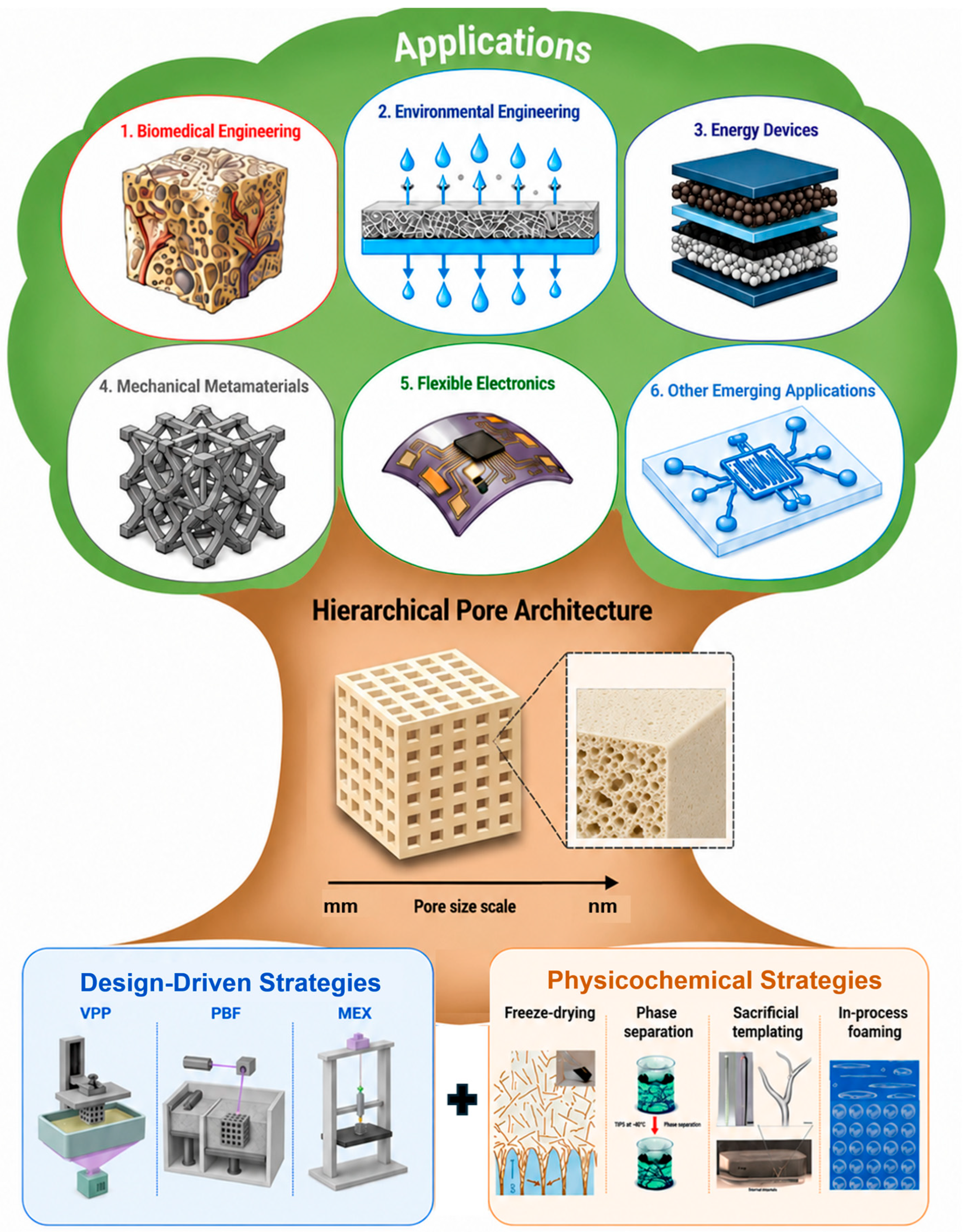
Figure 1: Tree-structured overview of the fabrication strategies, pore architecture, and application domains surveyed in this review. The roots represent two pore-forming paradigms: design-driven strategies from three-dimensional printing platforms (VPP, PBF, and MEX) and physicochemical strategies (freeze-drying, phase separation, sacrificial templating, and in-process foaming). The trunk shows their convergence into a hierarchical pore architecture spanning from approximately 1 nm to several millimetres. The canopy depicts the six resulting application domains: (1) biomedical engineering, (2) environmental engineering, (3) energy devices, (4) mechanical metamaterials, (5) flexible electronics, and (6) emerging applications. The schematic of freeze-drying was reproduced with permission from Ref. [30]. Copyright 2020, Wiley-VCH. The schematic of phase separation was reproduced with permission from Ref. [31]. Copyright 2012, The Royal Society of Chemistry. The schematic of sacrificial templating was reproduced with permission from Ref. [32]. Copyright 2022, Wiley-VCH. The schematic of in-process foaming was reproduced with permission from Ref. [33]. Copyright 2025, The Royal Society of Chemistry. The six application domain icons in the canopy were generated using GPT Image 2 (OpenAI) based on prompts describing each application scenario, and were reviewed by the authors for accuracy.
3D printing techniques for porous polymer fabrication differ by their raw materials and solidification mechanisms. The materials include light-cured liquid resins, thermally or rheologically set melts and inks, and laser-fused powder beds. As each technique has its own resolution, throughput, and material limits, the achievable pore size and structure is also different. Since resolution and throughput are inversely related across all platforms, choosing a specific printing technology always forces a compromise between material options and pore architecture design. Table 1 compares these platforms in terms of resolution, compatible polymers, and the principal advantages and limitations that govern platform selection for porous polymer fabrication.
Table 1: Comparison of 3D printing platforms for porous polymer fabrication.
| Platform | Technique | Resolution (XY; Z) | Compatible Polymers | Key Advantages | Key Limitations | Ref. |
|---|---|---|---|---|---|---|
| VPP | SLA | XY ~140 μm; Z 12–150 μm | Photocurable acrylates; GelMA | Near-isotropic mechanics; Ra 2.28 μm | Point-by-point; throughput ∝ area; photopolymer-only | [34,35,36,37] |
| DLP | XY 1 μm–100 μm; Z cure-depth dep. | GelMA; PEGDA; acrylates | Flash-per-layer; geometry-independent speed | Resolution–area trade-off; radical diffusion | [38,39,40,41] | |
| TPP | XY 15 nm–120 nm; axial 2–3× lateral | Photopolymers; GelMA | Highest AM resolution; true 3D freeform | Slow; ~25× parallelization limit | [42,43,44] | |
| MEX | FDM | XY 100–300 μm; Z 50–300 μm | PCL, PLA, PEEK, TPU, PA12, PLGA | High throughput; low cost; broad thermoplastic library | Thermoplastic-only; die swell; warping; z-anisotropy | [45,46,47] |
| DIW | XY 100–1200 μm; Z variable | GelMA, alginate, collagen, silicone; cell-laden hydrogels | Room-temp.; widest material diversity; cell-laden bioinks | kPa construct moduli; strict rheological control | [47,48,49] | |
| PBF | SLS | XY 20–80 μm; Z 100–150 μm | PA12, PA11, PCL, TPU, PEEK | Support-free fabrication; tensile strength 43–55 MPa | PA12/PA11 ~95% of output; powder aging; z-anisotropy | [50,51,52] |
| MJF | XY ~45–90 μm; Z layer 80–100 μm | PA12, PA11, TPU, PP | 8–10× SLS throughput; UTS 40–55 MPa | Residual fusing agent; porosity inconsistent (0.15–6.75%) | [53,54,55] | |
| Material jetting | PolyJet | XY ~15 μm; Z 16–30 μm | Proprietary photopolymers; multi-material blends | Multi-material voxel control (Shore 27–95 A); Ra 0.5–4 μm | Proprietary closed system; high cost; limited fatigue resistance | [56,57,58] |
2.1 Vat Photopolymerization (VPP)-Based 3D Printing
VPP encompasses a family of light-driven additive manufacturing technologies, including stereolithography (SLA), digital light processing (DLP) and its continuous variant Continuous Liquid Interface Production (CLIP), and two-photon polymerization (TPP), in which a liquid photopolymer resin is selectively solidified layer by layer through photoinitiated polymerization via either free-radical or cationic mechanisms. Cure depth is governed by the Jacobs working curve, relating solidification depth to incident energy dose and two resin-specific parameters that must be calibrated per formulation [35,38]. VPP operates exclusively on photocurable liquid resins, restricting the accessible material set to photopolymerizable thermosets and constraining feedstock diversity relative to extrusion- or powder-based platforms. As a direct consequence, the achievable pore size in VPP is governed almost entirely by the optical resolution of the platform rather than by a separate pore-forming step. Porous architectures are accordingly realized through design-driven approaches, specifically CAD-defined lattices and channels, with feature size set directly by the exposure mechanism described below. VPP is nonetheless commonly paired with two secondary pore-forming strategies to access pore scales below this optical limit. In hydrogel-based formulations, freeze-drying of the printed construct has produced porous cryogels with microporosity tunable from 68% to 96% [59]. In acrylate-based resins, sacrificial templating blends water-soluble particulates into the resin before printing and leaches them out after curing to yield multiple pore sizes [60].
SLA and DLP both cure a liquid photopolymer layer by layer but differ in exposure strategy. SLA scans an ultraviolet (UV) laser point by point across the resin surface via a galvanometric mirror, and after each exposure the build platform descends by one layer thickness, allowing fresh resin to flood the cured surface before the next scan begins (Fig. 2a). This point-by-point exposure limits SLA to an XY resolution of approximately 140 μm and a layer thickness of 12 to 150 μm [34,36], setting a corresponding floor on the smallest fabricable pore feature. DLP replaces the scanning laser with a digital micromirror device (DMD) that projects a complete binary layer pattern upward through a transparent window in a single flash, decoupling print time from geometric complexity. Because every pixel of the projected image is addressed simultaneously, DLP achieves a finer and more tunable XY resolution of 1 to 100 μm [40,41], allowing smaller pore features to be resolved than with SLA at comparable build volumes (Fig. 2b). A continuous variant, CLIP, eliminates the layer-separation force at the window through a persistent oxygen-rich dead zone, enabling withdrawal speeds roughly 100-fold faster than conventional DLP without altering this resolution [34,41,51,61,62]. This resolution distinction matters directly for porous fabrication, since SLA throughput scales linearly with cross-sectional area, penalizing high-open-area scaffolds, while DLP and CLIP expose each layer, whether dense or porous, in equal time, favoring periodic lattice architectures with fine pore features. All three remain restricted to photopolymerizable thermosets, and particle or cell loading substantially degrades resolution through scattering, further narrowing the achievable pore size in loaded resins [34,35,36,41].
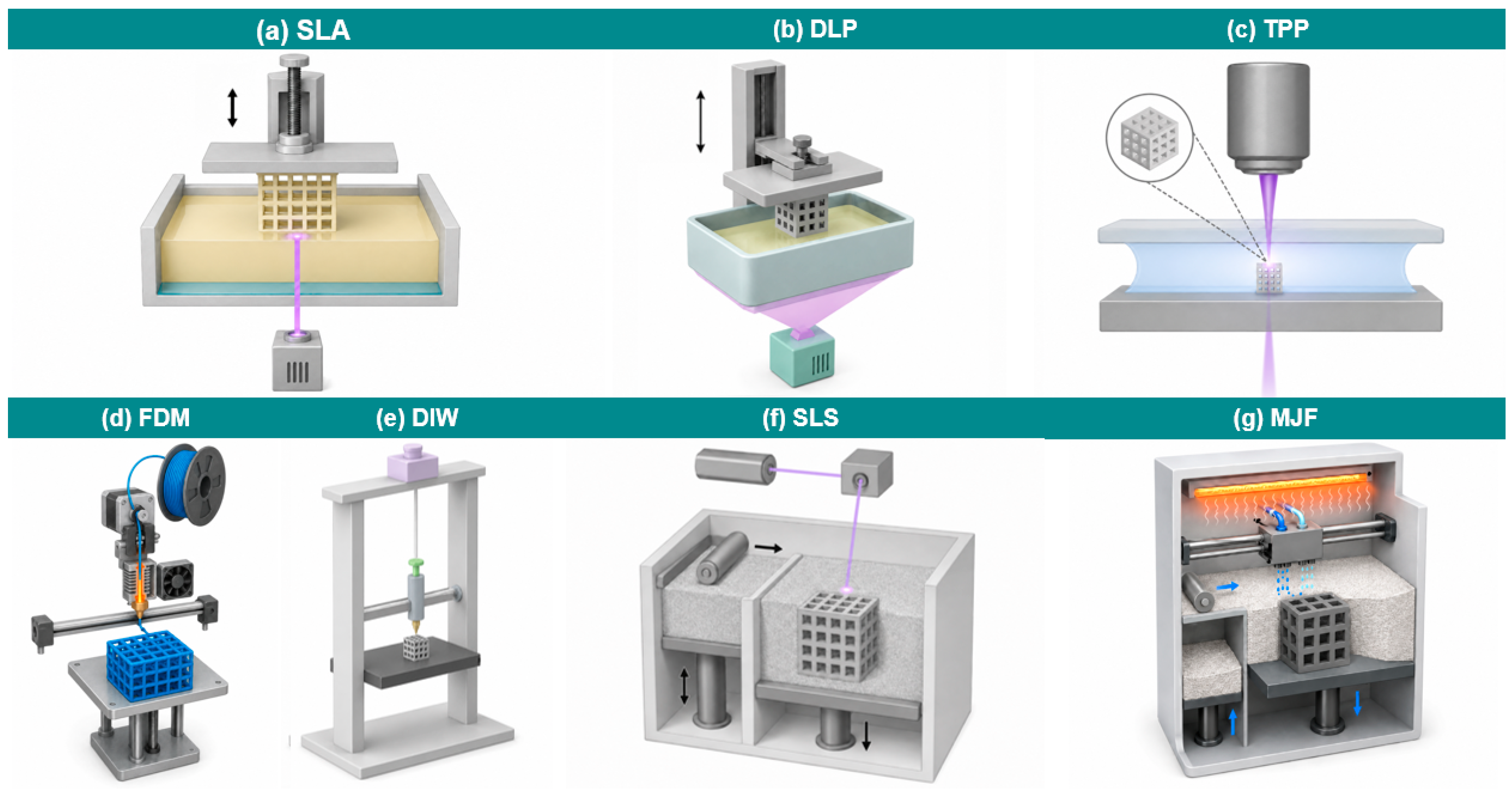
Figure 2: Schematic illustrations of representative additive manufacturing platforms for porous polymer fabrication, encompassing vat photopolymerization, material extrusion, and powder bed fusion. (a) SLA. (b) DLP. (c) TPP. (d) FDM. (e) DIW. (f) SLS. (g) MJF.
While SLA and DLP are practically limited by laser spot size, DMD pixel size, and resin curing characteristics, TPP (Fig. 2c) breaks through the optical diffraction limit by exploiting a nonlinear two-photon absorption mechanism. The simultaneous absorption of multiple photons confines radical generation to the focal volume of a tightly focused femtosecond laser, achieving an XY resolution of 15 to 120 nm with an axial resolution two to three times the lateral value [42,63]. Therefore, the smallest resolvable pore feature of TPP is roughly three orders of magnitude below that of SLA or DLP, making it suitable for fabricating submicron-pore optical metamaterials and high-resolution tissue scaffolds inaccessible to either platform. However, the fabrication time scales unfavorably with structure volume, confining its application to small, high-precision constructs [42,43,63].
2.2 Material Extrusion (MEX)-Based 3D Printing
MEX additive manufacturing, particularly Fused Deposition Modeling (FDM) and Direct Ink Writing (DIW), shares a fundamental operating principle. Material is continuously deposited through a fine nozzle under applied pressure in a layer-by-layer fashion, with resolution governed primarily by nozzle geometry and feedstock rheology. FDM and DIW differ fundamentally in the physical state of the extruded material, thermoplastic melts in the former and near-ambient viscoelastic inks in the latter, a distinction that directly determines material compatibility, achievable resolution, and the range of pore-forming strategies accessible to each [47,64,65]. Unlike vat photopolymerization, neither platform requires photocuring as a primary solidification mechanism. Both platforms are nonetheless commonly paired with a secondary, material-driven pore-forming strategy to access pore scales below the nozzle-defined resolution floor. In FDM, sacrificial templating extrudes filaments pre-loaded with a leachable component and removes the sacrificial phase after printing to reveal a microporous surface beneath the printed microarchitecture [66]. In-process foaming offers a comparable route for FDM, introducing gas-derived porosity through post-print supercritical fluid treatment. In DIW, freeze-drying and phase separation exploit the wet, unset state of the ink immediately after printing, with freeze-drying producing a printing-defined macroscale grid combined with micrometre-scale pores [67], while phase separation generates both inter-filament and intra-filament pore populations within the same printed structure [68].
In FDM, a thermoplastic filament is heated to working temperature and the melt is extruded through a nozzle, where rapid thermal cooling consolidates each road. Die swell during extrusion sets an XY resolution floor of approximately 100 to 300 μm and a layer thickness of 50 to 300 μm [45,47], directly bounding the smallest fabricable pore feature (Fig. 2d). Because each layer bonds to the one beneath through brief interfacial remelting rather than continuous polymerization, FDM parts exhibit pronounced z-axis anisotropy, with interlayer strength typically well below the in-plane strength of the extruded filament itself. The platform is restricted to thermoplastics, which precludes hydrogels and cell-laden systems [45,47,69]. DIW instead drives near-ambient viscoelastic inks by pneumatic or mechanical pressure, relying on shear-thinning for extrudability and post-deposition solidification via solvent evaporation, crosslinking, or gelation for shape retention [49] (Fig. 2e). Solidification through crosslinking or gelation produces thermoset-like, non-remeltable networks, in contrast to the remeltable thermoplastic filaments used in FDM. Because ink rheology rather than a fixed melt-flow die governs strand formation, DIW spans a substantially wider XY resolution range of 100 to 1200 μm than FDM [48,49], at the cost of less predictable strand geometry. This material universality, including living-cell-laden hydrogels, makes DIW the dominant platform for soft-tissue scaffolds. Support-bath embedded printing variants, of which Freeform Reversible Embedding of Suspended Hydrogels (FRESH) is the most widely adopted, extend this further by surrounding deposited filaments in a yield-stress medium until solidification, enabling ultrasoft hydrogels that would otherwise collapse during open-air deposition to be shaped into defined porous architectures [47,48,70]. The trade-off is kPa-range construct mechanics and demanding rheological formulation. Both platforms define macroporosity through CAD. FDM supplements this with post-print porogen leaching to access pore scales below its resolution floor, while DIW enables additional material-driven routes, including freeze-drying and phase separation of the deposited hydrogel, to introduce microporosity within printed strands and thereby achieve hierarchical porous architectures [45,48,49,65].
2.3 Powder Bed Fusion (PBF)-Based 3D Printing
PBF encompasses a family of additive manufacturing processes in which a heat source selectively consolidates discrete regions of a powder layer deposited on a build platform. Surrounding unsintered powder provides structural support without scaffolding. The part is built layer by layer as the platform descends and fresh powder is spread after each pass [50,52,71]. Selective laser sintering (SLS) and Multi Jet Fusion (MJF) are two typical PBF processes for polymers. Both operate on thermoplastic powder feedstocks and share the support-free geometric freedom that enables complex porous architectures, including interconnected lattices and anatomically graded scaffold geometries, to be fabricated without topological constraints or support removal steps. Despite this shared platform principle, SLS and MJF differ markedly in the achievable pore resolution and the throughput at which it is delivered. Porosity in PBF parts is most directly introduced through computer-aided design alone, but SLS is also commonly combined with two further pore-forming strategies. The first is partial fusion, which is intrinsic to the sintering process itself, where reducing the laser energy density below the full consolidation threshold leaves inter-particle voids distributed throughout the part rather than confined to a CAD-defined geometry [72]. The second is sacrificial templating combined with SLS, in which a leachable porogen is blended directly into the powder feedstock before sintering and removed afterward to yield hierarchically porous parts spanning sintering-defined micropores and leaching-defined macropores [73].
In SLS, a focused CO2 laser scans a preheated powder bed, melting designated cross-sections while a blade or roller recoater spreads each successive layer (Fig. 2f). This point-by-point laser scanning achieves an XY resolution of approximately 20 to 80 μm and a layer thickness of 100 to 150 μm [50,51], the finest pore resolution among the PBF platforms surveyed here. The material library extends beyond Polyamide 12 (PA12) and Polyamide 11 (PA11) to include Polycaprolactone (PCL), Polyetheretherketone (PEEK), and hydroxyapatite-polymer composites (HA), establishing SLS as the primary PBF platform for tissue engineering scaffolds, drug delivery carriers, and bone-mimetic porous matrices [50,51,52,71,74]. Tensile strength in SLS-printed parts typically reaches 43 to 55 MPa in-plane, though z-direction mechanical properties remain lower due to incomplete inter-layer fusion. Pore-forming strategies in SLS operate across multiple scales. Designed macroporosity from CAD can be combined with intrinsic partial sintering, in which reducing laser energy density below the full consolidation threshold retains inter-particle voids for in-process pore control. PCL- and PCL/HA-based parts are also amenable to post-processing pore formation, whereas PA12 and PEEK offer limited post-processing options due to their chemical resistance [50,74]. Pore wall surface roughness is governed by powder particle size, and the material library remains restricted to semi-crystalline thermoplastics possessing a sufficiently wide sintering window, the temperature interval between crystallization onset and melting point [52,71,75,76].
MJF substitutes agent-mediated area irradiation for laser scanning, achieving throughput approximately 8 to 10 times higher than SLS at the cost of a coarser XY resolution of approximately 45 to 90 μm and a layer thickness of 80 to 100 μm [53,55], with porous applications concentrated in PA12 lattice geometries for energy absorption and structural lightweighting [77,78,79] (Fig. 2g). Tensile strength reaches a comparable 40 to 55 MPa, but residual fusing agent components raise biocompatibility concerns, and the material library is confined to PA12, PA11, thermoplastic polyurethane (TPU), and polypropylene. Thermoplastic polyurethane presents the greatest post-processing pore-forming potential among these through solvent extraction or physical foaming [55,77,80]. Where powder bed platforms of both types are bounded by thermoplastic sinterability and powder particle dimensions, material jetting platforms operate from liquid-phase photopolymer droplets, enabling simultaneous multi-material gradient control at the resolution of individual jetting arrays.
2.4 Other 3D Printing Techniques
In recent years, several emerging additive manufacturing strategies have expanded beyond conventional layerwise or nozzle-based fabrication for porous polymers. Representative examples include voxel-scale multi-material jetting, volumetric photopolymerization, and field-mediated assembly approaches. These methods extend the design space of porous architectures while introducing new constraints in material compatibility and process scalability. Unlike the platforms surveyed in Section 2.1, Section 2.2 and Section 2.3, these emerging techniques are used predominantly in their direct, design-driven form rather than in combination with secondary pore-forming strategies. PolyJet uses proprietary, fully cured photopolymer chemistry, and Computational axial lithography (CAL) requires optical homogeneity. Each of these constraints precludes the post-processing routes available to VPP, MEX, and PBF platforms. Ultrasound-assisted printing uses a hydrogel-based feedstock, which would in principle support freeze-drying or phase separation in the manner of DIW. However, the current literature has focused on demonstrating the compositional and structural capabilities of this platform rather than on combining it with a secondary pore-forming step.
In PolyJet printing, piezoelectric actuators eject photopolymer resin as picolitre-scale droplets from an arrayed printhead carriage. A co-mounted UV lamp cures each swathe immediately upon deposition, before the platform steps down to repeat the cycle. This achieves an XY resolution finer than most VPP and MEX platforms, at approximately 15 μm [58,81,82]. The defining capability of this platform for porous polymer applications is voxel-scale multi-material control. By varying the jetting ratio between rigid and elastomeric resins in real time, the printhead array can place different photopolymer compositions at adjacent voxels within the same build. This produces functionally graded porous constructs inaccessible to single-material platforms, with stiffness spanning Shore hardness values from roughly 27 A to 95 A as one example of the resulting property range [57,83,84].
CAL and ultrasound-assisted printing represent emerging non-layered fabrication strategies that extend porous polymer manufacturing beyond conventional nozzle- or mask-based approaches. In CAL, multiple dynamically projected light fields are superimposed onto a rotating vat of photocurable resin, enabling volumetric curing of the entire structure without layerwise discretization. This eliminates interlayer interfaces and enables the fabrication of complex 3D porous architectures. Like the other photopolymer-based platforms surveyed above, CAL remains restricted to thermoset resins, and is further limited to optically transparent, chemically homogeneous formulations, restricting its use to single-phase systems without particulate or cellular loading. In contrast, ultrasound-assisted printing employs acoustic fields for nozzle-free droplet ejection and particle or cell manipulation within hydrogel precursors, enabling field-directed organization of microstructure in porous hydrogels, a functional capability with no direct equivalent among the platforms discussed above.
3 Pore-Forming Strategies Based on 3D Printing
Porosity introduction in 3D-printed polymer systems proceeds through two distinct paradigms, namely design-driven and physicochemical routes. Design-driven pore formation pre-encodes pore geometry in the digital model and reproduces it deterministically by the printing platform. Physicochemical routes, in which void architectures arise as outcomes of processing conditions rather than geometric intent, encompass phase separation, freeze-drying and ice-templating, sacrificial templating, partial fusion, and a range of supplementary strategies including gas-based foaming and surface etching. Together these approaches span pore scales from sub-micrometre to several millimetres. They are frequently deployed in combination, since physicochemical strategies applied to geometrically defined printed constructs add material-level porosity at scales that direct geometric encoding alone cannot reach. This complementarity recurs throughout this section and the application chapters that follow.
3.1 Design-Driven Pore Formation
In design-driven pore formation, pore geometry is set in the digital model before printing begins. The printing platform must then reproduce that geometry as faithfully as its resolution allows. PBF can resolve features down to about 0.2 mm, and VPP down to about 0.1 mm or below, setting the lower bound on wall thickness and the porosity that can be achieved [85,86]. Material choice is accordingly governed by whether it flows and solidifies correctly during extrusion or cures accurately under light exposure, not by its foaming or phase-separation behaviour. Three main architecture families organize this design space. These are strut-based lattices, triply periodic minimal surfaces, and spatially graded versions of either.
Strut-based lattices arrange periodic unit cells as interconnected nodes and struts. Common topology variants include Body-Centered Cubic (BCC), Body-Centered Cubic Z-reinforced (BCCZ), and Rhombic Dodecahedron (RD) (Fig. 3a). Strut diameter gives direct control over relative density and pore size, making this family the simplest to design across a wide porosity range, and the most widely used for load-bearing bone scaffolds and lightweight structural parts. The main drawback is stress concentration at the nodes, where multiple struts meet and carry load well above the level seen mid-strut. Node failure triggers the sudden load drop that limits energy absorption in BCC and BCCZ lattices [87]. Printing defects at the nodes also cause real mechanical performance to fall below theoretical predictions in both FDM and SLS lattices, an effect cross-supports or increased cell count can only partly offset [88,89,90]. Triply periodic minimal surfaces (TPMS) avoid these node problems through smooth, continuously curved surfaces with no sharp junctions, spreading load uniformly across the surface instead of concentrating it at discrete points (Fig. 3b). This node-free geometry gives higher fluid permeability than strut-based lattices at matched porosity and helps cells attach and migrate more evenly, making TPMS the preferred choice for perfusable tissue scaffolds and filtration membranes. Among TPMS types, Gyroid, Diamond, and I-graph and Wrapped Package offer the widest manufacturable design space, the most even internal curvature, and the highest permeability within the family [91,92,93]. The curved walls bend under load rather than carrying it along their length, giving TPMS lattices lower stiffness than strut-based lattices at the same relative density. The same curvature that aids fluid flow can also trap uncured resin or unsintered powder inside narrow channels, reducing effective pore connectivity and biocompatibility. Spatially graded architectures form a third category, varying relative density or topology continuously through the construct so that a denser load-bearing core and a more porous interface can be optimised independently within a single printed part (Fig. 3c). This gradual transition avoids the interface mismatch seen in discrete multi-material parts, making it well suited to osteochondral and other implants that must bridge two distinct mechanical and biological environments. The transition zone between dense and porous regions is, however, harder to design and predict, since local stiffness and defect sensitivity both change across the structure, and this zone can become a weak point under repeated loading [94].
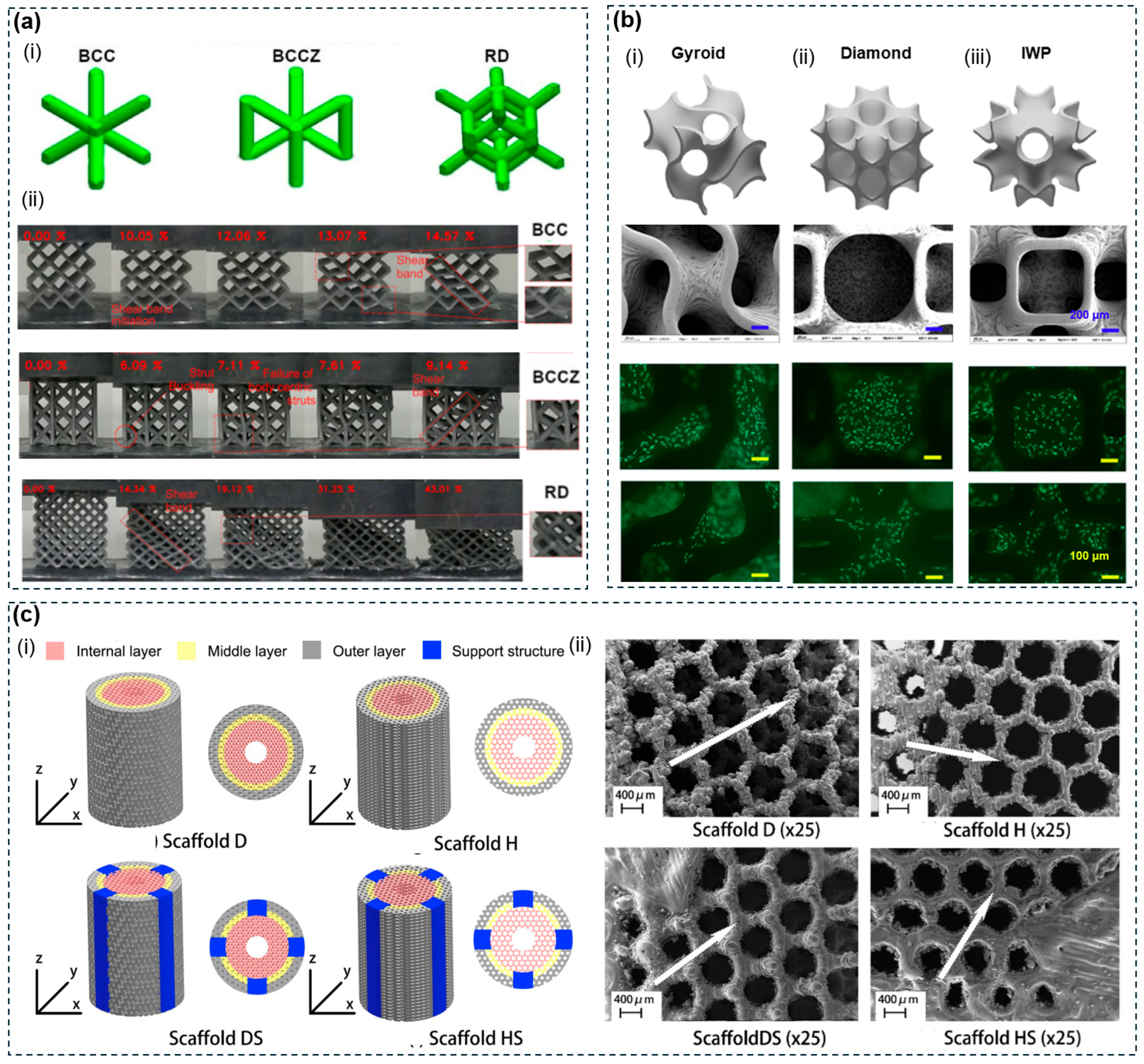
Figure 3: (a) SLM-printed AlSi10Mg strut-based lattice structures for mechanical characterization. (i) CAD models of BCC, BCCZ, and RD unit cells. (ii) Sequential compression deformation images showing shear band initiation and propagation in BCC, strut buckling and catastrophic failure in BCCZ, and shear band formation in RD. Reproduced with permission from Ref. [87]. Copyright 2026, Elsevier. (b) DLP-printed TPMS porous zirconia scaffolds for bone tissue engineering. CAD models and SEM images of Gyroid (i), Diamond (ii), and IWP (iii) unit cell architectures at two magnifications, showing interconnected porous networks and smooth sintered surfaces. Fluorescence images demonstrate rBMSC viability and cytoskeletal spreading on the three TPMS structures at days 1 and 3. Reproduced with permission from Ref. [92,93]. Copyright 2021, Elsevier and 2024, American Chemical Society. (c) SLM-printed Ti6Al4V functionally graded porous scaffolds for load-bearing orthopedic applications. (i) CAD models of four scaffold designs (D, H, DS, HS) showing radially graded porosity from high-porosity inner layer to dense outer layer, with and without embedded support structures. (ii) SEM images of the corresponding fabricated scaffolds showing gradient pore architecture and strut morphology. Reproduced with permission from Ref. [94]. Copyright 2020, Elsevier.
The pore architectures that design-driven printing can achieve vary widely across polymers, because the processability of each material, not pore design intent, sets the platforms it can access. Some polymers reach a broad design space because they print well on more than one platform. PCL, for example, is compatible with FDM, SLS, and melt electrowriting (MEW), each of which accesses a distinct portion of the achievable pore range [95]. On FDM, raster patterns deliver channel dimensions of 160 to 700 μm at porosities of 48 to 77% [92,93]. On SLS, feature enlargement during sintering can be corrected through geometric pre-scaling [96]. On MEW, ordered fibre arrays reach about 70% porosity with 140 to 160 μm pores at a resolution FDM cannot match [97]. TPU prints on both SLS and FDM [98], which together support auxetic lattices with strong negative Poisson’s ratio behaviour and gradient honeycomb structures with high energy absorption over repeated compression [99,100,101,102]. GelMA reaches Design-driven pore formation through VPP, where it forms tissue-relevant pore architectures near 100 μm resolution [103,104], and through DIW once blended with methylcellulose to restore filament fidelity [105,106]. Other polymers are confined to a single platform, which narrows the achievable pore range. PLA prints only by FDM, and its brittleness sets a hard lower bound on strut size, since thin pore walls in high-porosity lattices fail by sudden buckling rather than gradual deformation [98]. PEEK requires FDM at nozzle temperatures of 390 to 430°C and cannot be processed on other platforms [86], yet within this single route TPMS architectures reach cortical-bone-level compressive stiffness at 70% porosity with full pore interconnectivity [99]. PA12 is processed almost exclusively by SLS, where mechanical performance degrades as strut diameter shrinks, so printing-induced nodal defects must be accounted for to predict structural response accurately [90,103,104]. PEGDA prints only by VPP, but this single route delivers submillimetre feature reproduction, making it the main synthetic hydrogel choice for high-resolution TPMS fabrication.
Across these seven polymers, platform breadth and pore design freedom rise and fall together. Materials that print on multiple platforms, including PCL, TPU, and GelMA, access a wider pore size and architecture range, while materials restricted to one platform, including PLA, PEEK, PA12, and PEGDA, must work within whatever pore space that one platform allows.
Phase separation turns a single polymer solution into a porous solid by thermodynamic demixing into two phases. One phase becomes polymer-rich and solidifies into the scaffold matrix, while the polymer-lean phase leaves pores behind after the solvent is removed [17,107]. Diffusion-, vapor-, and thermally induced phase separations (DIPS, VIPS, and TIPS) are three main variants, which use a liquid nonsolvent bath, a nonsolvent vapor instead of a liquid, and cooling, respectively. Across these three approaches, the resulting pore structure depends on which region of the phase diagram the demixing process passes through. Spinodal decomposition produces a continuous open-pore network, while nucleation and growth yields closed pores. Faster cooling rates suppress nucleation and growth (NG) and favor fine spinodal decomposition (SD)-derived structures, while slower cooling allows NG-driven coarsening that enlarges pores but reduces uniformity [107,108,109,110]. All three variants share a common requirement that the target polymer be soluble in a processable organic solvent. Synthetic aliphatic polyesters such as PCL, PLA, and Poly(lactic-co-glycolic acid) (PLGA) meet this requirement. Chemically inert engineering thermoplastics such as PEEK [111], powder-bed materials such as PA12 [103,104], and crosslinking-dependent hydrogels such as GelMA and alginate do not [112,113].
In DIPS, the polymer solution is immersed in a liquid nonsolvent bath. Rapid exchange between the solvent and nonsolvent drives demixing (Fig. 4a). Pore shape depends on the solvent and nonsolvent pair and on the polymer concentration. Higher concentration raises the viscosity of the solution, slows nonsolvent diffusion, and produces smaller pores [17,68,107,114]. VIPS delivers the nonsolvent as vapor instead of liquid. This slows the exchange process and gives finer control over pore structure (Fig. 4b). In direct ink writing, this becomes an in-situ solidification method. The polymer solution is extruded into a nonsolvent mist, and relative humidity continuously controls the solidification rate. The resulting porous filament can be made even more porous by adding sacrificial porogens [68,114]. TIPS causes demixing by lowering temperature instead of adding a nonsolvent (Fig. 4c). This removes the need for an immersion bath or a vapor chamber. In binary systems such as PLA dissolved in dioxane, cooling triggers solid-liquid phase separation through solvent crystallization, producing anisotropic microtubular structures. Adding water as a nonsolvent turns this into a ternary system, which instead triggers liquid-liquid demixing above the solvent freezing point. This replaces the anisotropic structure with an isotropic, interconnected macroporous network. Pore size in this network grows continuously with demixing temperature and residence time, ranging from about 10 to 15 μm up to 50 to 90 μm. Longer residence time in the metastable region also produces secondary micropores of 1 to 2 μm within the macropore walls [17,108,109,110,115].
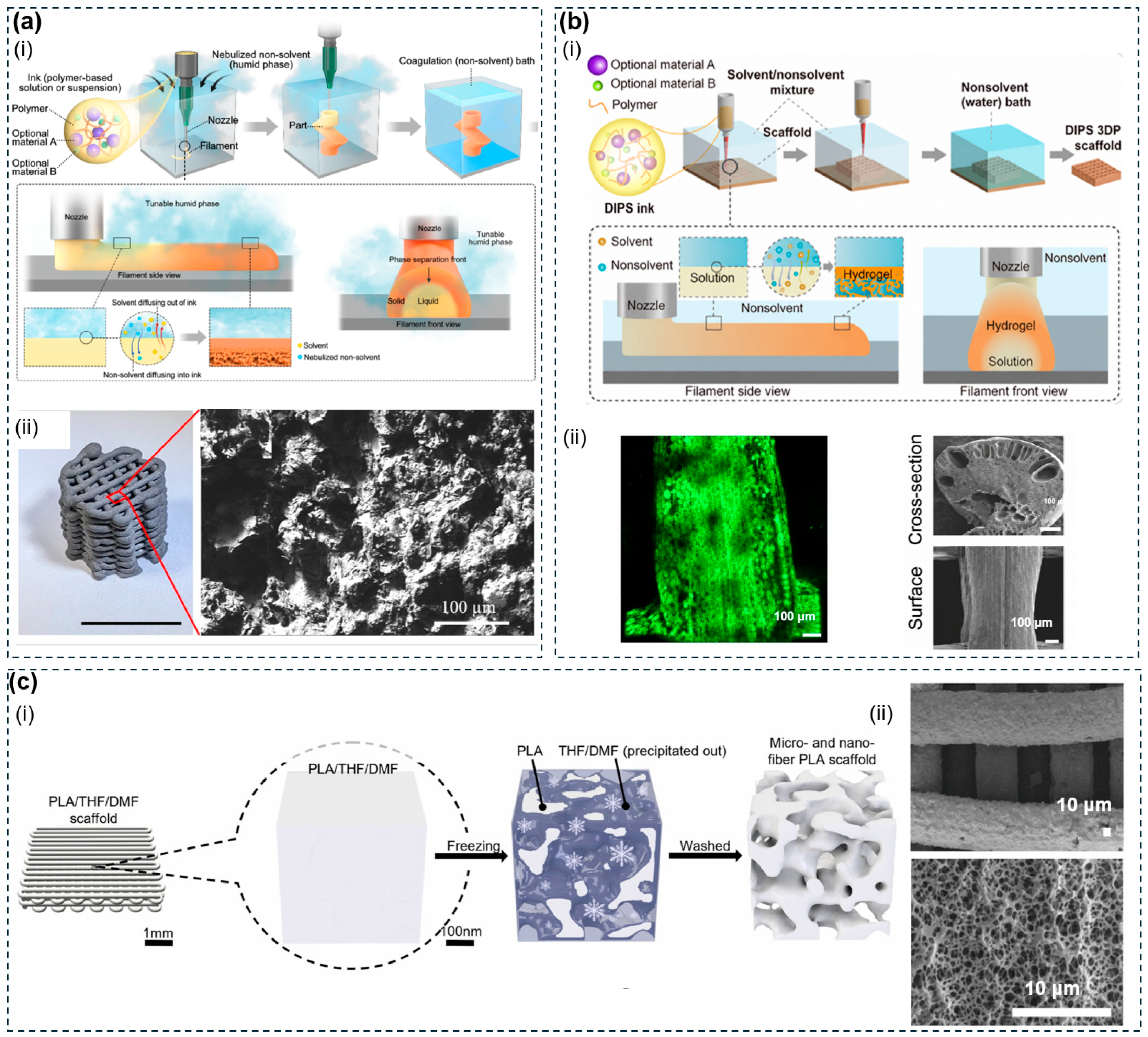
Figure 4: Phase separation-based solidification mechanisms for porous polymer fabrication. (a) VIPS solidification mechanism and its implementation in direct ink writing: (i) schematic of solvent/nonsolvent exchange dynamics in a nebulized environment driving an inward-propagating solidification front; (ii) SEM of intra-filament porous morphology and representative printed lattice. Reproduced with permission from Ref. [68]. Copyright 2024, Springer Nature. (b) DIPS-based direct ink writing for in-situ filament solidification. (i) Schematic showing polymer ink extruded into a liquid nonsolvent bath, driving solvent/nonsolvent exchange and phase separation from the filament surface inward. (ii) Confocal image and cross-sectional SEM micrographs show the resulting hierarchical porous morphology. Reproduced with permission from Ref. [114]. Copyright 2025, American Association for the Advancement of Science. (c) TIPS-derived micro/nanofibrous porous PLA scaffolds. (i) Freezing-induced phase separation of the printed PLA/THF/DMF scaffold precipitates a co-continuous solvent phase, which washing removes to yield a fibrous porous structure. (ii) SEM images of the resulting scaffold morphology. Reproduced with permission from Ref. [115]. Copyright 2025, Elsevier.
Which phase separation route a polymer can use depends on its solvent compatibility and thermal behaviour, and this in turn shapes the resulting pore architecture and function. PCL dissolves readily in a wide range of solvents, including dioxane, chloroform, and DMF. This broad solvent compatibility makes it suitable for both DIPS and TIPS. PCL/HA and PCL/HA/bioactive glass (BG) composites made through these routes show better bone-forming performance than unfilled PCL. Alkaline erosion after fabrication selectively exposes the HA at the surface, which speeds up bone regeneration in vivo [116,117,118]. PLGA can be processed by FDM at nozzle temperatures around 200–220°C, although careful control of the melt extrusion temperature is required to limit thermal degradation and chain scission [119]. Nevertheless, solvent-based routes such as DIPS remain attractive for PLGA because they avoid melt processing entirely and enable hierarchical pore architectures with patient-specific geometries. PLGA has been processed through DIPS combined with CAD/CAM negative molds and lyophilization, producing hierarchical architectures spanning 1 to 300 μm with patient-specific geometric accuracy [120]. PLGA/HA composites made by pneumatic direct-write extrusion followed by phase separation show sustained drug release and bone regeneration in vivo [121]. PLGA processed through TIPS also keeps its degradation rate tunable through the Lactide to Glycolide ratio (LA:GA), independent of the scaffold architecture [116]. PLA and its stereoisomers are the main substrate for TIPS. In a ternary dioxane and water system, this route produces isotropic, interconnected scaffolds with porosity above 95% and pore diameters ranging from 1 to 100 μm [108,109,110]. Solvent range and thermal stability determine which phase separation route each polymer can access, and the resulting pore size, architecture, and degradation behaviour follow directly from that choice.
3.3 Freeze-Drying and Ice-Templating
Freeze-drying and ice-templating use the solidification of solvent as a sacrificial structuring mechanism. As an aqueous polymer solution cools, ice crystals form and grow, pushing dissolved chains or suspended particles into the spaces between the crystals. This forms a continuous, polymer-rich network. Once the ice is removed by sublimation under reduced pressure, this network becomes a highly porous structure. Random freeze-drying, directional ice-templating, and cryogelation are three main variants of this approach, each producing a different pore architecture suited to different polymers. Thermodynamic and kinetic conditions must be met for this process to work. These two conditions limit this method to materials that can be processed in water, mainly water-soluble natural polymers and hydrogel precursors. Thermoplastics that are melted and consolidated, and light-cured synthetics such as PEGDA, cannot use this method [30,122].
Random freeze-drying has no single dominant direction of heat flow, so pores form with an even, equiaxed shape in all directions. Pore size decreases as supercooling increases, because ice crystals start forming faster than they can grow. Controlled cooling at 0.9°C per minute reduces the mean pore diameter from about 132 μm under quench conditions to about 96 μm, with much more even pore size and shape [122,123]. Directional freeze-drying, also called ice-templating, applies a set temperature gradient, making ice grow preferentially along the direction of heat flow (Fig. 5a). The result is column-shaped or layered pore channels aligned with the direction of heat flow. A lower freezing temperature increases undercooling and freezing speed, which narrows the channels and thins the walls. Freezing from one direction produces a layered structure with several domains. Freezing from two directions produces a single domain at the centimetre scale. Freezing radially produces anisotropy along both the long axis and the radius at the same time. When combined with direct ink writing, the polymer solution is extruded onto a substrate cooled by a thermoelectric plate. Substrate temperature directly controls local pore diameter, from about 185 μm at −5°C to about 24 μm at −20°C. A temperature gradient along the vertical axis also creates graded pore sizes, from 71 to 514 μm, within a single filament. At the same time, polymer chains concentrate around the ice crystals, which gives the structure stronger directional mechanical properties than an isotropic control [30,122,124]. Cryogelation crosslinks the polymer network during freezing at −5 to −20°C (Fig. 5b). Precursor molecules concentrate into a small unfrozen region around the ice crystals and crosslink there. After thawing, this leaves behind a macroporous network that is elastic, has shape-memory behaviour, and can be compressed by up to 90% without permanent damage. This is unlike conventional freeze-dried scaffolds, where uncrosslinked chains produce brittle structures. Pore size decreases with lower crosslinking temperature, faster cooling, or higher polymer concentration [30,125].
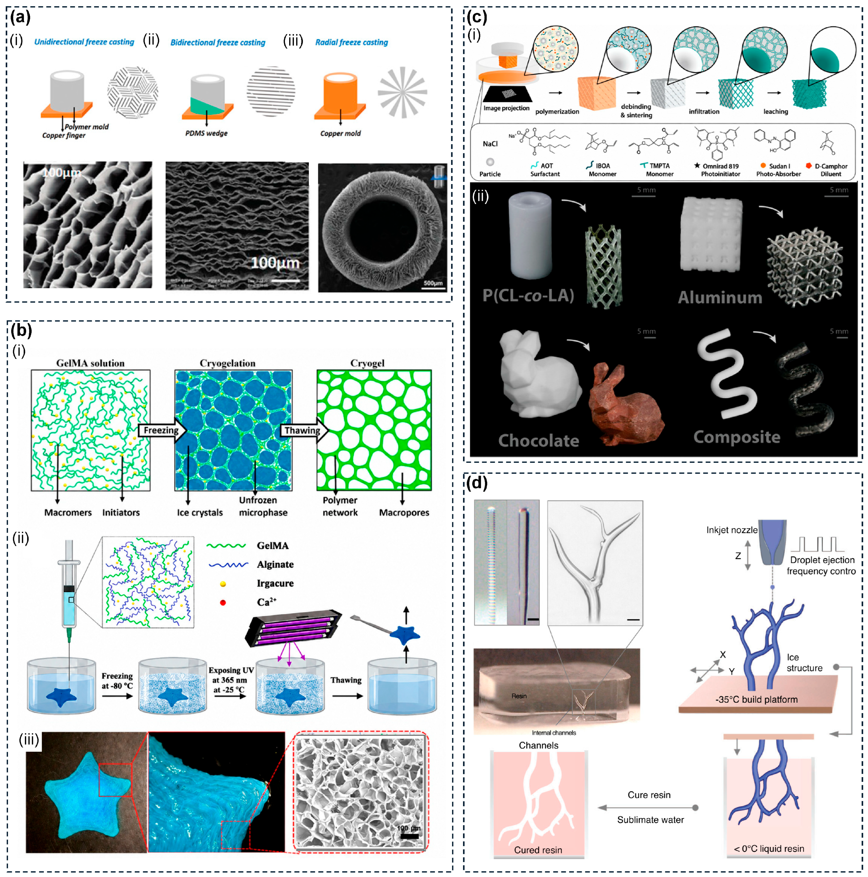
Figure 5: Freeze-based and sacrificial templating strategies for porous scaffold fabrication. (a) Directional freeze casting: schematics and SEM images of (i) unidirectional (columnar channels), (ii) bidirectional with PDMS wedge (lamellar pores), and (iii) radial with copper mold (longitudinal-radial anisotropy). Reproduced with permission from Ref. [30]. Copyright 2020, Wiley-VCH. (b) Cryogelation-based embedded 3D printing of GelMA scaffolds: (i) cryogelation mechanism; (ii) fabrication workflow via nanoclay support bath and subzero UV crosslinking; (iii) resulting scaffold and SEM of interconnected macropores. Reproduced with permission from Ref. [126]. Copyright 2023, American Chemical Society. (c) SLA-printed NaCl sacrificial templates: (i) fabrication workflow from printing through sintering, infiltration, and leaching; (ii) representative structures including polymer stents, aluminum lattices, and composites. Reproduced with permission from Ref. [127]. Copyright 2022, Wiley-VCH. (d) Freeform 3D ice printing: frequency-modulated droplet ejection builds branched ice templates that are resin-encapsulated, cured, and sublimated to yield internal channel networks. Reproduced with permission from Ref. [32]. Copyright 2022, Wiley-VCH.
Which freeze-drying or gelation route a polymer can use depends on its physical and chemical compatibility with the freezing or crosslinking process, and this in turn shapes the resulting pore architecture and intended application. Random freeze-drying is the main processing route for natural protein and polysaccharide scaffolds. Collagen-glycosaminoglycan composites produce equiaxed pores of about 96 μm under controlled cooling [123,126]. Chitosan and silk fibroin form similar isotropic porous architectures, used in bone, nerve, and cartilage tissue engineering [30,122]. Directional ice-templating, when combined with DIW, produces aligned rather than isotropic pores. GelMA has a physical gelation temperature of approximately 17 to 21°C, which stabilizes chain networks around growing ice crystals [105,128]. This allows myotubes to align much more strongly within the resulting anisotropic microchannels than in unfrozen, isotropic regions [124]. Collagen and silk fibroin blends printed onto a platform held at −20°C achieve improved mechanical properties and biomimetic aligned channels, demonstrated for spinal cord repair [129]. Cryogelation works for both natural and synthetic polymers and produces a third type of architecture suited to injectable delivery. GelMA crosslinked in a nanoclay composite bath of Pluronic F127, Laponite, and CaCl2 generates open porosity of 81 to 95% with pore sizes of 90 to 300 μm tunable by concentration [126,130]. Methacrylated alginate and hyaluronic acid crosslinked at subzero temperatures yield injectable, shape-memory macroporous scaffolds that can pass through 16 G needles for minimally invasive cell and drug delivery [125]. The choice of freezing or gelation strategy directly sets whether the resulting scaffold is isotropic, aligned, or injectable, and this choice follows from both the target application and the polymer being processed.
Sacrificial templating creates porous architectures by placing a removable phase inside a structural matrix, then removing it using a matched stimulus. The sacrificial phase sets the pore geometry, while the structural material fills the remaining space. Sacrificial templating takes three main forms, particle leaching, fugitive ink extrusion, and sacrificial filament casting, each placing the sacrificial component differently relative to the structural material. This method involves fabricating the sacrificial template, encapsulating it in scaffold prepolymer or melt, removing the template through temperature variation, water dissolution or solvent extraction, and rinsing the scaffold to form an internal structure that perfectly mirrors the inverse geometry of the sacrificial template [131,132,133]. In sacrificial templating, the structural material must survive this step while the sacrificial phase responds to it and dissolves or melts away. When the sacrificial phase is water-soluble, this approach is compatible with almost any natural polymer, synthetic hydrogel, or elastomer that can be processed in water. When the sacrificial phase is incorporated by melt blending instead, the method also extends to thermoplastic matrices.
Particle leaching spreads solid porogen particles throughout the structural matrix and removes them by selective dissolution after printing. When NaCl particles and water-soluble Polyvinyl Alcohol (PVA) are blended together into PLA filament for FDM printing, the PVA forms connecting pathways between the NaCl particles. This speeds up water access to the salt and stops particles from being sealed off inside the polymer. Ternary PLA, PVA, and NaCl blends reach porosities up to 70% with near-complete pore connectivity. Binary PLA and NaCl blends, without PVA, suffer from particles trapped below the surface that water cannot reach [127,134] (Fig. 5c). Fugitive ink extrusion prints a continuous sacrificial material as a defined channel template inside the structural matrix. Pluronic F127 is a free-flowing liquid at low temperatures and gels upon warming above approximately 10°C, allowing it to be removed by cooling below its gelation threshold. However, high concentrations are toxic to cells, low-temperature removal can damage cells, and the resulting structure is mechanically weak at larger scales. Gelatin avoids these problems because it melts at around 37°C instead. In embedded 3D printing, a gelatin sacrificial ink is deposited into a surrounding living tissue matrix. The gelatin ink has a much higher yield stress than this matrix. As the nozzle moves through the matrix, the matrix temporarily turns fluid around the nozzle tip and then heals itself back together behind it, while the firmer gelatin ink keeps its printed shape. Warming the construct afterward melts the gelatin away, leaving channels that allow fluid to flow through and keep cells alive at realistic tissue densities [32,131,134] (Fig. 5d). Solid printable sacrificial templates take this approach further, using a stiffer material than gelatin or Pluronic. A carbohydrate glass made from sucrose, glucose, and dextran has a Young’s modulus of about 1 GPa. This stiffness allows it to be printed into a free-standing lattice on its own. It dissolves within about 10 min in cell culture medium and works across six different types of extracellular matrix (ECM) crosslinking [132]. Sacrificial filament fabrication separates mould printing from scaffold casting entirely. A free-standing PVA mould is printed by FDM, encapsulated inside the structural material, and then dissolved away in water. This produces scaffolds whose porosity and channel spacing can be tuned through infill density, at sizes larger than 75 cm3 [131,135].
Particle leaching, fugitive ink extrusion, and sacrificial filament casting differ mainly in where the sacrificial component sits relative to the structural material, a distinction that governs which material systems and printing platforms each can support. Particle leaching works across many different material systems. In ternary PLA/PVA/NaCl FDM constructs, this method introduces micro-scale pores within the printed struts while keeping the overall structure strong enough for bone tissue engineering [134]. In alginate DIW scaffolds, methylcellulose blended into the ink leaches out after ionic crosslinking, creating microporosity that helps cells migrate [136]. In collagen prepolymer, sacrificial microparticles leave pores behind once removed, which support much better cell migration than GelMA controls [137]. Fugitive ink strategies work with the widest range of structural matrix materials, since the sacrificial component is printed as its own separate ink rather than mixed into the matrix. Carbohydrate glass lattices generate channels that allow fluid flow through GelMA, calcium-crosslinked alginate, fibrin, and Matrigel constructs, without needing to change the printing protocol [132]. In FRESH printing, gelatin microparticles formed by coacervation are suspended in a pH-buffered bath. These particles act both as a support medium during printing and as a sacrificial template afterward. They melt upon warming, leaving an interconnected microporous network within collagen constructs that allows full cell infiltration within three days [137]. In Sacrificial Writing Into Functional Tissue (SWIFT) embedded printing, gelatin is evacuated at 37°C from a collagen I and Matrigel matrix, producing channels that allow fluid flow across embryoid body, cerebral organoid, and cardiac spheroid matrices [138]. Sacrificial filament casting suits elastomeric and synthetic hydrogel matrices that cannot be co-printed directly, since the sacrificial component is shaped as a separate mould rather than printed alongside the structural material. Polydimethylsiloxane (PDMS) and Poly(2-hydroxyethyl methacrylate) (pHEMA) scaffolds made by dissolving a PVA mould support hepatocyte growth and function with near-complete cell viability. Because mould fabrication is separated from structural casting, this method extends to any mouldable polymer, regardless of how that polymer needs to be deposited [135].
The position of the sacrificial component relative to the structural material sets the limits of each form. Mixing it into the matrix, as in particle leaching, keeps the process simple but limits pore size to what the dispersed particles allow. Printing it separately, as in fugitive ink, gives more freedom over matrix chemistry but needs a printable sacrificial ink. Casting around it, as in filament casting, separates mould design from matrix deposition, extending the method to materials that cannot be printed in combination with anything else.
Partial fusion differs from design-driven pore formation in that pores form as a direct consequence of processing conditions rather than from geometry encoded in the digital model. The process is deliberately run short of full material consolidation by limiting thermal energy input below the level needed for complete particle or filament fusion, leaving voids that the printing parameters govern but cannot independently specify in shape or position. This mechanism operates primarily in selective laser sintering and fused deposition modelling. In PBF, the main mechanism is incomplete particle coalescence. Laser energy is delivered above the melting threshold but below the level needed for full densification. This leaves gaps between particles, which form the basis of the porous network. Particle coalescence is driven by surface tension and limited by melt viscosity and crystallization rate. Because of this competition, the gap between the onset of melting and the onset of crystallization works as a simple screening test for whether a material suits SLS [103,139,140]. In fused deposition modelling, the fusion window is much shorter, contracting to seconds after each layer is deposited. This means partial neck growth voids are a built-in feature of FDM parts rather than something that can be removed afterward [141]. A thermoplastic can be processed by FDM only if it exhibits a sufficiently wide thermal processing window between the onset of melting and the onset of decomposition. PLGA is excluded from the FDM route because its processing window is too narrow, and repeated heating during extrusion breaks down its polymer chains [119].
SLS based partial sintering is the most common and best studied route to this kind of porosity. It is controlled mainly through energy density, which increases with laser power and decreases with scan speed, layer thickness, and hatch spacing. Within the stable sintering range, increasing energy density reduces porosity and raises compressive strength. This sets up the basic trade-off in scaffold design, where lower porosity comes at the cost of higher strength. For PCL, varying energy density systematically spans porosity from about 24% to 64% across the usable processing window [142]. At the particle scale, scan speed has the strongest effect. Lower scan speed extends thermal exposure and drives more neck growth, which closes more pores. Higher scan speed keeps the microstructure only partly fused, sustaining an open, connected pore network [143,144] (Fig. 6a). Partially sintered scaffolds contain two pore types with different shapes. The first is irregular partial fusion, found at the boundaries between print tracks and between layers. The second is near-spherical gas entrapment pores, formed when gas trapped between particles cannot escape the viscous melt. For PA12, overall porosity drops substantially as laser power increases and scan speed decreases [145]. In FDM, nozzle temperature is the main parameter controlling the fraction of partial neck growth voids. Higher temperatures lower melt viscosity and extend the time the material stays above its glass transition temperature, giving adjacent printed lines more time to fuse together (Fig. 6b). Print speed and layer height offer additional adjustment, but over narrower effective ranges [86,98,141]. Direct ink writing through immersion precipitation offers a similar method at room temperature. Here, polymer concentration controls pore connectivity through how fast the solvent and nonsolvent exchange. Low-concentration inks produce fully interconnected porosity throughout the filament, while increasing concentration shifts the interior toward separate, disconnected pores [146].
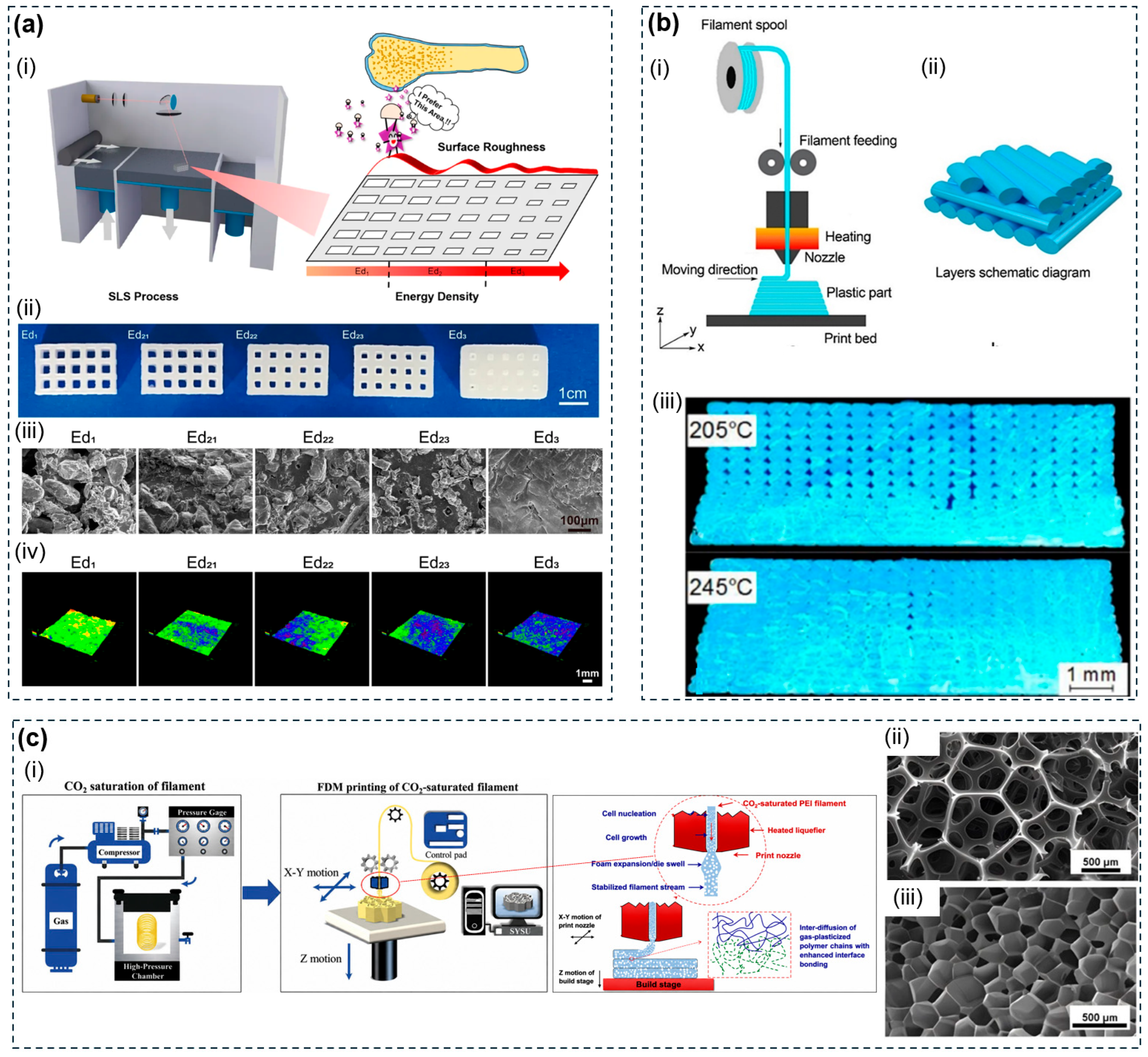
Figure 6: In-process pore formation strategies in powder bed fusion and extrusion-based printing. (a) SLS partial sintering of PCL scaffolds (i) schematic showing Ed-dependent surface roughness evolution; (ii) scaffold photographs, (iii) SEM surface morphology, and (iv) 3D topography maps from rough (Ed1) to dense smooth (Ed3). Reproduced with permission from Ref. [144] (CC BY 4.0). (b) (i) FDM void formation via partial neck growth; (ii) schematic of inter-raster contact geometry; (iii) cross-sectional micrographs at 205°C and 245°C showing void reduction at higher nozzle temperature. Reproduced with permission from Ref. [141]. Copyright 2021, Elsevier. (c) (i) In situ foam 3D printing of CO2-saturated filaments: CO2 saturation, FDM delivery, and nozzle depressurization-induced cell nucleation; SEM images of representative (ii) open-cell and (iii) closed-cell foam architectures. Reproduced with permission from Ref. [147]. Copyright 2020, Elsevier.
Among the materials surveyed in Section 3, how well a polymer forms pores during printing itself depends on its thermal behaviour and physical structure. This varies widely across PCL, PA12, TPU, PLA, and PEEK, and sets both the achievable pore size range and which platform each material can use. PCL and PCL/hydroxyapatite composites are the standard materials for SLS partial sintering. PCL has a low melting point and broad thermal stability, which together create a wide sintering window. Across FDM and SLS platforms, PCL scaffolds reach porosities of 48 to 77% with channel dimensions of 160 to 700 μm [96,97]. Adding 10 to 30 wt% HA narrows the usable range of scan speeds, because HA particles disrupt direct contact between PCL particles and require slower scanning to keep the structure coherent at the same energy input [143]. PA12 is the main engineering polymer used in SLS. Its sintering window of about 20 to 30°C is narrow enough to minimise warping, and its near-spherical powder packs well, together giving controllable pore sizes below 200 μm [90,103,104,139,145]. TPU is a segmented block copolymer, which allows it to form pores during printing on both SLS and FDM. Its sintering region of 171 to 242°C is wide enough to support partial sintering, and its elastomeric recovery allows printed pores to deform repeatedly without permanent damage. However, cryomilled TPU powder has an irregular shape and loses quality when reused, which limits how well it processes on SLS [100,101,148]. PLA and PEEK both use the FDM route. PEEK needs purpose-built hardware operating at 390 to 430°C, within a narrow thermal processing window between incomplete fusion and thermal degradation [86,98,141]. PLA can additionally use DIW immersion precipitation, where polymer concentration replaces thermal energy density as the main control over porosity. This allows pore formation at room temperature, which SLS and FDM cannot achieve [146]. The width of the usable thermal window sets how reliably each polymer can form pores during printing, while powder shape and polymer architecture set the platforms each can access and the pore sizes each can reach.
This section surveys three further physicochemical strategies, gas-based foaming, polymerized high internal phase emulsion (PolyHIPE) synthesis, and surface etching, that add porosity through agents or reactions applied as a separate processing step. Unlike the strategies surveyed in Section 3.2, Section 3.3, Section 3.4 and Section 3.5, each of these methods is restricted to a narrower set of materials or processing conditions, making them supplementary rather than broadly applicable routes to porosity. Gas-based foaming dissolves a blowing agent into the polymer under high pressure, then uses rapid depressurization or heating to create a supersaturated state in which gas bubbles, called cells, form and grow until the matrix solidifies. This approach only works for thermoplastics with a clear processing window between the onset of melting and thermal decomposition. PolyHIPE synthesis polymerizes the continuous phase of a high internal phase emulsion, which requires polymerizable precursors rather than an already formed thermoplastic part. Surface etching uses a chemical reagent to hydrolyse or dissolve bonds at the polymer surface, which restricts it to polymers containing the relevant reactive groups, such as ester bonds in polyesters.
Gas-based foaming takes two main forms, chemical foaming agents and supercritical CO2 foaming. Chemical foaming agents offer the simpler of the two routes. Solid additives blended into the feedstock decompose thermally during melt processing, releasing gas directly inside the material without any external gas injection [33,149]. Supercritical CO2 foaming is the most widely used strategy. CO2 has a critical point that is easy to reach, which lets it act as a blowing agent that leaves no residue behind and softens the polymer once dissolved [33,150,151] (Fig. 6c). Rapid depressurization creates supersaturation and triggers bubble nucleation. Saturation pressure is the main parameter controlling this process. Higher pressure increases the number of nucleation sites and reduces pore size. Temperature has an optimal middle value rather than a simple increasing or decreasing effect. If the melt is too viscous, bubble growth is limited. If it is too thin, bubbles merge together instead of staying separate [152,153,154]. Adding a plasticizer reduces melt strength and favours open-cell structures. Adding a chain extender instead favours closed-cell structures with better mechanical integrity [150,153]. PLGA has a narrow thermal processing window, which effectively excludes it from the FDM route [119]. This makes supercritical CO2 foaming a natural, solvent-free fabrication route for this polymer. PLGA scaffolds reinforced with hydroxyapatite reach porosities of 70 to 90%, with pore sizes matching what trabecular bone requires [152]. PLA tends to fracture brittlely and produce acidic degradation products when processed by FDM [98,155,156]. Combining supercritical CO2 foaming with poly(ethylene glycol) (PEG) as a plasticizer raises the open-cell content of PLA far above that of the unmodified polymer [153]. TPU has a segmented architecture that supports both SLS and FDM platforms [100,101]. It can also be processed by supercritical CO2 foaming, and a subsequent secondary supercritical N2 (ScN2) foaming step substantially reduces shrinkage after expansion [157].
Beyond the strategies described above, two further methods operate at opposite ends of the length scale. PolyHIPE synthesis is an emulsion-templated route in which the continuous external phase of an emulsion is polymerized while the dispersed internal phase, exceeding 74% of the volume, is later removed to leave an open-cell structure. Void size can be tuned from a few to over one hundred micrometres through surfactant concentration and internal phase fraction, and PCL-containing polyurethane variants made this way reach porosities above 70% with compressive strength suitable for tissue engineering [158]. NaOH etching instead works at the surface, hydrolysing ester bonds in PLA and PLGA to create micropits without affecting bulk mechanical properties, making it a post-print surface activation step rather than a bulk pore-forming method [98,119,159].
The strategies surveyed above fall into two broad paradigms. Design-driven architectures set pore geometry exactly before fabrication begins, while physicochemical strategies, including phase separation, freeze-drying, sacrificial templating, and partial fusion, introduce porosity through material-level processes below the printable resolution floor. Each strategy has its own strengths and limits. Design-driven pore formation allows patient-specific pore placement but cannot reach sub-100 μm scales. Phase separation extends into the low-micrometre range but only works for solution-processable polymers. Freeze-drying and ice-templating produce direction-aligned channels that mimic natural tissue but need water-compatible materials. Sacrificial templating works with the widest range of materials and uniquely creates channels open enough for fluid flow, at the cost of extra processing steps. Partial fusion needs no separate processing step but ties pore fraction directly to structural density. These strategies are not mutually exclusive. Hybrid combinations, most often post-print phase separation or freeze-drying applied to a printed scaffold, or sacrificial templating co-printed alongside the structural material, extend the pore scale range beyond what either paradigm reaches alone.
3.7 Strategy-Dependent Performance and Limitations
The five strategies surveyed in this section each occupy a distinct window of advantage, yet no single strategy simultaneously satisfies all the requirements that tissue engineering applications demand. Design-driven pore formation delivers spatial precision and mechanical integrity but cannot access pore scales below the printer resolution floor. Physicochemical strategies reach these finer scales and achieve higher porosity, but they cannot specify pore location or geometry with the same fidelity. Selecting among them requires weighing resolution against porosity, mechanical competence against material compatibility, and process simplicity against architectural control. Table 2 provides a consolidated comparison of these strategies across the key dimensions discussed below.
Table 2: Comparison of pore-forming strategies for 3D-printed porous polymers.
| Strategy | Pore Formation | Pore Geometry | Pore Size | Porosity | Representative Polymers | Key Feature | Mechanical Properties | Ref. |
|---|---|---|---|---|---|---|---|---|
| Design-driven pore formation | CAD-defined geometry reproduced by printing | Lattices; TPMS; graded architectures | ~100 μm–several mm | Medium–high | PCL, PLA, PEEK, TPU, PA12, GelMA, PEGDA | Deterministic spatial pore encoding | Hydrogels: kPa modulus; PBF: 40–55 MPa tensile strength; PCL SLS: 0.6–10 MPa compressive strength | [86,91,96,99] |
| Phase separation (DIPS/VIPS/TIPS) | Polymer-lean phase removal by solvent exchange or thermal quench | Sponge-like; bicontinuous; tubular | 1–300 μm; TIPS: 10–90 μm + 1–2 μm | up to 95% | PCL, PLA, PLGA | Sub-10 μm pores; dual-scale architecture | PLLA/PLGA/PCL: 0.067 MPa strength, 0.98 MPa modulus; PLA/nHA: 23 MPa modulus | [17,68,120] |
| Freeze-drying/Ice-templating | Ice template removal by sublimation | Random; lamellar; columnar; radial | 24–514 μm; cryogels: 90–300 μm | 81–95% | GelMA, alginate, collagen, chitosan, silk fibroin | Anisotropic channels; injectable cryogels | GelMA cryogels: 2–13 kPa modulus; 0.16–0.96 MPa strength; 90–94% compression | [30,124,125] |
| Sacrificial templating | Sacrificial phase removal by dissolution or melting | Spherical pores; perfusable channels; mould-defined pores | Particles: <500 μm; channels: 100 μm–1 mm | up to 70% | GelMA, alginate, collagen, PDMS, PLA, pHEMA | Perfusable channels; broad matrix compatibility | Matrix-dependent; hydrogels: kPa; PDMS/PLA/pHEMA: MPa | [132,134,138] |
| Partial fusion | Incomplete fusion during printing | Lack-of-fusion pores; gas pores; inter-raster voids | Sub-200 μm | 24–64% | PCL, PA12, TPU, PEEK, PLA | No post-processing; energy-density control | PCL/PCL-HA SLS: 0.1–10 MPa compressive/yield strength | [103,141,142] |
From the standpoint of achievable pore architecture and size range, design-driven pore formation is the only strategy enabling deterministic spatial pore encoding. Its minimum pore size is constrained by the intrinsic resolution of the printing platform, typically approximately 150 μm for FDM and approximately 100 μm or below for VPP. Physicochemical strategies operate independently of printer resolution [85]. Phase separation produces pores from 1 to 300 μm in sponge-like bicontinuous networks [110]. Freeze-drying and ice-templating generate pores from approximately 24 to 514 μm and uniquely provide directionally aligned anisotropic channels [30]. Sacrificial templating spans spherical voids below 500 μm to millimetre-scale perfusable channels suitable for convective fluid flow [133]. Partial fusion produces the narrowest range, typically below 200 μm, with irregular voids governed by process parameters rather than geometric intent [140]. Mechanical performance separates these strategies into two groups. Design-driven and partial fusion strategies deliver the highest structural competence, with partially sintered PCL maintaining structural integrity across 24 to 64 percent porosity and strength predictably decreasing as energy density is reduced. Phase separation and freeze-drying produce scaffolds with porosity up to 95 percent, which limits absolute strength, although directional ice-templating partially offsets this through anisotropic mechanical properties and cryogels offer reversible compression up to 90 percent strain without permanent damage. Design-driven architectures allow the porosity-strength trade-off to be rationally engineered through geometry selection, whereas physicochemical methods determine it indirectly through processing conditions that cannot be independently optimised for both pore fraction and mechanical integrity [91]. Material compatibility differs sharply across these strategies. Sacrificial templating accommodates the widest range of structural matrices, including GelMA, alginate, collagen, PDMS, PLA, and pHEMA. Freeze-drying is confined to water-soluble natural polymers and hydrogel precursors, excluding thermoplastics and photocurable synthetics such as PEGDA [31]. Phase separation is limited to synthetic aliphatic polyesters soluble in organic solvents, namely PCL, PLA, and PLGA [112]. Design-driven strategies accommodate a broad range of thermoplastics and photocurable resins, with the specific platform determining which material can be processed. Partial fusion demands a clear sintering window that excludes PLGA from the FDM route [119]. Each physicochemical strategy carries inherent limitations. Residual solvent in phase-separated constructs can compromise cytocompatibility, and pore reproducibility remains sensitive to demixing conditions. Incomplete porogen removal in sacrificial templating produces closed pores, while common fugitive inks can be cytotoxic at elevated concentrations. Partial fusion restricts the material library to semi-crystalline thermoplastics. Scalability further separates these strategies. Design-driven FDM and partial fusion SLS require little or no post-processing and are the most directly scalable. Freeze-drying is limited by long sublimation times, sacrificial templating by additional template fabrication and removal steps, and phase separation occupies an intermediate position because of solvent exchange and drying requirements.
The strategies surveyed in this section can be integrated with stimuli-responsive smart polymers to produce adaptive scaffolds whose pore architecture or function changes in response to external triggers. Design-driven pore formation is particularly compatible with shape memory polymers such as PLA/PEG blends and PCL-based copolymers, whose thermal transition temperature can be tuned near body temperature through composition control. This integration has been demonstrated in 4D-printed cardiac occluders [160], vascular stents [161], and intestinal stents [162], where the printed macrostructure is programmed into a compact temporary configuration and recovers its functional shape at the implantation site. Phase separation and freeze-drying are suited to stimuli-responsive hydrogels whose response mechanisms rely on hydrophilic–hydrophobic transitions or dynamic covalent bonds, including poly(N-isopropylacrylamide)-based thermoresponsive hydrogels and pH-responsive systems incorporating Schiff base or boronate ester linkages [163]. Because these strategies can be implemented under aqueous or other low-temperature conditions, they can preserve the chemical functionality required for stimulus responsiveness without exposing the material to melt-processing temperatures that may degrade thermally labile groups. Sacrificial templating can accommodate magnetically responsive composites by incorporating Fe3O4 or NdFeB particles into the structural matrix, enabling remote magnetic activation or actuation after the sacrificial phase is removed [164]. Partial fusion strategies are constrained to semi-crystalline thermoplastics with a sufficient sintering window, limiting the range of stimuli-responsive chemistries that can be directly printed, although post-printing surface modification can introduce responsiveness onto pre-formed porous architectures. The key consideration across all five strategies is that the fabrication conditions must not compromise the functional groups or phase structures that confer stimulus responsiveness. Selecting an appropriate integration route therefore requires matching the fabrication conditions to the specific physicochemical basis of the material’s responsiveness as well as the pore architecture to the biological target.
These comparisons point to a practical selection logic. For load-bearing hard tissues such as bone and cartilage, design-driven or partial fusion strategies are the natural first choice. Hybrid approaches combining a printed macrostructure with post-print phase separation or sacrificial templating can extend the pore scale downward when additional microporosity is required. For soft tissues including nerve, skin, and cardiac muscle, freeze-drying, ice-templating, and phase separation are better suited. For thick tissues demanding vascularisation, sacrificial templating through fugitive ink extrusion remains the only strategy capable of producing perfusable channel networks at physiologically relevant cell densities. For applications prioritising rapid production, design-driven FDM and partial fusion SLS offer the shortest process chains.
As established in the Introduction, the properties of porous polymers come from material chemistry working together with pore scale, connectivity, and spatial organisation, not from polymer chemistry alone. Different combinations of polymer material and pore architecture give rise to different physical and chemical property changes, and these changes activate different mechanisms across application domains. Open-cell macropores enable cell migration and drug release in biomedical applications. Periodic solid-void patterns interact with electromagnetic, acoustic, and thermal waves to enable shielding and absorption. Interconnected through-pores and pore wall surface area drive adsorption and selective transport in separation and catalysis. Pore topology and relative density constrain deformation under load, enabling energy absorption and sensing in mechanical metamaterials and flexible electronics. 3D printing makes it possible to set these pore-level parameters independently and at the same time within a single construct, something stochastic fabrication methods cannot achieve. This chapter introduces these applications across six functional domains, covering biomedical engineering, environmental and chemical engineering, energy devices, mechanical structures and metamaterials, flexible electronics, and emerging photonic, microfluidic, and robotic applications. Table 3 consolidates the pore size, connectivity, geometry, and material requirements relevant to each domain, serving as a practical reference for matching the strategies described in Section 2 and Section 3 to specific functional targets.
Table 3: Pore design parameters of 3D printed porous polymers across application domains.
| Application Domain | Porosity Function | Critical Pore Size | Preferred Pore Geometry | Materials/Requirements | Key References |
|---|---|---|---|---|---|
| Structural Mechanics | Load redistribution; auxeticity; energy absorption | Strut ~0.5 mm; unit cell 1–10 mm | Open lattices; octet-truss; re-entrant honeycomb | Photocurable resin; high stiffness-to-weight ratio; elastic recovery | [165,166,167] |
| Fluid & Interface | Selective permeation; capillary evaporation | Macro 10 μm–1 mm; meso 2–50 nm; micro <2 nm; pinhole ~14 μm | Open-cell foam; sponge; aerogel; zeolite-coated mesh; capillary network | WCA 0° or >150°; underwater OCA >150°; PDMS, PU, cellulose | [168,169,170,171] |
| Biomedical—Hard Tissue | Bone ingrowth; vascularization; nutrient transport | 100–400 μm; >600 μm for high porosity | Interconnected macro/micropores; trabecular-like; TPMS gyroid | PCL, PLGA, HA; stiffness 300–500 MPa; HA surface modification | [172,173,174] |
| Biomedical—Soft Tissue & Drug Delivery | ECM mimicry; cell infiltration; drug release | 100–300 μm channels; <10 μm drug release pores | Fibrous networks; aligned channels; interconnected pores | PCL, PLGA, PU; collagen; hydrogels; hydrophilic; stimuli-responsive | [2,175,176,177] |
| Chemistry, Catalysis & Adsorption | Active sites; mass transport | Micropores <2 nm; mesopores 2–50 nm | Interconnected micro/mesopores; amorphous microporous network | CMP, HCP, PAF, CTF; SSA 300–2500 m2/g; heteroatom functionalization | [178,179,180,181] |
| Optics | PBG; structural color | 400–700 nm; Bragg: mλ = 2d(n2eff − sin2θ)½ | Inverse opal; APC; CPC; lamellar 1D | PS, PMMA, PC; monodispersity; high RI contrast | [182,183] |
| Sensing—Piezoresistive | Resistance change under deformation | Salt template 60–550 μm; PVDF pores ~1 μm | 3D cubic pores; open porous network | PDMS; PVDF; AgNW coating | [184,185] |
| Electrochemical Energy Devices | Ion/electron/gas transport | Micro <2 nm; meso 2–50 nm; macro tens–hundreds μm; GDL porosity 30–70% | Hierarchical carbon pores; bicontinuous gas-diffusion network | Porous carbon; CNT composites; high conductivity; large SSA; PFPE | [186] |
| Acoustics & EMI Shielding | Acoustic dissipation; EM scattering | l ≫100 μm; MPP ~0.2 mm; Λ = 33–56 μm | Open-cell foam; MPP; spring–mass resonators; pores Df ≈ λ/8–λ/5 | PU; melamine foam; PDMS; σ ~104–105 Nm−4 s; CNT/MXene composites | [187,188,189,190] |
| Thermal Management | PDRC scattering; PCM encapsulation | PDRC 200 nm–7 μm; PCM <50 nm | Ordered micro/nanopores; aligned CNT arrays; interconnected PCM pores | PMMA, n = 1.49; porosity >90%; –NH2 functionalization; >2500 cycles | [191,192] |
Biomedical engineering is the most extensively explored application domain for 3D-printed porous polymers, driven by the recognition that biological tissues are themselves porous structures in which interconnected void networks govern mass transport, mechanical compliance, and cell-matrix signalling [193]. Conventional implant fabrication methods, such as injection moulding, subtractive machining, and particulate leaching, offer limited architectural resolution. They cannot produce patient-specific geometries with spatially programmed porosity gradients [194]. 3D printing resolves these constraints by depositing polymeric bioinks and thermoplastics layer by layer into structures with macropores of 100 μm to several millimetres, programmed directly through toolpath design and lattice topology, turning porosity from an incidental byproduct of manufacture into a precisely engineered functional variable that can be tailored independently across different regions of the same construct. Applications of 3D-printed porous polymers in biomedical engineering fall into several main areas, including bone and cartilage repair, vascular and neural regeneration, vascularized tissue construction and in vitro tissue modelling, and programmable drug delivery systems. The relationships among fabrication strategy, material compatibility, pore morphology, and biomedical function are summarized in Fig. 7.
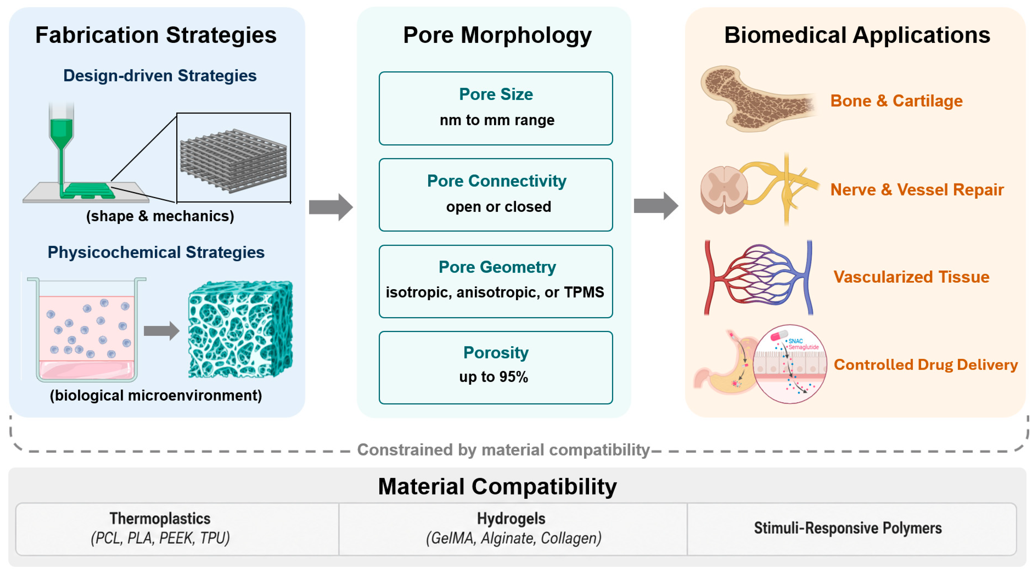
Figure 7: Structure–application framework for 3D-printed porous polymers in biomedical engineering. Design-driven strategies primarily define the macroscopic shape and mechanical architecture of printed constructs, whereas physicochemical pore-forming strategies generate biologically relevant porous microenvironments. Together with material compatibility, these fabrication routes determine pore size, connectivity, geometry, and porosity, which in turn govern the suitability of porous polymer constructs for bone and cartilage repair, nerve and vascular repair, vascularized tissue construction, and controlled drug delivery.
Bone and cartilage repair is the most mature application area within this domain, supported by extensive preclinical evidence across a range of polymer platforms and defect geometries. This maturity spans several design challenges, including the baseline pore architecture needed for osseointegration, oxygen delivery in large defects, mechanical demands in load-bearing sites, and the biphasic requirements of the osteochondral interface. Effective osseointegration requires open-cell macroporous architectures with interconnected pore diameters of 300 to 500 μm. This permits osteoblast infiltration, vascular ingrowth, and nutrient diffusion throughout the construct interior. Gradient porosity distributions further allow independent optimisation of mechanical stiffness at the load-bearing core and biological responsiveness at the implant-tissue interface [195]. In large volumetric defects, oxygen diffusion limitations constrain the osteogenic activity of transplanted cells within the scaffold interior. Farris et al. addressed this by embedding oxygen-releasing PVA-PLGA microtanks within porous PCL scaffolds fabricated by MEX printing. Even transient oxygen delivery over about 8 h was enough to enhance osteopontin deposition and mineral accumulation in murine defect models [196]. For load-bearing applications, Wei et al. fabricated porous PEEK scaffolds by FDM and coated them with polydopamine and Mg2+. The open-cell network supported osteoblast migration and offset the chemical inertness of PEEK, while Mg2+ release accelerated early angiogenesis in a rabbit model [197]. Critchley et al. met this requirement by reinforcing cell-laden alginate hydrogels with 3D-printed PCL fibre networks, and demonstrated that following chondrogenic priming (Fig. 8a), such biphasic constructs supported hyaline-like cartilage repair in critically sized caprine osteochondral defects over a six-month implantation period, with regenerated collagen fibre architecture more closely resembling that of native tissue than commercial scaffold controls [198]. In each case, pore connectivity, surface chemistry, and mechanical zonation were optimised for a single osseointegration phase rather than co-encoded within one printed pore network.
Vascular grafts and neural conduits represent a functionally distinct application category. Here the relevant structural feature is not isotropic interconnected macroporosity but open-cell, anisotropic, longitudinally aligned microchannels of 100 to 300 μm diameter. These channels guide regenerating cells and infiltrating vasculature directionally, replacing the random void networks of conventional porous scaffolds with spatially oriented conduits matched to the fascicular organisation of the target tissue. This principle has been demonstrated across peripheral nerve repair, spinal cord injury, and small-diameter vascular grafts. In peripheral nerve repair, Singh et al. [199] developed photocurable polymer conduits enclosing an aligned cryomatrix lumen, combining contact guidance from fibre orientation with biochemical stimulation from incorporated nerve growth factor. This design regenerated critical-sized rat sciatic nerve defects to a level comparable with autograft controls, a benchmark conventional hollow conduits consistently fail to reach. In the central nervous system, inhibitory glial scarring and the precise fascicular organisation of spinal white matter impose strict geometric constraints on scaffold design. Koffler et al. [200] used microscale continuous projection printing to fabricate PEGDA-GelMA hydrogel scaffolds individually matched to spinal cord lesion cavity geometries, incorporating microchannels aligned with host axonal tracts (Fig. 8b). When loaded with neural progenitor cells and implanted acutely after complete spinal cord transection in rats, these constructs supported the formation of functional neural relays. Animals recovered measurable motor evoked potentials and significant hindlimb locomotor scores over a six-month follow-up period. For small-diameter vascular substitutes, which remain clinically problematic due to compliance mismatch and thrombotic occlusion, Cui et al. [201] bioprinted catechol-functionalised GelMA constructs with co-axial smooth muscle and endothelial cell layers. These achieved autonomous vascular connection and tissue remodelling within about six weeks in a murine model, setting an in vivo benchmark for bioprinted vascular grafts. This directional guidance was established by matching channel orientation to host architecture at implantation. Whether channel-wall degradation during the months-long regeneration window preserves this alignment, or allows regenerating tissue to drift before native fascicular organisation is restored, remains untested.
The convergence of 3D bioprinting and organoid biology has enabled in vitro tissue models that bridge the gap between millimetre-scale self-organised organoids and macroscopic organ architectures relevant to drug screening and disease modelling [202,203]. Cell-laden hydrogel matrices function as open-cell microporous scaffolds whose sub-micrometre to micrometre pore networks support 3D cell encapsulation, nutrient diffusion, and morphogen-driven self-organisation, functions inaccessible to dense polymer constructs regardless of macroscopic geometry. This approach has produced models spanning the gastrointestinal tract, skin, cardiac tissue, and tumour microenvironments. Brassard et al. [204] deposited intestinal stem cells at high density directly into permissive extracellular matrices using a microscope-integrated extrusion bioprinter (Fig. 8c). This generated centimetre-scale epithelial tubes bearing self-organised crypt-villus domains and functional Paneth cell secretory responses with perfusable lumens. Sequential deposition of stomach and intestinal stem cells further enabled gastrointestinal boundary models that preserved region-specific gene expression profiles throughout extended culture. Toward clinically deployable tissue substitutes, Jin et al. [205] used multimaterial bioprinting with Acellular Dermal Matrix (ADM) and GelMA bioinks to reproduce full-thickness skin, including a keratinocyte epidermal layer, a fibroblast dermal compartment, and a human umbilical vein endothelial cells (HUVECs)-seeded vascular mesh. Transplanted onto murine wounds, the construct kept cells viable for at least one week while accelerating re-epithelialisation. The same paradigm has been extended to cardiac tissue. Noor et al. [206] fabricated thick vascularised cardiac patches from patient-specific bioinks derived from autologous omental tissue, supporting cardiomyocyte maturation after transplantation and demonstrating proof-of-concept printing of cellularised heart structures [207]. Beyond regenerative applications, Dey et al. [208] bioprinted a dynamic-flow vascularised breast tumour model, demonstrating dose-dependent doxorubicin cytotoxicity and approximately 70% tumour volume reduction following perfusion of human epidermal growth factor receptor 2 (HER2)-targeting chimeric antigen receptor T cells (CAR-T cells) through the vascular channels. These pore networks were sized at the time of printing to support nutrient and morphogen diffusion. Whether cell-deposited matrix progressively occludes these pathways as constructs mature toward the centimetre-scale, multi-week timepoints reported here has not been quantified.
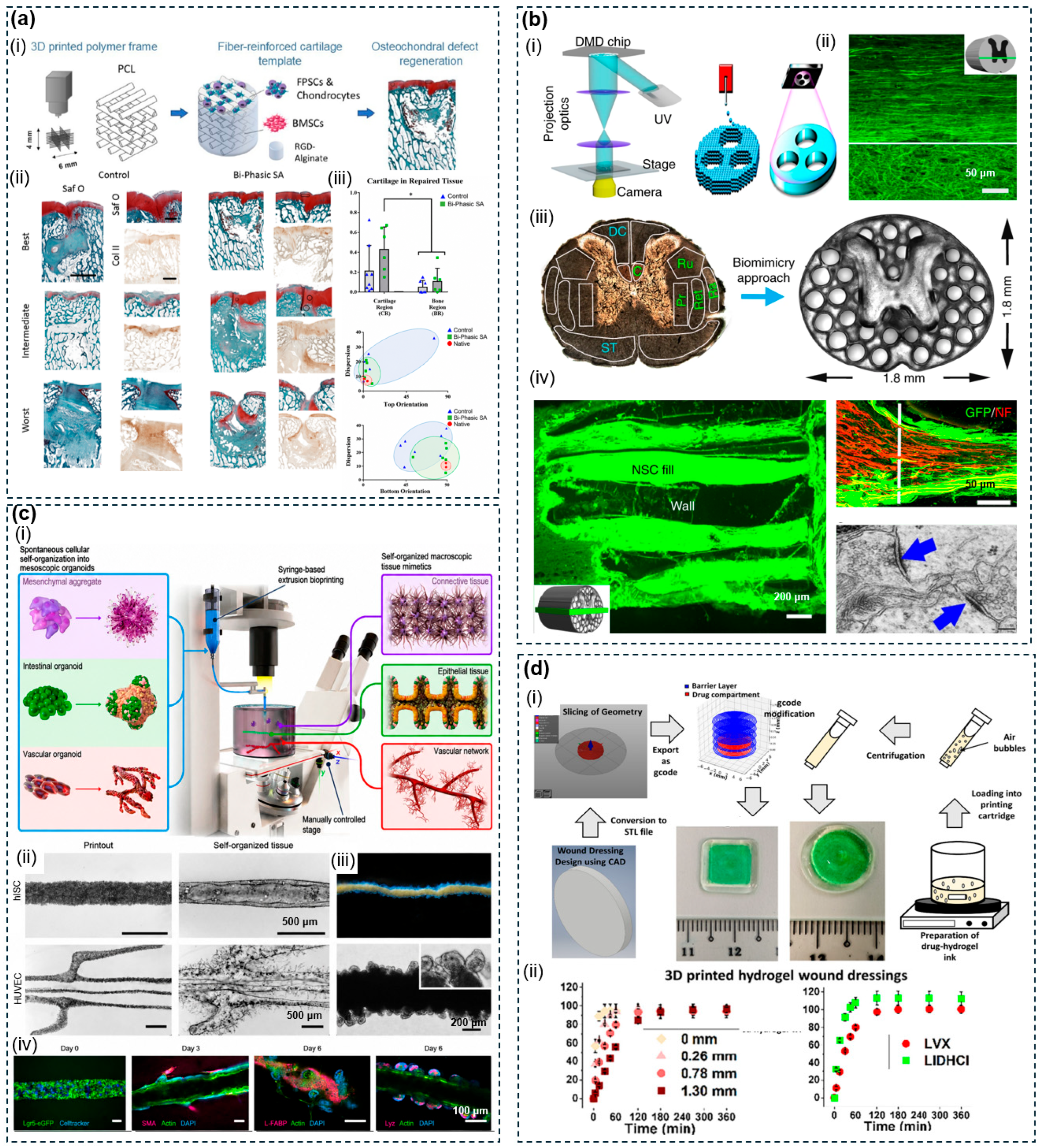
Figure 8: Representative biomedical applications of 3D-printed porous polymer scaffolds. (a) PCL-reinforced bi-phasic cartilaginous templates for osteochondral regeneration (i) construct schematic; (ii) histological staining of repair outcomes; (iii) cartilage quantification and collagen orientation analysis. Reproduced with permission from Ref. [198]. Copyright 2020, Elsevier. (b) μCPP-printed biomimetic PEG–GelMA scaffolds for spinal cord regeneration: (i) printing setup; (ii) axonal regeneration within channels; (iii) biomimetic scaffold design from rat spinal cord anatomy; (iv) NPC survival and synapse formation. Reproduced with permission from Ref. [200]. Copyright 2019, Springer Nature. (c) (i) BATE strategy for macroscale self-organized tissue construction; (ii) printout-to-tissue self-organization; (iii) intestinal epithelial tube; (iv) immunofluorescence of co-cultured constructs. Reproduced with permission from Ref. [204]. Copyright 2021, Springer Nature. (d) DIW-fabricated drug-loaded hydrogel wound dressings: (i) fabrication workflow; (ii) tunable drug release profiles under varying barrier layer thicknesses. Reproduced with permission from Ref. [209]. Copyright 2022, Elsevier.
Porous polymer architecture also functions as a programmable kinetic variable in controlled drug delivery. Pore diameter and open-cell connectivity set the surface-area-to-volume ratio and diffusion path length available to encapsulated therapeutics, independently of bulk polymer degradation rate. Smaller, denser pore networks sustain prolonged release, while macroporous open-cell geometries permit faster delivery, allowing a single printed construct to encode spatially distinct release kinetics across its volume. This capability spans systemic bone delivery, topical wound delivery, and local tumour delivery. Abu Awwad et al. [64] combined porogen-tuned PLGA microparticles with a PLGA/PEG paste into 3D-bioprintable scaffolds whose macroporosity was defined directly through printing. The construct delivered runt-related transcription factor 2 (RUNX2) within the first week, driving stem cell differentiation and bone formation in a murine model. Architectural programmability extends to topical delivery. Teoh et al. [209] printed photocurable chitosan methacrylate wound dressings with spatially discrete drug-loaded and drug-free layers printed in a single build (Fig. 8d), demonstrating that the thickness of overlying drug-free layers served as the primary determinant of diffusional resistance for independently tunable release of analgesic and antimicrobial agents, with Weibull-type kinetic modelling confirming that architectural parameters quantitatively described release behaviour across multiple design configurations. The local delivery rationale has been further validated in primary bone malignancies, where systemic chemotherapy carries severe off-target toxicity. Lahr et al. [210] implanted 3D-printed PCL scaffolds loaded with three doxorubicin dose levels into a humanised osteosarcoma defect in non-obese diabetic (NOD) scid gamma mice, achieving sustained local drug retention and tumour cell death without detectable systemic side effects.
Taken together, the four application areas surveyed above show that 3D printing can reliably translate pore geometry into specific biological functions, validated mainly in small-animal models spanning bone, nerve, vascular, gastrointestinal, skin, cardiac, and tumour applications. Most successes, however, remain limited to sub-centimetre constructs evaluated over weeks, with each scaffold engineered around a single function rather than several working together. Extending deterministic pore design to the larger, hierarchical, multi-phase architectures needed at clinically relevant scale remains the central challenge for this domain.
The four application areas surveyed above share a set of relationships between pore morphology and biological performance that operate across tissue types. Pore size governs the fundamental trade-off between cell infiltration capacity and mechanical competence. Interconnected pores of 300 to 500 μm permit osteoblast infiltration and vascular ingrowth in bone scaffolds, while channels of 100 to 300 μm provide directional guidance for regenerating axons and endothelial cells in nerve and vascular conduits [195,200]. Below approximately 100 μm, cell infiltration becomes physically restricted regardless of tissue type, as the pore diameter approaches the dimensions of mammalian cells themselves. Above approximately 500 μm, structural integrity degrades faster than the biological benefit of additional void space increases. Pore connectivity determines whether this size-dependent capacity is actually realised. Open-cell interconnected networks support convective and diffusive transport of oxygen, nutrients, and metabolic waste throughout the construct interior, sustaining cell viability over the weeks to months required for tissue regeneration [195,206]. Closed pores produce dead zones of hypoxia and necrosis that no biochemical modification of the pore wall surface can compensate. Pore geometry further dictates the spatial organisation of regenerated tissue. Isotropic pore networks produce randomly oriented tissue ingrowth suited to bone and dermis, while aligned anisotropic channels direct axonal and vascular growth along predetermined axes matched to the fascicular organisation of nerve and muscle [200]. TPMS surfaces eliminate the stress concentrations at nodal junctions that characterise strut-based lattices, distributing mechanical load and cell attachment uniformly across continuously curved surfaces. Gradient porosity extends this geometric control to the zonal level, enabling a single printed construct to transition from a chondral to an osseous pore regime [198]. These relationships point to a broader conclusion: pore size, connectivity, and geometry are not independent variables that can be optimised sequentially. They operate through the same set of transport and contact-guidance mechanisms, and changing one parameter inevitably shifts the effective range of the others. Recognising this coupling is a prerequisite for translating the empirical correlations surveyed here into predictive design rules.
4.2 Environmental and Chemical Engineering
Porous polymers, because their pore geometry can be precisely tuned for selectivity, throughput, and surface area, are well suited to addressing pressing environmental challenges [211]. These include the remediation of industrial effluents, the selective separation of immiscible phases, and the sustainable production of clean water, where selectivity, throughput, and long-term operational stability are all governed by the spatial organisation of void space within the functional medium. 3D printing translates computationally optimised pore architectures, including triply periodic minimal surfaces, ordered lattices, and hierarchical pore networks, directly into polymer structures with deterministic geometries. This replaces the empirical trial-and-error of conventional membrane and adsorbent fabrication with rational pore design [194,211]. This section surveys four application categories, covering oil-water separation, heavy metal and dye adsorption, enzyme and photocatalyst immobilisation, and solar-driven interfacial evaporation. Through these four categories, open-cell macropores of 0.1 μm to several millimetres govern convective transport and phase selectivity. Material-level micropores (below 2 nm) and mesopores (2–50 nm) maximise active surface area and adsorption capacity. The transition between open-cell and closed-cell configurations distinguishes architectures designed for fluid permeation from those optimised for thermal or acoustic isolation.
The selective separation of oil-water mixtures through porous polymer membranes exploits open-cell macropore networks, in which pore diameter and surface wettability together determine phase selectivity. Pores sized above the target droplet diameter, combined with superhydrophobic polymer surfaces, permit oil passage while blocking the aqueous phase, with tortuosity providing an additional handle on residence time and coalescence efficiency. This principle has been demonstrated through TPMS contactor geometry, ink-integrated superhydrophobic surfaces, and scalable powder-based manufacturing. Shimmery et al. [212] reported the first TPMS-based porous contactors for oil-in-water demulsification. Gyroid structures, with higher tortuosity and surface area than cylindrical or Schwarz-P counterparts, achieved 93% oil separation efficiency at only 10 mbar vacuum pressure, two orders of magnitude faster than natural coalescence. For membranes requiring intrinsic superhydrophobicity without post-fabrication surface treatments, Lv et al. [213] fabricated ordered porous PDMS structures by direct ink writing of nanosilica-filled viscoelastic ink (Fig. 9a). This integrated the superhydrophobic surface directly into the porous framework, eliminating the interfacial adhesion weakness typical of conventional coating-on-mesh approaches. The resulting membranes achieved over 99% oil-water separation efficiency and a flux of approximately 23,700 L·m−2·h−1, while retaining mechanical durability under abrasive damage. Extending this approach to scalable production, Zhang et al. [214] used selective laser sintering of unmodified TPU and TPE powders to produce flexible superhydrophobic membranes with micro-nano rough surfaces in a single step, achieving separation efficiency exceeding 99.3% and a gravity-driven flux of 3.23 × 10⁵ L·m−2·h−1.
The removal of heavy metal ions and synthetic dyes from wastewater through adsorption is constrained in practice by the aggregation, difficult recovery, and slow intraparticle diffusion of conventional powdered adsorbents. 3D printing addresses these limitations by consolidating functional materials into monolithic structures with designed pore networks that enable straightforward recovery and accelerated mass transfer. Wu et al. [181] integrated zeolitic imidazolate framework-67/-8(ZIF-67/ZIF-8) metal–organic framework (MOF) crystals and MXene sheets onto DLP-printed, polydopamine-coated scaffolds, achieving Congo red and Hg(II) removal rates of 99.13% and 94.10% and retaining above 80% efficiency after multiple regeneration cycles. At the material architecture level, Wu et al. [215] showed that replacing monolithic alginate-cellulose nanocrystal hydrogels with coaxially printed, hollow-filament grids increased Cu(II) adsorption capacity from about 48 to 68 mg·g−1 while tripling the adsorption rate constant, with access to ion-exchange, chelation, and electrostatic mechanisms across Cu, Zn, Cr, and Cd ions. Both adsorbents were evaluated under single-contaminant, short-cycle conditions. Competitive adsorption and regeneration in real multi-ion effluents remain untested.
Porous polymer scaffolds fabricated by 3D printing serve as mechanically stable, geometrically customisable carriers for both biological enzymes and inorganic photocatalysts [216]. In both cases, open-cell macroporous architectures with high surface-area-to-volume ratios maximise accessible catalytic sites, while interconnected pore channels reduce external mass transfer resistance. This delivers catalytic performance that free-catalyst suspensions and dense packed beds cannot sustain over repeated cycles. This principle has been applied to enzyme immobilisation, photocatalyst-loaded lattices, and dual-function photocatalytic membranes. Rybarczyk et al. [217] immobilised laccase onto open-structure PLA scaffolds by FDM. Confinement within the porous framework substantially improved enzyme stability, with immobilised laccase retaining 80% activity after 20 days versus 35% for free laccase, and improving estrogen removal from real wastewater by 10%. Kennedy et al. [218] compounded TiO2 nanoparticles at up to 34 wt% loading into PLA filament and printed lattices by FDM, where internal voids provided channels for water infiltration and photocatalyst contact, reducing microcystin half-lives from 36 to 3 h under solar simulation. Sreedhar et al. [219] adopted a complementary strategy, sintering polyamide PA2202 spacers with a crossed layers of parallels (CLP) TPMS architecture by SLS and mineralising β-FeOOH photocatalytic nanorods onto a polydopamine-polyethyleneimine intermediate layer (Fig. 9b). This dual-functional architecture achieved 98% removal of methylene blue and 4-nitrophenol within four hours as a photocatalytic degrader, while also recovering flux from three organic foulants without chemical intervention as a membrane surface cleaner. In each system the pore network was optimised for either reactant access or fouling resistance. The geometric overlap between these two design targets remains unexamined.
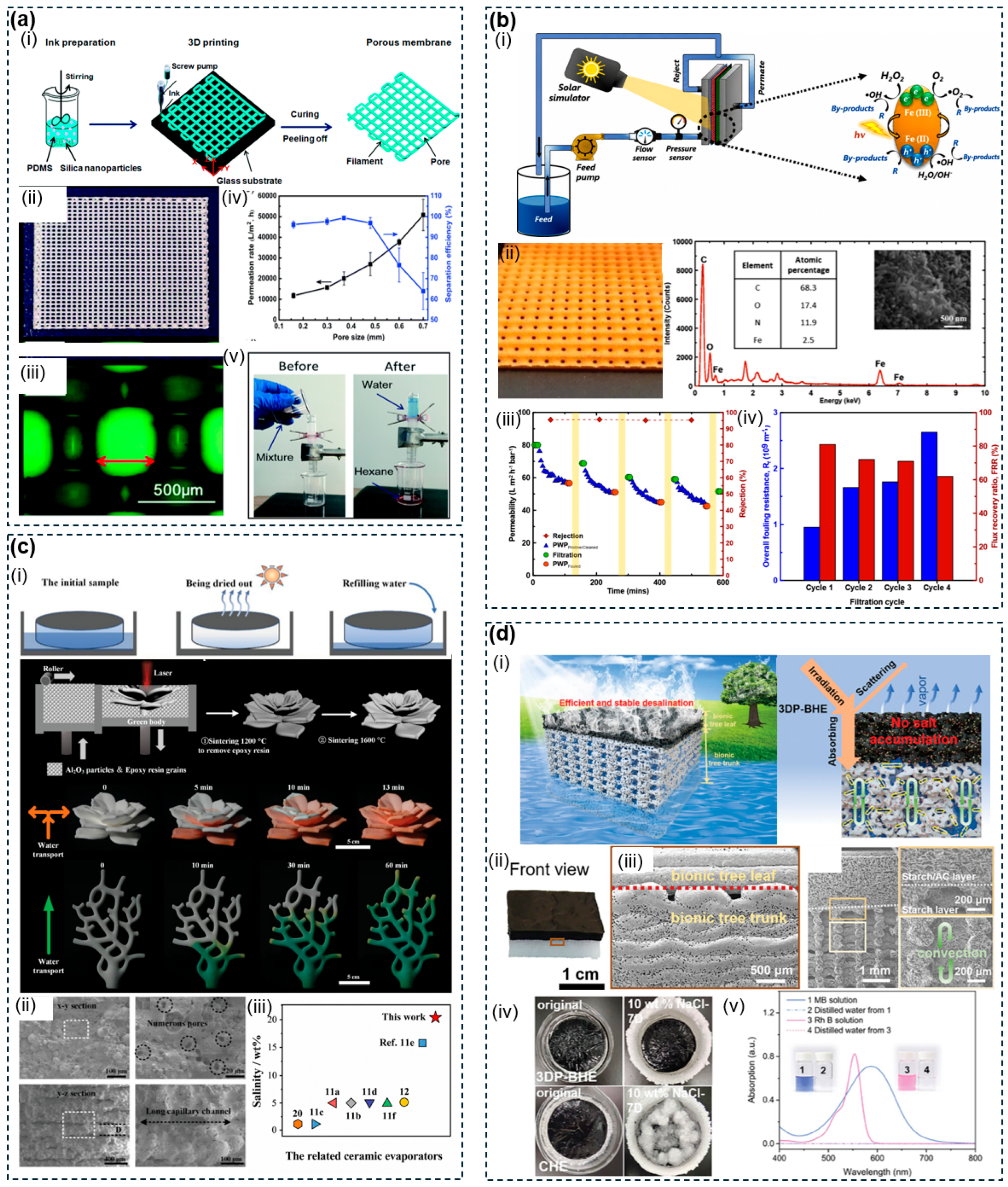
Figure 9: Environmental and energy applications of 3D-printed porous polymer structures. (a) DIW-fabricated superhydrophobic PDMS membranes for oil–water separation: (i) fabrication workflow; (ii) photograph; (iii) optical micrograph; (iv) permeation flux and separation efficiency vs. pore size; (v) gravity-driven separation demonstration. Reproduced with permission from Ref. [213]. Copyright 2017, The Royal Society of Chemistry. (b) SLS-printed TPMS photocatalytic spacer with β-FeOOH coating for membrane filtration: (i) crossflow photo-Fenton setup; (ii) spacer photograph and EDX/SEM; (iii) cyclic permeability and flux recovery; (iv) fouling resistance. Reproduced with permission from Ref. [219]. Copyright 2022, Elsevier. (c) SLS-printed superhydrophilic alumina ceramics for solar steam generation: (i) fabrication workflow and anisotropic water transport; (ii) micropore cross-sections; (iii) salinity handling comparison. Reproduced with permission from Ref. [220]. Copyright 2023, Wiley-VCH. (d) DIW-printed bionic hydrogel evaporator for solar desalination: (i) dual-layer design schematic; (ii) device photograph; (iii) cross-sectional SEM and salt convection pathways; (iv) salt accumulation comparison; (v) dye removal by UV-vis. Reproduced with permission from Ref. [171]. Copyright 2024, Wiley-VCH.
Solar-driven interfacial evaporation is a rapidly advancing application domain, in which the hierarchical pore network of 3D-printed polymer structures simultaneously serves as a capillary water transport pathway, a thermal insulation layer, and a photothermal conversion surface. Pore architecture directly determines evaporation rate, salt rejection capability, and long-term operational stability. This has been demonstrated across ceramic, hydrogel, and biomimetic polymer architectures. Wu et al. [220] used a sacrificial epoxy resin binder (12 wt%) in SLS-printed Al2O3/epoxy composite feedstock, which left a hierarchical micropore network after pyrolysis at 1200°C (Fig. 9c). Its anisotropic interlayer microslits achieved 5 μL water absorption in 14 ms and an evaporation rate of 2.6 kg·m−2·h−1. The rigid ceramic matrix maintained stable efficiency after ten drying cycles and salt-resistant operation in 20 wt% brine for 40 h, a performance inaccessible to purely polymeric evaporators. Wholly polymeric pore architectures can achieve competitive performance through hierarchical structural design. Zhang et al. [171] fabricated a bionic hydrogel evaporator from a biomass starch skeleton incorporating activated carbon as a solar absorber, with a bimodal porous trunk layer providing capillary water transport alongside salt ion convection and diffusion channels (Fig. 9d). The structure achieved an evaporation rate of 2.13 kg·m−2·h−1 at 90.5% energy efficiency under one-sun irradiation, maintaining 1.98 kg·m−2·h−1 over seven days in 10 wt% brine at a materials cost of $10.14·m−2. Zou et al. [221] addressed salt accumulation through a biomimetic bridge-arch evaporator with peristome-mimetic microchannels, establishing a double-layer liquid film that enabled continuous 200-h operation in 10 wt% saline water without post-cleaning, at an evaporation rate of 1.64 kg·m−2·h−1. Koh et al. [222] introduced regenerated cellulose as a hydratable plastic for solar evaporators. Architectural optimisation of the printed Woodpile structure overcame the rehydration saturation limit of hydrogel-based evaporators, achieving an evaporation rate of 7.35 kg·m−2·h−1 under three-sun irradiation with salinity reduced by four orders of magnitude below WHO drinking water standards.
The four application areas surveyed above share a common pattern. 3D printing translates pore geometry directly into separation, adsorption, catalytic, and evaporative performance that often exceeds conventional membranes and adsorbents on a single key metric. This progress remains mainly laboratory-scale, demonstrated with single contaminants and short test cycles, with each architecture optimised for one function rather than several working together. Validating these pore architectures under longer, more complex, field-relevant conditions remains the central challenge for this domain.
The performance of electrochemical energy storage and conversion devices is fundamentally constrained by the transport of ions, electrons, and reactive gases across electrode and membrane interfaces. The geometric complexity these interfaces require lies beyond the reach of conventional manufacturing methods. Electrode slurry casting produces planar films with limited thickness and uncontrollable porosity [223]. Commercial gas diffusion substrates offer little structural control over pore-size distribution or macroscale geometry. 3D printing addresses both limitations by enabling polymeric porous scaffolds with independently programmable porosity gradients, tortuosity, and external architecture [224]. Pore dimensions span from the nanoscale, for optical thermal management, to hundreds of micrometres, for electrochemical transport, and the same design logic extends to thermal management applications, where pore geometry governs heat storage capacity and radiative emission characteristics. 3D-printed porous polymers in energy devices are mainly applied in three areas. These are battery and supercapacitor electrodes, gas diffusion layers for electrochemical reactors, and thermal management systems.
3D porous polymer electrode architectures get around the fundamental trade-off in conventional thick electrodes between areal capacity and ion-transport rate. They replace randomly packed slurry films with deterministically structured networks in which electrolyte diffusion pathways are explicitly engineered. High-performance battery and supercapacitor electrodes require open-cell macroporous frameworks with interconnected porosities spanning 30 to 70% and pore dimensions of tens to hundreds of micrometres, ensuring complete electrolyte penetration across the active material depth while minimising ionic tortuosity at high discharge rates. The polymer binder scaffold maintains structural integrity without obstructing conductive pathways. This principle has been applied to battery cathodes, supercapacitor electrodes, and auxetic energy-storage lattices. Gupta et al. [225] fabricated hierarchically porous PLA/LiFePO4/CNT composite cathodes by FDM using in-house nanoengineered filaments (Fig. 10a). The CNT network spanning the pore framework sustained electron transport while the open porosity accommodated electrolyte penetration. Increasing the interconnected porosity from 10% to 70% in 300 μm-thick electrodes recovered specific capacity from 125 to 151 mAh·g−1 while maintaining an areal capacity of 4.4 mAh·cm−2, a direct consequence of reduced ionic tortuosity allowing electrolyte access throughout the full electrode depth. This shows that architectural engineering can decouple specific and areal capacities that conventionally move in opposition. For supercapacitor electrodes, Vaghasiya et al. [226] printed graphene/PLA composite filaments into 3D geometries using a handheld FDM device, then electrodeposited a Ti3C2@polypyrrole hybrid coating onto the porous scaffold surface (Fig. 10b). This achieved 81.4% capacitance retention after 6000 cycles at 5 A·g−1, since the rigid macroporous scaffold preserved ionic pathway geometry and suppressed delamination of the active coating under repeated volume changes. The device was integrated within the insulation voids of structural bricks as a proof-of-concept architectural energy storage system. Chang et al. [227] used SLA to print auxetic multicellular lattice frameworks, then deposited CoNi2S4/NiCo-layered double hydroxide (LDH) nanocomposites by electrodeposition. The structure stored elastic energy during deformation, sustaining a volumetric capacitance of 28.71 F·cm−3 across 55% tensile strain and 180° bending with 75.2% retention after 1000 cycles.
Gas diffusion layers fulfil an analogous mass-transport function in electrochemical flow reactors. The porous polymer scaffold must deliver gaseous reactants to the catalyst layer, evacuate products, and maintain hydrophobicity against electrolyte flooding, requirements that commercial PTFE-coated carbon paper substrates address with structurally fixed and compositionally uniform pore networks. Effective CO2 electroreduction requires open-cell polymer architectures with microporous to macroporous networks tuneable over several orders of magnitude in permeance, letting the diffusion layer act as the primary CO2-transport-limiting component while independently structured macroscale surface features control catalyst interfacial area and local species residence time. Wicks et al. [228] developed 3D-printable perfluoropolyether (PFPE) gas diffusion layers by large-area projection microstereolithography from UV-cured ternary monomer-porogen solutions (Fig. 10c), yielding homogeneous microporous and large-pore bicontinuous PFPE networks with CO2 permeances tunable over four orders of magnitude. Conformal copper nanoparticle catalyst layers spray-coated onto pyramid-array macrostructured layers showed that the surface morphology extended the diffusive residence time of locally produced CO near the catalyst, increasing the C2H4 to CO selectivity ratio 100-fold over the homogeneous equivalent, while the pyramidal macrostructure independently raised C2H4 partial current density 1.8-fold, reaching a total current density of 720 mA·cm−2.
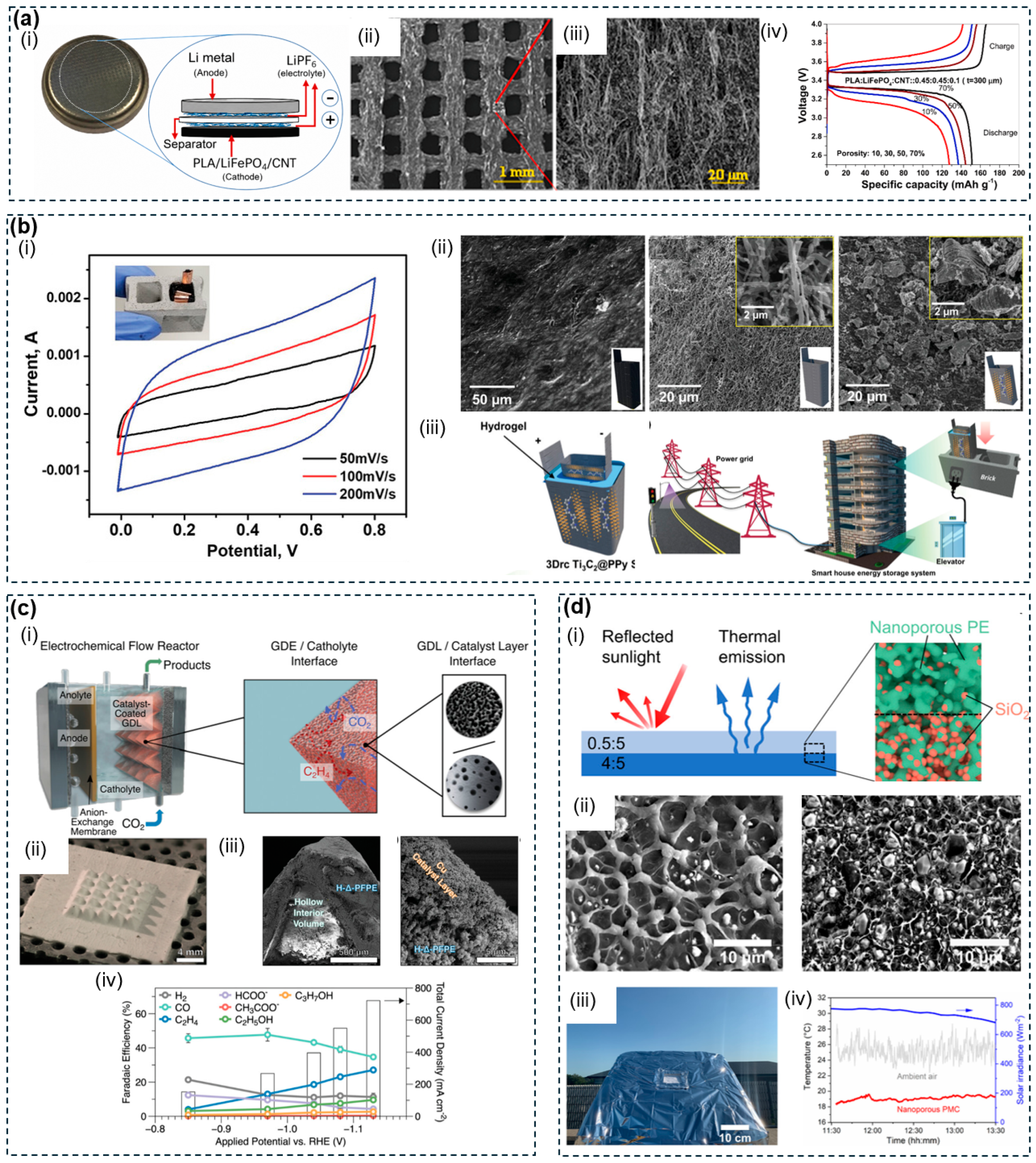
Figure 10: Energy storage and conversion applications of 3D-printed porous polymer structures. (a) FDM-printed PLA/LiFePO4/CNT microarchitected cathodes for Li-ion batteries (i) coin cell schematic; (ii) porous grid SEM; (iii) ligament surface showing LFP/CNT network; (iv) charge/discharge profiles at varying porosity. Reproduced with permission from Ref. [225]. Copyright 2021, Elsevier. (b) FDM-printed graphene/PLA electrodes with Ti3C2@PPy coating for brick-integrated supercapacitors: (i) CV curves with brick device inset; (ii) SEM of pristine, activated, and coated electrode surfaces; (iii) smart house energy storage concept. Reproduced with permission from Ref. [226]. Copyright 2021, Wiley-VCH. (c) 3D-printed porous PFPE GDLs for CO2 electroreduction (i) flow reactor schematic; (ii) pyramid GDL photograph; (iii) SEM of hollow interior and Cu catalyst layer; (iv) Faradaic efficiency and current density vs. applied potential. Reproduced with permission from Ref. [228]. Copyright 2021, Wiley-VCH. (d) Nanoporous PE/SiO2 PMC for daytime radiative cooling (i) bilayer design schematic; (ii) SEM of upper and lower layers; (iii) outdoor testing setup; (iv) subambient temperature measurement. Reproduced with permission from Ref. [229]. Copyright 2021, American Chemical Society.
Thermal management is a mechanistically distinct application mode for 3D-printed porous polymers, in which void networks serve as encapsulation matrices for phase-change materials or as optical scattering elements for passive radiative cooling, rather than as ion or gas transport conduits. Phase-change material (PCM)-based thermal regulation requires open-cell macroporous polymer lattices with void fractions of approximately 40% and millimetre-scale pore spacings, so the crosslinked polymer walls can conformally encapsulate melt-phase particles against leakage during repeated thermal cycling. Passive radiative cooling instead demands a closed-pore nanoscale network, with pore diameters of 200 nm to 2 μm tuned to match solar wavelengths and drive Mie-regime light scattering, a structural requirement opposite in both scale and connectivity to the open macroporosity needed for phase-change applications. This contrast has been demonstrated through PCM-loaded lattices and radiative-cooling composites. Wei et al. [230] formulated spherical paraffin microbeads as both rheological modifiers and active thermal storage elements in a photocurable DIW ink, printing 40%-infill cubic lattices that encapsulated up to 63 wt% PCM. The constructs maintained leakage below 1.8% over 200 melt-solidify cycles and maintained a markedly lower interior temperature than the external environment during heating, outperforming commercial acrylonitrile-butadiene-styrene (ABS) and PCM-free resin controls. In an orthogonal approach, Zhou et al. [229] exploited thermally induced phase separation to create an interconnected nanoporous polyethylene matrix incorporating uniformly dispersed SiO2 particles (Fig. 10d), with pore diameters of 200 nm to 2 μm tuned to match solar wavelengths and induce strong Mie scattering. The resulting composite achieved a solar reflectance of 96.2% and infrared emissivity exceeding 90% within the atmospheric transparency window, enabling a subambient temperature drop of 6.1°C and cooling power of 85 W·m−2 under direct sunlight. Thermoplastic processability was further demonstrated through fabrication of complex 3D cooling structures by standard MEX printing.
The three application areas surveyed above demonstrate that pore architecture can be engineered to govern ion transport, gas transport, or radiative behaviour, often outperforming conventional electrodes, gas diffusion substrates, and PCM matrices on a single key metric. This progress remains mainly component-level, with each architecture optimised for one transport mode rather than several working together. Integrating multiple transport modes into one pore network, and validating performance once assembled into full devices, remains the central challenge for this domain.
4.4 Mechanical Structures and Metamaterials
The mechanical response of a porous polymer architecture is governed not by material chemistry alone but by the geometry of its void network [231]. 3D printing exploits this principle with precision, engineering responses such as energy absorption, negative Poisson’s ratio deformation, and acoustic dissipation that monolithic or stochastically foamed materials cannot reproduce. Conventional energy absorbers rely on randomly distributed porosity whose collapse behaviour is neither predictable nor independently tuneable across spatial coordinates [232]. Commercial sound absorbers depend on fixed fibrous or foam microstructures with little architectural control over resonant frequency. 3D printing of polymeric lattices turns these parameters into programmable design variables. These include gradient open-cell macropore density fields of hundreds of micrometres, re-entrant unit cells with laterally contracting deformation modes, and hollow strut cavities of millimetre-scale dimensions whose geometry sets characteristic Helmholtz resonant frequencies. 3D-printed porous polymers in mechanical structures and metamaterials are mainly applied in three areas. These are energy-absorbing structures, auxetic and reconfigurable architectures, and acoustic dissipation panels.
In weight-critical structural applications, from unmanned aerial vehicle landing systems to primary aerospace panels, polymer components must absorb repeated impact loads without permanent deformation. Conventional homogeneous foams and lattices cannot reduce mass and maintain progressive energy dissipation at the same time, because uniform pore networks collapse synchronously under compression, coupling density reduction directly to capacity loss. Open-cell macroporous gradient architectures with pore dimensions of hundreds of micrometres resolve this conflict by spatially programming the buckling onset sequence across the structure. Compliant, larger-pore peripheral zones absorb initial impact, while denser, smaller-pore core regions provide residual load-bearing capacity and elastic recovery, distributing energy hierarchically without simultaneous collapse. This principle has been demonstrated in gradient lattice cores and sandwich panel structures. Wu et al. [102] fabricated five homogeneous and gradient TPU lattice configurations by FDM. Gradient specimen 3# achieved a specific energy absorption of 264.621 J·g−1, an energy recovery ratio of 51.44%, and a residual displacement ratio of only 12.16% after three loading cycles. Acanfora et al. [233] investigated FDM-printed polypropylene cores within Carbon Fiber Reinforced Polymer (CFRP) face-sheet sandwiches (Fig. 11a). Controlled reduction of infill density reduced panel mass by 27.6% while simultaneously raising the specific energy absorption index by 43.5% at a 20 J impact energy. This improvement came because the lower-density core switched the dominant dissipation mode from brittle strut fracture to progressive flexural deformation.
Protective liners, biomedical implants, and flexible devices each demand mechanical responses that conventional foams with a positive Poisson’s ratio cannot deliver. These responses include concentrating deformation at an impact site, replicating the nonlinear strain-stiffening of soft tissue, and enabling post-fabrication reconfiguration of stiffness. Fixed isotropic architectures expand laterally under compression and offer no mechanism for programmable adjustment of elastic response after manufacture. Open-cell macroporous re-entrant architectures, in which concave cell walls of hundreds of micrometres induce inward lateral contraction under axial loading rather than outward expansion, overcome this limitation. Shape memory polymers further extend this design space by enabling post-fabrication reprogramming of cell curvature, Poisson’s ratio, and modulus. This principle has been demonstrated in impact protection, biomimetic deformation, and reconfigurable chain mail structures. Shen et al. [234] fabricated concave-hexagon auxetic pads from lightweight expanded TPU and integrated them into a composite helmet liner with expanded polystyrene (EPS) foam. Fragment impact experiments at 714 m·s−1 showed a peak head pressure of only 25 kPa, more than six times lower than a conventional foam pad, with finite-element simulations confirming a 22.7% reduction in peak skull stress. Xin et al. [235] used 4D printing of chiral auxetic lattices from a shape memory polymer, generating a biomimetic J-shaped stress-strain response reaching approximately 90% strain. Thermal programming allowed the mechanical fingerprint to switch between distinct biological tissue signatures, including iliac artery and lens capsule. Tian et al. [236] scaled the re-entrant architecture from isolated unit cells to fabric-level chain mail structures (Fig. 11b), topologically interlocking 3D re-entrant PA12 cells in a dual-faced arrangement printed by MJF. The interlocked re-entrant topology yielded a specific energy absorption of 1530 J·kg−1 with approximately 80% recovery after 70% compressive strain. Vacuum jamming at 90 kPa raised the specific bending modulus by a factor of 67, enabling on-demand stiffness switching between flexible and load-bearing states for shape-adaptive body armour and medical fixation.
Noise reduction in aircraft cabins and automotive interiors demands absorbers that perform across wide frequency bands within tight spatial and weight constraints. Conventional fibrous mats and acoustic foams are ineffective under these conditions, because their stochastic porosity couples dissipation frequency inseparably to bulk material thickness. Open-cell macroporous lattice architectures instead implement acoustic dissipation through explicit pore channel geometry. Strut channel diameters of 1 to 2 mm, backed by defined millimetre-scale cavity volumes, establish Helmholtz resonant conditions at geometrically determined target frequencies [237]. Sub-millimetre open-cell pore networks of approximately 300 μm shift the dissipation mechanism from resonance-dominated to viscous frictional flow, extending effective absorption across complementary frequency ranges within a single material family. This principle has been demonstrated through hollow-truss resonators, interpenetrating microlattices, and viscous-flow log-pile scaffolds. Li et al. [238] used this relationship in DLP-fabricated hollow-truss metamaterials, hollowing the struts of a fluorite truss lattice so the outer solid phase contributed Helmholtz resonance while the inner hollow cavities provided an independent resonance mode. The configuration achieved an average broadband absorption coefficient of 0.72 across 1000 to 6300 Hz, with a 68% mass reduction relative to the solid equivalent. Building on this dual-mechanism concept, Li et al. [239] constructed an interpenetrating hollow microlattice by interweaving hollow octet-truss and hollow rhombic dodecahedron sublattices, establishing a six-layer cascaded Helmholtz resonant system. This drove the peak absorption coefficient above 0.99 and expanded the half-absorption bandwidth to 3.2 kHz, while raising plateau compressive stress by 217% over the constituent sublattice. In contrast to these resonance-based designs, Brzeski et al. [240] used direct ink writing of aerospace-grade epoxy composites incorporating hollow glass microspheres and fumed silica nanoparticles to fabricate log-pile micro-scaffolds with 300 μm square pores (Fig. 11c). Here, energy dissipation occurred through viscous frictional flow rather than cavity resonance, achieving peak absorption coefficients above 0.98 at 2100 to 2700 Hz at printing speeds of 110 to 175 mm·s−1, more than three times the rate previously reported for thermoset DIW. This established a scalable manufacturing route toward large-area acoustic panels for aerospace applications.
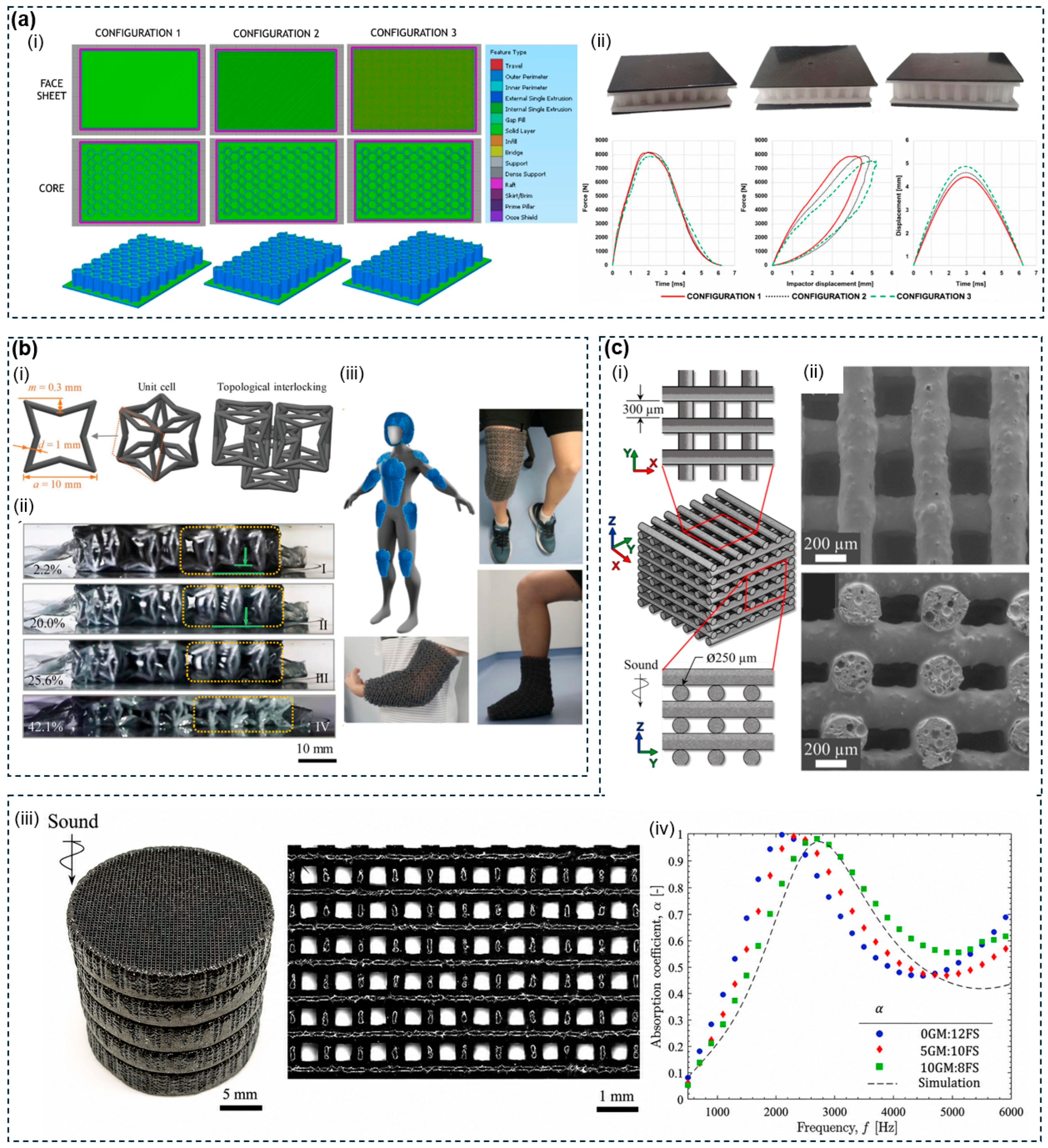
Figure 11: Mechanical and acoustic applications of 3D-printed porous polymer structures. (a) FDM-printed PP/CFRP sandwich structures for impact energy absorption: (i) slicer previews and 3D models of three graded infill configurations; (ii) fabricated samples and 20 J drop-weight impact curves. Reproduced with permission from Ref. [233]. Copyright 2023, Elsevier. (b) MJF-printed PA12 dual-faced chain mail fabric for shape-adaptive protection: (i) unit cell geometry and topological interlocking; (ii) sequential compression images from 2.2% to 42.1% strain; (iii) wearable protection applications. Reproduced with permission from Ref. [236]. Copyright 2023, Wiley-VCH. (c) DIW-printed epoxy composite micro-scaffolds for acoustic absorption: (i) log-pile scaffold architecture; (ii) SEM of filament top-view and cross-section; (iii) stacked cylindrical sample and pore structure; (iv) acoustic absorption coefficients vs. simulation across 500–6000 Hz. Reproduced with permission from Ref. [240]. Copyright 2021, Elsevier.
Pore geometry alone, independent of material chemistry, has proven sufficient to deliver predictable energy absorption, programmable auxetic deformation, and tuneable acoustic resonance in each of the application areas surveyed above, outperforming conventional foams and fibrous absorbers on each of these metrics. Most demonstrations, however, are confined to unit-cell or small-panel scale and cover only single impact events or a limited number of loading cycles. How these properties evolve under fatigue at printing-induced nodal defects, and whether they hold when geometries are scaled to full components, remain largely open questions for this domain.
Flexible electronics demand substrates that conform to curvilinear surfaces, transduce mechanical and chemical signals with high fidelity, and reversibly switch their conductivity in response to repeated deformation [241]. Rigid printed circuit boards and monolithic elastomers cannot fulfil all three requirements at once. Achieving broadband electromagnetic interference shielding within the same wearable or miniaturised platform compounds this challenge. Solid metal enclosures impede heat dissipation, while conventional polymer composites offer limited and non-tuneable shielding efficiency [190]. 3D printing of porous polymer scaffolds addresses both demands. Open-cell macroporous elastomer architectures of tens to hundreds of micrometres allow compression to directly and reversibly modulate embedded conductive network density. Periodic pore geometries scaled to electromagnetic wavelengths give tuneable shielding. Capillary-defined conductive networks integrate multiple electronic functions within a single printed structure. 3D-printed porous polymers in flexible electronics are mainly applied in two areas. These are wearable pressure and tactile sensors, and electromagnetic interference shielding.
Wearable health monitoring and robotic tactile perception require pressure sensors that replicate the mechanical compliance, spatial resolution, and breathability of human skin. Conventional film-based sensors suffer from narrow detection ranges, cross-sensitivity between compression and lateral stretch, and airtight substrates that impede long-term epidermal attachment. Open-cell macroporous elastomer architectures with programmable pore dimensions of tens to hundreds of micrometres resolve these limitations by converting pore geometry into the primary transduction variable. Compression reduces inter-channel spacing within the embedded conductive network, increasing network density and decreasing resistance in direct proportion to applied pressure, while open-cell connectivity allows air and moisture exchange for sustained epidermal comfort. This principle has been demonstrated in standalone pressure sensors, multimodulus devices, capillary-patterned lattices, and fully integrated multisensory e-skin. Kuang et al. [242] used direct bubble writing to fabricate silver-nanoparticle-embedded elastic polymer foams cell by cell in a single step, achieving sensitivities between 0.16 and 0.0014 kPa−1 across 1 to 125 kPa with mechanical hysteresis below 10% and stable response over 2000 loading cycles. Wang et al. [23] extended this approach with a hierarchically porous TPU sensing element formed by a NaCl sacrificial template, decoupling an out-of-plane pressure sensitivity of 5.54 kPa−1 across 10 Pa to 800 kPa from in-plane stretching disturbance, reduced to a 7% resistance change at 50% elongation. Gao et al. [243] demonstrated an architecturally distinct piezoresistive strategy by DLP-printing hierarchical open-cell lattice matrices, in which small cubic unit cells defined capillary infiltration paths and large unit cells provided air-permeable structural pores (Fig. 12a). Capillarity-driven infusion of liquid metal nanoparticle dispersion along the small-pore channels, followed by mechanical sintering, produced site-specific conductive films on selective lattice pillars. Compression-induced increase in inter-pillar contact area yielded a linear resistance decrease across 0 to 60% compressive strain, with a response time of 175 ms and stable performance over 1000 loading cycles, enabling assembly-free wristband, fingertip, and plantar sensors fabricated in a single DLP print. The full potential of all-printed porous e-skin was realised by Song et al. [244], who used semisolid extrusion printing and a phase elimination strategy to generate highly porous microstructures within a multimodal epifluidic electronic skin (Fig. 12b). This skin simultaneously monitored sweat glucose, alcohol, and pH alongside heart rate and body temperature. Coupled with machine learning, the platform predicted alcohol-induced behavioural impairments with over 90% accuracy during real-time daily activity surveillance, while operating without a battery via an integrated microsupercapacitor. Complementing this multisensory integration, Pei et al. [245] fabricated a fully DIW-printed e-skin with a porous silicone substrate, achieving a sensor density of 100 units cm−2 matching human fingertip tactile resolution and a sensitivity of 4 Pa−1 across 0 to 12 kPa.
Electronic devices require shielding from electromagnetic interference while maintaining ventilation and heat dissipation pathways. Solid metallic enclosures fail to meet these conditions, because their closed architecture traps heat and adds mass. Conventional polymer composites achieve only moderate and structurally uncontrolled shielding. Open-cell periodic macroporous polymer architectures, with pore dimensions scaled relative to the target electromagnetic wavelength and kept below the critical failure threshold of Df ≈ λ/8 to λ/5, address this by providing multiple internal scattering interfaces that dissipate electromagnetic energy while preserving airflow through the open pore network. Gradient pore conductivity further controls the absorption-to-reflection ratio independently of the structural geometry. This principle has been demonstrated through gradient-conductive aerogel frames and geometry-tuned honeycomb metamaterials. Xue et al. [246] constructed gradient-conductive MXene/CNT/polyimide aerogel frames by continuously 3D printing inks with increasing CNT content (Fig. 12c). This created a slightly conductive top absorption layer and a highly conductive bottom reflection layer, separated by a hierarchically porous interior that extended electromagnetic dissipation paths through multiple internal reflections. The resulting structure achieved an average EMI shielding efficiency of 68.2 dB with an ultra-low reflection coefficient of 0.23. Lv et al. [190] took a geometry-centred approach, FDM-printing periodic porous TPU/CNT metamaterials with varied pore shapes, sizes, and dislocation configurations. This established the hexagon-derived honeycomb unit as the most effective pore geometry, with thickness-optimised specimens reaching 85 to 95 dB and an absorption coefficient exceeding 0.87.
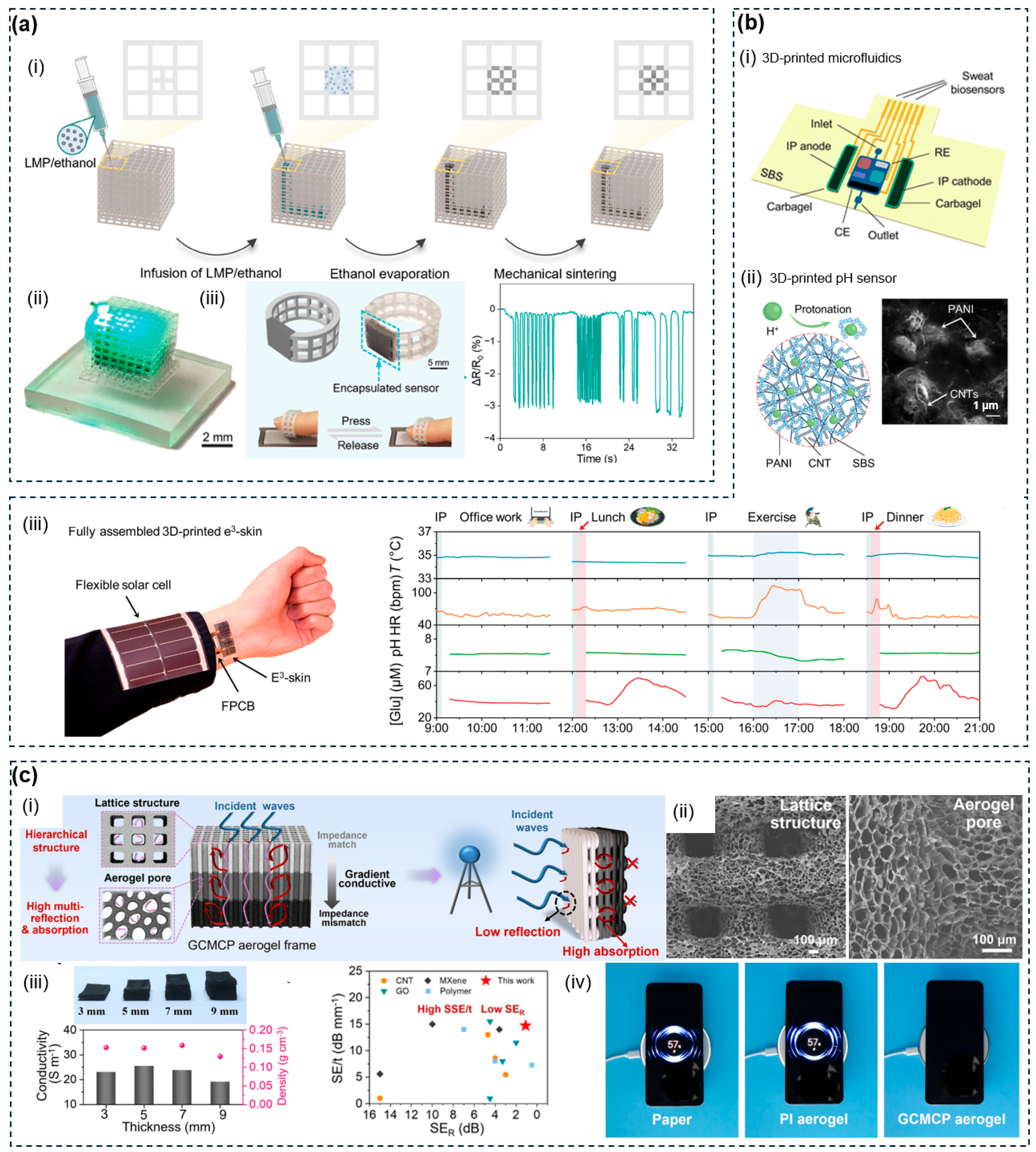
Figure 12: Flexible electronics applications of 3D-printed porous polymer structures. (a) Capillarity-assisted liquid metal patterning in DLP-printed flexible lattice matrices: (i) fabrication workflow from selective LMP infusion to mechanical sintering; (ii) LMP-deposited lattice photograph; (iii) ring-shaped sensor and piezoresistive response during fingertip pressing. Reproduced with permission from Ref. [243]. Copyright 2024, Wiley-VCH. (b) 3D-printed epifluidic electronic skin (e3-skin) for multimodal health surveillance: (i) microfluidic module schematic; (ii) CNT-SBS-PANI pH sensor mechanism and SEM; (iii) on-body 12-h monitoring of temperature, heart rate, pH, and glucose. Reproduced with permission from Ref. [244]. Copyright 2023, American Association for the Advancement of Science. (c) DIW-printed GCMCP aerogel frames for EMI shielding: (i) hierarchical porous structure and gradient-conductive shielding mechanism; (ii) SEM of lattice and aerogel micropores; (iii) conductivity, density, and SE comparison with reported materials; (iv) wireless charging blockage demonstration. Reproduced with permission from Ref. [246]. Copyright 2023, Springer Nature.
Both pressure sensing and electromagnetic shielding benefit from the same underlying principle: pore geometry, rather than bulk material composition, sets the primary transduction or dissipation variable, and 3D printing makes this geometry independently programmable in a way that film-based and metallic alternatives cannot match. In practice, each printed architecture is still optimised for a single function measured under static, flat conditions, leaving the question of how conductive networks and pore structures evolve under repeated bending and stretching as the most pressing open problem for this domain.
4.6 Other Emerging Applications
Beyond the domains surveyed in Section 4.1, Section 4.2, Section 4.3, Section 4.4 and Section 4.5, 3D printing of porous polymers has opened functional frontiers in optical metasurfaces, microfluidic analytical systems, and stimuli-responsive soft robotics. Among these, optical metasurfaces represent perhaps the most demanding architectural target, since sub-wavelength void features of 100 to 700 nm that modulate optical phase can only be realised in polymer systems through two-photon polymerization [247], the single additive manufacturing platform whose resolution reaches this regime. Open-cell microchannels of 20 to 500 μm govern laminar flow and analyte transport in lab-on-chip devices. Sub-micrometre hydrogel network pores enable water absorption and elastic recovery in soft actuators. These application areas are united by the demand for sub-micrometre to nanometre architectural precision that additive manufacturing uniquely provides, and in each case open-cell connectivity is essential, since closed-cell geometries would suppress the photon propagation, fluid flow, and solvent exchange on which these functions depend.
Two-photon polymerization combined with polymerization-induced phase separation generates polymer nanostructures whose open-cell nanoporous network can serve both as a light-scattering medium and as a matrix for impregnating functional optical components, with pore geometry governing optical function in both cases. Zhang et al. [248] used a thiol-ene photoresist with a polyethylene glycol porogen to print three-dimensional microarchitectures with pore diameters of approximately 420 nm, then demonstrated that fluorescent liquids of different colours could be selectively drawn into distinct regions of the same construct through capillary action, establishing a post-doping route to spatially programmable optical microstructures without photoresist reformulation. Mayer et al. [249] combined TPP with polymerization induced phase separation (PIPS) by formulating a Pentaerythritol Triacrylate (PETA)-porogen photoresist (Fig. 13a), in which laser-triggered PIPS, followed by solvent washing and supercritical CO2 drying, opened approximately 50 nm pores throughout the printed polymer. The resulting polymer-air scattering was harnessed to fabricate a functioning miniaturised Ulbricht integrating sphere of 800 μm outer diameter, whose nanoporous walls achieved near-unity diffuse reflectivity. Both demonstrations were confined to sub-millimetre build volumes; scaling these architectures to centimetre-scale components remains a throughput rather than a design limitation.
3D printing enables the direct monolithic fabrication of microfluidic channels, membranes, and porous functional elements without cleanroom infrastructure. Open-cell channel architectures with lateral dimensions of 20 to 500 μm define laminar flow paths, filtration cutoffs, and analyte contact zones, with geometrically distinct regions including mixing chambers, membrane valves, and extraction columns encoded within a single printed monolith. This capability has been demonstrated in organ-on-chip devices, solid-phase extraction columns, and integrated transport-and-reaction microlattices. Shafique et al. [250] introduced low-cost LCD photopolymerization printing with a PEGDA-based ink, achieving lateral features as small as 75 μm and 22 μm-thin embedded membranes. They demonstrated organ-on-a-chip devices with endothelial sprouting over five days, alongside fabrication of 3420 individual devices within a single eight-hour print run. Su et al. [251] fabricated solid-phase extraction columns by FDM printing of porous composite filaments, where the inherent open-cell polymer porosity provided ordered packing without frits. Integrated into an automated inductively coupled plasma mass spectrometry (ICP-MS) system, the columns achieved extraction efficiencies exceeding 99.2% for seven trace metals, with detection limits reaching 0.3 ng L−1. Beyond enclosed channel geometries, Li et al. [252] developed a programmable layer-reduction DLP printing strategy to fabricate transparent poly(acrylate) cellular microlattices with orthogonal open cells, integrated with bioinspired asymmetric re-entrant microstructures (Fig. 13b). The layer-reduction approach compensated for z-axis overcuring in transparent resins, yielding effective pore openings of 120 μm and overhanging dimensional deviations below 1%. The cellular lattice supported ethanol capillary rise to approximately 39.9 mm, while the asymmetric re-entrant geometry imposed Laplace pressure gradients driving unidirectional transport over 40.0 mm at a forward-to-reverse ratio approaching 700% without external actuation. Integrating both elements within a single printed monolith enabled spatially controlled acid-base neutralization reactions with in situ colourimetric readout under pump-free conditions.
3D printed porous polymer actuating bodies embed fluidic functionality through two distinct pore architectures. Designed thermally responsive pores mediate hydraulic fluid transport for autonomic thermoregulation. Process-formed micropores, whose compliance and macroscopic lattice topology together programme multidimensional pneumatic actuation, offer a complementary route. This principle has been demonstrated in a sweating thermoregulating hand and an emulsion-templated pneumatic gripper. Mishra et al. [253] used multimaterial stereolithography to print a PNIPAm actuating body capped with a porous PAAm dorsal layer. Above 30°C, pore dilation combined with PNIPAm contraction expelled hydraulic fluid evaporatively, cooling the actuator surface about 600% faster than a non-sweating counterpart, demonstrated in a five-finger perspiring hand. Joe et al. [254] 3D-printed water-in-oil emulsion inks by digital light processing, generating interconnected micropores of approximately 2 μm upon water evaporation (Fig. 13c). Gradient unit-cell tessellation distributed stiffness across the actuator body, producing pneumatic elastic lattice actuators with extensibility up to approximately 523%, a three-fingered gripper of 300 gf payload, and the first jointless continuum actuator encoding biaxial elongation and bidirectional bending from a single pneumatic source.
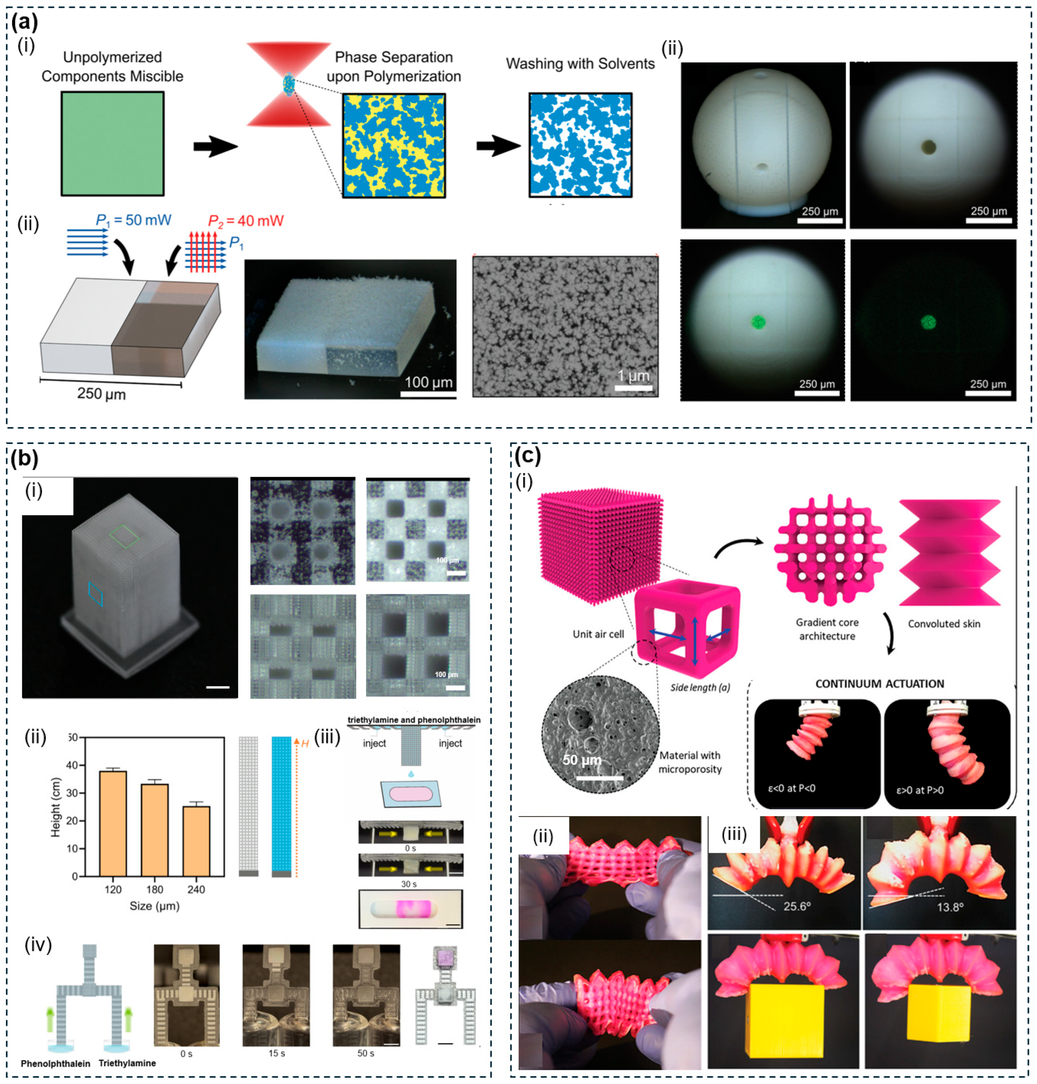
Figure 13: Emerging applications of 3D-printed porous polymer structures in optics, microfluidics, and soft robotics. (a) TPP-printed nanoporous polymer via PIPS for diffuse optical applications (i) PIPS mechanism schematic; (ii) double-exposure strategy and SEM of nanoporous microstructure; (iii) miniature Ulbricht integrating sphere under white light and 532 nm laser illumination. Reproduced with permission from Ref. [249]. Copyright 2020, Wiley-VCH. (b) DLP-printed cellular microlattices with asymmetric re-entrant microstructures for pump-free directional capillary microreactions: (i) lattice and open-cell microscopy images; (ii) ethanol capillary rise heights; (iii) acid–base neutralization triggered by simultaneous PP and TEA injection; (iv) time-lapse colourimetric readout. Reproduced with permission from Ref. [252]. Copyright 2026, American Chemical Society. (c) DLP-printed microporous polyurethane elastic lattice actuators for jointless soft robotics (i) dual-porosity design schematic; (ii) three-fingered gripper; (iii) continuum actuator bidirectional bending and object manipulation. Reproduced with permission from Ref. [254]. Copyright 2023, Wiley-VCH.
Optical metasurfaces, microfluidic analytical systems, and stimuli-responsive soft robotics collectively show what becomes accessible when pore resolution reaches the nanometre-to-micrometre scale, with demonstrated performance in diffuse optics, integrated lab-on-chip transport, and autonomic actuation that matches or exceeds cleanroom-fabricated and conventional alternatives. In each case, however, the results are standalone proofs of concept built at the resolution limit of the fabrication platform and confined to sub-millimetre or sub-centimetre build volumes. Throughput, batch reproducibility, and long-term stability under repeated use are reported for at most one system in each area, and translating these single-device demonstrations into reproducible, batch-produced components remains the step that none of the three areas has yet taken.
5 Discussion and Future Directions
3D printing has reframed pore architecture from an emergent consequence of stochastic physicochemical processes into a programmable design variable, addressable across material chemistry, pore scale, connectivity, and spatial distribution within a single fabrication workflow. This review has traced three interconnected dimensions behind this shift: (1) additive manufacturing platforms and their resolution-throughput trade-offs, (2) pore-forming strategies organised into design-driven and physicochemical paradigms whose hybrid combination extends the accessible pore scale range, and (3) application domains in which six functional areas are governed by four recurring pore-level mechanisms. Despite this progress, three challenges still constrain the field. First, achieving high-resolution pore control across a wide range of length scales within a single large-volume construct remains unsolved, because single platforms cannot span the full resolution range, post-processing pore formation cannot be spatially coordinated with the printed macroarchitecture, and pore morphology control breaks down below the platform resolution floor. Second, balancing printability against functional loading remains difficult for most printable polymer systems, and pore architectures fixed at the time of fabrication cannot adapt to environments that change over time. Third, conventional simulation methods cannot model the statistically distributed pore populations produced by hybrid fabrication strategies, and the nonlinear sensitivity of printed pore geometry to process parameters leaves batch-to-batch reproducibility unresolved. This chapter examines each of these challenges in turn and proposes future directions spanning multi-scale fabrication strategies, expansion of the functional material library, and AI-assisted pore design (Fig. 14), pointing toward constructs that are not only geometrically programmable but also functionally adaptive and computationally optimisable.
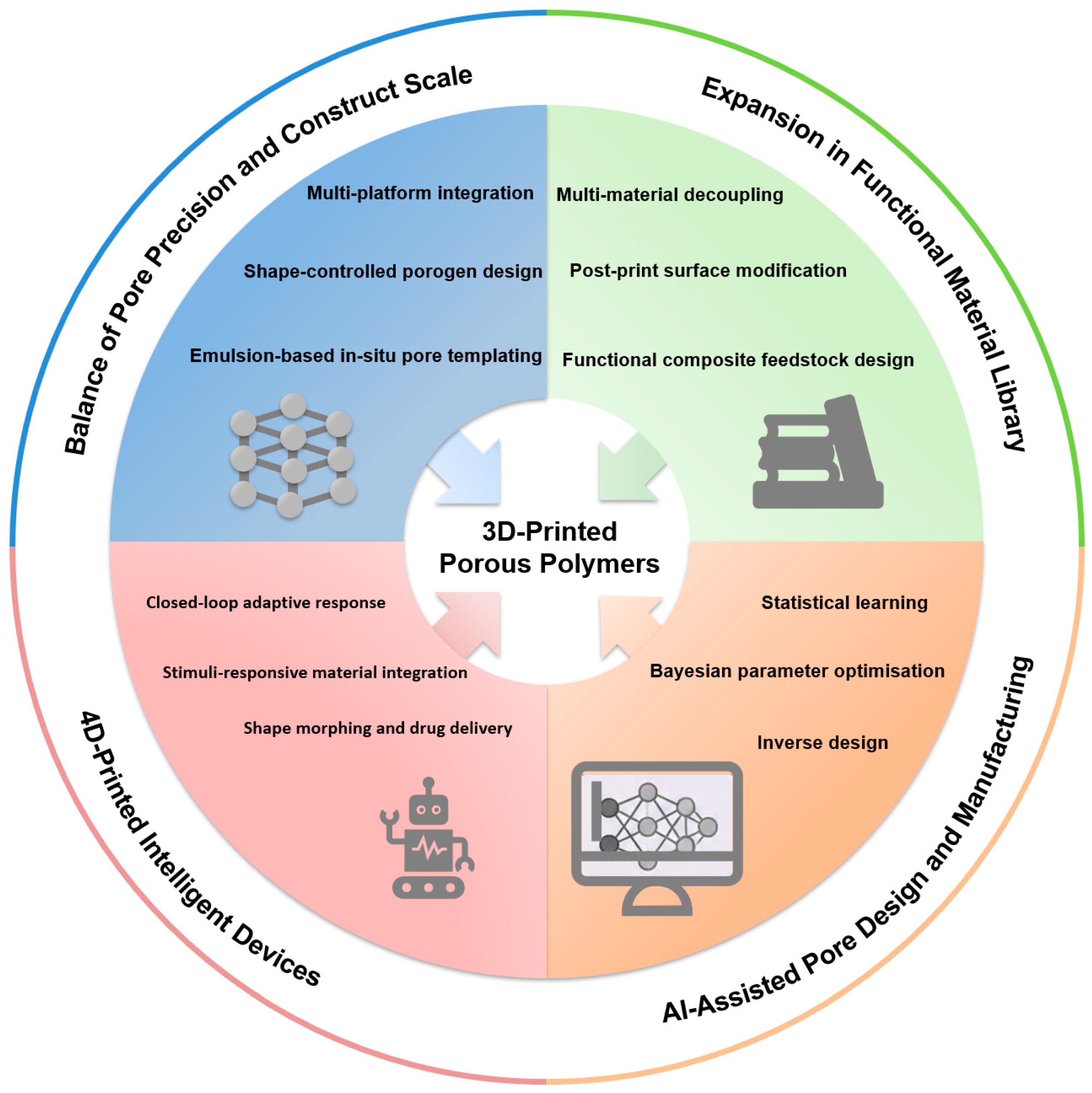
Figure 14: Proposed future directions for 3D-printed porous polymers across four challenge areas: balance of pore precision and construct scale (blue), expansion in the functional material library (green), AI-assisted pore design and manufacturing (orange), and 4D-printed intelligent devices (pink).
5.1 Balance of Pore Precision and Construct Scale
Achieving precise pore control across a wide range of length scales within a single large-volume construct presents three challenges spanning scale precision, spatial precision, and morphological precision. Scale precision is limited because no single platform spans the full resolution range required for hierarchical porous constructs. TPP reaches sub-100 nm feature resolution but only at millimetre construct scale, while DLP, MEX, and powder bed platforms recover throughput by processing larger areas at once but sacrifice resolution in doing so, and none can simultaneously match sub-micrometre precision and centimetre-scale build volume. Spatial precision is limited because even when micropores are successfully introduced into a large-volume construct, their spatial distribution cannot be precisely coordinated with the macroarchitecture, since the printing step and the pore-forming step are typically separated in time. Morphological precision is limited because when the target pore size falls below the resolution floor of the available platform, conventional sacrificial porogens such as salt particles have irregular shapes and broad size distributions, leaving pore morphology and connectivity poorly controlled regardless of how precisely the macrostructure is printed.
Multi-platform integration has emerged as the most direct route to addressing scale, spatial, and morphological precision, with one demonstrated strategy for each dimension. For scale precision, Spiegel et al. formulated a single shape memory polymer ink compatible with both DLP and direct laser writing, using DLP to fabricate centimetre-scale structures with 50 to 100 μm layer resolution and direct laser writing to produce microscale versions of the same geometries at sub-micrometre resolution, demonstrating that a single ink system can bridge macro- and microscale fabrication without requiring separate material development for each platform [255]. Peng et al. demonstrated a complementary multi-platform combination by integrating DLP and DIW in a single automated workflow, in which DLP defined the structural framework at 30 to 100 μm lateral resolution while DIW deposited functional materials at precisely specified locations within that framework [256]. For spatial precision, Kleger et al. used photo-curable Pickering emulsions as the printing feedstock for stereolithography, so that emulsion droplets acted as in-situ pore templates during printing, producing 0.5 to 2 μm micropores within the struts of a millimetre-scale lattice in a single fabrication step without any post-processing, ensuring that micropore location is governed by the printed geometry rather than by a separate post-processing step [257]. For morphological precision, Kim et al. showed that replacing irregular salt particles with shape-controlled hydrogel microparticles transferred pore morphology control from the printer to the template geometry entirely [258]. Each approach still has limits. Multi-platform strategies require ink compatibility across different curing mechanisms and careful process sequencing to prevent cross-contamination. In-situ emulsion templating is currently limited to photopolymerizable systems. Porogen shape control improves morphological precision but does not make pore size independently programmable across different regions of the same construct. A more productive direction would treat the three precision dimensions together within a single design framework, optimising macropore geometry, spatial pore-forming stimulus, and porogen morphology as jointly controlled variables rather than addressing them sequentially. Achieving this would require spatially resolved control over the local stimulus that triggers micropore formation, whether humidity in vapour-induced phase separation, cooling rate in ice-templating, or gas concentration in foaming, so that the micropore population can be actively coordinated with the macroarchitecture during deposition rather than applied uniformly afterward.
5.2 Expansion in Functional Material Library
The established printable polymer library constrains what functional performance porous architectures can deliver at two levels, namely the conflict between printability and functional loading within the feedstock itself, and the static nature of pore architecture after fabrication in environments that change over time. Balancing printability against functional performance remains difficult for most printable polymer systems. Materials with sufficient functional loading, such as conductive fillers, bioactive factors, or catalytic nanoparticles, often lose printability as filler content rises, while materials optimised for printability, including PCL, PLA, PA12, and GelMA, do not inherently possess the electrical, catalytic, ion-transport, or photothermal properties that sensor, electrode, separation, and evaporation applications require. Beyond this, pore architectures defined at the time of fabrication remain fixed in geometry and surface chemistry throughout the lifetime of the device, while most target environments change continuously over time.
Three directions have been demonstrated to work around these constraints, spanning multi-material printing, composite feedstock design, and post-print surface modification. Multi-material printing assigns structural and functional roles to different printed components within the same construct. Peng et al. integrated DLP and DIW in a single automated workflow, with DLP printing the high-resolution structural matrix while DIW deposited functional materials including liquid crystal elastomers and conductive inks into designated regions [256]. Composite feedstocks introduce functional fillers before printing, so that the scaffold inherits functional properties directly from the material. Kennedy et al. compounded TiO2 nanoparticles at up to 34 wt% loading into PLA filament and printed open-cell lattices by FDM, delivering photocatalytic activity across multiple reuse cycles [218]. Post-print surface modification introduces functional properties after fabrication without altering the printed architecture, including polydopamine coating, electrochemical deposition, and catalyst mineralisation [197]. Each direction has limits. Multi-material printing requires compatible ink-resin pairs. Composite feedstocks trade off functional loading against printability. Post-print modification cannot change bulk pore wall composition, coatings degrade over time, and diffusion-limited methods cannot reach deep interior surfaces. In each case, functional performance remains spatially uniform rather than deliberately patterned. Co-optimising filler distribution alongside the CAD-defined pore architecture, so that regions requiring high conductivity or catalytic density receive higher local filler concentration while load-bearing regions remain structurally uncompromised, represents a direction that none of the three approaches currently addresses.
5.3 AI-Assisted Pore Design and Manufacturing
Two specific gaps limit the predictive and manufacturing precision of hybrid porous polymer systems, namely the inability of conventional simulation tools to model statistically distributed pore populations, and the nonlinear sensitivity of printed pore geometry to process parameter variations. Conventional finite element simulations are well suited to geometrically defined pore architectures such as lattices and TPMS structures, where pore geometry can be described by deterministic equations. They perform poorly on the mixed pore populations produced by combining 3D printing with physicochemical strategies such as freeze-drying, phase separation, or foaming, because these strategies generate statistically distributed rather than geometrically defined pores. The pore size distribution, connectivity probability, and wall morphology of physicochemical pores do not map cleanly onto the mesh geometries that finite element solvers require, leaving the transport, mechanical, and biological performance of hybrid porous constructs without a reliable predictive framework. A separate gap concerns printing fidelity. The relationship between printing parameters and the resulting pore geometry is nonlinear and sensitive to small variations in feedstock and environment, making it difficult to match printed pore dimensions to design specifications and leaving batch-to-batch reproducibility as a persistent problem across FDM, SLS, and DIW platforms.
Machine learning addresses both gaps through complementary routes, applying statistical learning to model physicochemical pore populations, optimising printing parameters through iterative Bayesian methods, and extending inverse design frameworks to jointly specify pore geometry and process conditions. For hybrid pore structure prediction, de Oliveira et al. trained gradient boosting models on 252 freeze-casting studies spanning ceramics, polymers, and composites to predict porosity with an R2 of 0.81, with explainability analysis identifying solid loading as the dominant process variable [259]. This approach works because physicochemical pore formation follows statistical rather than deterministic rules, making it amenable to data-driven modelling in a way that finite element solvers are not. For printing fidelity, Chen et al. applied Bayesian optimisation to iteratively refine layer height, travel speed, and dispensing pressure in a DIW-based printing process, achieving substantial improvement in geometric fidelity within 46 iterations without human intervention [260]. At the design level, Pahlavani et al. demonstrated deep-learning inverse design of lattice architectures meeting specified mechanical targets while respecting platform-specific manufacturability constraints [261], and Patil et al. showed that generative AI can couple macro-scale pattern design with reactive-extrusion foaming to produce continuous pore hierarchy across nearly six orders of magnitude [262]. The gap that none of these approaches yet closes is the prediction of complete pore structural descriptors, including pore size distribution, three-dimensional connectivity, and pore morphology, from physicochemical process parameters in hybrid fabrication systems. These descriptors are the direct geometric inputs that finite element simulations require to predict transport, mechanical, and biological performance, yet current machine learning models for physicochemical pore formation predict only scalar approximations such as average porosity. Extending machine learning prediction from single scalar outputs to full structural distributions, and feeding these predicted distributions directly into simulation frameworks as geometric inputs, would close the chain from process parameters to functional performance in hybrid porous constructs, a capability that does not yet exist for systems combining design-driven and physicochemical pore-forming strategies.
5.4 4D-Printed Intelligent Devices
The preceding sections have established pore architecture as a programmable design variable. Yet a separate limitation cuts across all static constructs regardless of how precisely their internal architecture is controlled. Once fabricated, the geometry and function of a conventional 3D-printed implant remain fixed throughout its service life. This immutability creates two persistent problems for clinical translation. First, large or geometrically complex devices cannot be delivered through minimally invasive access routes without compromising their functional shape. Second, static constructs cannot adapt to the evolving biological environment after implantation, leaving them unable to participate actively in the dynamic processes of tissue repair and remodelling. 4D printing addresses both problems by introducing time as an additional programmable dimension. Stimuli-responsive smart materials, when integrated with additive manufacturing, enable printed structures to undergo programmed changes in shape, properties, or function in response to specific triggers. This capability opens new therapeutic strategies that are unavailable to static implants.
Recent advances have addressed these challenges through three complementary strategies, namely establishing a unified material-manufacturing-application framework for stimuli-responsive smart materials, coupling shape programming with localised multi-drug delivery in a single minimally invasive device, and adopting an application-driven design philosophy that integrates 4D printing, metamaterial engineering, and functional materials into a multilayered therapeutic platform [84]. Lin et al. constructed a unified framework connecting shape memory polymers, stimuli-responsive hydrogels, and liquid crystal elastomers across their stimulus-response mechanisms, 4D printing compatibility, and nine biomedical application domains, establishing that the convergence of 4D printing with stimuli-responsive materials enables a transition from passive structural replacement to active functional repair [263]. Wang et al. coupled shape memory-driven deployment with dual-drug delivery in a 4D-printed patent foramen ovale (PFO) occluder, whose biomimetic framework enabled catheter-based delivery and shape recovery within 20 s while the drug-loaded layer simultaneously released an anti-thrombosis agent and a pro-endothelialisation drug, demonstrating that programmable shape morphing and localised pharmacological function can be integrated within a single minimally invasive device [264]. Lin et al. adopted an application-driven design strategy by developing a sandwich-structured intestinal stent whose outer Janus layer performed synergistic photothermal-chemotherapy, intermediate 4D-printed gradient metamaterial layer matched the J-shaped mechanical response of native intestinal tissue, and hydrophobic inner layer inhibited restenosis, showing that multiple clinical needs can be addressed concurrently through architecturally encoded heterogeneous design [162]. The gap that none of these approaches yet closes is the coupling of programmable shape morphing with real-time feedback from the local tissue environment. Current 4D-printed devices execute pre-programmed shape changes and drug release profiles that are fixed at the design stage. Mechanical support, drug elution, and degradation proceed on predetermined schedules without sensing whether the surrounding tissue is regenerating as expected or has deviated from the anticipated repair trajectory. Closing this gap would require integrating sensing elements that detect local biological signals, such as enzymatic activity, mechanical compliance, or inflammatory markers, and using these signals to adapt device behaviour in situ.
3D printing has transformed porosity from an incidental byproduct of stochastic manufacturing into a programmable design variable that can be independently controlled across pore size, geometry, connectivity, and spatial distribution within a single construct. This review has examined this transformation through the lens of five pore-forming strategies, spanning design-driven architectures that encode pore geometry in the digital model and physicochemical routes that generate porosity through material-level processes including phase separation, freeze-drying, sacrificial templating, and partial fusion.
The central finding emerging from the comparative assessment of these strategies is that no single one simultaneously satisfies all the requirements that practical applications demand. Design-driven pore formation delivers deterministic spatial control and mechanical integrity but cannot access sub-100 μm pore scales. Physicochemical strategies reach these finer scales and achieve higher porosity, but they cannot specify pore location or geometry with comparable fidelity. Hybrid approaches that combine a printed macroarchitecture with post-print physicochemical treatment represent the most viable current solution to this resolution-porosity trade-off, although they introduce longer process chains and greater batch-to-batch variability. The integration of these strategies with stimuli-responsive smart polymers opens a further dimension in which pore architecture becomes not only geometrically programmable but also dynamically responsive to external triggers.
Across the application domains surveyed in this review, pore morphology governs biological and functional performance through a shared set of mechanisms. Pore size dictates the fundamental trade-off between cell infiltration capacity and mechanical competence. Pore connectivity determines whether transport pathways for oxygen, nutrients, and metabolic waste remain open throughout the construct. Pore geometry directs the spatial organisation of regenerated tissue, with anisotropic channels guiding axonal and vascular growth along predetermined axes and isotropic networks supporting randomly oriented ingrowth. These relationships are not independent: changing one pore parameter inevitably shifts the effective range of the others.
The most pressing direction for the field is closing the gap between the spatial precision of design-driven pore formation and the sub-printing-resolution porosity that physicochemical strategies provide, while maintaining throughput and reproducibility at clinically relevant scales. Achieving this will require tighter integration of multi-platform fabrication, computational prediction of hybrid pore populations, and systematic validation in orthotopic models under physiologically relevant conditions. Addressing these challenges will determine whether 3D-printed porous polymers can complete the transition from laboratory demonstrations to clinically and industrially deployable technologies.
Acknowledgement:
Funding Statement: This research was funded by the Natural Science Foundation of Jiangsu Province (BK20241268), the Open Research Fund of Southeast University and Jiangsu Province Hospital (2024-M02), the Jiangsu Province Youth Science and Technology Talent Support Project (JSTJ-2024-096), the Start-up Research Fund of Southeast University (RF028623292), the Fundamental Research Funds for the Central Universities (2242026K30018), the Young Scientists Fund of State Key Laboratory of Digital Medical Engineering, and the National Natural Science Foundation of China (52033002, 82227808). Xiaojiang Liu acknowledges the support by Research Program for Xiaomi Youth Scholars.
Author Contributions: Lihao Liu: Writing—original draft, Methodology, Investigation, Data curation, Formal analysis. Yiting Huang: Review & editing. Dong Wang: Review & editing. Zhongze Gu: Review & editing, Funding acquisition. Xiaojiang Liu: Review & editing, Funding acquisition. All authors reviewed and approved the final version of the manuscript.
Availability of Data and Materials: All data generated or analyzed during this study are included in this published article.
Ethics Approval: Not applicable.
Conflicts of Interest: Given his role as Editorial Board Member of this journal, Xiaojiang Liu had no involvement in the peer review of this article and had no access to information regarding its peer review. Full responsibility for the editorial process for this article was delegated to another journal editor. The authors declare no other conflicts of interest.
Nomenclature and Terminology
| Abbreviation | Definition |
| Manufacturing & Processes | |
| 2D | Two-Dimensional |
| 3D | Three-Dimensional |
| AM | Additive Manufacturing |
| CAD | Computer-Aided Design |
| CAM | Computer-Aided Manufacturing |
| CAL | Computational Axial Lithography |
| CLIP | Continuous Liquid Interface Production |
| DIW | Direct Ink Writing |
| DLP | Digital Light Processing |
| DMD | Digital Micromirror Device |
| FDM | Fused Deposition Modeling |
| FRESH | Freeform Reversible Embedding of Suspended Hydrogels |
| LCD | Liquid-Crystal Display |
| MEW | Melt Electrowriting |
| MEX | Material Extrusion |
| MJF | Multi Jet Fusion |
| PBF | Powder Bed Fusion |
| SLA | Stereolithography |
| SLM | Selective Laser Melting |
| SLS | Selective Laser Sintering |
| SWIFT | Sacrificial Writing Into Functional Tissue |
| TPP | Two-Photon Polymerization |
| UV | Ultraviolet |
| VPP | Vat Photopolymerization |
| μCPP | Microscale Continuous Projection Printing |
| X–Y/Z | In-Plane/Build Direction |
| Pore Formation, Architecture & Design | |
| APC | Amorphous Photonic Crystal |
| BCC | Body-Centred Cubic |
| BCCZ | Body-Centered Cubic Z-reinforced |
| CPC | Crystalline Photonic Crystal |
| DIPS | Diffusion-Induced Phase Separation |
| FCC | Face-Centred Cubic |
| IWP | I-Graph–Wrapped Package |
| NG | Nucleation and Growth |
| PBG | Photonic Band Gap |
| PIPS | Polymerization-Induced Phase Separation |
| RD | Rhombic Dodecahedron |
| SD | Spinodal Decomposition |
| TIPS | Thermally Induced Phase Separation |
| TPMS | Triply Periodic Minimal Surface |
| VIPS | Vapor-Induced Phase Separation |
| Materials & Chemicals | |
| ABS | Acrylonitrile Butadiene Styrene |
| AgNW | Silver Nanowire |
| BG | Bioactive Glass |
| CNT | Carbon Nanotube |
| CNT-SBS-PANI | Carbon Nanotube/Styrene–Butadiene–Styrene/Polyaniline |
| DMF | N,N-Dimethylformamide |
| F127 | Pluronic F127 (Poloxamer 407) |
| GelMA | Gelatin Methacryloyl |
| GCMCP | Gradient-Conductive MXene/Carbon Nanotube/Polyimide |
| GO | Graphene Oxide |
| HA (HAp) | Hydroxyapatite |
| LA:GA | Lactide-to-Glycolide Ratio |
| LFP | Lithium Iron Phosphate |
| MXene | Two-Dimensional Transition-Metal Carbide/Nitride |
| PA11 | Polyamide 11 |
| PA12 | Polyamide 12 |
| PA2202 | Commercial Polyamide 12 Powder Grade |
| PANI | Polyaniline |
| PC | Polycarbonate |
| PCL | Polycaprolactone |
| PDMS | Polydimethylsiloxane |
| PEG | Poly(Ethylene Glycol) |
| PEGDA | Poly(Ethylene Glycol) Diacrylate |
| PEEK | Polyetheretherketone |
| PFPE | Perfluoropolyether |
| PETA | Pentaerythritol Triacrylate |
| PLA | Polylactic Acid |
| PLGA | Poly(Lactic-co-Glycolic Acid) |
| PMMA | Poly(Methyl Methacrylate) |
| PolyHIPE | Polymerized High Internal Phase Emulsion |
| PP | Polypropylene |
| PS | Polystyrene |
| PTFE | Polytetrafluoroethylene |
| PU | Polyurethane |
| PVA | Poly(Vinyl Alcohol) |
| PVDF | Poly(Vinylidene Fluoride) |
| pHEMA | Poly(2-Hydroxyethyl Methacrylate) |
| TPE | Thermoplastic Elastomer |
| THF | Tetrahydrofuran |
| TPU | Thermoplastic Polyurethane |
| Characterization & Performance Metrics | |
| CV | Cyclic Voltammetry |
| D_f | Characteristic Feature Dimension |
| EDX | Energy-Dispersive X-Ray Spectroscopy |
| EMI | Electromagnetic Interference |
| GDL | Gas-Diffusion Layer |
| ICP-MS | Inductively Coupled Plasma Mass Spectrometry |
| JCA | Johnson–Champoux–Allard Model |
| MPP | Microperforated Panel |
| OCA | Oil Contact Angle |
| PDRC | Passive Daytime Radiative Cooling |
| PCM | Phase-Change Material |
| Ra | Arithmetic Mean Surface Roughness |
| RI | Refractive Index |
| SE | Shielding Effectiveness |
| SEM | Scanning Electron Microscopy |
| SSA | Specific Surface Area |
| UTS | Ultimate Tensile Strength |
| WCA | Water Contact Angle |
| Biomedical & Functional Applications | |
| ADM | Acellular Dermal Matrix |
| AI | Artificial Intelligence |
| BATE | Bioprinting-Assisted Tissue Emergence |
| CAR-T | Chimeric Antigen Receptor T Cell |
| ECM | Extracellular Matrix |
| HER2 | Human Epidermal Growth Factor Receptor 2 |
| HUVEC | Human Umbilical Vein Endothelial Cell |
| LMP | Liquid Metal Patterning |
| MOF | Metal–Organic Framework |
| NOD | Non-Obese Diabetic |
| NPC | Neural Progenitor Cell |
| PFO | Patent Foramen Ovale |
| RUNX2 | Runt-Related Transcription Factor 2 |
| WHO | World Health Organization |
| ZIF | Zeolitic Imidazolate Framework |
References
1. Liu Q , Xiong J , Lin W , Liu J , Wan Y , Guo CF , et al. Porous polymers: structure, fabrication and application. Mater Horiz. 2025; 12( 8): 2436– 66. doi:10.1039/d4mh01618a. [Google Scholar] [CrossRef]
2. Ahmed YW , Loukanov A , Tsai HC . State-of-the-art synthesis of porous polymer materials and their several fantastic biomedical applications: a review. Adv Healthc Mater. 2025; 14( 26): 2403743. doi:10.1002/adhm.202403743. [Google Scholar] [CrossRef]
3. Wu D , Xu F , Sun B , Fu R , He H , Matyjaszewski K . Design and preparation of porous polymers. Chem Rev. 2012; 112( 7): 3959– 4015. doi:10.1021/cr200440z. [Google Scholar] [CrossRef]
4. Zhao H , Li L , Ding S , Liu C , Ai J . Effect of porous structure and pore size on mechanical strength of 3D-printed comby scaffolds. Mater Lett. 2018; 223: 21– 4. doi:10.1016/j.matlet.2018.03.205. [Google Scholar] [CrossRef]
5. Tomin M , Kmetty Á . Polymer foams as advanced energy absorbing materials for sports applications—a review. J Appl Polym Sci. 2022; 139( 9): 51714. doi:10.1002/app.51714. [Google Scholar] [CrossRef]
6. Karageorgiou V , Kaplan D . Porosity of 3D biomaterial scaffolds and osteogenesis. Biomaterials. 2005; 26( 27): 5474– 91. doi:10.1016/j.biomaterials.2005.02.002. [Google Scholar] [CrossRef]
7. Zhianmanesh M , Varmazyar M , Montazerian H . Fluid permeability of graded porosity scaffolds architectured with minimal surfaces. ACS Biomater Sci Eng. 2019; 5( 3): 1228– 37. doi:10.1021/acsbiomaterials.8b01400. [Google Scholar] [CrossRef]
8. Han J , Gu H , Gu Z . Emerging engineering strategies in bone organoids: from biomimetic scaffolds to dynamic microenvironmental stimulation. Bioact Mater. 2026; 63: 682– 707. doi:10.1016/j.bioactmat.2026.04.008. [Google Scholar] [CrossRef]
9. Khajehmohammadi M , Azizi Tafti R , Nikukar H . Effect of porosity on mechanical and biological properties of bioprinted scaffolds. J Biomed Mater Res. 2023; 111( 2): 245– 60. doi:10.1002/jbm.a.37455. [Google Scholar] [CrossRef]
10. Izadyari Aghmiuni A , Heidari Keshel S , Sefat F , AkbarzadehKhiyavi A . Fabrication of 3D hybrid scaffold by combination technique of electrospinning-like and freeze-drying to create mechanotransduction signals and mimic extracellular matrix function of skin. Mater Sci Eng C Mater Biol Appl. 2021; 120: 111752. doi:10.1016/j.msec.2020.111752. [Google Scholar] [CrossRef]
11. Khoramgah MS , Ranjbari J , Abbaszadeh HA , Tabatabaei Mirakabad FS , Hatami S , Hosseinzadeh S , et al. Freeze-dried multiscale porous nanofibrous three dimensional scaffolds for bone regenerations. Bioimpacts. 2020; 10( 2): 73– 85. doi:10.34172/bi.2020.10. [Google Scholar] [CrossRef]
12. Kwon DY , Park JY , Lee BY , Kim MS . Comparison of scaffolds fabricated via 3D printing and salt leaching: in vivo imaging, biodegradation, and inflammation. Polymers. 2020; 12( 10): 2210. doi:10.3390/polym12102210. [Google Scholar] [CrossRef]
13. Adel IM , ElMeligy MF , Elkasabgy NA . Conventional and recent trends of scaffolds fabrication: a superior mode for tissue engineering. Pharmaceutics. 2022; 14( 2): 306. doi:10.3390/pharmaceutics14020306. [Google Scholar] [CrossRef]
14. Bhushan S , Singh S , Maiti TK , Sharma C , Dutt D , Sharma S , et al. Scaffold fabrication techniques of biomaterials for bone tissue engineering: a critical review. Bioengineering. 2022; 9( 12): 728. doi:10.3390/bioengineering9120728. [Google Scholar] [CrossRef]
15. Zhou C , Yang K , Wang K , Pei X , Dong Z , Hong Y , et al. Combination of fused deposition modeling and gas foaming technique to fabricated hierarchical macro/microporous polymer scaffolds. Mater Des. 2016; 109: 415– 24. doi:10.1016/j.matdes.2016.07.094. [Google Scholar] [CrossRef]
16. Tang X , Xu H , Liu X , Yang Y , Li Z . Advances in 3D-Printed scaffolds for bone defect repair: material strategies and synergistic functional performance. Front Bioeng Biotechnol. 2025; 13: 1707406. doi:10.3389/fbioe.2025.1707406. [Google Scholar] [CrossRef]
17. Capuana E , Lopresti F , Carfì Pavia F , Brucato V , La Carrubba V . Solution-based processing for scaffold fabrication in tissue engineering applications: a brief review. Polymers. 2021; 13( 13): 2041. doi:10.3390/polym13132041. [Google Scholar] [CrossRef]
18. Dehban A , Hosseini Saeedavi F , Kargari A . A study on the mechanism of pore formation through VIPS-NIPS technique for membrane fabrication. J Ind Eng Chem. 2022; 108: 54– 71. doi:10.1016/j.jiec.2021.12.023. [Google Scholar] [CrossRef]
19. do Amaral Montanheiro TL , Schatkoski VM , de Menezes BRC , Pereira RM , Ribas RG , de Sousa Martinez de Freitas A , et al. Recent progress on polymer scaffolds production: methods, main results, advantages and disadvantages. Express Polym Lett. 2022; 16( 2): 197– 219. doi:10.3144/expresspolymlett.2022.16. [Google Scholar] [CrossRef]
20. Chen H , Han Q , Wang C , Liu Y , Chen B , Wang J . Porous scaffold design for additive manufacturing in orthopedics: a review. Front Bioeng Biotechnol. 2020; 8: 609. doi:10.3389/fbioe.2020.00609. [Google Scholar] [CrossRef]
21. Guo W , Yang Y , Liu C , Bu W , Guo F , Li J , et al. 3D printed TPMS structural PLA/GO scaffold: process parameter optimization, porous structure, mechanical and biological properties. J Mech Behav Biomed Mater. 2023; 142: 105848. doi:10.1016/j.jmbbm.2023.105848. [Google Scholar] [CrossRef]
22. Hollister SJ . Scaffold design and manufacturing: from concept to clinic. Adv Mater. 2009; 21( 32–33): 3330– 42. doi:10.1002/adma.200802977. [Google Scholar] [CrossRef]
23. Wang Z , Guan X , Huang H , Wang H , Lin W , Peng Z . Full 3D printing of stretchable piezoresistive sensor with hierarchical porosity and multimodulus architecture. Adv Funct Mater. 2019; 29( 11): 1807569. doi:10.1002/adfm.201807569. [Google Scholar] [CrossRef]
24. Sultan S , Mathew AP . 3D printed scaffolds with gradient porosity based on a cellulose nanocrystal hydrogel. Nanoscale. 2018; 10( 9): 4421– 31. doi:10.1039/c7nr08966j. [Google Scholar] [CrossRef]
25. Levato R , Jungst T , Scheuring RG , Blunk T , Groll J , Malda J . From shape to function: the next step in bioprinting. Adv Mater. 2020; 32( 12): e1906423. doi:10.1002/adma.201906423. [Google Scholar] [CrossRef]
26. Dzobo K , Thomford NE , Senthebane DA , Shipanga H , Rowe A , Dandara C , et al. Advances in regenerative medicine and tissue engineering: innovation and transformation of medicine. Stem Cells Int. 2018; 2018: 2495848. doi:10.1155/2018/2495848. [Google Scholar] [CrossRef]
27. Sanz-Horta R , Elvira C , Gallardo A , Reinecke H , Rodríguez-Hernández J . Fabrication of 3D-printed biodegradable porous scaffolds combining multi-material fused deposition modeling and supercritical CO2 techniques. Nanomaterials. 2020; 10( 6): 1080. doi:10.3390/nano10061080. [Google Scholar] [CrossRef]
28. Shiran S , Nourbakhsh MS , Setayeshmehr M , Ali Poursamar S , Rafienia M . Improvement in surface morphology and mechanical properties of the polycaprolactone/hydroxyapatite/graphene oxide scaffold: 3D printing-salt leaching method. J Mater Res Technol. 2025; 36: 8731– 44. doi:10.1016/j.jmrt.2025.05.062. [Google Scholar] [CrossRef]
29. Khan AR , Grewal NS , Jun Z , Tawfiq FMO , Tchier F , Muhammad Zulqarnain R , et al. Raising the bar: progress in 3D-printed hybrid bone scaffolds for clinical applications: a review. Cell Transplant. 2024; 33: 09636897241273562. doi:10.1177/09636897241273562. [Google Scholar] [CrossRef]
30. Shao G , Hanaor DAH , Shen X , Gurlo A . Freeze casting: from low-dimensional building blocks to aligned porous structures—a review of novel materials, methods, and applications. Adv Mater. 2020; 32( 17): 1907176. doi:10.1002/adma.201907176. [Google Scholar] [CrossRef]
31. Lei B , Shin KH , Noh DY , Jo IH , Koh YH , Choi WY , et al. Nanofibrous gelatin–silica hybrid scaffolds mimicking the native extracellular matrix (ECM) using thermally induced phase separation. J Mater Chem. 2012; 22( 28): 14133. doi:10.1039/c2jm31290e. [Google Scholar] [CrossRef]
32. Garg A , Yerneni SS , Campbell P , LeDuc PR , Ozdoganlar OB . Freeform 3D ice printing (3D-ICE) at the micro scale. Adv Sci. 2022; 9( 27): 2201566. doi:10.1002/advs.202201566. [Google Scholar] [CrossRef]
33. Lee H , Chen G , Chang BP , Mekonnen TH . Recent progress in the development of porous polymeric materials for oil ad/absorption application. RSC Appl Polym. 2025; 3( 1): 43– 77. doi:10.1039/d4lp00211c. [Google Scholar] [CrossRef]
34. Zhang F , Zhu L , Li Z , Wang S , Shi J , Tang W , et al. The recent development of vat photopolymerization: a review. Addit Manuf. 2021; 48: 102423. doi:10.1016/j.addma.2021.102423. [Google Scholar] [CrossRef]
35. Zakeri S , Vippola M , Levänen E . A comprehensive review of the photopolymerization of ceramic resins used in stereolithography. Addit Manuf. 2020; 35: 101177. doi:10.1016/j.addma.2020.101177. [Google Scholar] [CrossRef]
36. Al Rashid A , Ahmed W , Khalid MY , Koç M . Vat photopolymerization of polymers and polymer composites: processes and applications. Addit Manuf. 2021; 47: 102279. doi:10.1016/j.addma.2021.102279. [Google Scholar] [CrossRef]
37. Pagac M , Hajnys J , Ma QP , Jancar L , Jansa J , Stefek P , et al. A review of vat photopolymerization technology: materials, applications, challenges, and future trends of 3D printing. Polymers. 2021; 13( 4): 598. doi:10.3390/polym13040598. [Google Scholar] [CrossRef]
38. Yu C , Schimelman J , Wang P , Miller KL , Ma X , You S , et al. Photopolymerizable biomaterials and light-based 3D printing strategies for biomedical applications. Chem Rev. 2020; 120( 19): 10695– 743. doi:10.1021/acs.chemrev.9b00810. [Google Scholar] [CrossRef]
39. Brown TE , Malavé V , Higgins CI , Kotula AP , Caplins BW , Garboczi EJ , et al. Voxel-scale conversion mapping informs intrinsic resolution in stereolithographic additive manufacturing. ACS Appl Polym Mater. 2020; 3( 1): 290– 8. doi:10.1021/acsapm.0c01090. [Google Scholar] [CrossRef]
40. Song H , Rodriguez NA , Seepersad CC , Crawford RH , Chen M , Duoss EB . Development of a variable tensioning system to reduce separation force in large scale stereolithography. Addit Manuf. 2021; 38: 101816. doi:10.1016/j.addma.2020.101816. [Google Scholar] [CrossRef]
41. Wang X , Liu J , Zhang Y , Kristiansen PM , Islam A , Gilchrist M , et al. Advances in precision microfabrication through digital light processing: system development, material and applications. Virtual Phys Prototyp. 2023; 18: e2248101. doi:10.1080/17452759.2023.2248101. [Google Scholar] [CrossRef]
42. O’Halloran S , Pandit A , Heise A , Kellett A . Two-photon polymerization: fundamentals, materials, and chemical modification strategies. Adv Sci. 2023; 10( 7): 2204072. doi:10.1002/advs.202204072. [Google Scholar] [CrossRef]
43. Jaiswal A , Rastogi CK , Rani S , Singh GP , Saxena S , Shukla S . Two decades of two-photon lithography: materials science perspective for additive manufacturing of 2D/3D nano-microstructures. iScience. 2023; 26( 4): 106374. doi:10.1016/j.isci.2023.106374. [Google Scholar] [CrossRef]
44. Arnoux C , Pérez-Covarrubias LA , Khaldi A , Carlier Q , Baldeck PL , Heggarty K , et al. Understanding and overcoming proximity effects in multi-spot two-photon direct laser writing. Addit Manuf. 2022; 49: 102491. doi:10.1016/j.addma.2021.102491. [Google Scholar] [CrossRef]
45. Daminabo SC , Goel S , Grammatikos SA , Nezhad HY , Thakur VK . Fused deposition modeling-based additive manufacturing (3D printing): techniques for polymer material systems. Mater Today Chem. 2020; 16: 100248. doi:10.1016/j.mtchem.2020.100248. [Google Scholar] [CrossRef]
46. Medellin-Castillo HI , Zaragoza-Siqueiros J . Design and manufacturing strategies for fused deposition modelling in additive manufacturing: a review. Chin J Mech Eng. 2019; 32( 1): 53. doi:10.1186/s10033-019-0368-0. [Google Scholar] [CrossRef]
47. Jiang Z , Diggle B , Tan ML , Viktorova J , Bennett CW , Connal LA . Extrusion 3D printing of polymeric materials with advanced properties. Adv Sci. 2020; 7( 17): 2001379. doi:10.1002/advs.202001379. [Google Scholar] [CrossRef]
48. Saadi MASR , Maguire A , Pottackal NT , Thakur MSH , Ikram MM , Hart AJ , et al. Direct ink writing: a 3D printing technology for diverse materials. Adv Mater. 2022; 34( 28): 2108855. doi:10.1002/adma.202108855. [Google Scholar] [CrossRef]
49. Rau DA , Williams CB , Bortner MJ . Rheology and printability: a survey of critical relationships for direct ink write materials design. Prog Mater Sci. 2023; 140: 101188. doi:10.1016/j.pmatsci.2023.101188. [Google Scholar] [CrossRef]
50. Han W , Kong L , Xu M . Advances in selective laser sintering of polymers. Int J Extreme Manuf. 2022; 4( 4): 042002. doi:10.1088/2631-7990/ac9096. [Google Scholar] [CrossRef]
51. Khudiakova A , Berer M , Niedermair S , Plank B , Truszkiewicz E , Meier G , et al. Systematic analysis of the mechanical anisotropy of fibre-reinforced polymer specimens produced by laser sintering. Addit Manuf. 2020; 36: 101671. doi:10.1016/j.addma.2020.101671. [Google Scholar] [CrossRef]
52. DePalma K , Walluk MR , Murtaugh A , Hilton J , McConky S , Hilton B . Assessment of 3D printing using fused deposition modeling and selective laser sintering for a circular economy. J Clean Prod. 2020; 264: 121567. doi:10.1016/j.jclepro.2020.121567. [Google Scholar] [CrossRef]
53. Raz K , Chval Z , Faitova P . Accuracy and powder removal limits in multi jet fusion 3D printing. Polymers. 2025; 17( 20): 2804. doi:10.3390/polym17202804. [Google Scholar] [CrossRef]
54. Rosso S , Meneghello R , Biasetto L , Grigolato L , Concheri G , Savio G . In-depth comparison of polyamide 12 parts manufactured by Multi Jet Fusion and Selective Laser Sintering. Addit Manuf. 2020; 36: 101713. doi:10.1016/j.addma.2020.101713. [Google Scholar] [CrossRef]
55. Khorasani M , MacDonald E , Downing D , Ghasemi A , Leary M , Dash J , et al. Multi Jet Fusion (MJF) of polymeric components: a review of process, properties and opportunities. Addit Manuf. 2024; 91: 104331. doi:10.1016/j.addma.2024.104331. [Google Scholar] [CrossRef]
56. Gülcan O , Günaydın K , Tamer A . The state of the art of material jetting-a critical review. Polymers. 2021; 13( 16): 2829. doi:10.3390/polym13162829. [Google Scholar] [CrossRef]
57. Elkaseer A , Chen KJ , Janhsen JC , Refle O , Hagenmeyer V , Scholz SG . Material jetting for advanced applications: a state-of-the-art review, gaps and future directions. Addit Manuf. 2022; 60: 103270. doi:10.1016/j.addma.2022.103270. [Google Scholar] [CrossRef]
58. Rafiee M , Farahani RD , Therriault D . Multi-material 3D and 4D printing: a survey. Adv Sci. 2020; 7( 12): 1902307. doi:10.1002/advs.201902307. [Google Scholar] [CrossRef]
59. Cafiso D , Bernabei F , Lo Preti M , Lantean S , Roppolo I , Pirri CF , et al. DLP-printable porous cryogels for 3D soft tactile sensing. Adv Mater Technol. 2024; 9( 10): 2302041. doi:10.1002/admt.202302041. [Google Scholar] [CrossRef]
60. Mu X , Bertron T , Dunn C , Qiao H , Wu J , Zhao Z , et al. Porous polymeric materials by 3D printing of photocurable resin. Mater Horiz. 2017; 4( 3): 442– 9. doi:10.1039/c7mh00084g. [Google Scholar] [CrossRef]
61. Wang Q , Wang J , Zhang Y , Liu Y , Zheng J , Li Y . Multi-physics simulation and experimental study of the reaction kinetics process of oxygen inhibition zone formation in constrained-surface stereolithography process. Addit Manuf. 2021; 47: 102280. doi:10.1016/j.addma.2021.102280. [Google Scholar] [CrossRef]
62. Lee BJ , Hsiao K , Lipkowitz G , Samuelsen T , Tate L , DeSimone JM . Characterization of a 30 μm pixel size CLIP-based 3D printer and its enhancement through dynamic printing optimization. Addit Manuf. 2022; 55: 102800. doi:10.1016/j.addma.2022.102800. [Google Scholar] [CrossRef]
63. Harinarayana V , Shin YC . Two-photon lithography for three-dimensional fabrication in micro/nanoscale regime: a comprehensive review. Opt Laser Technol. 2021; 142: 107180. doi:10.1016/j.optlastec.2021.107180. [Google Scholar] [CrossRef]
64. Abu Awwad HAM , Thiagarajan L , Kanczler JM , Amer MH , Bruce G , Lanham S , et al. Genetically-programmed, mesenchymal stromal cell-laden & mechanically strong 3D bioprinted scaffolds for bone repair. J Control Release. 2020; 325: 335– 46. doi:10.1016/j.jconrel.2020.06.035. [Google Scholar] [CrossRef]
65. Daghrery A , de Souza Araújo IJ , Castilho M , Malda J , Bottino MC . Unveiling the potential of melt electrowriting in regenerative dental medicine. Acta Biomater. 2023; 156: 88– 109. doi:10.1016/j.actbio.2022.01.010. [Google Scholar] [CrossRef]
66. Podgórski R , Wojasiński M , Małolepszy A , Jaroszewicz J , Ciach T . Fabrication of 3D-printed scaffolds with multiscale porosity. ACS Omega. 2024; 9( 27): 29186– 204. doi:10.1021/acsomega.3c09035. [Google Scholar] [CrossRef]
67. Zhu P , Yu Z , Sun H , Zheng D , Zheng Y , Qian Y , et al. 3D printed cellulose nanofiber aerogel scaffold with hierarchical porous structures for fast solar-driven atmospheric water harvesting. Adv Mater. 2024; 36( 1): e2306653. doi:10.1002/adma.202306653. [Google Scholar] [CrossRef]
68. Sole-Gras M , Ren B , Ryder BJ , Ge J , Huang J , Chai W , et al. Vapor-induced phase-separation-enabled versatile direct ink writing. Nat Commun. 2024; 15( 1): 3058. doi:10.1038/s41467-024-47452-9. [Google Scholar] [CrossRef]
69. Zhang P , Wang Z , Li J , Li X , Cheng L . From materials to devices using fused deposition modeling: a state-of-art review. Nanotechnol Rev. 2020; 9( 1): 1594– 609. doi:10.1515/ntrev-2020-0101. [Google Scholar] [CrossRef]
70. Fu Z , Naghieh S , Xu C , Wang C , Sun W , Chen X . Printability in extrusion bioprinting. Biofabrication. 2021; 13( 3): 033001. doi:10.1088/1758-5090/abe7ab. [Google Scholar] [CrossRef]
71. Sarabia-Vallejos MA , Rodríguez-Umanzor FE , González-Henríquez CM , Rodríguez-Hernández J . Innovation in additive manufacturing using polymers: a survey on the technological and material developments. Polymers. 2022; 14( 7): 1351. doi:10.3390/polym14071351. [Google Scholar] [CrossRef]
72. Yan R , Xie C , Zhao Z , Li J . Optimization of selective laser sintering process parameters based on PA12 powders for bone tissue scaffolds. 3D Print Addit Manuf. 2023; 10( 5): 1064– 71. doi:10.1089/3dp.2021.0111. [Google Scholar] [CrossRef]
73. Yan M , Tian X , Peng G , Cao Y , Li D . Hierarchically porous materials prepared by selective laser sintering. Mater Des. 2017; 135: 62– 8. doi:10.1016/j.matdes.2017.09.015. [Google Scholar] [CrossRef]
74. Lopes AC , Sampaio AM , Pontes AJ . The influence of the energy density on dimensional, geometric, mechanical and morphological properties of SLS parts produced with single and multiple exposure types. Prog Addit Manuf. 2022; 7( 4): 683– 98. doi:10.1007/s40964-021-00254-7. [Google Scholar] [CrossRef]
75. Lupone F , Padovano E , Casamento F , Badini C . Process phenomena and material properties in selective laser sintering of polymers: a review. Materials. 2021; 15( 1): 183. doi:10.3390/ma15010183. [Google Scholar] [CrossRef]
76. Bierwisch C , Mohseni-Mofidi S , Dietemann B , Grünewald M , Rudloff J , Lang M . Universal process diagrams for laser sintering of polymers. Mater Des. 2021; 199: 109432. doi:10.1016/j.matdes.2020.109432. [Google Scholar] [CrossRef]
77. Tey WS , Cai C , Zhou K . A comprehensive investigation on 3D printing of polyamide 11 and thermoplastic polyurethane via multi jet fusion. Polymers. 2021; 13( 13): 2139. doi:10.3390/polym13132139. [Google Scholar] [CrossRef]
78. Chen J , Tan P , Liu X , Tey WS , Ong A , Zhao L , et al. High-strength light-weight aramid fibre/polyamide 12 composites printed by Multi Jet Fusion. Virtual Phys Prototyp. 2022; 17( 2): 295– 307. doi:10.1080/17452759.2022.2036931. [Google Scholar] [CrossRef]
79. Cai C , Tey WS , Chen J , Zhu W , Liu X , Liu T , et al. Comparative study on 3D printing of polyamide 12 by selective laser sintering and multi jet fusion. J Mater Process Technol. 2021; 288: 116882. doi:10.1016/j.jmatprotec.2020.116882. [Google Scholar] [CrossRef]
80. Galati M , Calignano F , Defanti S , Denti L . Disclosing the build-up mechanisms of multi jet fusion: experimental insight into the characteristics of starting materials and finished parts. J Manuf Process. 2020; 57: 244– 53. doi:10.1016/j.jmapro.2020.06.029. [Google Scholar] [CrossRef]
81. Muthuram N , Madhav PS , Vasan DK , Mohan ME , Prajeeth G . A review of recent literatures in poly jet printing process. Mater Today Proc. 2022; 68: 1906– 20. doi:10.1016/j.matpr.2022.08.090. [Google Scholar] [CrossRef]
82. Han D , Lee H . Recent advances in multi-material additive manufacturing: methods and applications. Curr Opin Chem Eng. 2020; 28: 158– 66. doi:10.1016/j.coche.2020.03.004. [Google Scholar] [CrossRef]
83. Patpatiya P , Chaudhary K , Shastri A , Sharma S . A review on polyjet 3D printing of polymers and multi-material structures. Proc Inst Mech Eng Part C J Mech Eng Sci. 2022; 236( 14): 7899– 926. doi:10.1177/09544062221079506. [Google Scholar] [CrossRef]
84. Li S , Gao M , Shi W , Sun X , Zhou Y , Liu L , et al. Engineering stimuli-responsive shape-morphing through high-resolution 4D printing. Responsive Mater. 2025; 3( 4): e70025. doi:10.1002/rpm2.70025. [Google Scholar] [CrossRef]
85. Huo Y , Lu Y , Meng L , Wu J , Gong T , Zou JA , et al. A critical review on the design, manufacturing and assessment of the bone scaffold for large bone defects. Front Bioeng Biotechnol. 2021; 9: 753715. doi:10.3389/fbioe.2021.753715. [Google Scholar] [CrossRef]
86. Park SJ , Lee JE , Park J , Lee NK , Son Y , Park SH . High-temperature 3D printing of polyetheretherketone products: perspective on industrial manufacturing applications of super engineering plastics. Mater Des. 2021; 211: 110163. doi:10.1016/j.matdes.2021.110163. [Google Scholar] [CrossRef]
87. Syrlybayev D , Yankin A , Perveen A , Talamona D . Effect of topology on the mechanical properties and energy absorption of annealed lightweight SLM AlSi10Mg lattice structures. Int J Light Mater Manuf. 2026; 9( 3): 300– 13. doi:10.1016/j.ijlmm.2025.10.005. [Google Scholar] [CrossRef]
88. Guo S , Ma Y , Liu P , Chen Y . Mechanical properties of lattice structures with a central cube: experiments and simulations. Materials. 2024; 17( 6): 1329. doi:10.3390/ma17061329. [Google Scholar] [CrossRef]
89. Öztürk FH , Karamanlı İA , Temiz A . Strut-based design optimization for improving mechanical properties of lattice structures. Int J Mech Mater Des. 2025; 21( 3): 591– 608. doi:10.1007/s10999-025-09751-x. [Google Scholar] [CrossRef]
90. Rodríguez-Aparicio R , Alegre JM , Verbeeten WMH , Lorenzo-Bañuelos M , Cuesta II . Methodology to predict mechanical properties of PA-12 lattice structures manufactured by powder bed fusion. Addit Manuf. 2023; 78: 103864. doi:10.1016/j.addma.2023.103864. [Google Scholar] [CrossRef]
91. Montazerian H , Zhianmanesh M , Davoodi E , Milani AS , Hoorfar M . Longitudinal and radial permeability analysis of additively manufactured porous scaffolds: effect of pore shape and porosity. Mater Des. 2017; 122: 146– 56. doi:10.1016/j.matdes.2017.03.006. [Google Scholar] [CrossRef]
92. Poltue T , Karuna C , Khrueaduangkham S , Seehanam S , Promoppatum P . Design exploration of 3D-printed triply periodic minimal surface scaffolds for bone implants. Int J Mech Sci. 2021; 211: 106762. doi:10.1016/j.ijmecsci.2021.106762. [Google Scholar] [CrossRef]
93. Jiang C , Ding M , Zhang J , Zhu C , Qin W , Zhao Z , et al. 3D printed porous zirconia biomaterials based on triply periodic minimal surfaces promote osseointegration in vitro by regulating osteoimmunomodulation and osteo/angiogenesis. ACS Appl Mater Interfaces. 2024; 16( 12): 14548– 60. doi:10.1021/acsami.3c18799. [Google Scholar] [CrossRef]
94. Xiong YZ , Gao RN , Zhang H , Dong LL , Li JT , Li X . Rationally designed functionally graded porous Ti6Al4V scaffolds with high strength and toughness built via selective laser melting for load-bearing orthopedic applications. J Mech Behav Biomed Mater. 2020; 104: 103673. doi:10.1016/j.jmbbm.2020.103673. [Google Scholar] [CrossRef]
95. Ma Y , Shih CJ , Bao Y . Advances in 4D printing of biodegradable photopolymers. Responsive Mater. 2024; 2( 3): e20240008. doi:10.1002/rpm.20240008. [Google Scholar] [CrossRef]
96. Jang JW , Min KE , Kim C , Wern C , Yi S . Rheological properties and 3D printing behavior of PCL and DMSO2 composites for bio-scaffold. Materials. 2024; 17( 10): 2459. doi:10.3390/ma17102459. [Google Scholar] [CrossRef]
97. Malikmammadov E , Tanir TE , Kiziltay A , Hasirci N . Preparation and characterization of poly(ε-caprolactone) scaffolds modified with cell-loaded fibrin gel. Int J Biol Macromol. 2019; 125: 683– 9. doi:10.1016/j.ijbiomac.2018.12.036. [Google Scholar] [CrossRef]
98. Baptista R , Guedes M . Morphological and mechanical characterization of 3D printed PLA scaffolds with controlled porosity for trabecular bone tissue replacement. Mater Sci Eng C. 2021; 118: 111528. doi:10.1016/j.msec.2020.111528. [Google Scholar] [CrossRef]
99. Lambiase F , Pace F , Andreucci E . Design guidelines for FDM-processed PEEK lattice structures: mechanical properties, energy absorption under compressive loading, and process-induced defects. Int J Adv Manuf Technol. 2025; 141( 3): 2161– 81. doi:10.1007/s00170-025-16774-3. [Google Scholar] [CrossRef]
100. Marco V , Massimo G , Manuela G . Additive manufacturing of flexible thermoplastic polyurethane (TPU): enhancing the material elongation through process optimisation. Prog Addit Manuf. 2025; 10( 4): 2877– 91. doi:10.1007/s40964-024-00790-y. [Google Scholar] [CrossRef]
101. Blake P , El Hawary O , Myronidis K , Pinto F , Fallon C . 3D-auxetic elastomeric cellular structures for impact protection. Int J Prot Struct. 2025; 16( 4): 878– 901. doi:10.1177/20414196241284296. [Google Scholar] [CrossRef]
102. Wu Y , Wang L , Yi Z , Su Q , Qin Y , Cui B . Energy absorption and rebound behavior of 3D-printed TPU lattice structures. Sci Rep. 2026; 16( 1): 9072. doi:10.1038/s41598-026-36271-1. [Google Scholar] [CrossRef]
103. Josupeit S , Schmid HJ . Experimental analysis and modeling of local ageing effects during laser sintering of polyamide 12 in regard to individual thermal histories. J Appl Polym Sci. 2017; 134( 42): 45435. doi:10.1002/app.45435. [Google Scholar] [CrossRef]
104. Yang F , Zobeiry N , Mamidala R , Chen X . A review of aging, degradation, and reusability of PA12 powders in selective laser sintering additive manufacturing. Mater Today Commun. 2023; 34: 105279. doi:10.1016/j.mtcomm.2022.105279. [Google Scholar] [CrossRef]
105. Song P , Li M , Zhang B , Gui X , Han Y , Wang L , et al. DLP fabricating of precision GelMA/HAp porous composite scaffold for bone tissue engineering application. Compos Part B Eng. 2022; 244: 110163. doi:10.1016/j.compositesb.2022.110163. [Google Scholar] [CrossRef]
106. Song P , Gui X , Wu L , Su X , Zhou W , Luo Z , et al. DLP fabrication of multiple hierarchical biomimetic GelMA/SilMA/HAp scaffolds for enhancing bone regeneration. Biomacromolecules. 2024; 25( 3): 1871– 86. doi:10.1021/acs.biomac.3c01318. [Google Scholar] [CrossRef]
107. Ismail N , Venault A , Mikkola JP , Bouyer D , Drioli E , Tavajohi Hassan Kiadeh N . Investigating the potential of membranes formed by the vapor induced phase separation process. J Membr Sci. 2020; 597: 117601. doi:10.1016/j.memsci.2019.117601. [Google Scholar] [CrossRef]
108. Chen JS , Tu SL , Tsay RY . A morphological study of porous polylactide scaffolds prepared by thermally induced phase separation. J Taiwan Inst Chem Eng. 2010; 41( 2): 229– 38. doi:10.1016/j.jtice.2009.08.008. [Google Scholar] [CrossRef]
109. Pavia FC , La Carrubba V , Piccarolo S , Brucato V . Polymeric scaffolds prepared via thermally induced phase separation: tuning of structure and morphology. J Biomed Mater Res A. 2008; 86( 2): 459– 66. doi:10.1002/jbm.a.31621. [Google Scholar] [CrossRef]
110. Zeinali R , del Valle LJ , Torras J , Puiggalí J . Recent progress on biodegradable tissue engineering scaffolds prepared by thermally-induced phase separation (TIPS). Int J Mol Sci. 2021; 22( 7): 3504. doi:10.3390/ijms22073504. [Google Scholar] [CrossRef]
111. Oladapo BI , Zahedi SA , Ismail SO , Omigbodun FT . 3D printing of PEEK and its composite to increase biointerfaces as a biomedical material—a review. Colloids Surf B Biointerfaces. 2021; 203: 111726. doi:10.1016/j.colsurfb.2021.111726. [Google Scholar] [CrossRef]
112. Young AT , White OC , Daniele MA . Rheological properties of coordinated physical gelation and chemical crosslinking in gelatin methacryloyl (GelMA) hydrogels. Macromol Biosci. 2020; 20( 12): e2000183. doi:10.1002/mabi.202000183. [Google Scholar] [CrossRef]
113. Kong HJ , Lee KY , Mooney DJ . Decoupling the dependence of rheological/mechanical properties of hydrogels from solids concentration. Polymer. 2002; 43( 23): 6239– 46. doi:10.1016/s0032-3861(02)00559-1. [Google Scholar] [CrossRef]
114. Chai M , Bu H , Zheng R , Yang Z , Shi X . Immersion phase separation 3-dimensional printing for strain-stiffening hydrogel scaffolds. Research. 2025; 2025: 0742. doi:10.34133/research.0742. [Google Scholar] [CrossRef]
115. Yun X , Li Z , Yan Z , Li S , Dai Z , Hu J , et al. 3D printing combined with thermally induced phase separation for engineering hierarchical osteogenic PLA scaffolds. Mater Today Bio. 2025; 35: 102621. doi:10.1016/j.mtbio.2025.102621. [Google Scholar] [CrossRef]
116. Barbanti SH , Santos AR Jr , Zavaglia CA , Duek EA . Poly(ε-caprolactone) and poly(D, L-lactic acid-co-glycolic acid) scaffolds used in bone tissue engineering prepared by melt compression-particulate leaching method. J Mater Sci Mater Med. 2011; 22( 10): 2377– 85. doi:10.1007/s10856-011-4398-0. [Google Scholar] [CrossRef]
117. Fazeli N , Arefian E , Irani S , Ardeshirylajimi A , Seyedjafari E . 3D-printed PCL scaffolds coated with nanobioceramics enhance osteogenic differentiation of stem cells. ACS Omega. 2021; 6( 51): 35284– 96. doi:10.1021/acsomega.1c04015. [Google Scholar] [CrossRef]
118. Cho YS , Quan M , Lee SH , Hong MW , Kim YY , Cho YS . Assessment of osteogenesis for 3D-printed polycaprolactone/hydroxyapatite composite scaffold with enhanced exposure of hydroxyapatite using rat calvarial defect model. Compos Sci Technol. 2019; 184: 107844. doi:10.1016/j.compscitech.2019.107844. [Google Scholar] [CrossRef]
119. Kulker D , Pepin J , Rosa B , Laure B , Venin M , Biglione J , et al. Physico-mechanical characterization of 3D-printed PLGA for patient-specific resorbable implants in craniofacial surgery. Sci Rep. 2025; 15( 1): 22225. doi:10.1038/s41598-025-07617-y. [Google Scholar] [CrossRef]
120. Zhang M , Huang Z , Wang X , Liu X , He W , Li Y , et al. Personalized PLGA/BCL scaffold with hierarchical porous structure resembling periosteum-bone complex enables efficient repair of bone defect. Adv Sci. 2024; 11( 35): e2401589. doi:10.1002/advs.202401589. [Google Scholar] [CrossRef]
121. Ke re mu ALM , Liang ZL , Chen L , Tu xun AKBE , A bu li ke mu MMTAL , Wu YQ . 3D printed PLGA scaffold with nano-hydroxyapatite carrying linezolid for treatment of infected bone defects. Biomed Pharmacother. 2024; 172: 116228. doi:10.1016/j.biopha.2024.116228. [Google Scholar] [CrossRef]
122. Li WL , Lu K , Walz JY . Freeze casting of porous materials: review of critical factors in microstructure evolution. Int Mater Rev. 2012; 57( 1): 37– 60. doi:10.1179/1743280411y.0000000011. [Google Scholar] [CrossRef]
123. O’Brien FJ , Harley BA , Yannas IV , Gibson L . Influence of freezing rate on pore structure in freeze-dried collagen-GAG scaffolds. Biomaterials. 2004; 25( 6): 1077– 86. doi:10.1016/s0142-9612(03)00630-6. [Google Scholar] [CrossRef]
124. Luo Z , Tang G , Ravanbakhsh H , Li W , Wang M , Kuang X , et al. Vertical extrusion cryo(bio)printing for anisotropic tissue manufacturing. Adv Mater. 2022; 34( 12): e2108931. doi:10.1002/adma.202108931. [Google Scholar] [CrossRef]
125. Eggermont LJ , Rogers ZJ , Colombani T , Memic A , Bencherif SA . Injectable cryogels for biomedical applications. Trends Biotechnol. 2020; 38( 4): 418– 31. doi:10.1016/j.tibtech.2019.09.008. [Google Scholar] [CrossRef]
126. Bilici Ç , Altunbek M , Afghah F , Tatar AG , Koç B . Embedded 3D printing of cryogel-based scaffolds. ACS Biomater Sci Eng. 2023; 9( 8): 5028– 38. doi:10.1021/acsbiomaterials.3c00751. [Google Scholar] [CrossRef]
127. Kleger N , Fehlmann S , Lee SS , Dénéréaz C , Cihova M , Paunović N , et al. Light-based printing of leachable salt molds for facile shaping of complex structures. Adv Mater. 2022; 34( 32): 2203878. doi:10.1002/adma.202203878. [Google Scholar] [CrossRef]
128. Hoch E , Hirth T , Tovar GEM , Borchers K . Chemical tailoring of gelatin to adjust its chemical and physical properties for functional bioprinting. J Mater Chem B. 2013; 1( 41): 5675– 85. doi:10.1039/c3tb20745e. [Google Scholar] [CrossRef]
129. Jiang JP , Liu XY , Zhao F , Zhu X , Li XY , Niu XG , et al. Three-dimensional bioprinting collagen/silk fibroin scaffold combined with neural stem cells promotes nerve regeneration after spinal cord injury. Neural Regen Res. 2020; 15( 5): 959– 68. doi:10.4103/1673-5374.268974. [Google Scholar] [CrossRef]
130. Ning L , Mehta R , Cao C , Theus A , Tomov M , Zhu N , et al. Embedded 3D bioprinting of gelatin methacryloyl-based constructs with highly tunable structural fidelity. ACS Appl Mater Interfaces. 2020; 12( 40): 44563– 77. doi:10.1021/acsami.0c15078. [Google Scholar] [CrossRef]
131. Li S , Wang K , Hu Q , Zhang C , Wang B . Direct-write and sacrifice-based techniques for vasculatures. Mater Sci Eng C. 2019; 104: 109936. doi:10.1016/j.msec.2019.109936. [Google Scholar] [CrossRef]
132. Miller JS , Stevens KR , Yang MT , Baker BM , Nguyen DT , Cohen DM , et al. Rapid casting of patterned vascular networks for perfusable engineered three-dimensional tissues. Nat Mater. 2012; 11( 9): 768– 74. doi:10.1038/nmat3357. [Google Scholar] [CrossRef]
133. Shi W , Gao M , Zhou X , Sun X , Ding H , Zhou Y , et al. Advances of sacrificial strategies in additive manufacturing: towards precision 3D architectures and broad material adaptability. Int J Extreme Manuf. 2026; 8( 3): 032005. doi:10.1088/2631-7990/ae37ad. [Google Scholar] [CrossRef]
134. Shalchy F , Lovell C , Bhaskar A . Hierarchical porosity in additively manufactured bioengineering scaffolds: fabrication & characterisation. J Mech Behav Biomed Mater. 2020; 110: 103968. doi:10.1016/j.jmbbm.2020.103968. [Google Scholar] [CrossRef]
135. Mohanty S , Larsen LB , Trifol J , Szabo P , Burri HV , Canali C , et al. Fabrication of scalable and structured tissue engineering scaffolds using water dissolvable sacrificial 3D printed moulds. Mater Sci Eng C Mater Biol Appl. 2015; 55: 569– 78. doi:10.1016/j.msec.2015.06.002. [Google Scholar] [CrossRef]
136. Rastin H , Ramezanpour M , Hassan K , Mazinani A , Tung TT , Vreugde S , et al. 3D bioprinting of a cell-laden antibacterial polysaccharide hydrogel composite. Carbohydr Polym. 2021; 264: 117989. doi:10.1016/j.carbpol.2021.117989. [Google Scholar] [CrossRef]
137. Reynolds DS , de Lázaro I , Blache ML , Liu Y , Jeffreys NC , Doolittle RM , et al. Microporogen-structured collagen matrices for embedded bioprinting of tumor models for immuno-oncology. Adv Mater. 2023; 35( 33): 2210748. doi:10.1002/adma.202210748. [Google Scholar] [CrossRef]
138. Skylar-Scott MA , Uzel SGM , Nam LL , Ahrens JH , Truby RL , Damaraju S , et al. Biomanufacturing of organ-specific tissues with high cellular density and embedded vascular channels. Sci Adv. 2019; 5( 9): eaaw2459. doi:10.1126/sciadv.aaw2459. [Google Scholar] [CrossRef]
139. Chatham CA , Bortner MJ , Johnson BN , Long TE , Williams CB . Predicting mechanical property plateau in laser polymer powder bed fusion additive manufacturing via the critical coalescence ratio. Mater Des. 2021; 201: 109474. doi:10.1016/j.matdes.2021.109474. [Google Scholar] [CrossRef]
140. Chatham CA . Crystallization-coalescence relationships in laser powder bed fusion: moving beyond the “sintering window”. Addit Manuf. 2025; 100: 104668. doi:10.1016/j.addma.2025.104668. [Google Scholar] [CrossRef]
141. Tao Y , Kong F , Li Z , Zhang J , Zhao X , Yin Q , et al. A review on voids of 3D printed parts by fused filament fabrication. J Mater Res Technol. 2021; 15: 4860– 79. doi:10.1016/j.jmrt.2021.10.108. [Google Scholar] [CrossRef]
142. Eosoly S , Brabazon D , Lohfeld S , Looney L . Selective laser sintering of hydroxyapatite/poly-ε-caprolactone scaffolds. Acta Biomater. 2010; 6( 7): 2511– 7. doi:10.1016/j.actbio.2009.07.018. [Google Scholar] [CrossRef]
143. Wiria FE , Leong KF , Chua CK , Liu Y . Poly-ε-caprolactone/hydroxyapatite for tissue engineering scaffold fabrication via selective laser sintering. Acta Biomater. 2007; 3( 1): 1– 12. doi:10.1016/j.actbio.2006.07.008. [Google Scholar] [CrossRef]
144. Han J , Li Z , Sun Y , Cheng F , Zhu L , Zhang Y , et al. Surface roughness and biocompatibility of polycaprolactone bone scaffolds: an energy-density-guided parameter optimization for selective laser sintering. Front Bioeng Biotechnol. 2022; 10: 888267. doi:10.3389/fbioe.2022.888267. [Google Scholar] [CrossRef]
145. Tan P , Zhou M , Tang C , Su Y , Qi HJ , Zhou K . Multiphysics modelling of powder bed fusion for polymers. Virtual Phys Prototyp. 2023; 18( 1): e2257191. doi:10.1080/17452759.2023.2257191. [Google Scholar] [CrossRef]
146. Karyappa R , Liu H , Zhu Q , Hashimoto M . Printability of poly(lactic acid) ink by embedded 3D printing via immersion precipitation. ACS Appl Mater Interfaces. 2023; 15( 17): 21575– 84. doi:10.1021/acsami.3c00149. [Google Scholar] [CrossRef]
147. Li M , Jiang J , Hu B , Zhai W . Fused deposition modeling of hierarchical porous polyetherimide assisted by an in-situ CO2 foaming technology. Compos Sci Technol. 2020; 200: 108454. doi:10.1016/j.compscitech.2020.108454. [Google Scholar] [CrossRef]
148. Xu T , Shen W , Lin X , Xie YM . Mechanical properties of additively manufactured thermoplastic polyurethane (TPU) material affected by various processing parameters. Polymers. 2020; 12( 12): 3010. doi:10.3390/polym12123010. [Google Scholar] [CrossRef]
149. Damanpack AR , Sousa A , Bodaghi M . Porous PLAs with controllable density by FDM 3D printing and chemical foaming agent. Micromachines. 2021; 12( 8): 866. doi:10.3390/mi12080866. [Google Scholar] [CrossRef]
150. Villamil Jiménez JA , Le Moigne N , Bénézet JC , Sauceau M , Sescousse R , Fages J . Foaming of PLA composites by supercritical fluid-assisted processes: a review. Molecules. 2020; 25( 15): 3408. doi:10.3390/molecules25153408. [Google Scholar] [CrossRef]
151. Dong M , Wang G , Zhang X , Tan D , Jaya Prasanna Kumar D , Ren J , et al. An overview of polymer foaming assisted by supercritical fluid. Adv Compos Hybrid Mater. 2023; 6( 6): 207. doi:10.1007/s42114-023-00790-6. [Google Scholar] [CrossRef]
152. Carrascosa F , García MT , Ramos MJ , García-Vargas JM , Rodríguez JF , Gracia I . Enhancing polymeric bone scaffolds mechanical characteristics using supercritical CO2 foaming and reinforcing agents. J Taiwan Inst Chem Eng. 2024; 162: 105619. doi:10.1016/j.jtice.2024.105619. [Google Scholar] [CrossRef]
153. Ren Q , Zhu X , Li W , Wu M , Cui S , Ling Y , et al. Fabrication of super-hydrophilic and highly open-porous poly(lactic acid) scaffolds using supercritical carbon dioxide foaming. Int J Biol Macromol. 2022; 205: 740– 8. doi:10.1016/j.ijbiomac.2022.03.107. [Google Scholar] [CrossRef]
154. Zhang Y , Shi L , Li J , Chen S , Yi Q . Recent advances in supercritical carbon dioxide foaming technology for thermoplastic polyurethane foams: process optimization, property modulation, and emerging applications. J Polym Sci. 2025; 63( 15): 3047– 64. doi:10.1002/pol.20250171. [Google Scholar] [CrossRef]
155. da Silva D , Kaduri M , Poley M , Adir O , Krinsky N , Shainsky-Roitman J , et al. Biocompatibility, biodegradation and excretion of polylactic acid (PLA) in medical implants and theranostic systems. Chem Eng J. 2018; 340: 9– 14. doi:10.1016/j.cej.2018.01.010. [Google Scholar] [CrossRef]
156. Maduka CV , Alhaj M , Ural E , Habeeb OM , Kuhnert MM , Smith K , et al. Polylactide degradation activates immune cells by metabolic reprogramming. Adv Sci. 2023; 10( 31): e2304632. doi:10.1002/advs.202304632. [Google Scholar] [CrossRef]
157. Belmonte P , Céspedes M , Ramos MJ , Rodríguez JF , Garrido I , García MT , et al. Foaming of thermoplastic polyurethane using supercritical CO2 AND N2: Antishrinking strategy. J Supercrit Fluids. 2024; 211: 106311. doi:10.1016/j.supflu.2024.106311. [Google Scholar] [CrossRef]
158. Silverstein MS . PolyHIPEs: recent advances in emulsion-templated porous polymers. Prog Polym Sci. 2014; 39( 1): 199– 234. doi:10.1016/j.progpolymsci.2013.07.003. [Google Scholar] [CrossRef]
159. Schneider M , Fritzsche N , Puciul-Malinowska A , Baliś A , Mostafa A , Bald I , et al. Surface etching of 3D printed poly(lactic acid) with NaOH: a systematic approach. Polymers. 2020; 12( 8): 1711. doi:10.3390/polym12081711. [Google Scholar] [CrossRef]
160. Xiang Z , Zhang J , Zhou C , Zhang B , Chen N , Li M , et al. Near-infrared remotely controllable shape memory biodegradable occluder based on poly(l-lactide-co-ε-caprolactone)/gold nanorod composite. ACS Appl Mater Interfaces. 2023; 15( 36): 42341– 53. doi:10.1021/acsami.3c09852. [Google Scholar] [CrossRef]
161. Zhang C , Cai D , Liao P , Su JW , Deng H , Vardhanabhuti B , et al. 4D Printing of shape-memory polymeric scaffolds for adaptive biomedical implantation. Acta Biomater. 2021; 122: 101– 10. doi:10.1016/j.actbio.2020.12.042. [Google Scholar] [CrossRef]
162. Lin C , Yang M , Chen K , Liu Y , Liu F , Zou Z , et al. 4D-printed sandwich-structured metamaterial intestinal stents for palliative tumor therapy. Adv Mater. 2026; 38( 32): e18990. doi:10.1002/adma.202518990. [Google Scholar] [CrossRef]
163. Terriac L , Helesbeux JJ , Maugars Y , Guicheux J , Tibbitt MW , Delplace V . Boronate ester hydrogels for biomedical applications: challenges and opportunities. Chem Mater. 2024; 36( 14): 6674– 95. doi:10.1021/acs.chemmater.4c00507. [Google Scholar] [CrossRef]
164. Kanwar S , Vijayavenkataraman S . 3D-printed polycaprolactone-magnetic nanoparticles composite multifunctional scaffolds for bone tissue regeneration and hyperthermia treatment. Int J Bioprinting. 2024: 4538. doi:10.36922/ijb.4538. [Google Scholar] [CrossRef]
165. Bertoldi K , Vitelli V , Christensen J , van Hecke M . Flexible mechanical metamaterials. Nat Rev Mater. 2017; 2( 11): 17066. doi:10.1038/natrevmats.2017.66. [Google Scholar] [CrossRef]
166. Mohsenizadeh M , Gasbarri F , Munther M , Beheshti A , Davami K . Additively-manufactured lightweight Metamaterials for energy absorption. Mater Des. 2018; 139: 521– 30. doi:10.1016/j.matdes.2017.11.037. [Google Scholar] [CrossRef]
167. Veerabagu U , Palza H , Quero F . Review: auxetic polymer-based mechanical metamaterials for biomedical applications. ACS Biomater Sci Eng. 2022; 8( 7): 2798– 824. doi:10.1021/acsbiomaterials.2c00109. [Google Scholar] [CrossRef]
168. Di J , Li L , Wang Q , Yu J . Porous membranes with special wettabilities: designed fabrication and emerging application. CCS Chem. 2021; 3( 3): 2280– 97. doi:10.31635/ccschem.020.202000457. [Google Scholar] [CrossRef]
169. Scarratt LRJ , Steiner U , Neto C . A review on the mechanical and thermodynamic robustness of superhydrophobic surfaces. Adv Colloid Interface Sci. 2017; 246: 133– 52. doi:10.1016/j.cis.2017.05.018. [Google Scholar] [CrossRef]
170. Li S , Huang L , Wang D , Zhou S , Sun X , Zhao R , et al. A review of 3D superhydrophilic porous materials for oil/water separation. Sep Purif Technol. 2023; 326: 124847. doi:10.1016/j.seppur.2023.124847. [Google Scholar] [CrossRef]
171. Zhang S , Li M , Jiang C , Zhu D , Zhang Z . Cost-effective 3D-printed bionic hydrogel evaporator for stable solar desalination. Adv Sci. 2024; 11( 17): 2308665. doi:10.1002/advs.202308665. [Google Scholar] [CrossRef]
172. Toosi S , Javid-Naderi MJ , Tamayol A , Ebrahimzadeh MH , Yaghoubian S , Mousavi Shaegh SA . Additively manufactured porous scaffolds by design for treatment of bone defects. Front Bioeng Biotechnol. 2023; 11: 1252636. doi:10.3389/fbioe.2023.1252636. [Google Scholar] [CrossRef]
173. Mukasheva F , Adilova L , Dyussenbinov A , Yernaimanova B , Abilev M , Akilbekova D . Optimizing scaffold pore size for tissue engineering: insights across various tissue types. Front Bioeng Biotechnol. 2024; 12: 1444986. doi:10.3389/fbioe.2024.1444986. [Google Scholar] [CrossRef]
174. Murab S , Herold S , Hawk T , Snyder A , Espinal E , Whitlock P . Advances in additive manufacturing of polycaprolactone based scaffolds for bone regeneration. J Mater Chem B. 2023; 11( 31): 7250– 79. doi:10.1039/d2tb02052a. [Google Scholar] [CrossRef]
175. Lutzweiler G , Ndreu Halili A , Engin Vrana N . The overview of porous, bioactive scaffolds as instructive biomaterials for tissue regeneration and their clinical translation. Pharmaceutics. 2020; 12( 7): 602. doi:10.3390/pharmaceutics12070602. [Google Scholar] [CrossRef]
176. Elsner JJ , Kraitzer A , Grinberg O , Zilberman M . Highly porous drug-eluting structures: from wound dressings to stents and scaffolds for tissue regeneration. Biomatter. 2012; 2( 4): 239– 70. doi:10.4161/biom.22838. [Google Scholar] [CrossRef]
177. Tang Y , Varyambath A , Ding Y , Chen B , Huang X , Zhang Y , et al. Porous organic polymers for drug delivery: hierarchical pore structures, variable morphologies, and biological properties. Biomater Sci. 2022; 10( 19): 5369– 90. doi:10.1039/d2bm00719c. [Google Scholar] [CrossRef]
178. Chakraborty J , Nath I , Song S , Mohamed S , Khan A , Heynderickx PM , et al. Porous organic polymer composites as surging catalysts for visible-light-driven chemical transformations and pollutant degradation. J Photochem Photobiol C Photochem Rev. 2019; 41: 100319. doi:10.1016/j.jphotochemrev.2019.100319. [Google Scholar] [CrossRef]
179. Kaur P , Hupp JT , Nguyen ST . Porous organic polymers in catalysis: opportunities and challenges. ACS Catal. 2011; 1( 7): 819– 35. doi:10.1021/cs200131g. [Google Scholar] [CrossRef]
180. Kramer S , Bennedsen NR , Kegnæs S . Porous organic polymers containing active metal centers as catalysts for synthetic organic chemistry. ACS Catal. 2018; 8( 8): 6961– 82. doi:10.1021/acscatal.8b01167. [Google Scholar] [CrossRef]
181. Wu T , Karimi-Maleh H , Li Y , Zhang Z , Zhang D , Wen Y , et al. 3D printed monolithic nanocomposites as adsorbents to remove Congo red and mercury (II) from wastewater. Chem Eng J. 2024; 501: 157710. doi:10.1016/j.cej.2024.157710. [Google Scholar] [CrossRef]
182. Foelen Y , Schenning APHJ . Optical indicators based on structural colored polymers. Adv Sci. 2022; 9( 14): 2200399. doi:10.1002/advs.202200399. [Google Scholar] [CrossRef]
183. Li K , Li C , Li H , Li M , Song Y . Designable structural coloration by colloidal particle assembly: from nature to artificial manufacturing. iScience. 2021; 24( 2): 102121. doi:10.1016/j.isci.2021.102121. [Google Scholar] [CrossRef]
184. Jing M , Zhou J , Zhang P , Hou D , Shen J , Tian J , et al. Porous AgNWs/poly(vinylidene fluoride) composite-based flexible piezoresistive sensor with high sensitivity and wide pressure ranges. ACS Appl Mater Interfaces. 2022; 14( 49): 55119– 29. doi:10.1021/acsami.2c17879. [Google Scholar] [CrossRef]
185. Dan L , Shi S , Chung HJ , Elias A . Porous polydimethylsiloxane–silver nanowire devices for wearable pressure sensors. ACS Appl Nano Mater. 2019; 2( 8): 4869– 78. doi:10.1021/acsanm.9b00807. [Google Scholar] [CrossRef]
186. Li H , Yan Q , Li J , Qiu J , Zhang H . Porous carbon materials: from traditional synthesis, machine learning-assisted design, to their applications in advanced energy storage and conversion. Adv Funct Mater. 2025; 35( 33): 2504272. doi:10.1002/adfm.202504272. [Google Scholar] [CrossRef]
187. Yang M , Sheng P . Sound absorption structures: from porous media to acoustic metamaterials. Annu Rev Mater Res. 2017; 47: 83– 114. doi:10.1146/annurev-matsci-070616-124032. [Google Scholar] [CrossRef]
188. Nakayama M . Acoustic metamaterials based on polymer sheets: from material design to applications as sound insulators and vibration dampers. Polym J. 2024; 56( 2): 71– 7. doi:10.1038/s41428-023-00842-0. [Google Scholar] [CrossRef]
189. Fang Y , Zhang X , Zhou J . Acoustic porous metasurface for excellent sound absorption based on wave manipulation. J Sound Vibr. 2018; 434: 273– 83. doi:10.1016/j.jsv.2018.08.003. [Google Scholar] [CrossRef]
190. Lv Q , Peng Z , Pei H , Zhang X , Chen Y , Zhang H , et al. 3D printing of periodic porous metamaterials for tunable electromagnetic shielding across broad frequencies. Nanomicro Lett. 2024; 16( 1): 279. doi:10.1007/s40820-024-01502-5. [Google Scholar] [CrossRef]
191. Feng D , Feng Y , Qiu L , Li P , Zang Y , Zou H , et al. Review on nanoporous composite phase change materials: fabrication, characterization, enhancement and molecular simulation. Renew Sustain Energy Rev. 2019; 109: 578– 605. doi:10.1016/j.rser.2019.04.041. [Google Scholar] [CrossRef]
192. Wang T , Wu Y , Shi L , Hu X , Chen M , Wu L . A structural polymer for highly efficient all-day passive radiative cooling. Nat Commun. 2021; 12( 1): 365. doi:10.1038/s41467-020-20646-7. [Google Scholar] [CrossRef]
193. Picado-Tejero D , Mendoza-Cerezo L , Rodríguez-Rego JM , Carrasco-Amador JP , Marcos-Romero AC . Recent advances in 3D bioprinting of porous scaffolds for tissue engineering: a narrative and critical review. J Funct Biomater. 2025; 16( 9): 328. doi:10.3390/jfb16090328. [Google Scholar] [CrossRef]
194. Akoumeh R , Kacem E , Zadeh K , Kumar AS , Zubair M , Ponnamma D , et al. Advancement in 3D printing of polymer membranes: a breakthrough in sustainable membrane applications focusing on oil-water treatment. Sep Purif Technol. 2026; 388: 136837. doi:10.1016/j.seppur.2026.136837. [Google Scholar] [CrossRef]
195. Zhou J , Zhang Y , Fu Y , Li Q , Zhang J , Liu X , et al. Visualizing and quantifying dynamic cellular forces with photonic crystal hydrogels. Nanoscale. 2024; 16( 40): 19074– 85. doi:10.1039/d4nr02834a. [Google Scholar] [CrossRef]
196. Farris AL , Lambrechts D , Zhou Y , Zhang NY , Sarkar N , Moorer MC , et al. 3D-printed oxygen-releasing scaffolds improve bone regeneration in mice. Biomaterials. 2022; 280: 121318. doi:10.1016/j.biomaterials.2021.121318. [Google Scholar] [CrossRef]
197. Wei X , Zhou W , Tang Z , Wu H , Liu Y , Dong H , et al. Magnesium surface-activated 3D printed porous PEEK scaffolds for in vivo osseointegration by promoting angiogenesis and osteogenesis. Bioact Mater. 2023; 20: 16– 28. doi:10.1016/j.bioactmat.2022.05.011. [Google Scholar] [CrossRef]
198. Critchley S , Sheehy EJ , Cunniffe G , Diaz-Payno P , Carroll SF , Jeon O , et al. 3D printing of fibre-reinforced cartilaginous templates for the regeneration of osteochondral defects. Acta Biomater. 2020; 113: 130– 43. doi:10.1016/j.actbio.2020.05.040. [Google Scholar] [CrossRef]
199. Singh A , Asikainen S , Teotia AK , Shiekh PA , Huotilainen E , Qayoom I , et al. Biomimetic photocurable three-dimensional printed nerve guidance channels with aligned cryomatrix lumen for peripheral nerve regeneration. ACS Appl Mater Interfaces. 2018; 10( 50): 43327– 42. doi:10.1021/acsami.8b11677. [Google Scholar] [CrossRef]
200. Koffler J , Zhu W , Qu X , Platoshyn O , Dulin JN , Brock J , et al. Biomimetic 3D-printed scaffolds for spinal cord injury repair. Nat Med. 2019; 25( 2): 263– 9. doi:10.1038/s41591-018-0296-z. [Google Scholar] [CrossRef]
201. Cui H , Zhu W , Huang Y , Liu C , Yu ZX , Nowicki M , et al. In vitro and in vivo evaluation of 3D bioprinted small-diameter vasculature with smooth muscle and endothelium. Biofabrication. 2019; 12( 1): 015004. doi:10.1088/1758-5090/ab402c. [Google Scholar] [CrossRef]
202. Qiu J , Yang J , Liu L , Wen J , Yang J , Shi W , et al. Engineering organs-on-a-chip via multi-channel microfluidics. Lab Chip. 2026; 26( 5): 1739– 85. doi:10.1039/d5lc00598a. [Google Scholar] [CrossRef]
203. Wu X , Shi W , Liu X , Gu Z . Recent advances in 3D-printing-based organ-on-a-chip. EngMedicine. 2024; 1( 1): 100003. doi:10.1016/j.engmed.2024.100003. [Google Scholar] [CrossRef]
204. Brassard JA , Nikolaev M , Hübscher T , Hofer M , Lutolf MP . Recapitulating macro-scale tissue self-organization through organoid bioprinting. Nat Mater. 2021; 20( 1): 22– 9. doi:10.1038/s41563-020-00803-5. [Google Scholar] [CrossRef]
205. Jin R , Cui Y , Chen H , Zhang Z , Weng T , Xia S , et al. Three-dimensional bioprinting of a full-thickness functional skin model using acellular dermal matrix and gelatin methacrylamide bioink. Acta Biomater. 2021; 131: 248– 61. doi:10.1016/j.actbio.2021.07.012. [Google Scholar] [CrossRef]
206. Noor N , Shapira A , Edri R , Gal I , Wertheim L , Dvir T . 3D printing of personalized thick and perfusable cardiac patches and hearts. Adv Sci. 2019; 6( 11): 1900344. doi:10.1002/advs.201900344. [Google Scholar] [CrossRef]
207. Liu C , Wang Y , Zhou X , Dong L . Advances in liver engineering with cell, scaffold, and vascularization. EngMedicine. 2024; 1( 3): 100041. doi:10.1016/j.engmed.2024.100041. [Google Scholar] [CrossRef]
208. Dey M , Kim MH , Dogan M , Nagamine M , Kozhaya L , Celik N , et al. Chemotherapeutics and CAR-T cell-based immunotherapeutics screening on a 3D bioprinted vascularized breast tumor model. Adv Funct Mater. 2022; 32( 52): 2203966. doi:10.1002/adfm.202203966. [Google Scholar] [CrossRef]
209. Teoh JH , Tay SM , Fuh J , Wang CH . Fabricating scalable, personalized wound dressings with customizable drug loadings via 3D printing. J Control Release. 2022; 341: 80– 94. doi:10.1016/j.jconrel.2021.11.017. [Google Scholar] [CrossRef]
210. Lahr CA , Landgraf M , Sanchez-Herrero A , Dang HP , Wagner F , Bas O , et al. A 3D-printed biomaterials-based platform to advance established therapy avenues against primary bone cancers. Acta Biomater. 2020; 118: 69– 82. doi:10.1016/j.actbio.2020.10.006. [Google Scholar] [CrossRef]
211. Mohd Yusoff NH , Irene Teo LR , Phang SJ , Wong VL , Cheah KH , Lim SS . Recent advances in polymer-based 3D printing for wastewater treatment application: an overview. Chem Eng J. 2022; 429: 132311. doi:10.1016/j.cej.2021.132311. [Google Scholar] [CrossRef]
212. Al-Shimmery A , Mazinani S , Flynn J , Chew J , Mattia D . 3D printed porous contactors for enhanced oil droplet coalescence. J Membr Sci. 2019; 590: 117274. doi:10.1016/j.memsci.2019.117274. [Google Scholar] [CrossRef]
213. Lv J , Gong Z , He Z , Yang J , Chen Y , Tang C , et al. 3D printing of a mechanically durable superhydrophobic porous membrane for oil–water separation. J Mater Chem A. 2017; 5( 24): 12435– 44. doi:10.1039/c7ta02202f. [Google Scholar] [CrossRef]
214. Zhang J , Li Y , He B , Zhang T , Yang B , Yu W , et al. 3D-printed flexible thermoplastic polyurethane membrane for ultrafast oil/water separation. Chem Eng J. 2025; 503: 158500. doi:10.1016/j.cej.2024.158500. [Google Scholar] [CrossRef]
215. Wu Y , Tang C , Si P , Zhang D . 3D coaxial printing of porous construct stacked with hollow filaments for heavy metal removal. Sci Total Environ. 2023; 887: 164120. doi:10.1016/j.scitotenv.2023.164120. [Google Scholar] [CrossRef]
216. Sikiru S , Busari YO , Olutoki JO , Mahat MM , Kolawole SY . Recent advances in polymer-based photocatalysts for environmental remediation and energy conversion: a review. J Polym Mater. 2025; 42( 1): 1– 31. doi:10.32604/jpm.2025.058936. [Google Scholar] [CrossRef]
217. Rybarczyk A , Smułek W , Grzywaczyk A , Kaczorek E , Jesionowski T , Nghiem LD , et al. 3D printed polylactide scaffolding for laccase immobilization to improve enzyme stability and estrogen removal from wastewater. Bioresour Technol. 2023; 381: 129144. doi:10.1016/j.biortech.2023.129144. [Google Scholar] [CrossRef]
218. Kennedy AJ , McQueen AD , Ballentine ML , May LR , Fernando BM , Das A , et al. Degradation of microcystin algal toxin by 3D printable polymer immobilized photocatalytic TiO2. Chem Eng J. 2023; 455: 140866. doi:10.1016/j.cej.2022.140866. [Google Scholar] [CrossRef]
219. Sreedhar N , Kumar M , Al Jitan S , Thomas N , Palmisano G , Arafat HA . 3D printed photocatalytic feed spacers functionalized with β-FeOOH nanorods inducing pollutant degradation and membrane cleaning capabilities in water treatment. Appl Catal B Environ. 2022; 300: 120318. doi:10.1016/j.apcatb.2021.120318. [Google Scholar] [CrossRef]
220. Wu Z , Sun D , Shi C , Chen S , Tang S , Li Y , et al. Moisture-thermal stable, superhydrophilic alumina-based ceramics fabricated by a selective laser sintering 3D printing strategy for solar steam generation. Adv Funct Mater. 2023; 33( 45): 2304897. doi:10.1002/adfm.202304897. [Google Scholar] [CrossRef]
221. Zou M , Zhang Y , Cai Z , Li C , Sun Z , Yu C , et al. 3D printing a biomimetic bridge-arch solar evaporator for eliminating salt accumulation with desalination and agricultural applications. Adv Mater. 2021; 33( 34): e2102443. doi:10.1002/adma.202102443. [Google Scholar] [CrossRef]
222. Koh JJ , Lim GJH , Chakraborty S , Zhang Y , Liu S , Zhang X , et al. Robust, 3D-printed hydratable plastics for effective solar desalination. Nano Energy. 2021; 79: 105436. doi:10.1016/j.nanoen.2020.105436. [Google Scholar] [CrossRef]
223. Lou X , Zhao L , Gao Y , Nan X . 3D-printed electrode/electrolyte architectures for high-performance lithium-ion batteries: mechanisms, materials, and challenges. ACS Omega. 2025; 10( 27): 28630– 42. doi:10.1021/acsomega.5c02247. [Google Scholar] [CrossRef]
224. Niblett D , Guo Z , Holmes S , Niasar V , Prosser R . Utilization of 3D printed carbon gas diffusion layers in polymer electrolyte membrane fuel cells. Int J Hydrogen Energy. 2022; 47( 55): 23393– 410. doi:10.1016/j.ijhydene.2022.05.134. [Google Scholar] [CrossRef]
225. Gupta V , Alam F , Verma P , Kannan AM , Kumar S . Additive manufacturing enabled, microarchitected, hierarchically porous polylactic-acid/lithium iron phosphate/carbon nanotube nanocomposite electrodes for high performance Li-Ion batteries. J Power Sources. 2021; 494: 229625. doi:10.1016/j.jpowsour.2021.229625. [Google Scholar] [CrossRef]
226. Vaghasiya JV , Mayorga-Martinez CC , Pumera M . Smart energy bricks: Ti3C2@Polymer electrochemical energy storage inside bricks by 3D printing. Adv Funct Mater. 2021; 31( 48): 2106990. doi:10.1002/adfm.202106990. [Google Scholar] [CrossRef]
227. Chang P , Mei H , Tan Y , Zhao Y , Huang W , Cheng L . A 3D-printed stretchable structural supercapacitor with active stretchability/flexibility and remarkable volumetric capacitance. J Mater Chem A. 2020; 8( 27): 13646– 58. doi:10.1039/d0ta04460a. [Google Scholar] [CrossRef]
228. Wicks J , Jue ML , Beck VA , Oakdale JS , Dudukovic NA , Clemens AL , et al. 3D-printable fluoropolymer gas diffusion layers for CO2 electroreduction. Adv Mater. 2021; 33( 7): e2003855. doi:10.1002/adma.202003855. [Google Scholar] [CrossRef]
229. Zhou K , Li W , Patel BB , Tao R , Chang Y , Fan S , et al. Three-dimensional printable nanoporous polymer matrix composites for daytime radiative cooling. Nano Lett. 2021; 21( 3): 1493– 9. doi:10.1021/acs.nanolett.0c04810. [Google Scholar] [CrossRef]
230. Wei P , Cipriani CE , Pentzer EB . Thermal energy regulation with 3D printed polymer-phase change material composites. Matter. 2021; 4( 6): 1975– 89. doi:10.1016/j.matt.2021.03.019. [Google Scholar] [CrossRef]
231. Askari A , Jamalzadeh M . A review of structural diversity design and optimization for lattice metamaterials. AIP Adv. 2024; 14( 10): 100702. doi:10.1063/5.0229080. [Google Scholar] [CrossRef]
232. Jiang Y , Wang Q . Highly-stretchable 3D-architected mechanical metamaterials. Sci Rep. 2016; 6: 34147. doi:10.1038/srep34147. [Google Scholar] [CrossRef]
233. Acanfora V , Sellitto A , Russo A , Zarrelli M , Riccio A . Experimental investigation on 3D printed lightweight sandwich structures for energy absorption aerospace applications. Aerosp Sci Technol. 2023; 137: 108276. doi:10.1016/j.ast.2023.108276. [Google Scholar] [CrossRef]
234. Shen Z , Wen Y , Nie W , Wang H , Xu H . Protective performance of auxetic TPU pad for helmet: an investigation into design improvements for blunt impact protection. J Mech Behav Biomed Mater. 2026; 174: 107254. doi:10.1016/j.jmbbm.2025.107254. [Google Scholar] [CrossRef]
235. Xin X , Liu L , Liu Y , Leng J . 4D printing auxetic metamaterials with tunable, programmable, and reconfigurable mechanical properties. Adv Funct Mater. 2020; 30( 43): 2004226. doi:10.1002/adfm.202004226. [Google Scholar] [CrossRef]
236. Tian Y , Chen K , Zheng H , Kripalani DR , Zeng Z , Jarlöv A , et al. Additively manufactured dual-faced structured fabric for shape-adaptive protection. Adv Sci. 2023; 10( 21): 2301567. doi:10.1002/advs.202301567. [Google Scholar] [CrossRef]
237. Huang J . Additive manufacturing of polymer metamaterials for vibration isolation: a review. J Polym Mater. 2025; 42( 2): 307– 38. doi:10.32604/jpm.2025.062620. [Google Scholar] [CrossRef]
238. Li X , Yu X , Zhai W . Less is more: hollow-truss microlattice metamaterials with dual sound dissipation mechanisms and enhanced broadband sound absorption. Small. 2022; 18( 44): 2204145. doi:10.1002/smll.202204145. [Google Scholar] [CrossRef]
239. Li Z , Li X , Wang X , Wang Z , Zhai W . Interpenetrating hollow microlattice metamaterial enables efficient sound-absorptive and deformation-recoverable capabilities. ACS Appl Mater Interfaces. 2023; 15( 20): 24868– 79. doi:10.1021/acsami.3c02498. [Google Scholar] [CrossRef]
240. Brzeski D , Hia IL , Chauvette JF , Farahani RD , Piccirelli N , Ross A , et al. Design of thermoset composites for high-speed additive manufacturing of lightweight sound absorbing micro-scaffolds. Addit Manuf. 2021; 47: 102245. doi:10.1016/j.addma.2021.102245. [Google Scholar] [CrossRef]
241. Yang B , Zhu A , Li Y , Gao M , Gao X , Sun J , et al. Recent advances in 4D-printed bioelectronics: materials, structural design, fabrication, and applications. Mater Horiz. 2026; 13( 16): 7840– 69. doi:10.1039/d6mh00770h. [Google Scholar] [CrossRef]
242. Kuang G , Nguon O , Rastogi P , Mehrali M , Evert ten Elshof J , Rohlfs W , et al. Additive manufacturing of foam pressure sensors with controlled conductivity, stiffness, and shape. ACS Appl Mater Interfaces. 2025; 17( 51): 69983– 92. doi:10.1021/acsami.5c18168. [Google Scholar] [CrossRef]
243. Gao M , Liu X , Fan J , Gao J , Zhou Y , Zhou K . Capillarity-assisted 3D patterning of liquid metal. Adv Funct Mater. 2025; 35( 8): 2415819. doi:10.1002/adfm.202415819. [Google Scholar] [CrossRef]
244. Song Y , Tay RY , Li J , Xu C , Min J , Shirzaei Sani E , et al. 3D-printed epifluidic electronic skin for machine learning-powered multimodal health surveillance. Sci Adv. 2023; 9( 37): eadi6492. doi:10.1126/sciadv.adi6492. [Google Scholar] [CrossRef]
245. Pei Z , Zhang Q , Li Q , Ji C , Liu Y , Yang K , et al. A fully 3D printed electronic skin with bionic high resolution and air permeable porous structure. J Colloid Interface Sci. 2021; 602: 452– 8. doi:10.1016/j.jcis.2021.06.041. [Google Scholar] [CrossRef]
246. Xue T , Yang Y , Yu D , Wali Q , Wang Z , Cao X , et al. 3D printed integrated gradient-conductive MXene/CNT/polyimide aerogel frames for electromagnetic interference shielding with ultra-low reflection. Nano Micro Lett. 2023; 15( 1): 45. doi:10.1007/s40820-023-01017-5. [Google Scholar] [CrossRef]
247. Zhou X , Liu X , Gu Z . Photoresist development for 3D printing of conductive microstructures via two-photon polymerization. Adv Mater. 2024; 36( 48): 2409326. doi:10.1002/adma.202409326. [Google Scholar] [CrossRef]
248. Zhang J , Liu S , Kanokkanchana K , Kuzina M , Zhou M , Du X , et al. Fabrication of 3D functional nanocomposites through post-doping of two-photon microprinted nanoporous architectures. Small. 2025; 21( 5): 2403405. doi:10.1002/smll.202403405. [Google Scholar] [CrossRef]
249. Mayer F , Ryklin D , Wacker I , Curticean R , Čalkovský M , Niemeyer A , et al. 3D two-photon microprinting of nanoporous architectures. Adv Mater. 2020; 32( 32): 2002044. doi:10.1002/adma.202002044. [Google Scholar] [CrossRef]
250. Shafique H , Karamzadeh V , Kim G , Shen ML , Morocz Y , Sohrabi-Kashani A , et al. High-resolution low-cost LCD 3D printing for microfluidics and organ-on-a-chip devices. Lab Chip. 2024; 24( 10): 2774– 90. doi:10.1039/d3lc01125a. [Google Scholar] [CrossRef]
251. Su CK , Lin JY . 3D-printed column with porous monolithic packing for online solid-phase extraction of multiple trace metals in environmental water samples. Anal Chem. 2020; 92( 14): 9640– 8. doi:10.1021/acs.analchem.0c00863. [Google Scholar] [CrossRef]
252. Li S , Liu L , Zhou Y , Gao M , Shi W , Sun X , et al. Bioinspired integration of cellular microlattices and asymmetric re-entrant microstructures for directional capillary reaction. ACS Appl Mater Interfaces. 2026; 18( 12): 18561– 75. doi:10.1021/acsami.6c01313. [Google Scholar] [CrossRef]
253. Mishra AK , Wallin TJ , Pan W , Xu A , Wang K , Giannelis EP , et al. Autonomic perspiration in 3D-printed hydrogel actuators. Sci Robot. 2020; 5( 38): eaaz3918. doi:10.1126/scirobotics.aaz3918. [Google Scholar] [CrossRef]
254. Joe S , Bliah O , Magdassi S , Beccai L . Jointless bioinspired soft robotics by harnessing micro and macroporosity. Adv Sci. 2023; 10( 23): e2302080. doi:10.1002/advs.202302080. [Google Scholar] [CrossRef]
255. Spiegel CA , Hackner M , Bothe VP , Spatz JP , Blasco E . 4D printing of shape memory polymers: from macro to micro. Adv Funct Mater. 2022; 32( 51): 2110580. doi:10.1002/adfm.202110580. [Google Scholar] [CrossRef]
256. Peng X , Kuang X , Roach DJ , Wang Y , Hamel CM , Lu C , et al. Integrating digital light processing with direct ink writing for hybrid 3D printing of functional structures and devices. Addit Manuf. 2021; 40: 101911. doi:10.1016/j.addma.2021.101911. [Google Scholar] [CrossRef]
257. Kleger N , Minas C , Bosshard P , Mattich I , Masania K , Studart AR . Hierarchical porous materials made by stereolithographic printing of photo-curable emulsions. Sci Rep. 2021; 11( 1): 22316. doi:10.1038/s41598-021-01720-6. [Google Scholar] [CrossRef]
258. Kim J , Yaszemski MJ , Lu L . Three-dimensional porous biodegradable polymeric scaffolds fabricated with biodegradable hydrogel porogens. Tissue Eng Part C Methods. 2009; 15( 4): 583– 94. doi:10.1089/ten.TEC.2008.0642. [Google Scholar] [CrossRef]
259. de Oliveira RGB , Yudi J , da Silva EP , Bestard GA , Ribeiro Junior LA , Silva AMA . Predicting porosity in freeze casting with explainable machine learning. ACS Omega. 2025; 10( 29): 32246– 56. doi:10.1021/acsomega.5c04133. [Google Scholar] [CrossRef]
260. Chen ES , Ahmadianshalchi A , Sparks SS , Chen C , Deshwal A , Doppa JR , et al. Machine learning enabled design and optimization for 3D-printing of high-fidelity presurgical organ models. Adv Mater Technol. 2025; 10: 2400037. doi:10.1002/admt.202400037. [Google Scholar] [CrossRef]
261. Pahlavani H , Tsifoutis-Kazolis K , Saldivar MC , Mody P , Zhou J , Mirzaali MJ , et al. Deep learning for size-agnostic inverse design of random-network 3D printed mechanical metamaterials. Adv Mater. 2024; 36( 6): 2303481. doi:10.1002/adma.202303481. [Google Scholar] [CrossRef]
262. Patil D , Tian J , Jiang K , Westover C , Thummalapalli SV , Ramanathan A , et al. AI-directed 3D printing of hierarchical polyurethane foams. Adv Sci. 2026; 13( 8): e15122. doi:10.1002/advs.202515122. [Google Scholar] [CrossRef]
263. Lin C , Yang M , Zhang F , Liu Y , Leng J . Stimuli-responsive smart materials for biomedical applications. Mater Sci Eng R Rep. 2026; 167: 101126. doi:10.1016/j.mser.2025.101126. [Google Scholar] [CrossRef]
264. Wang J , Li S , Liu L , Liu F , Lin C , Liu Y , et al. 4D-printed intelligent reconfigurable occluder with anti-thrombotic and rapid-repairing capabilities for patent foramen ovale. Adv Healthc Mater. 2026; 15( 15): e05214. doi:10.1002/adhm.202505214. [Google Scholar] [CrossRef]
Cite This Article
 Copyright © 2026 The Author(s). Published by Tech Science Press.
This work is licensed under a Creative Commons Attribution 4.0 International License , which permits unrestricted use, distribution, and reproduction in any medium, provided the original work is properly cited.
 Downloads
 Citation Tools
