
 Submit a Paper
 Propose a Special lssue
 Open Access
ARTICLE

Performance Comparison of Deep and Machine Learning Approaches Toward COVID-19 Detection
1 Department of Software Engineering, Istanbul Sabahattin Zaim University, Istanbul, 34303, Turkey
2 Department of Software Engineering, Nisantasi University, Istanbul, 34398, Turkey
3 Department of Computer Science, College of Computer Engineering and Sciences, Prince Sattam bin Abdulaziz University, Al-Kharj, 11942, Saudi Arabia
4 Department of Electrical and Computer Engineering, New York University (NYU), Abu Dhabi, 129188, United Arab Emirates
5 College of Computer Engineering and Sciences, Prince Sattam bin Abdulaziz University, Al-Kharj, 11942, Saudi Arabia
6 Department of Computer Engineering, Istanbul Topkapi University, Istanbul, 34087, Turkey
7 Baqai Institute of Hematology, Baqai Medical University, Karachi, 75340, Pakistan
* Corresponding Author: Jawad Rasheed. Email:
Intelligent Automation & Soft Computing 2023, 37(2), 2247-2261. https://doi.org/10.32604/iasc.2023.036840
Received 13 October 2022; Accepted 24 February 2023; Issue published 21 June 2023
 View Full Text
 Download PDF
Abstract
The coronavirus (COVID-19) is a disease declared a global pandemic that threatens the whole world. Since then, research has accelerated and varied to find practical solutions for the early detection and correct identification of this disease. Several researchers have focused on using the potential of Artificial Intelligence (AI) techniques in disease diagnosis to diagnose and detect the coronavirus. This paper developed deep learning (DL) and machine learning (ML) -based models using laboratory findings to diagnose COVID-19. Six different methods are used in this study: K-nearest neighbor (KNN), Decision Tree (DT) and Naive Bayes (NB) as a machine learning method, and Deep Neural Network (DNN), Convolutional Neural Network (CNN), and Long-term memory (LSTM) as DL methods. These approaches are evaluated using a dataset obtained from the Israelita Albert Einstein Hospital in Sao Paulo, Brazil. This data consists of 5644 laboratory results from different patients, with 10% being Covid-19 positive cases. The dataset includes 18 attributes that characterize COVID-19. We used accuracy, f1-score, recall and precision to evaluate the different developed systems. The obtained results confirmed these approaches’ effectiveness in identifying COVID-19, However, ML-based classifiers couldn't perform up to the standards achieved by DL-based models. Among all, NB performed worst by hardly achieving accuracy above 76%, Whereas KNN and DT compete by securing 84.56% and 85% accuracies, respectively. Besides these, DL models attained better performance as CNN, DNN and LSTM secured more than 90% accuracies. The LTSM outperformed all by achieving an accuracy of 96.78% and an F1-score of 96.58%.Keywords
November 16, 2019, is an unforgettable date that marked human history and changed the world with the appearance of the first official case of coronavirus disease in Wuhan, the capital of Hubei province, Chin [1]. Coronaviruses, a virus from the coronaviridae family, is a highly infectious disease responsible for respiratory and digestive infections in humans [2]. On 11 March 2020, the World Health Organization (WHO) declared the epidemic coronavirus a global pandemic. Due to the high level of contagiousness, it affected the entire world's population in less than three months, thus forcing nations to implement strict quarantine regulations and countrywide lockdowns. Since the novel coronavirus COVID-19's initial incidence in China, it has spread to 230 nations, causing 648,731,533 new cases and 6,642,900 fatalities globally [3]. Thus, the pandemic greatly impacted the healthcare sector and caused panic around the globe.
Since its appearance, experts confirm that the best way to protect each other against coronavirus infection is to wear masks, wash hands frequently, avoid touching faces as much as possible and keep social distance. Early detection of the coronavirus helps to stop its global spread and increases the likelihood that infected individuals will recover. The RT-PCR test on respiratory specimens is currently the diagnostic method that is most frequently employed. However, when used in the early stages of the infection, the clinicians indicated that this procedure produces a low positive rate. Additionally, the required manual process is tiresome and difficult for the labor force involved. In addition, there is a significant risk of contamination for the medical staff, and it takes a while before the test findings can be disclosed [4]. Such restrictions and limitations encouraged the use of radiography imaging as a possible substitute for the accurate detection of COVID-19 cases. Various modalities such as lung ultrasonography, computed tomography (CT) and chest x-ray in particular have been adopted as quicker and more widely applicable diagnostics. However, due to the pressure placed on sanitary facilities and medical practitioners by the steadily rising number of cases, they are unable to “manually” diagnose and cater to patients at large. The deformity can be seen on lung ultrasonography as dense-shaped pleural lines with the presence of focal, varifocal, and confluent vertical B-lines, and can be seen as nodular opacities or ground-glass in other radiography imaging [5]. Additionally, the absence of prior knowledge and experience about the lung region affected by COVID-19 may have an impact on the accuracy of the diagnosis.
To put an end to the coronavirus pandemic, the race for the miracle cure has been launched, and scientific research has accelerated, focusing on finding reliable resources that will help the early and accurate detection of this disease. In this sense, Artificial Intelligence (AI) techniques such as Machine Learning (ML) or Deep Learning (DL) have been frequently used in the health field to predict diseases, help doctors make decisions, diagnose pathology, improve patient follow-up, etc., [6]. Today, a variety of issues in the medical industry are being addressed using ML/DL techniques. These innovative techniques have been exploited in different domains (such as [7] and [8]) and incorporated into smart medical applications for the diagnosis of diabetes [9], brain tumors [10], tuberculosis [11], breast cancer [12], pneumonia [13], and blood vessel extraction [14].
Similarly, many ML/DL-based computer-aided diagnosis (CAD) systems and other computer vision methodologies have been introduced to assist medical practitioners, professionals, clinical experts, and doctors in the diagnosis of COVID-19-affected people using different imaging modalities and clinical tests. Experts developed several intelligent diagnostic applications to detect and identify COVID-19 using x-ray imaging [15,16], CT scans [17], MRI [18], ultrasound imaging [19] and clinical tests [1]. Each of these imaging modalities has some pros and cons. Among these modalities, ultrasonography is most frequently used on children as it is less impacted by patient body movement [20]. However, ultrasound scans cannot show the entire lung and are unable to show consolidation that is deep within the lung parenchyma [21]. Additionally, the stomach spleen can occasionally be mistaken for lung consolidation [22]. Contrarily, a CT scan is considered highly reliable in many situations, however, due to hazardous radiations it is not advised for routine diagnosis for pregnant women and children. Additionally, being an expensive imaging technique, it is scarcely used in many neighborhood healthcare facilities [23]. Radiation risks associated with CT scanning can be avoided by using MRI. The cross-sectional imaging method used in MRI makes it beneficial for detecting severe pneumonia patients. The use of anesthesia is often necessary for MRI scanning on young kids who are unable to participate, which is strongly discouraged. Additionally, it is less useful due to the dearth of qualified radiologists at nearby healthcare facilities who can interpret the MRI results [24]. TheX-ray imaging technique is widely practiced due to easy access and availability in many medical centers for identifying various lung-related disorders. In comparison to CT and MRI, it is also faster and less expensive. However, X-ray is a two-dimensional imaging technique and provides less information overall than MRI. Therefore, for study utilized clinical test results to propose an intelligent decision-making system to identify COVID-19-positive cases.
Although there are numerous studies in the literature on the diagnosis of COVID-19-positive cases and several pneumonia cases using various medical modalities, the majority of these studies used small-to-medium radiography imaging samples of pneumonia cases that were COVID-19-infected. However, it is now possible to design and construct more precise and extremely efficient DL-based CAD systems because of the easy accessibility of advanced computational machines. Many CAD systems’ functions and contributions have already been well-documented in the literature. Besides these, scientists and doctors confirm that detecting COVID-19 at an early stage is essential for the treatment and control of the disease. The immediate isolation of infected patients from the healthy population is a first step in anticipating a possible rebound in the pandemic and limiting its health, social and economic impact [25]. Therefore, to diagnose and categorize instances into two classes—normal (uninfected) individuals and COVID-19-infected pneumonia—we propose an end-to-end ML/DL-based CAD model in this study. The objective is to develop diagnostic systems through ML models that can make independent decisions and deliver more precise and accurate results in the lowest amount of time feasible thanks to the ground-breaking engineering developments. Such models would greatly help doctors and clinicians in identifying COVID-19 that is excessively contagious.
The contribution of this study is as follows:
• The identification of the most relevant clinical test attributes that contribute to the diagnosis of COVID-19 cases.
• Presents LSTM-based model for better classification of COVID-19 positive cases from negative cases.
• A simple but effective diagnostic tool for COVID-19 diagnosis by comparing several ML and DL classifiers.
• A comprehensive comparison with prior works published in reputable journals.
The paper is divided into six sections. Section 2 outlines the recently published and strongly related work, while Section 3 briefly describes the technical background of methods used and the collection of datasets. Section 4 describes the experimental results whereas Section 5 presents the critical analysis and comparison with prior studies. The study ends with concluding remarks in Section 6.
Recently, scientific research has varied in predicting the famous COVID-19 virus by varying the methods or the type of data used. In some Works, DL-based models were used. Other works were interested in using ML techniques. In addition, other studies benefit from DL and ML techniques by combining them. A summary of these studies is shown in Table 1.
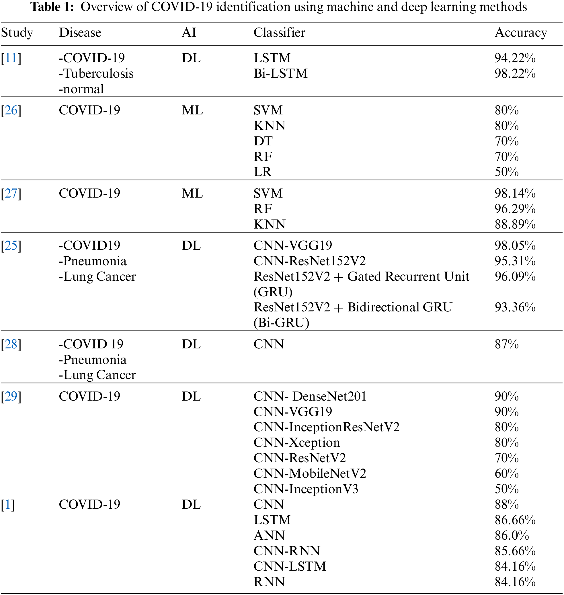
As an example of the study, authors in [1] used six different DL approaches, including Artificial Neural Network (ANN), Convolutional Neural Networks (CNN), Long-Short Term Memory (LSTM), Recurrent Neural Networks (RNN), CNNLSTM, and CNNRNN to predict COVID-19 infection. The authors used a dataset from the Israelite Albert Einstein Hospital in Sao Paulo, Brazil. The performance of these different systems has been tested in terms of accuracy, A-ROC curve, precision, recall, and f1-score. The obtained results were promising and proved their effectiveness in predicting COVID-19 by reaching an accuracy of 86.66% by the LTSM method. In [26], authors tested different ML techniques like Logistic Regression, KNN (with K = 5), Decision tree (DT), Random Forest, and the SVM to predict the clinical severity of the coronavirus among different COVID-19 cases. The authors used a private dataset from Wenzhou Central Hospital. The best result was obtained by the SVM method giving an accuracy of 80%. The authors of [25] present a combination of X-ray and CT (computerized tomography) images to predict and differentiate COVID-19 from pneumonia and lung cancer diseases using different DL approaches as classification methods. The authors used different combinations of models of the famous DL CNN methods. As a result, this research gave promoted results that reached an accuracy of 98%. Similarly, the authors in [28] proved the effectiveness of AI techniques in differentiating COVID-19 from Pneumonia and other lung cancer diseases by using some DL methods such as CNN-based models. The obtained results reached an accuracy of 87%. In [29], authors used a dataset of 50 X-Ray images with 25 confirmed positive COVID-19 cases to identify COVID-19 using DL models such as VGG19, DenseNet201, ResNetV2, InceptionV3, InceptionResNetV2, Xception and MobileNetV2 models. VGG19 model obtained the best accuracy to reach 90%. Table 1 Studies Overview for COVID-19 identification using ML and DL methods.
This section presents the different COVID-19 diagnosis techniques, a description of the dataset used, and our work methodologies to predict COVID-19 disease.
In this paper, different DL and ML models for COVID detection are proposed. The classification principle is shown in Fig. 1. As seen from this workflow, COVID classification is based on three main steps; the first step is feature selection by deleting, for example, unnecessary features. The second step is the classification task. The dataset will be split into training and test data and classified using different DL and ML models. The last step is to evaluate the performance of each model in terms of accuracy, precision, F1-score, and recall.
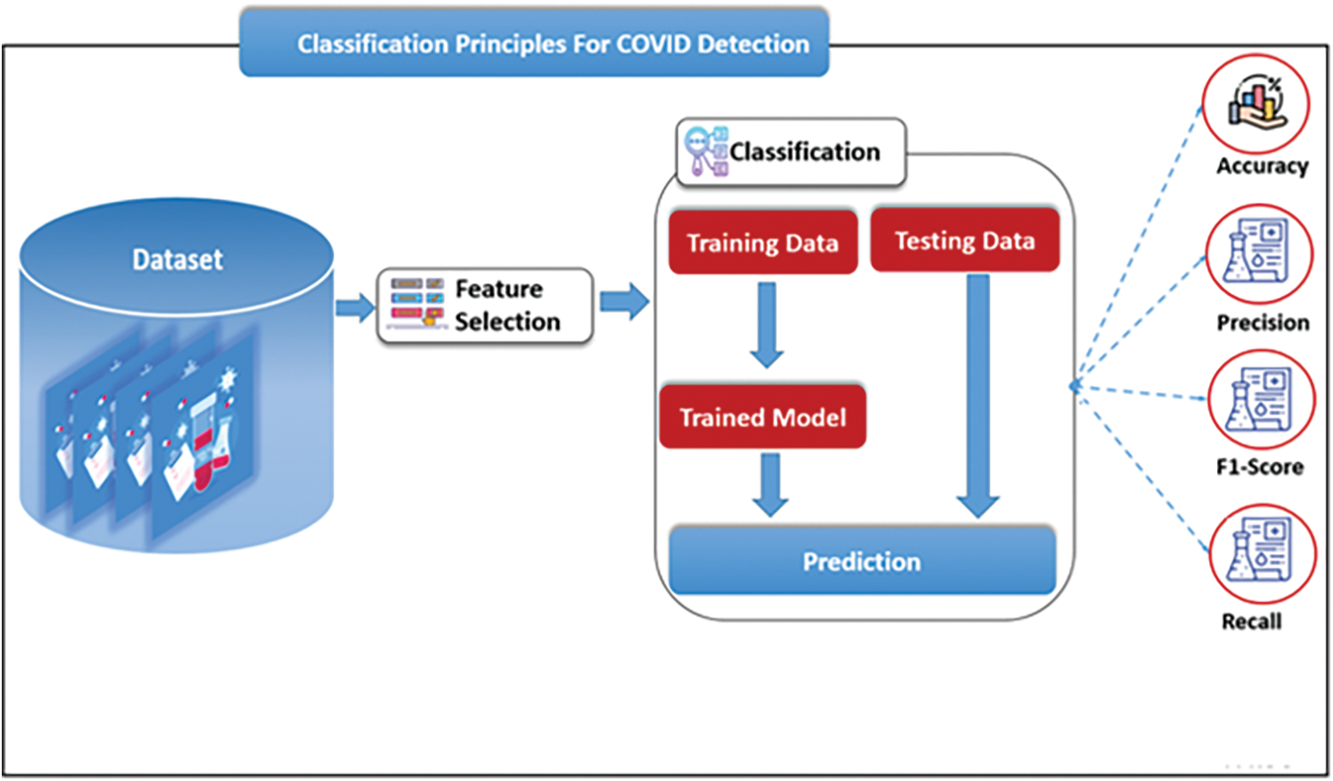
Figure 1: Classification principle for COVID-19 detection
3.1 COVID-19 Diagnostic Techniques
COVID-19 can be diagnosed by using three main techniques as shown in Fig. 2: RT-PCR test, CT-Scanner, and Numerical Laboratory Test. In our work, we used Numerical laboratory Tests as a diagnosis method. According to several types of research, this technique is the most accurate technique to diagnose COVID-19 [30].
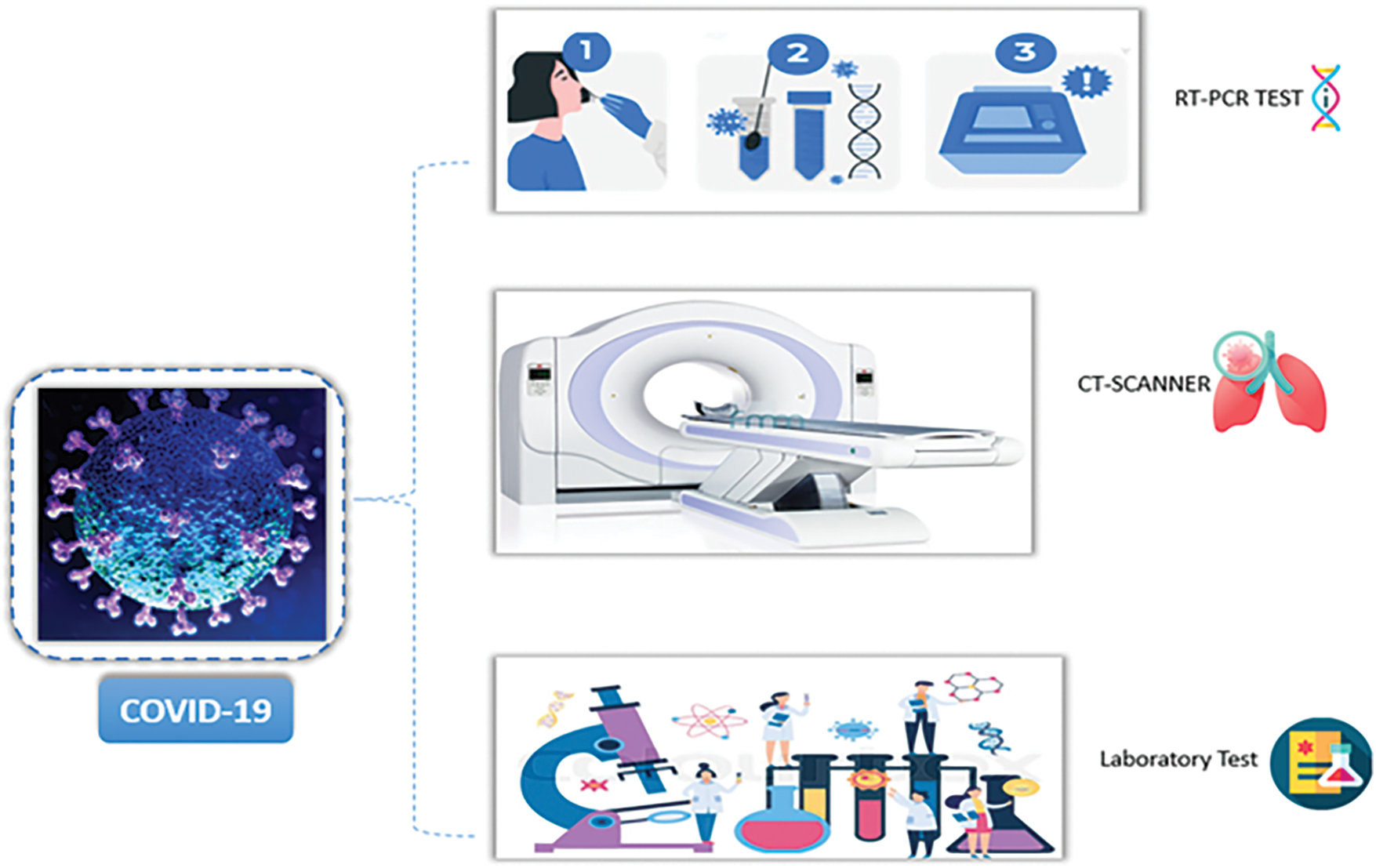
Figure 2: COVID-19 diagnosis techniques
For our experiments, we used a dataset provided by [1] obtained from the Israelita Albert Einstein Hospital in Sao Paulo, Brazil; this set of data is composed of 5644 laboratory results from different patients of which 10% are COVID-19 positive cases. The dataset includes 18 attributes that have a vital role in identifying COVID-19 disease and which are aspartate transaminase, alanine transaminase, sodium, potassium, C reactive protein, monocytes, urea, neutrophils, serum glucose, eosinophils, creatinine, basophils, red blood cells, leukocytes, platelets, hemoglobin, Hematocrit, and lymphocytes.
3.3 Classification Methods Used
ML and DL are two subsets of AI that have gained much attention recently. It refers to all theories and techniques for simulating human intelligence. Fig. 3 illustrates the main difference between ML and DL to perform a classification task. In ML, feature selection is a basic first step of manually extracting the most representative features that will be used to guide the classification task, while a DL system can learn these features without any human intervention.
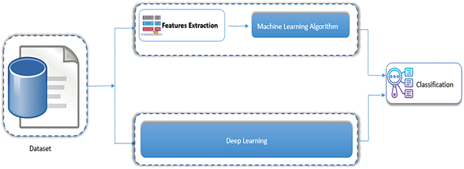
Figure 3: Machine learning vs. deep learning
In this work, and as shown in Fig. 4, we used various ML methods: DT, KNN and Naive Bayes, and some DL methods including DNN, CNN and LSTM.
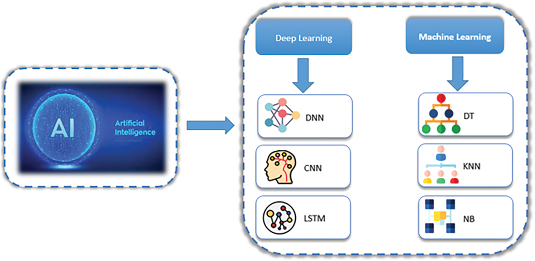
Figure 4: Method used
The ANN method is a model inspired by the structure of the human brain to solve ML problems. As shown in Fig. 5, ANN is a model composed of neurons connected to form three basic layers: an input layer, an output layer, and an intermediate layer.
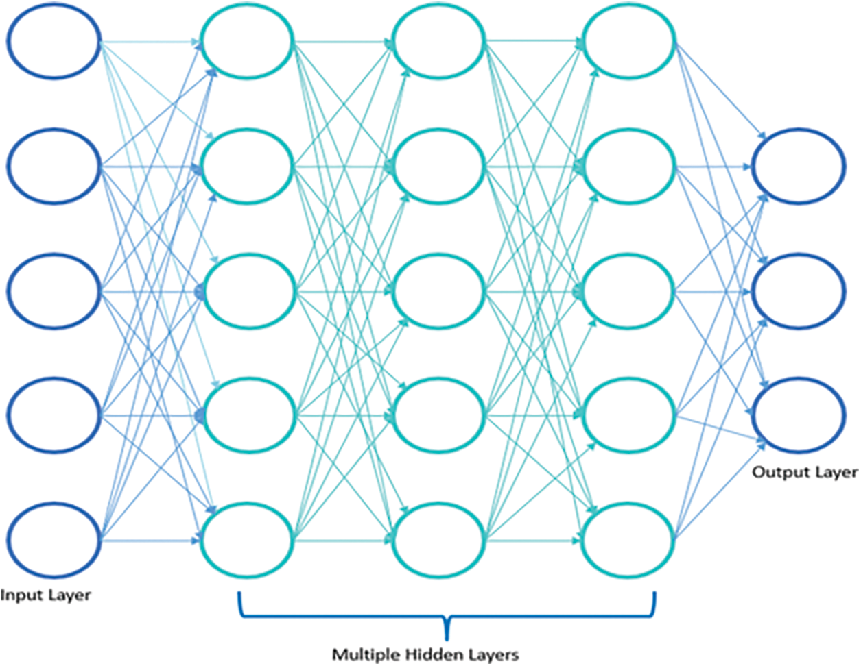
Figure 5: DNN architecture
In a classification task, the input layer contains the input data. Mathematical operations will transform these data from the input layer to the hidden layers. The number of hidden layers is determined according to the complexity of the classification task to be done. A DNN refers to an ANN with more than one hidden layer. The output layer contains the class label predicted by the model [31,32].
KNN is a ML technique widely used for classification tasks. KNN is one of the simplest algorithms based on the idea that similar things are close. It estimates the class of a new case by calculating its similarity with the K cases available in terms of Euclidean distance using the distance metrics
where D is the data and placing it in the category closest to the available categories using the probability,
The performance of this algorithm is based on the choice of parameter K, which is defined dynamically by carrying out specific tests [33].
A DT method is a tree-structured supervised learning method that predicts an output variable based on different input variables. This method decomposes the data set into hierarchical subsets by asking YES/NO questions to form a DT with decision nodes and leaf nodes. The nodes represent the different features used in the dataset, the branches are the decision rules used, and the leaf nodes are the final decision. Fig. 6 presents the general structure of a DT method [34].
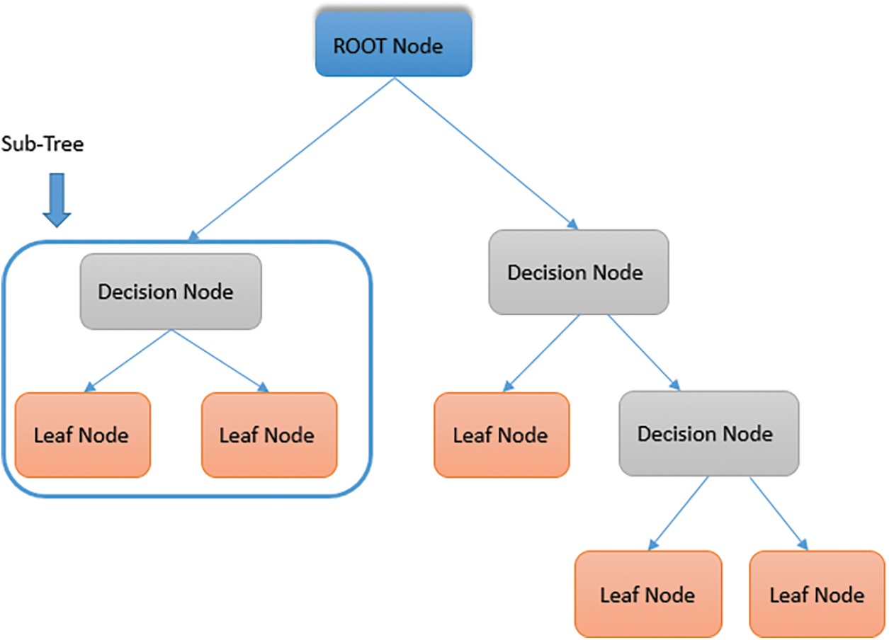
Figure 6: Decision tree architecture
AI is an exact science based on exact and accurate data. However, this assumption is not always valid in some situations due to measurement errors or external factors [35]. The NB method represents the data by their distribution probabilities rather than their exact values. In this sense, NB is a ML classifier used in medical diagnosis, speaker recognition, weather prediction, etc. It is a probabilistic algorithm based on the following Bayes theory (3) [36]:
With:
CNN is a DL method widely used for medical diagnosis. It is a variation of the ANN on a special technique called convolution, which involves multiplying a two-dimensional array of weights
As shown in Fig. 7, this method is characterized by an architecture based on three types of layers connected in sequence: convolutional, pooling, and fully connected. The convolution layer extracts the features, the pooling layer reduces the input parameters number. The final classification task will be achieved by the fully connected layers [37,38].
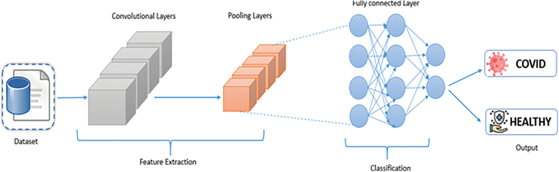
Figure 7: Decision tree architecture
LSTM is a variant of the ANN and in particular, a special type of Recurrent Neural Network (RNN). As its name suggests, LSTM is a DL method widely used when a temporal concept is involved in data [39]. In the LTSM architecture, the traditional hidden layer in the ANN architecture is replaced by a hidden layer with a particular unit called a memory block responsible for memorizing the temporal state of the network. Each Memory block is based on memory cells, special input, and output gates responsible for controlling the input-output flow of information [40]. It determines the next cell state
LTSM is widely used in speech recognition, language modeling, text generations field, medical diagnosis, etc.
In this section, we present the performance of the different experiments realized using several evaluation metrics as shown in Fig. 1: accuracy, precision, F1-score and recall. Table 2 summarizes the evaluation results of all ML and DL methods used in our work. Fig. 8 illustrates a comparison in terms of the evaluation metric used for the different methods. As shown in Table 2, the results show that the LTSM method gave the best classification accuracy, 96.78%, followed by the DNN method with 94.05%.
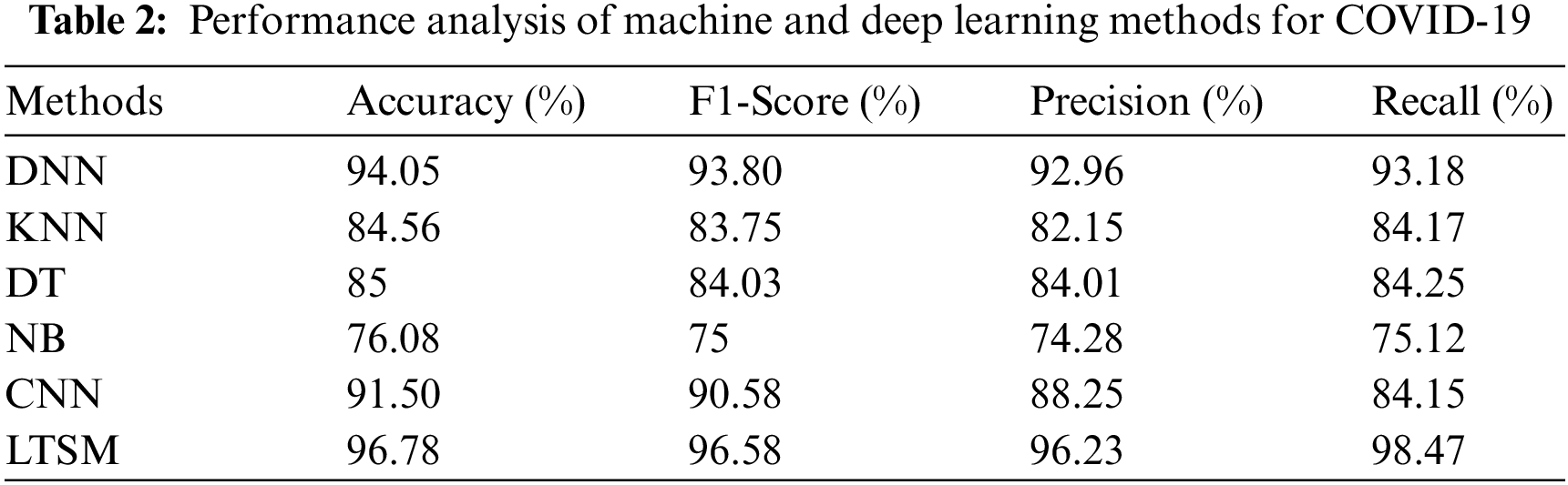

Figure 8: Performance metrics evaluation for the different methods
The ML methods, NB with 76.08% and the KNN method with 84.56% obtain the lowest classification accuracies. This can be explained by the efficacity of the DL methods in case of large dataset volume and especially to resolve complex classification tasks in the medical field. It can also be seen from Fig. 8 that the LTSM method outperforms the other methods in terms of F1-Score, precision, recall, and AUC metrics. The precision is the number of positive class predictions that are positive classes. In our cases, the best precision is given by the LTSM method with 96.23%, which comes to the perfect value equal to 1.
The F1-score metric indicates the false-positive and false-negative classification rate; in our cases, the obtained best F1-score is also given by the LTSM method with a value of 96.78%, which confirms the low false-positive and false-negative prediction. In addition, the recall metric, which provides us with details about the correctly positive predicted classes from all the prediction numbers, confirms the effectiveness of the LTSM method again with a result of 98.47%. All those metrics are very important and valuable in obtaining better COVID-19 identification. This study concludes that DL methods gave better results and performed better than ML methods. In addition, we can say that the LTSM method proves its effectiveness in identifying COVID- 19 disease.
By comparing our results, and the results obtained by previous similar studies that we already explained in the second section of related works, we can notice that:
• In general, the performance of the different methods of this study is close to those of previous works.
• From Table 1, it’s clear that our results are better than [1,3,10] and [27], but not better than [25] and [30]. The type of the database used in each work can explain this; for example, in some works, the data set type is CT images, X-ray images, or laboratory tests as our work; From this, in our future work, we will try to combine different type of databases and see the effect of that on the experimentation results.
• Moreover, as classes are imbalanced, the study also analyzed the performance of the system with F1-score, and among all LSTM model secured the better F1-score with 96.58%.
A detailed comparison is listed in Table 3.
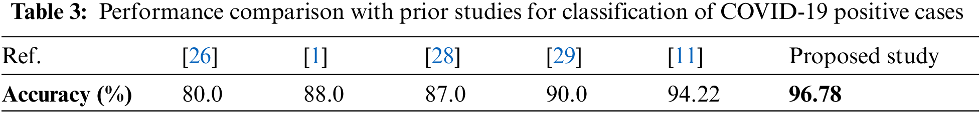
In this paper, a comparative study between ML and DL methods on COVID-19 detection is realized. For this, we used a dataset containing 5644 laboratory results obtained from Israelita Albert Einstein Hospital in Sao Paulo, Brazil. Each sample has 18 attributes, which we considered to train and test six different ML and DL-based methods: DNN, KNN, DT, NB, CNN, and LTSM. The best results were obtained by the LTSM method with an accuracy of 96.78%. Those results confirmed the effectiveness of the DL method in COVID-19 prediction by using laboratory findings. However, the limitation includes the scarcity of datasets related to COVID-19-positive cases. As the downloaded dataset contains only 10% of COVID-19-positive cases, whereas the rest belongs to non-positive COVID-19 cases, thus introduces a class imbalance problem, which may affect the performance of the ML and DL-based classifiers. In such a scenario, the f1-score is the best measure to analyze the performance of the proposed system, and in our case, the LSTM bypassed many prior well-established studies (published in renowned journals) by attaining an f1-score of 96.58%. Thus, the research presented in this paper can be practically implied in remote and rural areas where the availability of radiography imaging machines is yet not accessible.
Though the proposed study achieved better results, however, it has a few limitations. The availability of COVID-19 related clinical data is very limited, thus classes are imbalanced. In our further studies, we plan to combine the data set type with chest x-ray images, CT images and laboratory findings by applying other DL methods.
Funding Statement: The authors received no specific funding for this study.
Conflicts of Interest: The authors declare that they have no conflicts of interest to report regarding the present study.
References
1. T. B. Alakus and I. Turkoglu, “Comparison of deep learning approaches to predict COVID-19 infection,” Chaos, Solitons & Fractals, vol. 140, no. 11, pp. 110120, 2020. [Google Scholar]
2. J. Rasheed, A. Jamil, A. A. Hameed, F. Al-Turjman and A. Rasheed, “COVID-19 in the age of artificial intelligence: A comprehensive review,” Interdisciplinary Sciences: Computational Life Sciences, vol. 13,no. 2, pp. 153–175, 2021. [Google Scholar] [PubMed]
3. “COVID Live—Conronavirus Statistics—Worldometer,” 2022. https://www.worldometers.info/coronavirus/ (accessed 02 December 2022). [Google Scholar]
4. W. J. Wiersinga, A. Rhodes, A. C. Cheng, S. J. Peacock and H. C. Prescott, “Pathophysiology, transmission, diagnosis, and treatment of coronavirus disease 2019 (COVID-19),” JAMA, vol. 324, no. 8, pp. 782, 2020. [Google Scholar] [PubMed]
5. R. A. Abumalloh, M. Nilashi, M. Y. Ismail, A. Alhargan, A. Alghamdi et al., “Medical image processing and COVID-19: A literature review and bibliometric analysis,” Journal of Infection and Public Health, vol. 15, no. 1, pp. 75–93, 2022. [Google Scholar] [PubMed]
6. Y. Zhang, J. M. Gorriz and Z. Dong, “Deep learning in medical image analysis,” Journal of Imaging,vol. 7, no. 4, pp. 74, 2021. [Google Scholar] [PubMed]
7. E. -S. M. El-kenawy, A. Ibrahim, N. Bailek, K. Bouchouicha, M. A. Hassan et al., “Sunshine duration measurements and predictions in Saharan Algeria region: An improved ensemble learning approach,” Theoretical and Applied Climatology, vol. 147, no. 3–4, pp. 1015–1031, 2022. [Google Scholar]
8. M. S. Farooq, H. Khalid, A. Arooj, T. Umer, A. B. Asghar et al., “A conceptual multi-layer framework for the detection of nighttime pedestrian in autonomous vehicles using deep reinforcement learning,” Entropy, vol. 25, no. 1, pp. 135, 2023. [Google Scholar] [PubMed]
9. A. Yahyaoui, A. Jamil, J. Rasheed and M. Yesiltepe, “A decision support system for diabetes prediction using machine learning and deep learning techniques,” in 2019 1st Int. Informatics and Software Engineering Conf. (UBMYK), Ankara, Turkey, pp. 1–4, 2019. [Google Scholar]
10. A. Rahaman Wahab Sait and M. Khairi Ishak, “A novel handcrafted with deep features based brain tumor diagnosis model,” Intelligent Automation & Soft Computing, vol. 35, no. 2, pp. 2057–2070, 2023. [Google Scholar]
11. J. Rasheed and S. Alsubai, “A hybrid deep fused learning approach to segregate infectious diseases,” Computers, Materials & Continua, vol. 74, no. 2, pp. 4239–4259, 2023. [Google Scholar]
12. S. Khalid Alduraibi, “A novel convolutional neural networks-fused shallow classifier for breast cancer detection,” Intelligent Automation & Soft Computing, vol. 33, no. 2, pp. 1321–1334, 2022. [Google Scholar]
13. J. Rasheed and R. M. Shubair, “Screening lung diseases using cascaded feature generation and selection strategies,” Healthcare, vol. 10, no. 7, pp. 1313, 2022. [Google Scholar] [PubMed]
14. S. Dash, S. Verma, Kavita, S. Bevinakoppa, M. Wozniak et al., “Guidance image-based enhanced matched filter with modified thresholding for blood vessel extraction,” Symmetry, vol. 14, no. 2, pp. 194, 2022. https://doi.org/10.3390/sym14020194 [Google Scholar] [CrossRef]
15. J. Rasheed, “Analyzing the effect of filtering and feature-extraction techniques in a machine learning model for identification of infectious disease using radiography imaging,” Symmetry, vol. 14, no. 7, pp. 1398, 2022. [Google Scholar]
16. L. Fang and X. Wang, “COVID-RDNet: A novel coronavirus pneumonia classification model using the mixed dataset by CT and X-rays images,” Biocybernetics and Biomedical Engineering, vol. 42, no. 3, pp. 977–994, 2022. [Google Scholar] [PubMed]
17. Y. Chen, Y. Lin, X. Xu, J. Ding, C. Li et al., “Classification of lungs infected COVID-19 images based on inception-ResNet,” Computer Methods and Programs in Biomedicine, vol. 225, no. 7, pp. 107053, 2022. [Google Scholar] [PubMed]
18. G. Camastra, L. Arcari, F. Ciolina, M. Danti, G. Ansalone et al., “Characterization of COVID-19-related lung involvement in patients undergoing magnetic resonance T1 and T2 mapping imaging: A pilot study,” Journal of Imaging, vol. 8, no. 12, pp. 314, 2022. [Google Scholar] [PubMed]
19. M. La Salvia, G. Secco, E. Torti, G. Florimbi, L. Guido et al., “Deep learning and lung ultrasound for Covid-19 pneumonia detection and severity classification,” Computers in Biology and Medicine, vol. 136, pp. 104742, 2021. [Google Scholar] [PubMed]
20. D. Iuri, A. De Candia and M. Bazzocchi, “Evaluation of the lung in children with suspected pneumonia: Usefulness of ultrasonography,” La radiologia medica, vol. 114, no. 2, pp. 321–330, 2009. [Google Scholar] [PubMed]
21. P. Tomà and C. M. Owens, “Chest ultrasound in children: Critical appraisal,” Pediatric Radiology, vol. 43, no. 11, pp. 1427–1434, 2013. [Google Scholar]
22. V. P. Shah, M. G. Tunik and J. W. Tsung, “Prospective evaluation of point-of-care ultrasonography for the diagnosis of pneumonia in children and young adults,” JAMA Pediatrics, vol. 167, no. 2, pp. 119, 2013. [Google Scholar] [PubMed]
23. M. Studniarek, “The evaluation of the radiation dose delivered in different protocols of chest CT,” Polish Journal of Radiology, vol. 79, pp. 1–5, 2014. [Google Scholar] [PubMed]
24. K. S. Sodhi, N. Khandelwal, A. K. Saxena, M. Singh, R. Agarwal et al., “Rapid lung MRI in children with pulmonary infections: Time to change our diagnostic algorithms,” Journal of Magnetic Resonance Imaging, vol. 43, no. 5, pp. 1196–1206, 2016. [Google Scholar] [PubMed]
25. D. M. Ibrahim, N. M. Elshennawy and A. M. Sarhan, “Deep-chest: Multi-classification deep learning model for diagnosing COVID-19, pneumonia, and lung cancer chest diseases,” Computers in Biology and Medicine, vol. 132, no. 2, pp. 104348, 2021. [Google Scholar] [PubMed]
26. X. Jiang, M. Coffee, A. Bari, J. Wang, X. Jiang et al., “Towards an artificial intelligence framework for data-driven prediction of coronavirus clinical severity,” Computers, Materials and Continua, vol. 63, no. 1, pp. 537–551, 2020. [Google Scholar]
27. M. Ghaderzadeh and F. Asadi, “Deep learning in the detection and diagnosis of COVID-19 using radiology modalities: A systematic review,” Journal of Healthcare Engineering, vol. 2021, pp. 1–10, 2021. [Google Scholar]
28. L. Li, L. Qin, Z. Xu, Y. Yin, X. Wang et al., “Using artificial intelligence to detect COVID-19 and community-acquired pneumonia based on pulmonary CT: Evaluation of the diagnostic accuracy,” Radiology, vol. 296, no. 2, pp. E65–E71, 2020. [Google Scholar] [PubMed]
29. E. E. -D. Hemdan, M. A. Shouman and M. E. Karar, “CovidX-Net: A framework of deep learning classifiers to diagnose Covid-19 in X-ray images,” arXiv, 2020. [Google Scholar]
30. N. A. Mansour, A. I. Saleh, M. Badawy and H. A. Ali, “Accurate detection of Covid-19 patients based on feature correlated Naïve Bayes (FCNB) classification strategy,” Journal of Ambient Intelligence and Humanized Computing, vol. 13, no. 1, pp. 41–73, 2022. [Google Scholar] [PubMed]
31. S. Albawi, T. A. Mohammed and S. Al-Zawi, “Understanding of a convolutional neural network,” in 2017 Int. Conf. on Engineering and Technology (ICET), Antalya, Turkey, pp. 1–6, 2017. [Google Scholar]
32. A. Farizawani, M. Puteh, Y. Marina and A. Rivaie, “A review of artificial neural network learning rule based on multiple variant of conjugate gradient approaches,” Journal of Physics: Conference Series, vol. 1529, no. 2, pp. 022040, 2020. [Google Scholar]
33. H. Saadatfar, S. Khosravi, J. H. Joloudari, A. Mosavi and S. Shamshirband, “A new k-nearest neighbors classifier for big data based on efficient data pruning,” Mathematics, vol. 8, no. 2, pp. 286, 2020. [Google Scholar]
34. H. H. Patel and P. Prajapati, “Study and analysis of decision tree based classification algorithms,” International Journal of Computer Sciences and Engineering, vol. 6, no. 10, pp. 74–78, 2018. [Google Scholar]
35. J. Ren, S. D. Lee, X. Chen, B. Kao, R. Cheng et al., “Naive Bayes classification of uncertain data,” in 2009 Ninth IEEE Int. Conf. on Data Mining, Miami Beach, FL, USA, pp. 944–949, 2009. [Google Scholar]
36. J. Rasheed, A. A. Hameed, C. Djeddi, A. Jamil and F. Al-Turjman, “A machine learning-based framework for diagnosis of Covid-19 from chest X-ray images,” Interdisciplinary Sciences: Computational Life Sciences, vol. 13, no. 1, pp. 103–117, 2021. [Google Scholar] [PubMed]
37. S. H. S. Basha, S. R. Dubey, V. Pulabaigari and S. Mukherjee, “Impact of fully connected layers on performance of convolutional neural networks for image classification,” Neurocomputing, vol. 378, no. 7553, pp. 112–119, 2020. [Google Scholar]
38. V. P. Phung and E. J. Rhee, “A high-accuracy model average ensemble of convolutional neural networks for classification of cloud image patches on small datasets,” Applied Sciences, vol. 9, no. 21, pp. 4500, 2019. [Google Scholar]
39. M. Milivojević and A. Gavrovska, “Long short-term memory prediction for Covid19 time series,” Telfor. Journal, vol. 13, no. 2, pp. 81–86, 2021. [Google Scholar]
40. D. Lee, M. Lim, H. Park, Y. Kang, J. -S. Park et al., “Long short-term memory recurrent neural network-based acoustic model using connectionist temporal classification on a large-scale training corpus,” China Communications, vol. 14, no. 9, pp. 23–31, 2017. [Google Scholar]
Cite This Article
 Copyright © 2023 The Author(s). Published by Tech Science Press.
This work is licensed under a Creative Commons Attribution 4.0 International License , which permits unrestricted use, distribution, and reproduction in any medium, provided the original work is properly cited.
 Downloads
 Citation Tools